


Languages
Pages
Legal


Chromatin and Chromosome Structure
Assist. Prof. Dr. Betul Akcesme


1) Higher order structure of DNA
• Chromatine: material of which chromosomes are made of (DNA + proteins –histones)
• Nucleosomes are basic building units of chromatine
• Each nucleosome is consisted of: 8 histone molecules (histone octamer) and DNA (≈146 bp
long)
• Core histones: H2A, H2B, H3, H4
• “sealing histone” –H1 (lies outside of core histoneoctamer); purpose in connection of
nucleosomes in fibrous structures of higher order
• LINKER DNA–DNA molecule that “links” 2 nucleosomes (approx. 50 bp long, legth varies
between 8-114 bp)

Histones
• Histones: consisted mostly of basic, positively charged
amino acids like lysine and arginine
• Classified into 5 types: H1, H2A, H2B, H3and H4

Nucleosomes look like "beads on a string“ in the electron microscope

X-ray diffraction analyses of crystals
Structure of a nucleosome core
particle
• DNA bends sharply at
several places as it
wraps around the core
histone octamer
• Base sequence dictates
preferred nucleosome
positions along the DNA
Fig. 12.6
Higher order structure of DNA
• Secondary structure: the solenoid
• Solenoid structure has 6-7 nucleosomes per turn
• This is stabilized by interactions the highly variable C-and N-terminal regions
of the H1 histone and possibly the high mobility group (HMGs)
• This level of supercoiling produces a fibre of approximately 30nm in diameter
(solenoid fibre)

Secondary structure: the solenoid

Tertiary structure: loop structure
• When metaphase chromosomes are entirely depleted of histones–the residue
is an axial fibrous network –PROTEIN SCAFFOLD
• Protein scaffold is surrounded by DNA fibers which are organised into
LOOPS that radiate in all directions
• Loops contain between 5 –200 kb of DNA (63 kb)
• The loops are held together at their bases by non-histone proteins (HMGs)

Tertiary structure: loop structure
• What defines a DNA loop?
• SARs (scaffold-associated regions)
and MARs (matrix-associated
regions)
• Define loops of various sizes and are
usually found in non-transcribed regions
• Cleavage site for DNA topoisomerase II
(chromosome condensation –unwinding)

The radial loop-scaffold model for higher levels of
compaction
Several nonhistone proteins (NHPs) bind to chromatin every 60-100 kb and
tether the 300 Å fiber into structural loops
Other NHPs gather several loops together into daisylike rosettes
11
Fig. 12.7b

The radial loop-scaffold model for higher levels of compaction (cont)
Condensins may further condense chromosomes into a compact bundle for
mitosis
12
Fig. 12.7c
Quaternary structure: folding into chromosome
• Quaternary structure also involves the scaffold structure
• Scaffold structure contains NO histones, but amix of 30 non-
histoneproteins (HMGs)
• HMGs involved: Sc1 and Sc2
• Sc1: DNA topoisomeraseII (topoII)
• Sc2: no role has been assigned to it so far
• TopoII: involved in the release of stress during transcription and
replication (chromosome condensation?)
• Loopes–hexamerrosettes –coil (30 rosettes) –chromatides
13
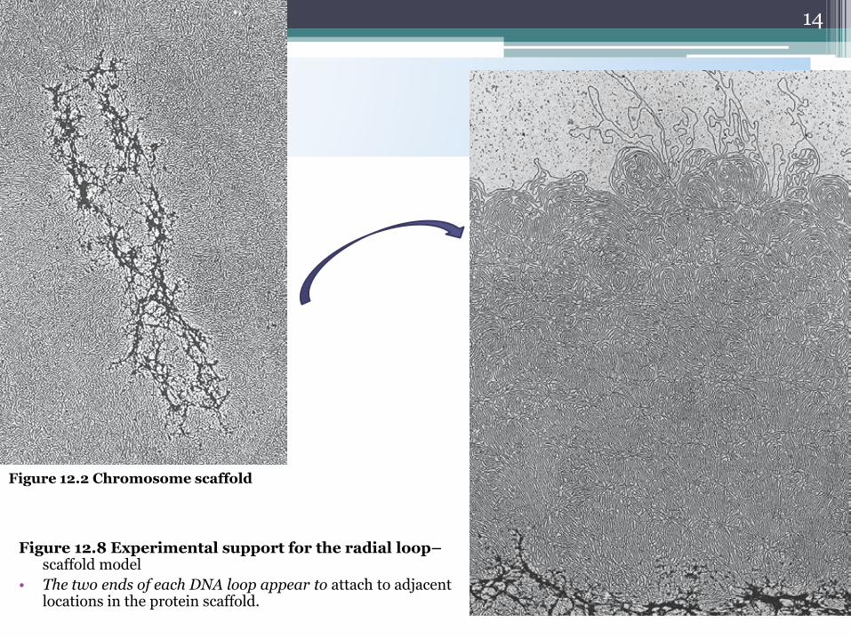
Figure 12.8 Experimental support for the radial loop–scaffold model
• The two ends of each DNA loop appear to attach to adjacent locations in the protein scaffold.
14
Figure 12.2 Chromosome scaffold

Chromosome compaction from interphase
to metaphase chromosomes.
initial looping and gathering compresses
the genetic material sufficiently to fit into the nucleus and to allow the placement of each chromosome
15
many structural loops, which are anchored together in rosettes in some areas.
By metaphase, the height of looping, gathering, and bundling achieves a 250-fold compaction of the roughly 40-fold-
compacted 300 Å fiber, giving rise to the highly condensed, rodlike shapes

Models of chromosomal structure and their fibrilary structure:
Nucleosomal fibre(11 nm)
Solenoid fibre(30 nm)
Chromatin fibre(300-700 nm)
Metaphase chromosome (1400 nm)

Levels of eukaryotic chromosomal organization
17
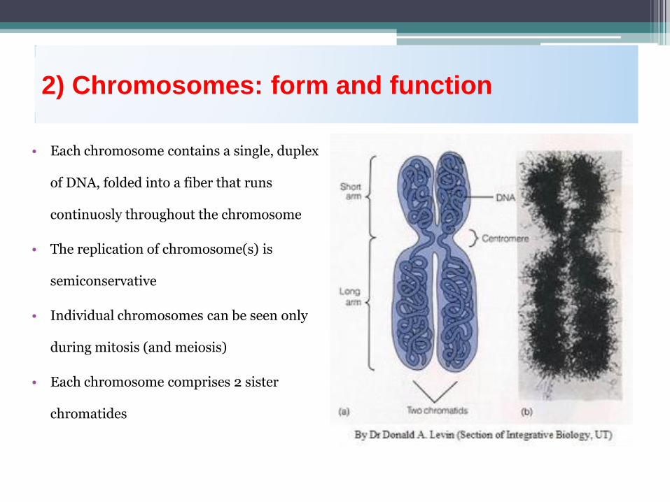
2) Chromosomes: form and function
• Each chromosome contains a single, duplex
of DNA, folded into a fiber that runs
continuosly throughout the chromosome
• The replication of chromosome(s) is
semiconservative
• Individual chromosomes can be seen only
during mitosis (and meiosis)
• Each chromosome comprises 2 sister
chromatides

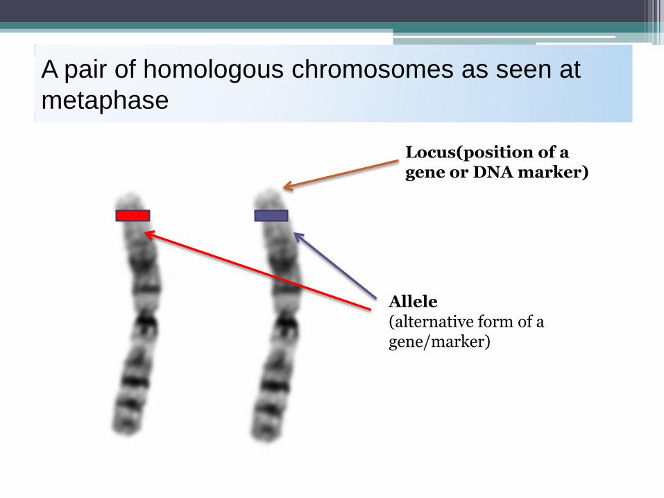
A pair of homologous chromosomes as seen at
metaphase
Allele (alternative form of a gene/marker)
Locus(position of a gene or DNA marker)

Chromosomes are cellular structures that transmit
genetic information
• Breeding experiments and microscopy provided evidence for the chromosome theory
of inheritance
• Proper development relies on accurate transmission of genes and accurate
maintenance of chromosome number
• The abstract idea of a gene was changed to a physical reality by the chromosome
theory
Copyright © The McGraw-Hill Companies, Inc. Permission required to reproduce or display Hartwell et
al., 4th ed., Chapter 4
Evidence that genes reside in the nucleus
1667 - Anton Van Leeuwenhoek
• Microscopy revealed that semen contain spermatozoa ("sperm
animals")
• Hypothesized that sperm may enter egg to achieve fertilization
1854 – 1874
• Direct observations of fertilization through union of nuclei of eggs
and sperm (frog and sea urchin)
• Conclusion: something in the nucleus must contain the hereditary
material
Copyright © The McGraw-Hill Companies, Inc. Permission required to reproduce or display Hartwell et
al., 4th ed., Chapter 4
22

Evidence that genes reside in chromosomes
• 1880s – improved microscopy and staining techniques
• Long, threadlike bodies (chromosomes) visualized in the nucleus
• Movement of these bodies followed through cell division
• Mitosis - nuclear division that generates two daughter cells
containing the same number and type of chromosomes as parent cell
• Meiosis - Nuclear division that generates gametes (egg and sperm)
containing half the number of chromosomes found in other cells
23

Diploid versus haploid: 2n versus n
• Most body cells are diploid (each
chromosome pair has one
maternal and one paternal copy)
• Meiosis haploid (n) gametes
• In Drosophila, 2n = 8, n = 4
• In humans , 2n = 46 and n = 23
Copyright © The McGraw-Hill Companies, Inc. Permission required to reproduce or display Hartwell et
al., 4th ed., Chapter 4
25
Fig. 4.2

Fertilization is the union of haploid gametes to
produce diploid zygotes
• Fertilized eggs carry matching sets of chromosomes, one set from
maternal gamete and one set from paternal gamete
• Gametes are haploid (n) – carry only a single set of chromosomes
• Zygotes are diploid (2n) – carry two matching set of chromosome
• Mitosis ensures that all cells of developing individuals have identical
2n chromosome sets
26

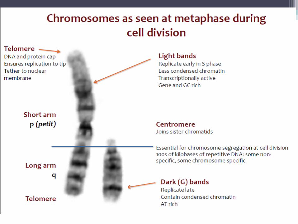
Metaphase chromosomes can be classified by centromere
position
Metacentric chromosome – centromere is in the middle
Acrocentric chromosome – centromere is near one end
Copyright © The McGraw-Hill Companies, Inc. Permission required to reproduce or display Hartwell et
al., 4th ed., Chapter 4
27
Fig. 4.3

Homologous chromosomes are matched in size,
shape , and banding patterns
• Homologs contain the same set of genes, but can have different alleles for
some genes
• Nonhomologs carry completely unrelated sets of genes
• Karyotype – micrograph of stained chromosomes arranged in homologous pairs
(see Fig 4.4)
• Sex chromosomes – unpaired X and Y chromosome
• Autosomes – all chromosomes except X and Y
• Cells of each species have a characteristic diploid number of chromosomes
e.g. D. melanogaster, 2n = 8; D. obscura, 2n = 10; D. virilis, 2n = 12; sweet peas, 2n = 14; goldfish, 2n =
94; dogs, 2n = 78
28

Karyotype of a human male
• Photos of metaphase human chromosomes (2n = 46, n = 23)
• Each homologous pair arranged in order of decreasing size
29
Fig. 4.4
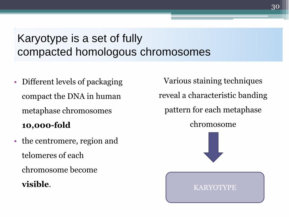
Karyotype is a set of fully
compacted homologous chromosomes
• Different levels of packaging
compact the DNA in human
metaphase chromosomes
10,000-fold
• the centromere, region and
telomeres of each
chromosome become
visible.
30
KARYOTYPE
Various staining techniques
reveal a characteristic banding
pattern for each metaphase
chromosome
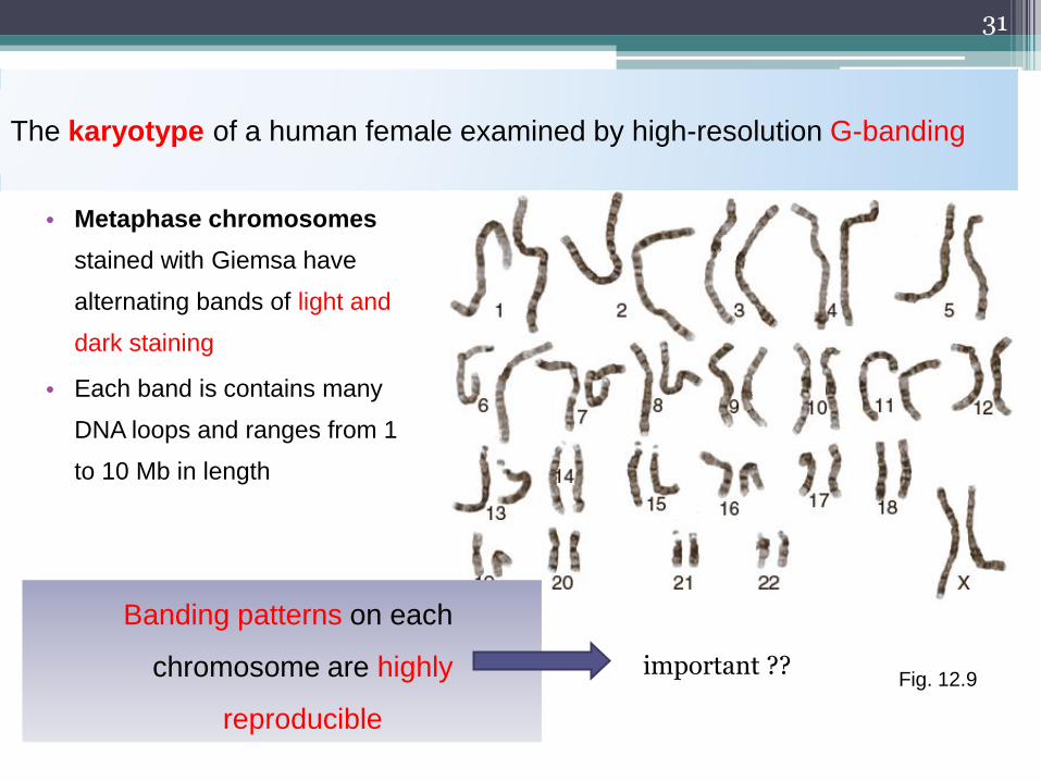
The karyotype of a human female examined by high-resolution G-banding
• Metaphase chromosomes
stained with Giemsa have
alternating bands of light and
dark staining
• Each band is contains many
DNA loops and ranges from 1
to 10 Mb in length
31
Fig. 12.9
Banding patterns on each
chromosome are highly
reproducible
important ??
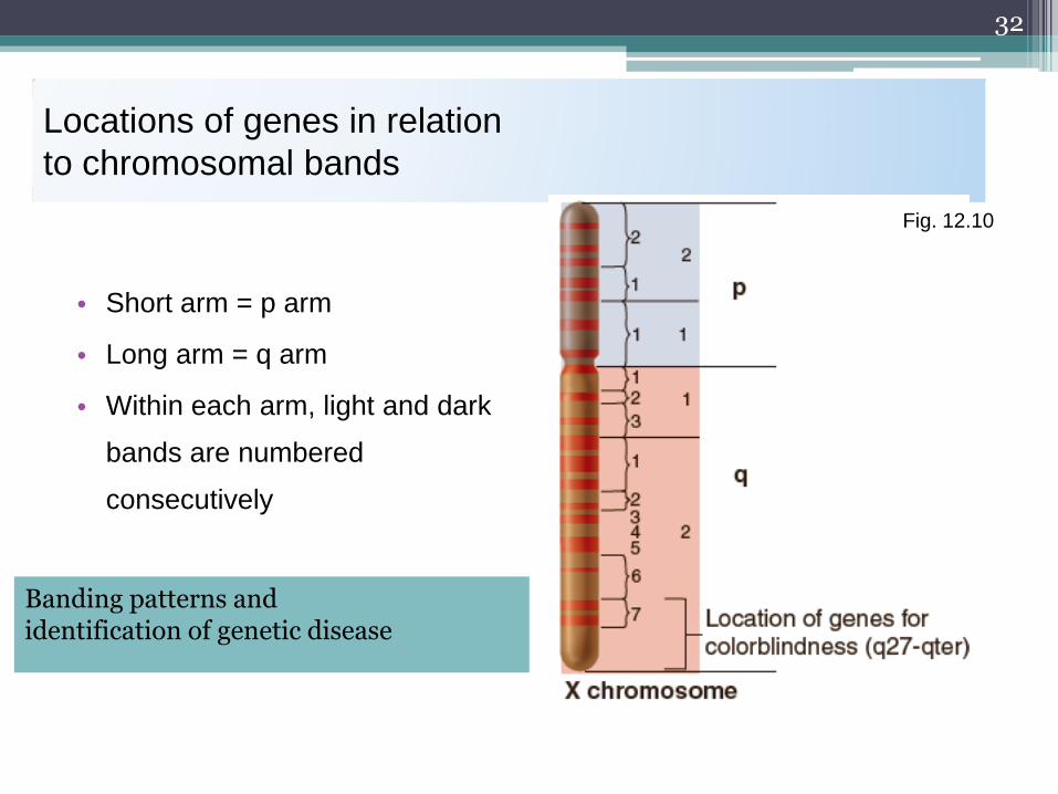
Locations of genes in relation
to chromosomal bands
• Short arm = p arm
• Long arm = q arm
• Within each arm, light and dark
bands are numbered
consecutively
32
Fig. 12.10
Banding patterns and identification of genetic disease
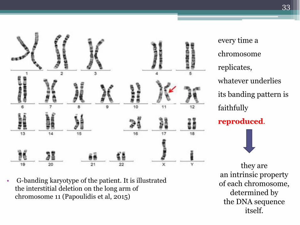
• G-banding karyotype of the patient. It is illustrated the interstitial deletion on the long arm of chromosome 11 (Papoulidis et al, 2015)
33
every time a
chromosome
replicates,
whatever underlies
its banding pattern is
faithfully
reproduced.
they are an intrinsic property of each chromosome,
determined by the DNA sequence
itself.

Standardization of human chromosomes –
banding patterns
Banding techniques – enabled correct identification of every
chromosomal pair in karyotype
The most often used technique is – G-banding (G-Giemsa) on
chromosomes
There are interrellated arrangement of light and dark bands along every
chromosome
This arrangment is characteristic for particular pair of chromosomes
G-banding became a standard procedure for identification of
metaphase chromosomes in human karyotype

Standardization of human chromosomes –
banding patterns
Chromosomes in metaphase can be identified using
certain staining techniques – called BANDING
Cells are cultured and stopped in metaphase to
maximize the number of suitable cells
They are then spread on a slide, stained with a suitable dye
and visualized in the microscope
Most conventional cytogenetic analyses depend on the
karyotyping of banded metaphase chromosomes

Human chromosomes classification
• KARYOTYPE : is the representation of entire metaphase
chromosomes in a cell
▫ The number and structure of the chromosomes in the nucleus of a cell
▫ The karyotype is identical in all the diploid cells of an organism
• KARYOGRAM: a graphic representation of a karyotype
• Graphic representation of chromosomal set where chromosomes are
arranged by their size
• IDIOGRAM: a diagram representing the characteristic features of
the chromosome set
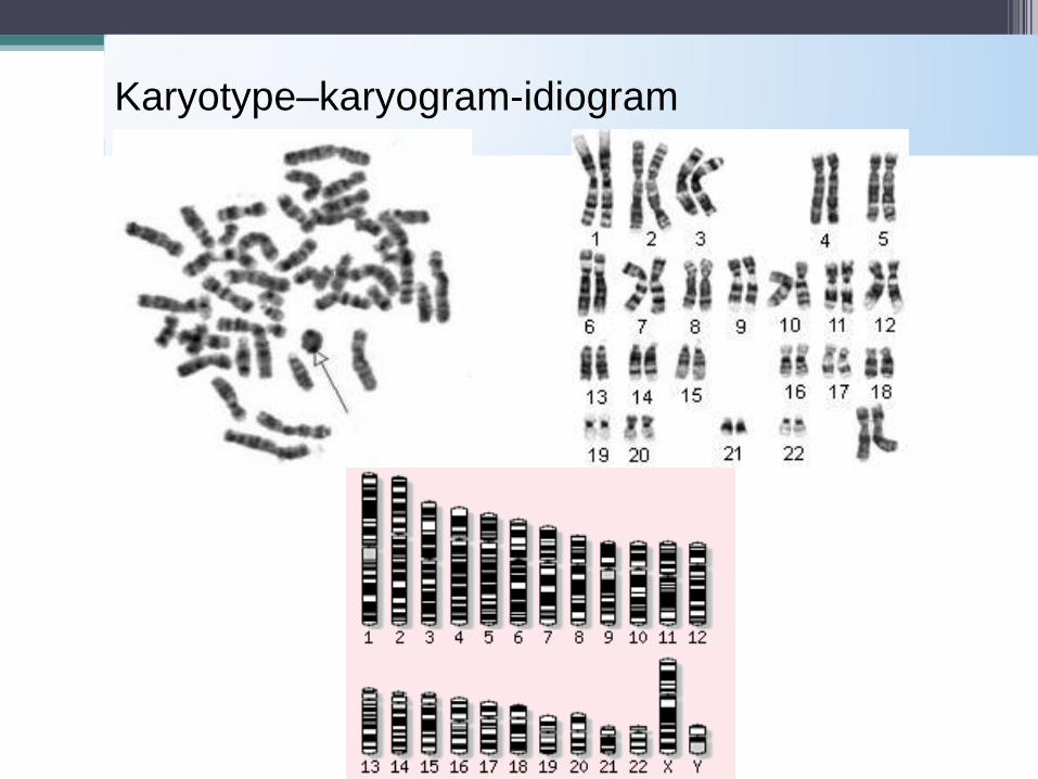
Karyotype–karyogram-idiogram
Compaction of DNA into chromatin presents a
problem
How do proteins access bases within the genome to perform
their functions?
• 1) chromatin structure is dynamic and can change to allow access
of specific proteins when they need to act.
• 2) variations exist in the molecules making up the basic
chromatin structure, and these variants recruit proteins that are
necessary for chromosomal functions.
38
3) Euchromatin and heterochromatin
Euchromatin and heterochromatin
• Genetically active • Less condensed
Is assumed that euchromatin is:
Location: chromosome arms
39
Heterochromatin
Made up of repeat sequences that vary in length
• Primary constriction (centromere) • Telomeres • Secondary constriction (nucleolar
organizing region)
Locations:
40
Heterochromatin
Inheritance is strictly mendelian
Contain tandemly repeated DNA
Some repeat sequences have significantly different DNA composition to the rest of the genome (being more GC or AT rich) – SATELLITE DNA
Made up of simple repeats
Doesn’t code for proteins
Little or no use for somatic cells but in some way important to germ cells!
41
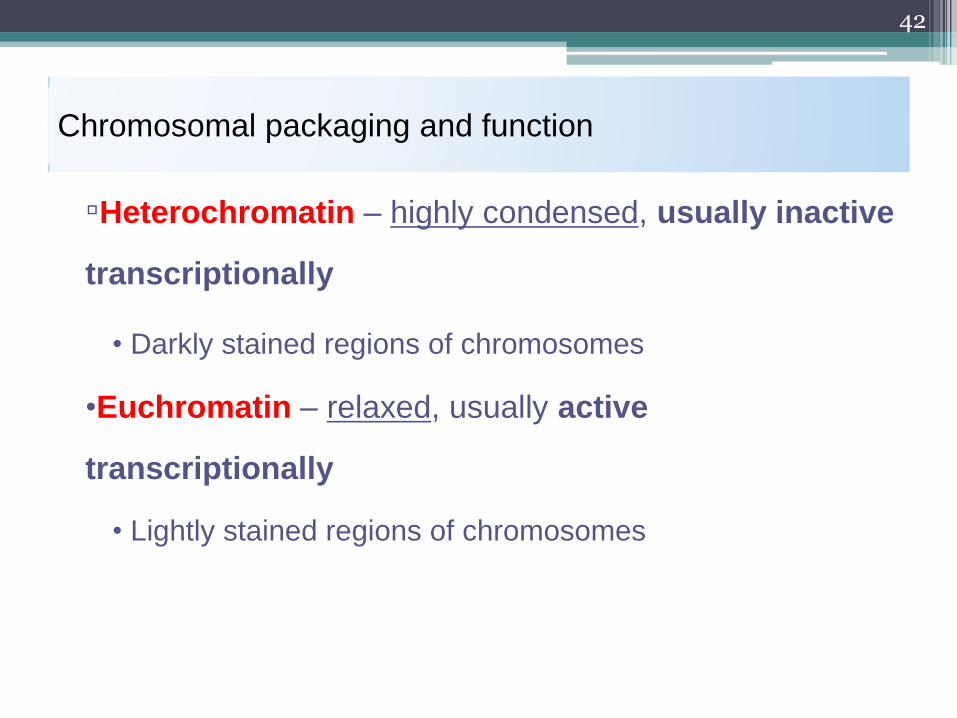
Chromosomal packaging and function
▫Heterochromatin – highly condensed, usually inactive
transcriptionally
• Darkly stained regions of chromosomes
•Euchromatin – relaxed, usually active
transcriptionally
• Lightly stained regions of chromosomes
42
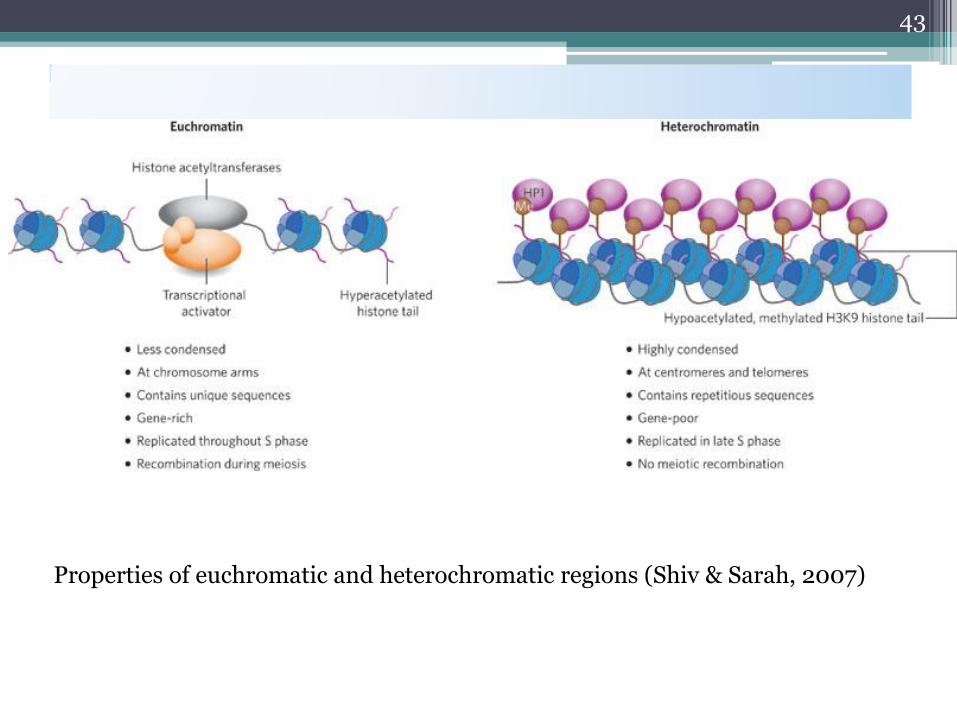
Properties of euchromatic and heterochromatic regions (Shiv & Sarah, 2007)
43

44
Figure 12.12 Stained heterochromatin.
human metaphase chromosomes were stained by a special C-banding
technique that darkens the heterochromatin, most of which localizes to
regions surrounding the centromere.

Structural parts of chromosomesTelomeres
Telomeres
Centromeres
Kinetochores
Nucleolar organizing regions (NORs)

Telomeres
The end of chromosomes
Made up of both DNA and proteins
Essential component in the control of chromosome integrity
Fundamental function of telomeres: to stop degradation of the chromosomes
during replication
sequence motif of telomeres: (TTAGGG)n is highly conserved across
phylogenies
46

Telomeres – mode of action
Prevent shortening of chromosomes
Without telomeres a chromosome would be shortened with every round of division
Telomeres are formed by enzyme telomerase
• To maintain cell and chromosome integrity • Involved in programming cellular senescence • Cause of cessation of cell division etc.
The effects of loosing telomeres:
47
Centromeres and kinetochores
Centromere – the primary constriction of
chromosomes
Characterized by particular repeat sequences of DNA
(satellite DNA) and specific associated proteins (Cenp)
This is the last point of separation of sister chromatids
during cell division!
48
Centromere: primary constriction
Made of satellite DNA of approx. 170 bp long
Size of repeats vary: from 5 bp – 170 bp
Often highly conserved repeats
DNA is associated with centromere proteins
Called: CENPs
Important for chromosome function and integrity
Site of attachment of the kinetochores
49

Kinetochores
• Is the anchor point for spindle fibers
• 2 kinetochores are present at each chromosome, facing each pole
• Characterized by the presence of CENTROMERE-ASSOCIATED PROTEINS:
▫ CENP-A
▫ CENP-B
▫ CENP-C* (vital for normal functioning)
▫ CENP-D
• The kinetochore is specifically the binding site for the microtubules which pull the
sister chromatids apart during anaphase of cell division!
50

Nucleolar organising regions (Nors)
51
• Are the site of ribosome formation!
• rRNA is transcribed and processed in the nucleolus
• In the human karyotype NORs are to be found associated with satellited chromosomes (ch 13, 14, 15, 21 and 22)
• NORs are essential for normal development

One chromosome pair determines sex in grasshoppers
W. S. Sutton studied meiosis in great lubber grasshoppers
Before meiosis, testes cells had 24 chromosomes
• 22 in matched pairs (autosomes) and 2 unmatched (large = X and smaller = Y)
After meiosis, two types of sperm were formed
• 1/2 of sperm had 11 chromosomes and an X
• 1/2 of sperm had 11 chromosomes and a Y
After meiosis, only one type of egg was produced
• All had 11 chromosomes plus an X
52
4) Sex chromosomes and sex determination

The great lubber grasshopper
• Fertilization of egg with sperm
carrying an X XX female
• Fertilization of egg with sperm
carrying a Y XY male
• Sutton concluded that the X and Y
chromosomes determine sex
53
Fig. 4.5
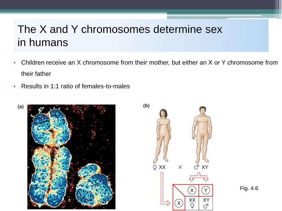
The X and Y chromosomes determine sex
in humans
• Children receive an X chromosome from their mother, but either an X or Y chromosome from
their father
• Results in 1:1 ratio of females-to-males
54
Fig. 4.6
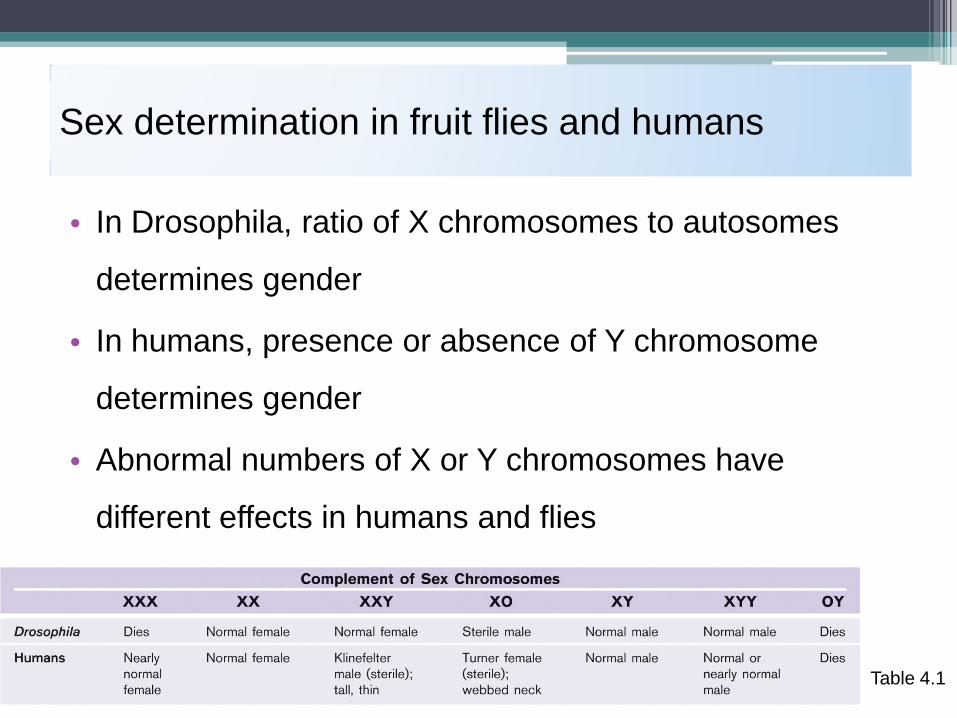
Sex determination in fruit flies and humans
• In Drosophila, ratio of X chromosomes to autosomes
determines gender
• In humans, presence or absence of Y chromosome
determines gender
• Abnormal numbers of X or Y chromosomes have
different effects in humans and flies
Copyright © The McGraw-Hill Companies, Inc. Permission required to reproduce or display Hartwell et
al., 4th ed., Chapter 4
55 Table 4.1
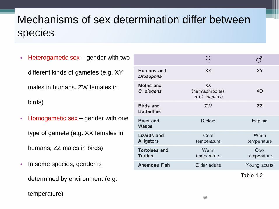
Mechanisms of sex determination differ between
species
• Heterogametic sex – gender with two
different kinds of gametes (e.g. XY
males in humans, ZW females in
birds)
• Homogametic sex – gender with one
type of gamete (e.g. XX females in
humans, ZZ males in birds)
• In some species, gender is
determined by environment (e.g.
temperature) 56
Table 4.2
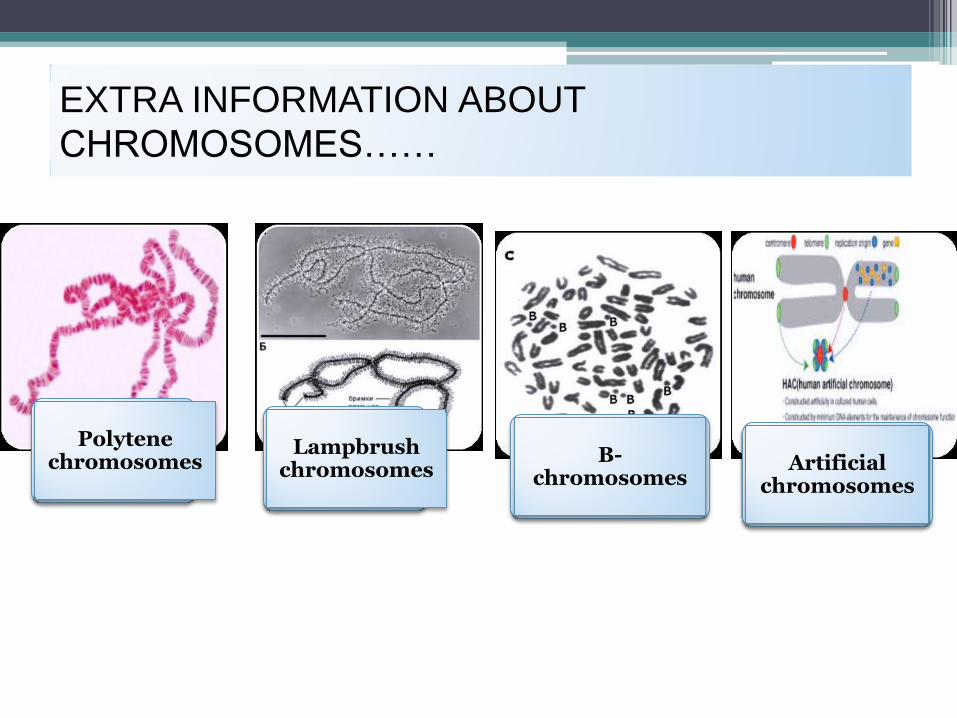
EXTRA INFORMATION ABOUT
CHROMOSOMES……
Polytene chromosomes
Lampbrush chromosomes
B-chromosomes
Artificial chromosomes

Polytenechromosomes and polyteny
• Special feature of dipteran flies
(e.g. Drosophyla)
• Clearly visible during interphase
• Constructed by
endoreduplication process
• Puffing: the formation of
decondensed DNA associated with
the bands – Balbiani rings
(actively transcribed sections of
DNA)
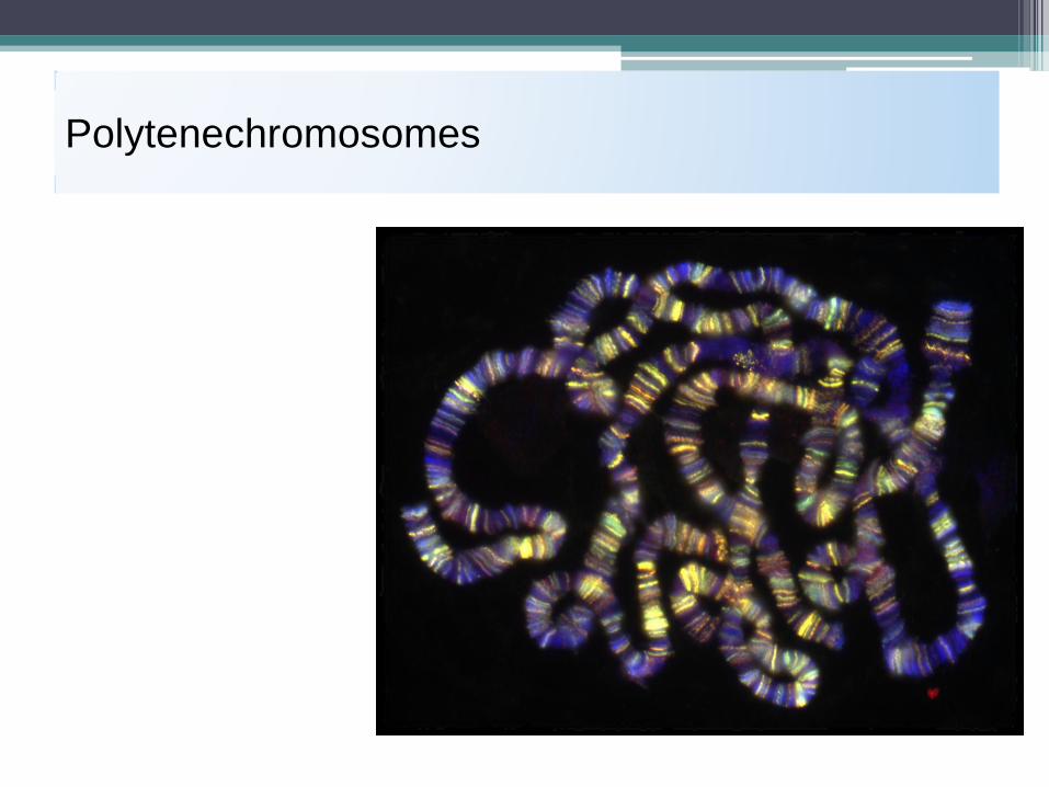
Polytenechromosomes

B chromosomes
They are typically smaller than the members of the regular complement
transmitted in a non-Mendelian pattern
not necessary for normal development and reproduction
Contain large proportions of heterochromatin
Don’t carry major genes
Don’t pair with A chromosomes during cell division
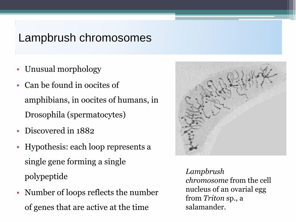
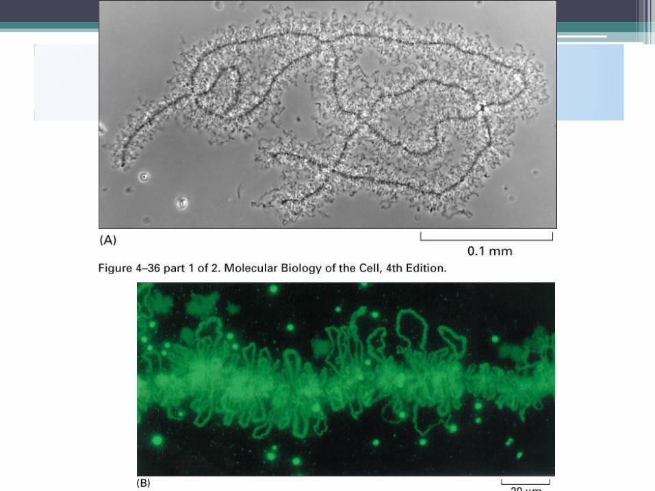
Lampbrush chromosomes
• Unusual morphology
• Can be found in oocites of
amphibians, in oocites of humans, in
Drosophila (spermatocytes)
• Discovered in 1882
• Hypothesis: each loop represents a
single gene forming a single
polypeptide
• Number of loops reflects the number
of genes that are active at the time
Lampbrush chromosome from the cell nucleus of an ovarial egg from Triton sp., a salamander.

• A model for the structure of a lampbrush chromosome
• Chromomeres: highly condensed and in general not expressed until unfolding

Artificial chromosomes
Artificially constructed
First artificial chromosome is YAC (yeast artificial chromosome)
BAC (bacterial artificial chromosome)
Can take up to 2 Mb of DNA

• REPLICATION AND SEGREGATION OF DNA
Top Related