


Languages
Pages
Legal


1
UNIVERSIDADE DE SÃO PAULO
CENTRO DE ENERGIA NUCLEAR NA AGRICULTURA
EDGARD FRANCO GOMES
Inhibition of infective larvae exsheathment and egg hatching of the
nematode Haemonchus contortus with extracts of tannin-rich plants
Piracicaba
2013


1
EDGARD FRANCO GOMES
Inhibition of infective larvae exsheathment and egg hatching of the
nematode Haemonchus contortus with extracts of tannin-rich plants
Versão revisada de acordo com a Resolução CoPGr 6018 de 2011
Dissertação apresentada ao Centro de Energia Nuclear na Agricultura da Universidade de São Paulo para obtenção do título de Mestre em Ciências Área de Concentração: Energia Nuclear na Agricultura e no Ambiente Orientador: Prof. Dr. Helder Louvandini
Piracicaba
2013
2
AUTORIZO A DIVULGAÇÃO TOTAL OU PARCIAL DESTE TRABALHO, POR QUALQUER MEIO CONVENCIONAL OU ELETRÔNICO, PARA FINS DE ESTUDO E PESQUISA, DESDE QUE CITADA A FONTE.
Dados Internacionais de Catalogação na Publicação (CIP)
Seção Técnica de Biblioteca - CENA/USP
Gomes, Edgard Franco
Inibições do desembainhamento de larvas infectantes e da eclodibilidade do nematoide Haemonchus contortus com extratos de plantas taniníferas / Inhibition of infective larvae exsheathment and egg hatching of the nematode Haemonchus contortus with extracts of tannin-rich plants / Edgard Franco Gomes; orientador Helder Louvandini. - - versão revisada de acordo com a Resolução CoPGr 6018 de 2011.- - Piracicaba, 2013.
66 f : il.
Dissertação (Mestrado – Programa de Pós-Graduação em Ciências. Área de Concentração: Energia Nuclear na Agricultura e no Ambiente) – Centro de Energia Nuclear na Agricultura da Universidade de São Paulo.
1. Etnobiologia 2. Fitoterapia 3. Leguminosas forrageiras 4. Metabólitos secundários 5. Nutrição animal 6. Parasitologia veterinária 7. Ruminantes I. Título
CDU (576.89 + 595.13) : 591.53

3
Dedico esse trabalho aos meus pais, Sr. Paulo e Sra.
Rosiléia Gomes pelo carinho, dedicação, cuidado, legado e
pelos bons ensinamentos. Aos meus preciosos irmãos
Hesddras e Hadassa Gomes (in memoriam) pela boa
companhia, pelas brincadeiras, pelos excelentes momentos.
Aos amados familiares e aos queridos amigos.

4

5
AGRADECIMENTOS
Em primeiro lugar ao Eterno, Bendito seja, o D’us de meus pais Avraham, Yitzchak e
Yaakov, pela vida, pela disposição e pelo intelecto. Ao Mashiach Yeshua ben Yosef pela
possibilidade de Teshuvá aberta por seu mérito.
Com carinho, agradeço aos meus bisavós, a quem alguns tive a honra de conhecer e
que a todos honrarei pelos meus dias. Agradeço aos esforços do Sr. Daniel Gomes (in
memoriam) e da Sra. Nartalona Maria de Azevedo (in memoriam); do Sr. Edgard do
Nascimento (in memoriam) e da Sra. Maria Salcedo do Nascimento (in memoriam); do Sr.
Mançoêto Alves Franco (in memoriam) e da Sra. Maria de Lourdes da Conceição; e do Sr.
Artur Ferreira Martins (in memoriam) e da Sra. Maria do Nascimento Martins (in memoriam).
Agradeço por eles terem deixado boas pistas para que as origens pudessem ser redescobertas.
Agradeço a todo carinho dos meus avós Sr. Aram Gomes e Sra. Zilda do Nascimento
Gomes (in memoriam) e ao Sr. Francisco Enéas Franco e Sra. Hamilta Ferreira Martins
Franco. Agradeço pelos excelentes momentos na infância, na adolescência e na juventude.
Agradeço aos meus queridos pais Sr. Paulo Nascimento Gomes e Sra. Rosiléia Martins
Franco Gomes, pelo carinho, dedicação, conselhos e por serem presentes. Agradeço por me
serem fonte de admiração, de exemplo e por estarem sempre ao meu lado.
Agradeço aos meus preciosos irmãos Hesddras Franco Gomes e Hadassa Franco
Gomes (in memoriam) pelos excelentes momentos que passamos juntos, pelo carinho e por
todos os sonhos que me fizeram sonhar.
Agradeço aos meus tios, a Sra. Rosinalva Ferreira Martins, a Sra. Rosa Dauria Ferreira
Martins, a Sra. Léia Martins Franco e o Sr. Wesley Newton Martins Franco e também à “vó”
Maria Victória Marinho Gomes por serem fontes constantes de inspiração e incentivo desde a
mais tenra infância. Agradeço pelos preciosos conselhos, pelos momentos de alegria e pelo
carinho.
Agradeço ao Prof. Dr. Helder Louvandini pela orientação que começou na graduação e
se estendeu no mestrado, pela dedicação e constante análise do trabalho experimental e
escrito. Também agradeço à Profa. Dra. Concepta McManus pela orientação e presença
6
quanto todas as dúvidas que me surgiram desde a graduação. Agradeço por esses anos de
contato com ambos.
Agradeço aos amigos da pós-graduação do LANA/CENA, o Prof. Dr. Bernardo
Berenchtein, o Dr. Amr Salah Morsy, o Sr. Dinesh Kumar, a Srta. Maria Barreal e ao Dr.
Ronaldo Lucas. Agradeço a este por ter me cedido as plantas utilizadas nesse experimento.
Agradeço aos funcionários Sr. Joaquim Everaldo Santos, Sr. Lécio Castilho e Sra. Maria
Regina Peçanha pela preciosa ajuda durante o experimento e pelos bons momentos. Agradeço
aos alunos Victor Guerrini e Egon Hion pela ajuda na preparação dos extratos.
Agradeço ao Dr. Hervé Hoste por todo o suporte técnico, por ter pessoalmente me
ensinado sobre as técnicas in vitro que foram utilizadas na parte prática do mestrado.
Agradeço também a Dra. Fotini Manolaraki pela pronta ajuda, mesmo à distância, em todas as
dúvidas que me surgiram quanto às técnicas in vitro e por ter me ensinado a preparar os
extratos de plantas.
Agradeço à Dra. Ana Carolina de Souza Chagas por ter me recebido na Embrapa
CPPSE, por ter pacientemente me ensinado e pelo treinamento sobre as técnicas in vitro
executadas nessa dissertação.
Agradeço à Dra. Luciana Katiki, a Sra. Márcia Lucena, a Sra. Elizabete, a Srta.
Luciana Dinato e a Srta. Érika Canova por terem me recebido no Instituto de Zootecnia de
Nova Odessa e por me auxiliarem no experimento.
Agradeço aos funcionários da CPG do CENA, o Sr. Fábio Antonio de Souza Oliveira,
a Sra. Sônia Aparecida Barrios de Campos, a Sra. Neuda Fernandes Oliveira e a Srta. Daiane
Vieira pela sempre pronta ajuda, pelo modo que sempre me atenderam de maneira muito
acolhedora e agradável.
Agradeço aos funcionários da Biblioteca do CENA, o Sr. Celso de Aguiar, a Sra.
Marília Henyei, a Sra. Adriana Moretti, a Sra. Raquel Carvalho, a Sra. Renata Fini e a Srta.
Sara Mondoni pelos bons momentos na biblioteca, pela ajuda que prontamente me ofereceram
e pelas boas conversas.
Agradeço ao Dr. Ruy Vieira por toda ajuda quanto ao tema “parasitologia”, pelas
ótimas conversas sobre diversos outros temas e pela companhia também. Agradeço a Sra. Ana
Marisa Fontoura Vidal por toda paciência e pelos grandes conhecimentos que me ensinou
7
com muito carinho. Aos “chaverim” da sinagoga Beit El Shamah de Piracicaba pelos
excelentes momentos juntos, pelo aprendizado, pelo companheirismo. Aos amigos Gustavo
Martins e Stela Sampaio pelos incentivos para que eu continuasse e pelos excelentes
momentos, mesmo à distância. Aos bons amigos Ricardo Maia, Victor Vitório, Lucas Pereira,
Saulo Braga, Rodrigo Sales, Taigleisson do Vale, Lucas Vicente, Isaac Cayna, Maycon da
Silva, Douglas Celis, Mayk Marlon, Jonathan Teixeira, Lucas Costa, Paulo Damasceno,
Alexandre Damasceno e Caio Rodrigues pelos excelentes momentos, pelos “dados que
rolaram”, pelas muitas risadas e por terem ajudado a carregar meus fardos.
Agradeço ao Sr. e Sra. Roberto e Marcione Santos e ao Sr. Pedro Botelho pela ajuda e
companhia nos momentos mais difíceis nesses últimos anos. Agradeço ao Sr. Cesar Marson e
sua família por terem me recebido muito bem assim que cheguei a Piracicaba. Agradeço a
Srta. Juliana Digestani pelos momentos agradáveis. Agradeço ao Sr. Tiago do Prado Paim e a
Srta. Aline Campeche Lopes pela ajuda nos experimentos desde a graduação e pela
importante ajuda nas disciplinas da pós-graduação.
Agradeço à CAPES e ao CNPq pela bolsa de mestrado.
Agradeço aos colegas da pós-graduação do LANA/CENA-USP pelo profissionalismo.
Agradeço a todos que direta ou indiretamente contribuíram para o presente trabalho.
Agradeço a todos que estiveram comigo desde o início da minha jornada para me
tornar um Mestre, seja aqui em Piracicaba ou em Brasília, de perto ou à distância.
Agradeço às preciosas ovelhas que aplicaram suas vidas para a execução desse
trabalho.

8

9
“Shemá Yisrael, HaShem Elokeinu, HaShem
ECHAD.” Devarim (“Deuteronômio”) 6:4
“O justo se preocupa com a vida de seus animais,
porém mesmo a compaixão do malévolo é cruel”
Mishlei (“Provérbios”) 12:10
“Para mim, não é qualquer questão que é digna de ser
questionada. Eu questiono apenas lá onde sou
questionado. Por pessoas sou questionado.” Franz
Rosenzweig
“Eu descobri em mim mesmo desejos os quais nada
nesta terra podem satisfazer, a única explicação lógica
é que eu fui feito para um outro mundo.” C. S. Lewis
“Com um pingo de mel se apanham mais moscas do
que com um barril de fel.” Abraham Lincoln
“Acorde cedo se você quiser a vida ou a terra de outro
homem. Nenhuma batalha é vencida deitado numa
cama. Nenhuma ovelha terá o lobo preguiçoso.”
Provérbio Viking
“Corte sua própria lenha. Assim, ela aquecerá você
duas vezes.” Henry Ford
“A maior recompensa para o trabalho do homem não
é o que recebe por ele, mas o que se torna por meio
dele.” John Ruskin
“O caminho que leva ao que realmente desejamos é
longo e difícil, mas só seguindo esse caminho é que
vamos alcançar nosso alvo.” M. Splinter

10

11
RESUMO GOMES, E. F. Inibições do desembainhamento de larvas infectantes e da eclodibilidade do nematoide Haemonchus contortus com extratos de plantas taniníferas. 2013. 66 f. Dissertação (Mestrado). – Centro de Energia Nuclear na Agricultura, Universidade de São Paulo, Piracicaba, 2013. Objetivou-se com o presente trabalho verificar a bioatividade dos extratos das plantas taniníferas Acacia mearnsii, Myracrodruon urundeuva, Caesalpinea bracteosa e Leucaena leucocephala contra os estágios de ovo e de larva L3 infectante de Haemonchus contortus. Para isso, dois ensaios in vitro foram realizados: o Teste da inibição da eclodibilidade dos ovos (TIEO) e o teste da Inibição do desembainhamento larvar (TIDL). O TIEO consiste na incubação de ovos recém recuperados das fezes de animais infectados em solução liquida de extrato de planta por 24 horas e posterior diferenciação entre larvas e ovos não eclodidos. As concentrações utilizadas para A. mearnsii foram de 50,00, 25,00, 12,50, 6,25, 3,12, 1,56, 0,78 e 0,39 mg/mL; 1,56, 0,78, 0,39, 0,19, 0,09 e 0,04 mg/mL para M. urundeuva; 6,25, 3,12, 1,56, 0,78, 0,39 e 0,19 mg/mL para C. bracteosa; e 6,25, 3,12 e 1,56 mg/mL para L. leucocephala. O TIDL consiste no desembainhamento artificial de larvas infectantes, obtidas através de coprocultura, que passaram por período de incubação de três horas em solução liquida de extratos de plantas nas concentrações 1.200, 600, 300 e 150 µg/mL. As doses letais (DL) 50 e 99 foram calculadas para ambos testes. Um efeito dose-dependente foi encontrado para os dois testes, exceto para a L. leucocephala no TIEO, onde não foi possível calcular o valor da DL50 e DL99 para o respectivo teste nas doses escolhidas. Os resultados da DL50 para TIEO foram de 0,18, 0,32 e 7,20 mg/mL e da DL99 foram de 4,31, 5,41 e 187,26 mg/mL respectivamente para M. urundeuva, C. bracteosa e A. mearnsii. Para o TIDL, a DL50 foi de 0,40, 0,52, 1,24 e 2,24 mg/mL e da DL99 foi de 2,37, 2,28, 19,99 e 2,53 x 10³ mg/mL respectivamente para M. urundeuva, A. mearnsii, L. leucocephala e C. bracteosa. Para o TIEO, as duas maiores concentrações de A. mearnsii e as três maiores de C. bracteosa foram efetivas (mais de 90% de bioatividade); as três maiores concentrações de M. urundeuva e a 0,78 mg/mL de C. bracteosa foram moderadamente efetivas (entre 80 e 90% de bioatividade); as concentrações de pouca efetividade (entre 60 e 80% de bioatividade) foram a 12,50 mg/mL (A. mearnsii), 0,19 mg/mL (M. urundeuva), e 0,39 mg/mL (C. bracteosa); as demais concentrações foram consideradas ineficientes (menos de 60% de bioatividade). Para o TIDL, apenas a maior concentração de A. mearnsii e M. urundeuva foram efetivas; a concentração de 600 µg/mL de M. urundeuva foi moderadamente efetiva; todas as outras concentrações foram ineficientes. Entretanto, deve-se observar que apesar de ineficientes em bloquear o desembainhamento, algumas concentrações atrasaram significativamente o desembainhamento das larvas, sendo esse um resultado de interesse. Assim, foi observado que os extratos, excetuando o extrato de L. leucocephala quanto a eclodibilidade, possuem bioatividade in vitro nas doses utilizadas contra a eclosão dos ovos e contra o desembainhamento das larvas de H. contortus. Palavras-chave: Verminose. Resistência anti-helmíntica. Haemonchus. Etnoveterinária. In vitro.

12

13
ABSTRACT GOMES, E. F. Inhibition of infective larvae exsheathment and egg hatching of the nematode Haemonchus contortus with extracts of tannin-rich plants. 2013. 66 f. Dissertation (Masters). – Centro de Energia Nuclear na Agricultura, Universidade de São Paulo, Piracicaba, 2013. The aim of this work was to assess the bioactivity of extracts of the tannin-rich plants Acacia mearnsii, Myracrodruon urundeuva, Caesalpinea bracteosa and Leucaena leucocephala against egg and infective larvae stages of Haemonchus contortus. Two in vitro assays were held: an Egg Hatch Assay (EHA) and a Larval Exsheathment Inhibition Assay (LEIA). The EHA consists of the incubation of previously recovered eggs from infected animal’s faeces in a solution of plant extract for 24 hours and later differentiation between larvae and non-hatched eggs. The concentrations used were 50.00, 25.00, 12.50, 6.25, 3.12, 1.56, 0.78 and 0.39 mg/mL for A. mearnsii; 1.56, 0.78, 0.39, 0.19, 0.09 and 0.04 mg/mL for M. urundeuva; 6.25, 3.12, 1.56, 0.78, 0.39 and 019 mg/mL for C. bracteosa; and 6.25, 3.12 and 1.56 mg/mL for L. leucocephala. The LEIA consists in the artificial exsheathment of infective larvae, obtained by previous coproculture, after a three hour incubation period with plant extract solution in the concentrations 1,200, 600, 300 and 150 µg/mL. The 50 and 99 lethal doses (LD) were calculated for both tests. A dose-dependent effect was found in the two tests, except for L. leucocephala in EHA, where it was not possible to calculate DL50 and DL99 with the chosen doses. The DL50 results for EHA were 0.18, 0.32, and 7.20 mg/mL and for DL99 were 4.31, 5.41, and 187.26 mg/mL, respectively for M. urundeuva, C. bracteosa, and A. mearnsii. For LEIA, the DL50 were 0.40, 0.52, 1.24, and 2.24 mg/mL and for DL99 these were 2.37, 2.28, 19.99 and 2.53 x 10³ mg/mL respectively for M. urundeuva, A. mearnsii, L. leucocephala and C. bracteosa. The two highest concentrations of A. mearnsii and the three highest for C. bracteosa were effective (more than 90% of bioactivity); the three highest concentrations of M. urundeuva and the 0.78 mg/mL level of C. bracteosa were moderately effective (between 80 and 90% of bioactivity); the concentrations with low effectiveness (between 60 and 80% of bioactivity) were the 12.50 mg/mL (A. mearnsii), 0.19 mg/mL (M. urundeuva), and 0.39 mg/mL (C. bracteosa); all other concentrations were ineffective (less than 60% of bioactivity). For the LEIA, only the highest dose from A. mearnsii and M. urundeuva were effective; the 600 µg/mL were moderately effective and all other doses were ineffective. It should be observed that even if the dose is ineffective against exsheathment, some concentrations were able to significantly delay the process. It was observed that the extracts had bioactivity in vitro within the chosen doses against the hatchability of eggs and exsheathment of larvae of H. contortus, except for L. leucocephala, which was not able to block the hatching of eggs.
Key-words: Parasitosis. Anthelmintic resistance. Haemonchus. Ethnoveterinary. In vitro.

14

15
SUMMARY 1. INTRODUCTION ................................................................................................................ 17
2. INTRODUÇÃO .................................................................................................................... 19
3. REVIEW OF THE LITERATURE ...................................................................................... 21
3.1. Developmental stages of larvae and feeding behavior ...................................................... 21
3.1.1. Habitat and ethology of nematodes ................................................................................ 24
3.2. Parasitism and related issues ............................................................................................. 25
3.2.1. Trichostrongylodea family ............................................................................................. 25
3.2.2. Parasitism origin ............................................................................................................ 26
3.2.3. Possible benefits of parasitism ....................................................................................... 28
3.2.4. Animal’s ethological defenses against parasites and pathogens .................................... 28
3.3. Ethnoveterinary ................................................................................................................. 30
3.3.1. Tannins ........................................................................................................................... 30
3.3.1.1. Leather tanning mechanisms ....................................................................................... 31
3.3.1.2. Tannin’s role against nematodes ................................................................................. 32
3.3.2. Plants .............................................................................................................................. 32
3.4. In vitro tests ....................................................................................................................... 33
3.5. Objectives .......................................................................................................................... 36
4. MATERIAL AND METHODS............................................................................................ 37
4.1. Plant samples ..................................................................................................................... 37
4.2. Laboratory facilities ........................................................................................................... 37
4.3. Bioactive extracts .............................................................................................................. 37
4.4. Egg Hatch Assay ............................................................................................................... 40
4.4.1. Egg recovery ................................................................................................................... 40
4.4.2. Preparation of concentrations of bioactive extracts for EHA ......................................... 41
4.4.3. Preparation of the 24-well plates .................................................................................... 42
4.4.4. Microscope readings for EHA ........................................................................................ 43

16
4.5. Larval Exsheathment Inhibition Assay .............................................................................. 43
4.5.1. Obtainment of larvae ....................................................................................................... 43
4.5.2. Preparation of concentrations of bioactive extracts for LEIA ........................................ 43
4.5.3. Preparation of the assay tubes ......................................................................................... 44
4.5.4. Microscope readings for LEIA ....................................................................................... 47
4.6. Statistical analyses ............................................................................................................. 48
5. RESULTS ............................................................................................................................. 49
6. DISCUSSION ....................................................................................................................... 54
7. CONCLUSIONS ................................................................................................................... 59
8. CONCLUSÕES .................................................................................................................... 60
REFERENCES.......................................................................................................................... 61
17
1. INTRODUCTION
The goat and sheep production are expanding in Brazil, according to data from
Brazilian Institute of Geography and Statistics (IBGE, 2011). But gastrointestinal parasites
still an ongoing issue against this production, being one of the greatest worldwide problems in
ruminants, particularly in sheep and goats. Under dosing, mass treatment, and use of the same
drug treatment for long periods might have worked together for the development of a drug
resistance in the parasites (LOUVANDINI et al., 2002; PIMENTEL NETO; FONSECA,
2002; WALLER, 2006).
Haemonchus contortus is one of the most common parasites in small ruminants. It is
haematophagous, and it is found worldwide. It is well known because of its high
pathogenicity. When animals acquire high infections, they become anaemic because of the
loss of blood and, because of that, a high loss of nutrients, such as proteins, lipids and
minerals. They also may also have oedema because of hypoproteinemia. For a long time
these infections have been treated with chemical anthelmintics, which has led to high levels of
drug resistance. Because of that resistance, alternative efforts to fight against parasites have
been investigated in different fronts, such as pasture rotation, selection for genetic resistant
hosts, biologic control of parasites, vaccination, and phytotherapy, among others. The
techniques aim at lowering the level of infection in the animals and, consequently, to lower
the tax of pasture contamination (BAIN, 1999; JACKSON et al., 2009; TAYLOR; COOP;
WALL, 2010).
Regarding phytotherapy, there is a vast amount of Brazilian tannin-rich plants, but the
anthelmintic properties of these are mostly unknown. A better way to screen these plants is by
doing in vitro assays, because they are cheaper, collected rapidly, show repeatability, and they
have sensitivity. Testing them first using in vivo would be costly and also too slow. Many
tests can be used to analyze tannin-rich plants such as the Egg Hatch Assay, Larvae Inhibition
Motility Assay, Larval Migration Inhibition Assay, Larval Exsheathment Inhibition Assay,
Larval Feeding Inhibition Assay, and others. These tests focus on different stages of the
nematode life cycle, and they show that tannin-rich plants do play a role similar to
anthelmintics. Once the in vitro tests are carried out, the plants that showed bioactivity may be
selected and tested in in vivo assays (POWERS et al., 1982; BRUNET; HOSTE, 2006;
GOMES; BANDEIRA, 2012).

18
Haemonchus contortus is a nematode specie. The general comprehension of
nematodes physiology is important to better understand the H. contortus physiology. Even
though this work focuses on H. contortus hatching and exsheathing, a brief description of
nematodes as a group will be given, highlighting the important differences found with
H. contortus when needed.

19
2. INTRODUÇÃO
A produção de caprinos e ovinos está em expansão no Brasil, segundo dados do
Instituto Nacional de Geografia e Estatística (IBGE, 2011). Entretanto, parasitas
gastrintestinais são um problema contra a produção, e são um dos principais problemas na
produção mundial de ruminantes, particularmente de ovelhas e cabras. Baixa dosagem,
tratamento em massa e uso do mesmo princípio ativo por períodos longos são fatores que
juntos contribuíram para o desenvolvimento de resistência anti-helmíntica dos parasitas
(LOUVANDINI et al., 2002; PIMENTEL NETO; FONSECA, 2002; WALLER, 2006).
Haemonchus contortus é um dos vermes mais comuns em pequenos ruminantes. É
hematófago, encontrado mundialmente e conhecido por conta de sua alta patogenicidade.
Quando animais têm altas infecções, eles se tornam anêmicos pela perda de sangue e, por
conta desta, também ocorre uma grande perda de nutrientes, como proteínas, lipídeos e
minerais, podendo ocasionar edema nos animais, por conta da hipoproteinemia. Por muito
tempo essas infecções foram tratadas com uso de moléculas comerciais, o que ocasionou, pelo
mal uso das moléculas, uma grande resistência aos medicamentos. Por conta da resistência,
tentativas de controle alternativo têm sido buscadas, como a rotação de pastagens, seleção de
animais geneticamente resistentes, controle biológico dos parasitas, vacinação, fitoterapia,
entre outras. Visa-se através das técnicas diminuir a infecção dos animais e,
consequentemente, diminuir a infestação dos pastos (BAIN, 1999; JACKSON et al., 2009;
TAYLOR; COOP; WALL, 2010).
Com relação à fitoterapia, existem muitas plantas taniníferas na flora brasileira, mas
suas atividades anti-helmínticas são pouco conhecidas. Uma forma viável para selecionar
essas plantas é através de testes in vitro, porque eles são baratos, são mais rápidos, possuem
repetibilidade e sensibilidade. Testar inicialmente in vivo pode ser muito caro e demorado.
Muitos testes podem ser usados para analisar plantas taniníferas tais como o Teste da Inibição
da Eclodibilidade dos Ovos (TIEO), o Teste da Inibição da Motilidade Larvar, o Teste da
Inibição da Migração Larvar, o Teste da Inibição do Desembainhamento Larvar (TIDL), o
Teste da Inibição da Alimentação Larvar, entre outros. Esses testes focam em diferentes
estágios do ciclo de vida do nematoide e através deles são mostrados o papel das plantas
taniníferas no combate aos helmintos. Depois de selecionar as plantas nos testes in vitro, as
plantas que apresentaram bioatividade são selecionadas e testadas in vivo (POWERS et al.,
1982; BRUNET; HOSTE, 2006; GOMES; BANDEIRA, 2012).

20
Haemonchus contortus é uma espécie de nematoide. Para a compreensão da fisiologia
do H. contortus, faz-se necessária a compreensão da fisiologia dos nematoides como uma
grupo. O foco desse trabalho é a eclodibilidade e o desembainhamento de H. contortus,
entretanto, uma breve descrição dos nematoides será apresentada, destacando o que é
especifico para o H. contortus quando necessário.
21
3. REVIEW OF THE LITERATURE
3.1. Developmental stages of nematode larvae and feeding behaviour
An egg and four juvenile stages precede the maturity of nematodes, i.e., the stages of
development are first stage larvae (L1), second stage larvae (L2), third stage larvae (L3), fourth
stage larvae (L4), and fifth stage, or adult, larvae (L5). Each stage has its own morphology and
feeding behavior, to ensure survival of the species. It is important to notice that even the
nematode’s eggs show remarkable adaptations. The egg shell is complex, having layers with
different permeability. They have an elliptical shape and have between 20 to 50 µm in width
and 50 to 100 µm in length. Haemonchus contortus eggs are 44 µm in width and 74 µm
length. Female length ranges from 300 µm (Bunonema and Paratylenchus) to 8.4 m
(Placentonema gigantissima, a whales’ parasite). The nematode’s egg has basically three
layers: the inner one is a thin membrane; in the middle there is a thick layer of chitin, and an
outer protein layer. The inner layer is responsible for the relatively impermeability of the egg
shell to polar compounds. The middle layer is not present at one or both ends of the egg in
many species, giving origin to an operculum. The last layer is secreted by the wall of the
uterus of the female (LEE, 1965; GAUGLER; BILGRAMI, 2004; TAYLOR; COOP; WALL,
2010).
Before hatching, environmental conditions such as temperature and moisture must be
optimal. Also, there is a partial control of hatching from the unhatched larva, since it starts to
attack the inner layer of the egg shell. It is known that the egg shell becomes permeable to
water, possibly because of that attack. The egg hatches only when the first stage larva is fully
formed for most parasitic nematodes (LEE, 1965; TAYLOR; COOP; WALL, 2010).
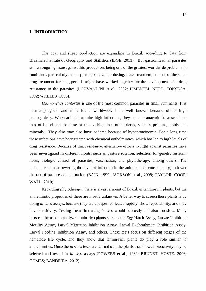
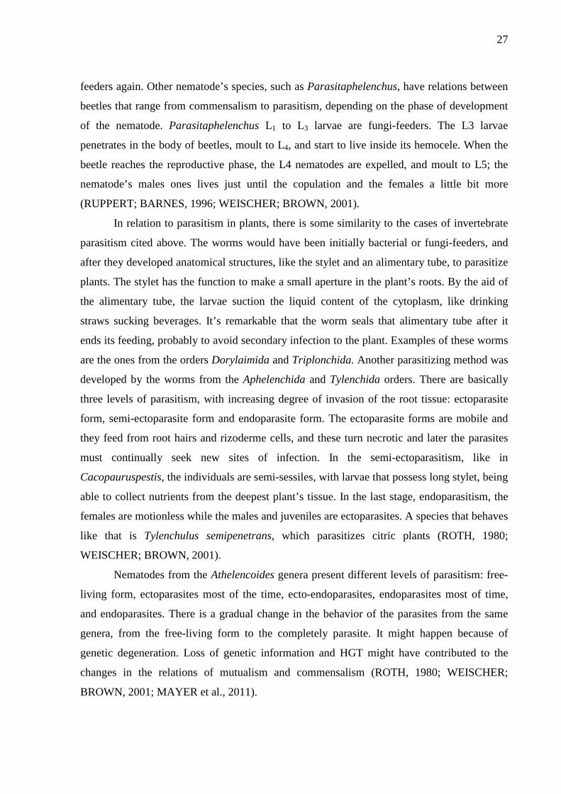
Moulting is the process in which the larva loses its cuticle and becomes the following
stage’s larva (Figure 1). The cuticle itself is a complex structure: it has three main layers, with
collagen in each one, and an epicuticle. The L5 stage does not moult, but its cuticle keeps
growing as the animal grows. In H. contortus, the L1 and L2 larval stage are bacterial-feeders,
and they feed from the bacteria from the faeces of the host. The cuticle of the L3 larvae is a
reminiscent of the L2’s cuticle. The L3 larva is fully encased in the L2’s sheath, with all anal,
oral, and excretory pore orifices being completely closed. So, the L3 larva does not feed. The
stimulus for the beginning of the L3’s exsheathment, in other words, the process of losing its
sheath, is the contact of the larvae with a solution of lower or higher pH. In an in vivo
situation, that solution may have soluble carbonic dioxide gas, undissociated carbonic acid, or

22
hydrochloric acid. With the beginning of the stimulus, the larvae respond with secretion of the
moulting fluid, and this fluid makes the cuticle detach from the larvae. The fluid is produced
close to the excretory pore, at the anterior end of the nematode, and distributed in a circular
pattern. Commonly, the cuticle detaches first of the anterior portion of the larvae like a cap,
and the larvae swims outside the cuticle. Part of the inner layer of the sheath is dissolved and
reabsorbed and the outer layer sheds. For H. contortus no enzyme from the host is necessary
for exsheathment. It all depends on the worm’s enzymes (LEE, 1965; ANDERSON, 1992;
RUPPERT; BARNES, 1996).
Figure 1 - The details vary from species to species, but the moulting process occurs basically in this sequence. (A) Intact cuticle. (B) Secretion of new epicuticle and digestion of the basal layer. (C) Secretion of the new outer and inner cortex, partial digestion of the old middle layer. (D) Secretion of the new basal layer to complete the new cuticle. (D) and (E) Discarding the old cuticle. Adapted from Ruppert and Barnes (1996)
According to Lee (1965), in in vivo conditions, the L3 larvae need 15 minutes of pH
lowering stimuli to start the exsheathment, but the whole process may take up to three hours.
In an in vivo experiment with cannulated sheep, Brunet et al. (2007) found that approximately
80% of larvae were exsheathed after 160 minutes, with rumen average pH of 5.72.
Haemonchus contortus larvae need a high concentration of H2CO3 in order to start the
exsheathment. That may explain why H. contortus has such a narrow host range, because
these conditions are hard to be found in species other than ruminants.
23
Parasites have specific dietary sources. Many adaptations have been occurring in order
for this to happen. There were some changes in feed behavior and in the response to the
environmental conditions. Morphologic changes in the cephalic region of the worms have
developed, allowing these animals to feed in different fonts and ways. For example, their
pharyngeal glands produce a secretion that is mixed with the food while it is passing through
the pharynx, and it is believed that this secretion is for helping in the digestion process; plant
nematodes have a stylet; bacterial-feeding nematodes from the digestive tract of invertebrates
have specific structures in the cephalic region; others feed from the food that the host has fed,
as seen in Ascaridoidea (LEE, 1965; GAUGLER; BILGRAMI, 2004).
The oral cavity of the parasites has a huge morphologic and function variation and also
varies to the presence of teeth. Small denticles may exist to break down food or to retain it.
These teeth can happen in different formats, the simplest being a single solid cuticular
protuberance of the stoma in the inner oral cavity. In this last case, the protuberance breaks
down food, and liberates small portions of it, so it can enter the oral cavity by lip or
pharyngeal suction. Nematodes that parasite plants usually pierce the cell wall and sucks the
content through its hollow stylet. The tooth of adult nematodes that lives in the digestive tract
of vertebrates is not just for feeding, they also help the nematode to be fixed in the sites they
live (for example, the Ancylostoma). Others, such as H. contortus, developed a lancet to cause
tissue lesions from where the parasites are fed (LEE, 1965; GAUGLER; BILGRAMI, 2004).
There is a phenomenon of sequestration of the L3 larvae when the population of adult
nematodes is high, in H. contortus. If the population is high, the immunological system is
being overstimulated, making harder for new larvae to fixate. So, the L3 larvae enter the
gastric mucosa and stay there until the adult population becomes senescent and dies. When
this happens, the immunological system will be less stimulated and the fixation will be easier.
So, the L3 larvae restart their development, by becoming L4 larvae, later adult larvae, and
fixing in the abomasum. Apparently, there is a relation between the number of adult
nematodes fixed and the number of larvae sequestrated. Another situation of sequestration
may happen is in severe drought periods or in temperate places, i.e., the larvae are
sequestrated during autumn and restart the development during spring. By the end of autumn,
the larvae stay in this situation of sequestration. In spring, the larvae become adults, when the
environment outside the animal is supportive to egg development. It has also been observed a
correlation between parasites egg laying and the period when sheep are lambing. It has been
observed that there is an increase in egg laying by nematodes in the peripartum and lactation
periods. It is known that there is an interrelation between the egg laying of nematode females
24
and the levels of prolactin produced by the host. So, as the levels of prolactin go higher
because of peripartum and lactation period, the nematode’s egg production also increases. The
young animals are more susceptible to infection, since their immunological systems are less
developed (ANDERSON, 1992; WEISCHER; BROWN, 2001).
3.1.1. Habitat and ethology of nematodes
Nematodes are distributed in vastly different environments. Free living forms can be
found in land, fresh water, and salt water. They can be found in deserts, Polar Regions, and in
the tropics as well as in high mountains and the depths of the ocean. Some of them are present
in places of extreme heat, such as hot springs of water, which can reach 53 °C. This
characteristic of the nematodes can be seen in a quote of Mr. Nathan A. Cobb, a famous
nematologist: “In short, if all the matter in the universe except the nematodes were swept
away, our world would still be dimly recognizable, and if, as disembodied spirits, we could
then investigate it, we should find its mountains, hills, vales, rivers, lakes, and oceans
represented by a film of nematodes. The location of towns would be decipherable, since for
every massing of human beings there would be a corresponding massing of certain
nematodes. Trees would still stand in ghostly rows representing our streets and highways. The
location of the various plants and animals would still be decipherable, and, had we sufficient
knowledge, in many cases even their species could be determined by an examination of their
erstwhile nematode parasites” (RUPPERT; BARNES, 1996, p. 283, our translation).
But different environments demand different nematode behavior. So, parasites with
riskier behavior – those who have to migrate, invade, infect the host, for example – usually
have larger females, which are capable of laying a greater amount of eggs. Haemonchus
contortus has a behavior of negative geotropism in the last free living stage, i.e., L3. After
living as bacterial-feeders (L1 and L2 stages), the L3 larvae migrate through a water film to the
top of the grass leaves, to make it easier to be eaten by the host (LEE, 1965; GAUGLER;
BILGRAMI, 2004).

25
3.2. Parasitism and related issues
Parasitism is a type of interaction overspread in the Animal Kingdom. For example, all
classes from the Chordates are parasitized. Particularly, animals from salt and fresh water, and
those from land, wild or domestic, are attacked by parasite nematodes. These nematodes can
be so harmful to the host that it may be killed (LEE, 1965; WEISCHER; BROWN, 2001).
Sheep may be affected by different species of nematode parasites, such as H. contortus
(abomasum), Teladorsagia circumcicta (abomasum), Trichostrongylus colubriformis
(duodenum), Strongyloides papillosus (small intestine), Oesophagostomum venulosum (large
intestine), among others. The adult forms of H. contortus have between 10 to 20 mm of length
for males and 18 to 30 mm of length for females. This parasite has a cephalic lancet, in order
to punch the internal wall of the abomasum and, by doing so, to suck blood from the host
(FORTES, 1987; WEISCHER; BROWN, 2001; TAYLOR; COOP; WALL, 2010).
Parasitosis may cause different type of issue to the host, depending on the tract it is
fixed, depending on the host, etc. For example, in the case of H. contortus, each adult worm
sucks from the host 0.05 mL of blood/day. So, an animal infected with a population of 5,000
adult larvae loses 250 mL of blood/day. If nothing is done, the animal would go under a
severe anemia, hypoalbuminemia due to great loss of plasma proteins (which leads to
oedema), vitamins, minerals, etc. If there are bad feeding conditions, the animal would die
quickly. It is known that, usually, adult parasites are found in only one organ of the
gastrointestinal tract of the host, i.e., adult H. contortus is found exclusively in the abomasum
(LEE, 1965; WEISCHER; BROWN, 2001; TAYLOR; COOP; WALL, 2010).
3.2.1. Trichostrongylodea family
The Trichostrongylodea family is the largest of the bursade nematodes. Most are
parasites of land vertebrates, such as bats, rats, ruminants, marsupials and monotremes. They
are rarely found in amphibians, reptiles and birds. The parasites of this family, one being H.
contortus, are usually found in the gastrointestinal tract of their hosts. Some can be found
parasitizing bronchi, biliary ducts, nasal cavity, and mammal glands. The hosts get infected by
ingesting the infective larva, i.e., L3 larvae (LEE, 1965; ANDERSON, 1992).

26
3.2.2. Parasitism origin
The studying of the origin of parasitism is a complex subject, since it’s hard to find
fossil record, at least for parasites. This happens because parasites are very small animals with
few mineral-inorganic structures, making it hard to be preserved as a fossil (ROTH, 1980;
WEISCHER; BROWN, 2001).
Some authors postulate that, initially, parasites held a mutualism relation with the
hosts, i.e., maybe at an old period in the history of life, the nowadays parasites were not in
this type of relation with their hosts. They were in mutualism or commensalism interactions,
and were probably bacterial-feeders. Nowadays, those types of interactions still exist, as seen
in the microbiota in the gastrointestinal tract of ruminants and non-ruminants or in the
microbiota in the roots of many plants. It can be postulated by the fact that parasites usually
have very specific sites, in very specific hosts. Hardly, for example, H. contortus would be
able to parasitize non-ruminant or plant species. So, maybe there was a change in the relations
of the parasite-host by loss of genetic variation or Horizontal Gene Transfer (HGT). By HGT,
genes from other species, such as bacteria and fungi, would be able to pass to new species,
like nematodes. It has been seen that plant parasitic nematode’s cellulase genes was gained by
HGT from different microbial donors, when phylogenetic reconstruction was held (ROTH,
1980; WEISCHER; BROWN, 2001; MAYER et al., 2011).
A characteristic favorable to animal and plant parasitism is the appearance of teeth-
like structures. Before the development of such structures, the parasites were bacterial-
feeders. For example, the parasite Rhabditis strongyloides have juvenile stages that penetrate
in the skin of small rodents and become inactive until the host dies. After it dies, the larvae
feed on bacteria. Also, in the case of animal hosts, the negative geotropism behavior was a
great advance leading to parasitism and the presence of epidermal syncytium was an
important feature to allow animals to live in stressful osmotic environments, such salt water
and inside host’s gastrointestinal tract. Other evidence is found in some fungi-feeders
nematodes. Many species of the genera Cryptaphelenchus have a commensal relation with
beetles that dwells in tree’s bark. When there’s deterioration of the fungi, for example, in the
case of drought, the worms start to produce juvenile L3 larvae, and these get fixed to adult
beetles and by that they are transported to new trees to start a new cycle, establishing a
commensalism relation. This way is only commensalism. But it is known that some species in
this genera started to invade the beetles, developing parasitism. They get installed in the
beetle’s excretory tract until the exsheath to the L4 stage, and after that they become fungi-
27
feeders again. Other nematode’s species, such as Parasitaphelenchus, have relations between
beetles that range from commensalism to parasitism, depending on the phase of development
of the nematode. Parasitaphelenchus L1 to L3 larvae are fungi-feeders. The L3 larvae
penetrates in the body of beetles, moult to L4, and start to live inside its hemocele. When the
beetle reaches the reproductive phase, the L4 nematodes are expelled, and moult to L5; the
nematode’s males ones lives just until the copulation and the females a little bit more
(RUPPERT; BARNES, 1996; WEISCHER; BROWN, 2001).
In relation to parasitism in plants, there is some similarity to the cases of invertebrate
parasitism cited above. The worms would have been initially bacterial or fungi-feeders, and
after they developed anatomical structures, like the stylet and an alimentary tube, to parasitize
plants. The stylet has the function to make a small aperture in the plant’s roots. By the aid of
the alimentary tube, the larvae suction the liquid content of the cytoplasm, like drinking
straws sucking beverages. It’s remarkable that the worm seals that alimentary tube after it
ends its feeding, probably to avoid secondary infection to the plant. Examples of these worms
are the ones from the orders Dorylaimida and Triplonchida. Another parasitizing method was
developed by the worms from the Aphelenchida and Tylenchida orders. There are basically
three levels of parasitism, with increasing degree of invasion of the root tissue: ectoparasite
form, semi-ectoparasite form and endoparasite form. The ectoparasite forms are mobile and
they feed from root hairs and rizoderme cells, and these turn necrotic and later the parasites
must continually seek new sites of infection. In the semi-ectoparasitism, like in
Cacopauruspestis, the individuals are semi-sessiles, with larvae that possess long stylet, being
able to collect nutrients from the deepest plant’s tissue. In the last stage, endoparasitism, the
females are motionless while the males and juveniles are ectoparasites. A species that behaves
like that is Tylenchulus semipenetrans, which parasitizes citric plants (ROTH, 1980;
WEISCHER; BROWN, 2001).
Nematodes from the Athelencoides genera present different levels of parasitism: free-
living form, ectoparasites most of the time, ecto-endoparasites, endoparasites most of time,
and endoparasites. There is a gradual change in the behavior of the parasites from the same
genera, from the free-living form to the completely parasite. It might happen because of
genetic degeneration. Loss of genetic information and HGT might have contributed to the
changes in the relations of mutualism and commensalism (ROTH, 1980; WEISCHER;
BROWN, 2001; MAYER et al., 2011).

28
3.2.3. Possible benefits of parasitism
Parasitism, in some cases, may bring some benefits to the host, but in general they are
not as perceptible as the damage. In the Persian Gulf, a research was held with sharks that
were in an area contaminated by lead and cadmium. Nine sharks were fished and it was
noticed that these were hosting two parasite species, Anthobothrium sp. and
Paraorigmatobothrium sp., both cestodes. They have collected some samples from the
sharks’ tissues (gonads, intestine, muscle and liver) and some samples from the cestodes for
heavy metal concentration. They realized that the parasites had about 278 and 332 times
higher cadmium and lead concentration, respectively, for Anthobothrium sp, and 455 and 438
times higher concentration respectively for cadmium and lead in Paraorigmatobothrium sp. if
compared to the shark’s muscle tissue (ROTH, 1980; MALEK et al., 2007).
In an experiment with Pseudacrisregilla (known as the Pacific Tree Frog), researchers
found that in the presence of higher parasite’s diversity, the hard effects of the most virulent
ones was kind of diluted by the competition with less virulent species. The opposite also
happened, if the diversity of parasite is lower, the hosts are more affected by the presence of
one more virulent, since there is no competition (JOHNSON; HOVERMAN, 2012).
One interesting characteristic of nematodes found in the soil or in the roots of plants is
that they play an important role in the mineralization and in the addition of nitrogenous
compounds to the soil. Also, there is a microbiota associated with the worms, and they also
mineralize the soil. So, plants that grow in these conditions have a higher growth rate than
others that do not (GAUGLER; BILGRAMI, 2004).
3.2.4. Animal’s ethological defenses against parasites and pathogens
Animals have developed ways to remove or avoid parasites or pathogens. These
ethological defenses include behaviors such as movements to remove ectoparasites, licking,
ingestion of medicinal herbs to eliminate worms, fumigation of the nests to expel flies,
quarantine (animal style), and immunization (animal style). These behaviors are used
sometimes as therapeutic or prophylactic measure. Studies on animal self-medication could be
a good way for discovering new medicines, both for humans and animals (HART, 2011; DE
DE ROODE; LEFÈVRE; HUNTER, 2013).
29
One interesting behavior is present in grazing animals. Ruminants and horses have
been known to avoid grazing on places close to recent dropped faeces. Carnivores do not
defecate in their den. They exit the den to defecate and urinate, in order to avoid contact with
the excrements. But the newborns are not so mobile. The female may practice coprophagy
quickly after one defecates. The female is not affected by parasites, since she is ingesting
eggs, not the infective hatched form. Licking may be another way to avoid diseases. It has
been shown that dog’s saliva is antibiotic, been effective against Streptococcus canis and
Escherichia coli. Female dogs lick their nipples, in a kind of bactericidal wash against
common disease-causing microorganisms, just before the newborn start lactating. Male rats
lick compulsively their penis after copulation, in order to avoid sexual transmitted diseases
(EZENWA, 2004; HART, 2011).
Some animals grab materials from the environment in order to fumigate their nest. For
example, it is has been noted that house sparrows and finches usually grab cigarette butts to
their nests. By increasing the levels of nicotine there, the mite infestation is lowered.
California dusky-footed wood rats bring leaves of bay to their nest, apparently as a mean of
control against an overabundance of fleas. Starlings also bring leaves to their nest when young
starlings are present. In this case it is believed that the leaves should retard the hatching of
louse eggs and have an antibacterial effect (HART, 2011; DE ROODE; LEFÈVRE;
HUNTER, 2013).
Animals are able to make an animal style self-immunization and self-medication. For
example, for more than just protecting the mates, nesting area and food resources, territorial
defenses may play a role on a type of quarantine, protecting a specific den against a
conspecific animal by peripheralizating it, since it may carry strange pathogens with it.
Another kind of quarantine not very accounted is the cannibalism of newborns. A sick
newborn may be a reservoir of pathogens against its siblings. So, by detecting ill signs like
hypothermia and inactivity, the female may cannibalize this animal. This behavior is observed
in dogs, cats, and rats. Peripheralizating of a stranger may also play a role on immunization.
Both the conspecific and the other animals will experience small samples of foreign
pathogens, because of the enforced space barrier. This immunization occurs by faecal
droppings and share of same water source. So, the foreign animal and the native animals will
experience each other pathogens slowly, being both sides immune to the new pathogens, in
case of the stranger is allowed in the group. Another way of slow immunization is seen in
mothers that brings prey to the young ones. It surely does help making a bridge between
weaning and hunting, but also presents new, yet local, pathogens to the animals. About self-
30
medication, some animals graze on specific medicinal herbs in order to eliminate parasites in
two ways, physically or chemically. It is well noted that dogs, cats, and chimpanzees would
eat whole rough leaves to maintain low worm loads, most times in a prophylactic way.
Sometimes, the leaves are seen associated with expelled parasites. Other studies show that
small-ruminants may graze on tannin-rich forages if they are available, when the infection is
present. This grazing of tannin-rich forages declines with the decline of the worm burdens in
the animals. So, it’s a type of self-medication by the animals. Animals must benefit by the
availability of tannin-rich fodder on its pasture, because they would self-medicate
(LISONBEE et al., 2009; HART, 2011; JUHNKE et al., 2012).
3.3. Ethnoveterinary
Ethnoveterinary can be categorized as an oral tradition, which has been passed down
generation to generation, mostly in places where the access to veterinary services is absent or
expensive (TOYANG et al., 2007). Most of the techniques use medicinal herbs, which are
easy to find. Plants utilized in the ethnoveterinarian’s treatment of livestock against
helminthes are rich in secondary metabolites from plants, most of them being tannins, and it’s
known that tannins play a role as an anthelmintic. An advantage of using tannin-rich plants is
that they leave no residual in meat, as chemical anthelmintic drugs would so (BAHUAUD et
al., 2006; HOSTE et al., 2006; BRUNET et al., 2007).
3.3.1. Tannins
Tannins are secondary compounds found in plants. They play a role in the plant’s
secondary metabolism, acting against herbivory and diseases, and also by mimetizing
hormones. They are polymers of phenolic monomers, arranged differently from plant to plant.
That is why different plant’s extracts have different responses in in vitro and in vivo assays.
There are a very wide array of phenolic compounds in plants, from the lignin in the wall of
the plant´s cells to the anthocyanidins (flower pigments). Tannins are polyphenolic
compounds with well-defined characteristics: they are able to interact and precipitate
macromolecules (for example, proteins and enzymes) such as the ones found in the animal
skin. Also, it is soluble in water, acetone, and alcohols (BRUNETON, 1999; HAGERMAN,
2002a; BRUNET; HOSTE, 2006).

31
Tannins are divided basically in two groups, Condensed Tannins (CT) and
Hydrolysable Tannins (HT). The CT are polymers formed by the condensation of monomers
of flavans. The HT are polymers formed by gallic and ellagic acid monomers and
carbohydrate (usually glucose). It is important to note that each plant has its own monomer
pattern, producing its own tannin molecule. That might explain why different plants have
different anthelmintic results against nematodes (BRUNETON, 1999; HAGERMAN, 2002b;
BRUNET; HOSTE, 2006; HAGERMAN, 2010).
Initially, tannin-rich plants were used by mankind in leather tanning process due to its
characteristic to bind and alter the spatial arrangement of proteins. Nowadays, it is being used
for ethnoveterinary practices, with interesting results. Also, since consumers are these days
more concerned about drug residues and animal green production, tannins may play an
important role in preventing or treating animals against nematodes in green animal production
farms. It also may be used as antimicrobial substance since inhibitory effects against
Staphylococcus aureus and Escherichia coli were found (BAHUAUD et al., 2006; LONE et
al., 2013).
3.3.1.1. Leather tanning mechanisms
Knowing the mechanisms of leather production is important to help us to understand
how tannins may work against nematodes, because the principles are similar, at least
hypothetically. The process aims at making the hides of animals a material heat-resistant, with
greater durability and also malleable, which are important characteristics of leather
(BRUNETON, 1999).
Briefly, the skin is divided in three layers, the epidermis, dermis, and subcutaneous.
About one third of the dermis is formed by collagen. Collagen is a triple helix protein rich in
glycine, alanine, arginine, proline, and hydroxyproline amino acids, and it is known that
tannins have great attraction to these last two amino acids. Collagen has also a loose
arrangement, giving it great motility. The tanning process changes the arrangement of the
molecules of collagen, making it rot proof by making new hydrogen and covalent bonds
between the collagen fibers, making it stick together. Briefly, it changes the molecular
arrangement, giving it new characteristics, different from the ones it had to have before,
physiologically speaking (HOSTE et al., 2006; MANN; MCMILLAN, 2013).
32
3.3.1.2. Tannin’s role against nematodes
Tannins play a role acting in different stages of the nematode life cycle. It can get
fixed to the proteins (or enzymes) from the egg shell or from the sheath, preventing the
hatching and moulting processes, by preventing the enzyme activity or its conjugation to
targeted proteins; it can agglutinate to the proline and hydroxyproline amino acids which are
found in high concentrations in the oral and vaginal region of the nematodes, preventing the
worms from feeding and reproducing. Another way tannins may work is by delaying the
exsheathment of the infective larvae. It was found that less than 40% of larvae exsheathed
after 160 minutes in cannulated animals fed with 100% diet of the tannin-rich plant sainfoin.
Tannins also may increase the amount of bypass protein for the animal, giving it a greater
protein supply for its immunological system. In a recent report, Martínez-Ortíz-de-Montellano
et al. (2013) showed the action of contact of adult females H. contortus’ larvae with
tanniferous fodders, both in vitro and in vivo. It was shown that there was aggregations to the
larva’s cephalic region, anus, vulva and alterations in the cuticle, such as the appearance of
longitudinal and transversal folds, thickening, and wrinkling for the in vitro test, and changes
in the cuticle, such as the folds, wrinkling, and thickening for the in vivo assay. This changes
may help understand the results found in in vivo experiments, like Faecal Egg Count (FEC)
reduction, either by damage to the reproductive tract and by damage in the cephalic region
which would led to malnutrition, and smaller nematode burdens (BAHUAUD et al., 2006;
HOSTE et al., 2006; BRUNET et al., 2007; LISONBEE et al., 2009; MANOLARAKI et al.,
2010; HOSTE et al., 2012).
3.3.2. Plants
The extracts utilized in the present work were from Acacia mearnsii, Myracrodruon
urundeuva, Caesalpinea bracteosa and Leucaena leucocephala.
Acacia mearnsii, from the Mimosoideae family, is a leguminous tree native to
Australia and is commonly known as Acácia Negra in Brazil. It has been utilized in nitrogen
fixation, in the recovery of degraded soil, as firewood, and also as a tannin source, being those
usually used in the leather industry (CENTRO DE INTELIGÊNCIA EM FLORESTAS,
2013). Also, some research has been held in in vivo and in vitro assays. The results from these
33
authors showed that the plants might have bioactive compounds against nematodes
(ALONSO-DÍAZ et al., 2011; CENCI et al., 2007).
Caesalpinea bracteosa, from the Caesalpinoideae family, is widely found in the
Brazilian Caatinga. Its common name in Brazil is Catingueira. It is a small slow growing
tree, and its wood is often used as flooring, its leaves as fodder for ruminants, and many parts
of the plant for ethnomedicine. It was the most cited plant in different population groups in
different types of treatment, from the cure of abdominal pain to the cancer treatment
(CENTRO NORDESTINO DE INFORMAÇÕES SOBRE PLANTAS, 2013; GOMES;
BANDEIRA, 2012).
Myracrodruon urundeuva (which has been classified before as Astroniam urundeuva),
from the Anacardiaceae family, is present both in Brazilian Caatinga and Cerrado, being
commonly known as Aroeira in Brazil. It has 17% of tannins in the bark, therefore considered
one of the most resistant woods against putrefaction from Brazilian flora (CARVALHO,
2003). All the tissues of the plant have abundant level of tannins. Those are responsible for
the protection of plants against herbivores and microorganisms. They have a healing,
antiseptic, antioxidant, and anti-inflammatory actions. The plant has great use in
Ethnomedicine in the treatment of gynecological issues, abdominal pain, diarrhea, and
conjunctivitis (GOMES; BANDEIRA, 2012).
Leucaena leucocephala, from the Mimosaceae family, is one of the most used
leguminous trees as fodders and its common name is Leucena in Brazil. Also, it is used as
wood, firewood, green fertilizer, shade, human food, and erosion control. The L. leucocephala
extract had a fungicidal effect against colonies of the fungus Leucoagaricus gongulophorus
(SOUZA et al., 2012). In an in vitro assay, Alonso-Diaz et al. (2008b) showed that the L.
leucocephala extract had a dose-dependent effect on the migration of H. contortus L3 larvae.
3.4. In vitro tests
The way compounds will interact with the animal can be assessed through in vivo
tests. But, in many places, these type of testing may be too expensive, too slow or have many
ethics related issues. So, in vitro testing may be a good, fast, less laborious, and more
economical manner of screening plants of ethnoveterinary potential use. But one must bear in
mind that in vitro tests may overestimate or underestimate what would happen if tested in the
animal, since in vitro tests do not respond to the complexity found in animal’s physiology. An
ideal in vitro test is one that has low cost, high sensibility, rapid collection of results and
34
repeatability. So, one can select plant samples that have showed effectiveness in in vitro and
test it later in in vivo (POWERS et al., 1982; JACKSON; HOSTE, 2010). Also, standard
protocols should be followed by different researchers, so the data from the assays may be
comparable. In order to determine such standards, Powers et al. (1982) published a guideline
to evaluate anthelmintic activity. These authors postulate that groups should have at least
three doses of treatment. This guideline was adopted by the World Association for the
Advancement of Veterinary Parasitology (WAAVP).
The Brazilian flora has a vast amount of tannin-rich plants that may have use for
ethnoveterinary purposes. Quantifying and qualifying the anthelmintic activity of those plants
may be valuable, but it may be too expensive to do it first with in vivo methods. In vitro
testing may be a better alternative. After in vitro screening, plants that showed great
anthelmintic activity would be selected and tested in vivo (GOMES; BANDEIRA, 2012;
SILVA et al., 2012; FERREIRA et al., 2013).
The Egg Hatch Assay (EHA) is an in vitro test that focuses on the egg stage and its
hatching. The egg solution are kept in contact with different pre-tested concentrations of plant
extracts for 24 hours with suitable conditions for egg hatching. The readings after 24 hours
are done in an inverted microscope and it’s possible to differentiate the eggs from the larvae.
The tannins interact with the collagen from the egg shell, and it may change the
morphological structure, making it not able to hatch. Thinking about the nematode cycle, eggs
would have contact with tannins in the intestines of the animal and also in its faeces. So, it
would not be able to hatch, or hatching taxes would be at least lowered, making it to drop the
contamination of the pastures with L3 larvae, and further to drop the infection taxes of the
animals (JACKSON; HOSTE, 2010; FERREIRA et al., 2013). Some examples of reports with
different plants tested in in vitro are found in Table 1. These reports may be an example of the
importance of the EHA in the screening of potential new anthelmintic material.
The Larval Exsheathment Inhibition Assay (LEIA) is an in vitro assay that focus on
the ecdysis process between the L3 and L4 larvae, also known as exsheathment. In the test, an
artificially exsheathment stimuli is given to L3 larvae previously exposed to tannin-rich
extracts for three hours. Tannins would interact with the collagen of the cuticle and alter that
in a way it wouldn’t be able to exsheath. In the in vivo process, if an animal is being fed with
a tannin-rich fodder, the tax of exsheathment will probably drop, depending on the tannin
molecules from the fodder. If the nematodes are not or just some of them are capable to
exsheath, the infection will not be established or it will be established, but at lower taxes,
which would be still a benefit for the animals (BRUNET et al., 2007; JACKSON; HOSTE,

35
2010). Some examples of reports in which were tested plant extracts or essential oils against
the exsheathment of L3 larvae are found in Table 1. This reports may show the importance of
the LEIA for the screening of tanniniferous plants.
Table 1 – Examples of reports that used in vitro assays for the screening of plants
Author Year Assay Plant
ADAMU; NAIDOO; ELOFF
2013 EHA
Brachylaena discolor, Melia azedarach, Cyathea dregei, Milletia grandis, Indigofera frutescens,
Apodytes dimidiate, Heteromorpha trifoliata, Maesa lanceolata, Leucosidae sericea, Zanthoxylum capense, Clerodendrum glabrum, and Strychnos mitis extracts
ALONSO-DÍAZ et al. 2008a LEIA Acacia pennatula, Leucaena leucocephala, Lysiloma
latisiliquum and Piscidia piscipula extracts
ALONSO-DÍAZ et al. 2008b LEIA Acacia pennatula, Leucaena leucocephala, Lysiloma
latisiliquum and Piscidia piscipula extracts
ALONSO-DÍAZ et al. 2011 LEIA Acacia gaumeri, Brosimum alicastrum, Havadia albicans, and Leucaena leucocephala extracts
BAHUAUD et al. 2006 LEIA Castanea sativa, Erica Erigena, Pinus sylvestres, and
Sarothammus scoparius extracts
BRUNET et al. 2007 LEIA Onobrychis viciifolia extract
DE OLIVEIRA et al. 2011 EHA Myracrodruon urundeuva extract
DE OLIVEIRA et al. 2011 LEIA Myracrodruon urundeuva extract
DOMINGUES et al. 2013 EHA Ananas comosus industrial residue
FERREIRA et al. 2013 EHA Annona muricata extract
KATIKI 2011 EHA Cymbopogon schoenanthus, Mentha piperita, and
Cymbopogon martinii essential oils
KATIKI 2011 LEIA Cymbopogon schoenanthus, Mentha piperita, and
Cymbopogon martinii essential oils
OLIVEIRA et al. 2011 LEIA Anadenanthera colubrine, Leucaena leucocephala, and
Mimosa tenuiflora extracts
PESSOA el al. 2002 EHA Ocimum gratissimum extract

36
3.5. Objectives
The objectives of the present work were to determine the bioactivity of the plant
extracts of A. mearnsii, M. urundeuva, C. bracteosa, and L. leucocephala against the hatching
and the exsheathment of the nematode H. contortus.
37
4. MATERIAL AND METHODS
4.1. Plant samples
Myracrodruon urundeuva, C. bracteosa, and L. leucocephala were harvested from the
semi-arid region of the Brazilian state of Pernambuco (latitude 8 ° 4 'S and longitude
34 ° 53' W) in four different counties, namely Floresta, Serra Talhada, Itacuruba and
Petrolândia. This region presents two distinct annual periods based in the rainfall rates. The
rainfall period lasts from three to four months, between February and May; the drought period
lasts from eight to nine months, between June and January. It is worth noting that this drought
period may last up to 18 months, because rainfall may be irregular and torrential.
The plants were collected from four different sites in each collection area. In each site,
material from five different plants from the same species were collected, emulating the
animal’s grazing behavior. Three kilograms of leaves and branches were collected from each
plant. The fresh material was dried in shade for 96 hours inside cardboard trays. After that, the
samples were stored in paper bags and were transported to Center for Nuclear Energy in
Agriculture, in Piracicaba, São Paulo, Brazil, where it were reduced to 1,0 mm powder by a
Wiley grinder. The powdered material where store inside plastic flasks in a light-free place at
room temperature. The plants utilized in the experiment were M. urundeuva,
L. leucocephala, A. mearnsii, and C. bracteosa. The A. mearnsii plant material was obtained
commercially from SETA S/A EXTRATIVA TANINO DE ACÁCIA.
4.2. Laboratory facilities
The extracting process and the EHA were held in the Bromatology Laboratory of the
Instituto de Zootecnia, in Nova Odessa, São Paulo, Brazil, between June 2012 and April
2013. The LEIA was held in the Animal Nutrition Laboratory of the Center for Nuclear
Energy in Agriculture, in Piracicaba, São Paulo, Brazil, between April 2013 and June 2013.
4.3. Bioactive extracts
For the in vitro tests, the tannin content from the plants was extracted following the
methodology of Barrau et al. (2005). The fresh plant were dried and ground fine, using sieves

38
of 1 mm drill. Five grams of this material were placed inside a beaker that was later covered
with a tinfoil for protection against the sunlight. This material were extracted with 50 mL of a
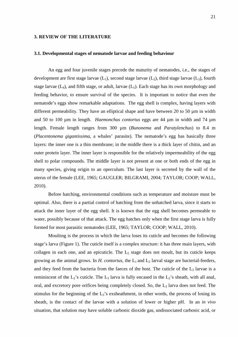
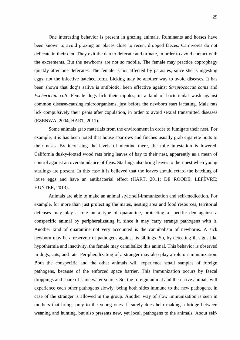
70:30acetone: distilled water solution for one hour in the magnetic stirrer (Figure 2).The
acetone causes cell lyses, letting the molecules of chlorophyll, lipids, and tannins free in the
solution (BARBIERI et al., 2010). After this phase, the material was filtrated in a number
two filter paper with the aid of a Buchner funnel, a vacuum pump, and a kitasato. The solid
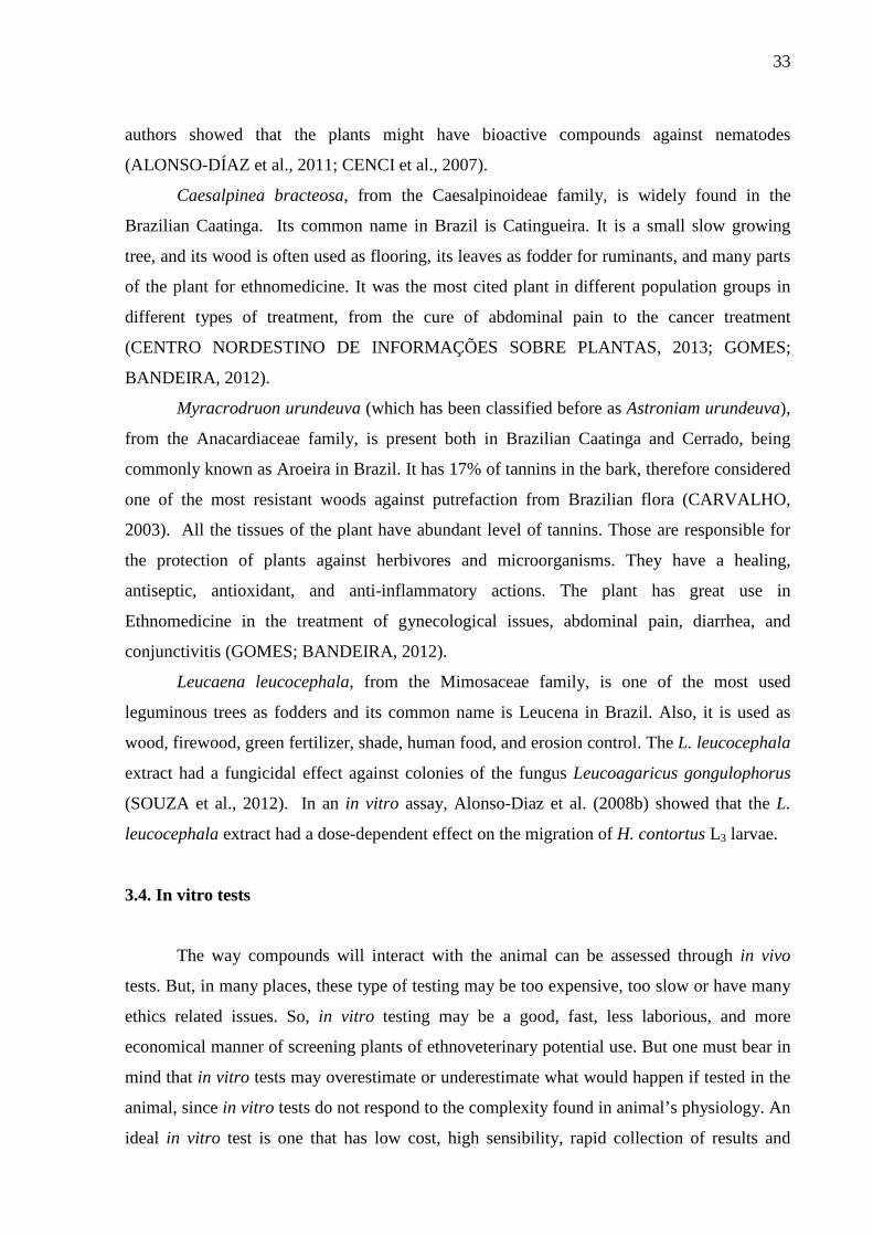
residues were discarded and the liquid solution were placed in the BÜCHI HB 140 rotary-
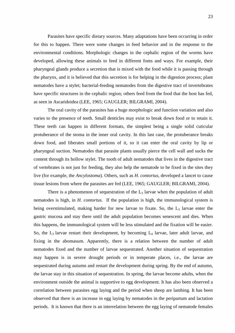
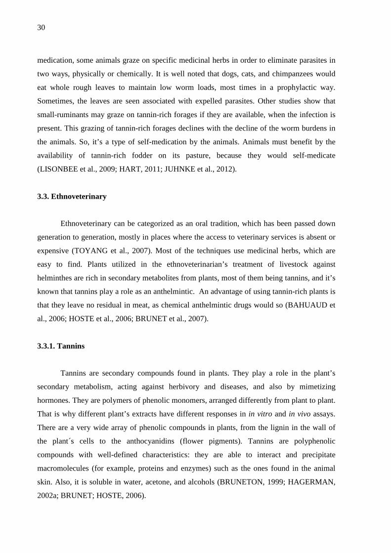
evaporator until the acetone evaporated (Figure 3). The water in the rotary-evaporator were at
35 °C. After the acetone evaporated, the resulting solution was placed inside a separating
funnel and washed three times in dichloromethane, with 50 mL of dichloromethane each time.
Since the dichloromethane is a non-polar solution and more dense (1.325 g/mL at 25 °C) than
the water, and the tannins being polar molecules, the tannins are kept in the water phase, and
lipids and chlorophyll (non-polar molecules) are discarded with the dichloromethane
(SIGMA-ALDRICH, 2012; FAO, 2009). After this washing phase, the extracts were placed in
bottles, frozen and then lyophilized in a Labconco - Freeze Dryer 5 lyophilizer
(LABCONCO CORPORATION, Kansas City CAT. N. 75050). The A. mearnsii extract was
bought from SETA S/A EXTRATIVA TANINO DE ACÁCIA. These lyophilized and
commercial plant extracts were evaluated in EHA and in LEIA.
Figure 2 - Beaker covered with tinfoil on the magnetic stirrer
39
Figure 3 - BÜCHI HB 140 rotary-evaporator
The M. urundeuva extract were brittle after lyophilization, and through time became
and remained a powder. The C. bracteosa and L. leucocephala were also brittle at first, but
within few days, C. bracteosa’s became a syrup and L. leucocephala’s became sticky. They
appeared to be highly hygroscopic.
After lyophilization, total phenols (TP), total tannins (TT), and condensed tannins
(CT) were determined according to Makkar et al. (2000). Two hundred mg from C. bracteosa
and L. leucocephala and 100 mg from A. mearnsii and M. urundeuva were taken and placed
inside a glass flask. Ten mL of aqueous acetone (70%) were added to each flask and a 20
minutes ultrasonic bath were held at room temperature. The contents were transferred to assay
tubes and centrifuged at 3,000 RMP at 4 °C for 10 minutes. The supernatant was collected.
From this supernatant all analyses were done.
For TP measurements, suitable aliquots from the supernatant were taken and placed
inside assay tubes. The volume was made up to 0.5 mL with distilled water; 0.25 mL of
Follin-Ciocalteu 1N, and 1.25 mL of 20% sodium carbonate solution were added. The tubes
were agitated and keep in dark for 40 minutes. After that, the absorbance were recorded at
725 nm in a spectrophotometer (DU-64 Beckman). The amounts were expressed in gram
equivalent of tannic acid per kilogram of dry matter.

40
Total tannins measurement was achieved by the binding of polyvinyl pyrrolidone
(PVPP) (Sigma P-6755) to the tannin molecules. One hundred mg were weighted and placed
in assay tubes, and 1.00 mL of distilled water and 1.00 mL of the supernatant were added and
the tubes were agitated. The assay tubes were centrifuged (at 4 °C 3,000 RPM for 10 minutes)
and the supernatant from this last solution was taken, and it contained only simple phenols
(SP), since the tannins precipitated with the PVPP. The SP solution was read in the
spectrophotometer with the same conditions as the last one. The SP measurement were
subtracted from the TP, and the result was the TT. Total tannins are also expressed in gram
equivalent of tannic acid per kilogram of dry matter.
For the measurement of CT, 0.50 mL of the diluted extract solution were taken and
placed inside assay tubes and 3.00 mL of the butanol-HCL reagent (95:5 n-butanol:HCl 37%)
and 0.10 mL of ferric reagent (2.00 g of ferric ammonium sulfate diluted in 100 mL of HCl
2N) were added. Tubes were agitated and covered with glass marbles. After, they were placed
inside a boiling water bath for 60 minutes. The tubes were cooled after this period and
readings were performed in the spectrophotometer, with readings at 550 nm of absorbance.
Condensed tannins are expressed in in gram equivalent of leucocyanidin/kg dry matter.
4.4. Egg Hatch Assay
The EHA was done following the methodology from Jackson and Hoste (2010).
4.4.1. Egg recovery
For the recovery of eggs, two animals were kept indoors following the
recommendations from “Animal Welfare Approved Standards for Sheep” (MELLOR;
HEMSWORTH, 2005) and were mono-infected with Haemonchus contortus.
About 15 g of feces were taken directly from the rectum of these sheep, according to
Bizimenyera et al. (2006). These feces were mixed to distilled water at 40 °C and a Fecal
Aqueous Solution (FAS) was obtained. The FAS solution was passed through a 1.0 mm sieve,
separating the solid portion – which was discarded – from the liquid portion (FAS) – which
was kept in a bucket. After that, the FAS were passed through a 106 µm sieve, discarding
again the solid portion. The remaining FAS were passed through a 53 µm sieve similarly to
the previous times and then through a 25 µm sieve and the eggs were kept in this last sieve’s
mesh.

41
The FAS was placed in assay tubes, and then centrifuged for three minutes at
3,000 RPM. After the centrifugation, the upper phase was discarded and it was added
saturated saline solution to the assay tubes and a new centrifugation under the same
conditions was done. The upper phase was collected in the 25 µm sieve and abundantly
rinsed with distilled water. Two new centrifugations process were carried out under the same
conditions: after the first, the upper phase was discarded and saturated saline solution was
added; after the second, the upper phase was kept in the 25 µm sieve and rinsed thoroughly
distilled with water. This last solution will be called Egg Solution (ES).
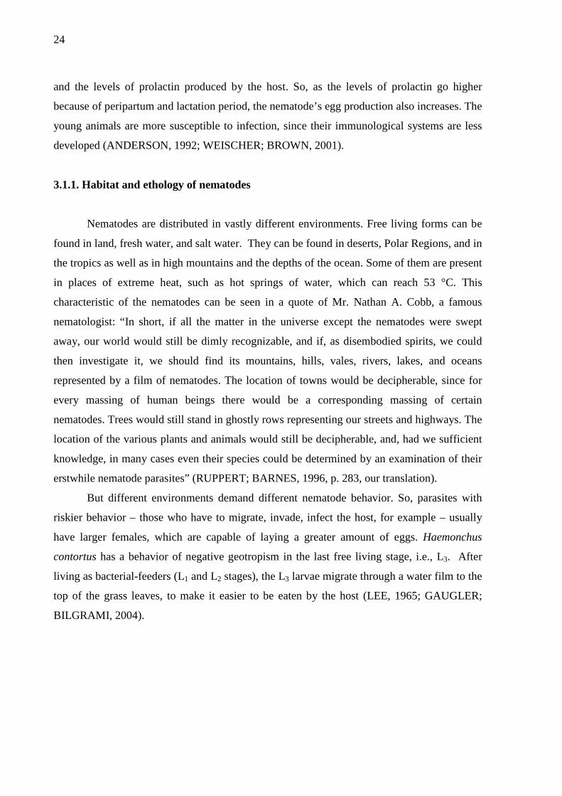
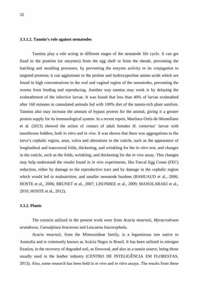
The ES was placed in a beaker. The concentration of eggs/mL was calculated by
reading of five fractions of 20 µL each in an optical microscope, at a magnification of 10x
(Figure 4). After that, the mean of number of eggs was calculated as eggs/mL.
Figure 4 - Haemonchus contortus eggs
4.4.2. Preparation of concentrations of bioactive extracts for EHA
The concentrations used in the test were obtained through a pre-test, intending that the
higher dose was the Lethal Dose 99% (LD99) and the lower dose was the Lethal Dose 0%
(LD0). So, the concentrations used for A. mearnsii were 50, 25, 12.5, 6.25, 3.12, 1.56, 0.78,
and 0.39 mg/mL; for M. urundeuva, 1.56, 0.78, 0.39, 0.19, 0.09, and 0.04 mg/mL; and for C.
bracteosa, 6.25, 3.12, 1.56, 0.78, 0.39, and 0.19 mg/mL. It was not possible to find the DL50
and DL99 for L. leucocephala through the pre-test, because the chosen concentrations were not
even reaching the DL50; if higher concentrations were chosen, the reading on the microscope

42
were not viable, since it was extremely dark to differentiate the eggs or larvae from the extract
solution. So, for this extract, three concentrations were chosen, namely 6.25, 3.12, and 1.56
mg/mL. Thirty repetitions were done for each concentration, for each extract.
4.4.3. Preparation of the 24-well plates
Twenty-four-well plates were prepared for each extract, with six wells for each
concentration at each reading round (Figure 5). An EHA’s stock solution (SSEHA) of
concentration two times higher the maximum concentration wanted for each extract was
prepared, in other words, 3.12 mg/mL, 12.5 mg/mL, 100 mg/mL, and 12.5 mg/mL for
M. urundeuva, L. leucocephala, A. mearnsii, and C. bracteosa, respectively. Methanol was
used to help on the dilution of extracts, on the proportion methanol: SSEHA of 1:50. Dilutions
of this SSEHA were done to obtain all other wanted concentrations for each extract. Two
hundred fifty microliters of solution with equal to twice higher the real concentration required
for that well. Then, 250 µL of an ES with 400 eggs/mL concentration was added to each plate
well. By doing that, each well reached the desired concentration. In each well there was
around 100 eggs and 500 µL of total solution. A negative and a positive control groups were
also prepared. The negative control consisted just of adding 250 µL of distilled water instead
of SSEHA. For the positive control a 0.78 mg/mL Thiabendazole solution were used. After
that, the well-plates were placed inside an incubator at 27 °C for 24 hours.
Figure 5 - Twenty-four-well plates

43
4.4.4. Microscope readings for EHA
The readings were performed with the aid of an inverted microscope, at magnitude
10x. The eggs were counted apart from the larvae, and this data was tabulated in files for
further statistical analysis.
4.5. Larval Exsheathment Inhibition Assay
The LEIA was done following the methodology from Jackson and Hoste (2010).
4.5.1. Obtainment of larvae
The infective larvae (L3) of H. contortus were obtained through coproculture process,
from feces of mono-infected sheep donors, according to Roberts and O’Sullivan (1950).
Between ten to 15 grams of feces were collected directly from the rectum of the donors. These
feces were moistened with distilled water and homogenized with vermiculite in a 50:50
proportion, in way it became a firm pasty material. This material was then placed inside glass
bottles and covered with a plastic film. The bottles were kept away from sunlight and stood at
room temperature (20 °C) for one week of incubation.
After seven days, each bottle was fulfilled with distilled water up to the rim, and a
Petri dish was placed on the bottle, to seal it. This set was turned upside down, allowing no or
just a little air inside it. The empty space of the Petri dish was filled with distilled water. After
six hours the distilled water from the Petri dish was collected inside assay tubes, called the
Larvae Solution (LS). The concentration in larvae/mL was calculated by reading five fractions
of 20 µL each with the aid of an optical microscope, at magnitude 10x.
4.5.2. Preparation of concentrations of bioactive extracts for LEIA
The concentrations for the LEIA test were chosen based on the standard
concentrations used in assays from papers on the same field of research. The concentration
used for all four plant extracts was 1.200, 600, 300, and 150 µg/mL and also a negative
control, that was prepared just with distilled water. Thirty repetitions for each concentration
were carried out (ALONSO-DÍAZ et al., 2008a; 2008b; MANOLARAKI et al., 2010).

44
4.5.3. Preparation of the assay tubes
In order to initiate the assay, plant extract was weighted in a precision balance in the
proportion of 2,400 µg/mL. Then, in order to help to dilute the plant extract, methanol was
added in the proportion of methanol:stock solution of 1:50. The volume of distilled water was
re-calculated and this new volume was added to the assay tube. This solution is now called
LEIA’s stock solution (SSLEIA) and it had 2,400 µg/mL.
Five assay tubes were taken and identified (Figure 6-A). It was added 1 mL of SSLEIA
in the first two tubes (Figure 6-B). Then, in one of them, 1 mL of distilled water was added,
dropping the initial concentration in half (Figure 6-C). One mL from this last tube was taken
and added in a third tube (Figure 6-D). One mL of distilled water was added to this third tube,
reducing the concentration, from 1,200 mg/mL to 600 µg/mL (Figure 6-E). This same
procedure was done as many times necessary to acquire all needed concentrations, as can be
seen in Figure 6-F to 6-H. One mL of distilled water was added to the tube destined to be the
negative control group (Figure 5-I). One milliliter of 1,000 L3/mL LS was added to each tube,
making the concentrations drop in half again, reaching the final desired concentrations (Figure
6-J). It is important to notice that each tube has two mL of solution. After this, the assay tubes
were placed inside an incubator at 23°C for 3 hours, each shaken every hour.
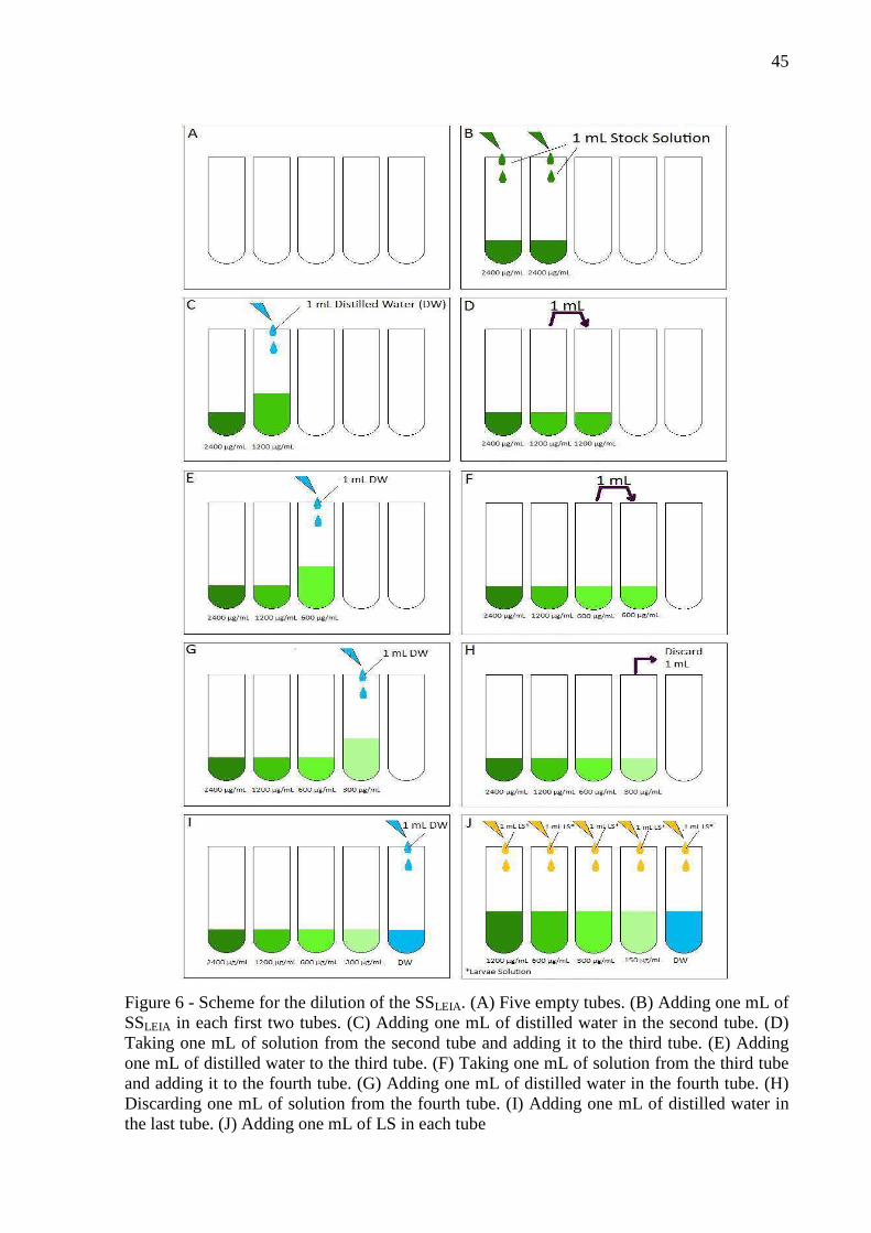
45
Figure 6 - Scheme for the dilution of the SSLEIA. (A) Five empty tubes. (B) Adding one mL of SSLEIA in each first two tubes. (C) Adding one mL of distilled water in the second tube. (D) Taking one mL of solution from the second tube and adding it to the third tube. (E) Adding one mL of distilled water to the third tube. (F) Taking one mL of solution from the third tube and adding it to the fourth tube. (G) Adding one mL of distilled water in the fourth tube. (H) Discarding one mL of solution from the fourth tube. (I) Adding one mL of distilled water in the last tube. (J) Adding one mL of LS in each tube
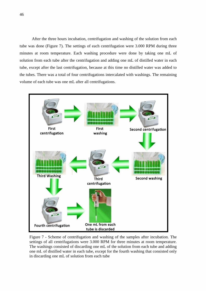
46
After the three hours incubation, centrifugation and washing of the solution from each
tube was done (Figure 7). The settings of each centrifugation were 3.000 RPM during three
minutes at room temperature. Each washing procedure were done by taking one mL of
solution from each tube after the centrifugation and adding one mL of distilled water in each
tube, except after the last centrifugation, because at this time no distilled water was added to
the tubes. There was a total of four centrifugations intercalated with washings. The remaining
volume of each tube was one mL after all centrifugations.
Figure 7 - Scheme of centrifugation and washing of the samples after incubation. The settings of all centrifugations were 3.000 RPM for three minutes at room temperature. The washings consisted of discarding one mL of the solution from each tube and adding one mL of distilled water in each tube, except for the fourth washing that consisted only in discarding one mL of solution from each tube

47
Four aliquots of 200 µL were taken from each tube and placed inside pre-identified
microtubes. There was four microtubes for each concentration of the assay and for the
negative control group. Each microtube consisted of a repetition of the assay.
4.5.4. Microscope readings for LEIA
In order to start the assay, 12 glass slides were organized on a plastic tray. These glass
slides were separated in four different time-groups, namely Starting Time, 20 Minutes, 40
Minutes and 60 Minutes, and each group had three slides. An aliquot of 30 µL were taken
from each microtube and placed on the glass slide, and so the remaining volume of the
microtubes was 170 µL. Each slide from the ST group was taken, and the larvae on it were
killed by heat stress, with the aid of a candle (Figure 8). The ashes from the bottom of the
slide that appeared after the heat stress were cleaned with toilet-paper. One hundred seventy
µL of Exsheathment Solution (ES) were added in each microtube. This ES consisted in 2.0%
sodium hypochlorite solution and distilled water in the proportion 1:74 (pH 11.02) and its
purpose was to emulate the physiological conditions of the gastric apparatus of the ruminants,
to stimulate the larvae to exsheath. The one hour period for the assay started at the time the
ES solution was added. The readings on the microscope where done at the Starting Time, 20
minutes, 40 minutes, and 60 minutes.
Figure 8 – Killing the larvae by heat stress with the aid of a candle
48
The readings for the other time-groups were done the same way of the first one:
30 µL of each microtube where taken followed by heat stress to kill the larvae. The material
from the slides was read in the optical microscope, at 10x magnitude. The L3 larvae were
differentiated in ensheathed and in exsheathed, and the data was tabulated in files for later
statistical analysis.
4.6. Statistical analysis
The SAS® Probit procedure was used to determine the dose effect and to calculate the
lethal dose (LD) of each plant extract in both in vitro tests. It was performed by fitting
regression using normal and logistic distribution. The parameters estimative of these
equations were obtained by the maximum likelihood method. Contrasts between doses and
negative control were carried out within the GLM procedure.

49
5. RESULTS
Concerning the yield of the plant extracts, M. urundeuva had 15.36%, C. bracteosa
had 5.79%, and L. leucocephala had 7.58% of yield (Table 2). The calculation of yield was
not done for A. mearnsii because it was obtained commercially. Regarding the condensed
tannins (CT), A. mearnsii had approximately 32.25%, M. urundeuva had approximately
7.27%, C. bracteosa had approximately 1.27%, and L. leucocephala had approximately
16.74%. These results are found in Table 3.
Table 2 - Yield of the plant extracts
Dry Plant Material (g) Bioactive Extract (g) Yield (%)
M. urundeuva 300.00 46.10 15.36
C. bracteosa 591.00 34.24 5.79
L. leucocephala 360.00 27.31 7.58
Table 3 – Tannin analysis of the extracts
Plant extract Total phenols* Total tannins* Condensed tannins**
A. mearnsii 751.73 700.49 322.5
M. urundeuva 766.61 618.5 72.72
C. bracteosa 434.34 407.68 12.71
L. leucocephala 238.01 177.69 167.49
*Amounts expressed in gram equivalent of tannic acid/kg dry matter.
**Amounts expressed in gram equivalent of leucocyanidin/kg dry matter.
The results for EHA are found in Table 4, Table 5, and Figure 9. Between 90 and 95%
of the eggs from the negative controls were hatched after the 24 hours incubation period.
From the positive controls, approximately less than 5% of the eggs have hatched in all EHAs
after the incubation period. The lowest LD50 was found for the M. urundeuva extract (0.18
mg/mL) and the highest for A. mearnsii (7.20 mg/mL). The same pattern was found for the
LD99, the lowest one for M. urundeuva extract (4.31 mg/mL) and the highest for A. mearnsii
extract (187.26 mg/mL). It was not possible to calculate the LD50 or LD99 value for
L. leucocephala extracts within the chosen doses.

50
Table 4 - LD50 (mg/mL) and fiducial limits of A. mearnsii, M. urundeuva extract, C.
bracteosa, and L. leucocephala extracts in Egg Hatch Assay (EHA)
EHA (mg/mL)
A. mearnsii M. urundeuva C. bracteosa L. leucocephala
DL50 7.20 (6.96 - 7.44) 0.18 (0.17 - 0.18) 0.32 (0.31 - 0.33) *
DL99 187.26 (141.70 - 258.20) 4.31 (3.90 - 4.78) 5.41 (4.98 - 5.93) *
* It was not possible to calculate this value with the chosen concentrations
Table 5 – Mean egg eclosion (%) of Haemonchus contortus eggs. Eggs were previously exposed during 24 hours to different concentrations (mg/mL) of tanniniferous plants extracts
Concentrations
(mg/mL) Plant extract
A. mearnsii M. urundeuva C. bracteosa L. leucocephala
Mean egg
eclosion (%)
Negative control 90.53 95.84 93.31 93.05 0.04 - 96.93 - - 0.09 - 63.21** - - 0.19 - 28.44** 87.18* - 0.39 91.91 14.40** 23.26** - 0.78 90.40 15.07** 10.69** - 1.56 89.73 11.69** 6.21** 97.14 3.12 81.68** - 6.15** 94.20 6.25 66.70** - 4.48** 96.69 12.50 34.17** - - - 25.00 5.28** - - - 50.00 1.46** - - -
- Dosage not calculated for this extract * Significantly different compared to the respective negative control (P<0.01) ** Significantly different compared to the respective negative control (P<0.0001)
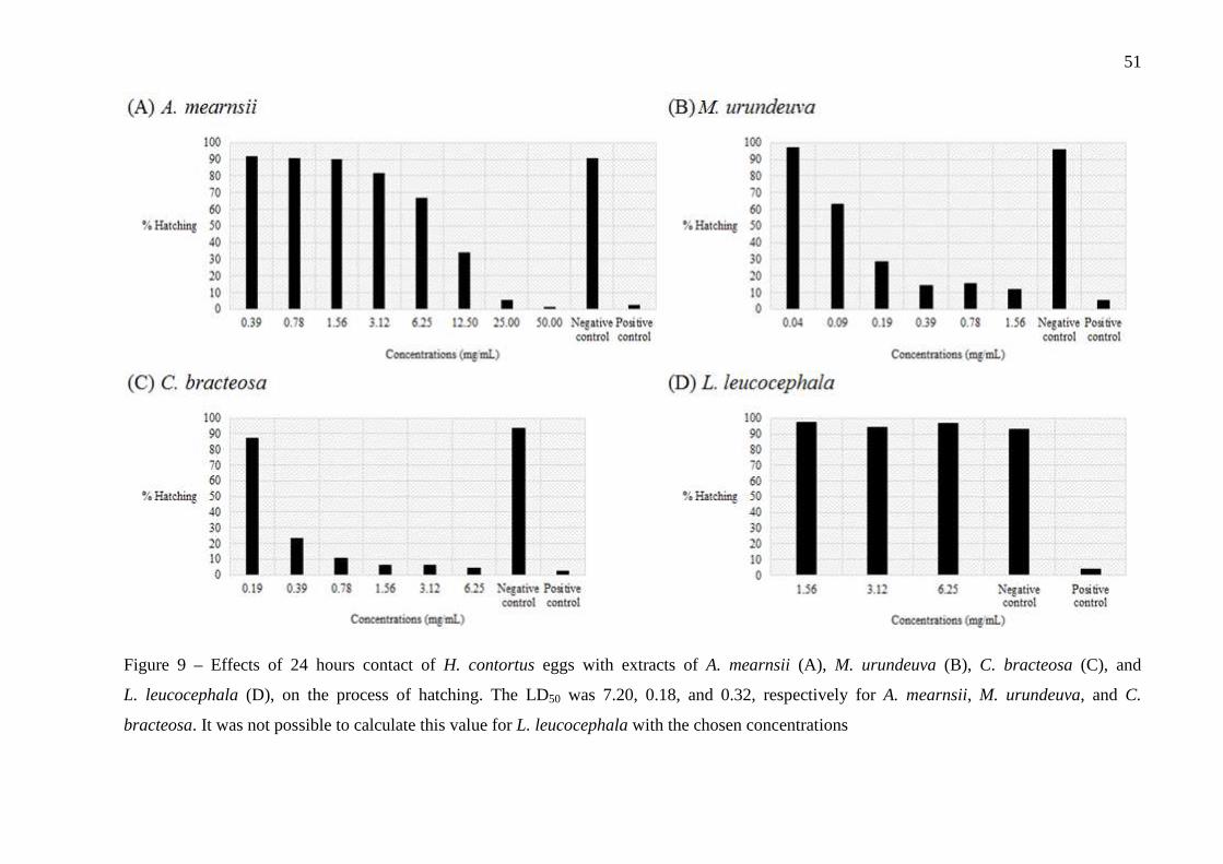
51
Figure 9 – Effects of 24 hours contact of H. contortus eggs with extracts of A. mearnsii (A), M. urundeuva (B), C. bracteosa (C), and
L. leucocephala (D), on the process of hatching. The LD50 was 7.20, 0.18, and 0.32, respectively for A. mearnsii, M. urundeuva, and C.
bracteosa. It was not possible to calculate this value for L. leucocephala with the chosen concentrations

52
The results for LEIA are found in Table 6, Table 7, and Figure 10. Between 95 and
99% of the L3 larvae from the negative controls exsheathed after the contact with the ES. The
lowest LD50 value found for LEIA was from M. urundeuva (0.40 mg/mL) and the highest for
C. bracteosa (2.24 mg/mL). A slightly different pattern was found for the LD99: A. mearnsii
had the lowest value (2.28 mg/mL) and C. bracteosa the highest (2,538.27 mg/mL).
Table 6 - LD99 (mg/mL) and fiducial limits of A. mearnsii, M. urundeuva extract, C. bracteosa, and L. leucocephala extracts in Larval Exsheathment Inhibition Assay (LEIA) against H. contortus
LEIA (mg/mL)
A. mearnsii M. urundeuva C. bracteosa L. leucocephala
DL50 0.52 (0.49 - 0.54) 0.40 (0.38 - 0.42) 2.24 (1.68 - 3.43) 1.24 (1.14 - 1.38)
DL99 2.28 (2.01 - 2.64) 2.37 (2.03 - 2.86) 2.53 x 10³ (5.81 x 10² - 2.39 x 10⁴) 19.99 (14.08 - 31.05)
Table 7 – Mean larval exsheathment (%) of Haemonchus contortus infective L3 larvae after one hour incubation with Exsheathment Solution. Larvae were previously exposed during 3 hours to different concentrations (µg/mL) of tanniniferous plants extracts
Plant extract Mean larval exsheathment (%) Negative control 150 300 600 1,200
A. mearnsii 99.52 97.11 84.56** 46.02*** 3.91*** M. urundeuva 99.84 88.61 76.74*** 29.85*** 1.78*** C. bracteosa 95.96 86.16 79.85* 74.21** 64.71***
L. leucocephala 97.78 94.95 90.64 85.65* 54.59*** * Significantly different compared to the respective negative control (P<0.05) ** Significantly different compared to the respective negative control (P<0.01) *** Significantly different compared to the respective negative control (P<0.0001)

53
Figure 10 - Effects of three hours contact of H. contortus L3 larvae with extracts of A. mearnsii (A), M. urundeuva (B), C. bracteosa (C), and
L. leucocephala (D), on the process of artificial exsheathment after addition of Exsheathment Solution. The LD50 was 0.52, 0.40, 2.24, and 1.24
mg/mL respectively for A. mearnsii, M. urundeuva, C. bracteosa, and L. leucocephala.
0
20
40
60
80
100
0 20 40 60
% exsheathment
Minutes
(a) A. mearnsii
1200 µg/mL 600 µg/mL 300 µg/mL
150 µg/mL Negative control
0
20
40
60
80
100
0 20 40 60
% exsheathment
Minutes
(b) M. urundeuva
1200 µg/mL 600 µg/mL 300 µg/mL
150 µg/mL Negative control
0
20
40
60
80
100
0 20 40 60
% exsheathment
Minutes
(c) C. bracteosa
1200 µg/mL 600 µg/mL 300 µg/mL
150 µg/mL Negative control
0
20
40
60
80
100
0 20 40 60
% exsheathment
Minutes
(d) L. leucocephala
1200 µg/mL 600 µg/mL 300 µg/mL
150 µg/mL Negative control

54
6. DISCUSSION
The extract’s bioactivity may rely on the CT content quality or quantity from each
plant extract. The extract with the highest percentage of CT was A. mearnsii (approximately
32.25%), followed by L. leucocephala (approximately 16.74%), M. urundeuva
(approximately 7.27%), and C. bracteosa (approximately 1.27%). A dose-dependent
inhibition of hatching was found for all extracts, except L. leucocephala, within the chosen
doses. A dose-dependent effect for the LEIA was also found, either by completely inhibiting
the exsheathment or delaying it, these results were more perceptible for A. mearnsii, M.
urundeuva, and L. leucocephala and less for
C. bracteosa.
No mortality was found in the negative control groups with the use of methanol as a
solvent. At first, we have tried to dilute the extracts with distilled water, but this was not
satisfactory. Later, we tried with Tween 80, but the solution became a foam, preventing the
differentiation of the eggs from the larvae. So we tried with a methanol:stock solution (1:50)
and it diluted the extracts without killing the eggs or the larvae from both assays.
Most reports use the DL50 and DL99 to interpret the results from assays (ALONSO-
DÍAZ et al., 2011; KATIKI, 2011; ADAMU; NAIDOO; ELOFF, 2013; DOMINGUES et al.,
2013). The same pattern from the DL50 concentrations has been found for the DL99 in the
EHA. Myracrodruon urundeuva had the lowest concentration for both DL50 and DL99 (0.18
and 4.31 mg/mL, respectively), followed by C. bracteosa (0.32 and 5.41 mg/mL,
respectively), and A. mearnsii (7.20 and 187.26 mg/mL, respectively). The extracts with the
lowest content of CTs were those with the lowest DL50 and DL99 concentrations, and that may
lead to two different assumptions: or the CTs from them have an elevated bioactivity, or the
CTs are not the molecules involved in these results. If the CT molecules of M. urundeuva
have a better bioactivity than the others, this may rely on the kind of monomers from that
tannins. According to Brunet and Hoste (2006), the amount of free hydroxyl groups in the
monomers of CTs is a key factor, i.e., monomers with more free hydroxyls were more potent
inhibitors of the exsheathment of L3; unfortunately, no test to characterize these monomers
was held. Silva et al. (2012) points another characteristic of M. urundeuva: it has also a rich
fraction content of chalcones, and these molecules also have presented bioactivity, such as
molluscicidal and antimicrobial, in the assays performed. Unfortunately, we cannot confirm
the role of tannins in present studies, since no test showed the presence of tannin binding
compounds, such as PVPP, in the experimental design. Alonso-Díaz et al. (2011) points that it

55
would be essential to detect if it is the action of tannins or other plant’s secondary metabolites.
Yet, De Oliveira et al. (2011) used PVPP to check the role of tannins in an experiment with
M. urundeuva extracts and they found that the bioactivity of the extract was lost after the
addition of PVPP. All five highest concentrations from A. mearnsii, M. urundeuva, and
C. bracteosa had significantly differed from the negative control (P<0.0001). For
L. leucocephala, no tested concentration had a significant difference from the negative
control.
In the LEIA, a different pattern has been found for the DL50 and DL99, with the change
being between the orders of M. urundeuva and A. mearnsii. For the DL50, M. urundeuva had
the lowest concentrations (0.40 mg/mL), followed by A. mearnsii (0.52 mg/mL),
L. leucephala (1.24 mg/mL), and C. bratecteosa (2.24 mg/mL). For the DL99, A. mearnsii had
the lowest concentration (2.28 mg/mL), followed by M. urundeuva (2.37 mg/mL),
L. leucocephala (19.99 mg/mL), and C. bracteosa (2,530 mg/mL). The CT content from
A. mearnsii and C. bracteosa were the highest ones and their DL50 and DL99 may be linked to
this content.
Basically, three different categories of results were found for LEIA after 60 minutes: a
significant delay in exsheathment may be also an interesting result (BAHUAUD et al., 2006).
In the first category, nearly total significant (P<0.0001) inhibition was achieved for the 1,200
µg/mL concentration; in the second, an evident delay was found (P<0.0001); in the third,
there was nearly no significant inhibition of exsheathment if compared to the negative control.
Only A. mearnsii and M. urundeuva achieved nearly total inhibition at 1,200 µg/mL
(P<0.0001). A delay was found for A. mearnsii (600 µg/mL), M. urundeuva (600 µg/mL),
C. bracteosa (1,200 µg/mL) and L. leucocephala (1,200 µg/mL) at 60 minutes (P<0.0001);
Caesalpinea bracteosa 600 µg/mL (P<0.01) and 300 µg/mL (P<0.05) concentrations also had
a delay if compared to the negative control (P<0.01). The other concentrations did not
inhibited the exsheathment of the larvae. Bahuaud et al. (2006) point that a delay in the
process may be also an important result. They found that E. eurigena and P. sylvestres
significantly delayed the exsheathment of H. contortus after 60 minutes of LEIA. According
to Lee (1965), if the process of exsheathment is delayed, the taxes of infection will be
dropped, since H. contortus has a specific time-frame (around three hours) to exsheath and
achieve infection of the host.
Another way of classifying the results from these tests is using the guideline adopted
by the WAAVP (POWERS et al., 1982; NERY; DUARTE; MARTINS, 2009; FERREIRA et
al., 2013). It has been postulated that a highly effective response from the in vitro tests are
56
those in which are found more than 90% inhibition. If the results ranged between 80 and 90%,
the concentration of the extract has moderate effectiveness; from 60 to 80%, low
effectiveness. If the results are less than 60%, that dose is ineffective. So, in the EHA, the
highly effective concentrations were 50.00 and 25.00 mg/mL (A. mearnsii) and 6.25, 3.12,
and 1.56 mg/mL (C. bracteosa). The moderately effective concentrations were 1.56, 0.78, and
0.39 mg/mL (M. urundeuva) and 0.78 mg/mL (C. bracteosa). Those with low effectiveness
were 12.50 mg/mL (A. mearnsii), 0.19 mg/mL (M. urundeuva), and 0.39 mg/mL (C.
bracteosa). All the other concentrations were ineffective according to W.A.A.V.P. guideline
(POWERS et al., 1984). About the LEIA, only the 1,200 µg/mL concentration from A.
mearnsii and M. urundeuva was highly effective, no concentration was moderately effective,
the 600 µg/mL concentration from M. urundeuva had low effectiveness and all other
concentrations from all plant extracts were ineffectiveby the standards of the above cited
guideline.
The doses in which M. urundeuva and C. bracteosa extracts showed effectiveness
against the hatching were lesser if compared to the effective doses from the extracts others
reports. For example, Pessoa et al. (2002) found 100% inhibition of hatching with 5 mg/mL of
Ocimum gratissimum. De Oliveira et al. (2011) found 97.73% of inhibition for M. urundeuva
with the dose of 1.25 mg/mL, a dose slightly slower if compared to the one found by us, so
we confirm the findings from this author; also, they used PVPP in the assay, in order to
confirm the role of CT for this plant. It was found that with the addition of PVPP, the
anthelmintic efficacy was lost. Adamu, Naidoo and Eloff (2013) found 100% inhibition with
12.50 mg/mL for Brachylaena discolor, Melia azedarach, Cyathea dregei, Milletia grandis,
and Indigofera frutescens, 100% with 6.25 mg/mL of Apodytes dimidiate, 100% with
3.13 mg/mL for Heteromorpha trifoliata, Maesa lanceolata, and Leucosidae sericea, 95%
with 12.50 mg/mL of Zanthoxylum capense, and 90% with 12.50 mg/mL of Clerodendrum
glabrum and Strychnos mitis. Ferreira et al. (2013) found that Annona muricata inhibited
84.91% of hatching at 0.5 g/mL. The effective dose we found for A. mearnsii was higher if
compared to most of the cited reports. That can emphasize that the quality of the biological
molecule may be different between extracts, at least for M. urundeuva. Caesalpinea bracteosa
extract showed low effectiveness in the LEIA and high effectiveness in the EHA; it may
imply that the bioactive molecules from it are not CTs, or at least not them alone. Other
molecules classes may have a role in this case.
Other reports showed similar or better results with other plants, using lower doses,
after 60 minutes of LEIA test, against H. contortus. Alonso-Díaz et al. (2011), testing Acacia
57
gaumeri, Brosimum alicastrum, Havadia albicans, and L. leucocephala at 600 µg/mL
concentration, found, respectively, 93.50%, 86.00%, 98.10%, and 78.90% of inhibition of
exsheathment. Bahuaud et al. (2006) found approximately 100.00%, 70.00%, 42.00%, and
18.00% inhibition, respectively with Castanea sativa, Erica Erigena, Pinus sylvestres, and
Sarothammus scoparius extracts at 600 µg/mL concentration. Brunet el al. (2007) found for
Onobrychis viciifolia extract, 50.00% and 90.00% inhibition, respectively for the doses of 600
and 1,200 µg/mL. Alonso-Díaz et al. (2008a) found inhibitions of 51.00%, 53.60%, 49.10%
and 63.80% respectively for Acacia pennatula, L. leucocephala, Lysiloma latisiliquum and
Piscidia piscipula at 1,200 µg/mL. Oliveira et al. (2011), testing the leaf’s and stem’s extract
of Anadenanthera colubrine, L. leucocephala, and Mimosa tenuiflora, found that all of them
inhibited completely the exsheathment of H. contortus at the dose of 300 µg/mL. De Oliveira
et al. (2011) found a 100.00% inhibition of exsheathment with 300 µg;mL of M. urundeuva;
this dose was lesser if compared with ours. The results that we found for L. leucocephala
were not those found in the literature. This may rely on the quality of the fresh leaves, or the
period of the year it was harvested.
Differences between the results from the two assays were expected to happen. The egg
shell makes the eggs less sensible to the bioactivity of the extracts than the L3 larvae. This
may rely on its structure, because it has a harder barrier if compared with the double sheath
from the L3 larvae. It is important to notice that the doses from A. mearnsii that showed
effective bioactivity in the EHA were around 200 times higher than those chosen used in
LEIA. For M. urundeuva, no dose reached the highly effective class from the guideline, but
three of them were moderately effective, i.e., had an efficacy between 80 and 90%, being the
lowest one the 0.39 mg/mL; for LEIA, the highest dose (1,200 µg/mL) was classified with
highest effectiveness (> 90%). Two things can be inferred from this: again, the eggshell
makes the eggs less sensible to the biomolecules, which would make necessary the use of
higher doses, and that the quality of CT found in M. urundeuva may be more bioactive than
the ones from others extracts, or maybe it is due to the presence of chalcones.
The variability in the results, for example for the LEIA’s DL99 dose from
C. bracteosa, may have happened because of the number of repetitions. Usually, for EHA
only six repetitions are done; for LEIA, only four. We have done 30 repetitions for each assay
for each extract. The DL50 may have varied day to day. Amarante et al. (1997), Álvarez-
Sánchez et al. (2005), and Katiki (2011) also found similar day to day variation in the DL50 in
in vitro assays, including EHA and LEIA among them.

58
Some authors showed the efficacy of A. mearnsii extract in in vivo trials. Cenci et al.
(2007), giving 18 g of A. mearnsii extract per week for the treated group, found a general
reduction for both FEC and worm burden (WD) after slaughter in lambs naturally infected
with H. contortus, Trichostrongylus colubriformis, Oesophagostomum columbianum,
Cooperia sp., Strongyloides papillosus, Trichuris globulosa, and Moniezia expansa. Minho et
al. (2008) gave 1.6 g/kg of live weight of A. mearnsii extract to the treated group of lambs in
lambs naturally infected with H. contortus and T. colubriformis, and also found a reduction in
FEC and WD. These are examples of what type of assays may be performed in order to test
the plant extracts in in vivo experiments; also, they show that tanninferous plants may play a
role against parasitic infections.
59
7. CONCLUSIONS
Anthelmintic activity was found for the extracts, being the M. urundeuva the one that
showed bioactivity at small doses for both EHA and LEIA. The extract from A. mearnsii
showed activity at lower doses for LEIA, but for EHA just in higher doses. Caesalpinea
bracteosa showed better activity for the EHA and L. leucocephala for the LEIA. More
research is needed to test the toxicity from the plants and then an in vivo trial, to see how the
polymers from the plants will work when interacting with the animals.
The use of in vitro assays may be a good alternative before testing in vivo, because
through that it is possible to screen potential anthelmintics plants rapidly and economically if
compared to the in vivo assays.
However, it is hard to produce the purified plant extract, because it demands time,
labor, dangerousness (for the working with dichloromethane), and the yield is too low. Also,
to obtain the fresh eggs and larvae is too laborious. These are maybe the hardest parts about
using in vitro assays.
An interesting anthelmintic effect was found for all extracts for at least one of the
assays, highlighting the M. urundeuva and A. mearnsii extracts for both assays. An advantage
of using these plants are that they have a potential use as forage, mostly at the North and
Northeast regions of Brazil, and also among small producers. Nevertheless, for nutraceuticals
use of the plants, toxicity and bioactivity in vivo assays must be done. Another option is the
use of the extracts as phytotherapy; for that, tests for calculation of dosages must be realized.

60
8. CONCLUSÕES
Foi encontrada atividade anti-helmíntica para os extratos, de forma que M. urundeuva
foi a que apresentou biotividade com menores doses tanto para o TIEO quanto para o TIDL.
O extrato de A. mearnsii mostrou atividade em menores doses para o TIDL, mas em doses
mais altas para o TIEO. Caesalpinea bracteosa mostrou melhor atividade para o TIEO e
L. leucocephala para o TIDL. É necessário a realização de mais pesquisas, visando testar a
toxicidade das plantas e posteriormente testes in vivo, visando-se conhecer como os polímeros
das plantas irão interagir com os animais.
O uso de testes in vitro, em um primeiro momento, é uma boa alternativa ao uso de
testes in vivo, no sentido da seleção de plantas com potencial anti-helmíntico mais rápida e
economicamente do que a opção de um teste in vivo inicialmente.
Entretanto, uma grande dificuldade é a produção dos extratos purificados, pois
demanda tempo, labor, possui periculosidade (por se trabalhar com diclorometano) e o
rendimento dos extratos é muito baixo. Além das dificuldades em relação a preparação dos
extratos, a obtenção de ovos e larvas é bastante trabalhosa. Esses são provavelmente os pontos
mais difíceis em se usar testes in vitro.
Foi encontrado efeito anti-helmíntico interessante para todos os extratos utilizados
pelo menos em um dos testes, destacando-se os extratos de M. urundeuva e de A. mearnsii
para ambos os testes. Uma vantagem em relação a essas plantas é o seu potencial uso
forrageiro, principalmente nas regiões Norte e Nordeste do Brasil, e também entre pequenos
produtores. Entretanto, para o uso nutracêutico das plantas, testes de toxicidade e de
bioatividade das plantas in vivo devem ser realizados. Uma possível opção é o uso dos
extratos purificados como fitoterápicos; para isso, testes devem ser realizados para o cálculo
das dosagens devem ser realizados.
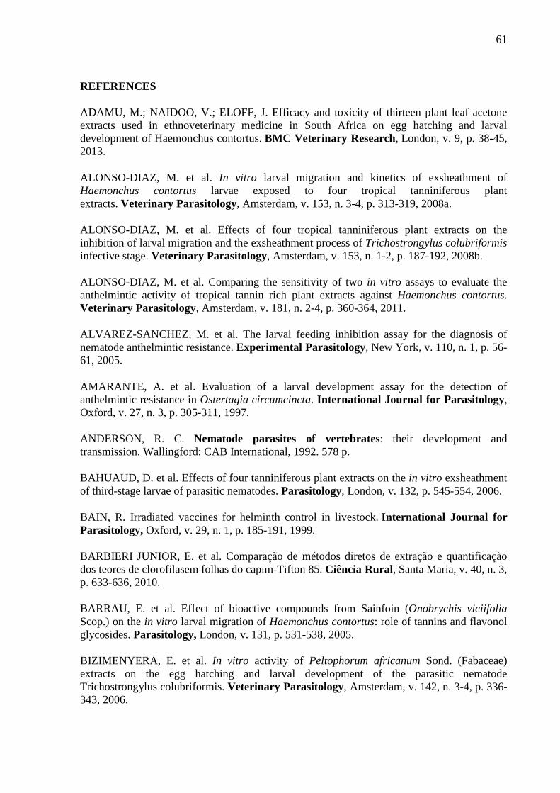
61
REFERENCES
ADAMU, M.; NAIDOO, V.; ELOFF, J. Efficacy and toxicity of thirteen plant leaf acetone extracts used in ethnoveterinary medicine in South Africa on egg hatching and larval development of Haemonchus contortus. BMC Veterinary Research, London, v. 9, p. 38-45, 2013.
ALONSO-DIAZ, M. et al. In vitro larval migration and kinetics of exsheathment of Haemonchus contortus larvae exposed to four tropical tanniniferous plant extracts. Veterinary Parasitology, Amsterdam, v. 153, n. 3-4, p. 313-319, 2008a.
ALONSO-DIAZ, M. et al. Effects of four tropical tanniniferous plant extracts on the inhibition of larval migration and the exsheathment process of Trichostrongylus colubriformis infective stage. Veterinary Parasitology, Amsterdam, v. 153, n. 1-2, p. 187-192, 2008b.
ALONSO-DIAZ, M. et al. Comparing the sensitivity of two in vitro assays to evaluate the anthelmintic activity of tropical tannin rich plant extracts against Haemonchus contortus. Veterinary Parasitology, Amsterdam, v. 181, n. 2-4, p. 360-364, 2011.
ALVAREZ-SANCHEZ, M. et al. The larval feeding inhibition assay for the diagnosis of nematode anthelmintic resistance. Experimental Parasitology, New York, v. 110, n. 1, p. 56-61, 2005.
AMARANTE, A. et al. Evaluation of a larval development assay for the detection of anthelmintic resistance in Ostertagia circumcincta. International Journal for Parasitology, Oxford, v. 27, n. 3, p. 305-311, 1997.
ANDERSON, R. C. Nematode parasites of vertebrates: their development and transmission. Wallingford: CAB International, 1992. 578 p.
BAHUAUD, D. et al. Effects of four tanniniferous plant extracts on the in vitro exsheathment of third-stage larvae of parasitic nematodes. Parasitology, London, v. 132, p. 545-554, 2006.
BAIN, R. Irradiated vaccines for helminth control in livestock. International Journal for Parasitology, Oxford, v. 29, n. 1, p. 185-191, 1999.
BARBIERI JUNIOR, E. et al. Comparação de métodos diretos de extração e quantificação dos teores de clorofilasem folhas do capim-Tifton 85. Ciência Rural, Santa Maria, v. 40, n. 3, p. 633-636, 2010.
BARRAU, E. et al. Effect of bioactive compounds from Sainfoin (Onobrychis viciifolia Scop.) on the in vitro larval migration of Haemonchus contortus: role of tannins and flavonol glycosides. Parasitology, London, v. 131, p. 531-538, 2005.
BIZIMENYERA, E. et al. In vitro activity of Peltophorum africanum Sond. (Fabaceae) extracts on the egg hatching and larval development of the parasitic nematode Trichostrongylus colubriformis. Veterinary Parasitology, Amsterdam, v. 142, n. 3-4, p. 336-343, 2006.

62
BRUNET, S.; HOSTE, H. Monomers of condensed tannins affect the larval exsheathment of parasitic nematodes of ruminants. Journal of Agricultural and Food Chemistry, Easton, v. 54, n. 20, p. 7481-7487, 2006.
BRUNET, S. et al. The kinetics of exsheathment of infective nematode larvae is disturbed in the presence of a tannin-rich plant extract (sainfoin) both in vitro and in vivo. Parasitology, London, v. 134, p. 1253-1262, 2007.
BRUNETON, J. Pharmacognosie: Phytochimie, plantes médicinales. Paris: Tec & Doc Lavoisier, 1999. 1119 p.
CARVALHO, P. Aroeira-verdadeira: Taxonomia e nomenclatura. Colombo, PR: Embrapa Florestas, 2003. 16 p. (Circular Técnica, 82). Disponível em: <http://www.infoteca.cnptia.embrapa.br/bitstream/doc/312989/1/CT0082.pdf>. Acesso em: 13 ago. 2013.
CENCI, F. et al. Effects of condensed tannin from Acacia mearnsii on sheep infected naturally with gastrointestinal helminthes. Veterinary Parasitology, Amsterdam, v. 144, n. 1-2, p. 132-137, 2007.
CENTRO DE INTELIGÊNCIA EM FLORESTAS. Acácia Negra. Viçosa, 2013. Disponível em: <http://www.ciflorestas.com.br/texto.php?p=acacia_negra>. Acesso em 13 ago. 2013.
CENTRO NORDESTINO DE INFORMAÇÕES SOBRE PLANTAS. Banco de Dados de Plantas do Nordeste - Catingueira. Recife, PE, 2013. Disponível em: <http://www.cnip.org.br/bdpn/fotosdb/Catingueira%20II.pdf>. Acesso em: 13 ago. 2013.
DE OLIVEIRA, L. M. et al. Effects of Myracrodruon urundeuva extracts on egg hatching and larval exsheathment of Haemonchus contortus. Parasitology Research, Heidelberg, v. 109, n. 3, p. 893-898, 2011.
DE ROODE, J. C.; LEFÈVRE, T.; HUNTER, M. D. Ecology. Self-medication in animals. Science, Washington, DC, v. 340, n. 6129, p. 150-151, 2013.
DOMINGUES, L. F. et al. In vitro and in vivo evaluation of the activity of pineapple (Ananas comosus) on Haemonchus contortus in Santa Inês sheep. Veterinary Parasitology, Amsterdam, 2013. doi: 10.1016/j.vetpar.2013.04.031. Disponível em: http://www.sciencedirect.com/science/article/pii/S0304401713002434.
EZENWA, V. Selective defecation and selective foraging: Antiparasite behavior in wild ungulates? Ethology, Berlin, v. 110, n. 11, p. 851-862, 2004.
FAO. Tannic acid. Rone, 2009. Disponível em: <http://www.fao.org/ag/agn/jecfa-additives/specs/monograph7/additive-454-m7.pdf>. Acesso em: 13 ago. 2013.
FERREIRA, L. et al. In vitro anthelmintic activity of aqueous leaf extract of Annona muricata L. (Annonaceae) against Haemonchus contortus from sheep. Experimental Parasitology, New York, v. 134, n. 3, p. 327-332, 2013.
FORTES, E. Parasitologia veterinária. Porto Alegre: Sulina, 1987. 453 p.
63
GAUGLER, R.; BILGRAMI, A. L. Nematode behavior. Wallingford: CAB International, 2004. 419 p.
GOMES, T.; BANDEIRA, F. Uso e diversidade de plantas medicinais em uma comunidade quilombola no Raso da Catarina, Bahia. Acta Botanica Brasilica, Porto Alegre, v. 26, n. 4, p. 796-809, 2012.
HAGERMAN, A. Condensed tannin structural chemistry. Oxford: Hagerman personal web page, 2002a. Disponível em: <http://www.users.muohio.edu/hagermae/Condensed%20Tannin%20Structural%20Chemistry.pdf>. Acesso em: 13 ago. 2013.
HAGERMAN, A. What is a tannin? Oxford: Hagerman personal web page, 2002b. Disponível em: <http://www.users.muohio.edu/hagermae/What%20Is%20a%20Tannin.pdf>. Acesso em: 13 ago. 2013.
HAGERMAN, A. Hydrolizable tannin structural chemistry . Oxford: Hagerman personal web page, 2010. Disponível em: <http://www.users.muohio.edu/hagermae/Hydrolyzable%20Tannin%20Structural%20Chemistry.pdf>. Acesso em: 13 ago. 2013.
HART, B. Behavioural defences in animals against pathogens and parasites: parallels with the pillars of medicine in humans. Philosophical Transactions of the Royal Society. B - Biological Sciences, London, v. 366, n. 1583, p. 3406-3417, 2011.
HOSTE, H. et al. The effects of tannin-rich plants on parasitic nematodes in ruminants. Trends in Parasitology, Oxford, v. 22, n. 6, p. 253-261, 2006.
HOSTE, H. et al. Direct and indirect effects of bioactive tannin-rich tropical and temperate legumes against nematode infections. Veterinary Parasitology, Amsterdam, v. 186, n. 1-2, p. 18-27, 2012.
INSTITUTO BRASILEIRO DE GEOGRAFIA E ESTATÍSTICA - IBGE. Produção da Pecuária Municipal, Rio de Janeiro, v. 39, p. 1-63, 2011. Disponível em: <ftp://ftp.ibge.gov.br/Producao_Pecuaria/Producao_da_Pecuaria_Municipal/2011/ppm2011.pdf >. Acesso em: 07 ago. 2013.
JACKSON, F. et al. Worm control in sheep in the future. Small Ruminant Research, Amsterdam, v. 86, n. 1-3, p. 40-45, 2009.
JACKSON, F.; HOSTE, H. In vitro methods for the primary screening of plant products for direct activity against ruminant gastrointestinal nematodes. In: VERCOE, P. E.; MAKKAR, H. P. S.; SCHLINK, A. C. In vitro screening of plant resources for extra-nutritional attributes in ruminants : nuclear and related methodologies. Dordrecht: Springer, 2010. p. 25-45.
JOHNSON, P.; HOVERMAN, J. Parasite diversity and coinfection determine pathogen infection success and host fitness. Proceedings of the National Academy of Sciences of the USA, Washington, DC, v. 109, n. 23, p. 9006-9011, 2012.

64
JUHNKE, J. et al. Preference for condensed tannins by sheep in response to challenge infection with Haemonchus contortus. Veterinary Parasitology, Amsterdam, v. 188, n. 1-2, p. 104-114, 2012.
KATIKI, L. M. Atividade anti-helmíntica in vitro e in vivo de compostos fitoquímicos para o controle de nematóides gastrintestinais de ovinos. 2011. 128 f. Tese (Doutorado) – Faculdade de Medicina Veterinária e Zootecnia, Universidade Estadual Paulista Julio de Mesquita Filho, Botucatu, 2011.
LEE, D. L. The physiology of nematodes. San Francisco: W. H. Freeman and Company, 1965. 154 p.
LISONBEE, L. et al. Tannins and self-medication: Implications for sustainable parasite control in herbivores. Behavioural Processes, Amsterdam, v. 82, n. 2, p. 184-189, 2009.
LONE, B. et al. Anthelmintic and antimicrobial activity of methanolic and aqueous extracts of Euphorbia helioscopia L. Tropical Animal Health and Production, Edinburgh, v. 45, n. 3, p. 743-749, 2013.
LOUVANDINI, H. et al. Effect of dietary protein intake on calf resilience to Haemonchus contortus. Brazilian Journal of Veterinary Research and Animal Science, São Paulo, v. 39, n. 5, p. 227-232, 2002.
MAKKAR, H. P. S. Quantification of tannins in tree foliage. Vienna: FAO; IEAE, 2000. (Laboratory Manual).
MALEK, M. et al. Parasites as heavy metal bioindicators in the shark Carcharhinus dussumieri from the Persian Gulf. Parasitology, London, v. 134, p. 1053-1056, 2007.
MANN, B.; MCMILLAN, M. The chemistry of the leather industry. Christchurch, New Zealand: New Zealand Institute of Chemistry, 2013. 17 p. Disponível em: <http://nzic.org.nz/ChemProcesses/animal/5C.pdf>. Acesso em: 13 de ago. 2013.
MANOLARAKI, F. et al. Anthelmintic activity of some Mediterranean browse plants against parasitic nematodes. Parasitology, London, v. 137, n. 4, p. 685-696, 2010.
MARTINEZ-ORTIZ-DE-MONTELLANO, C. et al. Scanning electron microscopy of Haemonchus contortus exposed to tannin-rich plants under in vivo and in vitro conditions. Experimental Parasitology, New York, v. 133, n. 3, p. 281-286, 2013.
MAYER, W. et al. Horizontal gene transfer of microbial cellulases into nematode genomes is associated with functional assimilation and gene turnover. BMC Evolutionary Biology, London, v. 11, p. 13-22, 2011.
MELLOR, D.; HEMSWORTH, P. Policy on the care and use of sheep for scientific purposes based on good practice. Victoria, Australia: Monash University; Animal Welfare Science Centre, 2005. 15 p. Disponível em: <http://www.animalwelfare.net.au/comm/download/sheepcare.pdf>. Acesso em: 13 de ago. 2013.
65
MINHO, A. et al. Effect of Acacia molissima tannin extract on the control of gastrointestinal parasites in sheep. Animal Feed Science and Technology, Amsterdam, v. 147, n. 1-3, p. 172-181, 2008.
NERY, P.; DUARTE, E.; MARTINS, E. Eficácia de plantas para o controle de nematóides gastrintestinais de pequenos ruminantes: revisão de estudos publicados. Revista Brasileira de Plantas Medicinais, Botucatu, v. 11, n. 3, p. 330-338, 2009.
OLIVEIRA, L. M. et al. Effect of six tropical tanniferous plant extracts on larval exsheathment of Haemonchus contortus. Revista Brasileira de Parasitologia Veterinária , Jaboticabal, v. 20, n. 2, p. 155-60, 2011.
PESSOA, L. et al. Anthelmintic activity of essential oil of Ocimum gratissimum Linn. and eugenol against Haemonchus contortus. Veterinary Parasitology, Amsterdam, v. 109, n. 1-2, p. 59-63, 2002.
PIMENTEL NETO, M.; FONSECA, A. Epidemiologia das helmintoses pulmonares e gastrintestinais de bezerros de baixada do Estado do Rio de Janeiro. Pesquisa Veterinária Brasileira, Seropédica, v. 22, n. 4, p. 148-152, 2002.
POWERS, K. G. et al. World Association of the Advancement of Veterinary Parasitology (WAAVP) guidelines for evaluating the efficacy of anthelmintics in ruminants (bovine and ovine). Veterinary Parasitology, Amsterdam, v. 10, p. 265–284, 1982.
ROBERTS, F.; O'SULLIVAN, P. Methods for egg counts and larval cultures for strongyles infesting the gastro-intestinal tract of cattle. Australian Journal of Agricultural Research, Melbourne, v. 1, n. 1, p. 99-102, 1950.
ROTH, A. Origem dos parasitos. Folha SCB, Brasília, DF, n. 22, p. 25-31, 1980.
RUPPERT, E.; BARNES, R. D. Zoologia dos invertebrados. São Paulo: Roca, 1996. 1029 p.
SIGMA-ALDRICH. Solvent center – Dichloromethane. St. Louis, 2012. Disponível em: <http://www.sigmaaldrich.com/chemistry/solvents/dichloromethane-center.html>. Acesso em: 04 abr. 2013.
SILVA, M. et al. Bioactivity and potential therapeutic benefits of some medicinal plants from the Caatinga (semi-arid) vegetation of Northeast Brazil: a review of the literature. Brazilian Journal of Pharmacognosy, Brasília, DF, v. 22, n. 1, p. 193-207, 2012.
SOUZA, M. et al. Desenvolvimento in vitro do fungo simbionte de Atta sexdens rubropilosa Forel, 1908 (Hymenoptera: Formicidae) em meio de cultura com diferentes extratos vegetais. Ciência Rural, Santa Maria, v. 42, n. 9, p.1603-1909, 2012.
TAYLOR, M. A.; COOP, R. L.; WALL, R. L. Parasitologia veterinária. Rio de Janeiro: Guanabara Koogan, 2010. 744 p.
TOYANG, N. et al. Ethnoveterinary medicine a practical approach to the treatment of cattle diseases in sub-Saharan Africa. Wageningen: Digigrafi, 2007. 88 p.

66
WALLER, P. Sustainable nematode parasite control strategies for ruminant livestock by grazing management and biological control. Animal Feed Science and Technology, Amsterdam, v. 126, n. 3-4, p. 277-289, 2006.
WEISCHER, B.; BROWN, D. J. F. Conhecendo os nematóides: nematologia geral. Sofia, Bulgária: Pensoft Publishers, 2001. 209 p.
Top Related