


Languages
Pages
Legal


Cellular Transport of Functionalized Gold
Nanoparticles
A Major Qualifying Project
Submitted to the Faculty of the
WORCESTER POLYTECHNIC INSTITUTE
in partial fulfillment of the requirements for the
Degree of Bachelor of Science
By
Jeffrey D. Peters
Date: April 25, 2013
Advisor: Prof. Germano S. Iannacchione
Department of Physics
Key Words: Gold nanoparticles, functionalized, model cells, lipid bilayer

Abstract
The biomedical industry is faced with the constant challenge of creating treatments
to help patients that are cost-effective and easy to synthesize and administer. Recent
advances in nanotechnology have sparked research in the use of nano-scale particles
for medical applications. Gold nanoparticles have become a popular choice of vector
in drug delivery research, however, the mechanism of cellular uptake of these particles
is still unknown. By attaching fluorescent functional groups to gold nanoparticles, and
observing how these nanoparticles transport through lipid bilayers, which mimic cell
membranes, I have gained a better understanding of the cellular transport properties.
Jeffrey Peters i Nanoparticle Transport

Acknowledgements
I would like to thank the WPI Department of Physics for the funding that made this project
possible. I would also like to thank Victoria Huntress, for her assistance with the fluores-
cent microscopy. Mostly, I would like to thank my advisor, Germano Iannacchione, for the
invaluable guidance he gave me throughout this project.
Jeffrey Peters ii Nanoparticle Transport

Contents
Abstract i
Acknowledgements ii
Contents iii
List of Tables vi
List of Figures vii
Executive Summary viii
1 Introduction 10
1.1 Nanotechnology in Medicine . . . . . . . . . . . . . . . . . . . . . . . . . . . 10
1.2 Determining the Uptake Mechanism of Gold Nanoparticles . . . . . . . . . . 10
1.3 Using Fluorescent Microscopy to Observe Transport of Nanoparticles Through
Model Cells . . . . . . . . . . . . . . . . . . . . . . . . . . . . . . . . . . . . 10
1.4 This Report . . . . . . . . . . . . . . . . . . . . . . . . . . . . . . . . . . . . 11
2 Literature Review 12
2.1 Usage of Nanotechnology in Biomedical Science . . . . . . . . . . . . . . . . 12
2.1.1 Drug Delivery Systems . . . . . . . . . . . . . . . . . . . . . . . . . . 12
2.1.2 Toxicity . . . . . . . . . . . . . . . . . . . . . . . . . . . . . . . . . . 13
2.2 Gold Nanoparticles . . . . . . . . . . . . . . . . . . . . . . . . . . . . . . . . 13
2.2.1 Properties of Gold Nanoparticles . . . . . . . . . . . . . . . . . . . . 13
2.2.2 Synthesis and Functionalization of Gold Nanoparticles . . . . . . . . 14
2.3 Lipid Bilayers . . . . . . . . . . . . . . . . . . . . . . . . . . . . . . . . . . . 15
2.4 Microscopy . . . . . . . . . . . . . . . . . . . . . . . . . . . . . . . . . . . . 16
2.4.1 Cross-Polarizing Microscopy . . . . . . . . . . . . . . . . . . . . . . . 16
Jeffrey Peters iii Nanoparticle Transport

2.4.2 Confocal Fluorescent Microscopy . . . . . . . . . . . . . . . . . . . . 17
3 Methods 19
3.1 Vesicle Preparation . . . . . . . . . . . . . . . . . . . . . . . . . . . . . . . . 19
3.1.1 Creating Lipid Solution . . . . . . . . . . . . . . . . . . . . . . . . . 19
3.1.2 Slide Preparation . . . . . . . . . . . . . . . . . . . . . . . . . . . . . 19
3.1.3 Cross-Polarizing Microscopy . . . . . . . . . . . . . . . . . . . . . . . 20
3.2 Functionalization of Gold Nanoparticles . . . . . . . . . . . . . . . . . . . . . 20
3.2.1 Creating the Buffer Solution . . . . . . . . . . . . . . . . . . . . . . . 20
3.2.2 Introducing the Nanoparticles . . . . . . . . . . . . . . . . . . . . . . 21
3.2.3 Washing Procedure . . . . . . . . . . . . . . . . . . . . . . . . . . . . 21
3.2.4 Fluorescent Microscopy . . . . . . . . . . . . . . . . . . . . . . . . . . 22
4 Results and Discussion 23
4.1 Results . . . . . . . . . . . . . . . . . . . . . . . . . . . . . . . . . . . . . . . 23
4.1.1 Vesicle Preparation . . . . . . . . . . . . . . . . . . . . . . . . . . . . 23
4.1.2 Gold Nanoparticles . . . . . . . . . . . . . . . . . . . . . . . . . . . . 24
4.2 Discussion . . . . . . . . . . . . . . . . . . . . . . . . . . . . . . . . . . . . . 26
4.2.1 Possible Problems with the Buffer Solution . . . . . . . . . . . . . . . 26
4.2.2 The Washing Procedure Affecting Functionalization . . . . . . . . . . 26
5 Summary and Future Work 28
5.1 Summary . . . . . . . . . . . . . . . . . . . . . . . . . . . . . . . . . . . . . 28
5.2 Future Work . . . . . . . . . . . . . . . . . . . . . . . . . . . . . . . . . . . . 28
5.2.1 Refining the Washing Procedure . . . . . . . . . . . . . . . . . . . . . 28
5.2.2 Collecting Data . . . . . . . . . . . . . . . . . . . . . . . . . . . . . . 29
5.2.3 Modifying the Gold Nanoparticles . . . . . . . . . . . . . . . . . . . . 29
5.2.4 Changing the Environment . . . . . . . . . . . . . . . . . . . . . . . . 29
Jeffrey Peters iv Nanoparticle Transport

References 33
Appendices 34
A Materials 34
Jeffrey Peters v Nanoparticle Transport
List of Tables
3.1 Properties of gold nanoparticles . . . . . . . . . . . . . . . . . . . . . . . . . 21
Jeffrey Peters vi Nanoparticle Transport

List of Figures
2.1 Color of nanoparticle solutions for different particle sizes . . . . . . . . . . . 14
2.2 Amine-functionalized Nanoparticle . . . . . . . . . . . . . . . . . . . . . . . 15
2.3 Structure of Liposomes . . . . . . . . . . . . . . . . . . . . . . . . . . . . . . 16
2.4 Cross-polarizing microscope . . . . . . . . . . . . . . . . . . . . . . . . . . . 17
2.5 Confocal microscope . . . . . . . . . . . . . . . . . . . . . . . . . . . . . . . 18
4.1 10mg/mL POPC, 200x magnification . . . . . . . . . . . . . . . . . . . . . . 23
4.2 Comparison of shaking techniques . . . . . . . . . . . . . . . . . . . . . . . . 24
4.3 Fluorescent microscope, sample with 60 nm nanoparticles . . . . . . . . . . . 25
Jeffrey Peters vii Nanoparticle Transport

Executive Summary
This report contains the introductory work to my Master’s thesis. I am studying how func-
tionalized gold nanoparticles transport through cell membranes. To reduce the unknowns
and complexities of using living cells, I am synthesizing ”model” cells that mimic the proper-
ties of cell membranes. This report contains the process I used to create model cells, and the
results of these experiments. To gather data for this experiment, I am functionalizing the
gold nanoparticles with fluorescent tags and collecting images using a confocal fluorescent
microscope. In this report, I describe the functionalization procedure and the results and
difficulties of the experiment. Finally, I discuss the work I will conduct as I progress to my
thesis.
Research in the use of nanotechnology for medical applications is becoming increasing
popular. There are some exciting possibilities of nanotechnology being used for cancer ther-
apy, drug delivery, and imaging. For example, it may be possible to attach chemotherapy
drugs to nanoparticles, and selectively target the cancerous cells without damaging the sur-
rounding healthy cells. Although many applications are being studied, it is still unknown
how nanoparticles transport through cell membranes. Although some studies have shown
the mechanism to be somewhat driven by endocytosis, this process doesn’t not completely
describe the phenomenon. With a better understanding of the cellular uptake mechanism, it
could be possible for researches to create effective medical treatments using nanotechnology.
To create the ”model” cells, I used POPC, a lipid found in eukaryotic cell membranes.
By dissolving this lipid in a solution of chloroform and methanol, and shaking the solution,
I created structures that self-assembled in to cell-like shapes. I am functionalizing the gold
nanoparticles with FITC, a fluorescent tag that can be seen on a fluorescent microscope.
The nanoparticles I am using have amine groups on the surface, and the functionalization
procedure I am using will covalently bond the FITC molecules to the amine groups. I then
introduced the nanoparticles in solution to the cells, which have been adhered to a glass mi-
croscope slide and rehydrated with water. The data collecting I will be conducting as part of
Jeffrey Peters viii Nanoparticle Transport

my Master’s thesis will involve using the fluorescent microscope to count the nanoparticles
inside and outside of the cells, and use the ImageJ software package to plot the concentration
of nanoparticles as a function of time.
The most successful aspect of my work thus far is the synthesis of the model cell mem-
branes. I can readily create cells that are relatively uniform in size and shape, and are well
distributed. They are also on the same order in size as human cells and are well suited for
using as a stand-in for living cells. I have also completed a majority of the functionalization
procedure for the nanoparticles. The problems I encountered will be resolved shortly in to
my Master’s work. I have also worked with both cross-polarizing and fluorescent microscopy
techniques, which will allow me to collect data for my thesis. The work I have completed
during my MQP has set a majority of the procedural work needed for my graduate studies.
Jeffrey Peters ix Nanoparticle Transport

1 Introduction
1.1 Nanotechnology in Medicine
Although medical technology is rapidly advancing, scientists are constantly searching for
drugs that will be more effective, cheaper to produce, and treat more diseases. For example,
despite having found medicines that can treat cancerous cells, specifically targeting the cancer
cells without harming surrounding cells has proven quite difficult. Nanoscience, the study of
particles on the scale of 10−9 meters in size, is a quickly growing field of research. Particles
of that size have unique properties that larger particles lack, and researchers are currently
exploring the possibility of using these particles for medical applications. By attaching drugs
to nanoparticles, it could be possible to deliver medicine directly to cells and sites of action.
Although applications of these particles are being intently studied, it is still unknown how
nanoparticles transport through cell membranes.
1.2 Determining the Uptake Mechanism of Gold Nanoparticles
Recent studies have shown that the effectiveness of a drug is determined by the dose and
duration of the drug at the cellular site of action. To create drugs using nanotechnology
that will act effectively, it is important to understand the mechanism that drives cellular
transport. The objective of this project is to better understand what the driving force of
cellular uptake of gold nanoparticles is.
1.3 Using Fluorescent Microscopy to Observe Transport of Nanopar-
ticles Through Model Cells
To address this problem, I have created ”model” cell membranes that mimic the properties of
living cells without the complexities of a living environment. By creating model cells, I will
be able to isolate the physics of the system without needing to worry about extra variables
Jeffrey Peters 10 Nanoparticle Transport

such as proteins embedded in the surface of the cell, or an additional cellular skeleton. I
have also attached fluorescent tags to gold nanoparticles that will allow me to view the
nanoparticles using a fluorescent microscope. By viewing the fluorescence of these particles,
I will be able to count the particles inside and outside of the cells, and observe how the
concentrations change with time and particle size. I can then further alter parameters such
as concentration and environment to better understand the uptake driving mechanism.
1.4 This Report
This report is a midway report to what will be my Master’s thesis. In this report, I have
included the procedures and methodology I have used thus far, which set the groundwork
for my graduate research. I have also included background information on research in this
field conducted thus far and details about the microscopy techniques I am using. Finally, I
discuss the work I will be continuing and the methods I will be using to collect and analyze
data.
Jeffrey Peters 11 Nanoparticle Transport

2 Literature Review
2.1 Usage of Nanotechnology in Biomedical Science
The biomedical industry faces the constant challenge of creating treatments that will not
only help patients, but that are cost-effective and easy to synthesize and administer[1].
The use of nanotechnology is expected to revolutionize the biomedical industry. ”Nano,”
meaning one billionth, defines the length scale (10−9 meters) of the systems being studied
in nanoscience[2]. Such small systems are of interest because at this length scale, properties
such as melting point, optical properties, and electrical properties change. Scientists are
beginning to exploit these properties to control matter on the nanometer scale. In the
medical field, one of the most important applications of nanotechnology is controlled drug
release systems, which offer numerous advantages over conventional medical treatments[3].
2.1.1 Drug Delivery Systems
Some of the many possible biomedical applications of nanotechnology include making it pos-
sible to improve the delivery of poorly water-soluble drugs, targeting specific cells or tissues,
delivering larger drug molecules directly to sights of action, and allowing for visualization
of drug delivery in real time[4]. One of the main problems with current cancer therapies is
that they lack the ability to target cancerous cells without also harming surrounding healthy
cells[5]. Carbon nanotubes have been studied as a possible vector that could be loaded with
drugs to target cancer cells and treat them. The bonds to the surface of the nanoparticle
can be either covalent or non-covalent. Covalent bonds are much stronger, and the bound
molecule will remain attached to the nanoparticle in most conditions. Non-covalent bonds
are less stable bonds, which are susceptible to environmental factors. This can be useful to
release drugs at a target location with an environment that will cause the bond to break.
According to a recent study, the efficacy of a drug is based on the dose and duration of
the drug at the intracellular site of action[6]. With the use of nanotechnology, it could be
Jeffrey Peters 12 Nanoparticle Transport
possible to control both dose and duration at the cellular level, and create extremely effective
drugs.
2.1.2 Toxicity
Nanotechnology holds great promise for biomedical uses, however, little is known about
potential short and long-term effects of nanomaterials on humans and the environment[7]. A
study conducted on mice showed that airborne single-walled carbon nanotubes (SWNTs) in
low concentrations triggered the formation of granulomas[8]. However, in a different study
with gold nanoparticles, they were shown to be fairly non-toxic to a line of leukemia cells[7].
In this study, gold nanoparticles of several sizes and with various surface modifiers were
studied. Although some of the precursors of the nanoparticles showed toxic effects, the
nanoparticles themselves seemed to have little effect on the heath of the cells. In comparison
to other nanoparticles, gold nanoparticles seem to be less toxic, and would likely be an
appropriate choice for medical applications.
2.2 Gold Nanoparticles
Gold nanoparticles are not a new concept. In fact, medieval glass workers unknowingly cre-
ated gold nanoparticles when they made red stained glass by mixing gold chloride in molten
glass[9]. More closely related to modern chemistry, Michael Faraday was the first scientist
to create gold colloids by vibrating particular fluids on an elastic surface[10]. Although gold
nanoparticles are colloids, the properties at the nano scale differ greatly from their larger
bulk gold counterparts[11].
2.2.1 Properties of Gold Nanoparticles
Physicists predicted that nanoparticles between 1-10 nm in diameter would display electronic
properties that would behave according to quantum mechanics[12]. As it turns out, the
properties behave neither as those of molecular compounds or bulk gold, but are strongly
Jeffrey Peters 13 Nanoparticle Transport

dependent on particle size, shape, and concentration. Optically, light interacts with free
electrons near the colloidal nanoparticles causing resonant oscillations known as surface
plasmons[13]. For nanoparticles around 30 nm in size, the surface plasmon resonance causes
light in the blue-green spectrum to be absorbed, and red light to be reflected. This yields
the rich red color as seen in Figure (2.1). As the particle size increases toward the bulk gold
limit, the color becomes clearer as the light reflected is in the infrared spectrum. Optical
properties for different applications can be obtained by varying the size or shape of the
nanoparticles.
Figure 2.1: Color of nanoparticle solutions for different particle sizes[14]
2.2.2 Synthesis and Functionalization of Gold Nanoparticles
The Brust-Schiffrin method of synthesizing gold nanoparticles, published in 1994, had a
considerable impact on the field of nanotechnology[12]. This method allowed for synthesis
of thermally stable and air-stable gold nanoparticles, and also controlled dispersity and size
for the first time. To create colloidal gold particles, chloroauric acid (HAuCl4) reacts with
tetraoctylammonium bromide in a solution of toluene and sodium borohydride. The HAuCl4
disassociates, and the gold particles aggregate. Depending on the chemical used to stabilize
the nanoparticles, various functional groups can be formed on the surface of the particles.
For example, various gold complexes such as gold(I) amine complexes have been used in
Jeffrey Peters 14 Nanoparticle Transport
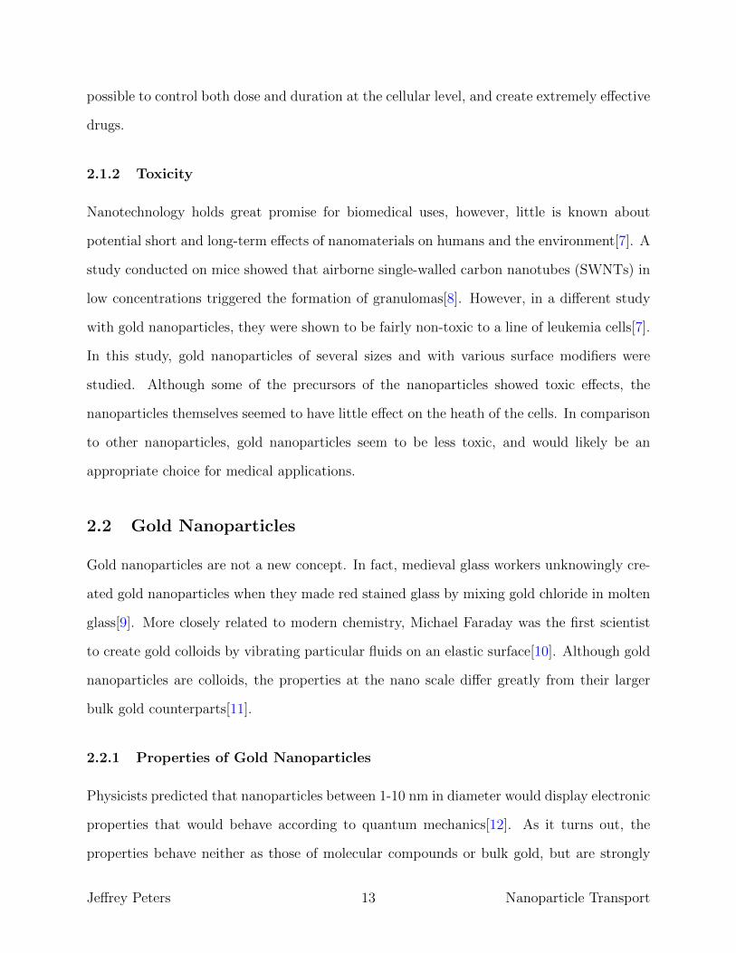
synthesis of amine-stabilized gold nanoparticles, which have NH2 groups on the surface, as
can be seen in Figure (2.2). Additional functional groups can be bonded with the functional
groups formed on the surface of the nanoparticles.
Figure 2.2: Amine-functionalized Nanoparticle[15]
2.3 Lipid Bilayers
A lipid bilayer is composed of lipids with hydrophobic tails, which drive the formation of
the bilayer[16]. In aqueous solutions, the lipids form micelles, which eliminate unfavorable
contacts between water, and the hydrophobic tails. A suspension of phospholipids can form
liposomes, which are closed vesicles bounded by a single bilayer, as can be seen in Figure
(2.3). These liposomes can serve as a model of a biological membrane.
Jeffrey Peters 15 Nanoparticle Transport

(a) Liposome[17] (b) Lipid bilayer[18]
Figure 2.3: Structure of Liposomes
2.4 Microscopy
2.4.1 Cross-Polarizing Microscopy
Polarized light is randomly oriented light that has been filtered such that the remaining light
waves all oscillate in the same plane[19]. In cross-polarizing microscopy, light passes through
a first polarizing filter, and is then blocked by a second filter oriented at a right angle to the
first. Polarizing microscopes use polarized light to enhance the contrast of images obtained
with birefringent (doubly refracting) materials[20]. The light from the source passes through
a polarizer before passing through the birefringent specimen, and then is passed through a
second polarizer, known as an analyzer, which recombines the light rays, as shown in Figure
(2.4).
Jeffrey Peters 16 Nanoparticle Transport

Figure 2.4: Cross-polarizing microscope[20]
The contrast in the image occurs when the birefringent specimen produces two wave
components of different velocities that, when recombine, interfere constructively and de-
structively. The contrast-enhancing properties of polarizing microscopy can provide detailed
information about the structure and composition of materials.
2.4.2 Confocal Fluorescent Microscopy
Fluorescent microscopy works on the principles of fluorescence, which means that when
an object is irradiated with light at a specific wavelength, the electrons in the object will
be excited and emit light at a certain detectable wavelength[21]. A properly configured
microscope will show only the fluorescent structures that will then be superimposed with a
dark background image. Confocal fluorescent microscopy provides several advantages over
conventional microscopy as the focal plane of the microscope can be moved[22]. Because
of this, the microscopes have a shallow depth of field and can collect images from thick
specimens without out-of-focus interference. To remove the out-of-focus aspect of the image,
the light emitted from the focal plane passes through a pinhole aperture, and the remaining
light is excluded, as shown in Figure (2.5).
Jeffrey Peters 17 Nanoparticle Transport

Figure 2.5: Confocal microscope[22]
Jeffrey Peters 18 Nanoparticle Transport

3 Methods
3.1 Vesicle Preparation
To isolate the exact physical properties of gold nanoparticles interacting with cell membranes,
I decided to use ”model” cell membranes, which will mimic the properties of an actual cell
membrane without the complexities of living cells. To create these lipid vesicles, I used 2-
Oleoyl-1-palmitoyl-sn-glycero-3-phosphocholine (POPC), obtained from Sigma-Aldrich (re-
fer to Appendix A for full description of materials). POPC is a lipid that occurs naturally in
eukaryotic cell membranes, and is an appropriate substitute that will mimic many properties
of a human cell.
3.1.1 Creating Lipid Solution
The most crucial step in the processes for creating the model cells is the preparation of
the lipid solution. The POPC must be carefully massed in a glass vial. A 2:1 solution of
chloroform to methanol is added to this. The ratio I found to work best is 2mg of POPC
to 1mL of solution. The sample must then be vigorously shaken for about 30 seconds. This
shaking process breaks the lipid structures up, allowing them to reform in cell-like shapes at
appropriate sizes.
3.1.2 Slide Preparation
After preparing the lipid solution, I placed a 10µL drop on to a well cleaned microscope slide.
The solution must then be dried on a hot plate at 40◦C. This causes the lipid structures to
adhere to the slide. The vesicles can then be re-hydrated with deionized water. This allows
the cell-like structures to reform.
Jeffrey Peters 19 Nanoparticle Transport

3.1.3 Cross-Polarizing Microscopy
I used cross-polarizing microscopy to observe the structure of the vesicles. The cells I had
synthesized were stained with diI, a red dye that adheres to lipids. This allowed me to view
the cells both with a bright-field view and under cross-polarizing conditions. One of the
next steps I will be conducting is to remove the dye, as it interferes with the fluorescent
microscopy. Because the cells will no longer be stained, the cross-polarizing microscope
will allow me to image the cells by viewing the birefringence of the bilayer. This technique
will show how the lipid structures are arranged and give me information about the surface
properties and makeup of the cells.
3.2 Functionalization of Gold Nanoparticles
To functionalize a nanoparticle means to attach some sort of molecule to the surface of the
nanoparticle. The nanoparticles I am using are functionalized with amine groups on the
surface. I am further functionalizing them with Fluorescein isothiocyanate isomer I (FITC).
This additional functional group will allow me to view the nanoparticles with a fluorescent
microscope, as the FITC molecules will fluoresce when excited by the proper wavelength
laser. To attach the FITC molecules to the nanoparticles, I am following a procedure similar
to one found in an Advanced Functional Materials (AFM) journal article[23].
3.2.1 Creating the Buffer Solution
A Britton-Robinson buffer solution is used to maintain the pH of the solution as the fluo-
rescent tags bond to the amine groups on the gold nanoparticles. This solution is made up
of equal parts 0.04M boric acid, 0.04M phosphoric acid, and 0.04M acetic acid. Then, the
solution is titrated with 0.1M sodium hydroxide, until a pH of 9.0 is reached. To do this, I
first created dilutions of each of the reactants to obtain the proper molarities. I then began
adding the sodium hydroxide to the acids, and checking frequently with pH strips until I
reached the correct pH.
Jeffrey Peters 20 Nanoparticle Transport

3.2.2 Introducing the Nanoparticles
In the procedure from the AFM article uses 100 mg of nanoparticles that are then dispersed
in 20 mL of the buffer solution. The gold nanoparticles I am using are expensive, and come
in much smaller quantities, so the procedure has to be adjusted. The nanoparticles are
in solution with water, so mass of the nanoparticles in solution must be determined by the
weight concentration of each solution. Properties of each nanoparticle size are given in Table
(3.1).
Nanoparticle size Weight Concentration Surface Area Molar Mass(nm) (mg/ml) (nm2) (g/mol)
20 2.66 1.26 × 10³ 4.68 × 10⁷
40 2.33 5.03 × 10³ 3.91 × 10⁸
60 2.15 1.13 × 10⁴ 1.32 × 10⁹
80 2.03 2.01 × 10⁴ 3.13 × 10⁹
Table 3.1: Properties of gold nanoparticles
Using the weight concentrations of the type of nanoparticle I was using, I calculated the
amount of buffer solution needed for 0.05 mL of nanoparticle solution. These two solutions
were then mixed with a solution with the fluorescent tag. The FITC tag used in this
procedure is in a powder form. To create the solution, the powder is massed in a glass vial,
and dissolved with methanol at a concentration of 0.25 mg/mL. This solution is added in a
1:20 FITC/buffer solution ratio. The final solution is then stirred over night.
3.2.3 Washing Procedure
To remove the unreacted dyes from the solution, I conducted a series of dilutions using a
centrifuge. I began by adding 1.0mM NaOH to the nanoparticle solution, and centrifuging
until the solution separated, and I could remove the liquid sitting on top of the nanoparticles.
I repeated this twice, and then twice more using 1.0mM HCl. Then I repeated the same
procedure twice using purified, deionized water, and added 0.5 mL of water to the final
solution. To prepare the sample for observation on the fluorescent microscope, I added a
Jeffrey Peters 21 Nanoparticle Transport

10µL drop to a slide prepared with rehydrated vesicles. I then sealed the slide with a cover
slip and incubated the sample at 37◦ to mimic the temperature of a human body until
observation.
3.2.4 Fluorescent Microscopy
Using the Leica SP5 Point Scanning Confocal at Gateway Park, I was able to take fluorescent
images of my sample. The FITC tag has an excitation wavelength of 492 nm, so I used the
argon laser on the microscope to excite the sample. Because the microscope is confocal, I
was able to adjust the focal plane and observe the sample directly on the glass slide and
throughout the sample. The data collection I will be conducting for my Master’s thesis with
this microscope is described in the Future Work section.
Jeffrey Peters 22 Nanoparticle Transport

4 Results and Discussion
4.1 Results
4.1.1 Vesicle Preparation
My first attempt used 5 mg of POPC to 0.5 mL of solution. The results of this are shown
in Figure (4.1).
Figure 4.1: 10mg/mL POPC, 200x magnification
In this image, the lipid, stained with 3,3’-Dihexyloxacarbocyanine Iodide (diI), did not as-
semble into cell-like structures. This was because the concentration of the POPC was too
high, and layers of vesicles were formed on top of each other. I determined that a more
suitable concentration of POPC was 2mg/mL of solution. Another important factor in the
creation of the vesicles is the shaking procedure. I discovered that a sample that had previ-
ously been successful did not yield cell-like structures when later reused. This is because I
had not re-shaken the sample. The vigorous shaking causes the lipid structures to break, and
reform in more uniform cell shapes. Figure (4.2) shows a comparison of shaking techniques.
Jeffrey Peters 23 Nanoparticle Transport

(a) 2mg/mL POPC, 200x, less shaken (b) 2mg/mL POPC, 200x, more shaken
Figure 4.2: Comparison of shaking techniques
In Figure (4.2a), the sample had been shaken enough to break the lipid structures in
to mostly separate shapes, however, this figure shows structures that have not completely
separated and are oblong in shape. The same sample was shaken more vigorously, and Figure
(4.2b) shows this resulted in individual cell-like structures that are more uniform in size and
shape.
4.1.2 Gold Nanoparticles
Following the procedure above for the functionalization of the gold nanoparticles, I created
a sample using the 60 nm nanoparticles. After introducing a drop of this solution to a
slide containing the vesicles, I sealed the solution with a cover slip and let the nanoparticles
interact with the vesicles for 8 hours at a temperature of 37◦C. I then observed the sample
on the fluorescent microscope, the results of which can be seen in Figure (4.3).
Jeffrey Peters 24 Nanoparticle Transport

Figure 4.3: Fluorescent microscope, sample with 60 nm nanoparticles
The material fluorescing in green is actually the dye used to stain the vesicles as well as
the FITC molecules left in the solution. The diI molecules fluoresce at a similar wavelength
to the FITC, and thus the dye on vesicles the would mask any nanoparticles inside. In
addition, there are no spots that are clearly brighter than the surrounding dye, indicating
that the nanoparticles were not functionalized. However, this sample is successful, in that
the vesicles are nearly spherical and similar in size. As the scale on the image indicates, the
vesicles are on the order of 30 µm, which is an approximately the size of a human skin cell,
and a little larger than a blood cell. This size vesicle is an appropriate analogue for medical
application. Also, as the confocal microscope can change focus planes, I observed that the
Jeffrey Peters 25 Nanoparticle Transport
remaining lipid seen around the cells was not floating freely in solution, but rather adhered
to the glass surface, and therefore will not interfere with the nanoparticles in the solution.
As a control test, I created a slide that contained only the solution with the gold nanopar-
ticles. All that appeared on this slide when looked at on the fluorescent microscope was a
green haze. Because there were again no obvious bright spots, it was clear that the nanopar-
ticles remained unfunctionalized. In addition, the washing procedure was not completely
effective, as FITC molecules were still floating in solution.
4.2 Discussion
4.2.1 Possible Problems with the Buffer Solution
One possible problem with the buffer solution that could have affected my functionalization
procedure is the titration. Unfortunately, a pH meter was not available while I was syn-
thesizing this solution. Instead, I used pH strips, which do not provide the same level of
accuracy as a pH meter. The pH of the solution has to be within a certain range for the
reaction to occur, so if my buffer solution was outside of this range, it could contribute to
the nanoparticles not being functionalized.
4.2.2 The Washing Procedure Affecting Functionalization
The most likely step in the functionalization procedure that would have caused the FITC
molecules to not be attached to the nanoparticles was the washing procedure. The test tube
with my first sample was destroyed by the centrifuge. Although the liquid had leaked out,
an important observation was that the gold had actually accumulated on the sides of the
test tube. Following this, I tried using a different centrifuge that would not destroy the test
tube, and had a variable speed control so I could reduce the centrifuging speed. This did
help, however, after the first several washes, the color of the solution began to change from
a red color to a gray color. Solutions containing larger gold nanoparticles tend to have a
grayer color, so this color change indicates that the nanoparticles were aggregating as the
Jeffrey Peters 26 Nanoparticle Transport

solution was being centrifuged. Shortly after this color change occurred, a slight gold layer
appeared on the test tube walls. If the gold particles were aggregating, not only were they
not ending up in the solution added to the vesicles, but any bonds made with the FITC
molecules were likely broken.
Jeffrey Peters 27 Nanoparticle Transport

5 Summary and Future Work
5.1 Summary
The work completed so far has set the groundwork for a successful Master’s thesis. I have
shown that I can synthesize model cells that are uniform in size in shape, and should act
appropriately as stand-ins for living cells. Furthermore, I have familiarized myself with both
cross-polarizing and confocal fluorescent microscopy, which I will be using to collect data. I
have worked through a majority of my functionalization procedure, and have the necessary
materials to create many samples to study. Although I have yet to perfect the procedure, I
have several ideas to test as I begin my thesis work. With most of the procedural work in
place, what remains is creating samples and data collection and analysis.
5.2 Future Work
As this research progresses, the remaining work will be focused on refining the procedures,
and analyzing data collected on the fluorescent microscope. Once the procedures are final-
ized, I will be able to vary the variables in the system such as concentration of nanoparti-
cles, functionalization, and environment, to observe what affects the cellular uptake of the
nanoparticles.
5.2.1 Refining the Washing Procedure
The first test to try is to observe the nanoparticles after being mixed with the FITC solution,
but before the washing procedure. This test will determine if the centrifuging is breaking the
bonds with the FITC molecules. Although the unreacted dye will show up on the fluorescent
microscope, it will only be as a green haze in the background. If the nanoparticles were
successfully functionalized, they should be clearly brighter than the fluorescing solution. If
the background fluorescence is uniform, and not interfering with seeing the nanoparticles, I
can collect data without having to wash the nanoparticle solutions. If the FITC is bonded
Jeffrey Peters 28 Nanoparticle Transport
to the nanoparticles, but the remaining dye interferes with data taking, I will have to adjust
my washing procedure to something gentler that will allow me to wash out the unreacted
dye without causing the nanoparticles to aggregate.
5.2.2 Collecting Data
To collect data, I will make a series of identical samples, and introduce the nanoparticles
to the vesicles at a series of different times. By counting the nanoparticles both inside
and outside of the vesicles in each sample, I will be able to plot the concentration of the
nanoparticles as a function of time. The rate at which the nanoparticles enter the cells, and
the equilibrium point of the system will provide insight as to how the nanoparticles enter the
cells. The analysis of the images I collect from the fluorescent microscope will be conducted
with ImageJ, a software package developed by the National Institutes of Health.
5.2.3 Modifying the Gold Nanoparticles
To better understand what affects the mechanisms of cellular uptake, I will begin varying
parameters of the system. In addition to adjusting the concentration of the nanoparticles,
modifications to the nanoparticles themselves could alter how they enter the cells. It is
possible to attach several functional groups to the nanoparticles, so a possible avenue to
explore would be adding hydrophobic and/or hydrophilic functional groups. The lipid bilayer
has both hydrophobic and hydrophilic properties, so the addition of these functional groups
would likely affect cellular uptake. If this is the case, it would indicate the the uptake process
is not purely dictated by endocytosis.
5.2.4 Changing the Environment
In addition to altering the nanoparticles, changes to the environment could affect the system
dynamics. Currently, the solutions containing both the cells and nanoparticles are composed
primarily of water. Depending on the biological environment being studied, water may not be
Jeffrey Peters 29 Nanoparticle Transport

the best substitute. Bovine serum albumen, for example, is often used in medical research, as
it more accurately mimics a human environment. By changing the environment the vesicles
are in to represent human systems, I could gain an understanding of how gold nanoparticles
might act if used on a living subject.
Jeffrey Peters 30 Nanoparticle Transport

References
[1] Bouzid Menaa. Importance of nanotechnology in biomedical sciences. Biotechnology
and Biomaterials, 1(5), 2011.
[2] Charles Martin and Punit Kohli. The emerging field of nanotube biotechnology. Nature
Reviews, 2:29–37, 2003.
[3] Kathryn Ulrich, Scott Cannizzaro, Robert Langer, and Kevin Shakesheff. Polymeric
systems for controlled drug release. Chemical Reviews, 99(11):3181–3198, 1999.
[4] Omid Farokhzad and Robert Langer. Impact of nanotechnology on drug delivery. AC-
SNano, 3(1):16–20, 2009.
[5] Elena Heister, Vera Neves, S.RaviP. Silva, Johnjoe McFadden, and HelenM. Coley.
Carbon nanotubes loaded with anticancer drugs: A platform for multimodal cancer
treatment. In Rudiger Klingeler and Robert B. Sim, editors, Carbon Nanotubes for
Biomedical Applications, Carbon Nanostructures, pages 223–245. Springer Berlin Hei-
delberg, 2011.
[6] Vinod Labhasetwar. Nanotechnology for drug and gene therapy: the importance of
understanding molecular mechanisms of delivery. Science Direct, 16:674–680, 2005.
[7] Ellen Connor, Judith Mwamuka, Anand Gole, Catherine Murphy, and Michael Wyatt.
Gold nanoparticles are taken up by human cells but do not cause acute cytotoxicity.
Small Nano Micro, 1(3):325–327, 2005.
[8] Robert Sercive. Nanomaterials show signs of toxicity. Science, 300(5617):243, 2003.
[9] Kenneth Chang. Tiny is beautiful: Translating ’nano’ into practical, February 22, 2005.
[10] Michael Faraday. On a peculiar class of acoustical figures; and on certain forms assumed
by groups of particles upon vibrating elastic surfaces. Phil Trans R Soc, 121:299–340,
1831.
Jeffrey Peters 31 Nanoparticle Transport

[11] Vanga Reddy. Gold nanoparticles: Synthesis and applications. Synlett, 11:1791–1792,
2006.
[12] Marie-Christine Daniel and Didier Astruc. Gold nanoparticles: Assembly, supramolec-
ular chemistry, quantum-size-related properties, and applications toward biology, catal-
ysis, and nanotechnology. Chemical Reviews, 104:293–346, 2004.
[13] Tanya Stuchinskaya, Miguel Moreno, Michael Cook, Dylan Edwards, and David
Russel. Targeted photodynamic therapy of breast cancer cells using anti-
body–phthalocyanine–gold nanoparticle conjugates. Photochemical & Photobiological
Sciences, 10:822–831, 2011.
[14] http://www.azonano.com/.
[15] http://cytodiagnostics.com/.
[16] Donald Voet, Judith Voet, and Charlotte Pratt. Fundamentals of Biochemistry. Wiley
& Sons, 3 edition, 2008.
[17] http://www.supplementclinic.com/.
[18] http://antranik.org/.
[19] Douglas Murphy, Kenneth Spring, and Michael Davidson. Introduction to polarized
light, 2013.
[20] Philip Robinson and Michael Davidson. Introduction to polarized light microscopy,
2013.
[21] Kenneth Spring and Michael Davidson. Introduction to fluorescence microscopy, 2013.
[22] Stephen Paddock, Thomas Fellers, and Michael Davidson. Basic concepts of confocal
microscopy, 2013.
Jeffrey Peters 32 Nanoparticle Transport

[23] Xu-dong Wang, Robert Meier, and Otto Wolfbeis. A fluorophore-doped polymer nano-
material for referenced imaging of ph and temperaturewith sub-micrometer resolution.
Advanced Functional Materials, 22:4202–4207, 2012.
Jeffrey Peters 33 Nanoparticle Transport

Appendices
A Materials
• Gold Nanoparticles: Obtained from Cytodiagnostics. Sizes of 20 nm, 40 nm, 60 nm,
and 80 nm. Nanoparticles functionalized with amine groups.
• 2-Oleoyl-1-palmitoyl-sn-glycero-3-phosphocholine (POPC): Obtained from Sigma-Aldrich.
A self-assembling lipid found in eukaryotic cell membranes.
• Fluorescein isothiocyanate isomer I (FITC): Obtained from Sigma-Aldrich. A fluores-
cent labeling reagent. Has an excitation wavelength of 492 nm and emission wavelength
of 518 nm.
• 3,3’-Dihexyloxacarbocyanine iodide (diI): Obtained from Sigma-Aldrich. A fluorescent
protein labeling dye. Has an excitation wavelength of 485 nm and emission wavelength
of 501 nm.
• Methanol: Obtained from Sigma-Aldrich. Anhydrous grade solvent. 99.8% assay.
• Chloroform: Obtained from Sigma-Aldrich. Anhydrous grade solvent. ≥ 99% assay.
• Ethanol: Obtained from Sigma-Aldrich. Anhydrous grade solvent. ≥ 99.5% assay.
• Acetic Acid: Obtained from Sigma-Aldrich. Volumetric grade acetic acid solution, 0.5
M.
• Boric Acid: Obtained from Sigma-Aldrich. Molecular biology grade buffer salt. ≥
99.5% assay.
• Phosphoric Acid: Obtained from Sigma-Aldrich. BioReagent, 85% assay.
• Hydrochloric Acid: Obtained from Sigma-Aldrich. Acid concentrate. Concentration
of 0.5 M.
Jeffrey Peters 34 Nanoparticle Transport

• Sodium Hydroxide: Obtained from Sigma-Aldrich. Base concentrate. Concentration
of 0.1 M.
Jeffrey Peters 35 Nanoparticle Transport
Top Related