


Languages
Pages
Legal

Cell Signal Transduction and Diseases
Jimin Shao ( 邵吉民 )Professor, Dept. Pathology and PathophysiologyTel: 88208209E-mail: [email protected]
1. Introduction: Cell signaling and Signal transduction (1) Cell signaling
(2) Types of cellular signals
(3) Functions
2. Gap junction and diseases
(1) Structure of gap junction
(2) Function of gap junction
(3) Related diseases
3. Receptor-Mediated Signal Transduction and diseases (1) Cell-surface receptors (2) Nuclear receptors (3) Regulatory mechanisms of signal transduction
(4) Disorders and related diseases
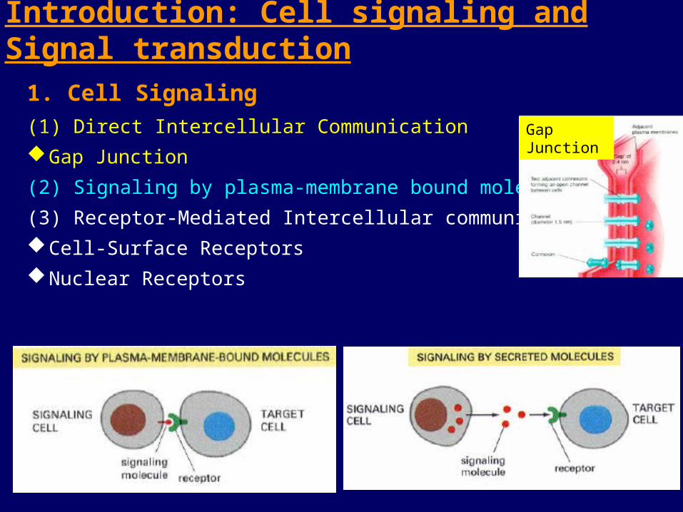
Introduction: Cell signaling and Signal transduction
1. Cell Signaling
(1) Direct Intercellular Communication
Gap Junction
(2) Signaling by plasma-membrane bound molecules
(3) Receptor-Mediated Intercellular communication
Cell-Surface Receptors
Nuclear Receptors
Gap Junction

2. Types of cellular signals
(1) Chemical signals
Hormones, neurotransmitters, growth factors, cytokines;
odor molecules; ATP, active oxygen; drugs, toxins, etc
(2) Physical signals Light, electronic, mechanic, UV, heat, volume, osmotic, etc
Endocrine Act on a far away organ via blood circulation, seen in most hormones
Paracrine Act on a nearby target, seen in GFs, CKs, etc Synaptic: Presynaptic to postsynaptic (neurotransmitters) Autocrine Act on itself after secreted, seen in GFs, especially in tumor tissues Intracrine Act on itself before secreted, seen in nuclear receptors
(3) Modes for the function of endogenous signals
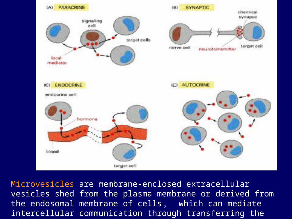
Microvesicles are membrane-enclosed extracellular vesicles shed from the plasma membrane or derived from the endosomal membrane of cells , which can mediate intercellular communication through transferring the vesicular components to recipient cells.

3. Cell Signal Transduction Pathways--Physiological functions--Metabolism--Cell cycle, growth, differentiation, and apoptosis--Responses to stress--etc.
LPSTNFIL-1

1. Structure of Gap Junction Connexins are four-pass transmembrane proteins,
six of which assemble to form a channel, a connexon. ~ 20 different isoforms of connexins in humans and mice
Gap junction
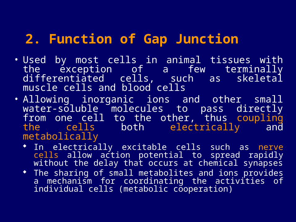
• Used by most cells in animal tissues with the exception of a few terminally differentiated cells, such as skeletal muscle cells and blood cells
• Allowing inorganic ions and other small water-soluble molecules to pass directly from one cell to the other, thus coupling the cells both electrically and metabolically
In electrically excitable cells such as nerve cells allow action potential to spread rapidly without the delay that occurs at chemical synapses
The sharing of small metabolites and ions provides a mechanism for coordinating the activities of individual cells (metabolic cooperation)
2. Function of Gap Junction
3. Gap Junction in Diseases
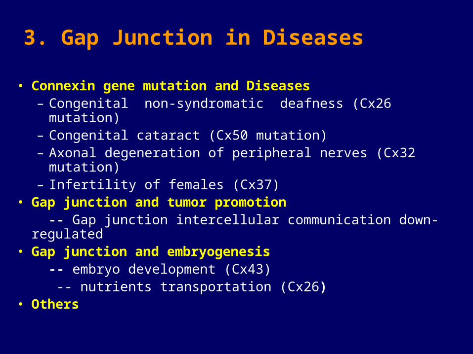
• Connexin gene mutation and Diseases – Congenital non-syndromatic deafness (Cx26 mutation) – Congenital cataract (Cx50 mutation) – Axonal degeneration of peripheral nerves (Cx32 mutation)– Infertility of females (Cx37)
• Gap junction and tumor promotion -- Gap junction intercellular communication down-regulated • Gap junction and embryogenesis -- embryo development (Cx43) -- nutrients transportation (Cx26)• Others

Receptors: Cell Surface Receptors; Nuclear Receptors
Receptor-Mediated Signal Transduction Systems
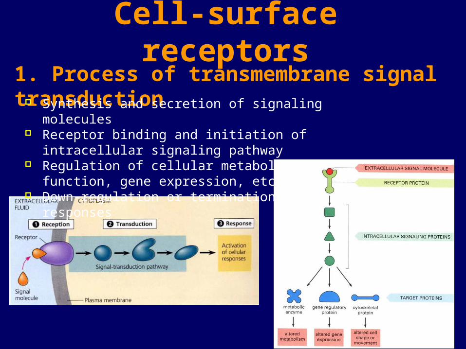
1. Process of transmembrane signal transduction
Cell-surface receptors
Synthesis and secretion of signaling molecules Receptor binding and initiation of intracellular signaling
pathway Regulation of cellular metabolism, function, gene expression, etc Down-regulation or termination of cellular responses
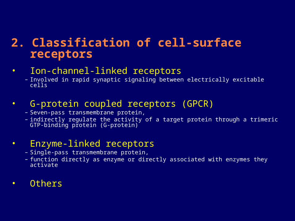
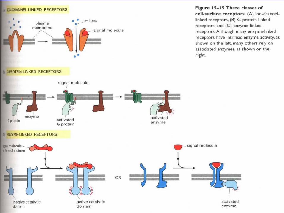
2. Classification of cell-surface receptors• Ion-channel-linked receptors
– Involved in rapid synaptic signaling between electrically excitable cells
• G-protein coupled receptors (GPCR)– Seven-pass transmembrane protein, – indirectly regulate the activity of a target protein through a trimeric GTP-binding
protein (G-protein)
• Enzyme-linked receptors– Single-pass transmembrane protein, – function directly as enzyme or directly associated with enzymes they activate
• Others

Ion Channel Linked Receptor(Ligand-gated Ion Channel)
1. Classification: Class I nAchR, GABAR, 5-HT3R, GlyR
Class II Glutamate/Aspartate Receptor
Class III cGMP/cAMPR, IP3R, ryanodine R
Class IV ATP/ADP gated channel (P2X)
2. Structure:

3. Involved in rapid synaptic signaling between electrically excitable cells--A model for iGluR activation and desensitization
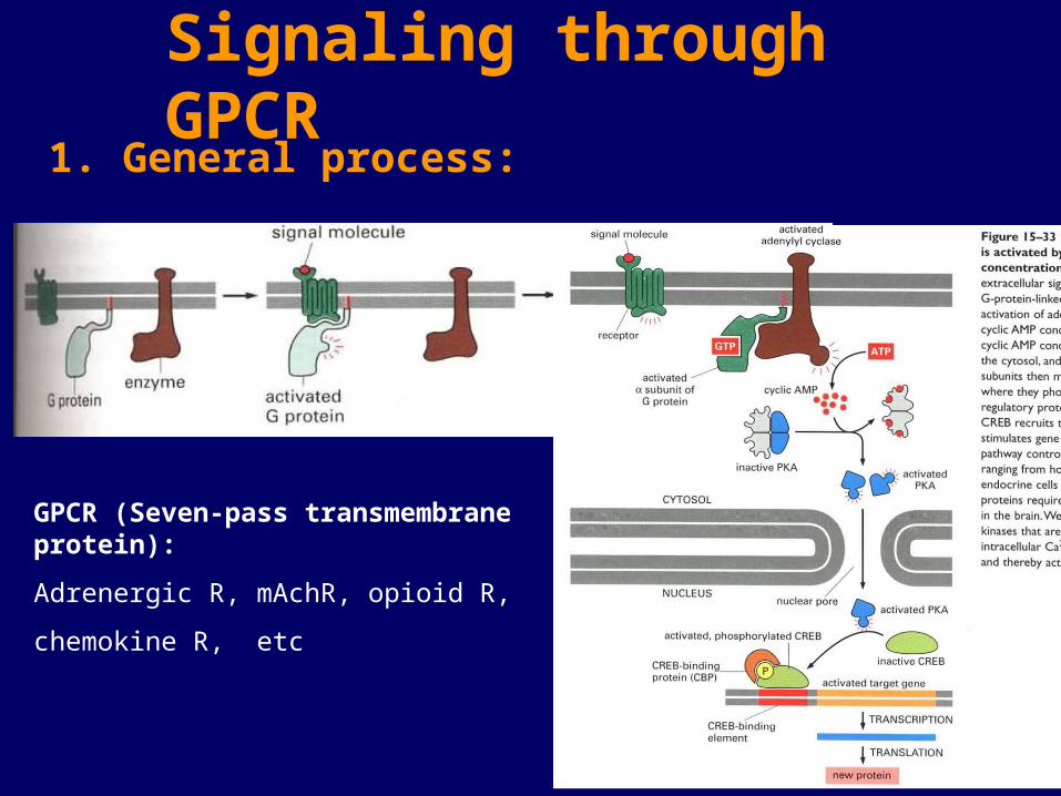
1. General process:
Signaling through GPCR
GPCR (Seven-pass transmembrane protein):
Adrenergic R, mAchR, opioid R,
chemokine R, etc
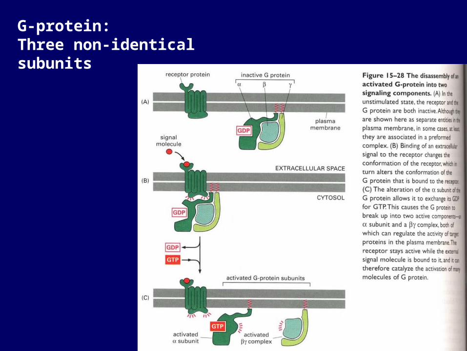
G-protein: Three non-identical subunits
(2) Gq: Gq activates the inositol phospholipid signaling pathway by activating phospholipase C- (PLC- PLC- acts on phosphatidylinositol 4,5-biphosphate [PI(4,5)P2] to generate: inositol (1,4,5)-triphosphate (IP3) ; diacylglycerol (DAG).
2. GαSignaling pathways
(1) Gs, Gi:Stimulatory G protein (Gs), activates Ac; Inhibitory G protein (Gi), inhibits Ac.cAMP is synthesized from ATP by adenylyl cyclase, and rapidly hydrolyzed by cAMP phosphodiesterase.Usually the α subunit regulates the cyclase, although βγ complex may does so as well.
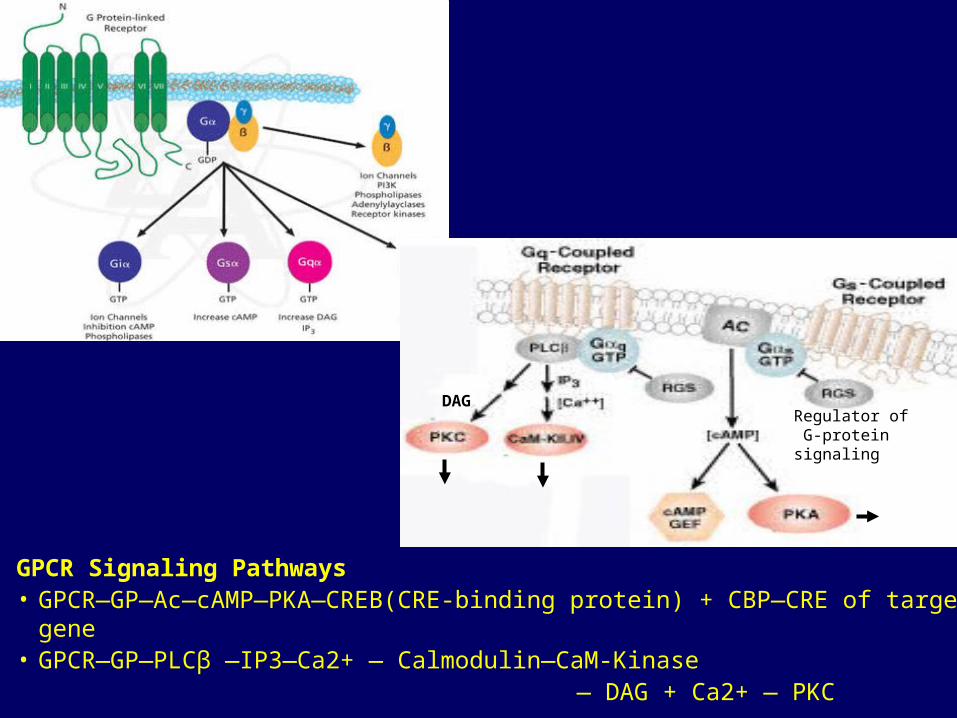
GPCR Signaling Pathways• GPCR—GP—Ac—cAMP—PKA—CREB(CRE-binding protein) + CBP—CRE of target gene• GPCR—GP—PLCβ —IP3—Ca2+ — Calmodulin—CaM-Kinase — DAG + Ca2+ — PKC
Regulator of G-protein signaling
DAG
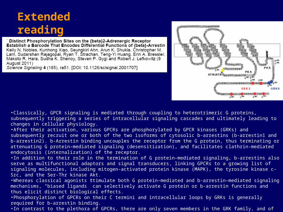
•Classically, GPCR signaling is mediated through coupling to heterotrimeric G proteins, subsequently triggering a series of intracellular signaling cascades and ultimately leading to changes in cellular physiology. •After their activation, various GPCRs are phosphorylated by GPCR kinases (GRKs) and subsequently recruit one or both of the two isoforms of cytosolic b-arrestins (b-arrestin1 and b-arrestin2). b-Arrestin binding uncouples the receptor from the G protein, thus terminating or attenuating G protein–mediated signaling (desensitization), and facilitates clathrin-mediated endocytosis (internalization) of the receptor. •In addition to their role in the termination of G protein–mediated signaling, b-arrestins also serve as multifunctional adaptors and signal transducers, linking GPCRs to a growing list of signaling molecules, including mitogen-activated protein kinase (MAPK), the tyrosine kinase c-Src, and the Ser-Thr kinase Akt.•Whereas classical agonists stimulate both G protein–mediated and b-arrestin–mediated signaling mechanisms, “biased ligands” can selectively activate G protein or b-arrestin functions and thus elicit distinct biological effects.•Phosphorylation of GPCRs on their C termini and intracellular loops by GRKs is generally required for b-arrestin binding. •In contrast to the plethora of GPCRs, there are only seven members in the GRK family, and of those, only GRKs 2, 3, 5, and 6 are ubiquitously expressed. •Studies suggest that distinct GRKs may contribute differently to the processes of receptor desensitization, endocytosis, and signaling.•We hypothesized that the different GRKs might phosphorylate distinct sets of sites on the C terminus and internal loops of the receptor, thereby establishing a “barcode” that would instruct or determine the conformation assumed by the b-arrestin, which would, in turn, determine its functional capabilities.
Extended reading

Signaling through enzyme-linked cell-surface receptors
1. Receptor tyrosine kinases
2. Tyrosine-kinase-associated receptors
3. Receptor serine/threonine kinases
4. Receptor guanylyl cyclases
5. others
1. Receptor tyrosine kinases• Many secreted growth factors and hormones act through receptor
tyrosine kinases:– Epithelial growth factor (EGF), – platelet-derived growth factor (PDGF), – fibroblast growth factors (FGFs), – hepatocyte growth factor (HGF), – insulin, insulinlike growth factor-1 (IGF-1), – vascular endothelial growth factor (VEGF), – macrophage-colony-stimulating factor (M-CGF), – neurotrophins
• Many cell-surface-bound signal proteins also act through receptor tyrosine kinases– Ephrins (Eph): regulates cell adhesion and repulsion response that guide the
migration of cells and axons during development– Eph receptors: receptor tyrosine kinases– Bidirectional signaling: binding to Eph receptor can cause the activation of
both Eph and Eph receptor, thus changing the behavior of both cells
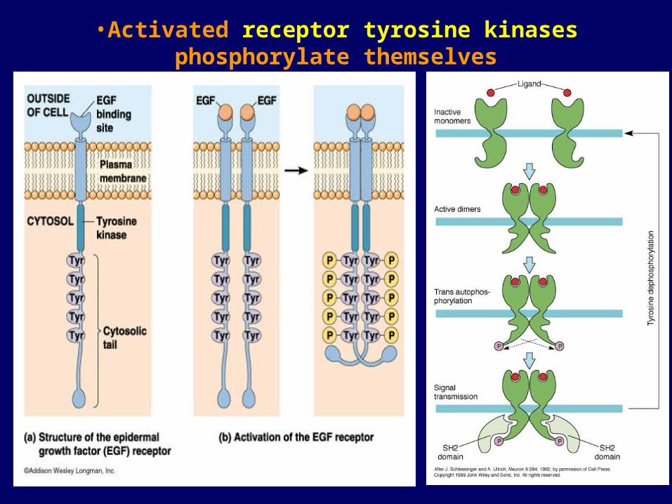
•Activated receptor tyrosine kinases phosphorylate themselves

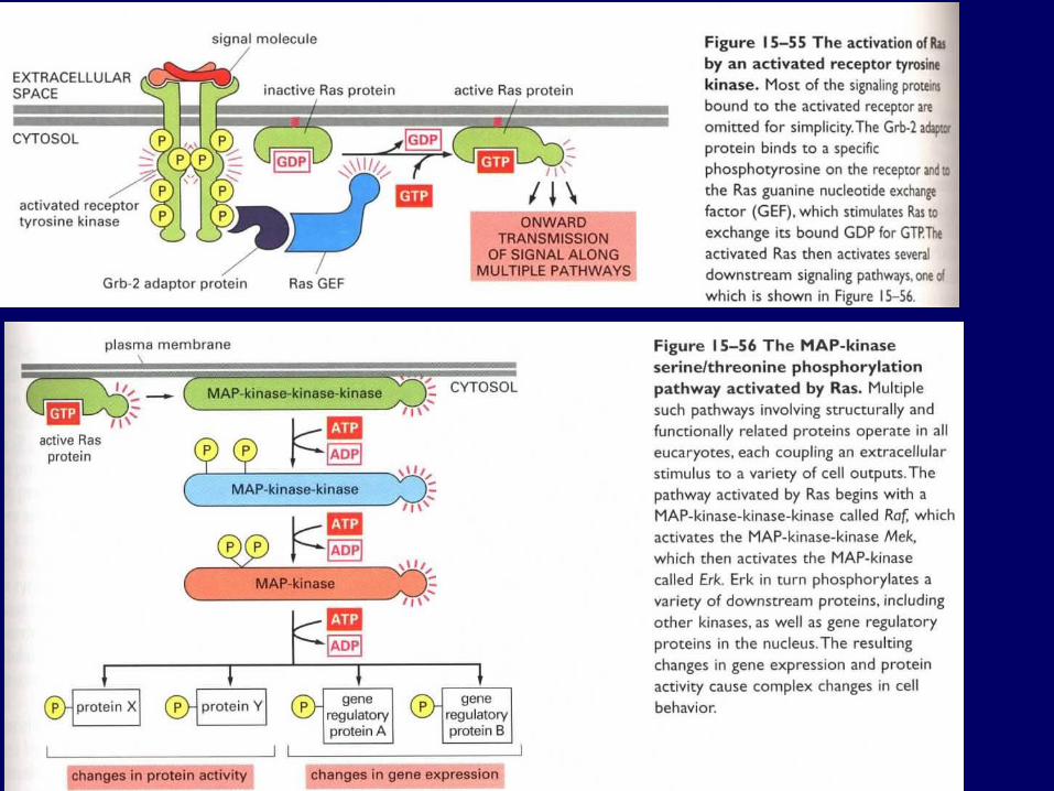
(1) RAS-MAPK PathwayGF--RTK--GRB2—Ras GEF—Ras –-MAPKKK—MAPKK--MAPK (Raf—MEK1/2—ERK1/2)
MAPKs:ERK
JNK/SAPK
p38MAPK
etc

(2) PI3-kinase/Protein kinase B pathway• PKB (or Akt) containing a PH domain --Binds to PI(3,4,5)P3 --Activated by a phosphatidylinositol-dependent protein kinase PDK1 --Phosphorylates and inactivates BAD, a pro-apoptosis factor, promotes cell survival
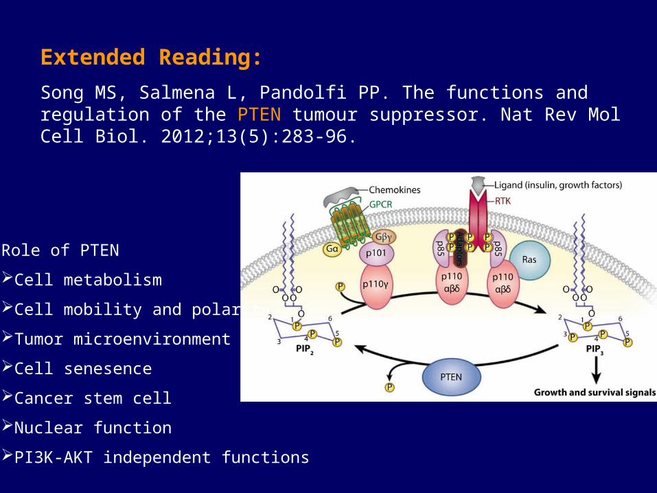
Extended Reading:
Song MS, Salmena L, Pandolfi PP. The functions and regulation of the PTEN tumour suppressor. Nat Rev Mol Cell Biol. 2012;13(5):283-96.
Role of PTEN
Cell metabolism
Cell mobility and polarity
Tumor microenvironment
Cell senesence
Cancer stem cell
Nuclear function
PI3K-AKT independent functions
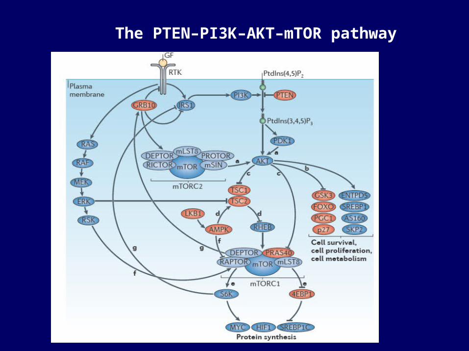
The PTEN–PI3K–AKT–mTOR pathway
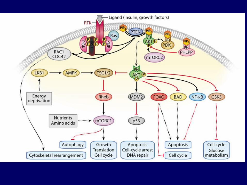

Functions of PTEN in the nucleus
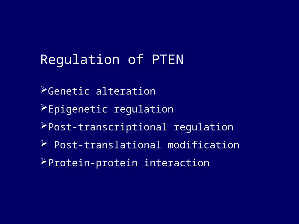
Regulation of PTEN
Genetic alteration
Epigenetic regulation
Post-transcriptional regulation
Post-translational modification
Protein-protein interaction

(3) Cross-talk between signaling pathways activated by GPCRs and RTKs
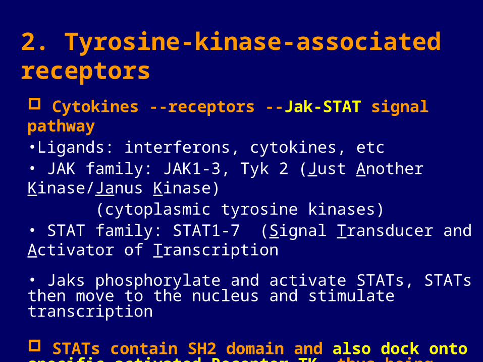
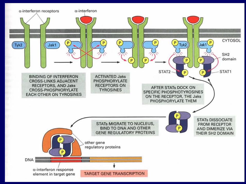
2. Tyrosine-kinase-associated receptors
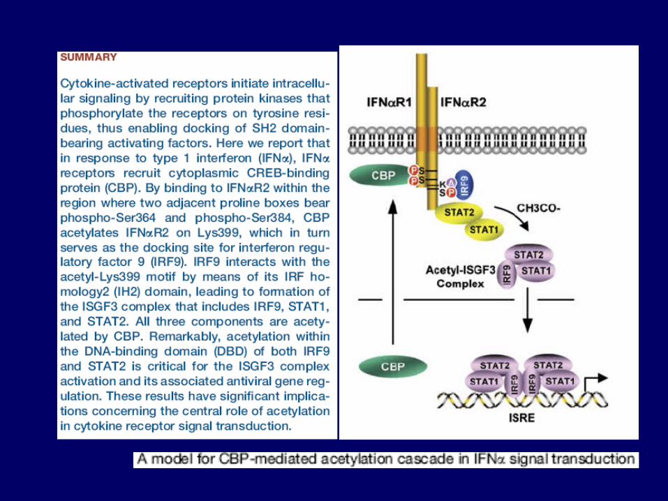
Cytokines --receptors --Jak-STAT signal pathway•Ligands: interferons, cytokines, etc • JAK family: JAK1-3, Tyk 2 (Just Another Kinase/Janus Kinase) (cytoplasmic tyrosine kinases)• STAT family: STAT1-7 (Signal Transducer and Activator of Transcription
• Jaks phosphorylate and activate STATs, STATs then move to the nucleus and stimulate transcription
STATs contain SH2 domain and also dock onto specific activated Receptor TK, thus being activated independent of Jaks.
3. Receptor serine/threonine kinasesTGF- superfamily--receptor serine/threonine kinases--Smads Signal Pathway
• Transforming growth factor- superfamily: TGF-s, activins, bond morphogenetic proteins (BMPs), etc• TGF-β Receptor Superfamily: Protein serine/threonine kinase; Single pass transmembrane receptor; Type I and II• The ligand first binds to and activates type II homodimer, which
recruits, phosphorylates, and activates a type I receptor dimer, forming a tetrameric receptor complex
• Smads


4. Receptor guanylyl cyclases• Receptor guanylyl cyclases: single pass transmembrane proteins.• Generates cGMP in response to stimuli. • cGMP binds/activates a cGMP-dependent serine/threonine protein kinase (PKG).• Atrial natriuretic peptides (ANPs) as signal molecules: regulate salt and water
balance and dilate blood vessels.
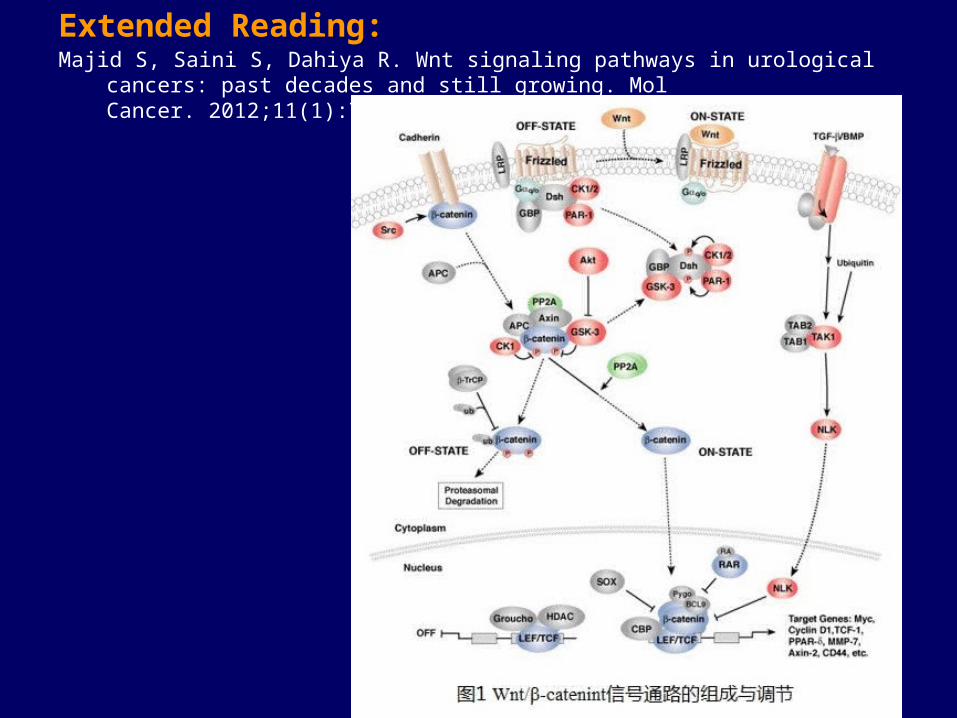
Extended Reading:Majid S, Saini S, Dahiya R. Wnt signaling pathways in urological cancers: past decades and still
growing. Mol Cancer. 2012;11(1):7.
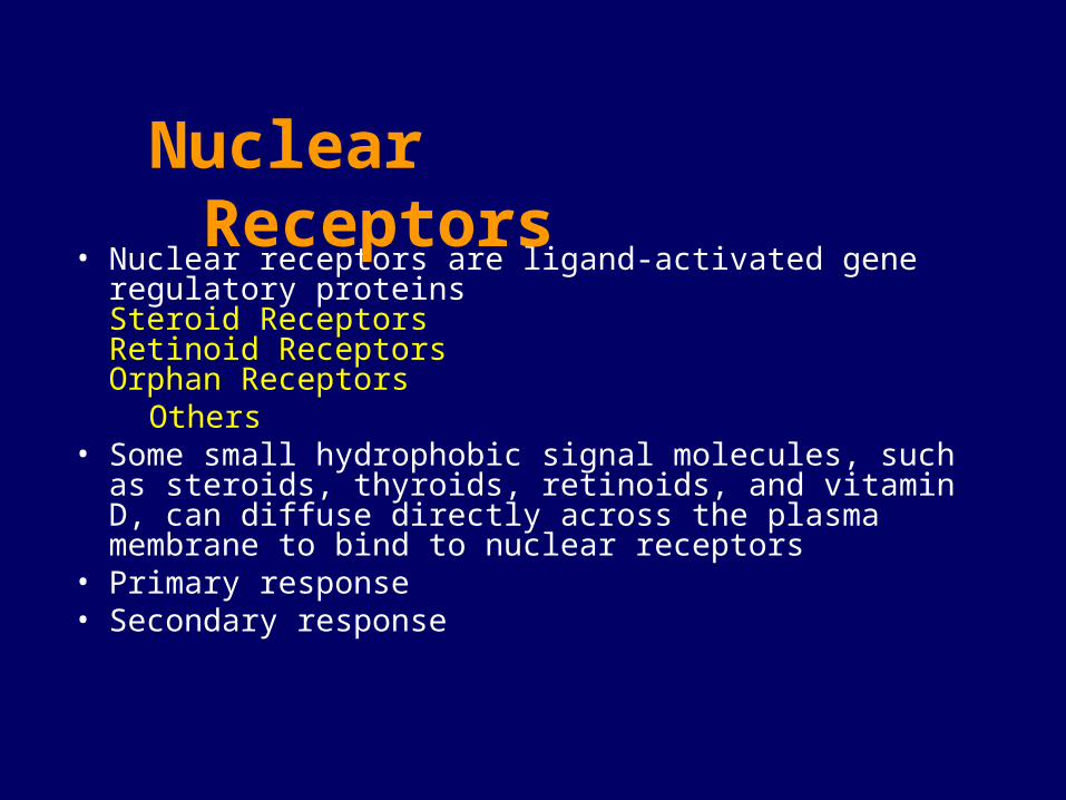
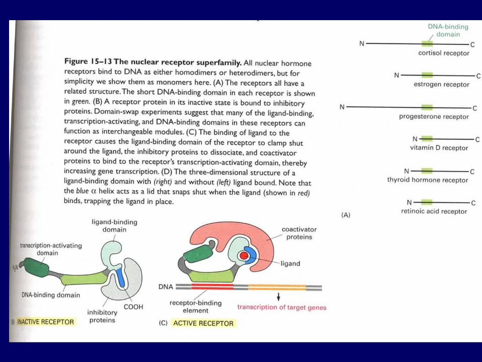
• Nuclear receptors are ligand-activated gene regulatory proteinsSteroid ReceptorsRetinoid ReceptorsOrphan Receptors
Others• Some small hydrophobic signal molecules, such as steroids,
thyroids, retinoids, and vitamin D, can diffuse directly across the plasma membrane to bind to nuclear receptors
• Primary response• Secondary response
Nuclear Receptors
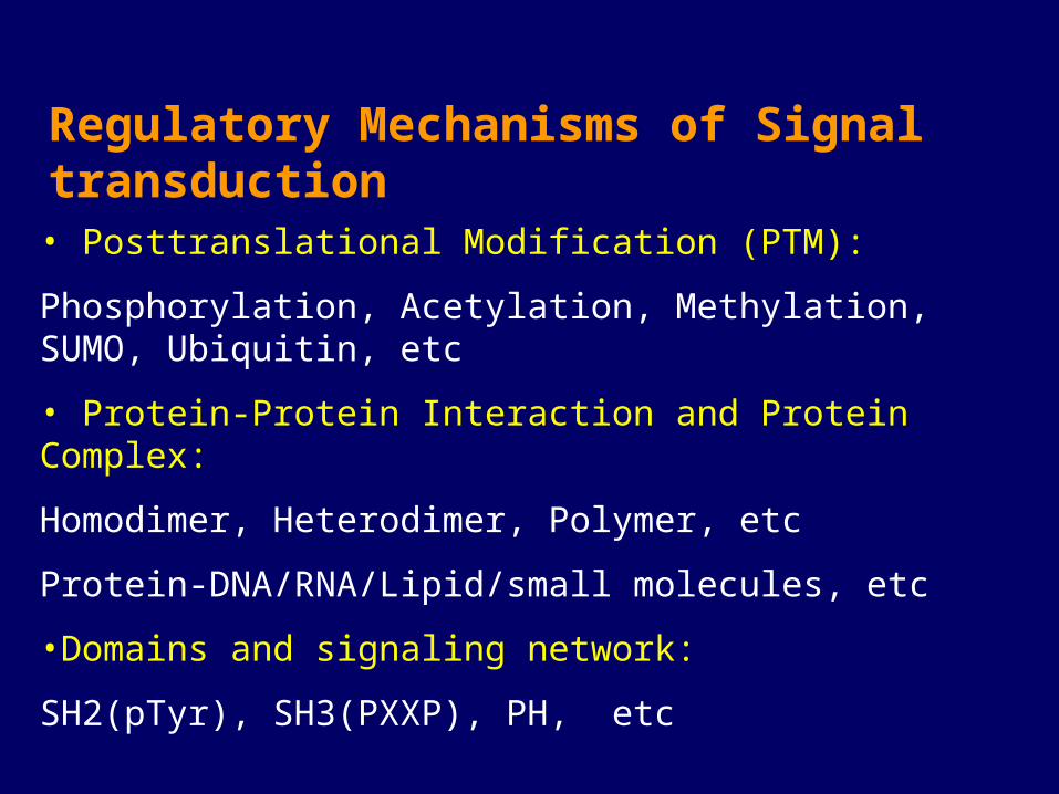
Regulatory Mechanisms of Signal transduction
• Posttranslational Modification (PTM):
Phosphorylation, Acetylation, Methylation, SUMO, Ubiquitin, etc
• Protein-Protein Interaction and Protein Complex:
Homodimer, Heterodimer, Polymer, etc
Protein-DNA/RNA/Lipid/small molecules, etc
•Domains and signaling network:
SH2(pTyr), SH3(PXXP), PH, etc
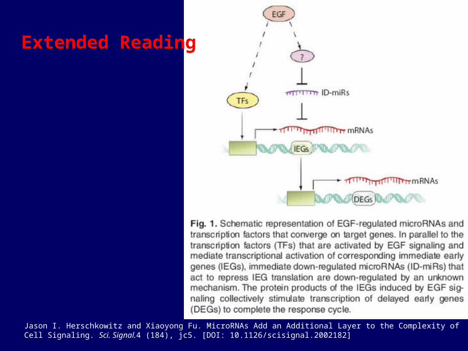
Jason I. Herschkowitz and Xiaoyong Fu. MicroRNAs Add an Additional Layer to the Complexity of Cell Signaling. Sci. Signal.4 (184), jc5. [DOI: 10.1126/scisignal.2002182]
Extended Reading
Top Related