


Languages
Pages
Legal


122Bonn,zool. Beitr.
Relationships of the Passerine Finches
(Passeriformes: Passeridae)
by
WALTER J. BOCK
Departement of Biological Sciences Columbia University
New York, N.Y., 10027, U.S.A.
and
Department of Ornithology
American Museum of Natural History
and
JOHN J. MORONY, Jr.
Department of Ornithology
American Museum of Natural History
New York, N.Y., 10024, U.S.A.
Introduction
The passerine finches are a small group of Old World seed-eating oscines
consisting of the genera Passer Brisson 1760, Montiiringilla Brehm 1828,
and Petronia Kaup 1829. Their relationships to other groups of oscine
finches have been a matter of disagreement for the past 75 years with no
clear resolution. Discovery of a unique skeletal structure — the preg-
lossale — in the tongue of the passerine finches (Bock and Morony, 1978)
prompted us to inquire further into the taxonomic position of these birds.
We would like to do so considering a series of hypotheses about groups
and about characters (Bock, 1977: 875—891) pertaining to the passerine
finches and to the preglossale and associated features. We accept, for this
study, the approach and methods of evolutionary classification (see Bock,
1974, 1977). In this approach, the phylogeny and the classification of a
group are not assumed to be redundant (i. e. redundant = the classification
is an exact reflection of the phylogeny based upon the definition of re-
lationship as being only that of phylogenetic branching), but that the
classification reflects both the phylogeny and the amount of evolutionary
© Biodiversity Heritage Library, http://www.biodiversitylibrary.org/; www.zoologicalbulletin.de; www.biologiezentrum.at

Heft 1—329/1978
Passerine Finches 123
modification. Thus a clear distinction must be made between classificatory
hypotheses about groups and phylogenetic hypotheses about groups. Afurther distinction must be made between these two types of hypotheses
about groups and the diverse set of hypotheses about characters. Mostimportant are the procedures used to test the hypotheses about characters
against empirical observations — the character analysis phase of any
taxonomic study. Once tested, the character hypotheses serve to test the
various hypotheses about groups (see Bock, 1977, for a fuller discussion).
In addition to providing clues about the affinities of the passerine finches,
the preglossale provides a good example to illustrate methods of phylo-
genetic analysis of taxonomic features. We would like to discuss the
arguments and evidence usend to test character hypotheses about the
preglossale and associated features.
The hypotheses to be considered in this study are:
A) Classificatory hypotheses about groups:
1) That the passerine finches (i. e. the genera Passer, Montifringilla and
Petronia) constitute a monophyletic taxon — the Passeridae or the
Passerinae depending, in part, upon acceptance of one of the next three
hypotheses — which includes no other genera of oscine birds.
2) That the passerine finches are members of the Fringillidae in the
broad sense (we include only the finch-like birds, not all of the NewWorld nine-primaried oscines).
3) That the passerine finches are members of the Ploceidae (either in
the narrow sense or in the broad, i. e. containing the estrildids, sense).
4) That the passerine finches are a family-level taxon of oscines that
have evolved features for seed-eating independently of the Fringillidae
and the Ploceidae, and hence are not members of either of these groups.
The closest relatives of the passerine finches must then be sought amongall oscine birds, and the New World nine-primaried oscines cannot be
eliminated from consideration.
B) Phylogenetic hypothesis about groups:
1) That the passerine finches had to evolve from an ancestral group
possessing the M. hypoglossus anterior (a tongue muscle).
C) Hypotheses about characters:
1) That the preglossale is an homologous feature in the genera possess-
ing it.
© Biodiversity Heritage Library, http://www.biodiversitylibrary.org/; www.zoologicalbulletin.de; www.biologiezentrum.at
124 W. J. Bock & J. J. Morony jr.Bonn,zool. Beitr.
2) That the preglossale is an apomorph compared to the lack of this
feature.
3) That the preglossale is a synapomorph (= homologous apomorph)
in the genera possessing it.
4) That the presence of the M. hypoglossus anterior is plesiomorphous
and its absence is apomorphous in the Passeriformes.
5) That the enlarged M. hypoglossus anterior with its sharply oblique
fibers is homologous and apomorphous in the passerine finches compared
to the usual structure (smaller with fibers arranged almost longitudinally)
of this muscle in other passeriform birds.
Methods and materials
With few exceptions, all observations were made on fluid specimens
preserved for gross anatomical dissection (preserved in 10 °/o formalin and
stored in 60—70 °/o alcohol). Most specimens examined were in the collec-
tion of the American Museum of Natural History, a few were obtained
especially for this study. A few skeletal specimens of passerine finches
were examined when no fluid specimens were available. All dissections
were made with the use of a Wild M5 dissecting microscope and all
figures were drawn directly from the dissections with the use of a drawing
tube (= camera lucida) attached to the microscope. The species examined
are listed in the section on "Comparison."
Abbreviations
Bones Mgg M. genioglossus
basihy basihyaleM hg a M. hypoglossus anterior
ceratob ceratobranchialeM hg o M. hypoglossus obliquus
para g paraglossaleM m h M. mylohyoideus
paragl paraglossaleMsh M. serpihyoideus
pre g preglossaleMsth M. stylohyoideus
Mtrh M. tracheohyoideus
Muscles Mtr 1 M. tracheolateralis
M b m M. branchiomandibularis Other
Meg M. ceratoglossus dp dermal papilla
t M c g tendon of M. ceratoglossus g gland
Mch M. ceratohyoideus n nerve
M er h M. circohyoideus s c sensory corpuscle
© Biodiversity Heritage Library, http://www.biodiversitylibrary.org/; www.zoologicalbulletin.de; www.biologiezentrum.at
Heft 1—329/1978
-v
Passerine Finches 125
Taxonomic history of the passerine finches
During the last century and the early years of the present century,
almost all workers regarded the passerine finches to be members of the
fringillid finches (in the broad sense) and to be closest to the fringilline-
cardueline group. Indeed the cardueline genus Leucosticte was included in
Montiiringilla. They were so treated by Sharp e in the "Catalogue of
Birds in the British Museum" (vol. XII, 1888). Sharpe separated Monti-
iringilla from Petronia and Passer by four genera. Further he included
Carpospiza brachydactyla in Petronia (see below). Lafresnaye (1850)
seems to be one of the very few early workers who disagreed with
Sharpe's arrangement and suggested that Passer may be allied to the
weaver-birds (Ploceidae) via the genus Plocepasser based on characters
of nest construction and breeding. Chap in (1917) did not mention
Passer and its close allies in his important paper on the classification of
the weaver-birds.
The major shift in thinking on the position of the passerine finches
came with the publication of a series of papers by S u s h k i n (1924, 1925,
1927). On the basis of the structure of the horny palate, molt pattern and
nest construction, he argued that the passerine finches should be removed
from the Fringillidae and placed in the Ploceidae. Further, he stated that
the genus Montiiringilla of the "Catalogue of Birds in the British Museum"is an artificial taxon and should be split into Montiiringilla (nivalis and
seven other species as recognized by Peters) which is allied to Passer,
and Leucosticte (the rosy finches, arctoa and two other species as
recognized by Peters) which is a true cardueline finch. The passerine
finches, comprising the currently recognized genera Passer, Montiiringilla
and Petronia, were placed in a distinct subfamily, the Passerinae. Sushkin
(1927, Fig. 16 and p. 28) believed the Passerinae to be an advanced group
within the Ploceidae and to have evolved from the Plocepasserinae.
Sushkin's conclusions were accepted immediately and have been followed
by most subsequent workers. Nevertheless, a careful reading of his three
papers revealed that Sushkin's conclusions were supported by little factual
evidence and that this factual material was not presented and discussed
in a systematic fashion.
Beecher (1953) and Tordoff (1954) concurred with Sushkin in
placing the Passerinae in the Ploceidae. Beecher included the Viduinae and
the Passerinae as advanced members of the Ploceidae and he separated
this family from the Estrildidae. Moreover he separates these two families
widely from the Fringillidae. Tordoff included the cardueline finches with
the weaver finches (ploceids plus estrildids) which partly obscures his
conclusions on the position of Passer. Few workers agreed with Tordoff's
treatment of the carduelines (e. g., Mayr, Andrew and Hinde, 1956; Ziswiler,
© Biodiversity Heritage Library, http://www.biodiversitylibrary.org/; www.zoologicalbulletin.de; www.biologiezentrum.at
126 W. J. Bock & J. J. Morony jr.Bonn,zool. Beitr.
1965). More interesting is Tordoffs (1954: 22) suggestion that the African
genus Poliospiza (e. g. leucopygia) should be placed in the Passerinae.
Currently, this genus is included in Serinus of the Carduelinae (Peters'
Check-list XIV: 208), but no one has checked Tordoffs suggestion using
additional anatomical material 1).
The most important recent studies on the relationships of the nine-
primaried finches and of the weaver finches are those by Ziswiler(1965, 1967 a, 1967 b, 1967 c, 1968) and his students (Ackermann, 1967, and
Foelix, 1970) based on the structure of the horny palate, method of seed-
husking and many details of the morphology of the alimentary canal. Heshowed that a sharp morphological distinction exists between the cha-
racters of the nine-primaried finches (e. g. the Fringillidae s. 1., which
includes the Carduelinae) and those of the Ploceidae and the Estrildidae.
And he concluded that these groups of finches are not closely related within
the oscines which should be reflected in the classification of the order.
Moreover. Ziswiler (1967 a) placed the ploceid and estrildid finches in
distinct families which he regards not to be closely related to one
another. The Passerinae are included in the Ploceidae s. s.ras a subfamily.
But Ziswiler also included those plocepasserine genera, Plocepasser,
Pseudonigrita and Philetairus, he studied in the Passerinae; these genera
had been placed by Sushkin and several workers in the Plocepasserinae,
a group distinct from and believed to be ancestral to the Passerinae.
Further, Ziswiler (1968) regarded the genus Sporopipes as a distinct,
specialized member of the Ploceidae that should be placed in a mono-typic subfamily, the Sporopipinae.
Ziswiler (1965) followed Beecher (1954), Steiner (1960) and Nicolai (1964)
in placing the Viduinae in the Ploceidae, stating that they originated from
a stock close to the genus Euplectes. However, Friedmann (I960,. 1962)
argued that the Viduinae should be placed in the Estrildidae or closely
allied to that group,
(One should note that problems exist in the nomenclature of these groups
depending on whether an author regards the Ploceidae and the Estrildidae as
distinct families or as subfamilies of the same family, the Ploceidae in the
broadest sense. Not all of the subfamilies of the Ploceidae s. s. become tribes
or other lower groups within the Pioceinae s. 1. if the estrildids are included in
the Ploceidae; some groups are sufficiently distinct that they would remainsubfamilies of an enlarged Ploceidae. These nomenciatural problems must bekept in mind when reading the literature to avoid misunderstanding the con-
clusions of the author.)
C o 1 1 i a s and C o 1 1 i a s (1964, 1977) retained Passer in the Ploceidae,
but stated that the nest of this genus does not show the weaving character-
*) In the meantime it has been shown that Tordoffs suggestions were based onthe examination of a misidentified skeleton of Vidua sp. (R. L. Zusi
r 1978,
Bull. Brit. Orn. CI. 98: 8). — W.
© Biodiversity Heritage Library, http://www.biodiversitylibrary.org/; www.zoologicalbulletin.de; www.biologiezentrum.at

Heft 1—329/1978
Passerine Finches 127
istic of the Ploceinae or even the close regular thatching of grass-stems
characteristic of the Ploceipasserinae.
Sibley (1970) on the basis of egg-white proteins and Bulatova(1973) on the basis of karyotypes differ from the general consensus of
opinion on' the affinities of these birds. Bulatova (1973: 238) concluded
that the karyotypes of Passer differ strongly from those of other Ploceinae
s. 1. and from those of the Estrildinae. Moreover, she concluded that the
karyotypes of Pyrgilauda (= Montifringilla), Montifringilla and Petronia
are much closer to those of finches (= Fringillidae) than to those of
Passer. These conclusions are difficult to evaluate because much of the
comparative data of karyotypes of the fringillid and ploceid finches used
by Bulatova is scattered in the Russian and the Japanese literature. Sibley
concluded that Passer is unlike the Pioceidae in egg-white proteins, but
may be close to the fringillid finches. Further, he doubted that Montifringilla
is closely related to Passer and concluded that is should be retained in
the Pioceidae. More interesting are his conclusions that the egg-white
pattern of Vidua is like that of Passer and unlike that of the ploceids,
and that Philetairus may be closer to Passer than to the ploceid in its
egg-white structure. Thus, both authors agree that Passer differs from the
other passerine finches, but disagree sharply on the affinities of Passer
and of the remaining passerine genera to the fringillid finches and to the
ploceid finches.
The arrangement of the Pioceidae followed in Peters' "Check-list" (vol. XV, 1962) groups the piocepasserine genera, including
Sporopipes and the passerine genera together in the subfamily Passerinae
following the Bubalornithinae.
A brief mention should be made of Petronia brachydactyia (Peters; vol.
XV, p. 23). This species was included by Sharpe in Petronia and retained
there by Sushkin when he argued that the passerine finches should be
removed from the Fringillidae to the Pioceidae. No mention was madeby Sushkin on whether he examined anatomical material of this species.
Recent observations by Professor H. Mendelssohn and his students at the
University of Tel Aviv (pers. comm.) on the breeding behavior of brachy-
dactyia convinced them that this species is erroneously included in
Petronia. They conclude that this bird is a cardueiine finch and should be
placed in the monotypic genus Carpospiza. Unfortunately we were unable
to examine any anatomical specimens on this species and cannot commentfurther on their conclusions which we accept. Comments on the morphology
and affinities of Petronia do not apply to brachydactyia pending the
publication of Professor Mendelssohn's findings.
Certainly the conclusions that can be reached about the monophyly of
the passerine finches and about their relationships to other finches on the
© Biodiversity Heritage Library, http://www.biodiversitylibrary.org/; www.zoologicalbulletin.de; www.biologiezentrum.at

128 W. J. Bock & J. J. Morony jr.Bonn,zool. Beitr.
basis of the evidence present in the above cited papers are inconclusive
at best. Indeed, with the exception of the series of papers by Ziswiler and
his students, most of the presented evidence is inconclusive. It should be
noted that the question of the affinities of the passerine finches wasperipheral to the central goal of most of these papers. The evidence
amassed by Ziswiler provides a far stronger argument for separating the
passerine finches from the Fringillidae than for including them in the
Ploceidae, although none of his comparative observations are in conflict
with the hypothesis that the Passerinae are members of the Ploceidae.
Nest construction remains one of the major pieces of evidence supporting
the relationships of the passerines to the weaver finches.
Taxonomic expressions for the relationships of the passerine finches
have ranged, in recent years, from placing them in a separate family, the
Passeridae which are usually placed next to the Ploceidae, to including
them in the Ploceidae as a subfamily. Some workers include the ploce-
passerine genera in the Passerinae while others exclude them. No recent
worker formally included the passerine finches in the Fringillidae or in the
New World nine-primaried oscines even if they had suggested that one
or more of these genera may be allied to these groups.
The preglossale
The preglossale (Figs. 1, 2) is a small skeletal element articulating with
the anterior tips of the paired paraglossalia of the tongue skeleton;
details of its morphology are given in Bock and Morony (1978).
,Msth
/ stM eg / M hg o
Fig. 1: Dorsal view of the tongue skeletomuscular system of Passer to show the
relationship of the preglossale to the rest of the tongue apparatus. The seed-cuphas been removed as have the muscles and the paraglossale from the right side.
The transverse and posterior longitudinal septa, the resulting anterior and twoposterior (one still containing the M. hg. anterior) chambers and the ligamentousconnection with the basihyale of the preglossale can all be seen. See text for
abbreviations.
© Biodiversity Heritage Library, http://www.biodiversitylibrary.org/; www.zoologicalbulletin.de; www.biologiezentrum.at

Heft 1—329/1978 Passerine Finches 129
Morphology: In structure, the preglossale is an elongated, dorsally open
trough or bowl (Fig. 1) that is broadly triangular in lateral view (Fig. 2).
Its dorsal concavity is divided into three chambers — a single anterior
one and a pair of posterior ones — by a transverse septum and a posterior
longitudinal septum; the transverse septum lies at the midpoint of the
preglossale, level with its paraglossal articulations. The preglossale is a
partly ossified endochondral bone. It articulates with the paraglossalia
by a pair of diarthroses and is connected to the basihyale by a stout
ligament. The paired hypoglossus anterior muscles originate from the twoposterior chambers of the preglossale and insert onto the ventromedial
surface of the anterior body of each paraglossale. The anterior chamber of
the preglossale contains the thick epidermal pad that comprises the seed-
cup (Fig. 2). Numerous dermal papillae project into the epidermal pad;
these are arranged in a regular pattern of about 15—20 rows each containing
6—8 papillae. Touch sensory corpuscles lie in the dorsal end of each
papilla just below the surface of the seed-cup.
Function: It is not possible to speak meaningfully of the function of the
preglossale as an isolated feature because this bone is part of a functional
complex including the anterior end of the paraglossalia, the paired Mm.hypoglossus anterior and the seed-cup. At the minimum, one must consider
the skeletomuscular system of the preglossale and M. hypoglossus anterior.
As such, the preglossal complex functions to support the seed-cup and to
modify its shape. The first is achieved by the presence of the preglossale
under the base of the heavy epidermal pad of the seed-cup. The second
is achieved by the whole preglossal skeletomuscular complex. Contraction
of the M. hg. anterior rotates the preglossale around its paraglossal articu-
lations, thereby depressing the anterior end of the preglossale. The result
would be a downward bending of the anterior part of the seed-cup with its
dorsal surface becoming more convex. The significances of this modifi-
cation of the seed-cup — whether it permits better seed orientation or
increases the sensitivity of the sensory corpuscles — for seed-husking
and of its possible role in the feeding habits of the passerine finches are
not known.
The seed-cup in the passerine finches, as in all finches, serves to orient
the seed and hold it in place between the jaws during husking. Ziswiler
(1965) has presented an excellent analysis of seed-husking in the major
groups and of the morphological adaptations associated with seed-husking.
The seed-cup of the tongue is a thick unkeratinized epidermal pad that
helps support the seed. The slightly yielding nature of the epidermal pad
conforms to the shape of the seed and permits distribution of force to the
tongue without damage to any of the tissues.
The regular pattern of dermal papillae with the contained touch sensory
organs provides a battery of sensory inputs by which the bird can detect
© Biodiversity Heritage Library, http://www.biodiversitylibrary.org/; www.zoologicalbulletin.de; www.biologiezentrum.at

130 W. J. Bock & J. J. Morony jr.Bonn,zool. Beitr.
the position of the seed on the seed-cup and hence in the mouth. Ziswiler
and his students (pers. comm.) in Zürich are currently studying the mor-
phology of the sensory organs associated with the seed-cup in the several
major groups of finches.
Comparison: A survey of the tip of the tongue in various groups of
finches was undertaken to compare the bone-muscle system of this region
and its relationship to the seed-cup. We are especially interested in the
presence or absence of the preglossale, the presence or absence of the
M. hypoglossus anterior, the structure of the M. genioglossus and the
structure of the paraglossale. We wished to sample a diversity of genera
from all groups of finches; time and material did not permit examination
of all genera. We did, however, make a special effort to obtain all genera
of passerine and plocepasserine finches. Our classification follows that
used in Peters' "Check-list" except that we separate the plocepasserines
from the passerines.
M g g
Fig. 2: The tongue of Passer seen in lateral view to show the skeletomuscularsystem against the background of the corneous tongue, including the seed-cup.The relationship of the preglossale to the seed-cup and to the paraglossale canbe seen. Note that contraction of the M. hg. anterior would rotate the anterior
end of the preglossale downward and thereby deform the seed-cup.
The illustrations were drawn directly from preparations of the tongue.
These were prepared by dissecting the tongue from the head and removing
the hyoid horns. A midsagittal cut with a sharp scalpel was made through
the epidermis and dermis of the tongue, but not into the muscles and bones.
The overlying epidermis, dermis and connective tissue were dissected
away from the right side of the tongue to leave the muscle-bone system
against the outline of the corneous tongue, including the seed-cup. The
preparation was pinned to a wax-bottom dish so that an exact lateral
view could be drawn.
Passeridae: Specimens of Passer, Montifringilla and Petronia were
examined. Unfortunately, only a skeleton of Petronia was available, but
the preglossale was still attached to the rest of the tongue skeleton.
© Biodiversity Heritage Library, http://www.biodiversitylibrary.org/; www.zoologicalbulletin.de; www.biologiezentrum.at

Heft 1—329/1978
Passerine Finches 131
Morphology of the tongue of Montiiringilla was identical in all important
aspects to that of Passer (Fig. 2). The preglossale of Passer articulates
with the paraglossale. A portion of the M. hg. anterior can be seen between
the two bones. The thick seed-cup with its dermal papillae and sensory
corpuscles lies dorsal to the preglossale. Mucous salivary glands lie dorsal
to the paraglossale and posterior to the seed-cup. Note that the anterior
tip of the paraglossale ends well dorsal in the corneous tongue; it does
not terminate close to the ventral surface of the tongue. The M. genio-
glossus which originates from the mandibular symphysis, divides into two
slips just before its insertion onto the posterior end of the paraglossale
and into the mucosa dorsal to the other jaw muscle in the area slightly
anterior to the glottis.
Fig. 3: The tongue of Dinemellia seen in lateral view.
Ploceidae ¡ Bubalornithinae: We were able to examine both Bubalor-
nis and Dinemellia which are similar in the morphology of their tongue;
the following description is based on Dinemellia (Fig. 3). The paraglossale
is large with a bulbous anterior body that extends to the ventral surface
of the tongue; the two paraglossalia meet at their anterior ends. A thick
epidermal pad of the seed-cup overlies the paraglossalia. It was not
possible to determine with certainty the arrangement of dermal papillae
and sensory organs in the epidermal pad in most of these preparations as
we worked on the gross morphological level. We will not consider this
aspect of the tongue morphology, but defer to the work of Ziswiler and
his students (in litt). The M. hg. anterior is absent and no sign of a
preglossale exists. The M. genioglossus divides into two slips that insert
onto the posterior tip of the paraglossale and into the dorsal mucosa.
Plocepasserinae: We were able to examine all genera of plocepasserine
finches recognized in Peters, namely Plocepasser, Histurgops, Pseudo-
nigrita (Fig. 4), Philetairus and Sporopipes (Fig. 5). Their morphology wasquite similar and much like that in the Bubalornithinae. The paraglossalia
are slender and longer with their anterior tips terminating close to the
© Biodiversity Heritage Library, http://www.biodiversitylibrary.org/; www.zoologicalbulletin.de; www.biologiezentrum.at

132 W. J. Bock & J. J. Mo ron y jr.Bonn,zool. Beitr.
ventral surface of the tongue. The seed-cup is rather thin. The M. hg.
anterior and the preglossale are absent. The M. genioglossus divides and
inserts onto the paraglossale and into the dorsal mucosa as in the Bubal-
ornithinae.
Ploceinae: We dissected only the genera Amblyospiza, Ploceus (Fig. 6),
Euplectes and Foudia in this subfamily. Much of the tongue morphology
is similar to that found in the plocepasserine genera. The paraglossalia are
long, slender and terminate at the ventral surface of the tongue, the
Fig. 4: The tongue of Pseudonigrita seen in lateral view.
Fig. 5: The tongue of Sporopipes seen in lateral view.
Fig. 6: The tongue of Ploceus seen in lateral view.
© Biodiversity Heritage Library, http://www.biodiversitylibrary.org/; www.zoologicalbulletin.de; www.biologiezentrum.at
Heft 1—329/1978
Passerine Finches 133
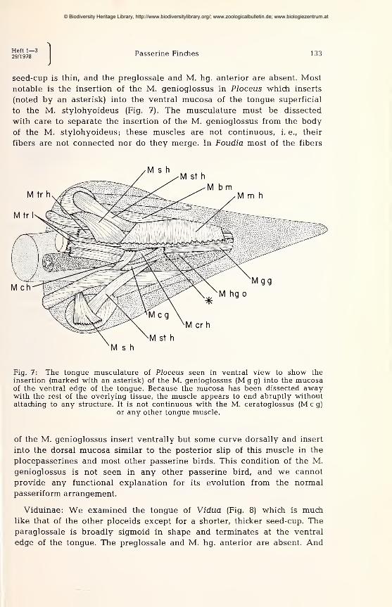
seed-cup is thin, and the preglossale and M. hg. anterior are absent. Most
notable is the insertion of the M. genioglossus in Ploceus which inserts
(noted by an asterisk) into the ventral mucosa of the tongue superficial
to the M. stylohyoideus (Fig. 7). The musculature must be dissected
with care to separate the insertion of the M. genioglossus from the body
of the M. stylohyoideus; these muscles are not continuous, i. e., their
fibers are not connected nor do they merge. In Foudia most of the fibers
Fig. 7: The tongue musculature of Ploceus seen in ventral view to show theinsertion (marked with an asterisk) of the M. genioglossus (M g g) into the mucosaof the ventral edge of the tongue. Because the mucosa has been dissected awaywith the rest of the overlying tissue, the muscle appears to end abruptly withoutattaching to any structure. It is not continuous with the M. ceratoglossus (M c g)
or any other tongue muscle.
of the M. genioglossus insert ventrally but some curve dorsally and insert
into the dorsal mucosa similar to the posterior slip of this muscle in the
plocepasserines and most other passerine birds. This condition of the M.
genioglossus is not seen in any other passerine bird, and we cannot
provide any functional explanation for its evolution from the normal
passeriform arrangement.
Viduinae: We examined the tongue of Vidua (Fig. 8) which is muchlike that of the other ploceids except for a shorter, thicker seed-cup. Theparaglossale is broadly sigmoid in shape and terminates at the ventral
edge of the tongue. The preglossale and M. hg. anterior are absent. And
© Biodiversity Heritage Library, http://www.biodiversitylibrary.org/; www.zoologicalbulletin.de; www.biologiezentrum.at
Fig. 8: The tongue of Vidua seen in lateral view.
the two insertions of the M. genioglossus are onto the paraglossale and
into the dorsal mucosa as normal for passeriform birds. It should be
noted, although we do not wish to draw any conclusions at this time, that
the morphology of the tongue (those parts examined in this study) in
Vidua is similar to those of the Plocepasserinae or the Estrildidae, not to
that of the Ploceinae.
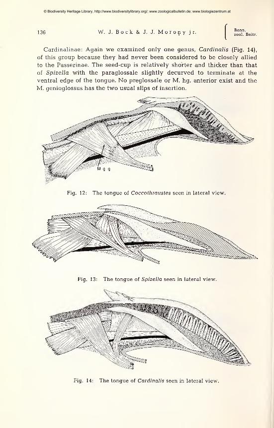
Estrildidae: Of the many genera in this family, we examined
only Estrilda (Fig. 9), Poephila, Lonchura (Fig. 10) and Amadina; these
were all similar to one another. The seed-cup is short and thick with a
short and strongly sigmoid paraglossale that terminates at the ventral
surface of the tongue. The preglossale and M. hg. anterior are absent. In
Lonchura, the M. genioglossus divides into two slips and inserts on the
paraglossale and into the dorsal mucosa. In Estrilda only the posterior
slip inserting into the dorsal mucosa remains. Little or no taxonomic
significance should be given to this difference.
Fig. 9: The tongue of Estrilda seen in lateral view.
© Biodiversity Heritage Library, http://www.biodiversitylibrary.org/; www.zoologicalbulletin.de; www.biologiezentrum.at

Heft 1—329/1978
Passerine Finches 135
Fig. 10: The tongue of Lonchura seen in lateral view.
Fringillidae : We dissected Fringilla (Fig. 11), as a representative
of the Fringillinae, and Coccothraustes ("Hesperiphona", Fig. 12), as a
representative of the Carduelinae. The tongue of Fringilla is clearly less
specialized than that of Coccothraustes. In Fringilla, the seed-cup is longer
and not as thick as in Coccothraustes. Correspondingly, the paraglossale of
Fringilla is more elongated and straighter while that of Cocothraustes is
shorter and curved ventrally. The preglossale and the M. hg. anterior
are absent in both. Both slips of insertion of the M. genioglossus exist
in Fringilla while only the posterior one is found in Coccothraustes.
Emberizidae ; Emberizinae: We dissected only Spizella (Fig. 13)
to have a representative of the buntings; this group had never been
considered to be closely related to the passerine finches. The seed-cup is
thick and elongated with a corresponding lengthening of the paraglossale.
The anterior end of this bone terminates close to the ventral surface of the
tongue. No preglossale or M. hg. anterior exists in Spizella. And the M.
genioglossus has the two usual slips of insertion.
Fig. 11: The tongue of Fringilla seen in lateral view.
© Biodiversity Heritage Library, http://www.biodiversitylibrary.org/; www.zoologicalbulletin.de; www.biologiezentrum.at
136 W. J. Bock & J. J. Morony jr.Bonn,zool. Beitr.
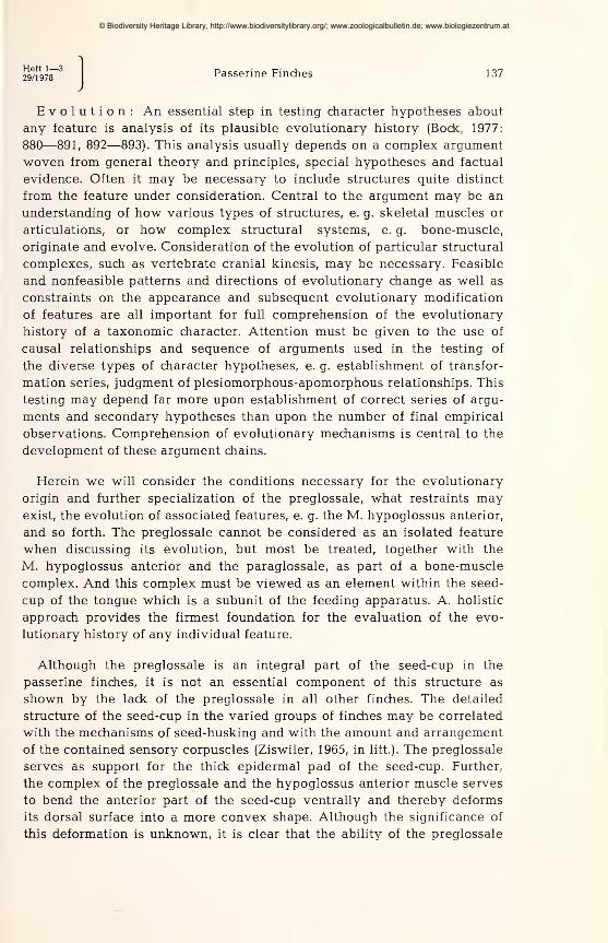
Cardinalinae: Again we examined only one genus, Cardinalis (Fig. 14),
of this group because they had never been considered to be closely allied
to the Passerinae. The seed-cup is relatively shorter and thicker than that
of Spizella with the paraglossale slightly decurved to terminate at the
ventral edge of the tongue. No preglossale or M. hg. anterior exist and the
M. genioglossus has the two usual slips of insertion.
Fig. 12: The tongue of Coccothraustes seen in lateral view.
Fig. 13: The tongue of Spizella seen in lateral view.
Fig. 14: The tongue of Cardinalis seen in lateral view.
© Biodiversity Heritage Library, http://www.biodiversitylibrary.org/; www.zoologicalbulletin.de; www.biologiezentrum.at
Heft 1—329/1978
Passerine Finches 137
Evolution : An essential step in testing character hypotheses about,
any feature is analysis of its plausible evolutionary history (Bock, 1977:
880—891, 892—893). This analysis usually depends on a complex argument
woven from general theory and principles, special hypotheses and factual
evidence. Often it may be necessary to include structures quite distinct
from the feature under consideration. Central to the argument may be an
understanding of how various types of structures, e. g. skeletal muscles or
articulations, or how complex structural systems, e. g. bone-muscle,
originate and evolve. Consideration of the evolution of particular structural
complexes, such as vertebrate cranial kinesis, may be necessary. Feasible
and nonfeasible patterns and directions of evolutionary change as well as
constraints on the appearance and subsequent evolutionary modification
of features are all important for full comprehension of the evolutionary
history of a taxonomic character. Attention must be given to the use of
causal relationships and sequence of arguments used in the testing of
the diverse types of character hypotheses, e. g. establishment of transfor-
mation series, judgment of plesiomorphous-apomorphous relationships. This
testing may depend far more upon establishment of correct series of argu-
ments and secondary hypotheses than upon the number of final empirical
observations. Comprehension of evolutionary mechanisms is central to the
development of these argument chains.
Herein we will consider the conditions necessary for the evolutionary
origin and further specialization of the preglossale, what restraints mayexist, the evolution of associated features, e. g. the M. hypoglossus anterior,
and so forth. The preglossale cannot be considered as an isolated feature
when discussing its evolution, but most be treated, together with the
M. hypoglossus anterior and the paraglossale, as part of a bone-muscle
complex. And this complex must be viewed as an element within the seed-
cup of the tongue which is a subunit of the feeding apparatus. A. holistic
approach provides the firmest foundation for the evaluation of the evo-
lutionary history of any individual feature.
Although the preglossale is an integral part of the seed-cup in the
passerine finches, it is not an essential component of this structure as
shown by the lack of the preglossale in all other finches. The detailed
structure of the seed-cup in the varied groups of finches may be correlated
with the mechanisms of seed-husking and with the amount and arrangement
of the contained sensory corpuscles (Ziswiler, 1965, in litt.). The preglossale
serves as support for the thick epidermal pad of the seed-cup. Further,
the complex of the preglossale and the hypoglossus anterior muscle serves
to bend the anterior part of the seed-cup ventrally and thereby deforms
its dorsal surface into a more convex shape. Although the significance of
this deformation is unknown, it is clear that the ability of the preglossale
© Biodiversity Heritage Library, http://www.biodiversitylibrary.org/; www.zoologicalbulletin.de; www.biologiezentrum.at

138 W. J. Bock & J. J. Morony jr.Bonn.zool. Beitr.
to rotate about the tips of the paraglossalia is an essential aspect of its
evolutionary history.
We conclude that the origin and specialization of the preglossale has
been as part of a movable system; it is not just a static support of the
seed-cup. If the latter were the case, then skeletal support of the
seed-cup most probably would have fused solidly to the anterior bodies
of the paraglossalia early in its evolution and would appear as a broad
anterior plate connecting the tips of these bones. Quite likely, it might
not have been recognized as a new feature unless detailed embryological
studies were conducted (see below). Once the preglossale was fused to
the tips of the paraglossalia, little cause would exist for the appearance
or enlargement of the M. hg. anterior. This muscle must be present as
a necessary prerequisite for the evolution of articulations between the
preglossale and the paraglossalia.
As a movable support for the seed-cup, the origin of the preglossale
was dependent upon the prior existence of the M. hypoglossus anterior.
Presumably the preglossale originated as a heterotopic ossification lying
along the origin of the paired Mm. hg. anterior. The primitive rod-like
preglossale served to transfer the force of the muscle to the entire length
of the seed-cup as well as supporting the latter structure; hence, the
early preglossale must be viewed as a mechanical lever but without any
direct attachment to the rest of the tongue skeleton. As this muscle in-
creased in size and as its function of deforming the seed-cup (if this
suggestion is correct) became more important, the need developed for a
stronger compression strut between the origin and insertion of the M. hg.
anterior. Proper functioning of any muscular system is dependent on the
existence of an antagonistic force to every muscle and on the presence
of an anticompression strut between the attachments of every muscle.
This strut was provided by enlargement of the preglossale which grew
until it abutted against the tips of the paraglossalia. Articulations evolved
at this stage. Further enlargement of the preglossale to support the whole
seed-cup would result in the dorsally opened-trough seen in the passerine
finches.
The presence of well-developed diarthroses between the preglossale
and paraglossalia supports, but does not prove, the conclusion that the
preglossale evolved as a movable bone relative to the existing tongue
skeleton. Articulations atrophy and the bones fuse during the life of an
individual organism if the bones do not move relative to one another
(Murray and Drachman, 1969). Yet, it is possible for articulations to develop
within the limits of an embryonic bone if the bone is subjected to repeated
bending as must have happened with the evoluation of a true diarthrosis
at the nasal-frontal hinge and/or at the anterior end of the jugal bar in
© Biodiversity Heritage Library, http://www.biodiversitylibrary.org/; www.zoologicalbulletin.de; www.biologiezentrum.at
Heft 1—329/1978
Passerine Finches 139
the skulls of parrots and some finches. Thus it is possible, but unlikely,
that the paraglossal articulations of the preglossale evolved after the
appearance of this bone as a fused anterior extension of the paraglossalia.
Most important is the presence of the M. hg. anterior as an essential
prerequisite for the evolutionary origin of a movable preglossale as
found in the passerine finches. Presence of articulations means that the
bone moves and movement means that a muscle is present. The evolu-
tionary history of this muscle in the passerine birds is thus of central
importance. Our discussion is based on the comparative studies of the
passerine tongue musculature by Bock and his associates (Bock, in litt.).
The M. hypoglossus anterior appears to be present in the passerine
birds as an ancestral feature inherited from their nonpasserine ancestors.
The M. hg. anterior is present in a broad diversity of groups within the
Passeriformes that are not necessarily closely related to one another within
the order. This muscle originates from a midventral raphe between the
anterior bodies of the paraglossalia and inserts onto the medioventral
surface of the paraglossale just anterior to its basihyale articulation.
Usually the insertion is close to or continuous with the insertion of the
M. ceratoglossus. Each M. hg. anterior is a unipinnate muscle; the pair
of muscles forms a single bipinnate unit. Often it is a strong muscle for
its mass as it contains many very short fibers. In a few groups, part of the
M. hg. anterior inserts on the anterior tip of the basihyale. The M. hg.
anterior varies greatly in size from a large muscle filling the space
between the anterior bodies of the paraglossalia to a minute vestigial
muscle that is difficult to find even with the aid of a dissecting microscope.
In Passer, it is very large (Bock and Morony, 1978). Moreover, only in the
passerine finches does the M. hg. anterior originate from a bony element
instead of a fibrous raphe.
The M. hg. anterior is absent in a number of genera and families of
passeriform birds; presumably it has been lost in the evolution of these
groups. Thus, evolution of this muscle has been from the ancestral condi-
tion to varying size, to different attachments (e. g. insertion on the basi-
hyale, and origin from the preglossale) and to total loss. Most important
is that once it is lost, the M. hg. anterior or a similar muscle in the same
position never reappears because there is no other muscle in this area
from which a new "M. hg. anterior" could evolve. The evolutionary appear-
ance of a new skeletal muscle is by splitting off of the new muscle from an
existing one. This mode of evolution provides the new muscle with the
necessary neural conections and skeletal attachments from those of the old
muscle. To be functional, it is essential for a muscle to have motor nerve
connections, sensory nerves and their endings within the muscle, the
© Biodiversity Heritage Library, http://www.biodiversitylibrary.org/; www.zoologicalbulletin.de; www.biologiezentrum.at

140 W. J. Bock & J. J. Morony jr.Bonn,zool. Beitr.
proper central neural connections, and attachments (resistance) to the
skeleton or other structures of the body. If any of these components are
absent, the muscle would not function. And functionless muscles (e. g.,
muscles which do not operate fully ) do not exist. Such muscles wouldatrophy during the life of the individual organism even if they started to
develop ontogenetically. This generalization is supported by numerousexperiments and accidents which show that whenever a muscle is de-
nervated or tenotomized (one of the attachments cut), the muscle will
atrophy. Hence, new muscles cannot appear "de novo" because all of
these neural and skeletal connections must be present simultaneously
with the muscular tissue. Because no other muscle lies in the tongue
anterior to the paraglossal-basihyal articulation, the evolutionary appear-
ance of a new muscle convergent to the lost M. hg. anterior would be
extremely unlikely. Thus we conclude that evolution of the M. hg. anterior
can only be in the direction of presence to loss; once lost, it is never
regained. Further, we conclude that in the evolutionary history of the
Passeriformes, a group possessing the M. hg. anterior could not have
evolved from an ancestral group lacking it.
Details of the evolution of the preglossale are difficult to provide be-
cause of the absence of any intermediate conditions. We suggest that it
appeared with the early stages in the evolution of the passerine seed-cup
and that it was always associated with the M. hg. anterior. The preglossale
became the origin for the already present M. hg. anterior. At this stage,
this muscle was probably much smaller and similar in appearance to
that seen in most other passeriform birds possessing it. The rudimentary
preglossale presumably strengthened the origin of the M. hg. anterior.
Extension of the preglossale anteriorly would connect it with the devel-
oping seed-cup and permit control of the position and shape (curvature
of its dorsal surface) of the seed-cup by the M. hg. anterior. With increased
force development of this muscle, the size of the preglossale increased and
more importantly it reached and abutted again the anterior tips of the
paraglossalia. At this point articulations developed between these bones.
Now the neomorphic bone could serve as a compression strut between
the origin and insertion of the M. hg. anterior, allowing a further increase
in size of the muscle and a shift in the position of its origin to a more
ventral position.
Discussion
The factural findings and arguments presented above permit the fol-
lowing conclusions on the diverse character hypotheses about the preglos-
sale and associated features and on the several classificatory and phylo-
© Biodiversity Heritage Library, http://www.biodiversitylibrary.org/; www.zoologicalbulletin.de; www.biologiezentrum.at

Heft 1—329/1978
Passerine Finches 141
genetic hypotheses about the passerine finches, presented in the introduc-
tion.
Character hypotheses: Five hypotheses were postulated in the introduc-
tion. Definite conclusions can be reached on each, as follows:
1) The preglossale is homologous in the three genera possessing it as
a skeletal element lying anterior to and articulating with the paraglossalia,
serving as the origin for the M. hg. anterior and supporting the seed-cup.
This hypothesis is tested and supported by the similar morphology of
the preglossale, including details of the transverse and longitudinal septa,
in these birds. Further, the preglossale has similar connections with other
features such as the origin of the M. hg. anterior from its posterior
chambers, the ligamentous connection to the anterior tip of the basihyale,
articulations with the anterior ends of the paraglossalia and support for
the base of the epidermal pad of the seed-cup. Therefore, we conclude
that a preglossale was present in the ancestor of the three genera of
passerine finches with a morphology similar to that seen in the living
forms.
2) The preglossale is an apomorph (= derived feature) with respect to
the ancestral condition of its absence in all other passerine birds, and
indeed in all other birds. The preglossale lies anterior to the rest of the
tongue skeleton which is part of the phylogenetically old hyoid gill arch
of vertebrates. The bones of the tongue skeleton, as do all bones of the
vertebrate gill arch series, develop ontogenetically from ectomesenchyme
which arises from neural crest cells (Horstadius, 1950). An excellent test
of the apomorphous nature of the preglossale is to examine its ontogenetic
development and ascertain whether it arises from ectomesenchyme or
from endomesoderm. Such observations are not available. However, the
available studies of the ontogeny of the tongue in Passer (Kallius, 1905;
Saayman, 1963) show that the preglossale first appears late in ontogeny,
just before hatching and long after the appearance of all other bones of
the hyoid skeleton. These observations indicate that the preglossale has
a different embryological development from that of the hyoid skeleton,
and hence is not of ectomenchymal (= neural crest) origin. On this basis
we conclude that the preglossale is an evolutionary neomorph or an
apomorph compared to its absence.
It is more difficult to show that the lack of the preglossale is plesiomor-
phous in all groups. It is possible for the preglossale and M. hg. anterior
to disappear completely leaving a tongue with a seed-cup, but lacking
this complex. We doubt this possibility because the preglossale appears
to be an integral part of the seed-cup in the passerine finches and wecannot comprehend a selection force that would favor the disappearance
© Biodiversity Heritage Library, http://www.biodiversitylibrary.org/; www.zoologicalbulletin.de; www.biologiezentrum.at

142 W. J. Bock & J. J. Mo ron y jr.Bonn,zool. Beitr.
of the preglossal complex in a finch. But we cannot offer any observations
to test our conclusion. More difficult to judge are the changes in the
tongue of a possible nonseed-eating descendent of the passerine finches.
If the passerine finches had an insectivorous descendent that lost the seed-
cup, then it is reasonable to expect a complete loss of the paraglossale
with or wihout loss of the M. hg. anterior. We know of no way to
distinguish between such a secondary loss of the preglossale — a
further apomorphous stage relative to the presence of the preglossale —from the original absence of this feature.
3) The preglossale is a synapomorph (= homologous apomorph) in the
genera of passerine finches; actually it is an autapomorph of this group.
This conclusion is simply a conjunction of the previous two conclusions.
We must emphasize that our conclusion that the preglossale is a synapo-
morph in the passerine finches is not because it is a shared derived
feature in these birds, but because we have tested critically the hypothesis
that the preglossale is homologous as an anterior tongue bone in these
genera as well as the hypothesis that it is an apomorph with respect to
its plesiomorphous absence. We have eliminated, as a real possibility,
convergent evolution of the preglossale.
Further we would conclude that the taxonomic value of the preglossale
is very great not because it is a synapomorph in the passerine birds, but
because it is a new feature of complex structure and possessing a complex
series of morphological connections with surrounding feature. We would
conclude that the possibility of the preglossale evolving independently two
or more times in the passerine finches is vanishingly low. In particular,
we would argue for a greater taxonomic value of the preglossale compared
to those shared apomorphs representing evolutionary loss of a structure
or those about which nothing is known about their function or their
possible evolutionary history.
4) On the basis of our discussion on the modes of evolutionary origin
of a new muscle we conclude that the presence of the M. hg. anterior
in passeriform birds is plesiomorphous and its absence is apomorphous.
Once lost, this muscle is not regained. Shared loss of the M. hg. anterior
is of little taxonomic value because of the great difficulty in separating
synapomorphous loss with convergent loss.
5) We would accept the hypothesis that the enlarged M. hg. anterior
with its sharply oblique fibers is homologous and apomorphous in the
passerine finches compared with this muscle in other passeriform birds.
The similar morphology of this muscle in passerine finches supports its
homologous nature, but we cannot provide strong arguments supporting
our conclusion of apomorphy and hence of synapomorphy.
© Biodiversity Heritage Library, http://www.biodiversitylibrary.org/; www.zoologicalbulletin.de; www.biologiezentrum.at
Heft 1—329/1978
Passerine Finches 143
Phylogenetic hypothesis about groups: Only one such hypothesis was
put forth, namely that the passerine finches had to evolve from an ancestral
group possessing the M. hg. anterior. We accept this hypothesis which
is tested by the character hypothesis (# 4) that the lack of this muscle is
apomorphous.
Classificatory hypotheses about groups: Several such hypotheses were
postulated. These are concerned with the reality of the passerine finches
as a taxon and the relationships of this taxon to other groups.
1) We accept the hypothesis that the passerine finches, the genera
Passer, Montiiringilla and Petronia, comprise a monophyletic and extremely
closely knit group based on the structure of the tongue (synapomorphy of
the preglossale, # 3, and of the passerine M. hg. anterior, # 5) and the
evidence presented earlier by Sushkin, Ziswiler and other workers. This
conclusion most likely does not hold for Carpospiza [Petronia] brachy-
dactyla, pending publication of their findings by Mendelssohn and his
associates (in litt.), which they believe to be a cardueline finch.
2) The evidence argues very strongly against the hypothesis that the
passerine finches are members of the Fringillidae in the broad sense or
in the sense of Peters' Check-list (vol. XIV, 1968). This follows from the
group phylogenetic hypothesis presented above as all fringillid finches
lack the M. hg. anterior. Moreover, the passerine finches could not be
members of any group of advanced New World nine-primaried oscines
because the M. hg. anterior was lost early in the evolution of the group.
3) In like fashion the evidence argues strongly against the hypothesis
that the passerine finches are advanced members of the Ploceidae as
concluded by Sushkin (1927: 28—29) and accepted by most workers be-
cause all members of the Ploceidae lack the M. hg. anterior.
4) The Passerinae of Peters' Check-list (vol. XV, 1962) are an artificial
taxon and must be divided into at least two groups, the passerine genera
and the rest. Several taxonomic solutions are possible. The most
reasonable one is to reestablish the subfamily Plocepasserinae for the
genera Plocepasser, Histurgops, Pseudonigrita, Philetairus and Sporopipes.
Another solution would be to unite the Plocepasserinae with the Bubalor-
nithinae. Attention should be given to Ziswiler's (1968) conclusion that
Sporopipes be placed in a monotypic subfamily which is in close agreement
with Sushkin's (1927: 29) conclusion on the position of this genus.
5) The evidence is consistent with the hypothesis that the passerine
finches are primitive members of the Ploceidae or of the New World nine-
primaried oscines (Fig. 15). A little evidence (e. g. rloss of the tenth
primary, some aspects of skull morphology) supports the latter suggestion
© Biodiversity Heritage Library, http://www.biodiversitylibrary.org/; www.zoologicalbulletin.de; www.biologiezentrum.at

144 W. J. Bock & J. J. Morony jr.Bonn,zool. Beitr.
Ploceidae
Pas
Fig. 15: Two schemes showing alternative ideas on the relationships of thepasserine finches. The Passeridae may be an early offshoot of the Ploceidae-Estrildidae group or an early offshoot of the New World nine-primaried oscines.
Although the evidence supporting either alternative is weak, more favors thefirst than any other available suggestion on the relationships of the passerine
finches.
which can be set aside, but not rejected, for now. More evidence (e. g.,
Sushkin, 1927; Ziswiler, 1965; Collias and Collias, 1964) supports the former
idea, but we do not believe that the available evidence is very convincing.
If the passerine finches are primitive members of the ploceid radiation,
then we would argue that these birds acquired their seed-eating adaptations
independently of the main complex of ploceid finches. The whole structure
of the seed-cup in the passerine finches is different from that of the ploceid
finches. The main feature uniting the passerines with the ploceids would
be nest construction. Collias and Collias (1964) argue that the weaving
abilities of Passer are very primitive compared to all ploceids which
supports the hypothesis that the passerine finches are primitive membersof the Ploceidae (see Ziswiler 1965: 42; Foelix, 1970: 584). But nest construc-
tion provides weak support on which to include the passerines in the
Ploceidae. It would appear that the passerine finches are no closer to
the Ploceidae than the latter are to the Estrildidae. If the argument of
Ziswiler (1967 a: 509), that the Estrildidae show, at best, basal relationships
to the Ploceidae but that these groups are distinct taxonomic families, is
accepted as most workers do, then it seems reasonable to treat the
passerine finches as a distinct family. This conclusion would be based on
the contention that each of these groups, ploceids, estrildids, and passerids,
acquired their seed-eating specializations independently. The alternative
solution would be to place all of these groups in a broad Ploceidae with
three subfamilies.
© Biodiversity Heritage Library, http://www.biodiversitylibrary.org/; www.zoologicalbulletin.de; www.biologiezentrum.at

Heft 1—329/1978
Passerine Finches 145
6) Thus, we accept the last of the classificatory hypotheses about
groups proposed in the introduction that the passerine finches be treated
as a distinct family — the Passeridae — and urge further intensive study
to ascertain their affinities to other oscines, be it with the primitive NewWorld nine-primaried oscines, the ploceid-estrildid complex, or some
other group.
Summary
1) The preglossale is an evolutionary neomorph providing new evidence onthe monophyly and relationships of the passerine finches. Analysis of this feature
also provides a good example of proper methods of phylogenetic analysis.
2) The passerine finches could have only evolved from an ancestor possessing
a M. hypoglossus anterior which excludes the Ploceidae, the Fringillidae and all
other higher New World nine-primaried oscines.
3) The passerine finches, Passer, Montifringilla and Petronia, are a monophyleticgroup that should be classified as a distinct family, the Passeridae.
Zusammenfassung
1) Das Praeglossale der Sperlinge ist evolutionsgeschichtlich ein neomorphesMerkmal, das neues Licht auf die verwandtschaftlichen Beziehungen dieser Vögelzu werfen vermag. Die Analyse der hier vorliegenden Situation bietet auch ein
gutes Beispiel für die einer phylogenetischen Auswertung angemessenen Methoden.
2) Die Sperlinge können nur von einem Vorfahr hergeleitet werden, der einen
M. hypoglossus anterior besaß. Das schließt die Ploceidae, Fringillidae und alle
höher entwickelten neunhandschwingigen Neuwelt-Oscines als Vorfahren aus.
3) Die Sperlinge, Gattungen Passer, Montifringilla und Petronia, bilden eine
monophyletische Gruppe, die im System als eine besondere Familie, Passeridae,
angesehen werden sollte.
Acknowledgements
We would like to thank Ms. D. Goldys for drawing most of the illustrations andMr. Taru Suzuki for drawing Figs. 1 and 7. Dr. N. Collias and Stuart Irwin
provided us with alcoholic specimens of otherwise unavailable genera of the
Plocepasserinae which permitted us to complete our comparative survey of this
group. Professor H. Mendelssohn kindly made available to us his findings andconclusions on the taxonomic positions of Carpospiza brachydactyla. Professor
V. Ziswiler discussed his ongoing studies on the tongue and sense organs of
finches which was essential to our understanding of the function of the seed-cup.
We would like to express our appreciation to all. This study was done with
support of a research grant (NSF BMS-73-06818 and DEB-76-14746) from the
National Science Foundation to Walter Bock.
© Biodiversity Heritage Library, http://www.biodiversitylibrary.org/; www.zoologicalbulletin.de; www.biologiezentrum.at
146 W. J. Bock & J. J. Morony jr.Bonn,zool. Beitr.
Literature cited
Ackermann, A. (1967): Quantitative Untersuchungen an körnerfressenden
Singvögeln. Journ. f. Ornith. 108: 430—475.
Beecher, W. J. (1953) : A phylogeny of the oscines. Auk. 70: 270—333.
Bock, W. J. (1974): The avian skeletomuscular system. In: Farner, D. S. andJ. R. King, eds., "Avian Biology," Acad. Press, New York, vol IV: 119—157.
— (1977): Foundations and methods of evolutionary classification. In: Hecht, M. K.,
P. C. Goody and B. M. Hecht eds., "Major patterns in vertabrate evolution,"
Plenum Press, New York, pp. 851—895.
— and J. Morony (1978): The preglossale of Passer (Aves: Passeriformes) — a
skeletal neomorph. J. Morph. 155: 99—110.
Bulatova, N. S. 1973): A cytotaxonomic study of three related families of
birds: Fringillidae, Emberizidae, Ploceidae. Zeitsch. f. Zool. Syst. Evolutions-
forschung 1 1 : 233—239.
Chapin, J. P. (1917): The classification of the weaver-birds. Bull. Amer. Mus.Nat. Hist. 37 (9) : 243—280.
Co 11 i as, N. E., and E. C. Colli as (1964): Evolution of nestbuilding in the
weaverbirds (Ploceidae). Univ. Calif. Publ. Zool. 73, viii + 162 pp.
— and — (1977): Weaverbird nest aggregation and evolution of the compoundnest. Auk 94: 50—64.
Foelix, R. (1970): Vergleichend-morphologische Untersuchungen an den Spei-
cheldrüsen körnerfressender Singvögel. Zool. Jb. Anat Bd. 87: 523—587.
Friedmann, H. (1960): The parasitic weaverbirds. Bull. U.S. Nat. Mus. # 223,
viii + 196 pp.
— (1962): The problem of the Viduinae in the light of recent publications. Smithson.
Misc. Cont. 145 (3): 1—10.
Hörstadius, (1950) : The neural crest. Oxford Univ. Press, London.
Kallius, E. (1905): Beiträge zur Entwicklung der Zunge. II. Teil. Vögel. (Anasboschas L., Passer domesticas L.). Anat. Hefte., vol. 28 (# 85—86): 305—586.
Lafresnaye, F. de (1850): Sur la nidification de quelques espéces d'oiseaux
de la famille ou sous-famille des Tisserins (Ploceinae). Rev. Mag. Zool. Pure
et Appliquée, 2nd S., vol. 2: 315—326.
M a y r,
E., R. I. Andrew and R. A. H i n d e (1956) : Die systematische Stel-
lung der Gattung Fringilla. Journ. f. Ornith. 97: 258—273.
Murray, P. D. F., and D. B. Drachman (1969): The role of movement in
the development of joints and related structures: The head and neck in the
chick embryo. J. Embryol. Exp. Morph. 22: 349—371.
Nicolai, J. (1964): Der Brutparasitismus der Viduinae als ethologisches Pro-
blem. Prägungsphänomene als Faktoren der Rassen- und Artbildung. Zeitsch.
Tierpsych. 21: 129—204.
Saayman.J. E. (1963): 'n Bydrae tot die Skedelmorfologie van Passer melanurusmelanurus. Ann. Univ. Stell., 138 (sect. A, No. 6): 180—213.
Sibley, S. G. (1970): A comparative study of the egg-white proteins of passerine
birds. Bull. Peabody Mus. Nat. Hist., 32, vi + 131 pp.
© Biodiversity Heritage Library, http://www.biodiversitylibrary.org/; www.zoologicalbulletin.de; www.biologiezentrum.at
Heft 1—329/1978
Passerine Finches 147
Steiner, H. (1960): Klassifikation der Prachfinken, Spermestidae, auf Grundder Rachenzeichnungen ihrer Nestlinge. Journ. f. Ornith. 101: 92— 112.
Sushkin, P. P. (1924): "On the classification of Fringillidae and Ploceidae."
Bull. Brit. Orn. Club 45: 36—39.
— (1925): The Evening Grosbeak (Hesperiphona), the only American genus of a
Palaearctic group. Auk, 42: 256—261.
— (1927): On the anatomy and classification of the weaver-birds. Bull. Amer.Mus. Nat. Hist. 57 (1): 1—32.
Tordoff, H. B. (1954): A systematic study of the avian family Fringillidae
based on the structure of the skull. Misc. Publ. Mus. Zool., Univ. Mich.
# 81: 1—41.
Ziswiler, V. (1965): Zur Kenntnis des Samenöffnens und der Struktur des
hörnernen Gaumens bei körnerfressenden Oscines. Journ. f. Ornith. 106: 1—48.
— (1967 a): Vergleichende morphologische Untersuchungen am Verdauungstraktkörnerfressender Singvögel zur Abklärung ihrer systematischen Stellung. Zool.
Jb. Syst. Bd. 94: 427—520.
— (1967 b): Die taxonomische Stellung des Schneefinken Montitringilla nivalis
(Linnaeus). Ornith. Beob. 64: 105—110.
— (1967 c): Der Verdauungstrakt körnerfressender Singvögel als taxonomischer
Merkmalskomplex. Rev. Suisse Zool., 74, p. 620—628.
— (1968): Die taxonomische Stellung der Gattung Sporopipes Cabanis. Bonn. zool.
Beitr. 19 (3/4): 269—279.
Addresses of the authors:
Dr. Walter J. Bock, Department of Biological Sciences, Columbia University, NewYork, N.Y., 10027, USA, and Department of Ornithology, American Museum of
Natural History, New York.
Dr. John J. Morony, Jr., Department of Ornithology, American Museum of NaturalHistory, New York, N.Y., 10024, USA.
© Biodiversity Heritage Library, http://www.biodiversitylibrary.org/; www.zoologicalbulletin.de; www.biologiezentrum.at
Top Related