


Languages
Pages
Legal

1
BJMP
Volume 5 Number 1
March 2012
www.bjmp.org
ISSN: 1757-8515
British Journal of Medical Practitioners
© BJMP.org
British Journal of Medical Practitioners Volume 5 Number 1 (March 2012)
http://www.bjmp.org
Editorial Board Managing Editors
• Dr Javed Latoo, UK
• Dr Nadeem Mazi-Kotwal, UK
Medical Editor
• Dr M.Y. Latoo, UK
Associate Editors
• Professor Ken Brummel-Smith, USA
• Dr Nasseer Masoodi, USA
• Dr Ramesh Mehta, UK
Assistant Editor
• Dr Minal Mistry, UK
• Dr Mehraj Shah, UK
Editorial Advisors
• Prof Raman Bedi, Director of Global Child Dental Health
Taskforce, UK
• Dr Francis Dunne, Consultant Psychiatrist and Honorary
Senior Lecturer, UK
• Prof Rajan Madhok,Medical Director of NHS Manchester,
UK
• Prof Elisabeth Paice, Dean Director of Postgraduate
Medical & Dental Education for London, UK
• Prof Arnie Purushotham, Professor of Surgery, UK
• Prof Khalid J Qazi, Professor of clinical Medicine, USA
• Dr Abid Rajah, Consultant Anaesthetics and Critical Care
Medicine, UK
• Prof A A Riaz, Professor of Surgery, UK
• Prof Robert Thomas, Professor of Oncology, UK
Editorial Board
Internal Medicine and allied Specialties
• Dr John Ellis Agens, Jr, Associate Professor of Medicine,
USA
• Dr Mohammed Azher, Consultant Physician, UK
• Dr Rajith deSilva, Consultant Neurologist, UK
• Dr Indrajit Gupta, Consultant Physician, UK
• Dr Amir Jaffer, Associate Professor of Medicine, USA
• Dr Roop Kaw, Assistant Professor of Internal Medicine,
USA
• Dr Ajay Kumar, Medical Director, Internal Medicine
Preoperative Center, US
• Prof Ghulam J Mufti, Professor and Head of
Haematological Medicine, UK
• Prof Claudio Puoti, Chief, Internal Medicine and Liver
Unit, Marino, Italy
• Prof G V Sherbet, Cancer and Molecular Medicine, UK
• Dr Yili Zhou, Neurologist and Interventional Pain
Management Specialist, USA
Surgery and allied Specialties
• Mr Habib Charfare, Consultant Surgeon, UK
• Prof Jorg Haier, Professor of Surgery, Germany
• Mr Patrick Omotoso, Consultant Surgeon, UK
• Mr Anup Kumar Saha MP, Laparascopic Surgeon and
Member of Parliament of India, India
• Mr Yadu K Shankarappa, Counsultant Trauma and
Orthopaedic Surgeon, UK
• Mr Harbinder Sharma, Consultant Surgeon and Urologist,
UK
• Mr Manoj Sood, Consultant Orthopaedic Surgeon, UK
Anaesthesia and Critical Care Medicine
• Dr Mehmood A Durrani, Vice Chair of Anaesthesia and
Chief of Cardiiac Anaesthesia, USA
• Dr Faisal Salim, Consultant Anaesthetics, UK
• Dr Asquad Sultan, Consultant Anaesthetics and Pain
Specialist, UK
Psychiatry
• Dr Charlotte Feinman, Consultant Psychiatrist, UK
• Dr Chris McEvedy, Consultant Psychiatrist, UK
• Dr Kabir Padamsee, Consultant Child Psychiatrist, UK
• Dr Saoud Sultan, Consultant Psychiatrist and College
Tutor, UK
• Prof Malcolm Weller, Emeritus Consultant Psychiatrist,
UK
Family Medicine
• Dr Anita Sharma, Family Physician, UK
Gynaecology & Obstetrics
• Mr Dilip Patil, Consultant Obstetrician & Gynaecologist,
UK
Research & Development Advisors
• Dr Sam Tothill, Associate Dean of the Faculty of Medicine
& Biosciences Crainfield University, UK
• Dr Mohammed Wasil,Assistant Director of Research &
Development & Clinical Fellow Crainfield University ,
UK
© BJMP.org
Legal Advisor
• Fazl Syed, Consultant International law, UK
Attorney at Law -New York USA, Solicitor-Supreme Court
of England & Wales-UK
Other Editorial Staff Marketing Advisors
• Dr Mohamed Abeid, Egypt
• Dr Shafi Shali, UK
Trainee Editors
• Dr Sripurna Basu, UK
• Dr Farida Jan, UK
• Dr Minaz Mazi Kotwal, UK
• Dr Prabhu Nesargarikar, UK
• Dr Daljit Sura, UK
Proof Readers
• Dr Diana Ayoola Mabayoje, UK
• Dr Tabassum Malik, UK
• Dr Cristal Oxley, UK
• Dr Claire Pocklington, UK
• Dr Natasha Quader, UK
• Dr Farheen Zulfiquer, UK
Instructions to authors Please visit: http://bjmp.org/content/guidance-authors
Submit an article Please visit: http://bjmp.org/content/submit-articles
Contact us Please visit: http://www.bjmp.org/contact
Publishers JMN Medical Education Ltd
1 Waltham Drive
Elstow
Bedford, United Kingdom
MK429FY
The British Journal of Medical Practitioners (BJMP) is a
quarterly peer-reviewed online international medical journal
published by JMN Medical Education Ltd UK. The
information, opinions and views presented in the British
Journal of Medical Practitioners reflect the views of the authors
and contributors of the articles and not of the British Journal of
Medical Practitioners or the Editorial Board or its publishers.
The British Journal of Medical Practitioners and/or its
publisher cannot be held responsible for any errors or for any
consequences arising from the use of the information contained
in this journal.

British Journal of Medical Practitioners Volume 5 Number 1 (March 2012)
BJMP BJMP BJMP BJMP March 2012March 2012March 2012March 2012 Volume Volume Volume Volume 5555 Number Number Number Number 1111
EditorialEditorialEditorialEditorial
Are opioids effective Are opioids effective Are opioids effective Are opioids effective and necessary for chronic nonand necessary for chronic nonand necessary for chronic nonand necessary for chronic non----malignant painmalignant painmalignant painmalignant pain
Yili Zhou and Bohdan Warycha
4
ResearchResearchResearchResearch ArticlesArticlesArticlesArticles
Effectiveness of Chlorhexidine oral decontamination in reducing the incidence of ventilator associated pneumonia: A Effectiveness of Chlorhexidine oral decontamination in reducing the incidence of ventilator associated pneumonia: A Effectiveness of Chlorhexidine oral decontamination in reducing the incidence of ventilator associated pneumonia: A Effectiveness of Chlorhexidine oral decontamination in reducing the incidence of ventilator associated pneumonia: A
metametametameta----analysis.analysis.analysis.analysis.
E Balamurugan, A Kanimozhi and Govinda Kumari
6
Barriers for Anaesthetists in Performing Nerve Blocks with Ultrasound GuidanceBarriers for Anaesthetists in Performing Nerve Blocks with Ultrasound GuidanceBarriers for Anaesthetists in Performing Nerve Blocks with Ultrasound GuidanceBarriers for Anaesthetists in Performing Nerve Blocks with Ultrasound Guidance
Asif Mahmood, Mohammed Auldin and Asquad Sultan
11
Are we managing acute knee effusion well?Are we managing acute knee effusion well?Are we managing acute knee effusion well?Are we managing acute knee effusion well?
A S Eid, V Burrows, J R M Murray, P Smitham, R Ahmad, R Miller and U Butt
14
Reminder letters to improve rate of attendance at Community Mental Health CentreReminder letters to improve rate of attendance at Community Mental Health CentreReminder letters to improve rate of attendance at Community Mental Health CentreReminder letters to improve rate of attendance at Community Mental Health Centre
Murali Krishna and Sreedharan Amarjothi
17
Review ArticlesReview ArticlesReview ArticlesReview Articles
REM Behavior Disorder (RBD) as an Early Marker for Development of Neurodegenerative REM Behavior Disorder (RBD) as an Early Marker for Development of Neurodegenerative REM Behavior Disorder (RBD) as an Early Marker for Development of Neurodegenerative REM Behavior Disorder (RBD) as an Early Marker for Development of Neurodegenerative DiseasesDiseasesDiseasesDiseases
Umesh Vyas and Rose Franco
20
Management of Drooling of salivaManagement of Drooling of salivaManagement of Drooling of salivaManagement of Drooling of saliva
Ganesh Bavikatte, Poh Lin Sit and Ali Hassoon
25
Benzodiazepines RevisitedBenzodiazepines RevisitedBenzodiazepines RevisitedBenzodiazepines Revisited
Tauseef Mehdi
31
Case Reports/SeriesCase Reports/SeriesCase Reports/SeriesCase Reports/Series
Bradyarrhythmias Associated with the Obstructive Sleep Apnoea Bradyarrhythmias Associated with the Obstructive Sleep Apnoea Bradyarrhythmias Associated with the Obstructive Sleep Apnoea Bradyarrhythmias Associated with the Obstructive Sleep Apnoea Syndrome: A Precursor to LifeSyndrome: A Precursor to LifeSyndrome: A Precursor to LifeSyndrome: A Precursor to Life----threatening threatening threatening threatening
Arrhythmias?Arrhythmias?Arrhythmias?Arrhythmias?
Amitasha Mann, Jean Karen Fleischman and Karen Mrejen-Shakin
39
Retroperitoneal Teratoma in an adult presenting with painful abdominal mass: case history and literature reviewRetroperitoneal Teratoma in an adult presenting with painful abdominal mass: case history and literature reviewRetroperitoneal Teratoma in an adult presenting with painful abdominal mass: case history and literature reviewRetroperitoneal Teratoma in an adult presenting with painful abdominal mass: case history and literature review
Sadaqat Ali Khan, Tariq Mahmood, Muhammad Zeeshan Sarwar, Syed Hamad Rasool, Muhammad Danish Siddique and
Zohaib Khan
42
Unusual presentation of thyrotoxicosis with paraparesis in a young male: A rare case reportUnusual presentation of thyrotoxicosis with paraparesis in a young male: A rare case reportUnusual presentation of thyrotoxicosis with paraparesis in a young male: A rare case reportUnusual presentation of thyrotoxicosis with paraparesis in a young male: A rare case report
Hakim Irfan Showkat, Arif Hussain Sarmast, Rubina Lone, Mehmood Iqbal Qadri and Manzoor Ahmed Wani 45
Clinical PracticeClinical PracticeClinical PracticeClinical Practice
A survey of aseptic technique when performing lumbar puncture: a comparison of medical and anaesthetic traineesA survey of aseptic technique when performing lumbar puncture: a comparison of medical and anaesthetic traineesA survey of aseptic technique when performing lumbar puncture: a comparison of medical and anaesthetic traineesA survey of aseptic technique when performing lumbar puncture: a comparison of medical and anaesthetic trainees
Rajiv Malhotra and Sara Kelly
47

British Journal of Medical Practitioners, March 2012, Volume 5, Number 1
BJMP.org
BJMP 2012;5(1):a510
Are opioids effective and necessary for chronic non-malignant pain
Yili Zhou and Bohdan Warycha
In recent years, increasing attention has focused on the
treatment of chronic pain with a considerable number of
research and publications about it. At the same time, opioid
prescription, use, abuse and death related to the inappropriate
use of opioids have significantly increased over the last 10
years. Some reports indicated that there were more than 100
‘pain clinics’ within a one-mile radius in South Florida, between
2009 and 2010, which led to the birth of new opioid
prescription laws in Florida and many other states to restrict the
use of opioids. In the face of clinical and social turmoil related
to opioid use and abuse, a fundamental question facing each
clinician is: are opioids effective and necessary for chronic non-
malignant pain?
Chronic low back pain (LBP) is the most common pain
condition in pain clinics and most family physician offices,
which ‘requires’ chronic use of opioids. Nampiaparampil et al
conducted a literature review in 20121 and found only one
high-quality study on oral opioid therapy for LBP, which
showed significant efficacy in pain relief and patient function.
Current consensus believes that there is weak evidence
demonstrating favourable effectiveness of opioids compared to
placebo in chronic LBP.2Opioids may be considered in the
treatment of chronic LBP if a patient fails other treatment
modalities such as non-steroidal anti-inflammatory drugs
(NSAIDs), antidepressants, physical therapy or steroid
injections. Opioids should be avoided if possible, especially in
adolescents who are at high risk of opioid overdose, misuse, and
addiction. It has been demonstrated that the majority of the
population with degenerative disc disease, including a disc
herniation have no back pain. A Magnetic Resonance Imaging
(MRI) report or film with a disc herniation should not be an
automatic ‘passport’ for access to narcotics.
Failed back surgery syndrome (FBSS) is often refractory to most
treatment modalities and sometimes very debilitating. There are
no well-controlled clinical studies to approve or disapprove the
use of opioids in FBSS. Clinical experience suggests oral
opioids may be beneficial and necessary to many patients
suffering from severe back pain due to FBSS. Intraspinal
opioids delivered via implanted pumps may be indicated in
those individuals who cannot tolerate oral medications. For
elderly patients with severe pain due to spinal stenosis, there is
no clinical study to approve or disprove the use of opioids.
However, due to the fact that NSAIDs may cause serious side
effects in gastrointestinal, hepatic and renal systems, opioid
therapy may still be a choice in carefully selected patients.
Most studies for pharmacological treatment of neuropathic pain
are conducted with diabetic peripheral neuropathy (DPN)
patients. Several randomized clinical controlled studies have
demonstrated evidence that some opioids, such as morphine
sulphate, tramadol,3 and oxycodone controlled-release,4 are
probably effective in reducing pain and should be considered as
a treatment of choice (Level B evidence), even though anti-
epileptics such as pregabalin should still be used as the first line
medication.5
Some studies indicate opioids may be superior to placebo in
relieving pain due to acute migraine attacks and Fiorinal with
codeine may be effective for tension headache. However there
is lack of clinical evidence supporting long-term use of opioids
for chronic headaches such as migraine, chronic daily headache,
medication overuse headache, or cervicogenic headache.
Currently there are large amounts of opioids being prescribed
for headaches because of patients' demands. Neuroscience data
on the effects of opioids on the brain has raised serious concerns
for long-term safety and has provided the basis for the
mechanism by which chronic opioid use may induce
progression of headache frequency and severity.6 A recent study
found chronic opioid use for migraine associated with more
severe headache-related disability, symptomology, comorbidities
(depression, anxiety, and cardiovascular disease and events), and
greater healthcare resource utilization.7
Many patients with fibromyalgia (FM) come into pain clinics to
ask for, or even demand, prescriptions for opioids. There is
insufficient evidence to support the routine use of opioids in
fibromyalgia.8 Recent studies have suggested that central
sensitization may play for role in the aetiology of FM. Three
central nervous system (CNS) agents (pregabalin, duloxetine
and milnacipran) have been approved by United States Food
and Drug Administration (US FDA) for treatment of FM.
However, opioids are still commonly prescribed by many
Editorial

British Journal of Medical Practitioners, December 2011, Volume 4, Number 4
BJMP.org
physicians for FM patients by ‘tradition’, sometimes even with
the combination of a benzodiazapine and muscles relaxant -
Soma. We have observed negative health and psychosocial
status in patients using opioids and labeled with FM. Opioids
should be avoided whenever possible in FM patients in face of
widespread abuse and lack of clinical evidence.9
Adolescents with mild non-malignant chronic pain rarely
require long-term opioid therapy.10 Opioids should be avoided
if possible in adolescents, who are at high risk of opioid
overdose, misuse, and addiction. Patients with adolescents
living at home should store their opioid medication safely.
In conclusion, opioids are effective and necessary in certain
cases. However, currently no single drug stands out as the best
therapy for managing chronic non-malignant pain, and current
opioid treatment is not sufficiently evidence-based. More well-
designed clinical studies are needed to confirm the clinical
efficacy and necessity for using opioids in the treatment of
chronic non-malignant pain. Before more evidence becomes
available, and in the face of widespread abuse of opioids in
society and possible serious behavioural consequences to
individual patients, a careful history and physical examination,
assessment of aberrant behavior, controlled substance
agreement, routine urine drug tests, checking of state drug
monitoring system (if available), trials of other treatment
modalities, and continuous monitoring of opioid compliance
should be the prerequisites before any opioids are prescribed.
Opioid prescriptions should be given as indicated, not as
‘demanded’.
Competing Interests None declared
Author Details Yili Zhou, MD, PhD and Bohdan Warycha, MD, Florida Pain and Rehabilitation
Center, 6830 NW 11th Place, Gainesville, FL 32605.
CORRESSPONDENCE: YiLi Zhou, MD, Ph.D., Florida Pain and
Rehabilitation Center 6830 NW 11th Place, Gainesville, FL 32605.
Email: [email protected]
REFERENCES
1. Nampiaparampil DE, Nampiaparampil GM, Nampiaparampil RG. Oral
opioid analgesics vs. spinal steroid injections in the treatment of low back
pain syndromes. Am.J.Phys.Med.Rehabil. 2012;91:162-76.
2. White AP, Arnold PM, Norvell DC et al. Pharmacologic management of
chronic low back pain: synthesis of the evidence. Spine (Phila Pa 1976.)
2011;36:S131-S143.
3. Ko SH, Kwon HS, Yu JM et al. Comparison of the efficacy and safety of
tramadol/acetaminophen combination therapy and gabapentin in the
treatment of painful diabetic neuropathy. Diabet.Med. 2010;27:1033-40.
4. Hanna M, O'Brien C, Wilson MC. Prolonged-release oxycodone
enhances the effects of existing gabapentin therapy in painful diabetic
neuropathy patients. Eur.J.Pain 2008;12:804-13.
5. Bril V, England J, Franklin GM et al. Evidence-based guideline:
Treatment of painful diabetic neuropathy: report of the American Academy
of Neurology, the American Association of Neuromuscular and
Electrodiagnostic Medicine, and the American Academy of Physical
Medicine and Rehabilitation. PM.R. 2011;3:345-52, 352.
6. Saper JR, Lake AE, III. Continuous opioid therapy (COT) is rarely
advisable for refractory chronic daily headache: limited efficacy, risks, and
proposed guidelines. Headache 2008;48:838-49.
7. Buse DC, Pearlman SH, Reed ML et al. Opioid use and dependence
among persons with migraine: results of the AMPP study. Headache
2012;52:18-36.
8. Ngian GS, Guymer EK, Littlejohn GO. The use of opioids in
fibromyalgia. Int.J.Rheum.Dis. 2011;14:6-11.
9. Ngian GS, Guymer EK, Littlejohn GO. The use of opioids in
fibromyalgia. Int.J.Rheum.Dis. 2011;14:6-11.
10. Kahan M, Wilson L, Mailis-Gagnon A et al. Canadian guideline for safe
and effective use of opioids for chronic noncancer pain: clinical summary for
family physicians. Part 2: special populations. Can.Fam.Physician
2011;57:1269-28.
British Journal of Medical Practitioners, March 2012, Volume 5, Number 1
BJMP.org
BJMP 2012;5(1):a512
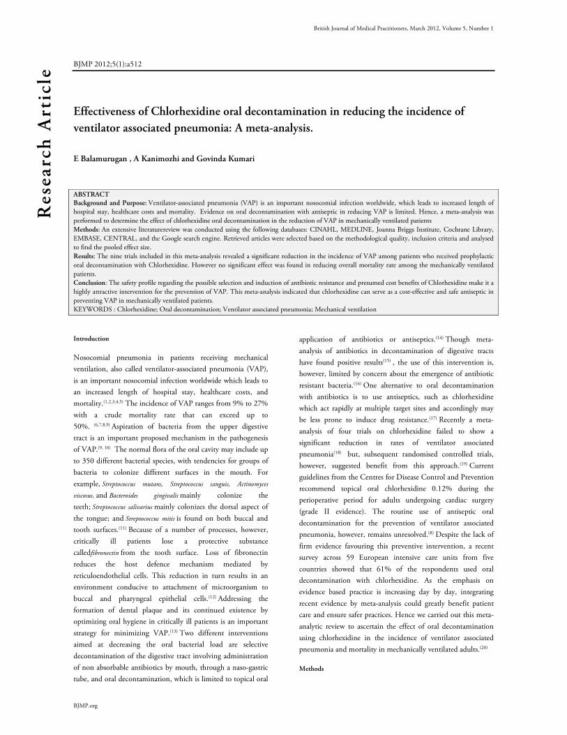
Effectiveness of Chlorhexidine oral decontamination in reducing the incidence of
ventilator associated pneumonia: A meta-analysis.
E Balamurugan , A Kanimozhi and Govinda Kumari
ABSTRACT Background and Purpose: Ventilator-associated pneumonia (VAP) is an important nosocomial infection worldwide, which leads to increased length of
hospital stay, healthcare costs and mortality. Evidence on oral decontamination with antiseptic in reducing VAP is limited. Hence, a meta-analysis was
performed to determine the effect of chlorhexidine oral decontamination in the reduction of VAP in mechanically ventilated patients
Methods: An extensive literaturereview was conducted using the following databases: CINAHL, MEDLINE, Joanna Briggs Institute, Cochrane Library,
EMBASE, CENTRAL, and the Google search engine. Retrieved articles were selected based on the methodological quality, inclusion criteria and analysed
to find the pooled effect size.
Results: The nine trials included in this meta-analysis revealed a significant reduction in the incidence of VAP among patients who received prophylactic
oral decontamination with Chlorhexidine. However no significant effect was found in reducing overall mortality rate among the mechanically ventilated
patients.
Conclusion: The safety profile regarding the possible selection and induction of antibiotic resistance and presumed cost benefits of Chlorhexidine make it a
highly attractive intervention for the prevention of VAP. This meta-analysis indicated that chlorhexidine can serve as a cost-effective and safe antiseptic in
preventing VAP in mechanically ventilated patients.
KEYWORDS : Chlorhexidine; Oral decontamination; Ventilator associated pneumonia; Mechanical ventilation
Introduction
Nosocomial pneumonia in patients receiving mechanical
ventilation, also called ventilator-associated pneumonia (VAP),
is an important nosocomial infection worldwide which leads to
an increased length of hospital stay, healthcare costs, and
mortality.(1,2,3,4,5) The incidence of VAP ranges from 9% to 27%
with a crude mortality rate that can exceed up to
50%. (6,7,8,9) Aspiration of bacteria from the upper digestive
tract is an important proposed mechanism in the pathogenesis
of VAP.(9, 10) The normal flora of the oral cavity may include up
to 350 different bacterial species, with tendencies for groups of
bacteria to colonize different surfaces in the mouth. For
example, Streptococcus mutans, Streptococcus sanguis, Actinomyces
viscosus, and Bacteroides gingivalis mainly colonize the
teeth; Streptococcus salivarius mainly colonizes the dorsal aspect of
the tongue; and Streptococcus mitis is found on both buccal and
tooth surfaces.(11) Because of a number of processes, however,
critically ill patients lose a protective substance
calledfibronectin from the tooth surface. Loss of fibronectin
reduces the host defence mechanism mediated by
reticuloendothelial cells. This reduction in turn results in an
environment conducive to attachment of microorganism to
buccal and pharyngeal epithelial cells.(12) Addressing the
formation of dental plaque and its continued existence by
optimizing oral hygiene in critically ill patients is an important
strategy for minimizing VAP.(13) Two different interventions
aimed at decreasing the oral bacterial load are selective
decontamination of the digestive tract involving administration
of non absorbable antibiotics by mouth, through a naso-gastric
tube, and oral decontamination, which is limited to topical oral
application of antibiotics or antiseptics.(14) Though meta-
analysis of antibiotics in decontamination of digestive tracts
have found positive results(15) , the use of this intervention is,
however, limited by concern about the emergence of antibiotic
resistant bacteria.(16) One alternative to oral decontamination
with antibiotics is to use antiseptics, such as chlorhexidine
which act rapidly at multiple target sites and accordingly may
be less prone to induce drug resistance.(17) Recently a meta-
analysis of four trials on chlorhexidine failed to show a
significant reduction in rates of ventilator associated
pneumonia(18) but, subsequent randomised controlled trials,
however, suggested benefit from this approach.(19) Current
guidelines from the Centres for Disease Control and Prevention
recommend topical oral chlorhexidine 0.12% during the
perioperative period for adults undergoing cardiac surgery
(grade II evidence). The routine use of antiseptic oral
decontamination for the prevention of ventilator associated
pneumonia, however, remains unresolved.(8) Despite the lack of
firm evidence favouring this preventive intervention, a recent
survey across 59 European intensive care units from five
countries showed that 61% of the respondents used oral
decontamination with chlorhexidine. As the emphasis on
evidence based practice is increasing day by day, integrating
recent evidence by meta-analysis could greatly benefit patient
care and ensure safer practices. Hence we carried out this meta-
analytic review to ascertain the effect of oral decontamination
using chlorhexidine in the incidence of ventilator associated
pneumonia and mortality in mechanically ventilated adults.(20)
Methods
Research Article

British Journal of Medical Practitioners, March 2012, Volume 5, Number 1
BJMP.org
Table 1: Brief summary of trials
Source Subjects Intervention ComparedWith Outcome with
respect to VAP
Outcome with
respect to
Mortality
C E C E
DeRiso et al., 1996
353- Open
Heart surgery
patients
Chlorhexidine 0.12% 15 ml preoperatively and twice
daily postoperatively until discharge from intensive care
unit or death
Placebo 9/180 3/173 10/180 2/173
Fourrier et al.,
2000
60- Medical
and surgical
patients
Chlorhexidine gel 0.2% dental plaque decontamination 3
times daily, compared with bicarbonate solution rinse 4
times daily followed by oropharyngeal suctioning until
28 days discharge form ICU or death
Standard
treatment 15/30 5/30 7/30 3/30
Houston et al.,
2002
561- cardiac
surgery
patients
Chlorhexidine 0.12% rinse compared with Listerine
preoperatively and twice daily for 10 days postoperatively
or until extubation, tracheostomy, death, or diagnosis of
pneumonia.
Standard
treatment 9/291 4/270 NA NA
MacNaughton et
al., 2004
194 – Medical
and surgical
patients
Chlorhexidine 0.2% oral rinse twice daily until
extubation or death Placebo 21/101 21/93 29/93 29/101
Fourrier et al.,
2005
228 –ICU
patients
Chlorhexidine 0.2% gel three times daily during stay in
intensive care unit until 28 days Placebo 12/114 13/114 24/114 31/114
Segers et al.,2005
954 – cardiac
surgery
patients
Chlorhexidine 0.12%, nasal ointment, and 10 ml
oropharynx rinse four times daily on allocation and
admission to hospital until extubation or removal of
nasogastric tube
Placebo 67/469 35/485 6/469 8/485
Boop et al., 2006
5- cardiac
surgery
patients as
pilot study
0.12% chlorhexidine gluconate oral care twice daily until
discharge
Standard
treatment 1/3 0/2 NA NA
Koeman et al.,
2006
385 –General
ICU patients
2 treatment group: 2%Chlorhexidine, chlorhexidine and
colistin, placebo four times daily until diagnosis of
ventilator associated pneumonia, death, or extubation
Placebo 23/130 13/127 39/130 49/127
Tontipong et al.,
2008
207 –General
medical ICU
or wards
2% chlorhexidine solution times per day until
endotracheal tubes were removed.
Standard
treatment 12/105 5/102 37/105 36/102
NA-Not available; C-Control group; E- Experimental group
Articles published from 1990 to May 2011 in English which
were indexed in the following databases were searched:
CINAHL, MEDLINE, Joanna Briggs Institute, Cochrane
Library, EMBASE, CENTRAL, and Google search engine. We
also screened previous meta-analyses and the references lists
from all the retrieved articles for additional studies. Further
searches were carried out in two trial registers
(www.clinicaltrials.gov/ and www.controlled-trials.com/) and on web
postings from conference proceedings, abstracts, and poster
presentations.
Articles retrieved were assessed for inclusion criteria by three
independent reviewers from the field of nursing with masters
degrees. The inclusion criteria set for this meta-analysis were as
follows:
a) VAP definition meeting both clinical and radiological criteria
b) Intubation for more than 48 hours in ICU.
We excluded the studies where clinical pulmonary infection
score alone was considered for diagnosing VAP. Thereafter the
articles were evaluated for randomisation, allocation
concealment, blinding techniques, clarity of inclusion and
exclusion criteria, outcome definitions, similarity of baseline
characteristics, and completeness of follow-up. We considered
randomisation to be true if the allocation sequence was
generated using computer programs, random number tables, or
random drawing from opaque envelopes. Finally, based on the
above characteristics, only 9 trials which fulfilled the inclusion
criteria was included for the pooled analysis. A brief summary
of the 9 trials were listed in Table 1. The primary outcomes in
this meta-analysis were incidence of VAP and mortality rate.
Data analysis
Meta-analysis was performed in this study by using Review
Manager 4.2 (Cochrane Collaboration, Oxford) with a random

British Journal of Medical Practitioners, March 2012, Volume 5, Number 1
BJMP.org
effect model. The pooled effects estimates for binary variables
were expressed as a relative risk with 95% confidence interval.
Differences in estimates of intervention between the treatment
and control groups for each hypothesis were tested using a two
sided z test. We calculated the number of patients needed to
treat (NNT, with 95% confidence interval) to prevent one
episode of ventilator associated pneumonia during the period of
mechanical ventilation. A chi-squared test was used to assess the
heterogeneity of the results. A Forest plot graph was drawn
using Stats direct software version 2.72 (England: Stats Direct
Ltd. 2008). We considered a two tailed P value of less than 0.05
as significant throughout the study.
Results
Effect of Chlorhexidine in reducing the Incidence of VAP
A total of nine trials were included in this meta-
analysis(19,21,22,23,24,25,26,27,28). Pooled analysis of the nine trials
with 2819 patients revealed a significant reduction in the
incidence of VAP using chlorhexidine (Relative risk 0.60, 0.47
to 0.76; P< 0.01) (Figure 1). In relation to the Number Needed
to Treat (NNT), 21 patients would need to receive oral
decontamination with Chlorhexidine to prevent one episode of
Ventilator associated pneumonia (NNT 21, 14 to 38).
Figure 1: Forest Plot showing the effect of Chlorhexidine oral
decontamination in preventing the incidence of ventilator-associated
pneumonia. Test for heterogeneity:χ2 =15.5, df =8, p < 0.01. Test for
overall effect: z =4.33, p <0.05.
Effect of Chorhexidine in overall mortality rate
For assessing the outcomes in terms of mortality, only seven out
of nine trials were included, since the other two(23,27)did not
report the mortality rate. Pooled analysis of the seven trials with
2253 patients revealed no significant effect in reducing the
overall mortality rate in patient who received chlorhexidine oral
decontamination.(Relative risk 1.02, 0.83 to 1.26; P= 0.781
(Figure 2).
Figure 2: Forest plot showing the effect of Chlorhexidine oral
decontamination in reducing overall mortality rate. Test for
heterogeneity:χ2 =0.05, df =6, p = 0.81. Test for overall effect: z =0.27,
p = 0.78
Discussion
The effectiveness of oral decontamination to prevent VAP in
patients undergoing mechanical ventilation has remained
controversial since its introduction, due to partly discordant
results of individual trials. In the present meta-analysis nine
trials were included to estimate the pooled effect size; the results
revealed a significant reduction in the incidence of VAP among
patients who were treated with oral chlorhexidine. But, it had
no effect in reducing the overall mortality rate among these
patients. There is a firm body of evidence that oropharyngeal
colonization is pivotal in the pathogenesis of VAP. More than
25 years ago, Johanson et al described associations between
increasing severity of illness, higher occurrence of oropharyngeal
colonization, and an increased risk of developing VAP
.(29,30)Subsequently, cohort and sequential colonization analyses
identified oropharyngeal colonization as a important risk factor
for VAP.(31,32,33) Our finding confirms the pivotal role of Oro-
pharyngeal colonization in the pathogenesis of VAP , since this
meta-analysis indicates that oral decontamination may reduce
the incidence of VAP. Chlorhexidine was proven to have
excellent antibacterial effects, with low antibiotic resistance rates
seen in nosocomial pathogens, despite long-term
use(34). Previous meta-analyses examining the effect of
prophylaxis using selective decontamination of the digestive
tract reported a significant reduction in the incidence of
ventilator associated pneumonia(35,36,37). The most recent meta-
analysis indicated that such an intervention combined with
prophylactic intravenous antibiotics reduces overall
mortality(38). In comparison our review suggests that oral
antiseptic prophylaxis alone can significantly reduce the
incidence of ventilator associated pneumonia, but not mortality.
A similar result was documented by Ee Yuee Chan et al
(2007)(14) who performed a meta-analysis with seven trials with
a total of 2144 patients and found a significant result (Odds
ratio 0.56, 0.39 to 0.81). Another comparable finding in the
present study was, Mortality rate was not influenced by use of
Chlorhexidine use, which was in line with the findings of Ee
Yuee Chan et al (2007)(14) . Our meta-analysis on Chorhexidine

British Journal of Medical Practitioners, March 2012, Volume 5, Number 1
BJMP.org
differs from the findings of Pineda et al, who pooled four trials
on chlorhexidine and did not report lower rates of ventilator
associated pneumonia (odds ratio 0.42, 0.16-1.06; P=0.07)(18) .
Our results also extend those of Chlebicki et al, who did not
find a statistically significant benefit using the more
conservative random effects model after pooling seven trials on
chlorhexidine (relative risk 0.70, 0.47- 1.04; P=0.07), although
their results were significant with the fixed effects model(39).
Our meta-analysis included larger data set with a total of 9 trials
including recent trials(28) which further adds strength to our
analysis.
Limitations
Though our literature search was comprehensive, it is possible
that we missed other relevant trials. Electronic and hand
searches do not completely reflect the extent of research
outcomes. For example, trials reported at conferences are more
likely than trials published in journals to contain negative
reports. In addition, more positive than negative results tend to
be reported in the literature. This failure to publish more
studies with negative outcomes is probably more due to authors’
lack of inclination to submit such manuscripts than to the
unwillingness of editors to accept such
manuscripts. Furthermore, many studies not published in
English were not included e.g. a study by Zamora Zamora F
(2011).(40) These limitations may lead to a risk for systematic
reviews to yield a less balanced analysis and may therefore affect
the recommendations resulting from the reviews. In addition,
the heterogeneity which we found among the trials with respect
to populations enrolled, regimens used, outcome definitions,
and analysis strategies, may limit the ability to generalize results
to specific populations.
Conclusion
The finding that chlorhexidine oral decontamination can
reduce the incidence of ventilator associated pneumonia could
have important implications for lower healthcare costs and a
reduced risk of antibiotic resistance compared with the use of
antibiotics. These results should be interpreted in light of the
moderate heterogeneity of individual trial results and possible
publication bias. It may not be prudent to adopt this practice
routinely for all critically ill patients until strong data on the
long term risk of selecting antiseptic and antibiotic resistant
organisms are available. Nevertheless, Chlorhexidine oral
decontamination seems promising. Further studies are clearly
needed in testing the effect of Chlorhexidine in specific
populations with standard protocols (which includes specific
concentration, frequency, and type of agents) to generalize the
findings. Studies also may be done to test the effect of different
oral antiseptics in reducing VAP, so as to enrich the body of
knowledge within this area.
Acknowledgements
The author is grateful to B.B Dixit Library, AIIMS, New Delhi India, for their
guidance in retrieving online journals for this meta-analysis.
Competing Interests
None declared
Author Details
E Balamurugan R.N, R.M, M.Sc., Lecturer, Vinayaka Missions College of
Nursing, Vinayaka Missions University, Pudhucherry, India. A Kanimozhi R.N,
R.M, M.Sc, Associate Professor , Vinayaka Missions College of Nursing, Vinayaka
Missions University, Pudhucherry, India. Govinda Kumari R.N, R.M, M.Sc.,
Lecturer, Vinayaka Missions College of Nursing, Vinayaka Missions University,
Pudhucherry, India.
CORRESSPONDENCE: E Balamurugan R.N, R.M, M.Sc., Lecturer, Vinayaka
Missions College of Nursing, Vinayaka Missions University, Pudhucherry, India.
Email: [email protected]
REFERENCES
1.Vincent JL, Bihari DJ, Suter PM. The prevalence of nosocomial infection
in intensive care units in Europe: results of the European Prevalence of
infection in Intensive Care (EPIC) Study: EPIC International Advisory
Committee. JAMA 1995; 274:639-644.
2.Richards MJ, Edwards JR, Culver DH, Gaynes RP. Nosocomial infections
in medical intensive care units in the United States. National Nosocomial
Infections Surveillance System. Crit Care Med 1999; 27:887-892.
3.Centers for Disease Control and Prevention. National Nosocomial
Infections Surveillance (NNIS) System report, data summary fromJanuary
1992 through June 2004, issued October 2004. Am J Infect Control 2004;
32:470-485.
4.Safdar N, Dezfulian C, Collard HR, Saint S. Clinical and economic
consequences of ventilator-associated pneumonia: a systematic review. Crit
Care Med 2005; 33:2184-2193.
5.Danchaivijitr S, Dhiraputra C, Santiprasitkul S, Judaeng T. Prevalence
and impacts of nosocomial infection in Thailand 2001. J Med Assoc Thai
2005; 88(suppl 10):S1-S9.
6.Chastre J, Fagon JY. Ventilator-associated pneumonia. Am J Respir Crit
Care Med 2002;165:867-903.
7.Rello J, Ollendorf DA, Oster G, Vera-Llonch M, Bellm L, Redman R.
Epidemiology and outcomes of ventilator-associated pneumonia in a large
US database. Chest 2002;122:2115-21.
8.Tablan OC, Anderson LJ, Besser R, Bridges C, Hajjeh R. Guidelines for
preventing health-care-associated pneumonia, 2003: recommendations of
CDC and the Healthcare Infection Control Practices Advisory Committee.
MMWR Recomm Rep 2004;53:1-36.
9.American Thoracic Society, Infectious Diseases Society of America.
Guidelines for the management of adults with hospital-acquired, ventilator-
associated, and healthcare-associated pneumonia. Am J Respir Crit Care
Med 2005;171:388-416.
10.Estes RJ, Meduri GU. The pathogenesis of ventilator-associated
pneumonia: Mechanisms of bacterial transcolonisation and airway
inoculation. Intensive Care Med 1995;21:365-83.
11.Bagg J, MacFarlane TW, Poxton IR, Miller CH, Smith AJ. Essentials of
Microbiology for Dental Students. 3rd ed.New York: Oxford University
Press, 1999:227-310.
12.Gibbons RJ. Bacterial adhesion to oral tissues: a model for infectious
diseases. J Dent Res 1989;68(5):750-760.
13.Angela M. Berry, Patricia M. Davidson, Janet Masters and Kaye Rolls.
Systematic Literature Review of Oral Hygiene Practices for Intensive care
Patients Receiving Mechanical Ventilation. Am J Crit Care 2007;16:552-
562
British Journal of Medical Practitioners, March 2012, Volume 5, Number 1
BJMP.org
14.Ee Yuee Chan, Annie Ruest, Maureen O Meade, Deborah J Cook. Oral
decontamination for prevention of pneumonia in mechanically ventilated
adults: systematic review and
meta-analysis. BMJ2007;334:861.
15.Selective Decontamination of the Digestive Tract Trialists’ Collaborative
Group. Meta-analysis of randomised controlled trials of selective
decontamination of the digestive tract. BMJ 1993;307:525-32.
16.Brun-Buisson C, Legrand P, Rauss A, Richard C, Montravers F, Besbes
M. Intestinal decontamination for control of nosocomial multiresistant
gram-negative bacilli. Study of an outbreak in an intensive care unit. Ann
Intern Med 1989;110:873-81
17.Pittet D. Improving compliance with hand hygiene. In: Wenzel RP, ed.
Prevention and control of nosocomial infections, 4th ed. Philadelphia:
Lippincott Williams, and Wilkins.;2003.p.532-3.
18.Pineda LA, Saliba RG, El Solh AA. Effect of oral decontamination with
chlorhexidine on the incidence of nosocomial pneumonia: a metaanalysis.
Crit Care 2006;10:R35.
19.Koeman M, van der Ven AJ, Hak E. Oral decontamination with
chlorhexidine reduces the incidence of ventilatorassociated pneumonia. Am J
Respir Crit Care Med 2006;
173(12):1348-1355.
20.Rello J, Koulenti D, Blot S. Oral care practices in intensive care units: a
survey of 59 European ICUs. Intensive Care Med. 2007 Jun;33(6):1066-70
21.DeRiso AJ II, Ladowski JS, Dillon TA, Justice JW, Peterson AC.
Chlorhexidine gluconate 0.12% oral rinse reduces the incidence of total
nosocomial respiratory infection and
nonprophylactic systemic antibiotic use in patients undergoing heart surgery.
Chest. 1996;109(6):1556-1561.
22.Fourrier F, Cau-Pottier E, Boutigny H, Roussel-Delvallez M, Jourdain
M, Chopin C. Effects of dental plaque antiseptic decontamination on
bacterial colonization and nosocomial
infections in critically ill patients. Intensive Care Med. 2000;26(9):1239-
1247
23.Houston S, Hougland P, Anderson JJ, LaRoccoM, Kennedy V, Gentry
LO. Effectiveness of 0.12% chlorhexidine gluconate oral rinse in reducing
prevalence of nosocomial pneumonia in patients undergoing heart surgery.
Am J Crit Care 2002;11:567-70.
24.MacNaughton P, Bailey J, Donlin N. , Intensive Care Med, A
randomized controlled trial assessing efficacy of oral chlorhexidine in
ventilated patients: European Society of Intensive Care
Medicine2004;30( suppl): S5–S18.
25.Fourrier F, Dubois D, Pronnier P. Effect of gingival and dental plaque
antiseptic decontamination on nosocomial infections acquired in the
intensive care unit: a double-blind
placebo-controlled multicenter study. Crit Care Med 2005; 33(8):1728-
1735.
26.Segers P, Speekenbrink RG, Ubbink DT. Prevention of nosocomial
infection in cardiac surgery by decontamination of nasopharynx and
oropharynx with chlorhexidine gluconate: a randomized controlled
trial.JAMA 2005; 296:2460–2466.
27.Bopp M, Darby M, Loftin KC, Broscious S.Effects of daily oral care with
0.12% chlorhexidine gluconate and a standard oral care protocol on the
development of nosocomial pneumonia in intubated patients: a pilot study.
J Dent Hyg 2006;80(3):9.
28.Hutsaya Tantipong, Chantana Morkchareonpong, Songyod Jaiyindee,
Visanu Thamlikitkul. Randomized Controlled Trial and Meta-analysis of
Oral Decontamination with 2% Chlorhexidine Solution for the Prevention
of Ventilator-Associated Pneumonia. Infection control and hospital
epidemiology 2008;29(1):345-350
29.Johanson WG Jr, Pierce AK, Sanford JP. Changing pharyngeal bacterial
flora of hospitalized patients: emergence of gram-negative bacilli. N Engl J
Med 1969;281:1137–1140.
30.Johanson WG Jr, Pierce AK, Sanford JP, Thomas GD. Nosocomial
respiratory infections with Gram-negative bacilli: the significance of
colonization of the respiratory tract. Ann Intern Med 1972;77:701–706.
31.Bonten MJM, Bergmans DCJJ, Ambergen AW, de Leeuw PW, van der
Geest S, Stobberingh EE, Gaillard CA. Risk factors for pneumonia, and
colonization of respiratory tract and stomach in mechanically ventilated ICU
patients. Am J Respir Crit Care Med 1996;154:1339– 1346.
32.Garrouste-Org M, Chevret S, Arlet G, Marie O, Rouveau M, Popoff N,
Schlemmer B. Oropharyngeal or gastric colonization and nosocomial
pneumonia in adult intensive care unit patients: a prospective study based on
genomic DNA analysis. Am J Respir Crit Care Med 1997; 156:1647–1655
33.Viola´n JS, Ferna´ndez JA, Bordes-Benı´tez A, Cardenosa-Cendrero JA,
de Castro FR. Impact of quantitative invasive diagnostic techniques in the
management and outcome of mechancally ventilated patients with suspected
pneumonia. Crit Care Med 2000;28:2737–2741.
34.Russell AD, Day MJ. Antibacterial activity of chlorhexidine. J Hosp
Infect 1993;25:229–238.
35.Stoutenbeek CP, van Saene HK, Miranda DR, Zandstra DF. The effect
of selective decontamination of the digestive tract on colonisation and
infection rate in multiple trauma patients. Intensive CareMed 1984;10:185-
92.
36.Vandenbroucke-Grauls CM, Vandenbroucke JP. Effect of selective
decontamination of the digestive tract on respiratory tract infections and
mortality in the intensive care unit. Lancet 1991;338:859-62.
37.Selective Decontamination of the Digestive Tract Trialists’ Collaborative
Group. Meta-analysis of randomised controlled trials of selective
decontamination of the digestive tract. BMJ 1993;307:525-32
38.LiberatiA,D’Amico R, Pifferi, Torri V,Brazzi L. Antibiotic prophylaxis to
reduce respiratory tract infections and mortality in adults receiving intensive
care. Cochrane Database Syst Rev 2004;(1):CD000022
39.Chlebicki MP, Safdar N. Topical chlorhexidine for prevention of
ventilator-associated pneumonia: a meta-analysis. Crit Care Med
2007;35:595-602.
40.Zamora Zamora F.(Effectiveness of oral care in the prevention of
ventilator-associated pneumonia. systematic review and meta-analysis of
randomised clinical trials.. Enferm Clin. 2011 Nov-Dec;21(6):308-19
British Journal of Medical Practitioners, March 2012, Volume 5, Number 1
BJMP.org
BJMP 2012;5(1):a508
Barriers for Anaesthetists in Performing Nerve Blocks with Ultrasound Guidance
Asif Mahmood, Mohammed Auldin and Asquad Sultan
ABSTRACT Aim: To review the potential barriers for clinicians in performing nerve blocks with appropriate resolution ultrasound (US) machines as recommended by
the National Institute for Health and Clinical Excellence (NICE).
Methods: A paper survey was handed out to anaesthetists of all grades. Information regarding nerve block competencies was gathered along with the
availability of ultrasound machines in their area of work, along with any training they may have received in its use.
Results: We gathered responses from 52 anaesthetists. Only 50% of respondents had completed a training course in ultrasound guided nerve blocks. 42%
of anaesthetists had their use of an ultrasound for nerve blocks limited by the lack of availability of an ultrasound in their area of work. Of the consultants
surveyed, 34% felt competent in performing ultrasound guided interscalene block vs 54% with the landmark technique.
Conclusions: The anaesthetists surveyed demonstrated a range of competencies in the use of ultrasound for the different nerve blocks; this could be due to
the lack of training for such blocks, the lack of availability of ultrasound machines or due to competency in performing nerve blocks without ultrasound.
This identifies potential deficits in training and the need for appropriate resolution ultrasound machines in the work place.
Background
Nerve blocks have a variety of applications in anaesthesia
enabling an extra dimension for patients with regards to their
pain control and anaesthetic plan. Anaesthetists can perform
nerve blocks by a range of methods including landmark
techniques and ultrasound guidance, with both of these
techniques having the potential to be used with a nerve
stimulator.
Nerve blocks are associated with complications including nerve
damage, bleeding, pneumothorax and failure. Ultrasound, if
used correctly, may help limit such complications.1 NICE
guidance on the use of ultrasound guidance for procedures, has
evolved over the years. Ultrasound guidance is now considered
an essential requirement for the placement of central venous
lines2 and is recommended when performing nerve blocks.3
Method
This survey aimed to assess the methods used by anaesthetists in
performing nerve blocks and audited the use and competencies
of clinicians in performing such blocks under ultrasound
guidance and landmark techniques. This survey also looked at
whether performing nerve blocks under ultrasound guidance
was hindered by the lack of availability of appropriate resolution
ultrasound machines in the workplace.
A paper survey was completed by anaesthetists of all grades at
Kettering general hospital, UK and Birmingham Heartlands
Hospital, UK between October and December 2011. The
survey consisted of a simple, easy to use, tick box table and a
generic area in which participants made further contributions.
From this we ascertained the following:
• Grade of clinician.
• Any courses undertaken in ultrasound guided nerve blocks.
• Which nerve blocks the clinicians felt they could perform
competently with either method (landmark versus
ultrasound guided).
• In the event the anaesthetist could perform a block with or
without ultrasound guidance; which method was used if
ultrasound equipment was available.
• Was the ability to perform ultrasound guided nerve blocks
limited by the availability of an ultrasound machine.
The term “landmark technique” is used when the landmark
technique is combined with or without a nerve stimulator and
the term “ultrasound technique” when ultrasound guidance is
used with or without a nerve stimulator.
Results
We surveyed a total of 52 anaesthetists, subdivided into
Consultants 26 (50%), ST/staff grade 17 (33%), CT trainees 9
(17%). Of all grades, only 50% had completed a course in
ultrasound guided nerve blocks. 42% of clinicians had
encountered situations when they could not use ultrasound
guidance for a nerve block because there was no ultrasound
machine available at the time of the procedure. The
competencies of clinicians with the landmark and ultrasound
technique varied depending on the type of nerve block and the
grade of clinician (figure 1).
Research Article
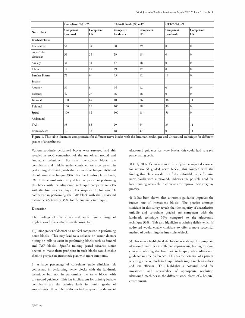
British Journal of Medical Practitioners, March 2012, Volume 5, Number 1
BJMP.org
Consultant (%) n-26 ST/Staff Grade (%) n-17 CT1/2 (%) n-9
Nerve block Competent
Landmark
Competent
US
Competent
Landmark
Competent
US
Competent
Landmark
Competent
US
Brachial Plexus
Interscalene 54 34 58 29 0 0
Supra/Infra
clavicular 31 23 29 18 0 0
Axillary 31 31 47 18 0 0
Elbow 12 19 29 12 0 0
Lumbar Plexus 73 0 65 12 11 0
Sciatic
Anterior 39 8 64 12 0 0
Posterior 42 27 76 18 0 0
Femoral 100 69 100 76 36 11
Epidural 100 19 100 18 36 0
Spinal 100 12 100 18 56 0
Abdominal
TAP 38 85 29 65 33 11
Rectus Sheath 19 35 18 47 0 11
Figure 1. This table illustrates competencies for different nerve blocks with the landmark technique and ultrasound technique for different
grades of anaesthetists
Various routinely performed blocks were surveyed and this
revealed a good comparison of the use of ultrasound and
landmark technique. For the Interscalene block, the
consultants and middle grades combined were competent in
performing this block, with the landmark technique 56% and
the ultrasound technique 33%. For the Lumbar plexus block,
0% of the consultants surveyed felt competent in performing
this block with the ultrasound technique compared to 73%
with the landmark technique. The majority of clinicians felt
competent in performing the TAP block with the ultrasound
technique, 65% versus 35%, for the landmark technique.
Discussion
The findings of this survey and audit have a range of
implications for anaesthetists in the workplace:
1) Junior grades of doctors do not feel competent in performing
nerve blocks. This may lead to a reliance on senior doctors
during on calls to assist in performing blocks such as femoral
and TAP blocks. Specific training geared towards junior
doctors to make them proficient in such blocks would enable
them to provide an anaesthetic plan with more autonomy.
2) A large percentage of consultant grade clinicians felt
competent in performing nerve blocks with the landmark
technique but not in performing the same blocks with
ultrasound guidance. This has implications for training because
consultants are the training leads for junior grades of
anaesthetists. If consultants do not feel competent in the use of
ultrasound guidance for nerve blocks, this could lead to a self
perpetuating cycle.
3) Only 50% of clinicians in this survey had completed a course
for ultrasound guided nerve blocks, this coupled with the
finding that clinicians did not feel comfortable in performing
nerve blocks with ultrasound, indicates the possible need for
local training accessible to clinicians to improve their everyday
practice.
4) It has been shown that ultrasonic guidance improves the
success rate of interscalene blocks.4 The practice amongst
clinicians in this survey reveals that the majority of anaesthetists
(middle and consultant grades) are competent with the
landmark technique 56% compared to the ultrasound
technique 36%. This also highlights a training deficit which if
addressed would enable clinicians to offer a more successful
method of performing the interscalene block.
5) This survey highlighted the lack of availability of appropriate
ultrasound machines in different departments, leading to some
clinicians utilising the landmark technique, when ultrasound
guidance was the preference. This has the potential of a patient
receiving a nerve block technique which may have been riskier
and less efficient. This highlights a potential need for
investment and accessibility of appropriate resolution
ultrasound machines in the different work places of a hospital
environment.

British Journal of Medical Practitioners, March 2012, Volume 5, Number 1
BJMP.org
The main limitation of this project was the small number of
clinicians in the respective hospitals the survey was performed
in. However, we feel the results reflect the practice of clinicians
across most anaesthetic departments. The recommendations
highlight a training need for anaesthetic trainees in the use of
ultrasound guided nerve blocks. This survey could form the
basis of a much larger survey of clinicians across the UK to
provide a more insightful review of the competencies and
preferences of anaesthetic trainees in performing nerve blocks
and the availability of appropriate resolution ultrasound
machines.
The difference in the number of clinicians in each category
limited comparisons between groups. A larger cohort of
participants would enable comparison of nerve block techniques
between different grades of clinicians.
This survey included all clinicians regardless of their sub-
specialist interest. This may result in a skewing of results,
depending on the area of interest of the clinicians surveyed.
This work only highlights the competencies and preferences of
clinicians in performing nerve blocks. No extrapolation can be
made to complications that arise from the choice of either
technique. Studies have shown an improved success rate when
performing nerve blocks with ultrasound.4 However this does
not directly apply to a specific clinician who may have
substantial experience in their method of choice in performing a
nerve block.
Competing Interests
None declared
Author Details Asif Mahmood CT2 Anaesthesia, MBChB, Anaesthetic Department, Kettering
General Hospital, Rothwell Road, Kettering UK Mohammed Auldin CT1
Anaesthesia, MBChB, Anaesthetic Department, Birmingham Heartlands
Hospital, Bordesley Green East, Birmingham UK Asquad Sultan MBBS,
FFARCSI, DESRA, Consultant Anaesthesia, Anaesthetic Department, Kettering
General Hospital, Rothwell Road, Kettering, NN16 8UZ, UK. CORRESSPONDENCE: Asif Mahmood CT2 Anaesthesia, MBChB, Anaesthetic
Department, Kettering General Hospital, Rothwell Road, Kettering, NN16 8UZ,
UK.
Email: [email protected]
REFERENCES
1. Soeding PE, Sha S, Royse CE et al. A randomized trial of ultrasound
guided brachial plexus anaesthesia in upper limb surgery. Anaesthesia
and Intensive Care 2005; 33: 719–25.
2. Guidance on the use of ultrasound locating devices for placing central
venous catheters. National Institute for Clinical Excellence. Technology
Appraisal Guidance. September 2002; Number 49
3. Ultrasound-guided regional nerve block. National Institute for Clinical
Excellence, January 2009; Number IPG285
4. Kapral S, Greher M, Huber G et al. Ultrasonographic guidance
improves the success rate of interscalene brachial plexus blockade. Reg
Anesth Pain Med 2008; 33:253-8.

British Journal of Medical Practitioners, March 2012, Volume 5, Number 1
BJMP.org
BJMP 2012;5(1):a504
Are we managing acute knee effusion well?
A S Eid, V Burrows, J R M Murray, P Smitham, R Ahmad, R Miller and U Butt
ABSTRACT Background: Non-traumatic knee effusion is a common referral to the on-call Orthopaedic team. The two most common causes of this presentation are
septic and crystal arthritis. Crystal-induced arthritis can easily be overlooked or misdiagnosed as septic arthritis resulting in patients having unnecessary
antibiotic therapy and surgical procedures.
Objectives: To review our management of patients with hot swollen knees, especially those due to crystal arthritis.
Materials and methods: We performed a retrospective study of patients presenting to the emergency department with acute non-traumatic knee effusion. A
total of 180 patients were identified; 60 patients were included in the study.
Results: All joints were aspirated and samples were sent for microscopy, culture and antibiotic sensitivity, and polarized light microscopy. Twenty six
patients were admitted and received antibiotic therapy based on clinical suspicion of infection, arthroscopic washout was performed on eight. Four patients
showed positive microscopic growth while eight had crystals identified on polarized light microscopy of joint aspirate. Only two (25%) patients with crystal
arthropathy received appropriate treatment and a rheumatology referral. Seven patients developed complications during their hospital stay.
Conclusion: Crystal arthritis is a common and serious cause of acute painful knee that can lead to joint damage if not treated properly. We should always
remember to follow up the results of polarized light microscopy of joint aspirates. Prompt diagnosis can avoid unnecessary antibiotic therapy and surgical
intervention. All patients with confirmed crystal arthritis should receive a rheumatology referral for further management and follow up.
KEYWORDS: Crystal arthritis, gout, hot swollen knee, Pseudogout, polarized light microscopy
Introduction
Acute non-traumatic knee effusion is a common condition
presenting to the Orthopaedic department which can be caused
by a wide variety of diseases(Table 1). Septic arthritis is the
most common and serious etiology. It can involve any joint; the
knee is the most frequently affected. Accurate and swift
diagnosis of septic arthritis in the acute setting is vital to prevent
joint destruction, since cartilage loss occurs within hours of
onset1,2. Inpatient mortality due to septic arthritis has been
reported as between 7-15%, despite improvement in antibiotic
therapy3,4. Crystal arthritis (Gout/Pseudogout) is the second
most common differential diagnosis. It is often under-diagnosed
and subsequently patients do not receive rheumatology referral
for appropriate treatment and follow-up. In addition, some
patients are misdiagnosed and treated as septic arthritis with
inappropriate antibiotics. Untreated crystal-induced
arthropathy has been shown to cause degenerative joint disease
and disability leading to a considerable health economic
burden.6,7
When the patient is systemically unwell, it is common practice
to start empirical antibiotic treatment after joint aspiration for
the fear of septic arthritis. This aims to minimize the risk of
joint destruction while awaiting gram stain microscopy and
microbiological culture results. In a persistent painful swollen
knee with negative gram stain and culture, antibiotic therapy
can be continued with or without arthroscopic knee washout
based on clinical suspicion of infection 8.
We have therefore undertaken a retrospective study to review
our management of patients with non-traumatic hot swollen
knees and in particular patients with crystal-induced arthritis.
Materials and methods:
We performed a retrospective review of 180 patients presenting
consecutively with acute non-traumatic knee effusion referred
to the on-call Orthopaedic team in the hospital of study
between November 2008 and November 2011. Sixty patients
were included in the study (Table 2). There were 43 males and
17 females, with a mean age of 36 years (range, 23- 93 years).
Patient demographics, clinical presentation, co-morbidities,
current medications and body temperature were recorded. The
results of blood inflammatory markers (WBC, CRP), blood
cultures, synovial fluid microscopy, culture and polarized
microscopy were also collected. Subsequent treatment (e.g.
antibiotics, surgical intervention), complications, and mortality
rates were reviewed.
Results:
On presentation, a decreased range of movement was evident in
all patients. Associated knee pain was reported by 55 patients
(92%), and 24 patients (40%) had fever (temperature ≥ 37.5º).
All joints were aspirated prior to starting antibiotics and samples
were sent for gram stain microscopy, culture and antibiotic
sensitivity, and polarized light microscopy.
Of the 60-patient cohort, 26 were admitted and started on
intravenous antibiotics based on clinical suspicion of infection
(Table 3). The median duration of inpatient admission was 4
days (range, 2 to 14 days). The median duration of antibiotic
Research Article

British Journal of Medical Practitioners, March 2012, Volume 5, Number 1
BJMP.org
therapy was 6 days (range, 2 to 25 days). Eighteen patients were
treated non-operatively by means of antibiotics and anti-
inflammatory medications. Arthroscopic washout was
performed in the remaining eight knees. In this group of
patients, leucocyte count in the joint aspirate ranged from 0-3
leucocyte/mm3, blood leucocyte count ranged from 4-20
leucocyte/mm3, while mean CRP was 37.8 mg/l (range, 1-275
mg/l).
Review of laboratory results revealed that four patients had
positive microscopic growth on gram stained films. Two
samples showed staphylococcus aureus growth and two grew
beta haemolytic streptococci. Eight patientshad crystals
identified on polarized light microscopy of joint aspirate. Three
showed monosodium urate (MSU) crystals while five had
calcium pyrophosphate (CPP) crystals. They received antibiotic
therapy for a mean duration of 10 days (range, 1-30 days). Two
patients were taken to theatre for arthroscopic lavage. Only two
patients received rheumatology referral.
Seven patients developed complications during their hospital
stay. Four contracted diarrhoea; three of which had negative
stool cultures but one was positive for clostridium difficile,
developed toxic megacolon and died. One patient with known
ischemic heart disease had a myocardial infarction and died.
Two further patients acquiredurinary tract infections.
Discussion:
Acute monoarthritisof the knee joint can be a manifestation of
infection, crystal deposits, osteoarthritis and a variety of
systemic diseases. Arriving at a correct diagnosis is crucial for
appropriate treatment 9. Septic arthritis, the most common
etiology, develops as a result of haematogenous seeding, direct
introduction, or extension from a contiguous focus of infection.
Joint infectionis a medical emergency that can lead to
significant morbidity and mortality. Mainstay of treatment
comprises appropriate antimicrobial therapy and joint
drainage 10,11. Literature reveals the knee is the most commonly
affected joint (55%) followed by shoulder (14%) in the septic
joint population12-13.
The second most common differential diagnosis is crystal-
induced monoarthritis. Gout and pseudogout are the two most
common pathologies 14. They are debilitating illnesses in which
recurrent episodes of pain and joint inflammation are caused by
the formation of crystals within the joint space and deposition
of crystals in soft tissue. Gout is caused by monosodium urate
(MSU) crystals, while pseudogout is inflammation caused by
calcium pyrophosphate (CPP) crystals, sometimes referred to as
calcium pyrophosphate disease (CPPD) 15,16. Misdiagnosis of
crystals arthritis or delay in treatment can gradually lead to
degenerative joint disease and disability in addition to renal
damage and failure 5. The clinical picture of acute crystal-
induced arthritis can sometimes be difficult to differentiate
from acute septic arthritis. It is manifested by fever, malaise,
raised peripheral WBC, CRP and other acute phase reactants.
Synovial fluid aspirate can be turbid secondary to an increase in
peripheral polymorphonuclear cells. Diagnosis can be
challenging and therefore crystal identification on polarized
microscopy is considered the gold standard 17, 18, 19. Rest, ice and
topical analgesia may be helpful but systemic non-steroidal anti-
inflammatory medications are the treatment of choice for acute
attacks provided there are no contraindications 20.
In this study, all joints were aspirated and samples were sent for
microscopy, culture and sensitivity, and polarized microscopy
for crystals in-line with the British Society of Rheumatology
and British Orthopaedic Association guidelines 8. Aspiration
not only helps diagnosis but in addition reduces the pain caused
by joint swelling. Twenty six patients were admitted, on clinical
and biochemical suspicion of septic arthritis. They presented
with acute phase response manifested by malaise, fever and
raised inflammatory markers and were treated with antibiotic
therapy and non steroidal anti-inflammatory medications while
awaiting the results of microbiology and polarized light
microscopy. Four of theses patients developed complications
secondary to antibiotic therapy including death due to
clostridium difficile infection and subsequent toxic megacolon.
Infection was confirmed to be underlying cause in four patients
(6%) who showed positive microscopic growth on gram stained
films. They underwent arthroscopic washout and continued
antibiotic therapy according to the result of culture and
sensitivity of their knee aspirate till their symptoms and blood
markers were normal. Arthroscopic washout was required for
four patients with negative microscopic growth due to
persistant symptoms despite antibiotic treatment, as
recommended by the British Society of Rheumatology and the
British Orthopaedic Association 8. Two patients showed
calcium pyrophosphate crystals on polarized microscopy and
two had no bacterial growth or crystals.
We retrospectively reviewed laboratory results and found that
eight patients (13%) were confirmed to have crystal arthritis as
crystals (MSU/CPP) were identified in their knee aspirates by
means of polarized microscopy. However, only two patients
(25%) received this diagnosis whilst in hospital. In both cases,
antibiotic therapy was discontinued and they were referred to a
rheumatologist for appropriate treatment and follow up. The
remaining six patients continued to receive antibiotics and two
of them were taken to theatre for arthroscopic lavage on clinical
suspicion of infection as symptoms did not improve
significantly with medications.
Our study shows that crystal-induced arthritis can easily be
overlooked or misdiagnosed as septic arthritis. This results in
patients having unnecessary antibiotic therapy, developing
serious complications and undergoing surgical procedures, all of
which can be avoided. Moreover, they were not referred to a
rheumatologist.

British Journal of Medical Practitioners, March 2012, Volume 5, Number 1
BJMP.org
Acute knee effusion is a common presentation to the
Orthopaedic department and although we seem to be providing
a good service for septic arthritis, patients with crystal
arthropathy are still slipping through the net. Clinicians should
always remember that crystal arthritis is almost as common as
septic arthritis and will eventually lead to joint damage if not
managed appropriately. It must be excluded as a cause of hot
swollen joints by routine analysis of joint aspirate using
polarized light microscopy. If crystal arthritis is proved to be the
underlying pathology, patients must be treated accordingly and
receive a prompt rheumatology referral for further management.
Acknowledgements This work was performed in Frenchay Hospital, North Bristol NHS Trust, UK. Competing Interests
None declared Author Details Ahmed Shawky Eid, MD, MRCS, Registrar Trauma & Orthopaedics, Yeovil
Hospital, Yeovil, UK. Victoria Burrows, MBChB, Foundation Training Year 2,
Yeovil Hospital, Yeovil, UK. James R M Murray, Consultant Trauma &
Orthopaedics, North Bristol NHS Trust, UK. Peter Smitham MBBS, MRCS,
Registrar Trauma & Orthopaedics, North Bristol NHS trust, UK. Riaz Ahmad,
Registrar Trauma & Orthopaedics, Weston Hospital, Weston-super-Mare, UK.
Roman Miller, MD, PhD, Consultant Trauma & Orthopaedics, Yeovil Hospital,
Yeovil, UK. Umer Butt MBBS, MRCS, Registrar Trauma & Orthopaedics,
Yeovil Hospital, Yeovil, UK.
CORRESSPONDENCE: Umer Butt MBBS, MRCS, Registrar Trauma &
Orthopaedics, Yeovil Hospital, Yeovil, UK.
Email: [email protected]
REFERENCES
1. Lawrence RC, Helmick CG, Arnett FC, et al. Estimates of the
prevalence of arthritis and selected musculoskeletal disorders in the
United States. Arthritis Rheum.1998;41:778-779
2. Baker DG, Schumacher HR. Acute monoarthritis. N Engl J Med.
1993; 329:1013-1020.
3. Kaandorp CJE, Krijnen P, Berelot Moens HJ, Habbema JDF, van
Schaardenburg D. The outcome of bacterial arthritis. A prospective
community-based study. Arthritis Rheum 1997; 40:884-92.
4. Weston VC, Jones AC, Bradbury N, Fawthrop F, Doherty M. Clinical
features and outcome of septic arthritis in a single UK Health District
1982-1991. Ann Rheum Dis 1999; 58:214-9.
5. Reginato A, Paul H, Schumacher HR. Crystal-induced arthritis. Arch
Phys Med Rehabil. 1982; 63: 401-408.
6. Mikuls TR, Farrar JT, Bilker WB, Fernandes S, Schumacher HR Jr,
Saag KG. Gout epidemiology: results from the UK general practice
research database. 1998-1999. Ann Rheum Dis 2005; 64:267-72.
7. K Kumar, E Daley, D.M. Carruthers et al. Delay in presentation to
primary care physicians is the main reason why patients with
rheumatoid arthritis are seen late by the rheumatalogists. Rheumatology
(Oxford). 2007 Sep;46(9):1438-40.
8. Coakley G, Mathews C, Field M, Jones A, Kingsley G, Walker
D, Phillips M, Bradish C, McLachlan A, Mohammed R, Weston
V; British Society for Rheumatology Standards, Guidelines and Audit
Working Group.
BSR & BHPR, BOA, RCGP and BSAC guidelines for management of
the hot swollen joint in adults. Rheumatology (Oxford). 2006
Aug;45(8):1039-41. Epub 2006 Jul 6.
9. Ma L, Cranney A, Holroyd-Leduc JM. Acute monoarthritis: what is
the cause of my patient's painful swollen joint? CMAJ. 2009 Jan
6;180(1):59-65.
10. Shirtliff ME, Mader JT. Acute septic arthritis. Review article. Clin
Microbiol Rev. 2002 Oct;15(4):527-44.
11. Pioro MH, Mandell BF. Septic arthritis. Rheum Dis Clin North Am.
1997; 23(2):239-58.
12. Kaandrorp CJ. et al. Risk factors for septic arthritis in patients with
joint disease. A prospective study. Arthritis Rheum 1995:38(12):1819-
25.
13. McCutchan HJ, Fisher RC, Synovial leukocytosis in infectious arthritis.
Clin Orthop Relat Res 1990 ;( 257):226-30.
14. Schumacher HR Jr, Moreno Alvarez JM. Clues to common crystal-
induced arthropathies. IM 1993; 14:35-47.
15. Martinon F, Glimcher LH. Gout: new insights into an old disease. J
Clin Invest. Aug 2006; 116(8):2073-5.
16. So A. Gout in the spotlight. Arthritis Res Ther. 2008;10(3):112.
17. Li SF, et al. Laboratory tests in adults with monoarticular arthritis: can
they rule out a septic joint? Acad Emerg Med 2004; 11(3): 276-80.
18. Till SH, Snaith MI. Assessment, investigation and management of
acute monoarthritis. J Accid Emerg Med 1999; 16(5):355-61.
19. Hamblen DL, Currey HLF, Key JJ. Pseudogout stimulating acute
suppurative arthritis. J Bone and Joint Surg 1996; 48B:533-53.
20. DieppePA.Investigation and management of gout in the young and the
elderly. Ann Rheum Dis. 1991; 50:263-266.
British Journal of Medical Practitioners, March 2012, Volume 5, Number 1
BJMP.org
BJMP 2012;5(1):a502
Reminder letters to improve rate of attendance at Community Mental Health Centre
Murali Krishna and Sreedharan Amarjothi
Abstract Objective: We carried out a naturalistic study to investigate whether reminder letters would improve the rate of attendance in a community-based mental
health outpatient clinic.
Methods: We prospectively compared the attendance rates between the experimental and control group over a period of 18 months.
Results: The results from this study confirm that reminder letters within a week before the appointment can improve attendance rates in community
mental health clinics for follow up patients.
Conclusion: Non-attendance is an index of severity of mental illness and a predictor of risk. The reasons for non-attendance in mental health clinic are
complex. More large, well-designed randomised studies are desirable. We also recommend periodic evaluation of outpatient non-attendance in order to
identify high-risk individuals and implement suitable measures to keep such severely mentally ill patients engaged with the services.
Introduction
Non-attendance in outpatient clinics accounts for a significant
wastage of health service resources. Psychiatric clinics have high
non-attendance rates and failure to attend may be a sign of
deteriorating mental health. Those who miss psychiatric follow-
up outpatient appointments are more ill with poor social
functioning than those who attend (1). They have a greater
chance of drop out from clinic contact and subsequent
admission (1). Non-attendance and subsequent loss to follow
up indicate possible risk of harm to the patient or to others (2).
Prompts to encourage attendance at clinics are often used and
may take the form of reminder letters (3), telephone
prompting(4) and financial incentives (5). Issuing a copy of the
referral letter to the appointee may prompt attendance for the
initial appointment (6). Contacting patients by reminder letters
prior to their appointments has been effective in improving
attendance rates in a number of settings, including psychiatric
outpatient clinics and community mental health centres (3).
Studies investigating the efficacy of prompting for improving
attendance have generated contrasting findings and non-
attendance remains common in clinical practice. We, therefore,
carried out a naturalistic, prospective controlled study to
investigate whether reminder letters would improve the rate of
attendance in a community-based mental health outpatient
clinic.
Design and Methods
The study was carried out at the Community Mental Health
Centres based in Runcorn and Widnes in Cheshire, UK. The
community mental health team (CMHT) provides specialist
mental health services for adults of working age. Both CMHTs
are similar in demographics, socio-economic need and, have
relatively higher non-attendance rates in the clinic. In the week
prior to the appointment, clerical staff from community mental
health team sent a standard letter to some patients reminding
the date and time of the appointment and name of the
consulting doctor. They recorded whether patient attended,
failed to attend or cancelled the appointment irrespective of
whether they had received a reminder letter or not.
We compared the attendance rates between experimental group
(those who had received the reminder letters) and the control
group ( those who had not received the reminder letters) over a
period of 18 months. Throughout the study period, the same
medical team held the clinics and there had been no major
change in the outpatients’ clinic setting or administrative and
procedural changes influencing outpatients’ attendance. Care
Planning Approach (CPA) was implemented and in operation
even before the introduction of reminding letters at both the
sites.
Attendance rates for all the clinics held during the study period
were obtained from medical records. For all subjects who failed
to attend, age and gender, was obtained from patients’ database.
Patients whose appointments were cancelled were also included
in the study.
Statistics and Data analysis
The data was analysed using SISA - Simple Interactive
Statistical Analysis (7). Chi -squared tests were used to
investigate the attendance rates between the groups, new
patients and follow-ups, with the P value for statistical
significance set at 0.05. Odds ratios were calculated to measure
the size of the effect. In addition, we examined how age and
Research Article

British Journal of Medical Practitioners, March 2012, Volume 5, Number 1
BJMP.org
gender may have influenced the effect of the text based
prompting on attendance.
Results
In the experimental group a total of 114 clinics were booked,
with clinic lists totalling 843 patients. Of these, 88 were new
referrals and 755 were follow-up appointments. 65 of 114
clinics had full attendance. A total of 228 patients failed to
attend the clinic. Of those who failed to attend, 25 patients
were new referrals and 203 were follow-up patients. 28 follow
up patients and 2 patients newly referred to the team called to
cancel their appointments.
In the control group, a total of 71 clinics were booked
amounting to a total of 623 patients. Of these, 86 were new
referrals and 537 were for follow-up patients. Only 25 out of 71
clinics had full attendance. A total of 211 patients failed to
attend. Of those who failed to attend, 32 were new referrals and
179 were follow-up patients. 55 follow up patients and 13
patients newly referred to the team called to cancel their
appointments.
Of those who failed to attend in the experimental group, 98
(43%) were women. The mean age of non-attendees was 38
years; with a range of 18-76 yrs .Of those who failed to attend
in the control group110 (52%) were women. The mean age of
non-attendees was 32 years; with a range of 19-70 yrs.
In our study, failure to attend was not distributed evenly but
had seasonal peaks at Christmas and during the summer
vacation period.
The outcome from prompting in the experimental group is
compared with the control group and displayed in Table 1.
Outcomes
Control
group n
(%)
Experimental
group n (%) 2 (df) P OR (CI)
No of clinics
with full
attendance
25 65 8.32 0.0039 2.44(1.32-
4.50
Total No of
Pts attended 344 585 15.05 0.0001
1.57(1.25-
1.98)
No of new Pts
attended 41 61 3.743 0.053
1.9 (0.98-
3.67)
No of follow
up Pts
attended
303 524 11.39 0.0007 1.52(1.19-
1.94)
No of
Cancellations 68 30 38.63 0
3.85(2.46-
6.04)
Table 1. χ2 = Chi square, df = degree of freedom, OR= Odds
Ratio, CI= Confidence Interval
The attendance rate in the experimental group was 71.95%
(585/813) as opposed to 56.57% (344/555) in the control
group (OR=1.57; p=0.0001).
The attendance rate for new patients in the experimental group
was 70.9%( 61/88) as opposed to 56.16 %( 41/ 86) in the
control group (OR=1.9; p=0.053).
The attendance rate for follow up patients in the experimental
group was 72.0%( 524/727) and 62.8% (303/482) in the
control group (0R=1.52; p=0.0007).
In addition, there were significantly more (by 22%) number of
clinics with full attendance in the experimental group (OR=
2.44, P=0.003).
The observed difference was not influenced by patient’s age or
gender.
Discussion
The results from this study confirm previous findings that
reminder letters within a week before the appointment can
improve attendance rates in community mental health clinics.
Our results are similar to those of the Cochrane systematic
review, which has suggested that a simple prompt in the days
just before the appointment could indeed encourage attendance
(8).
Although it has been reported elsewhere(8) that text based
prompting increases the rate at which patients keep their initial
appointments, our study did not show a similar result for new
patients.
It is already demonstrated that new patients and follow-up
patients in psychiatric clinics are distinct groups with different
diagnostic profiles, degrees of mental illness and with different
reasons for non-attendance. Follow-up patients are severely ill,
socially impaired and isolated than new patients. (1). Forgetting
the appointment and being too unwell are the most common
reasons given for non-attendance by follow-up patients, while
being unhappy with the referral, clinical error and being too
unwell are the most common reasons in the new patient groups
(1). In addition, it has also been observed that increased rate at
which patients keep their first appointments is more likely
related to factors other than simple prompting (4) This explains
our finding that prompting was more beneficial for follow-up
patients as opposed to new referrals to the Community Mental
Health Team.
We also identified several patients with severe mental illness
who ‘did not attend’ for three successive outpatient
appointments. Their care plans were reviewed and
arrangements made to follow up with their community
psychiatric nurses as domiciliary visits at regular intervals. Such
measures should reduce duplication of the services and shorten
British Journal of Medical Practitioners, March 2012, Volume 5, Number 1
BJMP.org
the waiting times for psychiatric consultation, which are well-
recognised factors associated with non-attendance (9).
Non-attendance is an index of severity of mental illness and a
predictor of risk (1). In addition to reminder letters, telephone
prompts are also known to improve attendance (4). Successful
interventions to improve attendance may be labour intensive
but they can be automated and, ultimately, prove cost effective
(8)
We noticed that there is limited research and lack of quality
randomised controlled trials in the area of non-attendance and
the effectiveness of intervention to improve attendance in
mental health setting. More large, well-designed randomised
studies are desirable. We also recommend periodic evaluation of
outpatient non-attendance in order to identify high-risk
individuals and implement suitable measures to keep such
severely mentally ill patients engaged with the services.
There was no randomisation in this study and we relied on
medical records. We have not directly compared the
characteristics of non-attendees with those patients who did
attend the clinics. We did not evaluate other clinical and socio-
demographic factors (e.g. travelling distance, financial
circumstances, etc) that are known to influence the attendance
rates in mental health setting. Hence, there may be limitations
in generalising the results beyond similar populations with
similar models of service provision.
Acknowledgements
Our sincere thanks to Suzanne Kippax, Informatics, Hollins Park Hospital,
Warrington for her help in preparing this manuscript.
Competing Interests
None declared
Author Details
MURALI KRISHNA, MBBS, MRCPsych Consultant Psychiatrist, Holdsworth
memorial Hospital, Mysore, India, and Associate Tutor , University of Edgehill,
Lancashire. SREEDHARAN AMARJOTHI, MBBS, MRCPsych Consultant
Psychiatrist at Liverpool.
CORRESSPONDENCE: MURALI KRISHNA, MBBS, MRCPsych Consultant
Psychiatrist, Holdsworth memorial Hospital, Mysore, India.
Email: [email protected]
REFERENCES
1.Killaspy, H., Banerjee, S., King, M & Lloyd, M. Prospective controlled
study on outpatient non-attendance. Characteristics and outcome. British
Journal of Psychiatry 2000;176:160-165.
2.Royal College of Psychiatrists.Steering committee of confidential inquiry
into homicides and suicides by mentally ill people: Report of the
confidential inquiry into homicides and suicides by the mentally ill
people.1996; London.
3.Kluger, M & Karras, A. Strategies for reducing missed initial appointments
in a community mental health centre. Community Mental Health Journal
1983,19(2): 137-143.
4.Burgoyne, R., Frank, A. & Yamamoto, J. Telephone prompting to
increase attendance at psychiatric outpatient clinic. American Journal of
Psychiatry 1983; 140:345-347.
5.Giuffrida, A. & Torgerson, D. Should we pay the patient? Review of
financial incentives to enhance patient compliance. British Medical Journal,
1997; 315: 703-707.
6.Hamilton, W., Round, A. & Sharp, D. Effect on hospital attendance rates
of giving patients a copy of their referral letter: randomised controlled trial.
British Medical Journal 1999; 318(7195): 1392–1395.
7.Uitenbroek & Daan, G. Binomial SISA. http://home.claa.net/sisa/
binomial.htm. 1997.
8.Reda, S. & Makhoul, S. Prompts to encourage appointment attendance
for people with serious mental illness. The Cochrane database of systemic
reviews.2001; (2): CD002085.
9.Gallucci, G., Swartz. & Hackerman, F. Impact of the wait for an initial
appointment on the rate of kept appointments at a mental health center.
Psychiatric Services 2005; 56: 344-346.

British Journal of Medical Practitioners, March 2012, Volume 5, Number 1
BJMP 2012;5(1):a506
REM Behavior Disorder (RBD) as an Early Marker for Development of
Neurodegenerative Diseases
Umesh Vyas and Rose Franco
ABSTRACT REM behavior disorder (RBD) is a parasomnia characterized by emergence of purposeful complex motor activity with an enactment of dream related
activities. This condition is associated with vivid often violent dreams. In normal adults during REM, diffuse hypotonia of muscles occur and on
polysomnography the limb and chin electromyographic (EMG) channels demonstrate a low voltage or even flat signal. In RBD, the EMG demonstrating
intermittent loss of electromyographic atoniais one of the criteria for diagnosis. Diagnostic polysomnographyrequire capturing the complex dream
behaviors on video and electroencephalography monitoring confirms that the behavior originated out of REM sleep. RBD can be either idiopathic or
symptomatic of various underlying conditions and may in fact be a prodromal symptom of neurodegenerative disease. It can present acutely which is almost
always induced by medications; or develop gradually over months to years. More than half of those with RBD will eventually exhibit signs and symptoms of
a degenerative neurologic disorder. A Polysomnogram (PSG) is necessary to diagnose RBD, showing absence of REM sleep atonia and related abnormal
behavior.
KEYWORDS: REM sleep; REM Behavior Disorder; Neurodegenerative diseases; Parkinson’s disease; Polysomnogram
Introduction
Normal sleep is divided into Non-REM and REM. REM
occurs every 90-120 minutes during adult sleep throughout the
night with each period of REM progressing in length such that
the REM periods in the early morning hours are the longest and
may last from 30-60 minutes. Overall, REM accounts for 20-
25% of the sleep time but is weighted toward the second half of
the night. During REM sleep with polysomnography
monitoring one observes a low voltage mixed frequency
amplitude EEG and low voltage EMG in the chin associated
with intermittent bursts of rapid eye movements. During the
periods of REM breathing becomes irregular, blood pressure
rises and the heart rate also increases due to excess adrenergic
activity. The brain is highly active during REM and the
electrical activity recorded in the brain by EEG during REM
sleep is similar to that of wakefulness.
Parasomnias are undesirable, unexpected, abnormal behavioral
phenomena that occur during sleep. There are three broad
categories in parasomnias. They are
Disorders of Arousal (from Non-REM sleep)
Parasomnias usually associated with REM sleep, and
Other parasomnias which also includes secondary type of
parasomnias.
RBD is the only parasomnia which requires polysomnographic
testing as part of the essential diagnostic criteria.
Definition of RBD
“RBD is characterized by the intermittent loss of REM sleep
electromyographic (EMG) atonia and by the appearance of
elaborate motor activity associated with dream mentation”
(ICSD-2).1 These motor phenomena may be complex and
highly integrated and often are associated with emotionally
charged utterances and physically violent or vigorous activities.
RBD was first recognized and described by Schenck CH et al.
in 1986.2 This diagnosis was first incorporated in the
International Classification of Sleep Disorders (ICSD) in 1990.
(American Academy of Sleep Medicine).
Figure 1
Revie
w A
rtic
le

British Journal of Medical Practitioners, March 2012, Volume 5, Number 1
BJMP.org
Figure 2
A defining feature of normal REM sleep is active paralysis of all
somatic musculature (sparing the diaphragm to permit
ventilation). This result in diffuse hypotonia of the skeletal
muscles inhibiting the enactment of dreams associated with
REM sleep. In RBD there is an intermittent loss of muscle
atonia during REM sleep that can be objectively measured with
EMG as intense phasic motor activity (figure 1 and 2). This loss
of inhibition often precedes the complex motor behaviors
during REM sleep. Additionally, RBD patients will report that
their dream content is often very violent or vigorous dream
enacting behaviors include talking, yelling, punching, kicking,
sitting, jumping from bed, arm flailing and grabbing etc. and
most often the sufferer will upon waking from the dream
immediately report a clear memory of the dream which
coincides very well with the high amplitude violent defensive
activity witnessed. This complex motor activity may result in a
serious injury to the dreamer or bed partner that then prompts
the evaluation.
Prevalence
The Prevalence of RBD is about 0.5% in general population.1,
3 RBD preferentially affect elderly men (in 6th and 7thdecade)
with ratio of women to men being 1 to 9.4 The mean age of
disease onset is 60.9 years and at diagnosis is 64.4 years.5 RBD
was reported in an 18 year old female with Juvenile Parkinson
disease,6 so age and gender are not absolute criteria.
In Parkinson disease (PD) the reported prevalence ranges from
13-50%,7, 14-19 LewyBody Dementia (DLB) 95%,8 and Multiple
System Atrophy (MSA) 90 %.9 The presence of RBD is a major
diagnostic criterion for MSA. RBD has been reported in
Juvenile Parkinson disease, and pure autonomic failure10-12 all
neurodegenerative disorders are synucleinopathies.13
Physiology
The neurons of locus coeruleus, raphe nuclei,
tuberomammillary nucleus, pedunculopontine nucleus,
laterodorsal tegmental area and the perifornical area are firing at
a high rate, and cause arousal by activating the cerebral cortex.
During REM sleep, the aforementioned excitatory areas fall
silent with the exception of the pedunculopontine nucleus and
laterodorsal tegmental areas. These regions project to the
thalamus and activate the cortex during REM sleep. This
cortical activation is associated with dreaming in REM.
Descending excitatory fibers from the pedunculopontine
nucleus and laterodorsal tegmental area innervate the medial
medulla, which then sends inhibitory projections to motor
neurons producing the skeletal muscle atonia of REM sleep.20-21
There are two distinct neural systems which collaborate in the
“paralysis” of normal REM sleep, one is mediated through the
active inhibition by neurons in the nucleus reticularis
magnocellularis in the medulla via the ventrolateral
reticulospinal tract synapsing on the spinal motor neurons and
the other system suppresses locomotor activity and is located in
pontine region.22
Pathophysiology
REM sleep contains two types of variables, tonic (occurring
throughout the REM period), and phasic (occurring
intermittently during a REM period). Tonic elements include
desynchronized EEG and somatic muscle atonia (sparing the
diaphragm). Phasic elements include rapid eye movements,
middle ear muscle activity and extremity twitches. The tonic
electromyogram suppression of REM sleep is the result of active
inhibition of motor activity originating in the perilocus
coeruleus region and terminating in the anterior horn cells via
the medullary reticularis magnocellularis nucleus.
In RBD, the observed motor activity may result from either
impairment of tonic REM muscle atonia or from increase
phasic locomotor drive during REM sleep. One mechanism by
which RBD results is the disruption in neurotransmission in the
brainstem, particularly at the level of the pedunculopontine
nucleus.23Pathogenetically, reduced striatal dopaminergic
mediation has been found24-25 in those with RBD.
Neuroimaging studies support dopaminergic abnormalities.
Types of RBD
RBD can be categorized based on severity:
1. Mild RBD occurring less than once per month,
2. Moderate RBD occurring more than once per month
but less than once per week, associated with physical
discomfort to the patient or bed partner, and
3. Severe RBD occurring more than once per week,
associated with physical injury to patient or bed
partner.
RBD can be categorized based on duration:
1. Acute presenting with one month or less,
2. Subacute with more than one month but less than 6
months,
3. Chronic with 6 months or more of symptoms prior
to presentation
British Journal of Medical Practitioners, March 2012, Volume 5, Number 1
BJMP.org
Acute RBD: In 55 - 60% of patients with RBD the cause is
unknown, but in 40 - 45% the RBD is secondary to another
condition. Acute onset RBD is almost always induced or
exacerbated by medications (especially Tri-Cyclic
Antidepressants, Selective Serotonin Reuptake Inhibitors,
Mono-Amine Oxidase Inhibitors, Serotonin Norepinephrine
Reuptake Inhibitors,26 Mirtazapine, Selegiline, and Biperiden)
or during withdrawal of alcohol, barbiturates, benzodiazepine
or meprobamate. Selegiline may trigger RBD in patients with
Parkinson disease. Cholinergic treatment of Alzheimer’s disease
may trigger RBD.
Chronic RBD: The chronic form of RBD was initially thought
to be idiopathic; however long term follow up has shown that
many eventually exhibit signs and symptoms of a degenerative
neurologic disorder. One recent retrospective study of 44
consecutive patients diagnosed with idiopathic RBD
demonstrated that 45% (20 patients) subsequently developed a
neurodegenerative disorder, most commonly Parkinson disease
(PD) or Lewy body dementia, after a mean of 11.5 years from
reported symptoms onset and 5.1 years after RBD diagnosis.27
The relationship between RBD and PD is complex and not all
persons with RBD develop PD. In one study of 29 men
presenting with RBD followed prospectively, the incidence of
PD was 38% at 5 years and 65% after 12 years.7, 28, 29Contrast
this with the prevalence of the condition in multiple system
atrophy, where RBD is one of the primary symptoms occurring
in 90% of cases.9 In cases of RBD, it is absolutely necessary not
only to exclude any underlying neurodegenerative disease
process but also to monitor for the development of one over
time in follow up visits.
Clinical manifestations
Sufferers of RBD usually present to the doctor with complaints
of sleep related injury or fear of injury as a result of dramatic
violent, potentially dangerous motor activity during sleep. 96%
of patients reporting harm to themselves or their bed partner.
Behaviors during dreaming described include talking, yelling,
swearing, grabbing, punching, kicking, jumping or running out
of the bed. One clinical clue of the source of the sleep related
injury is the timing of the behaviors. Because RBD occurs
during REM sleep, it typically appears at least 90 minutes after
falling asleep and is most often noted during the second half of
the night when REM sleep is more abundant.
One fourth of subjects who develop RBD have prodromal
symptoms several years prior to the diagnosis. These symptoms
may consist of twitching during REM sleep but may also
include other types of simple motor movements and sleep
talking or yelling.30-31 Day time somnolence and fatigue are rare
because gross sleep architecture and the sleep-wake cycle remain
largely normal.
RBD in other neurological disorders and Narcolepsy:
RBD has also been reported in other neurologic diseases such as
Multiple Sclerosis, vascular encephalopathies, ischemic brain
stem lesions, brain stem tumors, Guillain-Barre syndrome,
mitochondrial encephalopathy, normal pressure hydrocephalus,
subdural hemorrhage, and Tourette’s syndrome. In most of
these there is likely a lesion affecting the primary regulatory
centers for REM atonia.
RBD is particularly frequent in Narcolepsy. One study found
36% pts with Narcolepsy had symptoms suggestive of RBD.
Unlike idiopathic RBD, women with narcolepsy are as likely to
have RBD as men, and the mean age was found to be 41
years.32 While the mechanism allowing for RBD is not
understood in this population, narcolepsy is considered a
disorder of REM state disassociation. Cataplexy is paralysis of
skeletal muscles in the setting of wakefulness and often is
triggered by strong emotions such as humor. In narcoleptics
who regularly experienced cataplexy, 68% reported RBD
symptoms, compared to 14% of those who never or rarely
experienced cataplexy.32-33 There is evidence of a profound loss
of hypocretin in the hypothalamus of the narcoleptics with
cataplexy and this may be a link that needs further investigation
in the understanding of the mechanism of RBD in Narcolepsy
with cataplexy. It is prudent to follow Narcoleptics and
questioned about symptoms of RBD and treated accordingly,
especially those with cataplexy and other associated symptoms.
Diagnostic criteria for REM Behavior Disorder (ICSD-2:
ICD-9 code: 327.42)1
A. Presence of REM sleep without Atonia: the EMG finding
of excessive amounts of sustained or intermittent elevation
of submental EMG tone or excessive phasic submental or
(upper or lower) limb EMG twitching (figure 1 and 2).
B. At least one of the following is present:
i. Sleep related injurious, potentially injurious, or
disruptive behaviors by history
ii. Abnormal REM sleep behaviors documented during
polysomnographic monitoring
C. Absence of EEG epileptiform activity during REM sleep
unless RBD can be clearly distinguished from any
concurrent REM sleep-related seizure disorder.
D. The sleep disturbance is not better explained by another
sleep disorder, medical or neurologic disorder, mental
disorder, medication use, or substance use disorder.
Differential diagnosis
Several sleep disorders causing behaviors in sleep can be
considered in the differential diagnosis, such as sleep walking
(somnambulism), sleep terrors, nocturnal seizures, nightmares,
psychogenic dissociative states, post-traumatic stress disorder,
nocturnal panic disorder, delirium and malingering. RBD may
be triggered by sleep apnea and has been described as triggered
by nocturnal gastroesophageal reflux disease.
Evaluation and Diagnosis
• Detailed history of the sleep wake complaints

British Journal of Medical Practitioners, March 2012, Volume 5, Number 1
BJMP.org
• Information from a bed partner is most valuable
• Thorough medical, neurological, and psychiatric history
and examination
• Screening for alcohol and substance use
• Review of all medications
• PSG (mandatory): The polysomnographic study should be
more extensive, with an expanded EEG montage, monitors
for movements of all four extremities, continuous
technologist observation and continuous video recording
with good sound and visual quality to allow capture of any
sleep related behaviors
• Multiple Sleep Latency Test (MSLT): Only recommended
in the setting of suspected coexisting Narcolepsy
• Brain imaging (CT or MRI) is mandatory if there is
suspicion of underlying neurodegenerative disease.
Management
RBD may have legal consequences or can be associated with
substantial relationship strain; therefore accurate diagnosis and
adequate treatment is important, which includes non-
pharmacological and pharmacological management.
Non-pharmacological management: Acute form appears to be
self-limited following discontinuation of the offending
medication or completion of withdrawal treatment. For chronic
forms, protective measures during sleep are warranted to
minimize the risks for injury to patient and bed partner. These
patients are at fall risk due to physical limitations and use of
medications. Protective measure such as removing bed stands,
bedposts, low dressers and applying heavy curtains to windows.
In extreme cases, placing the mattress on the floor to prevent
falls from the bed has been successful.
Pharmacological management: Clonazepam is highly effective
in treatment and it is the drug of choice. A very low dose will
resolve symptoms in 87 to 90% of patients.4, 5, 7-
34 Recommended treatment is 0.5 mg Clonazepam 30 minutes
prior to bed time and for more than 90% of patients this dose
remains effective without tachyphylaxis. In the setting of
breakthrough symptoms the dose can be slowly titrated up to
2.0 mg. The mechanism of action is not well understood but
clonazepam appears to decrease REM sleep phasic activity but
has no effect on REM sleep atonia.35
Melatonin is also effective and can be used as monotherapy or
in conjunction with clonazepam. The suggested dose is 3 to 12
mg at bed time. Pramipexole may also be effective36-38 and
suggested for use when clonazepam is contraindicated or
ineffective. It is interesting to note that during holidays from
the drug, the RBD can take several weeks to recur.
Management of patients with concomitant disorder like
narcolepsy, depression, dementia, Parkinson disease and
Parkinsonism can be very challenging, because medications
such as SSRIs, selegiline and cholinergic medications used to
treat these disorders, can cause or exacerbate RBD. RBD
associated with Narcolepsy, clonazepam is usually added in
management and it is fairly effective.
Follow-up
Because RBD may occur in association with neurodegenerative
disorder, it is important to consult a neurologist for every
patient with RBD as early as possible, especially to diagnose and
provide care plan for neurodegenerative disorder, which
includes but not limited to early diagnosis and management,
regular follow up, optimization of management to provide
better quality of life and address medico-legal issues.
Prognosis
In acute and idiopathic chronic RBD, the prognosis with
treatment is excellent. In the secondary chronic form, prognosis
parallels that of the underlying neurologic disorder. Treatment
of RBD should be continued indefinitely, as violent behaviors
and nightmares promptly reoccur with discontinuation of
medication in almost all patients.
Conclusions
RBD and neurodegenerative diseases are closely interconnected.
RBD often antedates the development of a neurodegenerative
disorder; diagnosis of idiopathic RBD portends a risk of greater
than 45% for future development of a clinically defined
neurodegenerative disease. Once identified, close follow-up of
patients with idiopathic RBD could enable early detection of
neurodegenerative diseases. Treatment for RBD is available and
effective for the vast majority of cases.
Key Points
• Early diagnosis of RBD is of paramount importance
• Polysomnogram is an essential diagnostic element
• Effective treatment is available
• Early treatment is essential in preventing injuries to patient
and bed partner
• Apparent idiopathic form may precede development of
Neurodegenerative disorder by decades
Competing Interests
None declared
Author Details
UMESH VYAS, MD, Chair of Department of Psychiatry, Medical Director of
Behavioral Health Unit and Medical Director of Sleep Disorders Center,
Psychiatrist and Sleep Disorders Specialist, Mayo Clinic Health System, Mankato,
MN, USA. ROSE FRANCO, MD Sleep Medicine Fellowship Director and
Associate Professor in Medicine and Otolaryngology, Pulmonologist and Sleep
Disorders Specialist, Medical College of Wisconsin, Milwaukee, WI, USA.
CORRESSPONDENCE: UMESH VYAS, MD, Chair of Department of
Psychiatry, Medical Director of Behavioral Health Unit and Medical Director of
Sleep Disorders Center, Psychiatrist and Sleep Disorders Specialist, Mayo Clinic
Health System, Mankato, MN, USA.
Email: [email protected]
British Journal of Medical Practitioners, March 2012, Volume 5, Number 1
BJMP.org
REFERENCES
1. American Academy of Sleep medicine. International Classification of
Sleep Disorders 2nd ed.; Diagnostic and Coding Manual. Westchester,
Illinois; American Academy of Sleep Medicine, 2005.
2. Schenck CH, Bundlie SR, Ettinger MG et al. Chronic Behavioral
disorder of human REM sleep, a new category of Parasomnia. Sleep 1986; 9:
293-308.
3. Mahowald MW, Schenck CH. REM sleep parasomnias. In: Kryger MH,
Roth T, Dement WC. Principles and Practice of sleep medicine.
Philadelphia, Saunders; 2000: 724-42.
4. Oksenberg A, Radwan H, Arons E, et al. Rapid eye movement (REM)
sleep behavior disorder: a sleep disturbance affecting mainly older man. Isr J
Psychiatry Rel Sci 2002; 39: 28.
5. Olson EJ, Boeve BF, Silber MH. Rapid eye movement sleep behavior
disorder: demographic, clinical and laboratory findings in 93 cases. Brain
2000; 123: 331-9.
6. Rye DB, Johnston LH, Watts RL, et al. Juvenile Parkinson’s disease with
REM sleep behavior disorder, sleepiness and day time REM onset.
Neurology 1999; 53: 1868-70.
7. Schenck CH, Bundlie SR, Mahowald MW. Delayed emergence of a
parkinsonian disorder in 38% of 29 older men initially diagnosed with
idiopathic rapid eye movement sleep behavior disorder. Neurology 1996; 46:
388-393.
8. Boeve BF, Silber MH, Ferman JT, et al. REM sleep behavior disorder and
degenerative dementia: An association likely reflecting Lewy body disease.
Neurology 1998; 51: 363-370.
9. Plazzi G, Corsini R, Provini F, et al. REM sleep behavior disorder in
multiple system atrophy. Neurology 1997; 48: 1094-7.
10. Gagnon JF, Postuma RB, Mazza S, et al. Rapid-eye-movement sleep
behavior disorder and neurodegenerative diseases. Lancet Neurol 2006: 5;
424-432.
11. Iranzo A, Molinuevo JL, Santamaria J, et al. REM sleep behavior
disorder as an early marker for neurodegenerative disorder: a descriptive
study. Lancet Neurol 2006, 5: 572-577.
12. Weyer A, Minnerop M, Abele M, et al. REM sleep behavior disorder in
pure autonomic failure (PAF). Neurology 2006, 66: 608-609.
13. Galpern WR, Lang AE. Interface between tauopathies and
synucleinopathies: a tale of two proteins. Ann Neurol. 2006; 59: 449-458.
14. Rositsa GP, Zachari IZ. REM sleep Behavior disorder in patients with
Parkinson’s disease. Folia medica XLVII, 1/2005: 5-10.
15. Eisensehr I, Lindeiner H, Jager M, et al. REM sleep behavior disorder in
sleep-disordered patients with verses without Parkinson’s disease: is there a
need for polysomnography? J Neurol Sci 2001; 186: 7-11.
16. Oerlemans WGH, de Weerd AW. The prevalence of sleep disorders in
patients with Parkinson’s disease. A self-reported, community-based survey.
Sleep Medicine 2002; 3: 147-9.
17. Comella CL, Nardine TM, Diederich NJ, et al. Sleep-related violence,
injury, and REM sleep behavior disorder in Parkinson’s disease. Neurology
1998; 51: 526-9.
18. Comella CL, Tanner CM, Ristanovic RK. Polysomnographic sleep
measures in Parkinson’s disease patients with treatment-induced
hallucinations. Ann Neurol 1993; 34: 710-4.
19. Gagnon JF, Bedard MA, Fantini ML, et al. REM sleep behavior disorder
and REM sleep without atonia in Parkinson’s disease. Neurology 2002; 59:
585-9.
20. Espana RA, Scammell TE. Sleep neurobiology for the clinician. Sleep
2004; 27: 811-20.
21. Mignot E, Taheri S, Nishino S. Sleeping with the hypothalamus:
emerging therapeutic targets for sleep disorders. Nat Neurosci 2002;
5(Suppl): 1071-5.
22. Siegel JM (1994). Brainstem mechanisms generating REM sleep. In M
Kryger, T Roth and W Dement (Eds), Principles and practice of sleep
medicine (3rd Edn, pp 125-144). Philadelphia: W B Saunders Company.
23. Rye DB. Contributions of the pedunculopontine region to normal and
altered REM sleep. Sleep 1997; 20: 757-88.
24. Albin RL, Koeppe RA, Chervin RD, et al. Decreased striatal
dopaminergic innervation in REM sleep behavior disorder. Neurology 2000;
55: 1410-2.
25. Eisensehr I, Linke R, Noachtar S, et al. Reduced striatal dopamine
transporters in idiopathic rapid eye movement sleep behavior disorder.
Comparison with Parkinson’s disease and controls. Brain 2000; 123: 1155-
60.
26. Schenck CH, Mahowald MW. REM sleep parasomnias. Neurol Clin
2005; 23: 1107-1126.
27. Hickey MG, Demaerschalk BM, Caselli RJ et al. “Idiopathic” Rapid-
Eye-Movement (REM) sleep behavior disorder is associated with future
development of neurodegenerative diseases. The Neurologist 2007; 13: 98-
101.
28. Lee AJ, Blackburn NA, Campbell V. The nighttime problems of
Parkinson’s disease. Clin neuro pharmacol 1988; 11: 512-9.
29. Schenck CH, Bundlie SR, Mahowald MH. REM behavior disorder
(RBD): delayed emergence of Parkinsonism and/or dementia in 65% of
older men initially diagnosed with idiopathic RBD, and an analysis of the
minimum and maximum tonic and/or phasic electromyographic
abnormalities found during REM sleep. Sleep 2003; 26(suppl): A316.
30. Schenck CH, Hurwitz TD, Mahowald MW. Symposium: normal and
abnormal REM sleep regulation: REM sleep behavior disorder: an update on
a series of 96 patients and a review of the world literature. J Sleep Res 1993;
2: 224-31.
31. Sforza E, Krieger J, Petiau C. REM sleep behavior disorder: clinical and
physiopathological findings. Sleep Med Rev 1997; 1: 57-69.
32. Nightingale S, Orgill JC, Ebrahim IO, et al. The association between
Narcolepsy and REM Behavior Disorder (RBD). Sleep Med. 2005; 6(3):
253-8.
33. Frauscher B, Gschliesser V, Brandauer E, et al. REM sleep Behavior
Disorder in 703 sleep-disorder patients: The importance of eliciting a
comprehensive sleep history. Sleep Med. 2010; 11(2): 167-171.
34. Schenck CH, Mahowald MW. A polysomnographic, neurologic,
psychiatric and clinical outcome report on 70 consecutive cases with REM
sleep behavior disorder (RBD): sustained clonazepam efficacy in 89.5% of
57 treated patients. Clev Clin J Med 1990; 57 Suppl: 10-24.
35. Lapierre O, Montplaisir J. Polysomnographic features of REM sleep
behavior disorder: development of a scoring method. Neurology 1992, 42:
1371-1374.
36. Boeve BF, Silber MH, Ferman JT. Melatonin for treatment of REM
sleep behavior disorder in neurologic disorders: results in 14 patients. Sleep
Med 2003; 4: 281-284.
37. Fantini ML, Gagnon JF, Filipini D, et al. The effects of pramipexole in
REM sleep behavior disorder. Neurology 2003, 61: 1418-1420.
38. Schmidt MH, Koshal VB, Schmidt HS. Use of pramipexole in REM
sleep behavior disorder. Sleep Med 2006, 7: 418-423.

British Journal of Medical Practitioners, March 2012, Volume 5, Number 1
BJMP 2012;5(1):a507
Management of Drooling of saliva
Ganesh Bavikatte , Poh Lin Sit and Ali Hassoon
ABSTRACT Drooling, also known as ptyalism or sialorrhea can be defined as salivary incontinence or the involuntary spillage of saliva over the lower lip. Drooling
could be caused by excessive production of saliva, inability to retain saliva within the mouth, or problems with swallowing. Drooling can lead to functional
and clinical consequences for patients, families, and caregivers. Physical and psychosocial complication includes maceration of skin around the mouth,
secondary bacterial infection, bad odour, dehydration and social stigmatisation. People with drooling problems are also at increased risk of inhaling saliva,
food, or fluids into the lungs especially when body's normal reflex mechanisms, such as gagging and coughing are also impaired. Successful management of
sialorrhea can alleviate the associated hygienic problems, improve appearance, enhance self-esteem, and significantly reduce the nursing care time of these
sufferers.Chronic drooling can be difficult to manage; this article gives overview of the causes, effects and management of drooling of
saliva in general practice.
Saliva is the watery and usually frothy substance produced in
and secreted from the three paired major salivary (parotid,
submandibular and sublingual) glands and several hundred
minor salivary glands, composed mostly of water, but also
includes electrolytes, mucus, antibacterial compounds, and
various enzymes. Healthy persons are estimated to produce 0.75
to 1.5 liters of saliva per day. At least 90% of the daily salivary
production comes from the major salivary glands while the
minor salivary glands produce about 10%. On stimulation
(olfactory, tactile or gustatory), salivary flow increases five fold,
with the parotid glands providing the preponderance of saliva.1
Saliva is a major protector of the tissues and organs of the
mouth. In its absence both the hard and soft tissues of the oral
cavity may be severely damaged, with an increase in ulceration,
infections, such as candidiasis, and dental decay. Saliva is
composed of serous part (alpha amylase) and a mucus
component, which acts as a lubricant. It is saturated with
calcium and phosphate and is necessary for maintaining healthy
teeth. The bicarbonate content of saliva enables it to buffer and
produce the condition necessary for the digestion of plaque
which holds acids in contact with the teeth. Moreover, saliva
helps with bolus formation and lubricates the throat for the easy
passage of food. The organic and inorganic components of
salivary secretion have got a protective potential. They act as
barrier to irritants and a means of removing cellular and
bacterial debris. Saliva contains various components involved in
defence against bacterial and viral invasion, including mucins,
lipids, secretory immunoglobulins, lysozymes, lactoferrin,
salivary peroxidise, and myeloperoxidase. Salivary pH is about
6-7, favouring digestive action of salivary enzyme, alpha
amylase, devoted to starch digestion.
Image -1. (Source of this image- http://www.entdoctor.co.nz)
Salivary glands are innervated by the parasympathetic and
sympathetic nervous system. Parasympathetic postganglionic
cholinergic nerve fibers supply cells of both the secretory end-
piece and ducts and stimulate the rate of salivary secretion,
inducing the formation of large amounts of a low-protein,
serous saliva. Sympathetic stimulation promotes saliva flow
through muscle contractions at salivary ducts. In this regard
both parasympathetic and sympathetic stimuli result in an
increase in salivary gland secretions. The sympathetic nervous
system also affects salivary gland secretions indirectly by
innervating the blood vessels that supply the glands.
Drooling (also known as driveling, ptyalism, sialorrhea, or
slobbering) is when saliva flows outside the mouth, defined as
“saliva beyond the margin of the lip”. This condition is normal
in infants but usually stops by 15 to 18 months of age.
Revie
w A
rtic
le

British Journal of Medical Practitioners, March 2012, Volume 5, Number 1
BJMP.org
Sialorrhea after four years of age generally is considered to be
pathologic.
Table 1: Functions of saliva
Digestion and swallowing
Initial process of food digestion
Lubrication of mouth, teeth, tongue and food boluses
Tasting food
Amylase- digestion of starch
Disinfectant and protective role
Effective cleaning agent
Oral homeostasis
Protect teeth decay, dental health and oral odour
Bacteriostatic and bacteriocidal properties
Regulate oral pH
Speaking
Lubricates tongue and oral cavity
The prevalence of drooling of saliva in the chronic neurological
patients is high, with impairment of social integration and
difficulties to perform oral motor activities during eating and
speech, with repercussion in quality of lifeDrooling occurs in
about one in two patients affected with motor neuron disease
and one in five needs continuous saliva elimination7, its
prevalence is about 70% in Parkinson disease8, and between 10
to 80% in patients with cerebral palsy9.
Pathophysiology
Pathophysiology of drooling is multifactorial. It is generally
caused by conditions resulting in
Excess production of saliva- due to local or systemic causes
(table 2)
Inability to retain saliva within the mouth- poor head control,
constant open mouth, poor lip control, disorganized tongue
mobility, decreased tactile sensation, macroglossia, dental
malocclusion, nasal obstruction.
Problems with swallowing- resulting in excess pooling of saliva
in the anterior portion of the oral cavity e.g. lack of awareness
of the build-up of saliva in the mouth, infrequent swallowing,
and inefficient swallowing.
Drooling is mainly due to neurological disturbance and less
frequently to hyper salivation.Under normal circumstances,
persons are able to compensate for increased salivation by
swallowing. However, sensory dysfunction may decrease a
person’s ability to recognize drooling and anatomic or motor
dysfunction of swallowing may impede the ability to manage
increased secretion.
Depending on duration of drooling, it can be classified
as acute e.g. during infections (epiglottitis, peritonsilar abscess)
orchronicneurological causes.
Table 2 Aetiology of hypersalivation
Physiological
Pregnancy
Local causes
Oral inflammation- teething
Infection –oral cavity infection, dental caries, tonsillitis, peritonsilar
abscess
Systemic
Toxin exposure- pesticides, mercury, capsaicin, snake poisoning
Medication –tranquilizers, anticonvulsants, anticholinesterases, lithium
Neuromuscular –cerebral palsy, Parkinson’s disease, motor neuron
disease, bulbar/ pseudobulbar palsy, Stroke
Infection- rabies
Gastric- gastroesophageal reflux
Symptoms
Drooling of saliva can affect patient and/or their carers quality
of life and it is important to assess the rate and severity of
symptoms and its impact on their life.
Table 3 Effect of untreated Drooling of saliva
Physical Psychological
Perioral chapping (skin cracking)
Maceration with secondary
infection
Dehydration
Foul odour
Aspiration/ pneumonia
Speech disturbance
Interference with feeding
Isolation
Barriers to education (damage to
books or electronic devices)
Increased dependency and
level/intensity of care
Damage to electronic devices
Decreased self esteem
Difficult social interaction
Assessment
Assessment of the severity of drooling and its impact on quality
of life for the patient and their carers help to establish a
prognosis and to decide the therapeutic regimen. A variety of
subjective and objective methods for assessment of sialorrhoea
have been described3.
History (from patient and carers)
Establish possible cause, severity, complications and possibility
of improvement, age and mental status of patient, chronicity of
problems, associated neurological conditions, timing, provoking
factors, estimation of quantity of saliva – use of bibs, clothing
changing required/ day and impact on the day today life
(patient/carer)
Physical examination
Evaluate level of alertness, emotional state, hydration status,
hunger, head posture
Examination of oral cavity- sores on the lip or chin, dental
problems, tongue control, swallowing ability, nasal airway
obstruction, decreased intraoral sensitivity, assessment of health
status of teeth, gum, oral mucosa, tonsils, anatomical closure of
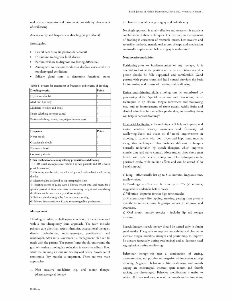
British Journal of Medical Practitioners, March 2012, Volume 5, Number 1
BJMP.org
oral cavity, tongue size and movement, jaw stability. Assessment
of swallowing
Assess severity and frequency of drooling (as per table 4)
Investigation
• Lateral neck x ray (in peritonsilar abscess)
• Ultrasound to diagnose local abscess
• Barium swallow to diagnose swallowing difficulties
• Audiogram- to rule out conductive deafness associated with
oropharyngeal conditions
• Salivary gland scan- to determine functional status
Table 4 : System for assessment of frequency and severity of drooling
Drooling severity Points
Dry (never drools) 1
Mild (wet lips only) 2
Moderate (wet lips and chins) 3
Severe (clothing becomes damp) 4
Profuse (clothing, hands, tray, object become wet) 5
Frequency Points
Never drools 1
Occasionally drools 2
Frequency drools 3
Constantly drools 4
Other methods of assessing salivary production and drooling
1) 1- 10 visual analogue scale (where 1 is best possible and 10 is worst
possible situation)
2) Counting number of standard sized paper handkerchiefs used during
the day
3) Measure saliva collected in cups strapped to chin
4) Inserting pieces of gauze with a known weight into oral cavity for a
specific period of time and then re-measuring weight and calculating
the difference between the dry and wet weights.
5) Salivary gland scintigraphy / technetium scanning
6) Salivary duct canulation 12 and measuring saliva production.
Management
Drooling of saliva, a challenging condition, is better managed
with a multidisciplinary team approach. The team includes
primary care physician, speech therapist, occupational therapist,
dentist, orthodontist, otolaryngologist, paediatrician and
neurologist. After initial assessment, a management plan can be
made with the patient. The person/ carer should understand the
goal of treating drooling is a reduction in excessive salivary flow,
while maintaining a moist and healthy oral cavity. Avoidance of
xerostomia (dry mouth) is important. There are two main
approaches
1. Non invasive modalities e.g. oral motor therapy,
pharmacological therapy
2. Invasive modalities e.g. surgery and radiotherapy
No single approach is totally effective and treatment is usually a
combination of these techniques. The first step in management
of drooling is correction of reversible causes. Less invasive and
reversible methods, namely oral motor therapy and medication
are usually implemented before surgery is undertaken5
Non invasive modalities
Positioning prior to implementation of any therapy, it is
essential to look at the position of the patient. When seated, a
person should be fully supported and comfortable. Good
posture with proper trunk and head control provides the basis
for improving oral control of drooling and swallowing.
Eating and drinking skills- drooling can be exacerbated by
poor eating skills. Special attention and developing better
techniques in lip closure, tongue movement and swallowing
may lead to improvements of some extent. Acidic fruits and
alcohol stimulate further saliva production, so avoiding them
will help to control drooling10
Oral facial facilitation - this technique will help to improve oral
motor control, sensory awareness and frequency of
swallowing. Scott and staios et al 18 noted improvement in
drooling in patients with both hyper and hypo tonic muscles
using this technique. This includes different techniques
normally undertaken by speech therapist, which improves
muscle tone and saliva control. Most studies show short term
benefit with little benefit in long run. This technique can be
practiced easily, with no side effects and can be ceased if no
benefits noted.
a) Icing – effect usually last up to 5-30 minutes. Improves tone,
swallow reflex.
b) Brushing- as effect can be seen up to 20- 30 minutes,
suggested to undertake before meals.
c) Vibration- improves tone in high tone muscles
d) Manipulation – like tapping, stroking, patting, firm pressure
directly to muscles using fingertips known to improve oral
awareness.
e) Oral motor sensory exercise - includes lip and tongue
exercises.
Speech therapy- speech therapy should be started early to obtain
good results. The goal is to improve jaw stability and closure, to
increase tongue mobility, strength and positioning, to improve
lip closure (especially during swallowing) and to decrease nasal
regurgitation during swallowing.
Behaviour therapy- this uses a combination of cueing,
overcorrection, and positive and negative reinforcement to help
drooling. Suggested behaviours, like swallowing and mouth
wiping are encouraged, whereas open mouth and thumb
sucking are discouraged. Behavior modification is useful to
achieve (1) increased awareness of the mouth and its functions,

British Journal of Medical Practitioners, March 2012, Volume 5, Number 1
BJMP.org
(2) increased frequency of swallowing, (3) increased swallowing
skills. This can be done by family members and friends.
Although there is no randomized controlled trial done, over 17
articles published in last 25 years, show promising results and
improved quality of life. No reported side effects make
behavioural interventions an initial option compared to surgery,
botulinum toxin or pharmaceutical management. Behaviour
interventions are useful prior and after medical management
such as botulinum toxin or surgery.
Oral prosthetic device- variety of prosthetic devices can be
beneficial, e.g. chin cup and dental appliances, to achieve
mandibular stability, better lip closure, tongue position and
swallowing. Cooperation and comfort of the patient is essential
for better results.
Pharmacological methods
Systematic review of anticholinergic drugs, show Benztropine,
Glycopyrrolate, and Benzhexol Hydrochloride, as being
effective in the treatment of drooling. But these drugs have
adverse side-effects and none of the drugs been identified as
superior.
Hyoscine - The effect of oral anticholinergic drugs has been
limited in the treatment of drooling. Transdermal scopolamine
(1.5 mg/2.5 cm2) offers advantages. One single application is
considered to render a stable serum concentration for 3 days.
Transdermal scopolamine has been shown to be very useful in
the management of drooling, particularly in patients with
neurological or neuropsychiatric disturbances or severe
developmental disordersIt releases scopolamine through the
skin into the bloodstream.
Glycopyrrolate studies have shown 70-90% response rates but
with a high side effect rate. Approximately 30-35% of patients
choose to discontinue due to unacceptable side effects such as
excessive dry mouth, urinary retention, decreased sweating, skin
flushing, irritability and behavior changes. A study on 38
patients with drooling due to neurological deficits had shown
up to a 90% response rateMier et al21 reported Glycopyrrolate
to be effective in the control of excessive sialorrhea in children
with developmental disabilities. Approximately 20% of children
given glycopyrrolate may experience substantial adverse effects,
enough to require discontinuation of medication.
Antimuscarinic drugs, such as benzhexol, have also been used,
but limited due to their troublesome side effects.
Antireflux Medication: The role of antireflux medication
(Ranitidine & Cisapride) in patients with gastro esophageal
reflux due to esophageal dysmotility and lower esophageal tone
did not show any benefits in a study 21.
Modafinil - One case study noticed decreased drooling in two
clients who were using the drug for other reasons, but no
further studies have been done.
Alternate medications: (Papaya and Grape seed extract) –
Mentioned in literature as being used to dry secretions but no
research in to their efficacy has been conducted.
Botulinum toxin It was in 1822 that a German poet and
physician, Justinus Kerner, discovered that patients who
suffered from botulism complained of severe dryness of mouth
which suggested that the toxin causing botulism could be used
to treat hypersalivation. However, it was only in the past few
years that botulinum toxin type A (BTx-A) has been used for
this purpose. BTx-A binds selectively to cholinergic nerve
terminals and rapidly attaches to acceptor molecules at the
presynaptic nerve surface. This inhibits release of acetylcholine
from vesicles, resulting in reduced function of parasympathetic
controlled exocrine glands. The blockade though reversible is
temporary as new nerve terminals sprout to create new neural
connections. Studies have shown that injection of botulinum
toxin to parotid and submandibular glands, successfully
subsided the symptoms of drooling 30,31. Although there is wide
variation in recommended dosage, most studies suggest that
about 30- 40 units of BTx-A injected into the parotid and
submandibular glands are enough for the symptoms to subside
The injection is usually given under ultrasound guidance to
avoid damage to underlying vasculature/ nerves. The main side
effects from this form of treatment are dysphagia, due to
diffusion into nearby bulbar muscles, weak mastication, parotid
gland infection, damage to the facial nerve/artery and dental
caries.
Patients with neurological disorders who received BTX-A
injections showed a statistically significant effect from BTX-A at
1 month post injection, compared with control, this
significance was maintained at 6 months. Intrasalivary gland
BTX-A was shown to have a greater effect than scopolamine.
The effects of BTx-A are time limited and this varies between
individuals.
Invasive modalities
Surgery can be performed to remove salivary glands, (most
surgical procedures focused on parotid and submandibular
glands). ligate or reroute salivary gland ducts, or interrupt
parasympathetic nerve supply to glands. Wilke, a Canadian
plastic surgeon, was the first to propose and carry out parotid
duct relocation to the tonsillar fossae to manage drooling in
patients with cerebral palsy. One of the best studied procedures,
with a large number of patients and long term follow up data, is
submandibular duct relocation 32, 33.
Intraductal laser photocoagulation of the bilateral parotid ducts
has been developed as a less invasive means of surgical therapy.
Early reports have shown some impressive results34.
Overall surgery reduced salivary flow and drooling can be
significantly improved often with immediate results – 3 studies
noted that 80 – 89% of participants had an improvement in
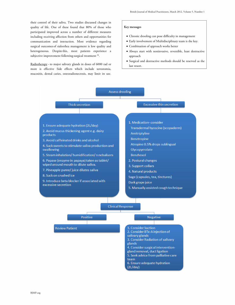
British Journal of Medical Practitioners, March 2012, Volume 5, Number 1
BJMP.org
their control of their saliva. Two studies discussed changes in
quality of life. One of these found that 80% of those who
participated improved across a number of different measures
including receiving affection from others and opportunities for
communication and interaction. Most evidence regarding
surgical outcomes of sialorrhea management is low quality and
heterogeneous. Despite this, most patients experience a
subjective improvement following surgical treatment 36.
Radiotherapy - to major salivary glands in doses of 6000 rad or
more is effective Side effects which include xerostomia,
mucositis, dental caries, osteoradionecrosis, may limit its use.
Key messages
• Chronic drooling can pose difficulty in management
• Early involvement of Multidisciplinary team is the key.
• Combination of approach works better
• Always start with noninvasive, reversible, least destructive
approach
• Surgical and destructive methods should be reserved as the
last resort.
British Journal of Medical Practitioners, March 2012, Volume 5, Number 1
BJMP.org
Competing Interests
None declared
Author Details
Ganesh Bavikatte, MBBS, MD (Medicine), MRCP(UK), MRCP(London),
Speciality Registrar Rehabilitation Medicine, Manchester, UK. Poh Lin Sit,
MBBch, Staff Grade in Rehabilitation Medicine, Central Manchester University
Hospitals, Manchester, UK. Ali Hassoon, MB ChB, FRCP (London), Consultant
Physician Rehabilitation Medicine, Central Manchester NHS Trust, Manchester,
UK.
CORRESSPONDENCE: Ganesh Bavikatte, MBBS, MD (Medicine),
MRCP(UK), MRCP(London, Speciality Registrar Rehabilitation Medicine, 9
averhill, worsley, Manchester, M281ZN.
Email: [email protected]
REFERENCES
1. Stuchell RN, Mandel ID. Salivary gland dysfunction and swallowing
disorders. Otolayngol Clin North Am 1988;21:649-61.
2. Costanzo, L. Text book of Physiology, 3rd edition. Saunders Elsevier.
ISBN 10:1-4160-2320-8.
3. Sochaniwskyj AE. Drool Quantification: noninvasive technique. Arch
Phys Med Rehabil 1982;63:605-7
4. Neil G. Hockstein, Daniel S. Samadi, Kristin Gendron, Steven D.
Handler, Am Fam Physician 2004;69:2628-34
5. Louise Cummings, Text book of Clinical Linguistics, edin univ press,
page 95-99, 2008
6. Hilary Johnson, Amanda Scott, Text book on A practical approach to
saliva control page31, 86.
7. Giles R, Naummann M, Werner E, Riemann M, Beck I, Puls I, Reinners
C, Toyka KV. Injection of botulinum toxin A in to salivary glands improve
sialorrhoea in amyotropic lateral sclerosis. J Neurol Neurosurg Psychiatry
200;69:121-3
8. Jongerius PH, Rotteveel JJ, Van Limbeck J, Gabreels FJM, Van Ilulst
KBS, Van Den Ilogen FJA. Botulinum toxin effect on salivary flow rate in
children with cerebral palsy. Neurology 2004;63:1371-5
9. Boothwell JE, Clarke K, Dooley JM, Gordon KE, Anderson R, Wood
Camfied CS, Camfield PR. Botulinum toxin A as a treatment for excessive
drooling in children. Paediatr Neurology 2002; 27(1):18-22
10. Johnson H, Scott A. 1993; Book on practical approach to saliva control
and communication; 1993
11. Scully C, Limeres J, Gleeson M, Tomás I, Diz P. Drooling, J Oral
Pathol Med. 2009 Apr;38(4):321-7. Epub 2009 Feb 23.
12. Suskind DL, Tilton A. Clinical study of botulinum toxin- A in the
treatment of sialorrhoea in children with cerebral palsy. Laryngoscope 2002.
112:73-81.
13. Crysdale WS, McCann C, Roske L. Saliva control issues in the
neurologically challenged: a 30 year experience in team management. Int J
Paedatr Otolarynol: 2006; 70: 519-527
14. Sullivan PB, Lambert B, Rose M. Prevalence and severity of feeding and
nutritional problems in children with neurological impairment: Oxford
Feeding study. Dev Med Child Neuol 2000;42:674-680
15. Meningaud JP, Pitak-Arnnop P, Chikhani L, Bertrand JC. Drooling of
saliva: A Review of the etiology and management options. Oral Surg Oral
Med Oral Pathol Oral Radiol Endod. 2006;101(1):48-57.
16. Hockstein NG, Samadi DS, Gendron K, Handler SD, Sialorrhoea: a
management Challenge, Am Fam Physician 2004; 69:2628-2634.
17. Jongerius PH, Van Tiel P, Van Limbeek J, A systematic review for
evidence of efficacy of anticholinergic drugs to treat drooling. Arch Dis
Child 2003; 88: 911-914
18. Scott, A., & Staios, G. (1993). Oral-facial facilitation. In J. Hilary & A.
Scott (Eds.), A practical approach to saliva control (pp. 32-42). San Antonio,
TX: Communication Skill Builders.
19. Potulska A, Friedman A. Controlling sialorrhoea: a review of available
treatment options. Expert Opin Pharmacother. 2005 Aug;6(9):1551-4.
20. Mato Montero A, Limeres Posse J, Tomás Carmona I, Fernández Feijoo
J, Diz Dios P.Med Oral Patol Oral Cir Bucal. 2008 Jan 1;13(1):E27-30.
21. Mier RJ, Bachrach SJ, Lakin RC, Barker T, Childs J, Moran M.
Treatment of sialorrhea with glycopyrrolate: A double-blind, dose-ranging
study. Arch Pediatr Adolesc Med. 2000 Dec;154(12):1214-8.
22. Blasco P.A. Glycopyrrolate treatment of chronic drooling. Archives of
paediatric adolescent medicine, vol 150, sept 1996:932-935.
23. Heine R.G. Effect of antireflux medication on salivary drooling in
children with cerebral palsy. Developmental medicine and child neurology,
1996, vol 38, 1030-36.
24. Blasco P.A. (2002) Management of drooling: 10 years after the
consortium on drooling, 1990. Dev. Med. Child Neurol. 44, 778–781
25. Camp-Bruno J.A., Winsberg B.G., Green-Parsons A.R. (1989) Efficacy
of benztropine therapy for drooling. Dev. Med. Child Neurol. 31, 309–319
26. Lloyd Faulconbridge R.V., Tranter R.M., Moffat V. Review of
management of drooling problems in neurologically impaired children: A
review of methods and results over 6 years at Chailey Heritage clinical
services. Clin. Otolaryngol. Allied Sci. (2001) 26, 76–81
27. Porta M., Gamba M., Bertacchi G. et al. (2001) Treatment of
sialorrhoea with ultrasound guided botulinum toxin type A injection in
patients with neurological disorders. J. Neurol. Neurosurg. Psychiatry 70,
538–540
28. Jongerius P.H., van den Hoogen F.J., van Limbeek J. et al. (2004) Effect
of botulinum toxin in the treatment of drooling: A controlled clinical trial.
Pediatrics 114, 620–627
29. Diamant H, Kumlien A. A treatment for drooling in children with
cerebral palsy. J Laryngol Otol. 1974;88(1):61-64.
30. Peter Misra. Botulinum toxin as a treatment for drooling of saliva.
ACNR; nov/dec 2002: v2 n2 11-12.
31. Dayse Manrique, application of botulinum toxin to reduce the saliva in
patients with amyotropic lateral sclerosis; Rev Bras Otorrinolaringol; sept-oct
2005, v.71, n.5, 566-69
32. Borg M, Hirst, the role of radiation therapy in the management of
sialorrhea international journal of radiation oncology, biology and physics;
1998 jul: 1113-9
33. Crysdale W.S. Management of drooling in individuals with
neurodisability: a surgical experience. Developmental medicine and child
neurology. 2001(43) 379- 383.
34. O’Dwyer T.P. the surgical management of drooling- a 15 year follow up.
Clinical Otolaryngology. 1997(22) 284-287.
35. Chang C. Intraductal laser photocoagulation of the bilateral parotid
ducts for reduction of drooling in patients with cerebral palsy. Plastic and
reconstructive Surgery. 2001(107) 907- 913.
36. Jeremy Reed, MD, MPH; Carolyn K. Mans, MD; Scott E. Brietzke,
MD, MPH Surgical Management of Drooling A Meta-analysis. Arch
Otolaryngol Head Neck Surg. 2009;135(9):924-931.
British Journal of Medical Practitioners, March 2012, Volume 5, Number 1
BJMP.org
BJMP 2012;5(1):a501
Benzodiazepines Revisited
Tauseef Mehdi
Abstract Up to 1 million people in the UK are currently long-term prescribed benzodiazepine users.1 Surveys of general practices show that there are over 180 long-
term prescribed users per general practice.2 Despite repeated recommendations to limit benzodiazepines to short-term use (2–4 weeks), doctors in the UK
and worldwide are still prescribing them for months or years. Dependence upon prescribed benzodiazepines is now recognised as a major clinical problem
and the National Performance Assessment Framework for the NHS makes it a national priority to reduce this within each health board area. Junior doctors
who have recently graduated from medical school are commonly placed in rotations where they have to manage patients on benzodiazepine prescriptions.
It is necessary for doctors in general to be aware of the essentials of benzodiazepines not only for the adequate management of patients on chronic
benzodiazepine prescriptions, but also for responsible prescription of this drug when it is appropriate.
History of benzodiazepines
The advent of benzodiazepines in the late fifties was met with
great excitement by the practicing physicians around the world.
Their range of actions – sedative/hypnotic, anxiolytic,
anticonvulsant and muscle relaxant – combined with low
toxicity and alleged lack of dependence potential seemed to
make them ideal medications for many common conditions.
The drugs were prescribed long term, often for many years, for
complaints such as anxiety, depression, insomnia and ordinary
life stressors. They began to replace barbiturates; drugs known
to be dangerous in overdose, which tended to cause addiction
and were associated with troublesome side-effects. Previous
compounds including opium, alcohol, chloral and bromides
were similarly burdened.
The first benzodiazepine, chlordiazepoxide (Librium), was
synthesized in 1955 by Leo Sternbach while working at
Hoffmann–La Roche on the development of tranquilizers. The
compound showed very strong sedative, anticonvulsant and
muscle relaxant effects when submitted for a standard battery of
animal tests. These impressive clinical findings led to its speedy
introduction throughout the world in 1960 under the brand
name Librium. Following chlordiazepoxide, diazepam was
marketed by Hoffmann–La Roche under the brand name
Valium in 1963.
The benefits of benzodiazepines and the apparent lack of
discouraging factors led an alarming rise of benzodiazepine
prescriptions. In the late 1970s benzodiazepines became the
most commonly prescribed of all drugs in the world.1 In1980,
Tyrer reported that each day about 40 billion doses of
benzodiazepine drugs are consumed throughout the world.3
This figure is staggering by any standards. However, towards
the end of the 1970s, awareness begin to grow that
benzodiazepines were being unnecessarily over-prescribed and it
was noticed that certain patients might become dependent on
benzodiazepines after chronic use.4 In particular, patients found
it difficult to stop taking benzodiazepines because of withdrawal
reactions and many complained that they had become
‘addicted’. Several investigations showed quite unequivocally
that benzodiazepines could produce pharmacological
dependence in therapeutic dosage.5-9
In 1988, the Committee of Safety of Medicines reacted to the
concerns by spelling out emphatic guidelines about the use of
benzodiazepines drugs. For anxiety and insomnia,
benzodiazepines are indicated for short term relief (two to four
weeks) only if the condition is severe, disabling and subjecting
the individual to extreme distress.10
Tolerance and dependence
Tolerance is a phenomenon that develops with many
chronically used drugs. The body responds to the continued
presence of the drug with a series of adjustments that tend to
overcome the drug effects. In the case of benzodiazepines,
compensatory changes occur in the GABA and benzodiazepine
receptors which become less responsive, so that the inhibitory
actions of the GABA and benzodiazepines are decreased. As a
result, the original dose of the drug has progressively less effect
and a higher dose is required to obtain the original effect.
Dependence is understood to be the inability to control intake
of a substance to which one is addicted. It encompasses a range
of features initially described in connection with alcohol abuse,
now recognised as a syndrome (see box 1) associated with a
range of substances.
Dependence has two components: psychological dependence,
which is the subjective feeling of loss of control, cravings and
preoccupation with obtaining the substance; and physiological
dependence, which is the physical consequences of withdrawal
and is specific to each drug. For some drugs (e.g. alcohol) both
Review A
rticle
British Journal of Medical Practitioners, March 2012, Volume 5, Number 1
BJMP.org
psychological and physiological dependence occur; for others
(e.g. LSD) there are no marked features of physiological
dependence.
Box 1: Dependence Syndrome*
Three or more of the following manifestations should have
occurred together for at least one month or if persisting for
periods of less than one month then they have occurred
together repeatedly within a twelve month period.
1. A strong desire or sense of compulsion to take the
substance.
2. Impaired capacity to control substance-taking behaviour in
terms of onset, termination or level of use, as evidenced by:
the substance being often taken in larger amounts or over a
longer period than intended, or any unsuccessful effort or
persistent desire to cut down or control substance use.
3. A physiological withdrawal state (see F1x.3 and F1x.4)
when substance use is reduced or ceased, as evidenced by
the characteristic withdrawal syndrome for the substance, or
use of the same (or closely related) substance with the
intention of relieving or avoiding withdrawal symptoms.
4. Evidence of tolerance to the effects of the substance, such
that there is a need for markedly increased amounts of the
substance to achieve intoxication or desired effect, or that
there is a markedly diminished effect with continued use of
the same amount of the substance.
5. Preoccupation with substance use, as manifested by:
important alternative pleasures or interests being given up
or reduced because of substance use; or a great deal of time
being spent in activities necessary to obtain the substance,
take the substance, or recover from its effects.
6. Persisting with substance use despite clear evidence of
harmful consequences, as evidenced by continued use when
the person was actually aware of, or could be expected to
have been aware of the nature and extent of harm.
* ICD 10 Classification of Mental and Behaviour disorder,
online version 2007.
Withdrawal syndrome and discontinuation syndrome
Any drug consumed regularly and heavily can be associated
with withdrawal phenomenon on stopping. Clinically
significant withdrawal phenomena occur in dependence to
alcohol, benzodiazepines, opiates and are occasionally seen in
cannabis, cocaine and amphetamine use. In general, drugs with
a short half-life will give rise to more rapid but more transient
withdrawal.
Discontinuation syndrome is a common phenomenon and
occurs with all classes of antidepressants. It is only experienced
when one tries to discontinue its use. The most common
symptoms are dizziness, vertigo, gait instability, nausea, fatigue,
headaches, anxiety and insomnia. Less commonly shock-like
sensations, paraesthesia, visual disturbances, diarrhoea and flu-
like symptoms have been reported. Symptoms usually begin 2-5
days after SSRI discontinuation or dose reduction. The
duration is variable (one to several weeks) and ranges from mild
to moderate intensity in most patients, to extremely distressing
in a small number. Tapering antidepressants at the end of
treatment, rather than abrupt stoppage, is recommended as
standard practice by several authorities and treatment
guidelines11-13.
The terms ‘antidepressant withdrawal syndrome’ and
‘antidepressant discontinuation syndrome’ are used
interchangeably in the literature. ‘Discontinuation’ is preferred
as it does not imply that antidepressants are addictive or cause a
dependence syndrome. The occurrence of withdrawal
symptoms does not in itself indicate that a drug causes
dependence as defined in ICD 10 (World Health Organisation
1992)14 and DSM –IV (American Psychiatric Association,
1994)15.
Understanding how benzodiazepines work and their effects
For the first 15 years after the introduction of benzodiazepines,
no clear picture emerged as to how these drugs might exert their
psychotropic effects. The great breakthrough in our
understanding in the mechanism of action of benzodiazepines
came in the mid 1970s when biologists at Hoffman-La Roche
demonstrated that benzodiazepines exert their psychotropic
effects by potentiating GABA neurotransmission.16
GABA, Gamma-Amino butyric acid, is the most important
inhibitory neurotransmitter in the mammalian brain
representing about 30% of all synapses in the whole brain.
GABAergic neurones mediate pre-synaptic inhibition by
depressing the release of neurotransmitter at excitatory input
synapse, and post-synaptic inhibition by depressing synaptic
excitation of the principal neuron. When benzodiazepines react
at their receptor site, which is actually situated on the GABA
receptor, the combination acts as a booster to the actions of
GABA making the neuron more resistant to excitation. Several
studies showed that benzodiazepines were able to facilitate both
types of inhibition, indicating that the effects of the
benzodiazepines were in fact due to an interaction with the
GABAergic transmission process17-19.
Various subtypes of benzodiazepine receptors have slightly
different actions. Alpha 1 is responsible for sedative effects.
Alpha 2 exerts anxiolytics effects. Alpha 1, Alpha 2 and Alpha 5
are responsible for anticonvulsant effects. As a consequence of
the enhancement of GABA’s inhibitory activity caused by
benzodiazepines, the brain’s output of excitatory
neurotransmitters including norepinephrine, serotonin,
dopamine and acetylcholine is reduced.

British Journal of Medical Practitioners, March 2012, Volume 5, Number 1
BJMP.org
The studies on the receptor binding of benzodiazepines and the
subsequent changes that occur in the central nervous system
have provided us with an adequate explanation for some or all
of the actions of benzodiazepines, which are listed in Box 2.
Box 2: Four principle biological properties of benzodiazepines
1. Anxiolytic and behavioural inhibition – The anxiolytic
effect is seen in animals as an increase of those behavioural
responses that are suppressed experimentally by punishment
or which are absent because of innate aversion20-23.
2. Anticonvulsant – Benzodiazepines are most potent against
chemically induced epileptiform activities. At higher doses
most, but not all, benzodiazepines also prevent seizures
induced by electric shock24.
3. Sedative/hypnotic – These effects of benzodiazepines are
most easily observed as a decrease of spontaneous locomotor
activity in rodents placed in an observation chamber.
Benzodiazepines will shorten sleep latency (amount of time
taken to fall asleep after the lights have been switched off)
which can be demonstrated by electroencephalogram25.
4. Muscle relaxant - Common tests on rodents show that
benzodiazepines impair performance at motor performance
tasks for example the rodent’s ability to balance on a
rotating drum. The cat shows marked ataxia at after
relatively low doses25.
What are benzodiazepines used for?
Sleep disorders
The benzodiazepines are used widely in the treatment of sleep
disorders and many have been developed and licensed for this
purpose. They are mainly known as hypnotic drugs (sleeping
pills) because insomnia is the main target use. Certain factors
are important in determining the choice of the hypnotic drug.
Ideally, the hypnotic should be effective at inducing sleep in the
individual, and should enhance objective and subjective
elements of sleep. It should have a fast onset with minimal side
effects and the absence of withdrawal symptoms.
The early benzodiazepine hypnotics were drugs such as
nitrazepam and flurazepam. After their introduction, it was
found that they had half-lives of more than a day, and
individuals suffered undesirable effects such as sedation, ataxia
or amnesia during the day. This was problematic especially for
those individuals who needed to drive or operate machinery.
Another consequence was of falls with subsequent hip fractures
in the elderly population because, due to slower metabolism,
they accumulated raised plasma levels of the drug. For these
reasons, benzodiazepines with shorter half lives were developed
so that plasma levels fall below the functional threshold
concentration by the next morning.
The first of the shorter half-life benzodiazepine hypnotics to be
introduced were temazepam and triazolam. Temazepam has a
half-life of 5 hours and is commonly used in primary, secondary
and tertiary settings for insomnia. A possible drawback of very
short half-life hypnotics is rebound insomnia. This is a state of
worsening sleep which commonly follows discontinuation of a
regularly used hypnotic.
An important point to note is that although the subjective
efficacies of benzodiazepines are widely reported, the use of
polysomnography (a sleep study that involves recording a
variety of physiological measures including
electroencephalograph, electro-oculogram and electromyogram)
has shown that sleep architecture in individuals with insomnia
is not normalised by benzodiazepines. The increase in sleep
duration can be accounted for by an increase in the time spent
in stage 2 of sleep, while the amount of time spent in slow-wave
sleep (deep) and REM (rapid eye movement) is actually
decreased26.
Anxiety disorders
It can be argued that the benzodiazepines are probably the most
efficacious and best tolerated pharmacological treatments of
anxiety. Numerous studies, many of them conducted under
stringent double-blind conditions, have consistently shown that
benzodiazepines produce significantly more improvement than
placebo in both somatic and emotional manifestations of
anxiety27-29.
Before the introduction of benzodiazepines, anxiety disorders
were treated either with the barbiturates or related drugs such as
meprobomate and glutethimide. These agents were highly likely
to be abused and led to a great deal of dependence. Moreover,
they were toxic in overdose and fatalities were high in
populations using them. The improved efficacy and safety
profile of benzodiazepines, aided by intense campaigns to
restrict use of barbiturate-type drugs, meant they rapidly
became the first choice drugs for anxiety within a few years of
them being introduced.
Much clinical practice and opinion suggests that
benzodiazepine can be used as first-line treatment for acute
anxiety episodes as long as CSM guidelines are adhered to. For
more intractable conditions such as established social phobia,
generalised anxiety disorder and panic disorder, they should
probably be reserved for adjunctive or second-line agents.
In contrast to the treatment of sleep disorders, it is important to
achieve a constant level of receptor occupation to maintain
anxiolysis throughout the day. So for anxiety, compounds with
longer elimination half-lives are preferred, whereas for sleep
induction, short half-life drugs are favoured. The principal
benzodiazepines used as anxiolytics include diazepam,
chlordiazepoxide, clonazepam, lorazepam, alprazolam and
oxazepam.
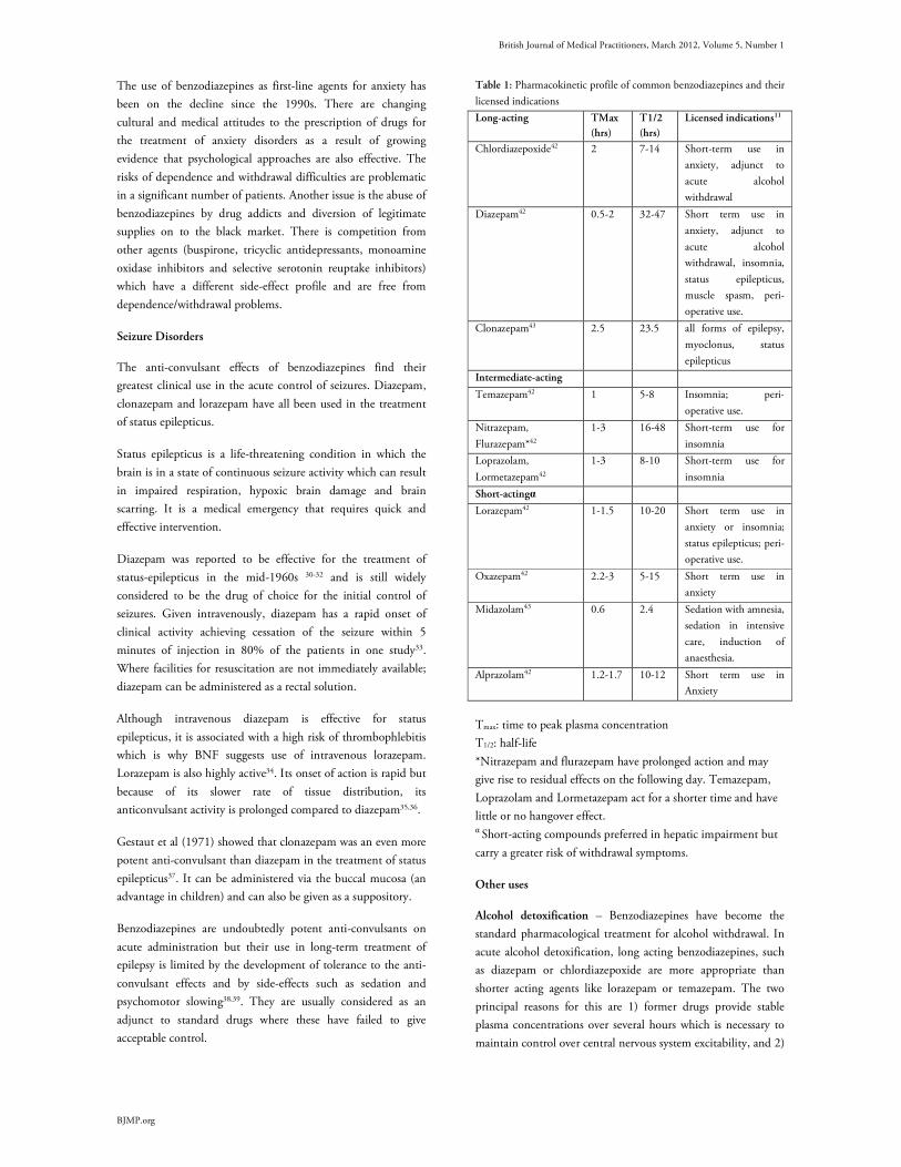
British Journal of Medical Practitioners, March 2012, Volume 5, Number 1
BJMP.org
The use of benzodiazepines as first-line agents for anxiety has
been on the decline since the 1990s. There are changing
cultural and medical attitudes to the prescription of drugs for
the treatment of anxiety disorders as a result of growing
evidence that psychological approaches are also effective. The
risks of dependence and withdrawal difficulties are problematic
in a significant number of patients. Another issue is the abuse of
benzodiazepines by drug addicts and diversion of legitimate
supplies on to the black market. There is competition from
other agents (buspirone, tricyclic antidepressants, monoamine
oxidase inhibitors and selective serotonin reuptake inhibitors)
which have a different side-effect profile and are free from
dependence/withdrawal problems.
Seizure Disorders
The anti-convulsant effects of benzodiazepines find their
greatest clinical use in the acute control of seizures. Diazepam,
clonazepam and lorazepam have all been used in the treatment
of status epilepticus.
Status epilepticus is a life-threatening condition in which the
brain is in a state of continuous seizure activity which can result
in impaired respiration, hypoxic brain damage and brain
scarring. It is a medical emergency that requires quick and
effective intervention.
Diazepam was reported to be effective for the treatment of
status-epilepticus in the mid-1960s 30-32 and is still widely
considered to be the drug of choice for the initial control of
seizures. Given intravenously, diazepam has a rapid onset of
clinical activity achieving cessation of the seizure within 5
minutes of injection in 80% of the patients in one study33.
Where facilities for resuscitation are not immediately available;
diazepam can be administered as a rectal solution.
Although intravenous diazepam is effective for status
epilepticus, it is associated with a high risk of thrombophlebitis
which is why BNF suggests use of intravenous lorazepam.
Lorazepam is also highly active34. Its onset of action is rapid but
because of its slower rate of tissue distribution, its
anticonvulsant activity is prolonged compared to diazepam35,36.
Gestaut et al (1971) showed that clonazepam was an even more
potent anti-convulsant than diazepam in the treatment of status
epilepticus37. It can be administered via the buccal mucosa (an
advantage in children) and can also be given as a suppository.
Benzodiazepines are undoubtedly potent anti-convulsants on
acute administration but their use in long-term treatment of
epilepsy is limited by the development of tolerance to the anti-
convulsant effects and by side-effects such as sedation and
psychomotor slowing38,39. They are usually considered as an
adjunct to standard drugs where these have failed to give
acceptable control.
Table 1: Pharmacokinetic profile of common benzodiazepines and their
licensed indications
Long-acting TMax
(hrs)
T1/2
(hrs)
Licensed indications11
Chlordiazepoxide42 2 7-14 Short-term use in
anxiety, adjunct to
acute alcohol
withdrawal
Diazepam42 0.5-2 32-47 Short term use in
anxiety, adjunct to
acute alcohol
withdrawal, insomnia,
status epilepticus,
muscle spasm, peri-
operative use.
Clonazepam43 2.5 23.5 all forms of epilepsy,
myoclonus, status
epilepticus
Intermediate-acting
Temazepam42 1 5-8 Insomnia; peri-
operative use.
Nitrazepam,
Flurazepam*42
1-3 16-48 Short-term use for
insomnia
Loprazolam,
Lormetazepam42
1-3 8-10 Short-term use for
insomnia
Short-actingα
Lorazepam42 1-1.5 10-20 Short term use in
anxiety or insomnia;
status epilepticus; peri-
operative use.
Oxazepam42 2.2-3 5-15 Short term use in
anxiety
Midazolam43
0.6
2.4
Sedation with amnesia,
sedation in intensive
care, induction of
anaesthesia.
Alprazolam42 1.2-1.7 10-12 Short term use in
Anxiety
Tmax: time to peak plasma concentration
T1/2: half-life
*Nitrazepam and flurazepam have prolonged action and may
give rise to residual effects on the following day. Temazepam,
Loprazolam and Lormetazepam act for a shorter time and have
little or no hangover effect. α Short-acting compounds preferred in hepatic impairment but
carry a greater risk of withdrawal symptoms.
Other uses
Alcohol detoxification – Benzodiazepines have become the
standard pharmacological treatment for alcohol withdrawal. In
acute alcohol detoxification, long acting benzodiazepines, such
as diazepam or chlordiazepoxide are more appropriate than
shorter acting agents like lorazepam or temazepam. The two
principal reasons for this are 1) former drugs provide stable
plasma concentrations over several hours which is necessary to
maintain control over central nervous system excitability, and 2)

British Journal of Medical Practitioners, March 2012, Volume 5, Number 1
BJMP.org
There is a higher risk of addiction with short-acting drugs in
this patient population.
In alcohol dependent patients with hepatic impairment,
oxazepam or lorazepam is more suitable as they are not
eliminated by hepatic oxidation through the Cytochrome P450
system. Cytochrome p450 (CYPs) is a collective generic term
use to describe a superfamily of membrane bound heme-
thiolate proteins of critical importance in the oxidative and
reductive metabolism of both endogenous and foreign
compounds. CYPs are the major enzymes in drug metabolism
accounting for 75% of the total metabolism40. Many of the
CYPs in humans are found in the liver and the gastrointestinal
tract. After the acute detoxification is over, many patients enter
rehabilitation programmes aimed at maintaining abstinence in
the community. There is no evidence that use of
benzodiazepines is useful in reducing alcohol craving or
facilitating abstinence.
Anaesthesia – The psychotropic effects of benzodiazepines
make them appropriate for use as anaesthetic agents or as
adjuncts to anaesthesia. Muscle relaxation, sedation and
retrograde amnesia are sought after properties in anaesthetic
agents. Midazolam is used as a sedative agent in patients
undergoing minor invasive practices considered as traumatic,
such as dental treatment or endoscopy.41
Muscle relaxants – The muscle relaxant properties of
benzodiazepines are an indication for their use in some
neurological disturbances for symptomatic relief of muscle
spasms and spasticity.
Assessment and management of patients with chronic
benzodiazepine dependence
Because of the adverse effects, lack of efficacy and
socioeconomic costs of continued benzodiazepine use, long-
term users have for many years been advised to withdraw if
possible or at least to reduce dosage.10,44 Echoing the CSM
advice, the Mental Health National Service Framework (NSF),
which was published in 1999, recommended that
benzodiazepines should be used for no more than two to four
weeks for severe and disabling anxiety. The Mental Health NSF
called upon health authorities to implement systems for
monitoring and reviewing prescribing of benzodiazepines
within local clinical audit programmes. Primary Care Trusts
(PCTs) should ensure that this recommendation is still being
implemented45.
In primary care, early detection and intervention are the main
principles of assessment. The initial assessment should
• Establish the pattern of benzodiazepine usage: onset,
duration, which benzodiazepine/s, dosage history, current
regime and any periods of abstinence.
• Check for evidence of benzodiazepine dependence (see box
3).
• If benzodiazepine dependence is present, determine the
type of benzodiazepine.
• Detail any history of previous severe withdrawal (including
history of seizures).
• Establish the level of motivation to change.
Dependence on benzodiazepines often indicates psychosocial
problems in a person. Benzodiazepines are increasingly used in
conjunction with other substance of abuse to enhance the
effects obtained from opiates, and to alleviate withdrawal
symptoms of other drugs of abuse such as cocaine,
amphetamines or alcohol. The patient needs to have an
individualised and a comprehensive assessment of their physical
and mental health needs and any co-morbid use of other drugs
and alcohol. Stable psychological health and personal
circumstances are desirable features for successful withdrawal
from benzodiazepines. Certain patients will be unsuitable for
withdrawal, e.g. those patients experiencing a current crisis or
having an illness for which the drug is required at the current
time. Referral to specialist teams may be appropriate for some,
e.g. if the patient is also dependent on other drugs or alcohol, if
there is co-existing physical or psychiatric morbidity or if there
is a history of drug withdrawal seizures. In some circumstances,
it may be more appropriate to wait until other problems are
resolved or improved.
Box 3 – Benzodiazepine Withdrawal Symptoms46
Psychological symptoms – excitability, sleep disturbances,
increased anxiety, panic attacks, agoraphobia, social phobia,
perceptual distortions, depersonalisation, derealisation,
hallucinations, misperceptions, depression, obsessions, paranoid
thoughts, rage, aggression, irritability, poor memory and
concentration, intrusive memories and craving (rare).
Physical symptoms – Headache, pain, stiffness, tingling,
numbness, altered sensation, weakness, fatigue, influenza-like
symptoms, muscles twitches, jerks, tics, “electric shocks”,
tremor, dizziness, light-headedness, poor balance, visual
problems, tinnitus, hypersensitivity to stimuli, gastrointestinal
symptoms, appetite change, dry mouth, metallic taste, unusual
smell, flushing, sweating, palpitations, over breathing, urinary
difficulties, skin rashes, itching, fits (rare).
This list is probably not inclusive. Not all patients get all the
symptoms. Different individuals get a different combination of
symptoms.
Management of benzodiazepine withdrawal
Withdrawal of the benzodiazepine drug can be managed in
primary care if the patients in consideration are willing,
committed and compliant. Clinicians should seek opportunities
to explore the possibilities of benzodiazepine withdrawal with

British Journal of Medical Practitioners, March 2012, Volume 5, Number 1
BJMP.org
patients on long-term prescriptions. Interested patients could
benefit from a separate appointment to discuss the risks and
benefits of short and long term benzodiazepine treatment47.
Information about benzodiazepines and withdrawal schedules
could be offered in printed form. One simple intervention that
has been shown to be effective in reducing benzodiazepine use
in long-term users is the sending of a GP letter to targeted
patients. The letter discussed the problems associated with long-
term benzodiazepine use and invited patients to try and reduce
their use and eventually stop48. Adequate social support, being
able to attend regular reviews and no previous history of
complicated drug withdrawal is desirable for successful
benzodiazepine withdrawal.
Switching to diazepam: This is recommended for some people
commencing a withdrawal schedule. Diazepam is preferred
because it possesses a long half-life, thus avoiding sharp
fluctuations in plasma level. It is also available in variable
strengths and formulations. This facilitates stepwise dose
substitution from other benzodiazepines and allows for small
incremental reductions in dosage. The National Health Service
Clinical Knowledge Summaries recommend switching to
diazepam for people using short acting benzodiazepines such as
alprazolam and lorazepam, for preparations that do not allow
for small reductions in dose (that is alprazolam, flurazepam,
loprazolam and lormetazepam) and for some complex patients
who may experience difficulty withdrawing directly from
temazepam and nitrazepam due to a high degree of
dependency49. See table 2 for approximate dose conversions of
benzodiazepines when switching to diazepam.
Gradual Dosage Reduction: It is generally recommended that
the dosage should be tapered gradually in long-term
benzodiazepine users such as a 5-10% reduction every 1-2
weeks1,49. Abrupt withdrawal, especially from high doses, can
precipitate convulsions, acute psychotic or confusional states
and panic reactions. As mentioned earlier, benzodiazepines’
enhancement of GABA’s inhibitory activity reduces the brain’s
output of excitatory neurotransmitter such as norepinephrine,
serotonin, dopamine and acetylcholine. The abrupt withdrawal
of benzodiazepines may be accompanied by uncontrolled release
of dopamine, serotonin and other neurotransmitters which are
linked to hallucinatory experiences similar to those in psychotic
disorders46.
The rate of withdrawal should be tailored to the patient's
individual needs and should take into account such factors as
lifestyle, personality, environmental stressors, reasons for taking
benzodiazepines and the amount of support available. Various
authors suggest optimal times of between 6-8 weeks to a few
months for the duration of withdrawal, but some patients may
take a year or more11,50. A personalised approach, empowering
the patient by letting them guide their own reduction rate is
likely to result in better outcomes.
Table 2: Approximate equivalent doses of benzodiazepines1
Benzodiazepine Approximate equivalent dosage (mg)a
Alprazolam 0.5
Chlordiazepoxide 25
Clonazepam 0.5
Diazepam 10
Flunitrazepam 1
Flurazepam 15-30
Loprazolam 1
Lorazepam 1
Lormetazepam 1
Nitrazepam 10
Oxazepam 20
Temazepam 20
a Clinical potency for hypnotic or anxiolytic effects may vary
between individuals; equivalent doses are approximate.
Patients may develop numerous symptoms of anxiety despite
careful dose reductions. Simple reassurance and encouragement
should suffice in most cases however, in a minority who are
experiencing significant distress, formal psychological support
should be available. Cognitive therapy, behavioural approaches
including relaxation techniques and breathing exercises for
anxiety management as well as other therapies such as massage
and yoga may alleviate difficulties during withdrawal.
Psychoeducation around withdrawal symptoms should be
offered and a referral to a support organisation or group is
helpful.
Resources
• The Ashton Manual, http://www.benzo.org.uk/manual/index.htm.
• NHS Clinical Knowledge Summaries,
http://www.cks.nhs.uk/benzodiazepine_and_z_drug_withdrawal.
• The Maudsley Prescribing Guidelines
Summary
Although prescriptions of benzodiazepines have declined
substantially since 1988, there is an ongoing challenge within
all sectors of the NHS to prevent benzodiazepine dependence.
This can be achieved by adhering to official recommendations
to limit prescriptions to 2-4 weeks, or for brief courses or
occasional usage. All health authorities should have clinical
audit programmes reviewing and monitoring prescribing rates
for benzodiazepines. Through this, increased awareness of CSM
guidelines amongst all health care professionals should aid in
more appropriate prescriptions and subsequent monitoring that
is required to prevent unnecessary prescriptions. Patients on
long-term prescriptions should be offered the opportunity for
controlled withdrawal and the relevant psychological and social
support.
British Journal of Medical Practitioners, March 2012, Volume 5, Number 1
BJMP.org
Competing Interests
None declared Author Details TAUSEEF MEHDI, MBBS, MRCPsych. Specialty Registrar in Psychiatry, Wells
Unit, Regional Secure Unit. West London Mental Health Trust, Uxbridge Road,
Southall Middlesex. UB1 3EU.
CORRESSPONDENCE: TAUSEEF MEHDI, MBBS, MRCPsych. Specialty
Registrar in Psychiatry, Wells Unit, Regional Secure Unit. West London Mental
Health Trust, Uxbridge Road, Southall Middlesex. UB1 3EU.
Email: [email protected]
REFERENCES
1. Ashton H. The diagnosis and management of benzodiazepine
dependence. Curr Opin Psychiatry 2005; 18:249–255.
2. Ashton, C H: All Party Action Group on Tranquilliser addiction.
London: House of Commons, November 7, 2006.
3. Tyrer P. Dependence on benzodiazepines. Brit J Psychiat. 1980; 137:
576-577
4. Lader M. Benzodiazepines – the opium of the masses. Neuroscience
1978;3:159-165
5. Pevnick JS, Jasinski DR, Haertzen CA. Abrupt withdrawal from
therapeutically administered diazepam. Arch Gen Psychiatry. 1978;
35:995-8.
6. Winokur A, Rickels K, Greenblatt DJ, Snyder PJ, Schatz NJ.
Withdrawal reaction from long term, low dosage, administration of
diazepam. Arch Gen Psychiatry 1980; 37:101-5.
7. Tyrer P, Rutherford D, Huggett T. Benzodiazepine withdrawal
symptoms and propranolol. Lancet 1981; i: 520-2.
8. Petursson H, Lader MH. Withdrawal from long-term benzodiazepine
treatment. Br Med J 1981; 283:643-5.
9. Tyrer P, Owen R, Dawling S. Gradual withdrawal of diazepam after
chronic therapy. Lancet 1983; i: 1402-6.
10. Committee on Safety of Medicines . Benzodiazepines, dependence and
withdrawal symptoms. 1988; Current Problems 21.
11. British National Formulary. 2009: British Medical Association/Royal
Pharmaceutical Society of Great Britain. BMJ Publishing Group &
RPS Publishing.
12. Drug and Therapeutics Bulletin. Withdrawing patients from
antidepressants. Drug and Therapeutics Bulletin, 1999; 37 (July), 49–52
13. Anderson, I. M., Nutt, D. J. & Deakin J. F. Evidence-based guidelines
for treating depressive disorders with antidepressants: a revision of the
1993 British Association for Psychopharmacology guidelines. Journal of
Psychopharmacology, 2000; 14, 3–20.
14. World Health Organization (1992) The ICD–10 Classification of
Mental and Behavioural Disorders. World Health Organization.
15. American Psychiatric Association (1994) Diagnostic and Statistical
Manual of Mental Disorders(4th edn) (DSM–IV). APA.
16. Polc P, Mohler H, Haefely W. The effects of diazepam on spinal cord
activities: possible sites and mechanisms of action. NS Arch Pharmacol
1988;34: 124-128.
17. Polc P, Haefely W. Effects of two benzodiazepines, phenobarbitone and
baclofen on synaptic transmission in the cat cuneate nucleus. Naunyn
Schmiedebergs Arch Pharmacol. 1976;294(2):121–131.
18. Wolf P, Haas HL. Effects of diazepines and barbiturates on
hippocampal recurrent inhibition. NS Arch Pharmacol 1977; 299: 211-
218.
19. Raab W, Gummit RJ. Anticonvulsant action of diazepam: Increase of
cortical post-synaptic inhibition. Epilepsia 1977; 18:117-120
20. Cook, L & Davidson, A. B. In The Benzodiazepines (eds S. Garattini, E.
Mussini, L 0.Randall). New York: Raven Press, 1973; pp 327-45.
21. Dantzer, R. Behavioural effects of benzodiazepines: a review.
Biobehavioural Reviews, 1977;1,71-86.
22. Haefely,W.E. Behavioural and neuropharmacological aspects of drugs
used in anxiety and related states. In Psychopharmacology: A Generation
of Progress (eds M. A. Upton, A. Di Mascio, K. F. Killam). New York:
Raven Press, 1978; pp 1359-74.
23. Cooper SJ. Benzodiazepines, barbiturates and drinking, In: Cooper SJ
ed. Theory in psychopharmacology. London: Academic Press 1983;
2:115-148.
24. Raabe, W. & Gumnit, R. J. Anticonvulsant action of diazepam:
increase of cortical postsynaptic inhibition. Epilepsia, 1977; 18, 11720.
25. Haefely, W.E. Central Actions of Benzodiazepines: General
Introduction. Brit.J.Psychiat, 1978; 133,231-238
26. Wheatley, D. Effects of drugs on sleep. In Psychopharmacology of sleep.
Wheatley D, ed. New York: Raven Press, 1981; 153-176.
27. Lader, M. The present status of benzodiazepines and psychiatry and
medicine. Drug Res 1980; 30:910-913.
28. Rickels, K. Use of anti-anxiety drugs and anxious outpatients.
Psychopharmacology 1978; 58:1-17.
29. Greenblatt, D.J & Shader RI: Benzodiazepines in Clinical Practice. New
York, Raven Press, 1974.
30. Gestaut H, Naquet R, Poire R, et al: Treatment of status-epilepticus
with diazepam (Valium). Epilepsia 1965; 6:167-182.
31. Lombroso CT: Treatment of status epilepticus with diazepam.
Neurology 1966; 16:629-634.
32. Prensky AL, Roff MC, Moore MJ, et al: Intravenous diazepam in the
treatment of prolonged seizure activity. N Engl J Med 1967; 276:779-
784.
33. Delgado-Escueta AV, Westertain C, Treiman DM, et al: Current
concepts in neurology: management of status epilepticus. N Engl J Med
1982; 306:1337-1340.
34. Leppick IE, Derivian AT, Homan RW, Walker J, Ramsay E and
Patrick B. Double blind study of lorazepam and diazepam in status
epilepticus. J. Am. Med. Assoc. 249, 1452-4.
35. Walker JE, Homan RW, Vasko MR, et al: Lorazepam in status
epilepticus. Ann Neurol 1979; 6:207-213.
36. Griffith PA, Karp HR: Lorazepam in therapy for status epilepticus. Ann
Neurol. 1980; 7:493.
37. Gestaut H, Coujon J, Poire R et al: Treatment of status epilepticus with
a new benzodiazepine more active than diazepam. Epilepsia 1971;
12:197-214.
38. Eadie MJ. Anti-convulsant drugs: an update. Drugs 1984; 27, 328-63.
39. Robertson MM. Current status of the 1,4- and 1,5-benzodiazepines in
the treatment of epilepsy: the place of clobazam. Epilepsia 1986; 27
(Suppl. 1), 27-41.
40. Guengerich FP (January 2008). "Cytochrome p450 and chemical
toxicology". Chem. Res. Toxicol. 2008; 21 (1): 70–83.
41. Whitwam JG, Al-Khudhairi D, McCloy RF. Comparison of
midazolam and diazepam in doses of comparable potency during
gastroscopy. Br J Anaesth 1983; 55:773-777.
42. Adam Noble, Ian Martin and David Nutt: Calming the Brain. 1st
Edition. UK. Martin Dunitz, Taylor and Francis Group, 2004; 135.
43. I.Hindmarch, G.Beaumont, S.Brandon, B.E.Leanord:
Benzodiazepines:Current Concepts. 1st Edition. Sussex, England. John
Wiley and Sons Ltd. 1990; 66-77.
44. CMO’s Update 37. Benzodiazepines warning. Department of Health;
January 2004. p. 4.
45. National Service Framework for Mental Health. London: Department
of Health; 1999. Available from: www.dh.gov.uk.
46. Prof H Aston: Benzodiazepines: How they work and how to withdraw
aka “The Ashton Manual”. 2002; Ch 3 Table1.
http://www.benzo.org.uk/manual/bzcha03.htm
47. Heather N, Bowie A, Ashton H, et al. Randomised controlled trial of
two brief interventions against long-term benzodiazepine use: outcome
of intervention. Addict Res Theory 2004; 12:141–154.
48. Cormack MA, Sweeney KG, Hughes-Jones H, et al. Evaluation of an
easy, cost-effective strategy for cutting benzodiazepine use in general
practice. Br J Gen Pract 1994; 44:5–8.
49. http://www.cks.nhs.uk/benzodiazepine_and_z_drug_withdrawal/mana
gement/scenario_benzodiazepine_and_z_drug_withdrawal/managing_s
omeone_who_wants_to_stop.

British Journal of Medical Practitioners, March 2012, Volume 5, Number 1
BJMP.org
50. Ashton H. Benzodiazepine dependence. In: Haddad P, Dursun S, Deakin B, editors. Adverse syndromes and psychiatric drugs. 2004
British Journal of Medical Practitioners, March 2012, Volume 5, Number 1
BJMP.org
BJMP 2012;5(1):a511
Bradyarrhythmias Associated with the Obstructive Sleep Apnoea Syndrome: A
Precursor to Life-threatening Arrhythmias?
Amitasha Mann , Jean Karen Fleischman and Karen Mrejen-Shakin
ABSTRACT Chronic Obstructive Pulmonary Disease (COPD) is a leading cause of global morbidity that is predicted to become the third most common cause of death
worldwide by the year 20201. Obstructive sleep apnoea syndrome (OSAS) is also highly prevalent and is estimated to affect 5% of adults in western
countries2. The co-existence of both COPD and OSAS in the same patient is termed the Overlap syndrome3,4. Overlap patients have more pronounced
nocturnal hypoxemia and appear to be at increased risk of death compared with COPD patients matched for Global Initiative for COPD stage without
OSA. We present a case of a patient with mild OSA and moderate COPD who was observed on CPAP titration polysomnography to have moderate
obstructive sleep apnoea during rapid eye movement (REM) sleep (REM apnoea-hypopnea index (AHI) 28/hr.), with associated Wenckebach second-
degree atrioventricular (AV) heart block observed during the nadir of oxygen desaturation associated with obstructive apnoeas. This led to further
investigation with a one month event recording which showed progression of a benign Type I Wenckebach second-degree AV heart block to life-
threatening Type II AV second-degree heart block and complete AV block with 3.9 seconds of ventricular asystole. Bradyarrhythmias during sleep
observed in patients with COPD and OSAS may be a precursor to more life-threatening arrhythmias.
Case Report
A 69 year old male with hypertension, body mass index 24
kg/m2, neck circumference 16 inches, and moderate COPD, on
home oxygen, presented to his pulmonary clinic appointment
with worsening complaints of fatigue, leg cramps, and
intermittent shortness of breath with chest discomfort. A
remote, questionable history of syncope five to ten years ago
was elicited. His vital signs were: temperature 98.80F, blood
pressure 119/76 mmHg, pulse 92/min and regular, and
respirations 20/min. Physical exam was significant for crowded
oropharynx with a Mallampati score of four, distant breath
sounds with a prolonged expiratory phase on lung exam with a
normal cardiac exam. Laboratory investigation showed normal
complete blood counts, haemoglobin 15 g/dL, and normal
chemistries. Compared to his previous studies, a pulmonary
function study showed stable parameters with a FEV1 1.47 L
(69%), FVC/FEV1 ratio 0.44 (62%), and a DLCO/alveolar
volume ratio of 2.12 (49%). A room air arterial blood gas
revealed pH 7.41, PCO2 44 mmHg, and PO2 61 mmHg, with
92% oxygen saturation. A six minute treadmill exercise test
performed to assess the need for supplemental oxygen showed
that he required supplemental oxygen at 1L/min via nasal
cannula to eliminate hypoxemia during exercise. His chest
radiograph was significant for hyperinflation and prominence of
interstitial markings. A high resolution computed tomography
of the chest demonstrated severe centrilobular and panacinar
emphysema only. A baseline electrocardiogram (EKG) showed
normal sinus rhythm with an old anterior wall infarct (Figure
1). Echocardiography of the heart revealed a normal left
ventricle with an ejection fraction of 65%. Right ventricular
systolic function was normal although elevated mean
pulmonary arterial pressure of 55 mmHg was noted. A
diagnostic polysomnogram performed for evaluation of daytime
fatigue and snoring at night revealed mild OSA with an AHI of
6/hr. with sleep time spent with oxygen saturation below 90%
(T-90%) of 19%. The EKG showed normal sinus rhythm. A
full overnight polysomnogram for continuous positive airway
pressure (CPAP) titration performed for treatment of sleep
disordered breathing was sub-optimal, however it
demonstrated an apnoea–hypopnea index (AHI) of 28 during
REM (rapid eye movement) sleep, and a T-90% of 93%. The
associated electrocardiogram showed Wenckebach second
degree AV heart block during REM sleep usually near the nadir
of oxygen desaturation. On a repeat positive airway pressure
titration study, therapy with Bilevel pressures (BPAP) of 18/14
cmH20 corrected the AHI and nocturnal hypoxemia to within
normal limits during Non REM (NREM) and REM sleep. His
electrocardiogram remained in normal sinus rhythm .A twenty-
four hour cardiac holter monitor revealed baseline sinus rhythm
and confirmed the presence of second degree AV block of the
Wenckebach type. A one month cardiac event recording
showed normal sinus rhythm with frequent episodes of second
degree AV block. These varied from Type I progressing to
Type II with a 2:1 and 3:1 AV block, during sleep. Progression
to complete heart block was noted with the longest pause
lasting 3.9 seconds during sleep. The patient underwent an
electrophysiology study with placement of a dual chamber
pacemaker. He was initiated on BIPAP therapy. Subsequently,
the patient was seen in clinic with improvements in his
Case Report

British Journal of Medical Practitioners, March 2012, Volume 5, Number 1
BJMP.org
Figure 1- Patient’s baseline EKG, normal sinus rhythm. Figure 2 -Progression to Mobitz Type II block 5:07 am. Figure 3 and 4- Sinus
pauses, longest interval 11:07 pm 3.9 seconds (Figure 4).
intermittent episodes of shortness of breath, fatigue, and
daytime sleepiness.
Discussion
In healthy individuals, especially athletes, bradycardia, Mobitz I
AV block, and sinus pauses up to 2 seconds are common during
sleep and require no intervention5. Cardiac rhythm is controlled
primarily by autonomic tone. NREM sleep is accompanied by
an increase in parasympathetic, and a decrease in sympathetic,
tone. REM sleep is associated with decreased parasympathetic
tone and variable sympathetic tone. Bradyarrhythmias in
patients with OSA are related to the apnoeic episodes and over
80% are found during REM sleep. During these periods of
low oxygen supply, increased vagal activity to the heart resulting
in bradyarrhythmias may actually be cardioprotective by
decreasing myocardial oxygen demand. This may be important
in patients with underlying coronary heart disease.
Some studies have found that Mobitz I AV block may not be
benign. Shaw6 et al studied 147 patients with isolated chronic
Mobitz I AV block. They inserted pacemakers in 90 patients,
74 patients were symptomatic and 16 patients received a
pacemaker for prophylaxis. Outcome data included five-year
survival, deterioration of conduction to higher degree AV block,
and new onset of various forms of symptomatic bradycardia.
They concluded that survival was higher in the paced groups
and that risk factors for poor outcomes in patients with Mobitz
I included age greater than 45 years old, symptomatic
bradycardia, organic heart disease, and the presence of a bundle
branch block on EKG.
The Sleep Heart Health Study7 found a higher prevalence of
first and second-degree heart block among subjects with sleep-
disordered breathing (SDB) than in those without (1.8% vs.
0.3% and 2.2 vs. 0.9%, respectively). Gami et al8observed
thatupon review of 112 Minnesota residents who hadundergone
diagnostic polysomnography and subsequentlydied suddenly
from a cardiac cause, sudden death occurred between the hours
of midnight and 6:00 AM in 46% of those with OSA, as
compared with 21% of those without OSA. In a study of
twenty-three patients with moderate to severe OSA who were
each implanted with an insertable loop recorder, about 50%
were observed to have frequent episodes of bradycardia and
long pauses (complete heart block or sinus arrest) during sleep9.
These events showed significant night-to-night intra individual
variability and their incidence was under-estimated, only 13%,
by conventional short-term EKG Holter recordings.
Physiologic factors predisposing patients with OSA to
arrhythmias include alterations in sympathetic and
parasympathetic nervous system activity, acidosis, apnoea’s, and
arousal2, 10, 11. Some patients with OSA may have an
accentuation of the ‘Diving Reflex’. This protective reflex
consists of hypoxemia-induced sympathetic augmentation to
muscles and vascular beds associated with increased cardiac
vagal activity which results in increased brain perfusion,
bradycardia and decreased cardiac oxygen demand. In patients
with cardiac ischemia, poor lung function (i.e. COPD), or
both, it may be difficult to differentiate between these
protective OSA-associated Bradyarrhythmias and those which
may lead to sudden death. It has been well established that
patients with COPD are at higher risk for cardiovascular
morbidity12 and arrythmias13. Fletcher14 and colleagues
reported that the effects of oxygen supplementation on AHI,
hypercapnea and supraventricular arrhythmias in patients with
COPD and OSA were variable. Out of twenty obese men with
British Journal of Medical Practitioners, March 2012, Volume 5, Number 1
BJMP.org
COPD studied, in most patients oxygen eliminated the
bradycardia observed during obstructive apnoea’s and
eliminated AV block in two patients. In some patients
supplemental oxygen worsened end-apnoea respiratory acidosis
however this did not increase ventricular arrhythmias.
CPAP therapy has been demonstrated to significantly reduce
sleep–related Bradyarrhythmias, sinus pauses, and the increased
risk for cardiac death 9, 15. Despite this, in certain situations
placement of a pacemaker may be required. These include
persistent life-threatening arrhythmias present in patients with
severe OSAS on CPAP, arrhythmias in patients who are non-
compliant with CPAP, and in patients who may have persistent
sympathovagal imbalance and hemodynamic fluctuations
resulting in daytime bradyarrhythmias16.
Our case is interesting since it highlights the importance of
recognizing the association between OSA, COPD, and life-
threatening cardiac arrhythmias. Primary care providers should
note the possible association of OSA-associated
bradyarrhythmias with life-threatening Type II
bradyarrhythmias and pauses. Since bradyarrhythmias related
to OSA are relieved by CPAP, one option would be to treat
with CPAP and observe for the elimination of these
arrhythmias using a 24hour holter or event recorder17.
Compliance with CPAP is variable and if life-threatening
bradycardia is present, placement of a permanent pacemaker
may be preferred18.
Our patient is unusual because most studies showing a
correlation with the severity of OSA and magnitude of
bradycardia have included overweight patients without
COPD19. This patient’s electrocardiogram revealed a Type II
AV block at 5am (Figure 2). This is within the overnight time
frame where patients with OSA have been observed to have an
increased incidence of sudden death. Figures 3 and 4 show
significant sinus pauses. In selected cases where patients have
significant co-morbidities (i.e. severe COPD with OSA), in
addition to treatment with positive airway pressure,
electrophysiological investigation with placement of a
permanent pacemaker may be warranted.
Competing Interests
None declared
Author Details
Amitasha Mann MD Resident, PGY-2, Department of Medicine Jean Karen
Fleischman MD, FCCP Associate Professor of Medicine Karen Mrejen-Shakin
M.Sc, MD, FCCP Assistant Professor of Medicine Department of Medicine,
Queens Hospital Centre N-Building, Room 707, 82-68 164th Street, Jamaica,
NY 11432
CORRESSPONDENCE: Amitasha Mann Department of Medicine, Queens
Hospital Centre N-Building, Room 707, 82-68 164th Street, Jamaica, NY 11432
Email: [email protected]
REFERENCES
1. Murray CJ, Lopez AD. Alternative projections of mortality and disability
by cause 1990-2020: Global Burden of Disease Study. Lancet 1997; 349:
1498-1504.
2. Caples SM, Gami AS, Somers VK. Obstructive Sleep Apnea. Ann Inter
Med 2005; 142: 187-197.
3. Lee R, McNicholas WT. Obstructive sleep apnea in chronic obstructive
pulmonary disease patients. Curr Opin Pulm Med. 2011; 17: 79–83.
4. Marin JM, Soriano JB, Carrizo SJ, et al. Outcomes in patients with
chronic obstructive pulmonary disease and obstructive sleep: The overlap
syndrome. Am J Respir Crit Care Med. 2010; 182: 325–331.
5. L.J.Gula, A.D. Krahn, A.C. Scanes, et al.Clinical Relevance of Arrythmias
During Sleep: Guidance for Clinicians. Heart 2004 ; 90: 347-352
6. Shaw DB, Gowers JL, Kekwick CA, et al. Is Mobitz type I atrioventricular
block benign in adults? Heart 2004 Feb; 90(2): 169-74.
7. Mehra R, Benjamin EJ, Shahar E, et al. Association of nocturnal
arrhythmias with sleep-disordered breathing: The Sleep Heart Health Study.
Am J Respir Crit Care Med 2006; 173: 910-916.
8. Gami AS, Howard DE, Olson EJ, et al . Day-night pattern of sudden
death in obstructive sleep apnea. N Engl J Med 2005; 352: 1206-1214.
9. Simantirakis EN, Schiza SI, Marketou ME, et al. Severe bradyarrythmias
in patients with sleep apnoea: the effect of continuous positive airway
pressure treatment. A long-term evaluation using an insertable loop
recorder. Eur Heart J. 2004 Jun; 25(12): 1070-6.
10. Narkiewicz K, van de Borne PJ, Pesek CA, et al. Selective potentiation
of peripheral chemoreflex sensitivity in obstructive sleep apnea. Circulation
1999; 99: 1183–1189.
11. Narkiewicz K, van de Borne PJ, Cooley RL, et al. Sympathetic activity in
obese subjects with and without obstructive sleep apnea. Circulation 1998;
98: 772–776.
12. S. Suissa, PhD et al. Cardiovascular Morbidity and Mortality in
COPD. Chest 2005; 128: 2640-2646.
13. VG Tirlapur, et al. Nocturnal Hypoxemia and Associated
electrocardiographic Changes in Patients with Chronic Obstructive Airways
Disease. NEJM 1982; 306(3): 125-130.
14. NJ Alford, EC Fletcher, and D Nickeson. Acute Oxygen in patients
with Sleep Apnea and COPD . Chest 1986; 89: 30-38.
15. Marin JJ, Carrizo SJ, Vicente E, et al. Long-term cardiovascular
outcomes in men with obstructive sleep apnoea–hypopnoea with or without
treatment with continuous positive airway pressure: an observational study.
Lancet 2005; 365:1046–53.
16. Ki-Hwan Ji, MD, et al. Severe Obstructive Sleep Apnea with
Symptomatic Daytime Bradyarrhythmia. Journal of Clinical Sleep
Medicine. 2009; 5(3): 246-7.
17. Voigt L, Saul BI, Lombardo G, et al. Correction of AV- nodal block in
a 27 year old man with severe obstructive sleep apnea – a case report.
Angiology 2003 May-June; 54(3): 363-7.
18. Becker H, Brandenburg U, Peter JH, et al. Reversal of sinus arrest and
atrioventricular conduction block in patients with Sleep Apnea during Nasal
continuous positive pressure. Am J Resp Crit Care Med 1995; 151: 215-8.
19. Roche F, Thanh Xuong AN, Court-Fortune, et al. Relationship among
severity of Sleep Apnea Syndrome, cardiac arrythmias, and autonomic
imbalance. Pacing Clin Electrophysiology 2003; 26: 669-77.

British Journal of Medical Practitioners, March 2012, Volume 5, Number 1
BJMP 2012;5(1):a509
Retroperitoneal Teratoma in an adult presenting with painful abdominal mass: case
history and literature review
Sadaqat Ali Khan, Tariq Mahmood, Muhammad Zeeshan Sarwar, Syed Hamad Rasool, Muhammad Danish Siddique
and Zohaib Khan
ABSTRACT Teratomas are congenital tumors that may contain derivatives of all three germ layers. They usually arise in the gonads and often occur in infancy and
childhood. A primary retroperitoneal teratoma is a relatively rare disease in adults. Here we report a case of retroperitoneal teratoma in an adult female. It
was benign but its wall was adherent to the aorta. It presented with right hypochondrial pain and examination revealed a mass in the abdomen.
Introduction:
Although one cell type may predominate, teratomas usually
comprise of tissue from all three embryonic germ
layers1.Generally arising from the gonads, they may be found in
extra-gonadal sites such as sacro-coccygeal region, mediastinum,
neck and retroperitoneum.2 Here we report a case of
retroperitoneal teratoma in an adult with successful surgical
treatment. Its clinical presentation, diagnosis and treatment are
reviewed.
Case Report:
A woman aged 28 years presented with pain in the right
hypochondrium of one year duration. There was no associated
bowel or urinary symptom. Examination showed minimal
fullness in the right hypochondrium. Routine blood tests and
urinalysis were within normal limits. A plain abdominal
radiograph showed calcification in the right side of the
abdomen (Fig. 1). Ultrasonography demonstrated 13.6 x 8.1
cm soft tissue mass in the retro-peritoneum between liver and
the right kidney. It was heterogeneous, well circumscribed with
sharply defined borders, and had some calcification and cystic
areas. CT abdomen revealed a hypo-dense lesion between liver
and the right kidney. It had fatty attenuation with internal
hyper-dense areas representing calcification. (Fig. 2).
Provisional diagnosis of a retroperitoneal teratoma was made
and an open exploration was performed with a right sub-costal
incision. There was a large cystic mass behind the ascending
colon, duodenum and the pancreas. It was located in the
retroperitoneal compartment. There were dense, fibrous
adhesions of the mass with aorta but entire cystic mass was
excised successfully.
Post operatively this tumor mass measuring 5 x 5 cm was
excised in vitro and found to be filled with yellowish creamy
material containing hair, sebum and bony tissue.
Microscopically it was confirmed to be a cystic teratoma with
no malignancy. Stratified squamous epithelium with sebaceous
and sweat glands, hair shafts, calcification, few bony spicules
and bone marrow elements were all demonstrated. (Fig. 3). The
post operative course was uneventful and she was well at the 2
months follow up.
Figure 1. Plain abdominal radiograph showing radio-opaque
shadow (arrow heads) in the right upper abdomen.
Case Report

British Journal of Medical Practitioners, March 2012, Volume 5, Number 1
BJMP.org
Figure 2: Computed Tomography showing an encapsulated
mass that contains multiple tissue elements including fat and
areas of calcification.
Figure 3: Microscopic examination of the tumor showing
squamous epithelium (SE), hair shaft (HS), sebaceous glands
(SBG)
Discussion:
Teratomas are congenital tumours arising from pluri-potential
embryonic cells and therefore have several recognizable somatic
tissues3, Teratomas are usually localized to the ovaries, testis,
anterior mediastinum or the retro-peritoneal area in descending
order of frequency.4 Teratomas constitute less than 10% of all
primary retroperitoneal tumours and hence are relatively
uncommon5. Furthermore, retroperitoneal teratomas occur
mainly in children and have been very rarely described in the
adults. Half of these cases present in children less than 10 years
of age and only a fifth of them present after 30 years of age.
Retroperitoneal teratomas are often located near the upper pole
of the kidney with preponderance on the left. The case
described here is therefore unusual in that it was a primary
retroperitoneal teratoma in an adult, on the right side and with
adhesions to the aorta.
Retroperitoneal teratomas are seen in females twice as
commonly than males. Teratomas are usually benign if they
are cystic and contain sebum or mature tissue. They are more
likely to be malignant if they are solid and have immature
embryonic tissue like fat, cartilage, fibrous and bony
elements.6 In these regards our case is similar to other described
cases as our patient is also female and as her teratoma was cystic,
it showed lack of malignancy.
Teratomas are usually asymptomatic as the retroperitoneal space
is extensive enough to allow for their free growth. When
compression of the surrounding structure occurs, patients may
get compression symptoms.The diagnosis of a retroperitoneal
teratoma cannot be made on clinical grounds alone. Ultrasound
and computed tomography are important in its diagnosis and
may show the presence of calcification, teeth or
fat. Calcification on the rim of tumour or inside the tumour is
seen in 53-62% of teratomas and although radiologically three
quarters of patients with a benign teratoma may have
calcification within it, a quarter of malignant cases may also
demonstrate calcification. Computed tomography is better
than Ultrasonography in defining the extent and spread of
teratoma to the surrounding organs.7
The prognosis is excellent for benign retroperitoneal teratoma if
complete resection can be accomplished.
Competing Interests
Dr Tariq Mahmood helped only in the scientific writing up of this case based
upon material provided by the co-authors. He was not involved in clinical
management and therefore cannot verify clinical details of the case.
Author Details
Sadaqat Ali Khan MBBS, MCPS, FRCSEd, FICS, Professor and Head of
Department, Surgical unit III, Services Institute of Medical Sciences, Lahore,
Pakistan. Tariq Mahmood Consultant Physician and Gastroenterologist
Ickenham, United Kingdom. (Involved only in helping to write up the case in
scientific manner. Not involved in management of the case) Muhammad Zeeshan
Sarwar MBBS, FCPS, Senior Registrar, Surgical Unit III, Services Institute of
Medical Sciences, Lahore. Syed Hamad Rasool MBBS, FCPS, Senior Registrar,
Surgical Unit III, Services Institute of Medical Sciences, Lahore. Muhammad
Danish Siddique MBBS, House Officer, Surgical Unit III, Services Institute of
Medical Sciences, Lahore. Zohaib Khan MBBS, Medical Officer, Surgical Unit
III, Services Institute of Medical Sciences, Lahore
CORRESSPONDENCE: Tariq Mahmood, Consultant Physician and
Gastroenterologist Ickenham, United Kingdom.
Email: [email protected]
REFERENCES
1. Muguti GI and Kanakambaran B. Retroperitoneal mature cystic
teratoma in an infant. Cen Afr J Med 1997; 43: 274-6
2. Engel RM, Elkins RC, and Fletcher BD. Retroperitoneal teratoma.
Review of the literature and presentation of an unusual case. Cancer
1968; 22: 1068-73

British Journal of Medical Practitioners, March 2012, Volume 5, Number 1
BJMP.org
3. Barbara W, Joseph LL and Scott W. Ultrasound and CT demonstration
of a benign cystic teratoma arising from the retroperitoneum. AJR
1979; 133: 936-38
4. Jean NB, Francois D, Jacques PD, Jean CS and Jean FT. Primary
retroperitoneal teratomas in adults. Radiology 1980; 134: 613-16
5. Taori K, Rathod J, Deshmukh A, Sheorain VS, Jawale R, Sanyal R, et
al. Primary extragonadal retroperitoneal teratoma in an adult. Br J
Radiol 2006; 79: 120-22
6. Pantoja E, Llobet R and Gonzalez-Flores B. Retroperitoneal teratoma: a
historical review. J Urol 1976; 115: 520-23
7. Davidson AJ, Hartman DS and Goldman SM. Mature teratoma of the
retroperitoneum: radiologic, pathologic and clinical correlation.
Radiology 1989; 172: 421-5
British Journal of Medical Practitioners, March 2012, Volume 5, Number 1
BJMP.org
BJMP 2012;5(1):a505
Unusual presentation of thyrotoxicosis with paraparesis in a young male: A rare case
report
Hakim Irfan Showkat, Arif Hussain Sarmast , Rubina Lone , Mehmood Iqbal Qadri and Manzoor Ahmed Wani
ABSTRACT Thyrotoxic Periodic Paralysis (TPP) is an uncommon disorder seen primarily in Asian males and caused by excessive thyroid hormones. This is an
endocrine emergency that can lead to respiratory failure, dysrhythmia, and death. The mainstay of therapy has been potassium replacement. However,
recent evidence suggests propranolol is a more effective therapy. We present a case of TPP in a 19-year male with rapidly progressive paraparesis &
hypokalemia.
INTRODUCTION:
Even though it is commonly seen in Graves' disease, TPP is not
related to the etiology, severity, and duration of thyrotoxicosis. 1
The pathogenesis of hypokalaemic periodic paralysis in certain
populations with thyrotoxicosis is unclear. Transcellular
distribution of potassium is maintained by the Na+/K+–ATPase
activity in the cell membrane, and it is mainly influenced by the
action of insulin and beta-adrenergic
catecholamines.2 Hypokalemia in TPP results from an
intracellular shift of potassium and not total body depletion. It
has been shown that the Na+/K+–ATPase activity in platelets
and muscles is significantly higher in patients with
TPP.3 Hyperthyroidism may result in a hyperadrenergic state,
which may lead to the activation of the Na+/K+–ATPase pump
and result in cellular uptake of potassium.2, 4, 5 Thyroid
hormones may also directly stimulate Na+/K+– ATPase activity
and increase the number and sensitivity of beta receptors.2,
6 Patients with TPP have been found to have hyperinsulinemia
during episodes of paralysis. This may explain the attacks after
high-carbohydrate meals.7
CASE REPORT:
A 19 year old male patient presented to our emergency room
with sudden onset weakness of lower limbs. He was not able to
stand or walk. Power of 0/5 in both lower limbs and 3/5 in
upper limbs was noticed on examination. Routine
investigations revealed to have severe hypokalemia with a serum
potassium of 1.6 meq/l (normal range 3.5-5.0 meq/l), a serum
phosphorus level of 3.4 mg/dl (normal range 3-4.5 mg/dl) and
mild hypomagnesemia with serum magnesium level of
1.5mg/dl (normal range 1.8-3.0 mg/dl). ECG showed
hypokalemic changes with prolonged PR interval, increased P-
wave amplitude and widened QRS complexes. He was managed
on intravenous as well oral potassium and history revealed
weight loss, increased appetite and tremors from past 4 months.
He had a multinodular goiter and radioactive iodine uptake
scan (Iodine 131) showed a toxic nodule (Toxic nodule shows
increased iodine uptake while the rest of the gland is
suppressed) with no exophthalmos, sensory or cranial nerve
deficits. Thyroid function tests revealed thyrotoxicosis with free
T4 of 4.3ng/dl (normal range 0.8-1.8ng/dl), T3 of 279 ng/dl
(normal range = 60 - 181 ng/dl) and a TSH level of
<0.15milliunits/L (normal range = 0.3 - 4 milliunits/L). He was
managed on intravenous potassium & propanolol. The patient
showed dramatic improvement of his symptoms. The patient
was discharged home on carbamazole with the diagnosis of TPP
secondary to toxic nodular goiter.
In this case there was a significant family history as one of his
elder brother had a sudden death (cause not known) and his
mother was primary hypothyroid on levothyroxin replacement
therapy.
DISCUSSION :
TPP is seen most commonly in Asian populations, with an
incidence of approximately 2% in patients with thyrotoxicosis
of any cause.1,8,9,10 The attacks of paralysis have a well-marked
seasonal incidence, usually occurring during the warmer
months.1 Pathogenesis of hypokalaemia has been explained by
some authors to be due to an intracellular shift of body
potassium, which is catecholamine mediated.11,12 Shizume and
his group studied total exchangeable potassium which revealed
that patients with thyrotoxic periodic paralysis were not
significantly different from controls when the value was related
Case Report

British Journal of Medical Practitioners, March 2012, Volume 5, Number 1
BJMP.org
to lean body mass.11 The paralytic symptoms and signs improve
as the potassium returns from the intracellular space back into
the extracellular space.13 The diurnal variation in potassium
movement where there is nocturnal potassium influx into
skeletal muscle would explain the tendency for thyrotoxic
periodic paralysis to occur at night.14 Hypophosphataemia and
hypomagnesaemia are also known to occur in association with
thyrotoxic periodic paralysis.14,15,16,17,18 The correction of
hypophosphataemia without phosphate administration supports
the possibility of intracellular shift of
phosphate.16 Electrocardiographic findings supportive of a
diagnosis of TPP rather than sporadic or familial periodic
paralysis are sinus tachycardia, elevated QRS voltage and first-
degree AV block (sensitivity 97%, specificity 65%).20 In
addition to ST-segment depression, T-wave flattening or
inversion and the presence of U waves are typical of
hypokalaemia.
The management is to deal with the acute attack as well as
treatment of the underlying condition to prevent future attacks.
Rapid administration of oral or intravenous potassium chloride
can abort an attack and prevent cardiovascular and respiratory
complications.4 A small dose of potassium is the treatment of
choice for facilitating recovery and reducing rebound
hyperkalaemia due to release of potassium and phosphate from
the cells on recovery.1,2,3 Rebound hyperkalaemia occurred in
approximately 40% of patients with TPP, especially if they
received >90 mmol of potassium chloride within the first 24
hours.4 Another mode of treatment is to give propranolol, a
nonselective b-blocker, which prevents the intracellular shift of
potassium and phosphate by blunting the hyperadrenergic
stimulation of Na+/K+–ATPase.20 Hence, initial therapy for
stable TPP should include propranolol.21,22,23 The definitive
therapy for TPP includes treatment of hyperthyroidism with
antithyroid medications, surgical thyroidectomy, or radioiodine
therapy.
Competing Interests
None Declared
Author Details
HAKIM IRFAN SHOWKAT, Postgraduate scholar, Dept of Internal medicine,
SKIMS, India. ARIF HUSSAIN SARMAST, Postgraduate scholar, Dept of
Surgery, SKIMS, India. RUBINA LONE, Assistant Professor, Dept of
microbiology, SKIMS Medical College, India. MEHMOOD IQBAL QADRI,
Senior resident, Dept of Internal medicine, SKIMS Medical college, India.
MANZOOR AHMED WANI, Senior resident, Dept of Internal medicine,
SKIMS Medical college, India.
CORRESSPONDENCE: HAKIM IRFAN SHOWKAT, Postgraduate scholar,
Dept of Internal medicine, SKIMS, India.
Email: [email protected]
REFERENCES
1. McFadzean AJ, Yeung R. Periodic paralysis complicating thyrotoxicosis in
Chinese. BMJ. 1967; 1(538):451–455.
2. Gennari FJ. Hypokalemia. N Engl J Med. 1998; 339 (7):451–458.
3. Chan A, Shinde R, Chow CC, Cockram CS, Swaminathan R. In vivo and
in vitro sodium pump activity in subjects with thyrotoxic periodic
paralysis. BMJ. 1991; 303(6810):1096–1099.
4. Lin SH. Thyrotoxic periodic paralysis. Mayo Clin Proc. 2005; 80(1):99–
105.
5. Levey GS, Klein I. Catecholamine-thyroid hormone interactions and the
cardiovascular manifestations of hyperthyroidism. Am J Med. 1990;
88(6):642–646.
6. Ginsberg AM, Clutter WE, Shah SD, Cryer PE. Triiodothyronine-
induced thyrotoxicosis increases mononuclear leukocyte beta-adrenergic
receptor density in man. J Clin Invest. 1981; 67(6):1785–1791.
7. Lee KO, Taylor EA, Oh VM, Cheah JS, Aw SE. Hyperinsulinemia in
thyrotoxic hypokalemic periodic paralysis. Lancet. 1991; 337(8749):1063–
1064.
8. Stedwell RE, Allen KM, Binder LS. Hypokalemic paralysis: a review of
the etiologies, pathophysiology, presentation, and therapy. Am J Emerg
Med. 1992; 10:143-8.
9. Magsino CH Jr, Ryan AJ Jr. Thyrotoxic periodic paralysis. South Med J.
2000; 93: 996-1003.
10. Mellgren G, Bleskestad HI, Aanderud S, Bindoff L. Thyrotoxicosis and
paraparesis in a young woman: case report and review of the literature.
Thyroid. 2002; 12:77-80.
11. Shizume K, Shishiba Y, Sakuma M, et al. Studies on electrolyte
metabolism in idiopathic and thyrotoxic periodic paralysis. II. Total
exchangeable sodium and potassium .Metabolism. 1966; 15: 145-52.
12. Shizume K, Shishiba Y, Sakuma M, et al. Studies of electrolyte
metabolism in idiopathic and thyrotoxic periodic paralysis. I. Arteriovenous
differences of electrolytes during induced paralysis. Metabolism. 1966; 15:
138-44.
13. Ober KP. Thyrotoxic periodic paralysis in the United States: report of 7
cases and review of the literature. Medicine. 1992; 71: 109-20.
14.Manoukian MA, Foote JA, Crapo LM. Clinical and 1. metabolic features
of thyrotoxic periodic paralysis in 24 episodes. Arch Intern Med. 1999;
159:601-6.
15. Tinker TD, Vannatta JB. Thyrotoxic hypokalemic periodic paralysis:
report of four cases and review of the literature. J Okla State Med Assoc.
1987; 80:76-83.
16.Norris KC, Levine B, Ganesan K. Thyrotoxic periodic paralysis
associated with hypokalemia and hypophosphatemia. Am J Kidney Dis.
1996; 28:270-3.
17. Nora NA, Berns AS. Hypokalemic, hypophosphatemic thyrotoxic
periodic paralysis. Am J Kidney Dis. 1989; 13:247-9.
18. Guthrie GP Jr, Curtis JJ, Beilman KM. Hypophosphatemia in
thyrotoxic periodic paralysis. Arch Intern Med. 1978; 138:1284-5.
19. Hsu Y, Lin Y, Chau T, Liou JT, Kuo SW, Lin SH. Electrocardiographic
manifestations in patients with thyrotoxic periodic paralysis. Am J Med Sci.
2003; 326:128-32.
20.Yeung RT, Tse TF. Thyrotoxic periodic paralysis: effect of propranolol.
Am J Med. 1974; 57:584-90.
21.Tassone H, Moulin A, Henderson SO. The pitfalls of potassium
replacement in thyrotoxic periodic paralysis: a case report and review of the
literature. J Emerg Med. 2004; 26:157-61.
22. Shayne P, Hart A. Thyrotoxic periodic paralysis terminated with
intravenous propranolol. Ann Emerg Med. 1994; 24:736-40.
23. Huang TY, Lin SH. Thyrotoxic hypokalemic periodic paralysis reversed
by propranolol without rebound hyperkalemia. Ann Emerg Med. 2001;
37:415-6.
24. Charness ME, Johns RJ. Hypokalemic periodic paralysis. Johns Hopkins
Med J. 1978; 143:48-53

British Journal of Medical Practitioners, March 2012, Volume 5, Number 1
BJMP 2012;5(1):a503
A survey of aseptic technique when performing lumbar puncture: a comparison of
medical and anaesthetic trainees
Rajiv Malhotra and Sara Kelly
Abstract Aim: To compare infection control measures taken by anaesthetic and acute medical trainees when performing lumbar puncture.
Methods: An online anonymous survey was sent to 50 anaesthetic and 50 acute medical trainees currently in training posts. Information on compliance
with infection control measures was gathered.
Results: The response rate was 71% (40/50 anaesthetic trainees, 31/50 medical trainees). All anaesthetic trainees complied with the components of aseptic
technique. In comparison to this, only 80.6% of medical trainees used sterile gloves, 38.7% used an apron and 77.4% used a dressing pack.
Conclusions: Levels of infection control during lumbar puncture differ between anaesthetic and medical trainees, particularly with the use of equipment as
part of an aseptic technique. The difference is likely to be due to a combination of factors including training and the clinical environment.
Introduction
Lumbar punctures are commonly performed by both medical
and anaesthetic trainees but in different contexts. Medically
performed lumbar punctures are often used to confirm a
diagnosis (meningitis, subarachnoid haemorrhage) whilst
lumbar puncture performed by anaesthetists are usually a
precedent to the injection of local anaesthetics into
cerebrospinal fluid for spinal anaesthesia. The similarity relies
on the fact that both involve the potential for iatrogenic
infection into the subarachnoid space. The incidence of
iatrogenic infection is very low in both fields; a recent survey by
the Royal College of Anaesthetists1 reported an incidence of
8/707 000 whilst there were only approximately 75 cases in the
literature after ‘medical’ lumbar puncture.2 However, the
consequences of iatrogenic infection can be devastating. It is
likely that appropriate infection control measures taken during
lumbar puncture would reduce the risk of bacterial
contamination. The purpose of the present study is to compare
infection control measures taken by anaesthetic and medical
staff when performing lumbar puncture.
Method
A survey was constructed online (www.surveymonkey.com)
and sent by email to 50 anaesthetic and 50 acute medical
trainees in January 2011. All participants were on an anaesthetic
or medical training programme and all responses were
anonymous. The survey asked whether trainees routinely used
the following components of an aseptic technique3when
performing lumbar puncture:
• Sterile trolley
• Decontaminate hands
• Clean patient skin
• Apron/gown
• Dressing pack
• Non-touch technique
• Sterile gloves
No ethical approval was sought as the study was voluntary and
anonymous.
Results
The overall response rate was 71% (40/50 anaesthetic trainees
and 31/50 medical). All anaesthetic trainees routinely used the
components of an aseptic technique when performing lumbar
puncture. All medical trainees routinely cleaned the skin,
decontaminated their hands and used a non-touch technique
but only 80.6% used sterile gloves. 61.3% of medical trainees
used a sterile trolley, 38.7% used an apron/gown and 77.4%
used a dressing pack.
Discussion
This survey shows that adherence to infection control measures
differ between anaesthetic and medical trainees when
performing lumbar puncture. The anaesthetic trainees have a
Cli
nic
al Pra
cti
ce
British Journal of Medical Practitioners, March 2012, Volume 5, Number 1
BJMP.org
100% compliance rate compared to 80% for the medical
trainees for all components of the aseptic technique. Both
groups routinely cleaned the patient’s skin, decontaminated
their hands and used a non-touch technique. However, there
were significant differences in the use of other equipment, with
fewer medical trainees using sterile gloves, trolleys, aprons and
dressing packs.
Although the incidence of iatrogenic infection after lumbar
puncture is low, it is important to contribute to this low
incidence by adopting an aseptic technique. There may be
differences with regards to the risks of iatrogenic infection
between anaesthetic and medical trainees. Anaesthetic lumbar
punctures involve the injection of a foreign substance (local
anaesthesia) into the cerebrospinal fluid and may therefore carry
a higher risk. Crucially however, both anaesthetic and medical
lumbar punctures involve accessing the subarachnoid space with
medical equipment and so the risk is present.
There are many reasons for the differing compliance rates
between the two specialties. Firstly, anaesthetic trainees perform
lumbar punctures in a dedicated anaesthetic room whilst the
presence of ‘procedure/treatment rooms’ is not universal on
medical wards. Secondly, anaesthetic trainees will always have a
trained assistant present (usually an operating department
practitioner, ODP) who can assist with preparing equipment
such as dressing trolleys.
The mechanism of iatrogenic infection during lumbar puncture
is not completely clear.4 The source of microbial contamination
could be external (incomplete aseptic technique, infected
equipment) or internal (bacteraemia in the patient); the fact
that a common cause of iatrogenic meningitis are viridans
streptococcus strains5 (mouth commensals) supports the notion
that external factors are relevant and an aseptic technique is
important.
It is very likely that improved compliance amongst acute
medical trainees would result from a dedicated treatment room
on medical wards, but this is likely to involve financial and
logistical barriers. The introduction of specific ‘lumbar
puncture packs’, which include all necessary equipment (e.g.
cleaning solution, aprons, sterile gloves) may reduce the risk of
infection; the introduction of a specific pack containing
equipment for central venous line insertion reduced
colonisation rates from 31 to 12%.6 The presence of trained
staff members to assist medical trainees when performing
lumbar puncture may assist in improved compliance, similar to
the role of an ODP for anaesthetic trainees.
The main limitation of this study is that the sample size is
small. However, we feel that this study raises important
questions as to why there is a difference in infection control
measures taken by anaesthetic and medical trainees; it may be
that the environment in which the procedure takes place is
crucial and further work on the impact of ‘procedure rooms’ on
medical wards is warranted.
Competing Interests
None declared
Author Details
RAJIV MALHOTRA ST4 Anaesthetics, MB ChB, FRCA, Department of
Anaesthetics, Royal Liverpool University Hospital, Prescot Street, Liverpool, L7
8XP. SARA KELLY CT1 Anaesthetics, MB ChB, Department of Anaesthetics,
Warrington Hospital, Lovely Lane, Liverpool, WA5 1QG.
CORRESSPONDENCE: RAJIV MALHOTRA ST4 Anaesthetics, Department
of Anaesthetics, Royal Liverpool University Hospital, Prescot Street, Liverpool, L7
8XP.
Email: [email protected]
REFERENCES
1. Cook T, Counsell D, Wildsmith JAW et al. Major complications of
central neuraxial block: report on the Third National Audit Project of the
Royal College of Anaesthetists. Br J Anaesth. 2009;102 (2):179-190.
2. Baer ET. Iatrogenic meningitis: the case for face masks. Clin Infect
Dis 2000; 31: 519-521.
3. Rowley S, Clare S. ANTT: a standard approach to aseptic technique.
Nurs Times. 2011; 107: 12-14.
4. Baer ET. Post-dural puncture bacterial meningitis. Anesthesiology.
2006;105 (2): 381-393.
5. Yaniv LG, Potasman I. Iatrogenic meningitis: an increasing role for
resistant viridans streptococci? Case report and review of the last 20 years.
Scand J Infect Dis. 2000; 32 (6): 693-696.
6. Mukerji S, Daniels R, Maung K, Mattin A. Central venous catheter
related infection: a cohort study evaluating dedicated central venous catheter
packs. Critical Care.2009; 13 (4): 22.
Top Related