


Languages
Pages
Legal


William N. Rom, MD, MPHSol and Judith Bergstein Professor of
Medicine and Environmental Medicine
NYU School of Medicine
NYU Global Institute of Public Health
Sabbatical, Environmental Protection
Agency
Biomarkers for the Early Detection of Lung Cancer

Biomass and Hut Lung• In Asia, Africa, and Latin America, cooking causes indoor air pollution
by burning wood, dung, charcoal and coal (Biomass) without
chimneys.
• Nearly half (2.8B) the people in the world use polluting inefficient
stoves to cook their food each day.
• Hut Lung: Increased COPD in women; pneumonia in children; lung
cancer especially in Chinese women.
• Global Burden of Disease Study 2010 estimates 3.2M deaths from
outdoor and 3.5M deaths from indoor pollution.
• Ranks 3rd (after high blood pressure and tobacco smoking) in Global
DALYs attributable to the 25 leading risk factors in 2010.
• IARC has declared particulate air pollution a class 1 carcinogen.
Gold JA, Jagirdar J, Hay JG Addrizzo-Harris D, Naidich DP, Rom WN. Hut Lung: A domestically acquired particulate lung disease. Medicine 2000; 79:310-317.


High-resolution chest CT-scan demonstrating numerous 2- to 3-mm nodules.

Bellevue Patient from Bangladesh with Indoor Cooking Exposure >35 Years: Alveolar Macrophages have Phagocytosed Anthracotic Particles
(X400)

A. Transbronchial biopsy specimen (40x). B. Extensive deposition of anthracotic
pigment with pneumocyte hyperplasia (400X).

Potential Use of a Serum Lung Cancer Biomarker Test
Early diagnosis
Avoid missing a cure
Avoid abortive
thoracotomies
Avoid anxiety/radiation
of prolonged follow-up
with CT-scan
Indeterminate Nodule on CT8-30mm
Serum test
PET, Bronchoscopy,
Fine Needle
Aspiration, VATS
Watchful Waiting
Repeat CT-scans
- +

NYU Lung Cancer Biomarker Center
• NYU Lung Cancer Biomarker Center recruited 1182 study
subjects (mean age 63 yr, smoking 42 p-y); studied risk factors
for presence of:
• 52% had Non-Calcified Nodules >4 mm at baseline (BENIGN).
Most were stable and 9.7% of solid and 26.2% of sub-solid resolved
• 30 lung cancers with 3 synchronous—for a total of 33 screen-
detected lung cancers (10 incident and 23 prevalent).
(MALIGNANT).
• A subgroup of 13 prevalent cancers were indolent—stable for a
prolonged period—and all stage I adenocarcinomas.
Greenberg AK, Lu F, Goldberg JD, Eylers E, Tsay JC, Yie TA, Naidich D, McGuinness GM, Pass H, Tchou-Wong KM, Addrizzo-Harris D, Chachoua A,
Crawford B, Rom WN. CT Scan screening for lung cancer: Risk factors for nodules and malignancy in a high-risk urban cohort. PLoS ONE 2012; 7(7):
e39403. Epub 2012 Jul 2. PMID: 22768300
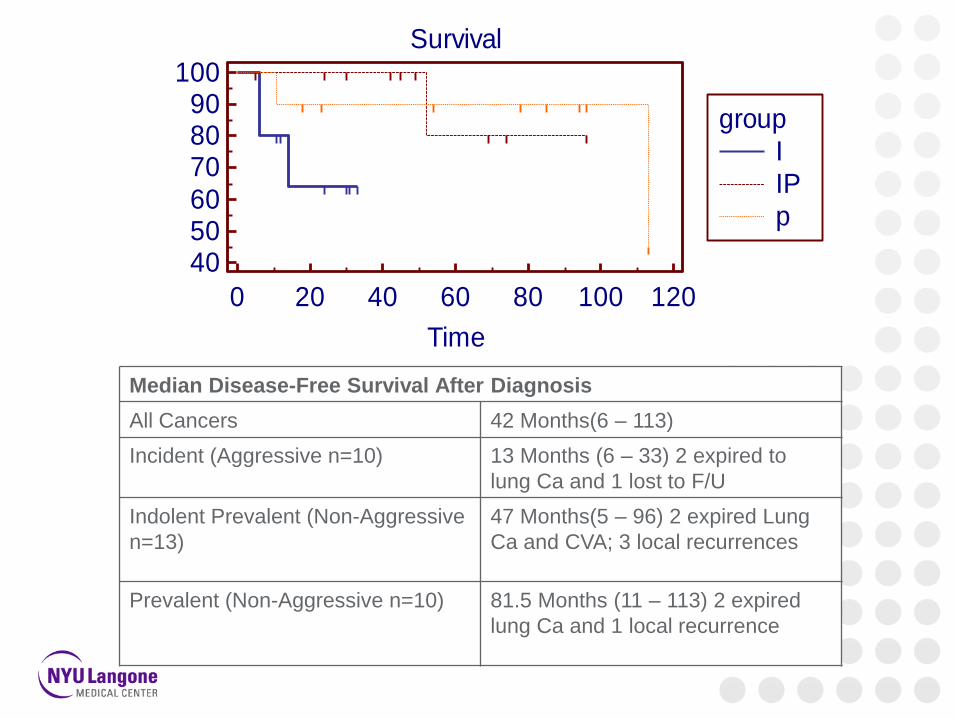
Survival
0 20 40 60 80 100 120
405060
708090
100
Time
Su
rviv
al p
rob
ab
ility
(%
)
Number at risk
Group: I
10 4 0 0 0 0 0
Group: IP
12 11 9 4 1 0 0
Group: p
10 8 7 6 5 2 0
group
I
IP
p
Median Disease-Free Survival After Diagnosis
All Cancers 42 Months(6 – 113)
Incident (Aggressive n=10) 13 Months (6 – 33) 2 expired to
lung Ca and 1 lost to F/U
Indolent Prevalent (Non-Aggressive
n=13)
47 Months(5 – 96) 2 expired Lung
Ca and CVA; 3 local recurrences
Prevalent (Non-Aggressive n=10) 81.5 Months (11 – 113) 2 expired
lung Ca and 1 local recurrence

Plasma osteopontin in screened lung cancers (n=10) and controls (n=33).
Joseph S, Harrington R, Walter D, Beck A, Litton T, Hirsch N, Blasberg J, Rom WN, Pass H, Donington J. Osteopontin velocity differentiates lung cancers from controls in a CT screening population. Cancer Biomarkers, in press.
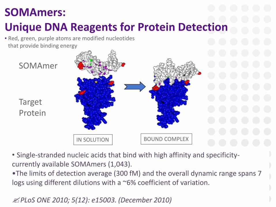
SOMAmers: Unique DNA Reagents for Protein Detection• Red, green, purple atoms are modified nucleotides
that provide binding energy
BOUND COMPLEXIN SOLUTION
• Single-stranded nucleic acids that bind with high affinity and specificity-currently available SOMAmers (1,043).•The limits of detection average (300 fM) and the overall dynamic range spans 7 logs using different dilutions with a ~6% coefficient of variation.
PLoS ONE 2010; 5(12): e15003. (December 2010)
SOMAmer
Target Protein

SomaLogic: Training and Verification Study Subjects

SomaLogic Aptamer Panel
PLoS One. 2010 Dec 7;5(12):e15003

Integrated Diagnostics: Scatter Plot of Nodule Size vs. Classifier Score of 247 Patients Using 13 Proteins
Li X, Hayward C, Fong P-Y, Dominguez M, Hunsucker SW, Lee LW, McLean M, Law S, Butler H, Schirm M, Gingras O, Lamontagne J, Allard R, Chelsky D,
Price ND, Lam S, Massion PP, Pass H, Rom WN, Vachani A, Fang KC, Hood L, Kearney P. A Blood-Based Proteomic Classifier for the Molecular
Characterization of Pulmonary Nodules. Science Translational Medicine 2013; 5: 207ra142.

Autoantibodies to Tumor Associated Antigens (TAAs)
136 Samples analyzed by enzyme immunoassay with recombinant
proteins of 10 TAAs in collaboration with Eng Tan (Scripps)
TAAs: p53, c-myc, IMP1, p62/IMP2, IMP3/Koc, Cyclin A, Cyclin B1, Cyclin D1,
CDK2, Survivin

ROC Curve Based on Stepwise Multiple Logistic Regression and Log Transformed Biomarkers to Classify Cancer vs. No Cancer
(including no nodules, solid nodules, and ground glass opacities groups)
Rom WN, Goldberg JD, Addrizzo-Harris D, Watson HN, Khilkin M, Greenberg AK, Naidich DP, Crawford B, Eylers E, Liu D, Tan EM. Identification of an
autoantibody panel to separate lung cancer from smokers and nonsmokers. BMC Cancer 2010; 10: 234.
Area Under Curve = 0.907
using c-myc, Cyclin A,
Cyclin B1, Cyclin D1,
CDK2, and Survivin.

Airway Genomics: A. Spira, BU
• Can profiles of gene expression in large-airway epithelial cells using
bronchial brushing RNA provide insights into how individual smokers
differ in their responses to cigarette smoke?
• Can such profiling detect smokers in whom the mutagenic effects of cigarette smoke have resulted in lung cancer presenting as a SPN?
Nat Med. 2007 Mar;13(3):361-6.

Affymmetrix Gene Microarrays of PBMCs • 29 gene classifier scores in 137 NSCLC patients and 91 high-risk smoker controls
with non-malignant lung disease. • A positive score indicates classification as cancer, a negative score as non-
malignant disease.

Gene Expression Pattern was Significantly Altered Post-Resection

Gene Classifier had Upregulated Genes in NK Cell Pathway, Ceramide and ErbB.

Wave Spectroscopic Microscopy of Cell Nanoarchitecture Determines Disorder Strength
Roy H et al. Optical detection of buccal epithelial nanoarchitectural alterations in patients harboring lung cancer: Implications for screening. Cancer Res 2010; 70: 7748.

ROC Curves for Ld Differentiating Lung Cancer vs. COPD (red) and
Lung Cancer vs. non-smokers (blue).

Breath Markers of Lung Cancer
• Lung cancer patients may have induction of cytochrome P450 enzymes accelerating catabolism of VOCs so that their abundance in 1.0 L of alveolar breath may provide an early detection biomarker.
• Chest 2003; 123: 2115, Cancer Biomarkers 2007; 3: 95.
Menssana Research’s
breath collection
apparatus collects about
200 VOCs in a breath
sample and gas
chromatography can
analyze picomolar
concentrations.

ROC Curves for Breath Markers of Lung Cancer

Breath Markers of Lung Cancer

Acknowledgements
•Katie Schliessman Research Administrator. Shanni Subryan Research Coord.
•EDRN: NYU Lung Cancer Biomarker Center: Study Nurse:
[email protected] or 263-6126 to enroll smokers >50 yr >20 pack-yrs;
Jim Messina Data manager, and Jackie Polenco,Scheduler.
•Pulmonary Faculty: Drs. James Tsay, John Munger, Michael Weiden, Leo Segal,
Eric Leibert, Doreen Addrizzo-Harris, Eric Tang PhD.
•Pulmonary Fellow: Vikram Mukherjee MD
•Research Laboratory Technician: Ting-An Yie MS.
•NYU SoM Varick GlycoLab: Ingrid Gils, Jennifer Thomson, Jordan Preiss
•Bioinformatics: Marko I. Vuskovic, D. Sci.
•Radiologists: David Naidich MD, Jane Ko MD, Maria Schiao MD, GeorgeAnn
McGuinness MD.
•Thoracic Surgery: Harvey Pass MD, Margaret Huflejt PhD, Jessica Donington
MD, Bernard Crawford MD.
•NCI Early Detection Research Network UO1 CA086137 Sudhir Srivastava PhD,
MPH, Karl Krueger PhD, Lynn Sorbara PhD, T32 NIEHS 007267. Con Edison.
Top Related