


Languages
Pages
Legal


UNIVERSITY OF CALIFORNIA
Santa Barbara
Biochemistry and Structure-Function Relationships in the Proteinaceous
Egg Capsules of Busycotypus canaliculatus
A Dissertation submitted in partial satisfaction of the
requirements for the degree
Doctor of Philosophy in Biochemistry and Molecular Biology
by
Stephen Scott Wasko
Committee in charge:
Professor J. Herbert Waite, Chair
Professor James Cooper
Professor Christopher Hayes
Professor Frank Zok
June 2010
UMI Number: 3428956
All rights reserved
INFORMATION TO ALL USERSThe quality of this reproduction is dependent upon the quality of the copy submitted.
In the unlikely event that the author did not send a complete manuscriptand there are missing pages, these will be noted. Also, if material had to be removed,
a note will indicate the deletion.
UMTDissertation Publishing
UMI 3428956Copyright 2010 by ProQuest LLC.
All rights reserved. This edition of the work is protected againstunauthorized copying under Title 17, United States Code.
ProQuest®
ProQuest LLC789 East Eisenhower Parkway
P.O. Box 1346Ann Arbor, Ml 48106-1346

Biochemistry and Structure-Function Relationships in the Proteinaceous
Egg Capsules of Busycotypus canaliculatus
Copyright ©2010
by
Stephen Scott Wasko
iii

ACKNOWLEDGEMENTS
I would like to first thank my father, Stephen, for instilling in me from a
young age a profound curiosity for how the natural world around me
operates, from the tiniest minutia up through the largest overall picture. My
mother, Lee, for keeping me grounded and focused so that I never lost track
of the value of my work. And to both of them, as well as my sister, Laura, for
their loving encouragement throughout the entire process of my life to date.
To my advisor, Herb, for guiding me along not only the finer points of
biochemistry and biophysics, but for guiding me along the path to being a
better scientist overall and better person with his eccentric style and allowing
me the freedom to learn from my own mistakes and direct my own project.
These were the primary reasons I had for joining his group, and I was
certainly not disappointed with that decision. To members, past and present,
of the Waite lab, and everyone else in the Marine Biotech Building for
assistance both technical and philosophical, especially Ali who worked so
hard to turn me into a materials scientist while I worked so hard to turn him
into a biologist. To my committee for keeping me on track and providing
valuable insights. And last, but certainly not least, to all of my wonderful
friends, both those I've met since coming to Santa Barbara, and those
who've kept in touch from previous times. You are too numerous to list here.
iv

All of the intellectual stimulation in the world could not replace the joy that
you bring me on a regular basis. I love you all.

Curriculum Vita, Stephen Scott Wasko
Aprii 2010
Education:
2004-20 1 0 PhD in Biomolecular Sciences and Engineering, 20 1 0 (expected)Univeristy of California, Santa Barbara, CA.Advanced to candidacy May, 2007Research advisor: J. Herbert Waite
1 997-200 1 BA in Biological Sciences, emphasis in Molecular and Cell BiologyCornell University, College ofArts and Science, Ithaca, NY.
Research experience:2005-20 1 0 Graduate Student Researcher, UC Santa Barbara
Biomolecular Sciences and Engineering ProgramAdvisor: J. Herbert Waite
2002 Field Research Technician, Cornell UniversityDepartment of Plant BreedingMentor: Julia Hansen
2000 Undergraduate Student Researcher, Cornell UniveristyDepartment of MicrobiologyMentor: Anthony Hay
Teaching Experience:2009 Teaching Assistant, UC Santa Barbara
MCDB IAL: Intro Biology Lab2009 Mentor, UC Santa Barbara
California Nanosystems Institute, Apprentice Researchers (AR)Mentee: Gabriel Li
2008 Mentor, UC Santa BarbaraMaterials Research Lab, Research Internships in Science andEngineering (RISE)Mentee: Christine F Carpenter
2005 Teaching Assistant, UC Santa BarbaraMCDB 109L: Biochemistry Lab
2001 Teaching Assistant, Cornell UniveristyBIOG 206 (now BIOG 1106): Introductory Biology
Fellowships/Scholarships:
Vl

2007-2009 University of California Biotechnology Research and EducationProgram (BREP) Graduate Research and Education in AdaptiveBio-Technology (GREAT) Grant: Two years $100,000
1 998-2000 Raytheon Incentive Scholars Award (undergraduate)
Awards:
• Chang Distinguished Alumni Award for Audience Participation at Friday NoonSeminar Series, 2009• Chang Distinguished Alumni Award for Outstanding Talk at Friday Noon SeminarSeries, 2008
Presentations:• S. Scott Wasko and J. Herbert Waite Recoverable yield in an extensible,proteinaceous material found in gastropod egg capsules, 2008Oral presentation at the Materials Research Society spring meeting in SanFrancisco.• S. Scott Wasko and J. Herbert Waite Recoverable-Yield in a PoteinaceousElastomer From Marine Whelks, 2007Poster presented at the International Conference on Self-healing Materials inNoordwijk, NL.
Publications:
Ali Miserez, S. Scott Wasko, Christine F. Carpenter and J. Herbert Waite. "Non-entropic and Reversible Long-Range Deformation of an EncapsulatingBioelastomer." Nature Materials, 8 (11): 910-916 (2009).(AM and SSW contributed equally to the manuscript)
S. Scott Wasko, J. Herbert Waite. Structural proteins from an egg capsule withnonentropic reversible extensibility," Biomacromolecules. (submitted/under review).
S. Scott Wasko, Matthew J. Harrington, Himadri S. Gupta, Peter Fratzl, J. HerbertWaite. "Characterizing the reversible a-helix ß-sheet transition in a shape-memorybiological material," (manuscript in preparation).
S. Scott Wasko, Matthew J. Harrington, Himadri S. Gupta, Peter Fratzl, J. HerbertWaite. "Supramolecular structure and nanoscale tensile behavior of a shape-memorybiological material," (manuscript in preparation).
VII
ABSTRACT
Biochemistry and Structure-Function Relationships in the Proteinaceous
Egg Capsules of Busycotypus canaliculatus
by
Stephen Scott Wasko
The designs of highly extensible soft materials in nature are of a
fundamental interest to engineers so that insights into the production of
modern, synthetic materials can be gleaned. Marine gastropods of the
genus Busycotypus produce a protein based elastomer which possesses a
unique combination of stiffness and extensibility. Furthermore, this material
displays shape-memory/self-healing properties that are unmatched in
synthetic engineering systems. Four variants of the precursor protein
components of the egg capsules are highly unique in their amino acid
sequences, showing no homology to any known protein families. These
proteins are strongly a-helicaì in nature, and can self-assemble into
nanometer scaled fibers in vitro. The remarkable reversibly extensibility of
viii

the bulk material is dictated not by entropie forces, but rather by a crystalline
phase transition within the protein components when the material is put
under tension. Proteins shift from a-helix to ß-sheet, and it is this uncoiling of
helices within the polymer backbone that allows for the extensibility of the
egg capsules. This transition is reversible, as when loads are removed and
the material is allowed to relax, it returns to its original a-helical
conformation. When examined more closely, it is shown that this a«->>ß
transition is a multi-step transformation which involves first the uncoiling of
crystalline a-helices into non-crystalline random coils before these then lock
into ß-sheets. These different steps dictate changes in the mechanical
properties of the material as this transition is occurring. Furthermore, the
supramolecular structure of how the individual proteins interact in the intact
material also changes throughout tension/relaxation cycles. These structural
changes also have effects on the bulk mechanical properties of the material.
This work explores in detail the structure-function relationships of
Busycotypus egg capsule material mentioned above.
IX
Table of contents
1. Introduction 1
A. Overview 1
B. Elastomeric Materials: Properties, Theories, and Applications 2
C. Biomimetics: Taking Inspiration from Nature 8
D. Fibrous Biological Materials 10
E. Whelk Egg Capsules as a Model System 15
F. Research in this Dissertation 21
G. References 22
2. Characterization of precursor proteins from Busycotypus canaliculatus. .25
A. Abstract 25
B. Introduction 26
C. Materials and Methods 30
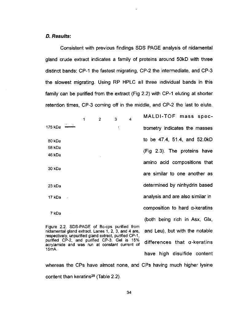
D. Results 34
E. Discussion 42
F. Conclusions 50
G. Accession Numbers 52
H. Acknowledgments 52I. References 54
X

3. Thermomechanical properties of Busycotypus canaliculatus egg capsules:
entropie vs non-entropic 56
A. Abstract 56
B. Introduction 57
C. Materials and Methods 59
D. Results and Discussion 62
E. Conclusions 78
F. Acknowledgments 79
G. References 80
4. Using wide angle x-ray scattering to characterize the phase transition
from a-helix to ß-sheet as a result of uniaxial tension 82
A. Abstract 82
B. Introduction 82
C. Materials and Methods 85
D. Results 86
E. Discussion 91
F. Conclusions 97
G. Acknowledgments 98
H. References 99
xi

5. The supramolecular hierarchical ordering of Busocotypus canaliculars
egg capsules and its role in tensile mechanics 102A. Abstract 102
B. Introduction 103
C. Materials and Methods 104
D. Results 106
E. Discussion 113
F. Conclusions 120
G. Acknowledgments 121H. References 122
6. Conclusions and future directions 124
A. Abstract 124
B. Conclusions from this Work 125
C. Future Directions 129
D. Materials and Methods for Preliminary Studies 131
E. Preliminary Results 133
F. Acknowledgments 139H. References 140
XIl

Chapter 1. Introduction
A. Overview
Research into soft elastomeric materials has been ongoing and active
for a considerable amount of time. With a broad range of functionalities and
mechanical properties, these materials can be utilized in a wide variety of
applications. Fibrous composite materials can, and often do add a level of
anisotropic structure which can further tune these functionalities and hence
applications.
In biologic systems, elastomers are frequently formed from naturally
occurring fibers such as protein or carbohydrate. The somewhat recent
notion of studying these kinds of natural materials and systems with the
intent of applying what is learned to manmade engineering strategies has
been dubbed biomimetics, and this field has gained considerable
momentum over the past few years. Research into biological fibers has
increased dramatically as a result.
One such biologic elastomer is the egg capsule that marine
prosobranch gastropods, commonly known as whelks, use to protect
embryos during development. This material has shown to not only have
strong biomimetic potential, but to also serve as a model system to further
elucidate theory regarding the thermodynamics of reversible extensibility,
and phase transition in materials. The characterization of the biochemistry
1

and structure-function relationships in this egg capsule material is the topic
of this dissertation.
B. Elastomeric Materials: Properties, Theories, and Applications
An elastomer has been defined as "a macromolecular material that
returns rapidly to approximately the initial dimensions and shape after
substantial deformation by a weak stress and release of the stress."1 Somechoose to establish more detailed conditions that the material must meet in
order to be classified an elastomer: it must be capable of being stretched at
least 100% of it's original length2, and that after being stretched to 100%,
held for five minutes, and then released, it must retract to within 10% of its
original length within five minutes after release3. Regardless of specifics,elastomers are materials which exhibit little plastic flow and demonstrate a
quick and nearly complete recovery from an extending force.
Such materials have countless applications in both manmade as well
as natural systems. In synthetic applications, elastomers are employed as
protective coatings, adhesives, composite components to increase
extensibility and durability, insulators (both thermal and electrical), textiles,
implants, foams, the list goes on and on2·5. Numerous fields employelastomeric materials in their respective systems, from construction to
automotive to biomedical and beyond. Natural systems have also developed
elastomeric polymers which serve roles equally as diverse as their
2

manmade counterparts: as shock absorbers in, for example, hydrated
dragline spider silks6, for elastic energy storage capacity in the resilin of
jumping and flying insects7 and to ensure adequate elasticity in the
integument and arteries of various organisms8-10. With such varied uses andfunctional environments, these materials, both manmade and natural, are
also varied in the properties that they possess.
The properties that must be considered when selecting an
appropriate elastomer for a given system include mechanical properties
(extensibility, hardness, strength, stiffness, resilience, scratch and abrasion
resistance, creep, compressibility, etc) as well as environmental compatibility
(temperature performance at both low and high temps, resistance to
solvents and oils, behavior in aqueous environments, biocompatibility, flame
resistance, UV resistance, age related degradation, etc)2. All of these must
be examined to find the perfect match for a specific application.
Many of the mechanical properties are dictated by the
thermodynamic nature of extensibility in each material. The primary forces
that contribute to this can be grouped as either entropie or internal. Entropie
force is dictated by the second law of thermodynamics which states that
systems have a tendency to maximize entropy (disorder) within that system.
Internal force, or bond energy, is the sum of energy in the chemical bonds as
well as the weak force interaction between atoms within a system. Simply
put, while a system may seek to maximize disorder, the forces associated
3

with bond energies in that system resist that tendency. The interplay of these
forces is represented by the Hemholtz free energy relationship2·4.
A=U-TS (1.1)
Wherein A is the Hemholtz free energy, U is the internal energy, T is
absolute temperature and S is entropy. The differential of the maximum workfunction dA can be written to include the work done on an elastomer by the
application of a force, /, over the change in length, dL.
dA = -PdV - SdT + fdL (1.2)
Where P is pressure and V is volume. Accordingly, the force is comprised of
two components, and internal energy component, /u, and an entropy
component, /s.
/ = /u + /s (1.3)
The partial differential of A with respect to length at constant V, T, and
composition, n, can be rewritten as
/u// = -T(3\n[f/T\/dT)v,L,n (1 -4)
4

which allows for an experimental estimate of the fu/f ratio from the slope of
a plot of ln(//T) versus temperature under conditions of constant V1 L, and
composition, n, so long as appropriate correction terms are applied.
Many elastomers, both synthetic and natural rubbers2·5·11·12 as well as
some biological materials such as elastin9 and resilin7, exhibit classic
entropie elasticity wherein /s is far greater than /u. In this model, anelastomeri material in a relaxed state is composed of a network of random
chains which with a low crosslink density. The extension of this material from
the relaxed state by an external force causes a decrease in entropy,
governed by the Boltzmann relationship2·4, as chains become aligned in an
anisotropic manner.
AS = (Se - Sr) = R In(WW) (1 .5)
Wherein R is the Boltzmann constant. This decrease in entropy largely
overshadows any internal energy component and, and once the external
force is removed, the system seeks to reestablish the thermodynamic
equilibrium of the relaxed state by the polymer chains randomly orienting
themselves again, causing a contraction of the material (Fig 1.1)
5

HighEntropy
External force-----------------*-
Entropie drive
LowEntropy
Figure 1.1. Schematic of classic entropically driven elastomeric behavior
The flip side of this thermodynamic relationship has internal forces
dominating reversible extensibility. The sequential rupturing of weak
chemical bonds in a material without compromising the polymer backbone
can allow for an increase in overall length of the material. Furthermore, if the
ruptured bonds are both capable of, and are allowed to reform, the material
may recover initial dimensions and shape after deformation, thereby
qualifying it as an elastomer. This paradigm has been dubbed "sacrificial
bonds and hidden length," and allows for reversible, long range extensibility
that is governed by internal rather than entropie energy13 (Fig 1.2).
6
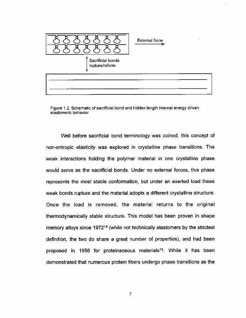
Sacrificial bondsrupture/reform
Figure 1 .2. Schematic of sacrificial bond and hidden length internal energy drivenelastomeric behavior
Well before sacrificial bond terminology was coined, this concept of
non-entropic elasticity was explored in crystalline phase transitions. The
weak interactions holding the polymer material in one crystalline phase
would serve as the sacrificial bonds. Under no external forces, this phase
represents the most stable conformation, but under an exerted load these
weak bonds rupture and the material adopts a different crystalline structure.
Once the load is removed, the material returns to the original
thermodynamically stable structure. This model has been proven in shape
memory alloys since 197214 (while not technically elastomers by the strictest
definition, the two do share a great number of properties), and had been
proposed in 1956 for proteinaceous materials15. While it has been
demonstrated that numerous protein fibers undergo phase transitions as the

result of an applied physical load, no dedicated studies into the
thermodynamics of those protein fibers' elasticity have been performed.
C. Biomimetics: Taking Inspiration from Nature
Every biological organism employs a variety of systems and materials
to successfully fill its ecological niche. Ultimately these material designs are
geared towards maximizing functionality, and frequently these functions
include a form of load or impact bearing material that is designed to meet an
organism's unique demands. However, it is not uncommon that the specific
functions that these naturally occurring materials are designed for are also
desired in manmade synthetic systems. As such, these materials can be
studied and the lessons learned can be applied to modern engineering
problems.
There are many intuitive advantages in taking inspiration from nature.
Natural materials are invariably built under ambient conditions (temperature,
pressure, pH, etc) using non-toxic, biologically friendly components. In the
natural world, the overarching functionality of any system is to increase the
individuals' evolutionary fitness, and materials systems are no different. The
result of this is two-fold. Firstly, these biomaterials have been honed by
generations upon generations of evolutionary pressure to perform its
specific function very well. Secondly, as high energy consumption is an
evolutionary liability, biomaterials are synthesized in an energetically efficient
8

manner. Nature rarely over-engineers its systems. Biomimetics can offer
insights not only into designing systems and materials with desiredfunctionalities, but also provide a fabrication process that is more
environmentally friendly, with low energy costs, furthering "green"technologies.
In addition to load and impact bearing materials, biomimetic research
has resulted in advances in numerous specific fields. Some of these include
energy production and storage16, adaptive materials17, adhesives18, self-healing/shape-memory materials19, and biomedical systems20. It is a proventruth that by studying the structure function relationships of these kinds ofsystems, humans can apply the same principles that nature does todeveloping modern high-performance material systems.
However, it must also be stated that there are inherent limitations in
this process. As mentioned before, evolutionary pressures direct theprogression and functionality of these natural systems, but that does notmean that these systems are optimized. Instead they merely perform their
intended functions at a satisfactory level for the evolutionary pressures
applied, given the limited resources available to the organism (buildingcomponents, synthesis conditions, energy allocation, etc). Furthermore, thedemands that human engineered systems have may be greater than those
that natural systems are adapted to perform. Another challenge is the factthat many biological systems are multifunctional and, as such, contain many
9
components, each designed to perform its own task in a synergistic manner
with the other components. This makes deciphering exact structure-function
relationships very difficult. Evolution has spent hundreds of millions of years
designing a specific material, yet biomimeticists will attempt to elucidatetheir secrets and fabricate a suitable mimic in a few short years. This can be
a daunting task indeed.
Despite these potential setbacks, the exploration of natural systems
is still worthwhile. As mentioned above, great strides have already been
made in numerous systems, and the adaptation of design strategies from
natural to engineered materials continues to show more and more promise.
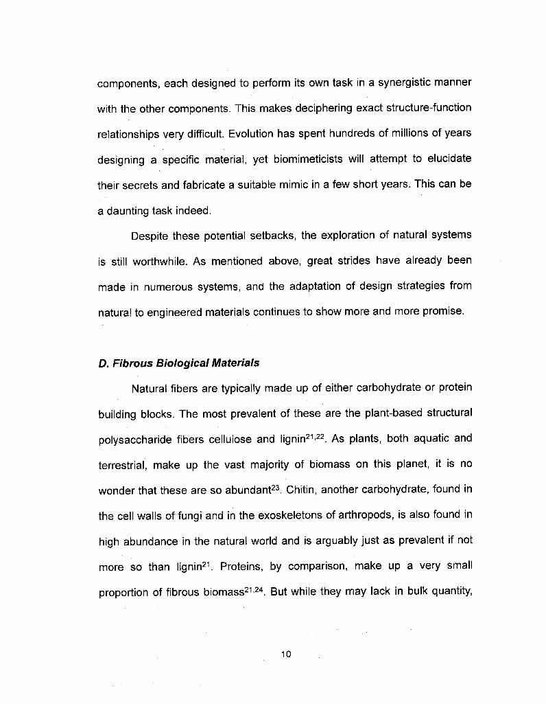
D. Fibrous Biological Materials
Natural fibers are typically made up of either carbohydrate or protein
building blocks. The most prevalent of these are the plant-based structural
polysaccharide fibers cellulose and lignin21·22. As plants, both aquatic andterrestrial, make up the vast majority of biomass on this planet, it is nowonder that these are so abundant23. Chitin, another carbohydrate, found in
the cell walls of fungi and in the exoskeletons of arthropods, is also found in
high abundance in the natural world and is arguably just as prevalent if notmore so than lignin21. Proteins, by comparison, make up a very small
proportion of fibrous biomass21·24. But while they may lack in bulk quantity,
10

the variety of different functionalities that protein materials possess is
staggering.
Protein fibers are found in all kingdoms of life. They are located
intracellular^, extracellularly, and extraorganismally and are involved in load
bearing scaffolds such as the skeleton and cytoskeleton25·26; in cellular
transport and cellular division27; in wear resistant tools such as teeth,
mandibles, and radulae25·28·29; in locomotion of all kinds24; in tissue
encapsulation, both internal30 and external31'33; in shelter and protection34·35;in wound healing and response36; in holdfasts and prey capture6·37; and in a
variety of other functions. As there is such a broad range of demands, the
fibers themselves must also be highly varied as they are tuned to perform
each specific task. Keratins in horns and nails primarily function in dryenvironments and must be stiff and wear resistant, whereas the fibrins
involved in wound healing must allow for flexibility around the damaged area
to maintain a tight seal while functioning in aqueous conditions. Microtubules
must be programmable highways with controlled destinations so that
transporters can guide their molecular cargo to specific targets, while spider
silks must be able to withstand the forces of an entire organism impacting a
web. As all proteins are composed of the same amino acid building blocks,
differences in sequence and structure are responsible for the differences in
functionality.
11
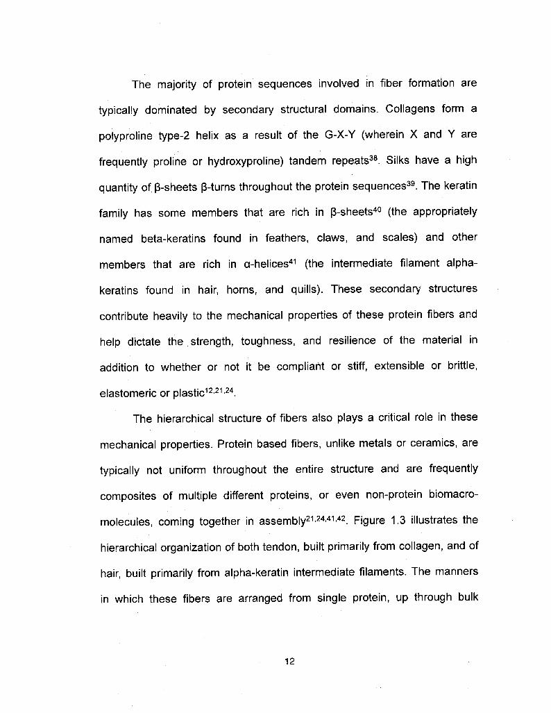
The majority of protein sequences involved in fiber formation are
typically dominated by secondary structural domains. Collagene form a
polyproline type-2 helix as a result of the G-X-Y (wherein X and Y are
frequently proline or hydroxyproline) tandem repeats38. Silks have a highquantity of ß-sheets ß-turns throughout the protein sequences39. The keratinfamily has some members that are rich in ß-sheets40 (the appropriatelynamed beta-keratins found in feathers, claws, and scales) and other
members that are rich in a-helices41 (the intermediate filament alpha-
keratins found in hair, horns, and quills). These secondary structures
contribute heavily to the mechanical properties of these protein fibers and
help dictate the strength, toughness, and resilience of the material inaddition to whether or not it be compliant or stiff, extensible or brittle,
elastomeric or plastic12'21·24.
The hierarchical structure of fibers also plays a critical role in these
mechanical properties. Protein based fibers, unlike metals or ceramics, are
typically not uniform throughout the entire structure and are frequentlycomposites of multiple different proteins, or even non-protein biomacro-molecules, coming together in assembly21 ·24·41·42. Figure 1.3 illustrates the
hierarchical organization of both tendon, built primarily from collagen, and ofhair, built primarily from alpha-keratin intermediate filaments. The manners
in which these fibers are arranged from single protein, up through bulk
12
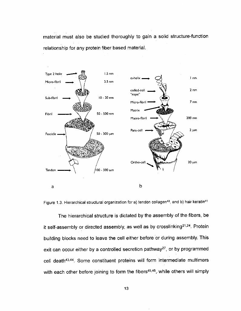
material must also be studied thoroughly to gain a solid structure-function
relationship for any protein fiber based material.
Type 2 helix
Micro-fibril
Sub-fibril
Fibril
Fascicle
Tendon ?
3.5 nm
O- 20 nm
50 - 500 nm
50 - 300 µ??
a-helix —
coiled-coil"rope"
100 -500 urn
Micro-fibril
Matrix
Macro-fibril
Para-cell
Ortho-eel
2 nm
7 nm
200 nm
2 µ??
20 um
Figure 1.3. Hierarchical structural organization fora) tendon collagen42, and b) hair keratin41
The hierarchical structure is dictated by the assembly of the fibers, be
it self-assembly or directed assembly, as well as by crosslinking21·24. Protein
building blocks need to leave the cell either before or during assembly. This
exit can occur either by a controlled secretion pathway27, or by programmed
cell death43·44. Some constituent proteins will form intermediate multimers
with each other before joining to form the fibers45·46, while others will simply
13

add on single proteins to the structure47. Some materials will spontaneously
self-assemble under the proper conditions (pH, ionic strength, etc)43·44,
others must be modified (frequently by protein cleavage) before assembly48,still others require additional assistance from molecular chaperones47·49.Such variety in assembly processes leads to a variety of structures.
Additionally the methods for crosslinking are just as varied. Crosslinks cancome in the form of covalent bonds (disulfide bonds, desmosine, and
glutamyl-lysine linkages are some of the more prevalent)38, or in the form ofweak interactions such as metal coordination complexes and hydrogen
bonding5053. The location of crosslinks throughout the fiber, whether theyjoin the proteins end to end, end to middle, or middle to middle, also criticallyaffects mechanical properties54.
Many of the more extensible protein materials such as elastin9 andresilin7 have been shown to behave like classic rubbers from both a
structural and a thermodynamic standpoint. That is the protein constituents
are seemingly randomly oriented with a low level of crosslinks, and theextensibility and recovery of the material is dictated by entropie forces ratherthan internal bond energies. It had been proposed that a crystaline
transformation can be responsible for long-range, reversible deformation in
protein materials15, greater than in the shape memory alloys. However, asmentioned above, while there have been numerous theoretical candidates
for this non-entropic elasticity, either due to the complex nature of protein
14

materials or possibly a perceived lack of interest in the field, there has been
no experimental conformation of the theory.
E. Whelk Egg Capsules as a Model System
Busycotypus canaliculatus (formerly known as Busycon
canaliculatum) is a marine prosobranch gastropod that is endemic to the mid
to northern regions of the North American Atlantic coast, and has been
introduced to San Francisco Bay. Commonly known as the "channeled
whelk", this organism is valued as a food resource. The species has maleand female individuals and fertilization is internal as opposed to external
spawning like some other aqueous organisms. Once fertilization is
complete, the female will lay long strings of interconnected disk-shaped egg
cases, colloquially known as "mermaids necklaces" (Fig 1.4), in shallow
ocean waters, anchored by burying one end in the sand. These strings may
be in excess of 1 meter in length, and can contain hundreds of individual
egg cases, each case containing up to 25 developing embryos. After aincubation period of as many as 10 months, the embryos come to term as
completely developed whelks and exit the egg cases55.
15

During this time period, the
egg cases are subjected to haz-
ards such as extreme hydro-
dynamic forces generated by water
velocities in excess of 10 msec-1 56,
abrasion from sand and other
particulates in the surrounding
water, as well as prédation57. In
addition to physical protection, the
diffusion of certain molecules, such
as metabolic waste products, is
also necessary57. As such, the
organism has been forced to
develop a very robust, yet permeable material to use as the egg capsule
wall.
An acellular material composed of >90% protein, the egg capsule wall
is highly ordered and possesses unique tensile properties. It has been
previously established that multiple processing steps are required to
produce the mature form of the material. Each one of these steps alters the
tensile properties, physical appearance, and biochemical composition of the
material58-61. The precursor proteins are produced in the nidamental gland
(also known as egg capsule gland). Here, they are stored in secretory
Figure 1.4. Channeled whelk with a string ofegg capsules, "mermaid's necklace"
16

granules for use during the reproductive cycle62. These precursors are
soluble, and there is evidence that they are stored in a liquid crystalline
array58·62. During egg capsule deposition, the proteins are secreted into the
reproductive tract where they self-assemble into fibers and sheets to
surround the embryos. At this stage, the egg capsule has a white
appearance, no definitive shape, and limited structural stability. This
"immature" capsule is soluble in detergents and denaturing agents, and is
susceptible to enzyme-mediated proteolytic cleavage63. The unfinishedproduct is then passed along the reproductive duct to the ventral pedalgland located on the ventral surface of the foot. Here, the capsuleundergoes a sclerotization process for approximately 1-2 hours during whichthe capsule gains a yellowish tint, is molded into the disk-like shape, anddevelops the mechanical robustness necessary for protecting the embryos
during development55·57'59. The material is now insoluble in even theharshest dénaturants, and resists enzymatic cleavage58·63. This "mature"
capsule is incorporated onto the string of capsules that came before it and isnow anchored to the seabed.
While observed and described since the late 1800's 64, research into
the material itself did not begin until the 1960s, when researchers at the
University of Leeds, England, performed the first structural studies. Using
wide-angle x-ray scattering it was determined that whelk egg case proteins
resides primarily in a coiled-coil a-helix conformation. The same group used
17

TEM work to show that multiple laminate sheets make up the biaxial
material, and that fibers are highly ordered in these sheets with a visible
periodicity 50 nm60·65.
In the 70s, work on the egg cases shifted to biochemical
characterization of bulk material, as well as the protein precursors. Amino
acid analysis of intact capsule wall indicated levels of the key structuralresidues glycine, proline, alanine, and valine in much lower proportions than
those of collagene and elastins, suggesting that this material has a different
structural composition. Charged residues (aspartate, glutamate, lysine, and
arginine) dominate the amino acid content, making up >40% of the protein.Helix formers comprise 50% of the content with 25% helix indifferent and
25% helix breakers, which corroborates the x-ray studies of the 60s 62,66,67
Over all, the amino acid composition of the capsule wall is very similar to
that of a-keratin intermediate filaments (IF), with one notable difference: IFs
have a high disulfide cystine content, where as the capsules have none.
Therefore, a different crosslinking mechanism is probable in the latter
system21 .
By comparing analyses of capsule wall and precursor proteins, it wasshown that the lysine content drops significantly during processing. So it is
thought that lysine based crosslinks could be involved62. Furthering thistheory was the fact that borohydride reducible compounds were found in thebulk material61. Using tritium labeled borohydride, it was possible to isolate
18

these compounds for further study and evidence suggested that
lysinonorleucine crosslinks were involved61·62. However, the results were
never conclusively proven, and research on that topic stopped abruptly.It was not until the late 90s and into the 00s that research on the
material started again. Scientists at the Scripps Research Institute at UC
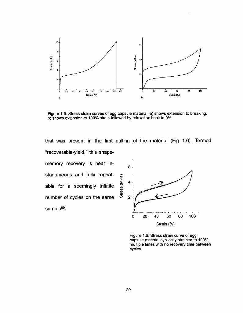
San Diego examined tensile mechanical properties. Their stress-strainstudies (using engineering stress and strain) indicate three regions withdistinctly different elastic moduli58·59. At low strain there is high modulus inthe range of 160 MPa. However, after being extended past ~3% strain, thereis a sudden and dramatic drop in flow modulus, down to the 2 MPa range,
which is maintained until -80% strain. At this point it increases again to 11
MPa and is maintained until the breaking strain of -170% . When the
material is cyclically strained to a point below its breaking strain, itundergoes an energy absorbing hysteresis of 50% at 100% extension (Fig1.5). Upon subsequent strain, recovers the initial high modulus
19
2wOT
O 20 40 100 120 140 160 180
Strain (%) Strain (%)
Figure 1.5. Stress strain curves of egg capsule material, a) shows extension to breaking,b) shows extension to 100% strain followed by relaxation back to 0%.
too.H 4tnInF
that was present in the first pulling of the material (Fig 1.6). Termed
"recoverable-yield," this shape-
memory recovery is near in-
stantaneous and fully repeat-
able for a seemingly infinite
number of cycles on the same °° 2
sample59.0 20 40 60 80 100
Strain (%)
Figure 1 .6. Stress strain curve of eggcapsule material cyclically strained to 100%multiple times with no recovery time betweencycles
20

G. Research in this Dissertation
The overarching goal of this research is to further elucidate the
structure-function relationships in whelk egg capsule material in terms of
biochemistry, molecular ordering, and mechanical properties. This
necessitates work that crosses multiple disciplines. Firstly, characterization
of the precursor proteins of the capsule wall will be discussed. This
characterization includes physical properties such as mass and amino acid
composition, documentation and analysis of the complete primary sequence
of four of these precursors, as well as structural studies of how they behave
in solution. Next, the thermomechanical nature of egg capsule extensibility
(entropie vs internal energies) will be examined, and experimental evidence
for crystalline transitions in protein secondary structure will be presented.Following that, a more thorough characterization of this secondary structure
transition, as well as shifts in hierarchical structure, derived from synchrotron
source x-ray scattering, and how they relate to the stress-strain properties ofthe material will be introduced. Lastly, potential directions for future research
and preliminary results will be outlined.
21

H. References
1 . Am. Soc. for Testing of Materials Standards, Part 28, 19672. Harper, C. A. Handbook of Plastics and Elastomers. McMraw-Hill Book Company,
New York, NY, 1975.3. Pickett, A. G., & Lemcoe, M. M. Handbook of Design Data on Elastomeric Materials
Used in Aerospace Systems. U.S. Air Force Communication, Wright-Patterson AirForce Base, Dayton, OH, 1961.
4. Treloar, L. R. G. The Physics of Rubber Elasticity. Oxford University Press, Oxford,U.K., 2005
5. Holden, G. Understanding Thermoplastic Elastomers. Hanser Gardner Publications,Inc. Cincinnati, OH, 2000
6. Gosline, J. M., Denny, M. W. & DeMont, M. E. Spider Silk as Rubber. Nature 309,551-552(1984).
7. Weis-Fogh, T. A Rubber-Like Protein in Insect Cuticle. Journal of ExperimentalBiology 37, 889-907 (1 960).
8. LiINe, M. A. & Gosline, J. M. The Effects of Hydration on the Dynamical MechanicalProperties of Elastin. Biopolymers 29, 1147-1160 (1990).
9. Urry, D. W. et al. Elastin: A Representative Ideal Protein Elastomer. PhilosophicalTransactions of the Royal Society of London, Series B: Biological Sciences 357,169-184(2002).
10. Shadwick, R. E. & Gosline, J. M. Physical and Chemical Properties of Rubber-LikeElastic Fibres from the Octopus Aorta. Journal of Experimental Biology 114,239-257(1985).
11. Tatham, A. S. & Shewry, P. R. Elastomeric Proteins: Biological Roles, Structuresand Mechanisms. Trends in Biochemical Sciences 25, 567-571 (2000).
12. Shewry, P R., Tatham, A. S., & Bailey, A. J. Elastomeric Proteins: Structures,Biomechanical Properties, and Biological Roles. Cambridge University Press,Cambridge, U.K. 2002.
13. Fantner, G. E., Oroudjev, E., Schitter, G., Golde, L. S., Thurner, P., Finch, M. M.,Turner, P., Gutsmann, T, Morse, D. E., Hansma, H., & Hansma, P. K. SacrificialBonds and Hidden Length: Unraveling Molecular Mesostructures in ToughMaterials. Biophysical Journal 90, 1411-141 8 (2006).
14. Jackson, C. M., H. J. Wagner, & R. J. Wasilewski. 55-Nitinol- The Alloy With aMemory: Its Physical Metallurgy, Properties, and Applications: A Report.Washington: NASA, 1972.
15. Flory, P. J. Theory of Elastic Mechanisms in Fibrous Proteins. Journal of theAmerican Chemical Society, 5222-5235 (1956).
16. Christodoulou, L., & Venables, J. D. Multifunctioal material systems: the firstgeneration. J. Min. Met. Mater. Sci. 55, 39-45 (2003).
17. Varadan, V. K., Varadan, V. V. Microsensors, microelectromechanicai systems(MEMS) and electronics for smart structures and systems. Smart Mat. Str. 9,953-972 (2000).
18. Hui Shao, H., Bachus, K. N., & Stewart, R. J. A Water-Bome Adhesive Modeledafter the Sandcastle Glue of P californica. Macromol. Biosci. 9, 464-471 (2009)
19. Kushner, A. M., Gabuchian, V., Johnson, E. G., & Guan, Z. Biomimetic design ofreversibly unfolding cross-linker to enhance mechanical properties of 3D networkpolymers. J. Am. Chem. Soc. 129, 14110-14111 (2007).
20. Lee, H., Lee, B. P., & Messersmith, P. B. A reversible wet/dry adhesive inspired bymussels and geckos. Nature 448, 338-341 (2007).
21. Vincent, J. Structural Biomaterials, Revised ed; Princeton University Press,Princeton, NJ, 1990.
22. Holtman, K. M., Chang, H., Jameel, H., & Kadla, J., F. Elucidation of lignin structurethrough degradative methods: comparison of modified DFRC and thioacidolysis. JAgrie Food Chem 51 , 3535-40 (2003).
22

23. Botkin, D. B., & Keller, E. A. Environmental Science: Earth as a Living Planet, 6thed; John Wiley & Sons, Hoboken, NJ. 2005
24. Vogel, S. Comparative Biomechanics Life's Physical World. Princeton UniversityPress, Princeton, NJ, 2003.
25. Currey, J. D. Bones: Structure and Mechanics. Princeton University Press,Princeton, NJ, 2002.
26. Khurana. S. /Advances in Molecular and Cell Biology #37: Aspects of theCytoskeleton, Eiselvier LTD, London, U.K. 2006.
27. Alberts, B., Johnson, A., Lewis, J., Raff, M., Roberts, K., & Walter, P. MolecularBiology of the Cell, 5th ed; Garland Publishing Inc. New York, NY, 2008.
28. Broomell, C. C, Chase, S. F., Laue, T., & Waite, J. H. Cutting edge structuralprotein from the jaws of Nereis virens. Biomacromolecules 9 669-677 (2008).
29. Miserez, ?., Schneberk, T., Sun, C, Zok, F. W., & Waite, J. H. The transition fromstiff to compliant materials in squid beaks. Science 319,1816-1819 (2008).
30. Heiden, T. C1 Haines, A. N., Maniré, C, Lombardi, J., & Koob, T. J. Structure andpermeability of the egg capsule of the bonnethead shark, Sphyrna tiburo. J. Exp.Zool. A. Comp. Exp. Biol. 303, 577-589 (2005).
31. Lavelin, i., Meiri, N., & Pines, M. New insight in eggshell formation. Poult Sci. 79,1014-1017(2000).
32. Hu, X., Kohler, K., Falick, A. M., Moore, A. M., Jones, P. R., Sparkman, O. D., &Vierra, C Egg case protein-1 . A new class of silk proteins with fibroin-like propertiesfrom the spider Latrodectus hesperus. J Biol Chem. 280, 21220-21230 (2005).
33 Knupp, C, Chew, M., & Squire, J. Collagen Packing in the Dogfish Egg Case Wall.J Struct. Biol. 122, 101-110 (1998)
34. Zhao, H., Sun, C, Stewart, R. J., & Waite, J. H. Cement proteins of thetube- building polychaete Phragmatopoma californica. J Biol Chem. 280,42938-42944 (2005).
35. Baden, H. P, & Maderson, P. F. Morphological and biophysical identification offibrous proteins in the amniote epidermis. J Exp Zool 174, 225-232 (1970).
36. Doolittle, R. F. Fibrinogen and Fibrin. Annu Rev Biochem 53, 195-229 (1984).37. Waite, J. H., Vaccaro, E., Sun, C & Lucas, J. M. Elastomeric gradients: a
hedge against stress concentration in marine holdfasts? Phil. Trans. R. Soc. Lond.8 357,143-153(2002).
38. Creighton, T E. Proteins: Structures and Molecular Properties, 2nd ed; W H.Freeman and Company: New York, NY, 1 993.
39. Hayashi, C. Y, & Lewis, R. V. Evidence from flagelliform silk cDNA for the structuralbasis of elasticity and modular nature of spider silks. J. MoI. Biol 275, 773-84(1998).
40. Lee, L. D., & Baden, H. P. Chemistry and composition of the keratins. Int. J.Dermatol. 14,161-171 (1975).
41. Feughelman, M. Mechanical Properties and Structure of Alpha-Keratin Fibres:Wool, Human Hair, and Related Fibres, UNSW Press: Sydney, Australia, 1997.
42. Kastelic, J., & Baer, E. Deformation in Tendon Collagen. Symp. Soc. Exp. Biol 34,397-435(1980).
43. Rogers, G. E. Biology of the wool follicle: an excursion into a unique tissueinteraction system waiting to be re-discovered. Exp. Dermatol 15, 931-949 (2006).
44. Koch, E. A., Spitzer, R. H., Pithawalla, R. B., & Downing, S. W Keratin-likecomponents of gland thread cells modulate the properties of mucus from hagfish(Eptatretus stouti). Cell Tiss. Res 264, 79-86 (1991).
45 Coulombe, P. & Fuchs, E. Elucidating the early stages of keratin filament assembly.J. Cell. Biol 1990, 111, 153-169.
46. Kreplak, L, Aebi, U., & Herrmann, H. Molecular mechanisms underlying theassembly of intermediate filaments. Exp. Cell. Res 2004, 301 , 77-83.
47. Pollard, T. D., Actin and actin-binding proteins. A critical evaluation of mechanismsand functions. Ann. Rev. Biochem. 55, 987-1035 (1986).
23
48. Fessier, J. H., & Fessier, L. I. Biosynthesis of procollagen. Ann. Rev. Biochem.47,129-162(1978).
49. Weigarten, M. D., Lockwood, A. H., Hwo, S. Y, & Kirschner, M. W. A protein factoressential for microtubule assembly. Proc Natl Acad Sci USA. 172, 1858-1862(1975).
50. Broomell, C. C, Zok, F W., & Waite, J. H. Role of transition metals in sclerotizationof biological tissue. ActaBiomater. 4, 2045-2051 (2008).
51. Pontin, M. G., Moses, D. N., Waite, J. H., & Zok, F W. A nonmineralized approachto abrasion- resistant biomaterials. Proc Natl Acad Sci USA. 104, 13559-13564(2007).
52. Harrington, M. J., & Waite, J. H. pH-dependent locking of giant mesogens in fibersdrawn from mussel byssal collagens. Biomacromolecules 9, 1480-1486 (2008).
53. Gosline, J. M., Guerette, P. A., Ortlepp, C. S., & Savage, K. N. The mechanicaldesign of spider silks: from fibroin sequence to mechanical function. J. Exp. Biol202,3295-3303(1999).
54. Fudge, D. S., & Gosline, J. M., Molecular design of the a-keratin composite:insights from a matrix-free model, hagfish slime threads, Proc. R. Soc. Lond. B 271,291-299 (2004).
55. Magalhaes, H. An Ecological Study of Snails of the Genus Busycon at Beaufort,North-Carolina. Ecol. Monograph 18, 377-409 (1948)
56. Denny, M. W Biology and the Mechanics of the Wave-Swept Environment.Princeton Univ. Press: Princeton, NJ, 1988.
57. Rawlings, T.A., Adaptations to physical stresses in the intertidal zone: The eggcapsules of neogastropod molluscs. American Zoologist 39, 230-243 (1999).
58. Rapoport, H. S., Biomechanics, Biochemistry, and Molecular Biology of aMolluscen Scleroprotein Elastomer: Whelk Egg Capsule Biopolymer, in MarineBiology. PhD thesis. University of California San Diego (2003).
59. Rapoport, H. S., & Shadwick R. E. Mechanical characterization of an unusualelastic biomaterial from the egg capsules of marine snails (Busycon spp.)Biomacromolecules 3, 42-50 (2002).
60. Flower, N. E., Geddes, A. J., & Rudall, K. M. Ultrastructure of Fibrous Protein fromEgg Capsules of Whelk Buccinum Undatum. J Ultrastructure Research 26, 262-273(1969).
61. Price, N. R. & Hunt, S. Occurrence of Reducible Compounds in an InvertebrateProtein of Buccinum-Undatum (L). Cell. MoI. Life 32, 557- 558 (1976).
62. Goldsmith, L. A., & Lindberg, K. A. Nidamental Gland Precursor of Egg CapsuleProtein of Gastropod Mollusk Busycon-Carica. Comp. Biochem. Physiol. B 59,133-138(1978).
63. Rapoport, H. S., & Shadwick, R. E. Reversibly labile, sclerotization-induced elasticproperties in a keratin analog from marine snails: whelk egg capsule biopolymer(WECB). J. Exp. Biol 210, 12-26 (2007).
64. Cunningham, J. T. Formation of egg-capsules in Gastropoda. Nature 59, 557(1899)
65. Gathercole, L. J. Studies on the protein of the egg capsule of whelks. PhD thesis,University of Leeds: Leeds, UK (1969).
66. Hunt, S., Carbohydrate and amino-acid composition of egg capsule of whelkBuccinum undatum L. Nature 21 0, 436-437 (1 966).
67. Hunt, S., Comparison of 3 extracellular structural proteins in gastropod molluscBuccinum undatum L, Periostracum, egg capsule and operculum. Comp. Biochem.and Physiol. 4.0. 37-46(1971).
24

Chapter 2. Characterization of precursor proteins from
Busycotypus canaliculars
Adapted from S.S. Wasko, and J.H. Waite, "Structural proteins from an
egg capsule with nonentropic reversible extensibility,"
Biomacromolecules, under review.
Reproduced with permission from ACS Publications.
A. Abstract:
The robust, proteinaceous egg capsules of marine prosobranch
gastropods (genus Busycotypus) are extraordinary examples of shapememory polymers. Capsule material possesses long-range extensibility thatis fully recoverable and is the result of a secondary structure phase
transition from a-helix to ß-sheet rather than from entropie elasticity. We
report here the characterization of several precursor proteins that make up
this materia!. Three different proteins have been purified and analyzed, and
complete protein sequences deduced from mRNA transcripts. Circulardichroism spectra indicate that the proteins are strongly a-helical in solution
and primary sequence analysis suggests these proteins have a propensity
to form coiled-coil trimers. This is in agreement with previous wide-angle x-
25

ray analysis of intact egg capsules. TEM images of the purified proteins self-assembling into fibers of the same dimension as those observed in the bulk
material further strengthen the hypothesis that these proteins are the main
structural elements in the ëgg capsules. Finally, a structure-function model
that is consistent with Flory's original theory on non-entropic elasticity at the
scale of an individual protein is proposed.
Keywords: Egg capsule, elasticity, coiled-coil, a-helix ß-sheet transition,shape memory
Abbreviations: Bc-cp, Busycotypus canaliculars capsule protein; CP,
capsule protein; IF, intermediate filament.
B. Introduction:
Rubbery proteins such as elastin, resilin, and abductin are wellstudied from a variety of organisms in which they endow specialized tissuessuch as aorta, insect wings, and scallop hinge ligaments with low modulus,
high extensibility (>100%), high resilience (>90%), and an entropically drivenelastic recovery13. Accordingly, the entropy of a rubbery protein chain at restis much greater than of a chain pulled taut, hence its spontaneous recovery
upon unloading.
26

In 1957, Flory4 proposed another paradigm for protein stretchiness, in
which a loaded protein or protein assembly extends by undergoing a
transition from one crystalline structure such as alpha helix to another such
as beta sheet. This paradigm parallels the rubbery proteins in providing
large extensions but differs significantly in that the energy driving recovery isprovided by internal energy rather than entropie changes. The originalexample cited, wool keratin, does indeed undergo an alpha to beta transitionduring extensions of up to 40%, but recovery is slow and incomplete. Abetter paradigm would be a material that exhibits a completely reversiblestructural transition during a large extension. Such a material actually exists
and consists of proteins that prosobranch gastropod mollusks or whelks ofthe genus Busycotypus (formerly Busycon) use to encapsulate embryosduring their extended development period5. Meter-long strings (mermaidnecklaces) containing hundreds of interconnected egg capsules aredeposited on the seabed, and rely chiefly on this protein casing to protectthe developing embryos from the hydrodynamic forces associated with wavevelocities in excess of 25m sec1, abrasion from sand and other particulate
debris, prédation, and damaging UV light6"8.
Fully processed, or mature, whelk egg capsule wall is a highlyextensible, self-assembled, proteinaceous material which possesses
remarkable self-healing/shape-memory properties. The egg capsules
approach the extensibility of the biological rubbers elastin and resilin but
27

have an initial stiffness two orders of magnitude higher. Strain energy
density (toughness) of capsule wall is more than five times greater and
resilience two thirds that of elastin9 (Table 2.1). Furthermore, even after the
material is extended beyond a mechanical yield point, upon relaxation it
returns to its original size and shape and within seconds recovers its initial
high stiffness15 (Fig 2.1 and Table 2.1). This is a rare and highly desirablecombination of properties in polymer fibers16.
Previous studies have
detailed the tensile properties
of the capsule wall15'17'18 as -g-CL2. 4
well as the structural con- "^f?.
formation of the material19·20, « 2
and have modeled the thermo-
dynamics of the self-healing
behavior5. It is suggested that a
reversible protein secondaryFigure 2.1. Typical engineering stress-strain curve
... ... for whelk egg capsule material in uniaxial tension.structure transition from a-helix Samp|e was cyclicly strained multiple times at arate of 5mm/min with no rest between cycles.
to ß-sheet, similar to that Recovery of initial stiffness is complete and rapid.
observed in hard a-keratin
materials10·21·22, rather than entropie elasticity of protein filaments, similar to
elastin and resilin, accounts for this behavior. It is of critical importance to
characterize the constituent proteins of this material, hereafter referred to as
20 40 60 80 100
Strain (%)
28

the Busycotypus canaliculatus capsule proteins (Bc-cp), and whereverpossible to reconcile the biochemistry with the observed tensile properties.Protien E¡ Yield Breaking Yield Breaking Toughness ResilienceMaterial (MPa) Stress Stress Strain Strain (MJm ) (%)
(MPa) (MPa) (%) (%)
Whelk Egg Capsule3 110 25 10.0 5 1® 8.5 49Wool Keratin6 3000 30 150 5 52 37.4 55
Resilin* 2 NA 4 NA 190 4 92
Elastin" 1 NA 2 NA 150 1.6 90
Tendon Collagen* 1200 20 39 1.5 12 4.1 90Dragline Silk' 10000 160 1100 4 30 160 35
Table 2.1. A comparison of some mechanical properties of whelk egg capsules with otherprotein materials. The values presented here for egg capsules are taken from the authors'studies and differ slightly from previously published work. Values for other materials aretaken from the following references- wool keratin: resilience10 all others11, elastin9, resilin2tendon collagen: resilience (preyield)12 all others (3 month rat tail)13, dragline silk14. Strainrates for egg case, keratin, collagen, and silk are all comparable, but are unavailable forelastin and resilin.
Capsule proteins undergo at least two separate post-translationalprocessing steps in the female whelk before the mature end product isformed: soluble precursor proteins first self-assemble into an immature
scaffold for the egg capsule (which possesses neither high extensibility norself-healing properties); then this scaffold is covalently crosslinked beforethe egg strings are released and abandoned on the ocean floor17.Consequently, it is extremely difficult to extract full-length, intact protein fromthe mature capsule. However, precursor capsule proteins are readilyextractable from the female nidamental gland and easily purified. Previous
29

work has determined that the precursors extracted from gland tissue are the
same as the proteins present in the immature capsules16. Using nidamental
gland derived precursors, we determined the protein masses, amino acid
composition, primary amino acid sequence, secondary structure in solution,and self-assembly behavior. From these data, as well as data from previous
works, potential structure-function models for these proteins are proposed.
C. Materials and Methods:
All materials were purchased from Sigma-Aldrich or Fisher Scientificunless otherwise stated.
Live whelks {Busycotypus canaliçulatus) were purchased from the
Marine Biological Laboratories (Woods Hole, MA). Females were sacrificedby immersion in a cold ethanol bath and were immediately dissected toremove the nidamental gland which was placed at -8O0C. Crude extract was
prepared by treating gland material with 6M urea in 5% acetic acid and5OmM TCEP to reduce disulfide bonds. Reverse phase HPLC was
performed on a C-8 Brown Lee Aquapore column, 7pm (Perkin Elmer,Norwalk, CT) with a Varian Pro-Star modular HPLC system (Varian Ine, PaloAlto, CA). Crude extract and purified protein HPLC fractions were assayedvia SDS-PAGE using 15% acrylamide gels.
Henceforth in the Methods section, unless otherwise stated, when a
solution is described as "CP-1" it is a mixture of CP-Ia and CP-Ib.
30

M/Z was determined by matrix-assisted laser desorption and
ionization with time of flight (MALDI-TOF) mass spectrometry using a
PerSeptive Biosystems Voyager DE model (AB Biosystems, Foster City, CA)as described elsewhere23.
Lyophilized HPLC fractions were hydrolyzed in vacuo with 6N HCl at
11O0C for 24 hours. Following hydrolysis, the samples were evaporated and
washed with high purity water and methanol, twice each. Amino acid
compositions were determined using a Beckman Coulter 6300 Amino AcidAnalyzer using ninhydrin-based post column derivatization as describedelsewhere24.
Lyophilized HPLC fractions were subjected to Edman sequencingreactions using a Portón Instruments Pl 2020 as described elsewhere25. ForCP-2 and CP-3 N-terminal Edman sequencing was also performed by
transferring protein from SDS PAGE gel to Immobilon-P PVDF membrane(Qiagen, Valencia, CA), excising bands, and submitting to the ProteinFacility of Iowa State University (Ames, IA) for analysis.
RNA was purified from fresh (non-frozen) nidamental gland tissue
immediately following dissection with Trizol reagent following themanufacturer's instructions. Total RNA was reverse transcribed to RACE
ready cDNA using the GeneRacer Kit (Invitrogen, Carlsbad, CA), primerswere purchased from Integrated DNA Technologies (Coralville, IA) and PCRreagents were purchased from Novagen (Darmstadt, Germany). PCR was
31
carried out on an Mastercycler Gradient thermocycler (Eppendorf,
Hauppauge, NY). PCR products were purified on 1.1% agarose gels,
visualized by ethidium bromide, and bands were excised and purified with a
Qiagen Gel Extraction Kit. PCR products were directly sequenced byGenewiz Corp (La JoIIa, CA).
Far UV (260-1 90nm) CD measurements were carried out on an OLIS
RSM 1000 spectrophotometer (OLIS, Bogart, GA) in a 0.5 mm quartz cell at1 nm resolution. Protein solutions between were prepared in 50 mM acetate
buffer, pH 4.0. 10 spectra were taken at room temperature and the resultswere averaged. Spectra are presented in mean residue ellipticity withmolecular weight determined by MALDI-TOF, and protein concentrationdetermined by quantitative ninhydrin based amino acid analysis (see above)as described elsewhere26. Secondary structure analysis was performed by
the Selcon3 algorithm using the SP2 37 soluble protein standard setprovided by the OLIS Globalworks software package.
Based off of the output from the Selcon3 CD analysis and the number
of residues per individual protein, the number of amino acids present in eachsecondary structure conformation was calculated for CP-1 and CP-2. Thesenumbers were input into translational rise per amino acid relationships forthose secondary structures. These relationships are as follows: 1.5A perresidue for a-helix, 3.2Ä per residue for parallel ß-sheet, and 3.4A perresidue for antiparallel ß-sheet. For random coil length, statistical analysis of
32

steric restrictions were taken into account and a characteristic ratio, C-, of
9.0 was used as described by Creighton (1993)27. This results in a
calculation dictated by the following equation
<r2>o1/2 = (130n)1/2 (1)
Wherein the square root of the root mean square is taken as length.
These relationships are based on the assumption that the secondary
structures are fixed at their ends in a scaffold versus tumbling freely in
solution which is the case in the fully processed egg capsule material.
Maximum end to end protein length takes the sum of the lengths of
secondary structures and assumes no folding back within the protein.Small aliquots of the protein samples used for CD measurements
were diluted 1:5 v/v in 50 mM acetate buffer, pH 4.0. 5 mL of each solution
were applied to Formar and Carbon Type-B coated Copper grids (Ted PellaInc. Redding, CA) and allowed to dry. Samples were then washed withnanopure water, fixed with 2% gluteralydehyde and paraformaldehyde for 2hours at room temperature, washed again, stained with uranyl acetate for 15
minutes, and then washed a final time. Imaging was performed using a FEI
Tecnai G2 Sphera Microscope, 200 kV (FEI, Hillsboro, OR). Minimalcurrents were used to avoid significant beam damage.
33
D. Results:
Consistent with previous findings SDS PAGE analysis of nidamental
gland crude extract indicates a family of proteins around 5OkD with three
distinct bands: CP-1 the fastest migrating, CP-2 the intermediate, and CP-3
the slowest migrating. Using RP HPLC all three individual bands in this
family can be purified from the extract (Fig 2.2) with CP-1 eluting at shorter
retention times, CP-3 coming off in the middle, and CP-2 the last to elute.
MALDI-TOF mass spec-I c. O t·
175 kDa -——- trometry indicates the masses
80kDa t0 be 47·4- 51-4. and 52.OkD58 kDa
(Fig 2.3). The proteins have46 kDa
amino acid compositions that3OkDa
are similar to one another as
23 kDa determined by ninhydrin based
17 kDa analysis and are also similar in
composition to hard a-keratins7 kDa
(both being rich in Asx, GIx,Figure 2.2. SDS-PAGE of Bc-cps purified fromnidamental gland extract. Lanes 1, 2, 3, and 4 are, and Leu), but with the notablerespectively, unpurified gland extract, purified CP-1,purified CP-2, and purified CP-3 Gel is 15% differences that a.keratinsacrylamide and was run at constant current of15mA.
have high disulfide content
whereas the CPs have almost none, and CPs having much higher lysine
content than keratins28 (Table 2.2).
34

CP-1 CP-2
47461
oO
oQ
20 40 60 80 100 120m/z X 1000
51550
JLyoO
20 40 60 80 100 120m/z X 1000
CP-3
52129
U20 40 60 80 100 120
m/z X 1000
Figure 2.3. MALDI-TOF mass spectrum of purified Bc-cps. Figs a), b), and c) are CP-1,CP-2, and CP-3 respectively. Peaks from left to right are [M + 2H]2+, [M + H]+, and [2M + H]+. Delayed extraction (200 ns) in positive ion mode with an accelerating voltage of 25 000 V,a grid voltage at 93%, and a guide wire voltage at 0.1%. The spectrum represents anaverage of 254 scans.
N-terminal amino acid AminoAcid
sequence of the first 10Ala
residues was obtained by As^Cys
automated Edman reaction GIxGIy
from the HPLC purified HisHeLeu
fractions. CP-1 has a unique N- LysMet
terminal sequence where as PhePro
CP-2 and CP-3 share the first SerThrTyr
Bulk EggCP-1 CP-2 CP-3 Capsule a-Keratin
10 amino acids. This sequence Val
9.04.710.10.018.65.12.25.99.512.00.02.40.97.25.00.37.1
6.44.210.80.016.15.93.45.49.112.20.04.12.97.94.70.56.4
6.34.110.70.016.76.33.76.18.612.60.05.61.67.53.50.06.7
7.53.913.50.015.68.01.54.99.59.40.84.61.98.05.00.45.5
7.77.99.66.016.95.20.63.810.24.10.62.03.38.14.82.76.4
identity for CP-2 and CP-3 was Total 100 100 100 100 100
verified by repeating the Table 2.2. Amino acid composition of purified Bc-T rrs 1
(1990)28.
cr.0 cps in mole percent (residues per 100 residues).Edman reaction on ÒDÒ values for a-keratin are taken from J. Vincent
PAGE membrane transfers of
crude extract.
35

10 20 30 40 50
Bc -cp la MKVQLLLALI VLMQPLRGVF GDLDFDVPTE AAKKGRGFGD FKTSSPSDLKBc -cp Ib MKVQLLLALI VLMQPLRGVF GALDFDVTKE AAKKGRGFGD FKTSSPSDLKBc -cp 2 MKLCWLALV LLTREALGLF QGFNFGKKDG PFNLLHKHLD I LGSGVDSALBc -cp 3 MKLCWLALV LLTREALGLF QGFNFGKKDG PFNLLHKHLD ILGSGVDSAL
Bc-cp la GLGQVEAKVK DIIVHTGKLV DIMTGNSRRQ DFFLDLCEKV AESQKWKDS 100Bc -cp Ib GLGQVEAKVK DIIVHTGKLV DIMTGNSRRQ DFFLDLCEKV AESQKWQNSBc-cp 2 HLAAFMTDQH REFSKSLDNV WKNQDKI VED VSTAES IASR VLQGTKDILNBc-cp 3 HLAAFMTDQH REFSKSLDNV WKNQDKIVED VSTAESIASR VLQGTKDILN
Bc-cp la LKKVIETSTA ADSNTAQIKS NQDAMLKNQE GILNLVSGQH KITRKNIAQA 150Bc-cp Ib LRKVIETSTA AESNTAQIKS NQDAMLKNQE GILNLVLGQH KITRKNIAQABc-cp 2 KQTQIFKGQS SIQQLINNEH RVTREQVADL QKNLQNNIDQ MGNNQKSILIBc-cp 3 KQTQIFKGQS SIQQLINNEH RVTREQVADL QKNLQNNIDQ MGNNQKSILI
Bc-cp la ETTLVNGMGK LIESQGVIIS SLQKSEDFLA AGFNRIVEAE RRTQVLNSQV 200Bc-cp Ib ETTLVNGMQK LIQTQGDIDS SLQKSGDFLA AGFNRIVEAE RQTQVLNSQVBc-cp 2 KIAQGRSKAG NFFEQFLKVK AQTDNQVRVL LKTVLSSQAA NAKGQSRILEBc-cp 3 KIAQGRSKAG NFFEQFLKVK AQTDNQVRVL LKTVLSSQAA NAKGQSRILE
Bc-cp la LKAVEESAAA NALGQANITA ELKDIRQETV EALNKALDQQ EIILNQISTA 250Bc-cp Ib LKAVEESAAA NALGQANITA ELKDIRQETV EALNKALDQQ ENTLNQISTABc-cp 2 WFSLRKDVA DRLSALSQSQ GTVLKRLASS QKNVENSIDK VIKQQAVIANBc-cp 3 WFSLRKDVA DRLSALSQSQ GTVLKRLASS QKNVENSIDK VIKQQAVIAN
Bc-cp la EKSQLSKLDQ VQSNLLDILK LVGQSQKAVQ QSIHEANAIE RETQSLVKVS 300Bc-cp Ib EKSQLSKLDQ VQSNLLDILK LVGQSQKAVQ QSIHEANAIE RETQSLVKVSBc-cp 2 AVWSQNAEL ADLEELKSEQ GKTQVRVKES TSAILGDISD IRNNEAGTQHBc-cp 3 AVWSQNAEL ADLEELKSEQ GKTQVRVKES TSAILGDISD IRNNEAGTQH
Bc-cp la QKVIQKELGE HKKDQKETQH LAKKCLENTK CDLDKVNDAL VQLVKLNTAS 350Bc-cp Ib QKVIQKELGE HKKDQKETQH LAKKCLENTK CDLDKVNDAL VQLVKLNTASBc-cp 2 ILHKCKGSNC DPTKTINLIK HILKQVEEIE MEHKEHIHEI LAAESKTFSEBc-cp 3 ILHKCKGSNC DPTKTINLIK HILKQVEEIE MEHKEHIHEI LAAESKTFSE
Bc-cp la RDSIREGLKE IRSKQSATQG RVKSTEKTVI KAIGAGFNTI NRNAAALVER 400Bc-cp Ib RDSIREGLKE IRSKQSATQG RVKSTEKTVI KAIGAGFNAI NRNAAALVERBc-cp 2 VGELEQNVLE VFSSNAAGTI HTLEDVENRL TDFLEQNEGI IGDFGREVHTBc-cp 3 VGELEQNVLE VFSSNAAGTI HTLEDVENRL TDFLEQNEGI IGDFGREVHT
Bc-cp la LESSRSQSQN QFKVIEKNIR DTRNSQENAR GLIHQIHSII NKLLKH 450Bc-cp Ib LESSRSQSQN QFKVIEKNIR DTRHSQENAR GLIHEIHSII NKLLKHBc-cp 2 LEQIQESIEH ELKKLLHKLK DLHFPKGGKK KSDGFGKKFD FGKKDFDFDKBc-cp 3 LEQIQESIEH ELKKLLHKLK DLHFPKGGKK KSDGFGKKFD FGKKDFDFDK
Bc-cp 2 KDFVKKDFS F HKKDSDFGKK SFDFP 500Bc-cp 3 KDFVKKDFS F HKKDSDFGKK SFDFPRFKF
Figure 2.4. Complete amino acid sequence of Bc-cps 1a, 1b, 2, and 3 as deduced fromcDNA. Red text at N-terminus represents signal peptide sequence. Green sequencerepresents heptad repeat regions that are predicted coiled-coils from the Lupas methodusing MTIDK matrix and scoring values >0.8.
36

From the partial N-terminal sequence, degenerate primers were
designed for RT-PCR. First 5' RACE was performed to give sequence of the5' untranslated region, then non-degenerate primers were designed for 3'
RACE to amplify full-length transcripts. 3' RACE of the CP-1 cDNA transcript
produced two PCR products, one at -1500 bp and a second at -2400 bp,whereas the reaction for the CP-2/CP-3 cDNA transcript produced only one
PCR product at -2400 bp (supplementary data). These three PCR productswere gel excised and sequenced directly without cloning.
All three transcripts contain an open reading frame, which translates
to a full length gene with start methionine and termination codon. Despitethe difference in the size of the PCR products, the two CP-1 transcripts both
translate to proteins of identical length (446 amino acids) and have 95%sequence identity. Hereafter these variants are referred to as CP-Ia andCP-Ib. The transcript for CP-2 and CP-3 translates to a single 479 amino
acid protein (Fig 2.4). The fact that this transcript is responsible for theproduction of two different proteins can be explained by the potential of earlytermination in the translation process. Since sequencing was performed
directly on gel purified PCR products without cloning, multiple variants maybe present, resulting in different nucleotide sequences at the same positionin the transcript. The primary codon at position 476 is AGA (arginine),however, in the raw sequencing chromatogram there is a lesser, but still
significant, thymine peak in the first position. This would result in TGA, a
37
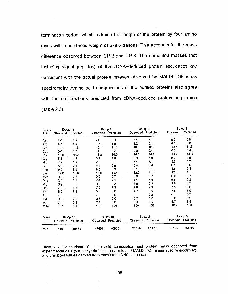
termination codon, which reduces the length of the protein by four amino
acids with a combined weight of 578.6 daltons. This accounts for the mass
difference observed between CP-2 and CP-3. The computed masses (not
including signal peptides) of the cDNA-deduced protein sequences are
consistent with the actual protein masses observed by MALDI-TOF mass
spectrometry. Amino acid compositions of the purified proteins also agree
with the compositions predicted from cDNA-deduced protein sequences
(Table 2.3).
Amino Bc-cp 1aAcid Observed Predicted
Bc-cp 1 bObserved Predicted
Bc-cp 2Observed Predicted
Bc-cp 3Observed Predicted
Ala
ArgAsxCysGIxGIyHislieLeuLysMetPheProSerThrTrpTyrValTotal
9.04.710.10.018.65.12.25.99.512.0002.40.97.25.0
0.37.1100
8.54.511.50.716.24.91.97.59.610.60.72.10.58.25.40.00.07.1100
9.04.710.10.018.65.12.25.99.512.00.02.40.97.25.0
0.37.1100
8.94.511.60.716.94.92.16.89.910.40.72.10.27.85.60.00.06.8100
6.44.210.80.016.15.93.45.49.112.20.04.12.97.94.7
0.56.4100
5.73.112.90.414.85.93.76.69.411.40.75.90.97.93.90.20.06.6100
6.34.110.70.016.76.33.76.18.612.60.05.61.67.53.5
0.06.7100
5.63.311.80.414.85.93.76.59.311.50.76.30.98.83.90.20.06.5100
Mass Bc-cp 1aObserved Predicted
m/z 47461 46880
Bc-cp 1bObserved Predicted
Bc-cp 2Observed Predicted
47461 46962 51550 51437
Bc-cp 3Observed Predicted
52129 52016
Table 2.3. Comparison of amino acid composition and protein mass observed fromexperimental data (via ninhydrin based analysis and MALDI-TOF mass spec respectively),and predicted values derived from translated cDNA sequence.
38

In addition, all three transcripts code for a signal peptide sequence,
indicating that these proteins are secreted via secretory vesicle pathways.Cleavage between G-21 and D/A-22 for the two CP-1 variants and betweenG-18 and L-19 in CP-2/CP-3 is predicted by "SIGNALP" (EXPASY) and
matches the N-terminal sequence derived by Edman sequencing. BLAST
searches (NCBI) resulted in no matches with E values of less than 109 forCP-Ia and CP-Ib and 10"5 for CP-2 and CP-3 in the database of sequences
from eukaryotic species, indicating that the capsule proteins are not strongly
homologous to any known eukaryotic proteins.Far-ultraviolet (UV) CD spectroscopy was used to examine the
secondary structure of CP-I and CP-2 in solution. CP-3 was unable to bepurified in large enough quantities to perform CD on. Lyophilized HPLCpurified capsule proteins were resuspended in 5OmM acetate buffer pH 4.0(lyophilized proteins do not resolubilize at more neutral pH values), andspectra collected at room temperature. The CD spectra of both CP-1 andCP-2 (Fig 2.5) were characteristic for an single coil a-helical structure,exhibiting minima at 208 and 222nm and a [?]222:[?]208 ratio of <1 29. Analysis
by the Selcon3 algorithm to derive secondary structure composition wasperformed and the results tabulated (Table 2.4). Selcon3 was chosen over
39

other algorithms because it
most accurately predicted the
CP-1OO oO J
I 1
X!X -2CD
190 200 210 220 230 240 250 260
a.Wavelength (nm)
CP-2
secondary structure com- ?' OE
TJ
position of human fibrinogen, a <n oE
soluble protein trimer with §> "?"
coiled-coil structures (data not
shown).
CD was also performed
on a 1:1 molar concentration
mixture of CP-1 and CP-2. This
also produced similar a-helical
spectra with a [?]222:[?]208 ratio? -1
of <1 which would indicate a 2.
single coil conformation (data ~ % 200 210 220 230 240 250 260Wavelength (nm)
not shown).
Purified protein sol- Figure 2.5. Circular dichroism spectra of purified, Bc-cps. Figs a) and b) are CP-1 and CP-2
utions, upon the evaporation Of respectively. Bc-cp 3 was unable to be purified insufficient quantities to measure with CD. Minima at
solute self-assemble into 208nm and 222nm and a [?]222:[?]208 ratio of <1are representative of single coil a-helix. The
_. spectrum represents an average of 10 scans,anisotropic nano-filaments < 20
nm in diameter, which bundle into fibers that are 200-500 nm across (Fig
2.6). This is similar in size to the fibers observed in mature egg capsules5.
40
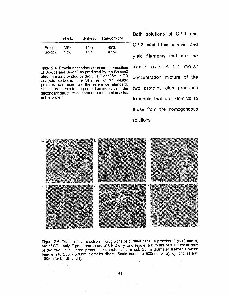
a-helix ß-sheet Random coil
Bc-cp1 36% 15% 49%Bc-cp2 42% 15% 43%
Both solutions of CP-1 and
CP-2 exhibit this behavior and
yield filaments that are the
Table 2.4. Protein secondary structure composition same size. A 1:1 molarof Bc-cp1 and Bc-cp2 as predicted by the Selcon3algorithm as provided by the Olis GlobalWorks CD concentration mixture of theanalysis software. The SP2 set of 37 solubleproteins was used as the reference standard.Values are presented in percent amino acids in the two proteins also producessecondary structure compared to total amino acidsin the protein. filaments that are identical to
those from the homogeneous
solutions.
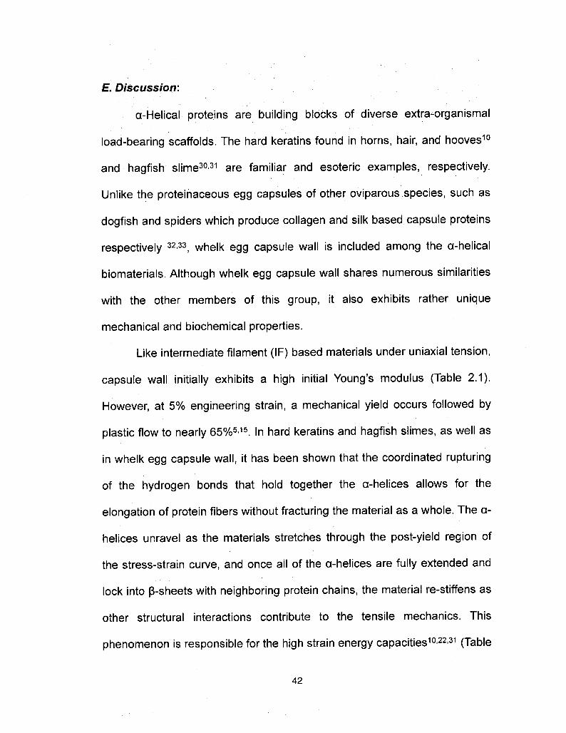
* '
d.
W
MC
Figure 2.6. Transmission electron micrographs of purified capsule proteins. Figs a) and b)are of CP-1 only, Figs c) and d) are of CP-2 only, and Figs e) and f) are of a 1:1 molar ratioof the two. In all three preparations proteins form sub 20nm diameter filaments whichbundle into 200 - 500nm diameter fibers. Scale bars are 500nm for a), c), and e) andIOOnmforb), d), and f).
41
E. Discussion:
a-Helical proteins are building blocks of diverse extra-organismal
load-bearing scaffolds. The hard keratins found in horns, hair, and hooves10and hagfish slime30·31 are familiar and esoteric examples, respectively.
Unlike the proteinaceous egg capsules of other oviparous species, such as
dogfish and spiders which produce collagen and silk based capsule proteinsrespectively 32·33, whelk egg capsule wall is included among the a-helical
biomaterials. Although whelk egg capsule wall shares numerous similarities
with the other members of this group, it also exhibits rather unique
mechanical and biochemical properties.
Like intermediate filament (IF) based materials under uniaxial tension,
capsule wall initially exhibits a high initial Young's modulus (Table 2.1).However, at 5% engineering strain, a mechanical yield occurs followed by
plastic flow to nearly 65%5·15. In hard keratins and hagfish slimes, as well asin whelk egg capsule wall, it has been shown that the coordinated rupturing
of the hydrogen bonds that hold together the a-helices allows for the
elongation of protein fibers without fracturing the material as a whole. The a-
helices unravel as the materials stretches through the post-yield region of
the stress-strain curve, and once all of the a-helices are fully extended and
lock into ß-sheets with neighboring protein chains, the material re-stiffens asother structural interactions contribute to the tensile mechanics. This
phenomenon is responsible for the high strain energy capacities10'22·31 (Table
42

100.
coQ.
è 10U)(Oo>
55 Hagfish SlimeaMerino Wool aEgg Capsule0
0 50 100 150 200 250
Strain (%)
2.1, Fig 2.7). However, it is the
extensibility and recoverability
of whelk capsule wall that sets
it apart from these other
materials. Although not quite as
extensible as hagfish slime,
capsule wall has the ability toFigure 2.7. Engineering stress vs strain curves forwhelk egg capsule material, hagfish slime, and ¡t oriainai COnforma-wool. Values for hagfish slime and wool are taken recover its originai conrormafrom Fudge and Gosline (2004)11.
tion when the load is removed
whereas hagfish slime remains extended even after unloading. Hydrated a-
keratins also relax slowly, dissipating energy, but are not as extensible as
the whelk capsule wall. Most remarkable is the capsule wall's ability to
rapidly and completely recover initial stiffness following multiple cycles of
tensile strain. Biochemical differences between these materials may help
explain their differences in mechanical properties.
Proteins comprise over 90% of the dry weight in whelk egg capsule
wall5·15 and the CPs characterized in this work represent the most abundant
of these18. The amino acid composition is consistent between both individual
CPs as well as the bulk capsule wall material and is predominantly made up
of helix forming and helix indifferent residues. The composition is also very
similar to the filament component of hard keratins with the major difference
coming in the cystine and lysine contents. Cystine is the primary crosslinking
43

molecule in keratins10, and previous studies have suggested lysine as a
potential crosslinker in capsule walls34·35.
In agreement with the helix favoring amino acid composition, circulardichroism data presented here indicate that these precursors are strongly a-
helical in solution. Very probably, these are the same proteins that contribute
to the anisotropic a-helix x-ray scattering patterns and, hence, the a-helix «-»
ß-sheet transition and associated mechanical properties reported earlier.Using the composition results from Selcon3 analysis geometric predictionsof CP dimensions have been made using calculations detailed in the
Methods section of this paper.
The maximum end-to-end distances for CP-1 and CP-2 in a relaxed
state are 50.3 nm and 56.4 nm, respectively. This assumes that the intra
protein ß-sheet domains are antiparallel and are folded over on themselvesto create this secondary structure. When egg capsule material is stretched
a-helices are unraveled and ß-sheets form between neighboring proteins. If
these newly formed ß-sheets are parallel in conformation, the end-to-endlengths increase to 76.3 nm and 89.1 nm, resulting in a 51.7% and 58.0%increase in end-to-end length. If the ß-sheets are antiparallel, the lengths
would increase to 79.3 nm and 93.0 nm respectively, causing a 57.7% and
64.9% gain respectively (Fig 2.8a). All of these values are close to the -60%post-yield flow strain observed in bulk egg capsule material and further
support the hypothesis that this is the result of an a-helix «-» ß-sheet44

transition. Potential inter and intra-protein crosslinks in the random coil
domains of the proteins would prevent unfolding during extension of the bulk
material (Fig 2.8b). This is also hypothesized in the case of hard keratin
materials, where disulfide linkages prevent the unraveling of the 1B and 2Bhelical domains22.
Like IF materials, x-ray scattering studies have suggested that the a-
helical filaments of whelk capsule walls are in a coiled-coil motif5. This
tertiary structure is the result of a heptad repeat (amino acids A through G)in which apolar residues at positions A and D form a hydrophobic band thatbrings the two helices together, and the flanking charged residues E and Gprovide electrostatic stabilization27. Sequence analysis via the Lupasmethod36 indicates several short patches of heptad repeat domains in each
of the four capsule proteins (Fig 2.3). Additional analysis by Berger'smethod37, suggests that these areas are more likely to form coiled-coiltrimers in contrast to the dimers found in hagfish slime and keratin materials
(Fig 2.8b).
45

CP-1
50.3 nm
Relaxed EWfflfflülHE
Extension a-helix «-» ß-sheet transition
Parallel ß-sheets: 76.3 nm 51 .7% increase
CP-256.4 nm
Relaxed I—— t-Tfl
Extension a-helix <-+ ß-sheet transition
Parallel ß-sheets: 76.3 nm 51.7% increase
Anti-parallel ß-sheets: 79.3 nm 57.7% increase Anti-parallel ß-sheets: 79.3 nm 57.7% increase
Figure 2.8. Schematics for the structure function model of both individual proteins, as wellas predicted protein trimers. Fig a) shows single protein a-helical domains unraveling intoß-sheets during extension. Secondary structures are presented in singular domains forclarity. The random coil and ß-sheet domains in the relaxed protein remain unchangedunder extension. Depending on whether the a-helices transition into parallel or antiparallelß-sheets, which is unknown as of this point, the increases in end to end length of theproteins are slightly different. Regardless of the ß-sheet conformation, the percent increasein length of the individual proteins is close to the -60% increase in flow strain that ishypothesized to be the result of an a-helix <->¦ ß-sheet transition in the bulk material. Fig b)shows a model for the capsule protein multi-domained coiled-coil trimer. Inter and intra-protein crosslinks prevent random coil regions from extending under tension. End to endcrosslinks connect trimers to each other to maintain a continuous covalently linked protein
These heptad repeat domains are comparatively small, between 22
and 34 residues long for CPs 1a and 1b and between 15 and 30 residues
long for CPs 2 and 3, and are separated by long stretches of non multi-coil
sequence. This is far shorter than the >100 residue coiled-coil domains
commonly found in IFs38. Additionally, the circular dichroism data suggest
that in solution, purified capsule proteins preferentially adopt single coils
rather than coiled-coils as indicated by the [?]222:[?]2?ß ratio of <1. Either the
quantity of coiled-coil helices is too small compared to the single coiled
46

helices to shift the [?]222:[?]208 ratio from less than 1 to greater than 1 , or the
transition from single to multi-coils occurs during filament assembly.
During the egg laying process, egg capsule proteins first assemble
into filaments in the immature egg capsule before they are crosslinked18.
However, as with purified IF proteins, where it is postulated that changes in
the pH and/or ionic strength of the solution cause electrostatic interactions incritical amino acid side-chains39·40, egg capsule filament assembly can occur
in vitro without the aid of additional accessory proteins/molecules. Based on
the similar morphologies of the fibers created in vitro observed here by TEM,
and those of the naturally produced fibers observed previously by SEM5,
purified capsule proteins form the filaments that the mature egg capsulematerial is composed of.
Intermediate filaments that form heterodimeric coiled-coil structures
such as keratins in vivo, will not form filaments in vitro under any conditions
unless both proteins are present in the solution40. It is therefore likely thatthe filaments formed in whelk capsule wall are not heterogeneous, as
purified homogenous solutions of CP-1 and CP-2 are capable of formingfilaments on their own. However, as both CPs form assemble into filaments
of identical size, the exact roles of the two different proteins and whether
both are necessary to form egg capsules in vivo remain to be determined.Proteins from IF based materials form coiled-coil tetramere, and sometimes
octamers, in solution before assembling into filaments39·40. A more thorough
47
investigation into the self-assembly of capsule proteins will be necessary to
determine whether they also form intermediate multimers in solution prior to
filament assembly.
After assembly, crosslinking is the final processing step for the egg
capsules before they are deposited on the ocean floor. It is only aftercrosslinking that the egg capsules exhibit the robust mechanical propertiesof the mature capsule that have been described. Previous work on both inthe egg capsule material itself34 as well as in the gland where this finalprocessing step occurs35 have suggested that these crosslinks are lysinederived. As mentioned above, all of the Bc-cps have high quantities of lysine
in the amino acid composition. In CPs 1a and 1b, the distribution of lysine is
fairly constant throughout the primary sequence, with some patches ofhigher density. In CPs 2 and 3, there is a very high proportion of lysine in theC-terminal region. CP-2 and CP-3 have 53 and 54 lysines in 475 and 479amino acids respectively, but 20 and 21 of those lysines between residue413 and the C-terminus (Fig 2.9). This region of the primary sequence
seems a likely position for end-to-end crosslinking of these capsule proteins.However, until a chemical structure for the crosslink is determined, nothing
definitive can be said about its location in the primary sequence of any of the
capsule proteins.
48

Although there are Lysine locationss Agnnine locationswi "13 _ . ______ „ ..„...„
striking similarities between cp lbwhelk egg capsule wall and IF ?^2materials in terms of mech- cp-3anics, amino acid composition
Figure 2.9. Distribution of lysine and arginine, .. . . . residues throughout the primary sequence ofof the precursor proteins, and capsU|e proteins. Lysine is a suspected
crosslinking amino acid in capsule wall material. Itthe ability to self-assemble into ¡s shown here that there are distinct clusters of not
only lysine but also arginine, which preliminary data. suggests may also contribute to crosslinking, infilaments m vitro with no each of the four capsule protein sequences.
accessory molecules needed,
primary sequence analysis, indicates that the capsule proteins are very
different from intermediate filament proteins. An NCBI BLAST analysis of
similar protein sequences results in no eukaryotic proteins in the databasewith E values of less than 10"9for CP-Ia and CP-Ib, less than 10"5 for CP-2
and CP-3, and no homology to known IF proteins at all. Additionally each of
the CPs contains a signal peptide sequence at the N-terminus. This short
sequence, 21 residues long in CP-Ia and CP-Ib and 18 residues long in
CP-2 and CP-3, indicates that the capsule proteins are exported from the
cell via a controlled vesicle based secretory pathway. This is in direct
contrast to hard keratins and hagfish slime both of which reach the extra
cellular region by the programmed death of the specialized cells in which
they are accumulated41·42. Based on the ClustalW2 alignment program43 ofthe primary sequence and the strong suggestion of coiled coil trimers, the
49
capsule proteins may actually be more closely related to the fibrinogenfamily, an extracellular matrix protein involved in blood clotting and wound
response, than it is to either a-keratin or hagfish slime filament proteins(data not shown).
There remain additional unanswered questions regarding whelk egg
capsule wall processing and structure. The mature material is over 90%protein by dry weight, but the composition of the remaining mass isunknown. Furthermore, SDS PAGE of immature capsules and crude
nidamental gland extract indicate that traces of additional proteins are
present in this material, although in far lesser quantities than Bc-cp 1-3 18,and the structure and function of these needs to be investigated. A potential
role for these proteins involves composite matrix components. Hard keratinmaterials have globular, high-sulfur content matrix proteins which contributesignificantly to mechanical properties10·22, whereas hagfish slime does not11.Once these proteins are fully characterized, it will be possible to develop amodel for how they interact with the CPs characterized here, and of the
structure/function of the egg capsule material as a whole.
F. Conclusions:
In the present study the precursors of the primary structural proteinsfor whelk egg capsules were purified and characterized. The amino acidcomposition is very similar to intermediate filament proteins with the
50

exception of lysine and cystine, which are significantly higher in egg capsulewalls and in IFs respectively. Circular dichroism and TEM studies indicate
that, much like intermediate filament proteins, these precursors are strongly
a-helical in solution and are capable of self-assembling into filaments on
their own without the assistance of accessory molecules.
Using reverse genetics, the primary amino acid sequence for fourvariants was deduced from cDNA. The primary sequences are very unique
as BLAST homology searches do not produce any significant matches to
known proteins, including intermediate filaments. Analysis of thesesequences indicates that the proteins are likely to form coiled-coil trimers,which is consistent with previous x-ray scattering studies. Furthermore, the
end regions of the sequence are dense with lysine and arginine, both ofwhich are suspected to play a role in the covalent crosslinking of eggcapsule fibers.
From this data, as well as previous mechanical and structural studies,
a structure-function model for the single protein unit of this material is
proposed. In this model, the predicted increase in contour length of proteinsdue to an a-helix «-» ß-sheet structure transition ranges from 51.7% to
64.9%. This change corresponds well to the increase in overall length of theegg capsule material during the post-yield region of extension. This is theregion in which the flow modulus decreases dramatically before restiffeninglater and is where the a-helix *-* ß-sheet transition is shown to take place.
51

This model further supports Flory's theory on non-entropic elasticity in
proteins.
G. Accession Numbers:
The sequence data of Bc-cp1a, Bc-cp1b, Bc-cp2, and Bc-cp3 have
been deposited in the NCBI database with the accession numbersGU205809, GU205810, GU205811, and GU205812 respectively.
H. Acknowledgments:
The authors would like to thank Krystyna Brzezinska and James R
Neilson (Materials Research Lab, UC Santa Barbara) for assistance with theCD spectrometery and TEM imaging respectively; Douglas Fudge(University of Guelph, Ontario, Canada) for providing tensile data for Merinowool and Hagfish slime; Rebekah Ashley (Molecular Cellular andDevelopmental Biology, UC Santa Barbara) for assistance with cDNAsequencing; and Robert Shadwick (University of British Columbia,Vancouver, Canada) for insightful advice and communications through out
the project.
This work was supported by a University of California Biotechnology
Research and Education Program (UC BREP) Graduate Research and
Education in Adaptive Biotechnology (GREAT) grant (#2007-02) and NIH
grant #R01DE018468 OHW). The work made use of MRL Central Facilities
52
supported by the MRSEC program of the NSF under Award #DMR05-20415.

/. References:
1. Urry, D. W., Hugel, T., Seitz, M., H. Gaub, E., Sheiba, L1 Dea, J., Xu, J. & Parker, T.(2002). Elastin: a representative ideal protein elastomer. Phil. Trans. R. Soc. Lond[Biol]. 357, 169-184.
2. Weis-Fogh, T. (1961). Molecular interpretation of the elasticity of resilin, arubber- like protein. J. MoI. Biol. 3, 648-667.
3. Alexander, R. M. (1966). Rubber-like properties of the inner hinge-ligament ofPectinidae. J. Exp. Biol. 44,119-130.
4. Flory, P. (1956). Theory of Elastic Mechanisms in Fibrous Proteins. J. Am. Chem.Soc. 78, 5222-5235.
5. Miserez, ?., Wasko, S. S., Carpenter C. F. & Waite, J. H. (2009). Non-entropic andreversible long-range deformation of an encapsulating bioelastomer. Nat. Mater. 8,910-916.
6. Denny, M. W. (1988). Biology and the Mechanics of the Wave-Swept Environment,Princeton Univ. Press, Princeton.
7. Magalhaes, H. (1948). An Ecological Study of Snails of the Genus Busycon atBeaufort, North Carolina. Ecol. Monograph. 18, 377-409.
8. Rawlings, T. A. (1999). Adaptations to physical stresses in the intertidal zone: Theegg capsules of neogastropod mollusks. Am. Zool. 39, 230-243.
9. Aaron, B. B. & Gosline, J. M. (1981). Elastin as a random-network elastomer: amechanical and optical analysis of single elastin fibers. Biopolymers. 20,1247-1260.
10. Feughelman, M. (1997). Mechanical Properties and Structure of Alpha-KeratinFibres: Wool, Human Hair, and Related Fibres. UNSW Press, Sydney.
11. Fudge, D. & Gosline, J. (2004). Molecular design of the a-keratin composite:insights from a matrix-free model, hagfish slime threads. Proc. R. S. Lond [Biol].271,291-299.
12. Pollock, C. M. & Shadwick, R. E. (1994). Relationship between body mass andbiomechanical properties of limb tendons of adult mammals. Regul. IntegrativeComp. Physiol 35, R1016-R1021.
13. Kastelic, J & Baer, E. (1980). Deformation in Tendon Collagen. Symp. Soc. Exp.Biol. 34, 397-435.
14. Denny, M. (1976). The physical properties of spider's silk and their role in thedesign of orb webs. J. Exp. Biol. 65, 483-506.
15. Rapoport, H. S. & Shadwick, R. E. (2002). Mechanical characterization of anunusual elastic biomaterial from the egg capsules of marine snails {Busycon spp.).Biomacromolecules. 3, 42-50.
16. Lendlein, A. & Kelch, S. (2002) Shape-memory polymers. Angew. Chem. Int. Ed.Engl. 41,2035-57
17. Rapoport, H. & Shadwick, R. (2007). Reversibly labile, sclerotization-induced elasticproperties in a keratin analog from marine snails: whelk egg capsule biopolymer(WECB). J. Exp. Biol. 210, 12-26.
18. Rapoport, H. S. (2003) Biomechanics, Biochemistry, and Molecular Biology of aMolluscan Scleroprotein Elastomer: Whelk Egg Capsules. PhD Thesis, University ofCalifornia San Diego
19. Flower, N. E., Geddes, A. J. & Rudall, K. M. (1969). Ultrastructure of the fibrousprotein from the egg capsules of the whelk Buccinum undatum. J. Ultrastruct. Res.26, 262-273.
20. Goldsmith, L. A. & Lindberg K. A. (1978). Nidamental Gland Precursor of the EggCapsule Protein of the Gastropod Mollusc Busycon carica. Comp. Biochem.Physiol. B. 59B, 133-138.
21. Bendit, E. G. (1957) The a-ß Transformation in Keratin. Nature. 179, 535.
54

22. Hearle, J. W. S. (2000). A critical review of the structural mechanics of wool and hairfibres. Int. J. Biol. Macromol. 27, 123-138.
23. Vincent, J. (1990). Structural Biomaterials (revised ed). Princeton: PrincetonUniversity Press, Princeton.
24. Zhou, N. E., Zhu, B. Y, Kay, C. M. & Hodges, R. S. (1992). The two-strandedalpha- helical coiled-coil is an ideal model for studying protein stability and subunitinteractions. Biopolymers. 32, 419-426.
25. Koch, E. A., Spitzer, R. H., Pithawalla, R. B. & Parry, D. A. D. (1994). An UnusualIntermediate Filament Subunit From the Cytoskeletal Biopolymer ReleasedExtracellularly Into Sea Water by the Primitive Hagfish (Eptatretus stouti). J. CellSci. 107 3133-3144.
26. Fudge, D., Gardner, K. H., Forsyth, V. T. & Riekel, C. (2003). The MechanicalProperties of Hydrated Intermediate Filaments: Insights from Hagfish SlimeThreads. Biophys. J. 85, 2015-2027.
27. Gathercole, L. J., Atkins, E. D. & Goldbeck-Wood, E. (1993). Molecular Bendingand Networks in a Basement Membrane-like Collagen: Packing in Dogfish EggCapsule Collagen. Int. J. Biol. Macromol. 15, 81-88.
28. Hu, X., Köhler, K., Falick, A. M., Moore, A. M., Jones, P. R., Sparkman, O. D. &Vierra, C. (2005) Egg case protein-1. A new class of silk proteins with fibroin-likeproperties from the spider Latrodectus hesperus. J. Biol. Chem. 280, 21220-21230
29. Price, N. R. & Hunt, S. (1976). The occurrence of reducible compounds in aninvertebrate structure protein of Buccinum undatum (L.). Cell. MoI. Life. Sci. 32,557-558.
30. Price, N. R. & Hunt, S. (1976). An unusual type of secretory cell in the ventral pedalgland of the gastropod Buccinum undatum (L. ). Tissue and Cell. 8, 217-228.
31. Creighton, T E. (1993). Proteins: Structures and Molecular Properties (2nd ed). W.H. Freeman and Company, New York.
32. Lupas, ?., Van Dyke, M. & Stock, J. (1991). Predicting coiled coils from proteinsequences. Science. 252, 1162-1164.
33. Wolf, E., Kim, P S. & Berger, B. (1997). MultiCoil: A Program for Predicting Two-and Three-Stranded Coiled Coils. Prof. Sci. 6, 1179-1189.
34. Parry, D. A. & Steinert, P M. (1999). Intermediate filaments: molecular architecture,assembly, dynamics and polymorphism. Q. Rev. Biophys. 32, 99-187.
35. Coulombe, P & Fuchs, E. (1990). Elucidating the early stages of keratin filamentassembly. J. Cell. Biol. 111, 153-169.
36. Kreplak, L, Aebi, U. & Herrmann, H. (2004). Molecular mechanisms underlying theassembly of intermediate filaments. Exp. Cell Res. 301, 77-83.
37. Koch, E. A., Spitzer, R. H., Pithawalla, R. B. & Downing, S. W. (1991). Keratin-likecomponents of gland thread cells modulate the properties of mucus from hagfish(Eptatretus stouti). Cell Tiss. Res. 264, 79-86.
38. Rogers, G. E. (2006). Biology of the wool follicle: an excursion into a unique tissueinteraction system waiting to be re-discovered. Exp. Dermatol. 15, 931-949
39. Larkin, M. A., Blackshields, G., Brown, N. P., Chenna, R., McGettigan, P. A.,McWilliam ,H., Valentin, F, Wallace, I. M., WiIm, A., Lopez, R., Thompson, J. D.,Gibson, T J. & Higgins, D. G.(2007). Clustal W and Clustal X version 2.0.Bioinformatics. 23, 2947-2948
40. Zhao, H. & Waite, J. H. (2005). Coating Proteins: Structure and Cross-Linking infp-1 from the Green Shell Mussel Perna canaliculus. Biochemistry. 44,15915-15923
41. Paz, M., Flückinger, R., Boak, ?., Kagan, H. M. & Gallop, P M. (1991). Specificdetection of quinoproteins by redox-eycling staining. J. Biol. Chem. 266, 689-692.
42. Waite, J. H. (1991). Detection of peptidyl-DOPA by amino acid analysis andmicrosequencing techniques. Anal. Biochem. 192, 429-433.
43. Greenfield, N. (2006). Using circular dichroism spectra to estimate proteinsecondary structure. Nat. Protoc. 1 , 2876-2890.
55

Chapter 3. Thermomechanical properties of Busycotypus
canaliculatus egg capsules: entropie vs non-entropic
Adapted from A. Miserez, S.S. Wasko, CF. Carpenter, J.H. Waite, "Non-
entropic and reversible long-range deformation of an encapsulating
bioelastomer," Nature Materials, 8 (11): 910-916 (2009).
Reproduced with permission from Nature Publishing Group.
A. Abstract
Encapsulation is a widespread biological process particularly in theformation of protective egg cases of oviparous animals. The egg capsule
wall of the channelled whelk Busycotypus canaliculus is an effective shock
absorber with high reversible extensibility and a stiffness that changes
significantly during extension. Here we show that post-stretch recovery inegg capsules is not driven by entropie forces as it is in rubber. Indeed, atfixed strain, force decreases linearly with increasing temperature, whereas
in rubber elasticity the force increases. Instead, capsule wall recovery is
associated with the internal energy arising from the facile and reversible
structural a-helix^ß-sheet transition of egg capsule proteins during
extension. This behavior is extraordinary in the magnitude of energy
56

dissipated and speed of recovery and is reminiscent of strain-induced
crystallization in some polymeric fibers and of superelastic deformations
associated with diffusionless phase transitions in shape memory alloys.
B. Introduction
Engineering versatile encapsulants for pharmaceuticals and the
transplantation of delicate cells and tissues is a very active area of medical
research 1·2. Surprisingly, engineers are largely unaware of how exquisitely
well-tuned naturally occurring encapsulation strategies are. Oviparous
animals, for example, all produce eggs that possess diverse protective
capsules or shells specifically adapted to their anticipated wear and tear in
the external environment. The familiar calcified eggshell of bird eggs is
adequate protection in a nest of straw, but it would be a deathtrap for the
eggs of the channeled whelk Busycotypus canaliculus, a large marine snail.
Busycotypus eggs are released in meter-long helical strands known as
mermaid necklaces that link up to 160 capsules (Figs. 3.1a and b). These
are buffeted about for months by breakers at velocities up to 10 m/sec along
the seashores of the east coast of North America 3. Given the punishing
environment, how the Busycotypus capsules shield the delicate embryos
from damage is of fundamental interest. Recent investigations of the
Busycotypus capsule wall have found it to consist of cross-plied sheets of
protein fibers (Fig. 3.1c) with effective shock absorbing tensile properties 4·5.
57

The large and reversible extension of biopolymers in Busycotypus egg
capsule wall is strongly suggestive of an elastomer. Elastomeric proteins
play an essential role in a variety of extracellular structural tissues 6 and theirlong-range elastic properties are useful for instance as shock absorbers(e.g. in hydrated dragline spider silks 7), for elastic energy storage capacity
in jumping and flying insects 8, and to ensure adequate elasticity in the
integument and arteries of various organisms 9_11. Bioelastomers classically
exhibit rubber-like entropie elasticity 10. In this well-established framework 12,
the force / acting on a strained specimen is expressed as the sum of the
internal energy contribution and the entropie contribution, with the latter
prevailing at sufficiently large extensions (larger than 10 %) 7·10 and typically
contributing to 80-90 % of the total elastic force for rubbery materials. Our
thermomechanical and x-ray scattering analysis shows that the Busycotypus
egg capsule, albeit elastomeric in its high and reversible extensibility, is not
dominated by entropie forces. Instead, our experimental data support the
notion that high extensibility is due to a fully reversible a-helix «-» ß-sheet
structural transition upon loading. This behaviour can be explained by simple
thermodynamic equilibrium concepts and provides a clear experimental
validation of the Flory 13 model predicting that forces arising from internal-
energy can dominate long-range elasticity in polymorphic fibers. Whereas
a <-? ß structural transitions have previously been described in a-keratin
fibres, Busycotypus egg capsules show that it can be exploited to create
58
bioelastomers with higher extensibility and much less time-dependent
recovery than previously recognized. This paradigm could prove useful indesigning novel bioencapsulants for delicate tissue implants.C. Materials and methods
Busyctypus canaliculus egg capsules were purchased from theMarine Biological Laboratory (Woods Hole, MA). Individual cases were splitapart, the gelatinous contents including the embryos discarded, and theinteriors were cleaned by repeated rinses with filtered water. Gentle physical
disruption was necessary to dislodge the mucous lining. Rectangular stripsabout 10 mm long, and a 3-6 mm wide were cut from egg capsules with a
razor blade, and the cross-sectional area was measured by light microscopy.
Stress-strain analysis was performed on an MTS (MTS, Eden Prairie,
MN) Bionix 200 universal testing machine, at nominal strain rate of 10"2 s_1,using a 50 N load cell and a built-in optical encoder to measure the load anddisplacement, respectively. All specimens were tested in seawater using acustom-made stainless steel container. The temperature of the seawater
bath was regulated over the range -1 to 80 0C using an external
thermocontroller unit. The seawater bath temperature was brought to
prescribed levels by connecting the heating/coolant fluid from thethermocontroller to an inner copper tubing wrapped inside the water, hence
allowing heat transfer by conduction. Styrofoam was used to insulate theseawater container from the ambient environment, and the temperature was
59

measured within 1 cm of the specimen with a thermocouple (accuracy +/-
0.50C) during testing. Thermal expansion was corrected by re-setting the
initial gauge length to zero before each test. In addition, swelling -which is
known to significantly affect the actual volume of elastin samples at high
temperatures 11- was measured by heating an unstrained sample in water
and observing size changes under an stereomicroscope. No detectable
swelling was noticed. Control experiments were performed on a natural latex
material under identical conditions.
Poisson's ratio was measured independently using a custom-made
micrometer tensile frame. Fully hydrated samples were gripped and pulled
to strains ranging from 20 to 100 % under a stereo-microscope. Width and
thickness reductions were obtained in separate measurements, in which the
micro-tensile grips were rotated by 90° to enable focus on the thickness or
the width, respectively. All images were acquired with a Nikon D70 digital
camera and analyzed with Adobe Photoshop software (Adobe Ine, San
Jose, Ca, USA). Six measurements of width and thickness were obtained
and averaged at each strain.
The elastic "Hookean" modulus was monitored at selected strains by
partial unloading/reloading cycles as illustrated in Fig. 3.2a, at a strain rate
during the unloading section that was half of that during monotonie loading
(i.e. 5?0"3 s-1). The modulus at a given strain cycle was measured on true
stress/strain curves as the slope of the tangent of an unloading cycle at the
60
unloading strain. This tangent modulus is a more accurate measure of the
true elastic modulus at the instant of unloading; it also better captures micro-
damage or stiffening due to structural changes during straining as observed
in engineering composites and metal foams 30·31. Each loading/unloading
series was repeated three times per temperature and the relevant values for
data analysis (force, modulus, stress, strain) were averaged.
X-ray diffraction patterns were obtained from dry specimens using a
Huber Four-Circle goniometer Wide Angle X-ray Spectrometer (WAXS),
equipped with a Cu rotating anode x-ray generator (Rigaku UltraX18,
wavelength =1.54 A), and a MAR345 image plate area detector. Spectra
were acquired on pristine (unstrained), loaded, and cycled (loaded/
unloaded) specimens with a sample-to-detector distance of 282.5 mm. For
loaded specimens, the hydrated specimen was deformed to a given strain,
and then dried, thereby "locking" the specimen at that strain.
Diffraction patterns were acquired on fully hydrated samples using a
high intensity synchrotron source at BESSY (Berlin Elektronenspeicherring
Gesellschafft m.b.h, Berlin Germany) with x-ray wavelength of 1 A. Thebeam line was equipped with a custom-made in situ tensile testing
equipment 32 allowing to continuously monitor diffraction patterns during
extension. A comprehensive description of the set-up can be found
elsewhere 33. In brief, experiments were conducted with a MarMosaic 225
2D CCD detector (MAR USA, Evanston, II, USA), a having a 3072 ? 3072
61

pixel resolution, a 30 µ?? beam size, and a sample-to-detector distance of
256.4 mm. Diffraction patterns were acquired at regular strain intervals for
30 sec, as well as at complete unloading.
Specimens for SEM imaging were freeze-fractured following
immersion in liquid nitrogen. Fixation was done in a mixture of 1%
glutaraldehyde and 1% paraformaldehyde, followed by serial de-hydration in
ethanol/water mixtures up to 100% ethanol. Final dehydration was done in a
hexamethyldisilazane overnight. Fixed samples were gold-coated for 1 min.
Low magnification imaging was performed using a Vega Tescan TS5130-
MM thermionic emission SEM (Brno, Czech Republic), whereas high-
resolution imaging was done on a field emission gun FEI XL-40 microscope
(FEI, Hillsboro, Oregon). Working-distance was set at 5 mm for high
resolution imaging.
For amino acid copositional analysis, egg capsules were thoroughly
washed in milli-Q water and subsequently hydrolyzed in vacuo in a 6M HCl/
5% phenol solution at 11O0C for 24 hours. The supernatant was separated
from the solid residue by centrifugation, flash-evaporated and subsequently
washed with water and methanol to remove traces of acid and the amino
acid composition was obtained with and analyzed in a ninhydrin-based
Beckman Autoanalyzer (Beckman-Coulter, Fullerton, CA)
D. Results and Discusson
62

The microstructure of a mature Busycon capsule wall resembles
plywood in consisting of stacked sheets, each about 10-20 Mm thick 14. Each
sheet is an array of aligned fibers in which the preferred fiber orientation is
rotated by 90° from one sheet to the next (Fig. 3.1c) and results in
orthotropic mechanical properties when measured in the plane of the
capsule. At higher magnification, individual fibers with diameters of 200-500
nm and characteristic surface ridges are evident (Fig. 3.1d). Capsule
microstructure and formation is described at length elsewhere 4.
(a) (b)-xV &Z-Â
V| \x '¦ ggf' ,.
20µp?
(C)
Figure 3.1. Busycon canaliculum photographs and SEM micrographs, a, Heiical strand('mermaid's necklace') containing dozens of interconnected capsules, with a total length ofabout 1 m. b, Close-up view of a dissected capsule from which tensile specimens wereextracted, c, Low-magnification SEM image after freeze-fracture illustrating the cross-plywood structure, with the preferred fibre orientation at about 90° between adjacentsheets, d, High-magnification SEM micrograph of a few individual fibres within one sheet;fibres are typically 200-500 nm in diameter and contain characteristic surface ridges.
63

Atypical stress-strain curve (strain rate 10'2 s1) with both engineering
and true properties reproducibly illustrates the four regimes reported earlier 5(Fig. 3.2a): (1) an initial stiff region to about 5 % strain with an elastic
modulus of about 100 MPa; (2) a pseudo-yield region with a much lower
increase of flow stress with strain; (3) at strains beyond 70 %, increased
stiffening; and (4) a large hysteresis loop upon unloading during which up to
40 % of the total energy is dissipated.
The material exhibits a quasi-instantaneous recovery of strain and
modulus with the residual strain at zero load attributed to viscoelastic effects
(the residual strain was recovered in fully unloaded specimens after a few
minutes). The representative stress-strain curves in the temperature range -
1 to 800C clearly show diminished initial stiffness as well as decreased flow
stress and hysteresis with increasing temperature (Fig. 3.2c). Additional
tests (data not shown) performed at various strain rates confirmed earlier
data 5 regarding a slight reduction of hysteresis (10 %) at higher strain rates
(10-1 s-1).
64

True stress-straincurve
Engineering stress-straincurve¡Transition stiain. r I
Strain %40 SO 80
Strain [%]
0.8
0.7
0.6
— 0.5OId·- 0 4en
I 0.3°- o.a
0.1
O
(b)
..>--^--L
ThtcknGss direction
""f--·*---
-4 4 * A
Width direction
20 40 60 80 100
Strain [%] (d)
%,] 60 PCl.i ,., r* ·
[70PtIj- "¦¦' -"MOpCt!^—-"·- - -
1 go pet L/.
20 40 60
Temperature [" C]
Figure 3.2. Thermomechanical properties of whelk egg capsule material, a, Typical whelkegg capsule stress-strain curves (engineering curve and true tensile curve corrected for themeasured Poisson's ratios). At fixed engineering strains during loading, unloading/reloadingcycles were carried out, and the true modulus was obtained from the tangent of theunloading portion of a cycle on the true stress-strain curve, as illustrated. Forwardtransition stress and transition strain (defined in text) are calculated with reference to theengineering properties. The slight residual deformation on unloading is due to viscoelasticeffects, and full recovery was observed by letting the sample rest for a few minutes beforethe next cycle, b, Poisson's ratio versus strain (engineering values); each data point is theaverage of two samples measured at six different locations (n = 12) and error bars are thestandard deviation; note the distinct anisotropy of Poisson's ratios in the thickness and thewidth directions, c, Stress-strain curves with full unloading at various temperatures (±0.5°).Unloading/reloading loops at given strain intervals as illustrated in a were also carried outfor each temperature but are omitted for clarity, d, Force versus temperature plots atvarious values of strains, measured from plots in b. For both c and d, each data point is theaverage of three measures and error bars are the standard deviation.
Wide angle X-ray scattering (WAXS) analysis Busycon capsule wall
65

during deformation resulted in distinctly recognizable diffraction patterns
(Fig. 3.3a). At 0 % strain, two classical features of a-helices are detectable
15·16, i.e. (1) a broad equatorial peak centered at 1.03 nm corresponding to
the mean distance between a-helical axes (the presence ofthat reflection on
the meridian axis is attributed to the orthotopic arrangement of the egg
capsules), and (2) the 0.52 nm peak along the meridian due to projection of
the a-helix along the coiled-coil axis 15. At 70 % strain, a ß-sheet formation is
indicated by (1) the 0.465 nm equatorial reflections, due to lateral periodicity
between chains in the ß-sheet 17, and (2) the 1.03 nm peak which is now
due to the mean inter-sheet distance. Upon further stretching to 120 %, the
0.465 nm reflection on the equatorial arc was intensified, as were the
1.03 nm spots. The faint peak at 0.33 nm on the meridian approximates the
translational rise between successive residues along the protein chain in a
ß-sheet 17. At full unloading the diffraction pattern returns to that of a pristine
unstretched specimen; notably, the 0.465 nm ß-sheet feature is absent.
Time-resolved diffraction patterns acquired by synchrotron radiation of fully
hydrated samples during in-situ extension confirm earlier trends in the
0.52 nm meridian and equatorial 0.465 nm reflections (Fig. 3.3b), and the
diffraction pattern returns to that of the unstrained material upon unloading.
The slightly higher D-spacing corresponding to inter-helix and inter-sheet
distances, respectively, in hydrated samples (1.26 nm vs. 1.03 nm for dry
samples) is attributed to swelling. Taken together, the patterns are consistent
66

with a structural and reversible transition between a-helices and ß-sheets
during a stretch/relaxation cycle.
A ¦ i
0% strain
(a)
0% strain
(b)
\- 1 ? nrni
Í
70% strain
mm nm
!l2Sï&ïtî
60% strain
ioiI»*^^;°je5nmiIr"' -
?
120%, strain
1.03 ntam
H 26 nro
100%, strain
120%, strainback to 0%
100% strainback to 0%
Figure 3.3. WAXS diffraction patterns at various strains and after full unloading, a, 'Driedfrozen' sample, diffraction pattern acquired with a standard X-ray tube (0, 70, 120%, back to0%). b, Time-resolved diffraction pattern of a hydrated sample acquired with a high-intensitysynchrotron beam. Diffraction patterns were integrated with the Fit2D software and peakswere assigned according to refs 14, 15. The larger inter-helical and inter-sheet distancesobserved in the hydrated specimen (1.26 nm versus 1.03 nm in the dry state) are attributedto swelling.
Entropie elasticity manifests distinct thermoelastic properties. In force
vs. temperature plots (/ vs. T), the force at a fixed strain increases with
temperature, with the values of force being nearly proportional to absolute
temperature. This behavior is described by established thermodynamic
concepts relating the total elastic force / with its internal (/u) and entropie
67

energy (/s) components as:
f fU+fS \dllVT 7\di)VT KdIJyx + 7KdTJyJ
(3.1)
where U and S are the internal energy and the entropy of the system,
respectively; the right-hand side is from the Maxwell relationship -
(dS/dl) ={df/d T)vr On / vs. T plots at constant strain, the slope at any
given point on the curve thus corresponds to the entropy change per unit
extension, whereas the intercept at T= 0 is equal to the change of U per unit
extension. Capsule wall stress-strain curves at various temperatures (Fig.
3.2b) re-plotted as corresponding / vs. T plots for engineering strains in the
range 10 to 100 % (Fig. 3.2d) unequivocally show that force decreases with
temperature at all strains tested, contrary to rubbery elastomer behavior:
latex rubber at various temperatures conformed to the classical increase of
force with temperature (Supplementary Materials). Poisson's ratios showed
significant dimensional anisotropy (Fig. 3.2b), with values around 0.48 and
0.27 for capsular thickness and width dimensions, respectively, at
extensions of 50 % and above (calculated in engineering strain, with
corresponding true strain values of 0.83 and 0.43, respectively).
Interestingly, this results in a 15 % volume decrease during uniaxial
68

extension, a phenomena quite unusual for synthetic elastomers, but not
unexpected in biological materials 18. A correction to Eq. (1) for the observed
volume change shows that the internal energy contribution is slightly
overestimated at constant pressure 12 but does not alter the inverse stress/
temperature trend apparent from our experiments. The implication is thatvis-à-vis most known elastomers, the extensibility of Busycon egg capsules
is not dominated by entropie forces.
X-ray analysis supports a transition from a-helices present at low
strain to ß-sheets structures at high strain (Fig. 3.3), as previously
speculated in 5 . In keratin fibers, a-helical coiled-coils start to unravel at
roughly 5 % strain 16, resulting in an intermediate random coil structure as
well as some a and ß structures between 20 and 40 % engineering strain.
We measured the change in tangent modulus with strain to gain additional
insight into structural reorganization occurring during deformation (see
Materials and Methods) (Fig. 3.4). At low temperature (< 3O0C), the modulusdecreased from its initial value to a minimum at about 20 % strain. The
modulus then increased slightly to 65 % strain. Upon further stretching,
noticeable and rapid stiffening occurred. Based on the x-ray and modulus
data and similarity with results reported for keratin 16, four distinct structural
domains were identified during extension, and schematically illustrated in
Fig. 3.5 for fibers oriented along the loading axis. In domain 1, fromunstrained to 5 % strain, a-helices dominate in the capsule wall. At the
69

initiation of pseudo-yielding (5 %), unfolding of a-helices into random coils
occurs, leading to a semi-amorphous structure that is accompanied by a
decrease in stiffness. Domain 2 is present from 5 to 20 % strain. The
observed modulus is lower300
250
_ 200W
Q.¡g^ 150
50
V
? Tt^cI...... s /
........a··_'.*^*-*Gt - 50"·?G .—".''''
4 and consistent with semi-
crystalline polymers and elas-
tomers in which amorphous
domains leads to a reduction
of stiffness 12·19. The third0 20 40 60 80 100
En9. strain [pet.] domain prevails at 20 toroughly 60 % strain; here
Figure 3.4. Tangent modulus versus strain atvarious temperatures. In the temperature range ranc|om co¡|s anc| R-sheets0-30 ° C, the modulus initially decreases up to20%. strain and then increases on further straining.At T > 30 ° C, the modulus is initially constant, and coexist with the latter prog-tangent modulus versus strain then follows thetrends at lower temperatures. Each data point is ressive|y rep|acing the formerthe average of three measurements and error bars a r »are the standard deviation.
(transition regime 3). Domain 4
is a ß-sheet dominated structure, in which random coils have mostly
disappeared and re-crystallized into ß-sheets. At such high strains, the
increased modulus results from the inherently stiffer nature of ß-sheets as
well as sheet alignment along the loading axis during stretching - a stiffening
mechanism recognized for semi-amorphous polymer fibers 19. The much-
reduced hysteresis during partial loading cycles is further suggestive of a
single-phase material in this domain (see Fig. 3.2). At higher temperatures,
70

the a-helices are thermodynamically less stable, consequently, the initial
decrease in modulus was not observed at T > 30C°. Instead the modulus
remained constant up to 60 % strain, where stiffening then occurred as at
lower temperatures.
"*^!**?????£^ e=0 Native state
Domain 1 : unraveling of a-helicesinto random coils
Domain 2 : Extension ofrandom coils
Domain 3: Formation ofj^
/S-sheets from random coilsT-
Domain 4: extension andalignment of /3-sheets
•^MW^yfjJj&jJJJ^jffrfÇ^'^ « = 0 Unloading: reformation of a-helices
Figure 3.5. Schematic diagram of a-helix <-> ß-sheet transition during straining. For fibrilsoriented along the loading axis, transition is accommodated by the presence of intermediaterandom coils. Fibrils oriented at 90° relative to the loading axis (not shown) do not undergothe structural transition. The coiled coil dimer was arbitrarily chosen to illustrate the
A stress dependent interconvertibility of secondary structures is
thermodynamically analogous to that of axially oriented polymer fibers with
both crystalline and amorphous phases 19. For this type of polymorphic
system, it is convenient to use the Gibbs free energy G as the
thermodynamic potential, from which it can be shown that the melting
temperature Tis a function of applied force f, 13 and given by:
71

'iL) _/ AHKdT)P T TAL
(3.2)
with AL equal to the total length increment during crystallization and ??, the
enthalpy of crystallization. In the present case AL can be seen as the length
increment necessary to fully transform a-helices into ß-sheets, whereas ??
is the enthalpy of the a->ß phase transformation under a tensile force f.
Equation (2) is simply a restatement of the Clausius-Clapeyron relation, with
force and length replacing pressure and volume. Interestingly, this reversible
behavior is reminiscent of shape memory effects in metallic alloys in which
high reversible deformation is obtained through a reversible, diffusionlesssolid-phase transformation - the martensitic transformation 20·21 - and is alsomodeled by the Clausius-Clapeyron relation. Recalling that a-helices start to
unravel at the yield point in Busycon egg capsules (Figs. 3.3 and 3.4), it is
tempting to interpret our results within this framework, using / as the force atinitiation of yield for the conformational phase transition. Rewriting equation
(2) as:
'd(f/T)\ AH (33){ à{yi) ? AL
?
,or equivalent^ in terms of stress and strain:
'djojT)) AH (34){¿(VT) )p ~ S1-V072

where at is defined as the "forward transition stress". According to equation
(4), a plot of (at / T) vs. (1/7) should be linear with the enthalpy oftransformation or ?? per. unit volume Vo deduced from the slope. This
analysis is shown in Fig
specimens are closely
comparable, although
there are some differ-
ences in stress inten-
sity (due to uncertain-
ties in specimen thick-
ness and slight varia-
tions in orientation).
Equation (3) implicitly
requires the transform-
ation strain £t to be
temperature-independ-
ent. Plots of experimental e? vs 1/T confirm this independence (Fig. 3.6). The
whelk capsule wall is thus an unequivocal validation of the theoretical
proposition by Flory 13 that high extensibility in some elastic proteins couldbe achieved via an internal-energy dominated mechanism. It must be noted
that the entropie forces are not fully absent, especially in the regime ofrandom-coils extension. However their contribution is much lower than that
3.6, which indicates that the slopes for 3 different
1.8 x 10*
1.6x10*
1.4x10*
„ 1.2x10*
I 1 ? 10*~^„ 8x10*
6x10*
4x10*
2 ? 10*
*-?~y-y v-rj-
[ sample 2 1''
?¡? ? , ^ - ? sample "1I^-" ¦¦¦'
??/(e.?a),·' _ ,y I sample 3 1
70
65
60
55
60 -
45
40
35
302.8 ? 1 0* 3x1 0* 3.2 X 1 0* 3.4 ? 1 0* 3.6 X 1 0* 3.8 ? 1 0*
1/T[K-1]
Figure 3.6. Transition stress and strain at varioustemperatures.(Ot/T) versus (1/T) plot (left y axis) with atbeing the forward transition stress, and Et versus (1/T) plot(right y axis) with et being the transition strain, for threedifferent samples. Each data point is the average of threecycles and error bars are the standard deviation. The linearrelationship (Ot/T) versus (1/T) agrees with equation (4),whereas u is essentially independent of T.
73

of the internal energy of the phase transition happening simultaneously, and
are hence not distinguished in the experiments. Solving Eq. 3 for the capsule
data yields an enthalpy of transformation ?? = 1 .26 cal/cm3. Approximatingthe density of the egg capsules as 1.45 g/cm3 22 and the average molecular
weight of Busycon egg capsule proteins as 50 kDa 23, and finally assumingthat only half of initial coiled-coils undergo the structural transition because
of the orthotropic microstructure, we calculate a value ?? of 86.8 kcal/mol or
207 cal/mol residue (average residue mass 119 g/mole). This is in the range
of the 170 cal/mol residue reported for the a -> ß transition of poly-L-lysine
measured by differential scanning calorimetry at pH « lysyl pKa 24 at whichthe prevailing secondary structure is a-helical below a critical temperature. It
should be noted that our approximation of the a -ȧ transition enthalpy is
contingent on its being a first-order transition, an assumption that has been
considered at some depth elsewhere in connection with the a —> ß transition
of hair keratins 25 and is accepted 26.
In the a-ȧ transition regime, the stress increases with strain (Figs.
3.2a and b). This behavior is not consistent with simple thermodynamic
equilibrium concepts discussed above and merits further scrutiny. In a one-
component ideal system (i.e. fibers composed of a single protein with
uniform sequence and cross-link density) and with all fibers aligned along
the loading axis, a constant transformation stress would be expected. The
actual material deviates from ideality in three ways, and the combined
74

deviations reshape the stress/strain curve. First, nanofibrils are not uniquely
oriented to the loading axis. Fibers in different layers of the orthotropic
cross-lamellar structure experience at least two levels of local stresses
along their longitudinal axis, oy. Consider a sample with one family of fibers
oriented at an angle ? from the loading axis. At a given external load oa,
these fibers will start transforming into ß-sheets when oy equals the critical
stress, st. This condition hence is satisfied by sa =oT/cos6. A second group
of fibers oriented at 90° relative to the first group will require an external load
sa = ar/cos(90 - ?) , which differs from the first group. The presence of fibers
at various angles in the structure would necessarily lead to the coexistence
of a and ß forms in the intermediate region, spreading the transition over a
range of tensile forces rather than at a unique force.. Keeping also in mind
that the capsules do not exhibit a perfect orthotropic organization across
their thickness, one will expect a distribution of angles ? and, in turn, a
distribution of critical transformation stresses in the intermediate two-phase
region. Second, the diameter distribution of the capsule nanofibrils is not
monodisperse. At a given external force, the local stress sustained byindividual fibrils of various diameters varies, and such variations will lead to
different critical transformation stresses. A third deviation from ideality
pertains to the assumption of a one-component system. Protein extraction
from the capsules and nidamental glands suggests three electrophoretically
75

distinct proteins 23, all of which may undergo a <->ß transitions. The Gibbs
free energy for a multi-component system under uniaxial force and constant
pressure accounts for an additional chemical potential term:
dG = -SdT + fdL + JM*,- (3·5)i
where S is the entropy, n¡ the concentration of component / in the mixture
and µ? its chemical potential. At equilibrium, AG is at a minimum and
impacted by the relative concentrations and chemical potential of each
phase in the system. Under such conditions, it can be shown that equation
(3) is re-written as 19:
(HîM) -H (3.6)
where ? denotes the fraction of fibers in the a state, Mi now consists of the
enthalpy of transformation plus the enthalpy of dilution of the a-phase, and
?? is similarly defined. Because the ?? and AL terms are dependent on
the composition, the critical force / (or critical stress at) is not constant at a
given temperature. Instead, the force varies with the length as thetransformation progresses.
The large macroscopic deformation associated with egg capsule a <->
ß íransitions is consistent with the behavior of a number of coiled-coilproteins examined by atomic force microscopy. In myosin 27·28 for example,
76

reversible extensibility as high as 200 % has been reported in individual
fibers, and is explained in terms of unraveled coiled-coils. Indeed, the
myosin force-extension curves reveal a plateau of constant force that
resembles the present macroscopic experiments with Busycon capsule.
Reversible extension to 65 % during unfolding of helical to amorphous
structure prior to formation of ß-sheets is therefore not uncommon. The
large capsule hysteresis, however, in combination with instantaneous
recovery of initial modulus is rare. Myosin exhibits almost no hysteresis
(< 5 %); and keratin, which does have significant hysteresis, shows only
slow recovery 25. The slow recovery of keratin, as well as its more limited
extensibility, can be attributed to the high density of disulfide cross-links,
which are absent in whelk egg capsules 5. Disulfide bonds are also
permanently broken at high strains and temperature, resulting in incomplete
strain recovery under those conditions. Hagfish slime also exhibits an a —> ßtransition 29 but this transformation is irreversible.
The implications of these subtleties for engineering protective
encapsulants for delicate tissues such as a developing embryo are
significant. Imagine an embryo in a fluid filled rubbery sac. Given the high
resilience of rubbers, extensive deformation of the capsule walls by buffeting
would result in release of most of the stored energy, thus the fluid and
embryo would be vigorously jostled. This is not possible in Busycon
capsules because only a fraction of the energy invested in deforming the
77

capsule walls is released and the rest dissipated. Like an ideal rubber,
however, egg capsule elastomer will be as good as new for the next
challenge.
E. Conclusions
The Busycon egg capsule wall is endowed with high extensibility,
durability, and shock-absorbing capability. Thermomechanical and structural
analyses indicate that the long-range elasticity of the capsules is not
entropically dominated. Instead internal energy changes associated with a
fully reversible a <-» ß phase transition seem to be the main determinant.
Capsule thermomechanical behavior conforms to the Clausius-Clapeyron
relation as applied to phase equilibrium previously developed to model
crystallization of polymer fibers under stress, and deviations from an ideal
system are invoked to explain the increase of stress during the structural
transition. Although a <-> ß transitions during extension are not unique to
Busycon egg capsules, the capsules are extraordinarily distinctive in the
magnitude of reversible strain and their almost instantaneous strain
recovery. Complete characterization and recombinant expression of
Busycon egg capsule proteins should ultimately enable the engineering of
capsule-like materials that combine high modulus, reversible extensibility
(> 100 %), and impact-absorbing properties for the insulation of damage
prone tissues.
78

F. Acknowledgements
Thanks are due to B. Shadwick (University of British Columbia,
Vancouver, Canada) and F. Zok (Materials Department, UCSB) for helpful
criticism. Y. Li (Materials Research Laboratory (MRL), UCSB) and H. Gupta
(Max-Planck Institute for Colloids and Interfaces, GoIm, Germany) provided
critical guidance for WAXS and time-resolved synchrotron experiments,
respectively. K. Field (Mechanical Engineering Department, UCSB) helped
in developing the microtensiometer. S.W. was financially supported by aCalifornia Sea Grant # R/MP-97B and UC BREP GREAT traineeship
#2007-02. This work made use of MRL Central Facilities supported by the
MRSEC Program of the National Science Foundation under award No.DMR05-20415.
79
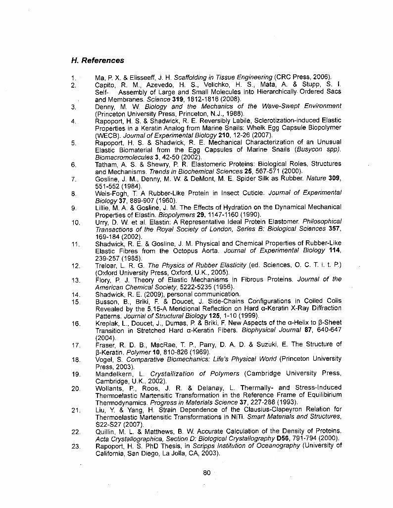
H. References
1 . Ma, P. X. & Elisseeff, J. H. Scaffolding in Tissue Engineering (CRC Press, 2006).2. Capito, R. M., Azevedo, H-. S., Velichko, H. S., Mata, A. & Stupp, S. I.
Self- Assembly of Large and Small Molecules into Hierarchically Ordered Sacsand Membranes. Science 319, 1812-1816 (2008).
3. Denny, M. W. Biology and the Mechanics of the Wave-Swept Environment(Princeton University Press, Princeton, N.J., 1988).
4. Rapoport, H. S. & Shadwick, R. E. Reversibly Labile, Sclerotization-induced ElasticProperties in a Keratin Analog from Marine Snails: Whelk Egg Capsule Biopolymer(WECB). Journal of Experimental Biology 210, 12-26 (2007).
5. Rapoport, H. S. & Shadwick, R. E. Mechanical Characterization of an UnusualElastic Biomaterial from the Egg Capsules of Marine Snails (Busycon spp).Biomacromolecules 3, 42-50 (2002).
6. Tatham, A. S. & Shewry, P. R. Elastomeric Proteins: Biological Roles, Structuresand Mechanisms. Trends in Biochemical Sciences 25, 567-571 (2000).
7. Gosline, J. M., Denny, M. W & DeMont, M. E. Spider Silk as Rubber. Nature 309,551-552(1984).
8. Weis-Fogh, T. A Rubber-Like Protein in Insect Cuticle. Journal of ExperimentalBiology 37, 889-907 (1960).
9. LiINe, M. A. & Gosline, J. M. The Effects of Hydration on the Dynamical MechanicalProperties of Elastin. Biopolymers 29, 1147-1160 (1990).
10. Urry, D. W et al. Elastin: A Representative Ideal Protein Elastomer. PhilosophicalTransactions of the Royal Society of London, Series B: Biological Sciences 357,169-184(2002).
11. Shadwick, R. E. & Gosline, J. M. Physical and Chemical Properties of Rubber-LikeElastic Fibres from the Octopus Aorta. Journal of Experimental Biology 114,239-257(1985).
12. Treloar, L. R. G. The Physics of Rubber Elasticity (ed. Sciences, O. C. T. i. t. P.)(Oxford University Press, Oxford, U.K., 2005).
13. Flory, P. J. Theory of Elastic Mechanisms in Fibrous Proteins. Journal of theAmerican Chemical Society, 5222-5235 (1956).
14. Shadwick, R. E. (2009), personal communication.15. Busson, B., Briki, F. & Doucet, J. Side-Chains Configurations in Coiled Coils
Revealed by the 5.15-A Meridional Reflection on Hard a-Keratin X-Ray DiffractionPatterns. Journal of Structural Biology 125, 1-10 (1999).
16. Kreplak, L., Doucet, J., Dumas, P. & Briki, F. New Aspects of the a-Helix to ß-SheetTransition in Stretched Hard a-Keratin Fibers. Biophysical Journal 87, 640-647(2004).
17. Fraser, R. D. B., MacRae, T P, Parry, D. A. D. & Suzuki, E. The Structure ofß-Keratin. Polymer 10, 810-826 (1969).
18. Vogel, S. Comparative Biomechanics: Life's Physical World (Princeton UniversityPress, 2003).
19. Mandelkern, L. Crystallization of Polymers (Cambridge University Press,Cambridge, U.K., 2002).
20. Wollants, P., Roos, J. R. & Delanay, L. Thermally- and Stress-InducedThermoelastic Martensitic Transformation in the Reference Frame of EquilibiriumThermodynamics. Progress in Materials Science 37, 227-288 (1993).
21. Liu, Y. & Yang, H. Strain Dependence of the Clausius-Clapeyron Relation forThermoelastic Martensitic Transformations in NiTi. Smart Materials and Structures,S22-S27 (2007). .
22. Quillin, M. L. & Matthews, B. W Accurate Calculation of the Density of Proteins.Acta Crystallographica, Section D: Biological Crystallography D56, 791-794 (2000).
23. Rapoport, H. S. PhD Thesis, in Scripps Institution of Oceanography (University ofCalifornia. San Diego, La JoIIa, CA, 2003).
80
24. Chiou, J. -S. et al. The a-Helix to ß-Sheet Transition in Poly(L-lysine): Effects ofAnesthetics and High Pressure. Biochimica et Biophysica Acta 1119, 211-217(1992).
25. Feughelman, M. Mechanical Properties and Structure of Alpha-keratin Fibres:Wool, Human Hair, and Related Fibres (University of South Wales Press, 1997).
26. Hearle, J. W. S. A Critical Review of the Structural Mechanics of Wool and HairFibres. International Journal of Biological Macromolecules 27, 123-138 (2000).
27. Schwaiger, I., Sattler, C, Hostetter, D. R. & Rief, M. The Myosin Coild-Coil is a TrulyElastic Protein Stucture. Nature Materials 1, 232-235 (2002).
28. Root, D. D., Yadavalli, V. K., Forbes, J. G. & Wang, K. Coiled-Coil Nanomechanicsand Uncoiling and Unfloding of the Superhelix and a-helices of Myosin. BiophysicalJournal 90, 2852-2866 (2006).
29. Fudge, D. S., Gardner, K. H., Forsyth, V. T, Riekei, C. & Gosline, J. M. TheMechanical Properties of Hydrated Intermediate Filaments: Insights from HagfishSlime Threads. Biophysical Journal 85, 2015-2027 (2003).
30. Kouzeli, M., Weber, L., SanMarchi, C. & Mortensen, A. Quantification ofMicrodamage Phenomena During Tensile Straining of High Volume FractionParticle Reinforced Aluminium. Acta Materialia 49, 497-505 (2001).
31. Despois, J. -F., Mueller, R. & Mortensen, A. Uniaxial Deformation of MicrocellularMetals. Acta Materialia 54, 4129-4142 (2006).
32. Gupta, H. S. et al. Cooperative Deformation of Mineral and Collagen in Bone at theNanoscale. Proceedings of the National Academy of Sciences of the United Statesof America 103, 17741-17746 (2006).
33. Paris, O. et al. A New Experimental Station for Simutaneous X-Ray MicrobeamScanning for Small- and Wide-Angle Scattering and Fluorescence at BESSY.Journal ofApplied Crystallography 40, S466-S470 (2007).
81

Chapter 4. Using wide angle x-ray scattering to characterize
the phase transition from a-helix to ß-sheet as a result ofuniaxial tension
A. Abstract:
Synchrotron source x-ray radiation has been a valuable tool in the
structural analysis of biological materials. This tool can be coupled with in
situ tensile testing to provide additional information on how structural
changes directly effect the mechanical properties of such materials. The
following study uses this approach to analyze the a-helix <-* ß-sheet
transition in a proteinaceous material that marine gastropods use in egg
capsules. This material displays remarkable shape memory behavior and it
has been theorized that an a-helix <*-> ß-sheet transition is responsible for
the mechanical properties associated with that behavior. Evidence is
presented that the cooperative rupturing and reforming of the hydrogen
bonds that stabilize these two protein secondary structure is directly related
to changes in modulus, as well as hysteresis throughout an entire shape
memory cycle.
B. Introduction:
82

Shape memory materials are being developed for a wide range of
applications including heavy construction (in bridges, for example)1·3,
switches and actuators4-6, and implanted biomedical devices (vascular
stents7'9 and encapsulants10). Reversible crystalline transitions have been
implicated in a variety of shape memory materials including metal alloys11,
synthetic polymers12'14, and biological materials15. In these systems, when
an external force is placed on the material, a shift in the atomic lattice occurs
to accommodate it, resulting in a change in crystal structure to a metastable
state. Once the force is removed, the material returns to the original, more
thermodynamically favorable crystalline conformation.
The strain induced a-helix <-> ß-sheet transition has been observed
in keratin fibers via x-ray scattering since 193116. As coiled-coil a-helices are
subjected to a load along the fiber axis, the hydrogen bonds responsible for
the stabilization of the helices rupture and the peptide unravels and
elongates. During this elongation, new hydrogen bonds are created between
adjacent fibers, forming a ß-sheet crystalline structure. When load is
removed the process reverses, hydrogen bonds stabilizing the ß-sheets
rupture, and fibers return to the preferred a-helical conformation. This
reversible transition contributes to a degree of shape memory properties in
keratins1517.
This phenomenon has been observed in a variety of other
intermediate filament-like fibers such as vimentin18 and hagfish slime
83

threads19, and has been conjectured to occur in other materials such as
desmin20 and fibrin in blood clots21. Once thought to be a one step transition
wherein ß-sheet content increased in direct correspondence with a-helix
destruction and vice-versa22, new evidence indicates that the transition is
actually a two-step mechanism wherein a-helices unravel into non-
crystalline random coils before a critical strain is reached, at which point the
peptides will form hydrogen bonds and adopt a ß-sheet conformation23.Numerous examples of human diseases are believed to result from the
irreversible structural transition of a-helices to ß-sheets24 and of mutations to
intermediate filament proteins25·26. As such, it is of fundamental importance
that this phase transition be thoroughly examined.
The effect that a crystalline phase transition has on the tensile
mechanical properties of a material has been reported for non-biological
systems such as shape memory alloys11 and synthetic shape memory
polymers12. However, although there is considerable speculation on the
subject17·23, how the a-helix ++ ß-sheet transition affects the tensile
properties of protein fibers has not been directly investigated. One model
system that is particularly well suited for doing this is the egg capsule wall ofmarine prosobranch gastropods, commonly know as whelks. This material
demonstrates long-range reversible extensibility and recent studies have
shown that this property is the result of the a-helix <-? ß-sheet transition
rather than entropie elasticity10. It is suggested that this transition contributes
84

heavily to the remarkable shape memory properties of this material which
have been studied extensively10·27·28. The following report utilizes wide angle
x-ray scattering (WAXS) with in situ tensile testing to directly correlate
changes in protein secondary structure to mechanical properties in real time
and on the same sample.
C. Materials and Methods:
Busycotypus canaliculars egg capsules were purchased from the
Marine Biological Laboratories (Woods Hole, MA). Capsules were cut with a
razor into approximately 5 X 10 mm strips and washed repeatedly in Dl
water using gentle physical disruption to remove the mucous lining. The
exact thickness and width of each strip was measured by micrometer.
A custom-made, portable uniaxial tensile testing apparatus called the
Micro-mechanical Tensile Apparatus (MiTA) was used29. A nebulizer directed
water vapor to ensure complete hydration of the sample throughout the
complete tensile cycle. A strain rate of 8 µ??/sec was used for both extension
and relaxation and grip separation distance of the device calipers was used
for sample length. There was no pause between extension and relaxation.
Results are given in Engineering stress vs strain.
Data collection was performed on the MuSpot beamline at the
BESSY synchrotron source (Berlin Elektronenspeicherring Gesellschaft
m.b.H., Berlin, Germany)30 with an X-ray wavelength of 1.0 A and a beam
85

size of 30 µ?t?. Beamline calibration was carried out with a corundum (AI2O3)
standard, giving a sample to detector distance of 256.39 mm. A 2D CCD
detector (MarMosaic 225, Mar USA, Evans- ton, USA) with pixel size of
73.24 pm and resolution of 3072 X 3072 pixels was used to acquire the
WAXS frames.The MiTA was situated so that the sample was directly in the
beamline to perform realtime in situ tensile testing. Samples were exposed
to x-rays for 30 second durations while the MiTA was actively recordingtensile data. As the strain rate was so low, the engineering strain of the
sample sample changed by no more than 2% during exposure times.
Analysis was performed using the Fit2D program31. Intensity around
the meridian and equator was integrated radially using the CAKE command,
and the shift and change in peaks were tracked as a function of externally
applied stress and strain. Background (empty beam) intensities were at least
an order of magnitude smaller than the intensity from the sample at the
wavevectors corresponding to both the a-helix 0.53 nm and ß-sheet 0.47 nm
reflections, and were not subtracted separately but were included in the
linear background term. To account for the decrease in sample thickness
during stretching, a scaling factor that allowed the intensity profiles to
superimpose at wide diffraction angles was applied, as described
previously23.
D. Results:
86

0.53 nm
\
When a protein material is exposed to x-rays, the two dimensional
scattering pattern can indicate the secondary structure crystallinity, as wellas the orientation of these structures in the material. The the WAXS data
presented below focus on the a-helix and ß-sheet specific peaks
corresponding to 0.53 nm along the
meridian and 0.47 nm along the
equator respectively (Fig 4.1). These
values have been previously estab-
lished and detailed23. The weak ß-
sheet specific 0.33 nm peak along the
meridian was unfortunately obscured
by an amorphous halo in the scat- 0% Straintering data.
WAXS of an unstrained sample
produces a double peak around 0.53 _ ._y 0.47 nmnm along both the meridian and the \equator indicative of coiled-coil a- ^t 'helices. (Fig 4.2). These data suggest
that this material has coiled-coil a-
helical fibers running orthogonally at100% Strain
87

90° to each other, producing these Figure 4. 1. Two dimensional WAXS framesof whelk egg capsules at 0% and 100%strain. At 0%, the material displays banding
spacings along both equatorial and patterns characteristic of a-helical fibersorthogonal to each other (0.53 nm along
meridianal axes which is in agree- both axes). At 100% the pattern changesto displaying ß-sheet structure along theaxis of extension (0.47 nm along the
ment with previously published equator).
findings10.
WECB is comparatively stiff at strains up to ~4% with a modulus of
69.67 MPa. After this point, there is a yield event as flow modulus drops
dramatically to 1.69 MPa. This results in a post-yield plateau between 4%and 45% where this stiffness is maintained. Just past 45% strain, flow
modulus increases to 12.31 MPa and is maintained until 100% strain which
is in agreement with previous work32.
The recovery curve differs drastically from the deformation curve,
resulting in an energy dissipating hysteresis. First there ¡s a very steep dropin stress as strain decreases from 100% to 65%. Then there is a recovery
plateau wherein stress does not decrease much as strain lowers further to10%. Below this point, stress decreases rapidly until it reaches 0 at 2.5%
strain, indicating some non-recoverable plastic deformation in the material.
(Fig 4.3).
Previous studies have detailed the repeatability of multiple load
cycles on the same sample (Fig 4.3 inset)32. As the tensile data here areconsistent with these previous works through one complete cycle, multiple
cycles on these samples were not conducted.
88

3.0
ftf*>*i
"«saa
3.5 4.0 4.5 5.0
D-space (Â)
SSco
¿r\f>ésf** 0%X
2%
4%
!^BS* 25%£&*&£&&&· 40%
SW ^^«s 60%Hfäs^5* 80%
100%
3.0 3.5 4.0 4.5 5.0
D-space (A)5.5 6.0
100%¿ss 80%
^%js^äs 60%40%
CD?
CO
*»*8»??
100%80%
60%
40%
3.0 3.5 4.0 4.5
D-space
5.0
(A)5.5 6.0
Figure 4.2. One dimensional WAXS plots of whelk egg capsules as they are being extendedto 100% strain, and as they are being relaxed back to 0% after this extension, a) and b)represent the meridian and equator respectively under tension. The 0.53 nm meridian peakcorresponding to a-helix structure decreases while the 0.47 nm equator peakcorresponding to ß-sheet structure increases, c) and d) are the meridian and equator duringrelaxation after extension. Here the transition reverses with a-helix intensity increasingwhile ß-sheet intensity decreases.
89

16
14
12
£ 10
t/1
S 6 ¦{4
2
0
- ß-sheet scattering intensity- a-helix scattering intensity
itress (??)
20 40 60-----1 1 1 1 1 1 | ' G- > ! ' I
80 100 80 60 40 20 0Strain (%)
?
?'
i-t*
Ol
C
Figure 4.3. Tensile and structural data for a complete shape memory cycle, extension to100% strain and relaxation back down to 0% strain, a-helix unraveling during extension andformation during relaxation occurs at a fairly constant rate while ß-sheet formation anddestruction happens in a stepwise manner, supporting the two step a-helix -<-? ß-sheettransition model. Inset is a stress strain curve of whelk egg capsules undergoing sequentialshape memory cycles to demonstrate the repeatability of the tensile behavior.
As the material is uniaxially strained from 0% - 4%, the intensity and
location of the double peak at 0.53 nm does not change (Figs 4.2a, b and
4.3). However, once the material is strained beyond the yield point, the
intensity of the meridian 0.53 nm peak begins to decrease indicating that the
a-helices aligned along this axis of stress are unraveling. However, there is
no indication of ß-sheet formation as there is no increase in the 0.47 nm
equatorial peak. This peak does not appear until the material reaches 40%
strain, just before the material starts to restiffen at 45% strain. As it is
extended past 45% strain, this ß-sheet specific peak increases in intensity
90

until 100% strain is reached. By this point the 0.53 nm meridian peak has
almost completely vanished.
It should also be noted that during this loading cycle that the 0.53 nm
peak on the equator, corresponding to the a-helices that are perpendicular
to the axis of tension decreases in intensity. This is not due to unraveling of
a-helices, but is likely due to the decrease in a-helical fiber density along this
axis. A model for this has been previously published27.
Upon relaxation after 100% strain, the order of events is similar to a
step by step reversal of the a —* ß transition that occurred under tension
(Figs 4.2c,d and 4.3). First the 0.47 nm equator peak decreases in intensityas the material relaxes towards 40% strain while, simultaneously, the 0.53
nm meridian peak intensity increases. Once the material relaxes further, the
0.47 nm peak disappears entirely, but the 0.53 nm does not return to
maximum intensity until strain drops below 10%. This corresponds in the
downward "jump" in the stress strain relationship at ~9%.
E. Discussion:
Recently synchrotron source x-ray radiation has been combined with
in situ tensile mechanical testing to directly investigate the change in
morphological crystallinity in materials while they are being put underuniaxial load. Studies have focused on both wide angle and small angle
scattering on synthetic polymers33'35 as well as biological materials such as
91
collagen36, silk37, and the mussel byssus38. Although synchrotron x-ray
studies have been applied to study the a-helix «<->. ß-sheet transition in
keratins while extended23, this is the first study to couple WAXS with in situ
mechanical testing on the same sample to examine the secondary structure
changes throughout a complete shape memory cycle (extension and
relaxation).
Data presented here are in agreement with previous studies
suggesting that the a-helix —? ß-sheet transition is a two step mechanismwherein the a-helices first unravel into an non-crystalline intermediate before
locking into ß-sheets after extended further23, a-helix structure is completely
maintained throughout the pre-yield region up to -5% strain and it is only
after the mechanical yield point that the a-helix content begins to decrease.
This provides clear evidence that helix-stabilizing hydrogen bonds remain
intact during the initial high modulus region, and that the rupturing of these
bonds directly corresponds with the decrease in stiffness of the material, and
supports the model that some supramolecular mechanism, such as fibril
sliding, is responsible for the initial high modulus, but that when the
hydrogen bonds rupture under load, the unzippering of the a-helices causes
the dramatic decrease in stiffness observed at the yield point (Fig 4.4).
92

Helix stabilizing hydrogen bonds
lnterfibrillar connections
Extension prior to yieldlnterfibrillar links contributeto stiffness as helices remainintact
tandrc
-bonds rupture sequentiallyExtension during yield | and helices unravel causing
drop in stiffness
Figure 4.4. Schematic for the contributions of lnterfibrillar interactions and hydrogenbonding to modulus before and during yielding. The exact nature of the interfibrillarinteractions (direct crosslinks, matrix interactions, etc) is unknown as of this point.
Stiffness does not increase again until ß-sheet formation begins. A
previous model contends that this restiffening in keratin is the result of a-
helix unraveling reaching high density cystine crosslink regions in the fiber.
93

Although this cannot be discounted in the keratin system, whelk egg capsule
proteins do not have cystine crosslinks. Additional work into the precise
nature of covalent crosslinks in these proteins will be needed to apply this
model to the whelk egg capsule system. However, data here provide direct
evidence to support a hypothesis that the increase in hydrogen bond density
resulting from ß-sheet formation causes this increase in modulus.As the material relaxes from 100% to 0% strain, the secondary
structure transition reverses. It is proposed here that this is also a two step
mechanism wherein ß-sheets first break down into random coils before
forming a-helices. This is supported by the fact that although ß-sheet
destruction is complete somewhere between 40% and 25% strain, a-helix
formation continues until past 10% strain, indicating that helices are being
formed from random coils at this point. However, although this ß-sheet—>
a-helix crystalline phase transition is seemingly a mirror image of the a-helix
—+ ß-sheet transition observed during extension, the stress strain profile is
vastly different upon relaxation, resulting in a large hysteresis loop.
Hysteresis is typically attributed to either internal friction of material
components, the existence of metastable equilibria associated with the
system dynamics, or a combination thereof 1·39·40. Although there is a greatbody of literature on the mechanics of the a-helix—? ß-sheet transition in
keratins, there a relative dearth regarding the ß-sheet—? a-helix transition
and the hysteresis that accompanies it. One model suggests that the
94
reforming of stabilizing electrostatic interactions is slowed during relaxationand that this combined with interfibrillar friction causes the observed
hysteresis, and is based off of tensile testing in media at varied pH41. As thepH is lowered further, the proteins shift to positive net charges, disruptingnative electrostatic interactions and reducing hysteresis. However, the same
effect is observed when whelk egg capsules are exposed to heated water10
and non-charged dénaturants such as urea42, both of which should have no
effect on electrostatics. Likely, the acidity of the media changes the charge
structure of helix forming residues' side chains, such as glutamate, and
render them less prone to forming a-helices.
Additionally, this model does not take into account that once ß-sheets
are formed and stabilized by hydrogen bonds, it will take an energy input to
rupture these bonds. The amount of force required to rupture a hydrogen
bond is -A pN, and force spectroscopy studies on ß-amyloid fibers has
shown that upwards of several hundred pN is required to rupture all of the ß-
sheet structure of a single molecule43. Figure 4.5 shows that during
relaxation, ß-sheets are stable at much lower stresses than they are during
extension indicating that additional energy is required to rupture thesebonds.
Although the energy required to rupture the ß-sheet stabilizing
hydrogen bonds does contribute to the hysteresis of the material, there must
be an additional component. This is evidenced by the fact that during
95

relaxation the stress values at a given strain are always less than those
values at the same strain during extension. If the energy to break ß-sheets
was the only contributing factor, the relaxation stress values would equal the
corresponding extension values once all of the ß-sheets were destroyed.However, this is not the case, and it is possible that interfibrillar friction is
responsible for this.
m- ß-sheet scattering intensityD- a-helix scattering intensity
... __ ?~ " ¦ 1Y'''
' 'V
..... —. 1 . r—+—l ' ? ' ' ' ' 1 '0 2 46 88 64 2 0
Extension Relaxation
Stress (MPa)
Figure 4.5. Plot of a-helix and ß-sheet scattering intensities versus stress. During therelaxation part of the cycle, ß-sheets exist at lower stresses suggesting that they are morestable and require additional energy be destroyed.
Figure 4.6 summarizes the current model for how the a-helix ++ ß-sheet transition directly affects tensile mechanics.
C
£rTOi—
?TO
C .B_c
Ç
TO?in
96
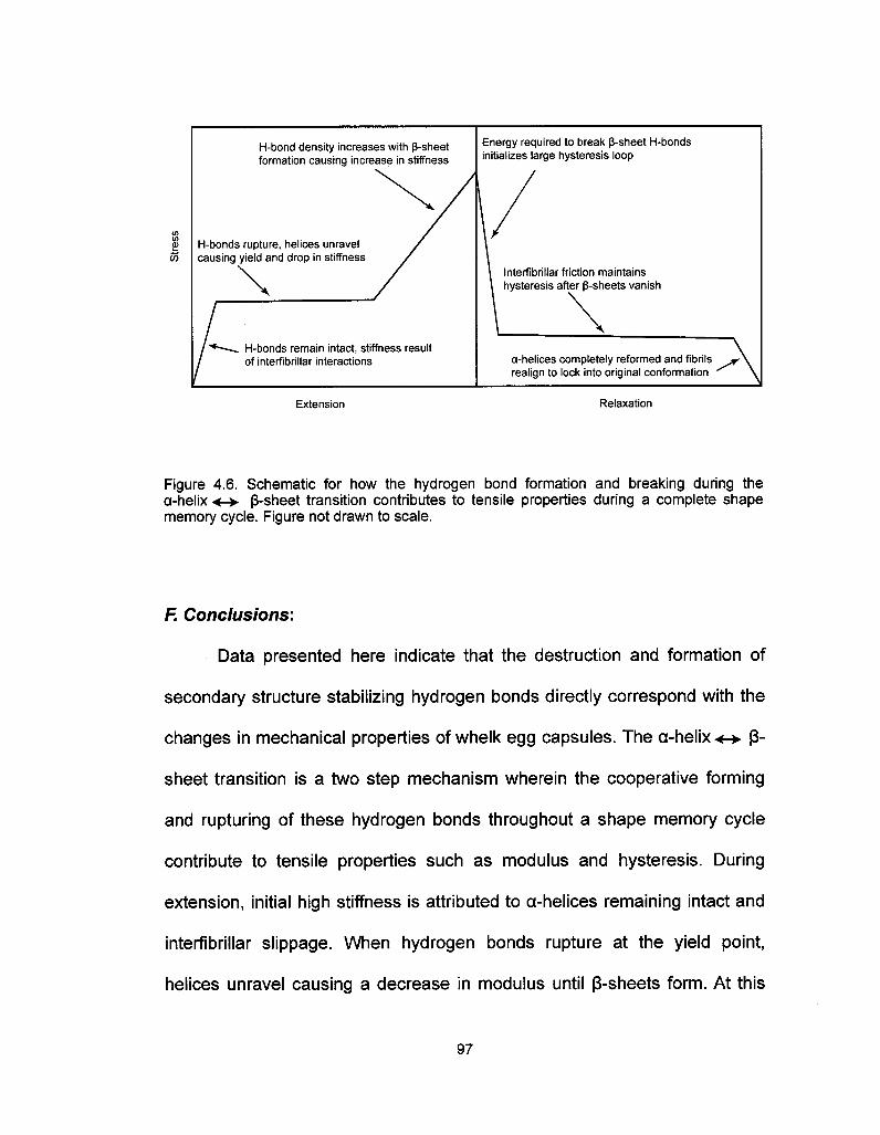
?-bond density increases with ß-sheetformation causing increase in stiffness
?-bonds rupture, helices unravelcausing yield and drop in stiffness
?-bonds remain intact, stiffness resultof interfibrillar interactions
Energy required to break ß-sheet H-bondsinitializes large hysteresis loop
Interfibrillar friction maintainshysteresis after ß-sheets vanish
a-helices completely reformed and fibrilsrealign to lock into original conformation
Extension Relaxation
Figure 4.6. Schematic for how the hydrogen bond formation and breaking during thea-helix ·*-> ß-sheet transition contributes to tensile properties during a complete shapememory cycle. Figure not drawn to scale.
F. Conclusions:
Data presented here indicate that the destruction and formation of
secondary structure stabilizing hydrogen bonds directly correspond with the
changes in mechanical properties of whelk egg capsules. The a-helix <<-? ß-
sheet transition is a two step mechanism wherein the cooperative forming
and rupturing of these hydrogen bonds throughout a shape memory cycle
contribute to tensile properties such as modulus and hysteresis. During
extension, initial high stiffness is attributed to a-helices remaining intact and
interfibrillar slippage. When hydrogen bonds rupture at the yield point,
helices unravel causing a decrease in modulus until ß-sheets form. At this
97

point, hydrogen bond density increases again, increasing stiffness. During
relaxation, the energy required to break these ß-sheet stabilizing hydrogen
bonds initiates the hysteresis loop. Once all ß-sheets have broken down,
interfibrillar friction likely contributes to the continued hysteresis until the
material relaxes back to 0% strain.
G. Acknowledgements
Thanks are due to H. Gupta (Max-Planck Institute for Colloids and
Interfaces, GoIm, Germany) for assistance with the time-resolved WAXS
synchrotron experiments. S.W. was financially supported by a California SeaGrant # R/MP-97B and UC BREP GREAT traineeship #2007-02.
98

/. References:
1. DesRoches, R., & and Delemont, M. Seismic retrofit of simply supported bridgesusing shape memory alloys, Eng. Struct. 42, 325-332 (2002).
2. Song, G., Ma, N., & Li, H. N. Applications of shape memory alloys in civil structures.Eng Struct. 28, 1266-1274 (2006).
3. Desroches, R., & Smith, B. Shape memory alloys in seismic restraint design andretrofit: A critical review of their potential and limitations. J. Earthquake Eng. 7, 1-15(2003).
4. Seelecke, S., & Müller, I. Shape memory alloy actuators in smart structures:Modeling and simulation. Appi. Mech. Rev. 57, 23-46 (2004).
5. Liang, C, & Rogers, C. A. Design of Shape Memory Alloy Actuators. J. Intell. Mater.Syst. Struct. 8, 303-313 (1997).
6. Tabib-Azar, M., Sutapun, B., & Huff, M. Applications of TiNi thin film shape memoryalloys in micro-opto-electro-mechanical systems. Sens. Actuators A 77, 34-38(1999).
7. Wache, H. M., Tartakowska, D. J., Hentrich, A., & Wagner, M. H. Development of apolymer stent with shape memory effect as a drug delivery system. J. Mater. Sci. -Mater. Med. 14, 109-112 (2003).
8. Morgan, N. B. Medical shape memory alloy applications—the market and itsproducts. Mater. Sei. Eng. A. 378, 16-23 (2004).
9. Lendlein, ?., & Langer, R. Biodegradable, Elastic Shape-Memory Polymers forPotential Biomedical Applications. Science 296, 1673 - 1676 (2002).
10. Miserez, ?., Wasko, S. S., Carpenter, C. R, & Waite, J. H. Non-entropic andreversible long-range deformation of an encapsulating bioelastomer. Nat Mater 8,910-916(2009).
11 . D. C. Lagoudas (Editor). Shape Memory Alloys. Springer Science Media LLC. NewYork, NY. (2008)
12. Pereira, I. M., & Oréfice, R. The morphology and phase mixing studies onpoly(ester-urethane) during shape memory cycle. J. Mater. Sci. 45, 511-522 (2010).
13. Ichikawa, Y, Washiyama, J., Moteki, Y, Noguchi, K., & Okuyama, K. CrystalTransition Mechanisms in Poly(tetramethylene succinate). Polym. J. 27, 1230 -1238(1995)
14. Apostolov, A. A., Fakirov, S., Stamm, M., Patii, R. D., & Mark, J. E. Alpha-BetaTransition in Poly(butylene terephthalate) As Revealed by Small-Angle X-rayScattering. Macromolecules 33, 6856-6860 (2000).
15. Feughelman, M. Mechanical Properties and Structure of Alpha-Keratin Fibres:Wool, Human Hair, and Related Fibres, UNSW Press: Sydney, Australia, 1997.
16. Ashbury, W. T, & Street, A. X-ray studies of hair, wool, and related fibres - I:General. Philos. Trans. R. Soc. London, Ser. A 230, 75-101 (1931).
17. Hearle. J. A critical review of the structural mechanics of wool and hair fibres. Int. J.Biol. Macromol. 27, 123-138 (2000).
18. Qin, Z., Kreplak, L., & Buehler, M. J. Nanomechanical properties of vimentinintermediate filament dimers. Nanotechnology 20, 425101-9 (2009).
19. Fudge, D., Gardner, K., Forsyth, V., & Riekel, C. The Mechanical Properties ofHydrated Intermediate Filaments: Insights from Hagfish Slime Threads. Biophys. J.85,2015-2027(2003).
20 Kiss, B., Karsai, ?., & Kellermayer, M. S. Z. Nanomechanical properties of desminintermediate filaments. J. Struct. Biol. 155, 327-339 (2006).
21. Lim, B. B.C., Lee, E. H, Sotomayor, M., & Schulten, K. Molecular basis of fibrin clotelasticity. Structure 16, 449-59 (2008).
22. Bendit, E. G. A quantitative x-ray diffraction study of the alpha-beta transformationin wool keratin. Text. Res. J. 30, 547-555 (1960).
99

23. Kreplak, L., Doucet, J., Dumas, P., & Briki, F. New Aspects of the a-Helix to ß-SheetTransition in Stretched Hard a-Keratin Fibers. Biophys. J. 87, 640-647 (2004).
24. Stefani, M., & Dobson, C. M. Protein aggregation and aggregate toxicity: newinsights into protein folding, misfolding diseases and biological evolution. J. MoI.Med. 81,678-699(2003).
25. Lazarides, E. Intermediate filaments as mechanical integrators of cellular space.Nature 283, 249-256 (1980).
26. Bonne, G:, Di Barletta, M. R., Varnous, S., Becane, H. M., Hammouda, E. H.,Merlini, L. et al. Mutations in the gene encoding lamin A/C cause autosomaldominant Emery-Dreifuss muscular dystrophy. Nat Genet 21, 285-288 (1999).
27. Rapoport, H. S., & Shadwick, R. E. Reversibly labile, sclerotization-induced elasticproperties in a keratin analog from marine snails: whelk egg capsule biopolymer(WECB). J. Exp. 6/?/210, 12-26 (2007).
28. Rapoport, H. S., Biomechanics, Biochemistry, and Molecular Biology of a MolluscanScleroprotein Elastomer: Whelk Egg Capsule Biopolymer, in Marine Biology. PhDthesis. University of California San Diego (2003).
29. Gupta, H. S., Seto, J., Wagermaier, W., Zaslansky, P., Boesecke, P, Fratzl, P,Cooperative deformation of mineral and collagen in bone at the nanoscale. Proc.Natl. Acad. Sci. USA 103, 17741-17746 (2006).
30. Paris, O., Li, CH. , Siegel, S., Weseloh, G., Emmerling, F., Riesemeier, H., Erko, A.,Fratzl, P. A new experimental station for simultaneous X-ray microbeam scanningfor small- and wide-angle scattering and fluorescence at BESSY II. J. Appi.Crystallogr. 40, S466-S470 (2007).
31. Hammersley, A. FIT2D: an introduction and overview. ESRF Internal ReportESRF97HA02T. (1997)
32. Wasko, S. S., & Waite, J. H. Structural proteins from an egg capsule withnonentropic reversible extensibility. Submitted to: Biomacromolecules, under review(2010).
33. Feldman A. Y, , Wachtel, E., Zafeiropoulos, N. E., Schneider, K., Stamm, M., R. J.,DavieS' Weinberg, A., & Marom, G. In situ synchrotron microbeam analysis of thestiffness of transcrystallinity in aramid fiber reinforced nylon 66 composites.Compos. Sci. Technol. 66, 2009-20015 (2006).
34. Davies, R. J., Zafeiropoulos, N. E., Schneider, K., Roth, S. V., Burghammer,M., Riekel, C, Kotek, J. C, & Stamm, M. The use of synchrotron X-ray scatteringcoupled with in situ mechanical testing for studying deformation and structuralchange in isotactic polypropylene. Colloid. Polym. Sci. 282, 854-866 (2004).
35. Wu, J., Schultz, J. M., Yeh, F, Hsiao, B, S., & Chu, B. In-Situ SimultaneousSynchrotron Small- and Wide-Angle X-ray Scattering Measurement ofPoly (vinylidene fluoride) Fibers under Deformation. Macromolecules, 33,1765- 1777(2000).
36. Gupta, H. S., Seto. J., Krauss, S., Boesecke, P., & Screen H. R. C. In situ multi-levelanalysis of viscoelastic deformation mechanisms in tendon collagen. J. Struct Biol.169,183-191(2010)
37. Seyde!, T, KoIIn, K., Krasnov, I., Diddens, I., Hauptmann, N., Helms, G., Ogurreck,M., Kang, S., Koza, M. M., & Müller, M. Silkworm Silk under Tensile StrainInvestigated by Synchrotron X-ray Diffraction and Neutron Spectroscopy.Macromolecules 40, 1035-1042 (2007).
38. Harrington, M. J., Gupta, H. S., Fratzl, P., Waite, J. H. Collagen insulated fromtensile damage by domains that unfold reversibly: In situ X-ray investigation ofmechanical yield and damage repair in the mussel byssus. J. Struct. Biol. 167,47-54 (2009).
39. Sethna, J. P., Dahmen, K., Kartha, S., Krumhansl, J. A., Roberts, B. W, & Shore, J.D. Hysteresis and Hierarchies: Dynamics of Disorder-Driven First-Order PhaseTransformations. Phys. Rev. Lett. 70, 3347 (1993)
100

40. Dahmen, K., Kartha, S., Krumhansl, J. ?., Roberts, B. W., Sethna, J. P., & Shore, J.D. Disorder-driven first-order phase transformations: A model for hysteresis, KarinDahmen, Sivan Kartha, J. Appi. Phys. 75, 5946 (1994)
41. Feughelman. M. Mechanical hysteresis in wool keratin fibers. J. Macromol. Sci. PanB Phys. 7, 569-582(1973).
42. Non-published observations.43. Kellermayer, M. S. Z., Grama, L., Karsai, ?., Nagy, ?., Kahn, ?., Datki, Z. L., &
Penke, B. Reversible Mechanical Unzipping of Amyloid ß-Fibrils. J. Biol Chem. 280,8464-8470 (2005)
101
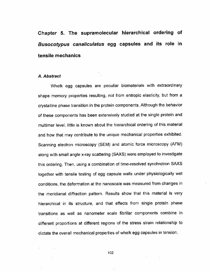
Chapter 5. The supramolecular hierarchical ordering of
Busocotypus canaliculatus egg capsules and its role intensile mechanics
A. Abstract
Whelk egg capsules are peculiar biomaterials with extraordinary
shape memory properties resulting, not from entropie elasticity, but from a
crystalline phase transition in the protein components. Although the behaviorof these components has been extensively studied at the single protein and
multimer level, little is known about the hierarchical ordering of this material
and how that may contribute to the unique mechanical properties exhibited.
Scanning electron microscopy (SEM) and atomic force microscopy (AFM)
along with small angle x-ray scattering (SAXS) were employed to investigate
this ordering. Then, using a combination of time-resolved synchrotron SAXS
together with tensile testing of egg capsule walls under physiologically wetconditions, the deformation at the nanoscale was measured from changes in
the meridianal diffraction pattern. Results show that this material is very
hierarchical in its structure, and that effects from single protein phase
transitions as well as nanometer scale fibrillar components combine in
different proportions at different regions of the stress strain relationship to
dictate the overall mechanical properties of whelk egg capsules in tension.
102

B. Introduction
The proteinaceous egg capsules produced by marine prosobranch
gastropods (whelks) are remarkable examples of shape memory/self healingmaterials. The tensile properties have been studied extensively1"3 and
models for the long range, reversible extensibility have been presented4.Briefly, the material undergoes a crystalline phase transition from a-helix to
ß-sheet when placed under tension. When the load is removed, the materialrelaxes back to the original a-helical conformation.
Although the unraveling of a-helices makes a major contribution to
the extensibility and shape memory of this material, the involvement oflarger (nanometer rather than A) scale ordering has not yet beeninvestigated. The supramolecular ordering of protein fibers into hierarchicalassemblies is a common theme in biological materials5-7 and is known to
play a critical role in the mechanics of these systems. Indeed in somekeratin models, while an a-helix ** ß-sheet phase transition is certainly
responsible for long range deformation, changes in the surpamolecularordering also play significant role in tensile properties7·8. Therefore, it is ofgreat importance to look into the hierarchical arrangement of the eggcapsule fibers in order to gain a more complete understanding of thestructure-property relationships of this material under tension, from thesingle protein level, up through the fiber level, and eventually to the bulkmaterial.
103
Utilizing the same methods that have greatly advanced theunderstanding of tendon collagen structure under loading9"12, this studycombines scanning electron microscopy and tapping mode atomic forcemicroscopy with high-resolution synchrotron based small angle x-ray
scattering (SAXS) with real time tensile testing to investigate the structuralreorganization occurring at the nano-length scales of the egg capsule fiberhierarchy during uniaxial loading, and builds a more detailed understandingof the local material mechanics. This, combined with what is already known
about this material on a single protein level will provide a much more
thorough knowledge of the structure/function relationship of egg capsulesunder mechanical tension.
C. Materials and Methods
Busycotupus canaliculars egg capsules were obtained and preparedfor experimentation exactly as described in the Materials and Methodssection of Chapter 4 of this dissertation.
SEM sample preparation and image acquisition was performed asdescribed in Chapter 3 of this dissertation. Analysis of the distances
between bands in the micrographs was performed using the Vega TC
software program (Tescan USA, Cranberry Twp., PA).
Egg capsule samples were cut into narrow strips with a razor andwashed repeatedly with deionized water using gentle physical agitation to
104

remove the mucous lining. Measurements were taken using an MFP-3D
AFM system (Asylum Research, Santa Barbara, CA) in tapping mode
according to the manufacturers instructions. Dry measurements were taken
by applying a wetted strip of egg capsule on a glass slide so that the cross
section was exposed to the AFM probe. When the water evaporated, the
sample adhered to the glass surface. Silicon probes were used for these
measurements (Olympus America, Lakewood, CO). Wet measurements
were taken by applying a strip of egg capsule to double-sided tape on top of
a glass slide so that the cross section was exposed to the AFM probe.
Silicon nitride probes were used for the measurements (Olympus America,
Lakewood, CO). Data were analyzed by the Argyle software program
provided by Asylum Research.
Tensile testing during x-ray analysis was conducted using the Micro-
mechanical Tensile Apparatus (MiTA) described in the Chapter 4, using
identical parameters. All other tensile testing was preformed using an MTS
Bionix 200 (MTS, Eden Prairie, MN) tensile apparatus as described in
Chapter 3. X-ray data collection was performed on the MuSpot beamline at
the BESSY synchrotron source (Berlin Elektronenspeicherring Gesellschaftm.b.H., Berlin, Germany) also described in the Materials and Methods
section of Chapter 4 using the same parameters for data acquisition, with
the exception that the sample to detector distance was determined to be
906.39 mm using a corundum (AI2O3) standard.
105

Analysis of 2D SAXS frames was performed using the CAKE
command of the Fit2D program, as described in Chapter 4. This resulted in
a one-dimensional intensity profile of peak reflections from the 4th to the 9th
order that are visible on both sides of the primary beam. The peaks
corresponding to the different orders were fitted to a series of Gaussian
functions to determine changes in D-spacing and measure fibril strain, zf, as
described previously11·12. The current analysis only examines the meridianreflections.
D. Results
SEM images clearly show the fibrous nature of this material, with fiber
strands typically ranging from 200 - 500 nm in diameter with some >1 pm
(Fig 5.1a). These fibers occasionally, but not always, appear to be braided toform larger rope-like structures. Higher magnification of these fibers revealsa lateral banding pattern with elevated ridges of different widths occurring at
repeated intervals (Fig 5.1b). The width of these bands was determined byanalysis with the Vega TC software program and these numbers along withthe pattern of the banding repeats is detailed in (Fig 5.2). Cross sections ofthe fibers were also imaged. These show a smaller scale ordering within the
fibers that appear to be between 10 and 20 nm in diameter as well as some
pores/voids (Fig 5.1c, d).
106
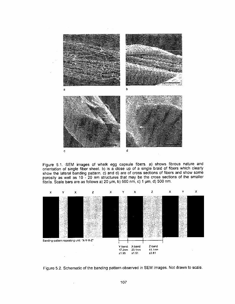
Figure 5.1. SEM ¡mages of whelk egg capsule fibers, a) shows fibrous nature andorientation of single fiber sheet, b) is a close up of a single braid of fibers which clearlyshow the lateral banding pattern, c) and d) are of cross sections of fibers and show someporosity as well as 1 0 - 20 nm structures that may be the cross sections of the smallerfibrils. Scale bars are as follows a) 20 µ??, b) 500 nm, c) 1 µ??, d) 500 nm.
Banding pattern repeating unit: "X-Y-X-Z"Y-band: X-band17.2nm 23.1nm±1.95 ±1.51
Z-band41.1nm±3.81
Figure 5.2. Schematic of the banding pattern observed in SEM images. Not drawn to scale.
107
Tapping mode AFM in air produced similar images to those taken by
SEM, revealing fibers that are several hundred nanometers in diameter and
with lateral ridges going across the fiber (Fig 5.3a). However, the banding
patterns of these ridges is noticeably different than those observed by SEM.
Here, there is no complex periodicity, rather there is just a constant
sigmoidal ridge that is 93.8 ±8.5 nm (STDEV) across from peak to peak (Fig
5.3b). Furthermore, when AFM is performed under fully hydrated conditions,
the pattern persists, but the peak to peak width changes to 98.0 ± 1.2 nm
(STDEV) (Fig 5.4). One possible explanation for the inconsistencies
between imaging methods is that sample preparation is vastly different: SEM
requires a series of dehydration treatments as well as gold coating of the
sample; dry AFM does not require coating of the sample and while the
dehydration steps are not as thorough or harsh as those for SEM, the
sample is still in a non-native (dehydrated) state; wet AFM keeps the sample
in the most biologically native condition. As the SAXS studies also kept the
samples fully hydrated during testing, all comparisons and analysis
combining microscopy images with x-ray data will use the wet AFM images.
108
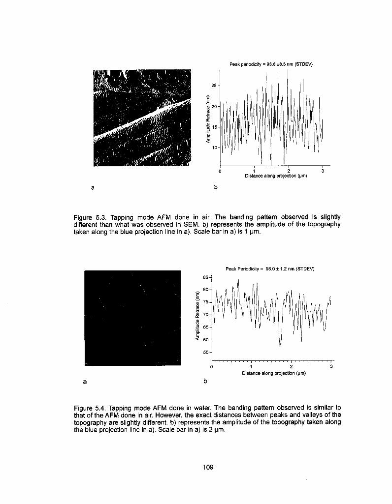
Peak periodicity = 93.8 ±8.5 nm (STDEV)
25
20
cdo>¦o 15
e<
10
1 2 3Distance along projection (µ?t?)
Figure 5.3. Tapping mode AFM done in air. The banding pattern observed is slightlydifferent than what was observed in SEM. b) represents the amplitude of the topographytaken along the blue projection line in a). Scale bar in a) is 1 µ?t?.
Peak Periodicity = 98.0 ± 1.2 nm (STDEV)85
80
75
£ 70
I 65Q.E< 60
55
1 2 3
Distance along projection (pm)
Figure 5.4. Tapping mode AFM done in water. The banding pattern observed is similar tothat of the AFM done in air. However, the exact distances between peaks and valleys of thetopography are slightly different, b) represents the amplitude of the topography taken alongthe blue projection line in a). Scale bar in a) is 2 \im.
109

X-ray scattering prod-
uced visible reflections from
orders 4-9 along both the
meridian and equator (Fig 5.5).
To calculate the lateral dia-
meter, R1 of the scattering unit
the relationship R « 1/d?± was
applied. Using values from the
? = 5 reflection for the equation
1000
.E 100
S03
N = 5 peak
N = 4 peak N = 7 peak
0.2 0.3 0.4
Q-space (nm1)
Figure 5.5. One dimensional plot of x-rayscattering intensity along the meridian axisshowing the N = 4 - 7 peak reflections.
5qj. = 2qm * sin(5Xi/2) (5.1)
Wherein ? is the angular coordinate of the reflection. Oq^ = 2 ? 0.315 ? sin(20°/
2) = 0.11 nnr1. Previous studies13 indicate that this value corresponds to a
coherent length of -9.1 nm for the radius of a single fibril (Fig 5.6). These
may correspond to the 10 - 20 nm structures observed in the SEM cross
sectional images of the fibers. This suggests that the several hundred
nanometer diameter structures that were observed by microscopy are
bundles of these fibrils.
110

D = 99.8 nm
R = 9.1 nm
To examine the axial ordering of
these fibrils the relationship qn = 2p n/D
was applied to each of the visible
reflections. All resulted in a D space
value of 99.8 nm which corresponds to
the distances between lateral bands
across fibrils (Fig 5.6). This is consistent
with the 98.0 nm value measured by wet
AFM.Figure 5.6. Schematic of fibril
T .. , . . . , diameter and band distances asTensile mechanical data for derived from SAXS data. Image is notsamples undergoing SAXS analysis were
nearly identical in every way to the samples that underwent WAXS analysis
in Chapter 4. No significant differences between the two data sets were
noted.
As the material is extended in the pre-yield region (from 0% - ~5%
engineering strain) there is little change in the intensity and location of the
SAXS reflections. Once extended into the yield region (~5% - ~45%
engineering strain) there is a significant decrease in peak intensity, but a
negligible shift in peak position suggests a decrease in the crystalline
ordering within these fibrils. As the material restiffens, the peak intensity
increases again, and there is a significant shift in peak position suggesting a
111

new crystalline order has been established and is contributing to the
mechanics (Fig 5.7).
o(?
N = 5 peak
/?
restiffening40 60Strain (%)
Figure 5.7. One dimensional SAXS plotsalong the meridian and how theychange with tensile extension of thematerial. There are clear changes in thelocation, intensity, and integrated area ofpeaks as strain increases towards 100%
0.2 0.3 0.4
Q-space (nm')
Fibril strain, ef, was calculated by the relationship:
Ef = (D(e)-Do)/Do (5.2)
wherein D0 is the 99.8 nm D-spacing of the fibril at rest and ?(e) is the D-
spacing of a fibril at any given engineering strain, ef increases slightly during
112
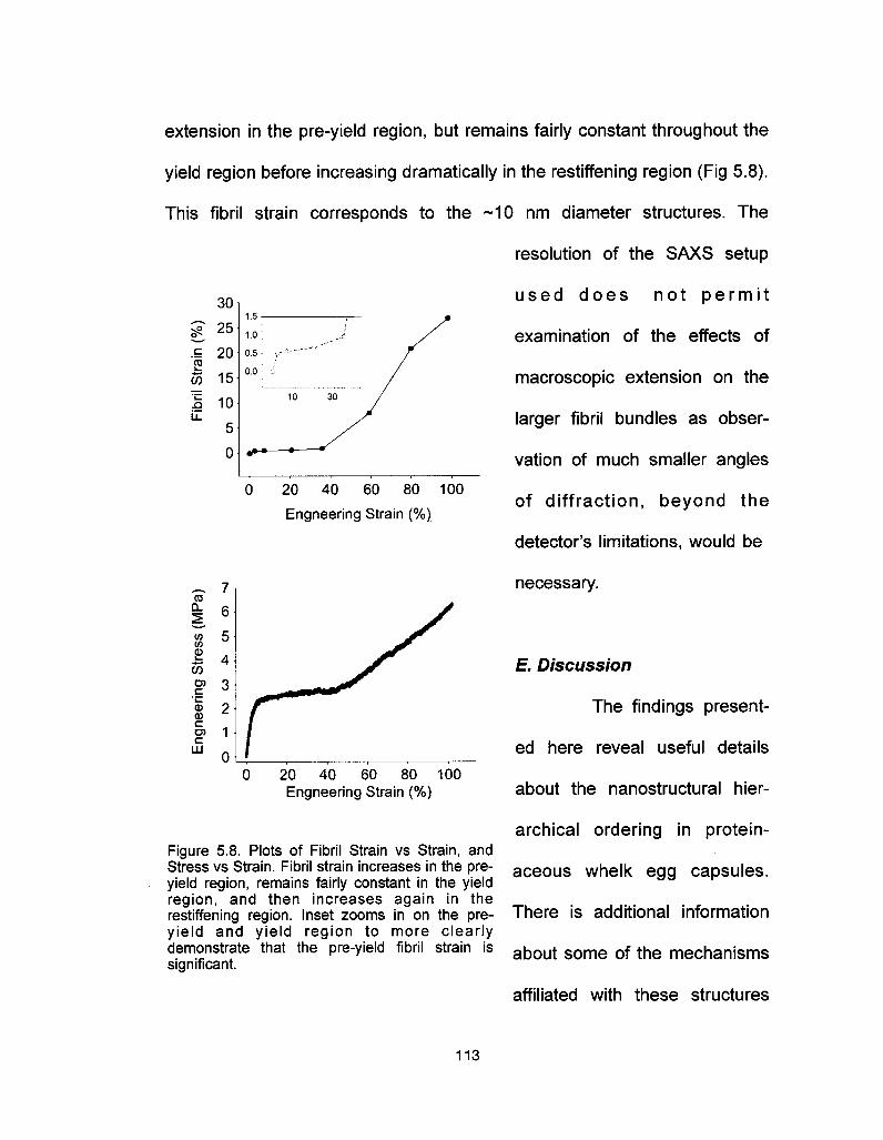
extension in the pre-yield region, but remains fairly constant throughout the
yield region before increasing dramatically in the restiffening region (Fig 5.8).
This fibril strain corresponds to the ~10 nm diameter structures. The
resolution of the SAXS setup
30
g 25.£ 20 \ 0.5S
OT 15
I ioj5
0 ·»
0 20 40 60 80 100
Engneering Strain (%)
ccOl
</)(?
%OTO)C
"u.f(DCO)C
IXI
20 40 60 80 100Engneering Strain (%)
Figure 5.8. Plots of Fibril Strain vs Strain, andStress vs Strain. Fibril strain increases in the pre-yield region, remains fairly constant in the yieldregion, and then increases again in therestiffening region. Inset zooms in on the pre-yield and yield region to more clearlydemonstrate that the pre-yield fibril strain issignificant.
used does not permit
examination of the effects of
macroscopic extension on the
larger fibril bundles as obser-
vation of much smaller angles
of diffraction, beyond the
detector's limitations, would be
necessary.
E. Discussion
The findings present-
ed here reveal useful details
about the nanostructural hier-
archical ordering in protein-
aceous whelk egg capsules.
There is additional information
about some of the mechanisms
affiliated with these structures
113

and how they are involved in the capsule's remarkable mechanical
properties in uniaxial tension14. Building upon previous work indicating that
individual a-helical protein components, 1.2 nm in diameter15, form coiled-
coil trimers16 which are 4 - 5 nm in diameter17. The data suggest that these
are then organized into -10 nm fibrils which then bundle into the 200 - 500
nm fibers, with the occasional fiber being >1 µ??, that further braid in co-
alignment to make up the laminar sheet structure (Fig 5.9).
The lateral D-spacings of ~100 nm that are observed at the fibril level
by x-ray scattering may be caused by the alignment of secondary structures
in the individual proteins making up the fibrils, as in intermediate filaments
like nuclear lamins18, or could potentially be caused by gaps in between the
end to end linkages of proteins within the fibrils, as is the case with tendon
collagen19. It is the stretching and lengthening of the distances between
these structures and/or gaps that causes increases in D-space reflections
and, hence, fibril strain. The banding that is visible on the fiber level in via
microscopy (wet mode AFM) has the same dimensions as the fibril D-
spacings, ~100 nm, suggesting that the two are possibly related. However,
the amplitude of the bands on the fiber surface can be in excess of 20 nm
(Fig 5.4b), whereas the diameter of the individual fibrils is only -10 nm,
indicating that this banding pattern cannot be the result of single fibrils on
the surface, but rather must be caused by a ordered stacking of multiple
114

fibrils. Further studies will be needed to determine the relationship, if any,
between the fibril D-spacings and the fiber banding pattern.
a-helix proteins (~1 nm)
Goiled-coil trimers (-4-5 nm)
Nanofibrils (-10 nm)
Fibers (-200 - 500 nm)
Braided fiber ropes (>1 µ??)
Sheets of aligned fibersarranged 90° to one another(30 µ?? thick)
%
WAXS
SAXS/SEM
SEM
Light Microscopy
Figure 5.9 Schematic of the hierarchical ordering of whelk egg capsules
115

When an external load is applied, an initial slight increase in fibril
strain is observed in the pre-yield region. At the protein level, coiled-coil a-
helix trimers are being strained and the hydrogen bonds stabilizing these
structures remain intact7. Many biological fibers demonstrate supercoiling in
their relaxed state, which can cause crimping of the fibers20-22. The observed
pre-yield increase in fibril strain may be the result of unraveled supercoiling
and the fiber crimps being straightened out.
As the egg capsule is pulled beyond the mechanical yield point at
-5% engineering strain, fibril strain does not increase much as the material
extends further. Typically this is the result of either a sliding between fibrils
caused by delamination of the structures connecting them, or by an
inhomogeneous destruction of the crystalline elements of the fibril after the
yield point, corresponding to strain localization. SAXS data presented here
are suggestive of the second mechanism, and indicate that both the
intensity as well as the integrated area of the peaks corresponding to fibril
crystallinity decrease while the location of these peaks remains constant.
Furthermore, previous studies detail how the a-helix ^ß-sheet phase
transition occurs in this region and how this transition is a two step
mechanism which begins with the destruction of the a-helix crystal structure.
However, some degree of interfibrillar slipping cannot be completely ruled
out. Any actual slippage must be reversible to account for the recoverability
and shape memory of this material.
116

Once the egg capsule is strained beyond 45%, it begins to restiffen.
At this point there is a shift in the peak location in the SAXS profile as well
as an increase in peak intensity and integrated area. This indicates an
increase in crystalline content as well as a change in D-spacings of the
crystal structures of the fibril, corresponding perfectly to the formation of ß-
sheets at the single protein level detailed before4·14. As these structures
propagate throughout the material the fibril strain increases as well. In
collagen fibrils, two different models for this increase have been presented.
1) a homogeneous stretching in the crystalline poly-proline helix during
elongation leads to changes in the D-spacings, and 2) a side by side gliding
of neighboring molecules within the fibrils causing an increase in gap sizeand distance9.
In egg capsules the first model would translate to a homogeneous
increase in ß-sheet content in the restiffening region. The second model
would relate to a sliding of the protein trimers which form the fibrils along
each other resulting in an increase in D-spacings due to either a realignment
of secondary structures, or an increase in gap sizes (Fig 5.10). This second
model would also imply that covalent crosslinks between trimers would
either be ruptured, or that there is an additional matrix component in
between trimers that accommodates this slipping without rupturing covalent
bonds.
117

\ > II ?-?? > JH
M > VV ?
£e ^•1 ^-
3?Ea
Figure 5.10. Schematic of two models for increase in fibril strain in restiffening region, a)represents a homogenous formation and elongation of ß-sheet structures in unisonresponsible for an increase in D-spacings due to alignment of secondary structures andrandom coils, b) shows a heterogeneous increase in ß-sheets with a sliding of neighboringtrimers causing changes in gap size and location resulting in increased D-spacings. It is notyet determined whether there are gaps in this material, and the sliding model shown in b)could also be applied to a realignment of secondary structures and random coils causing D-space changes.
In tendon collagen, the second model has been experimentally
confirmed as molecular shearing causes changes in the gap/overlap
patterns9 with a proteoglycan matrix allowing for this slippage without
rupturing crosslinks until critical strains are met23. While it is not yet known if
118

the egg capsule material has
matrix components, the
rupturing of covalent cross-
links would imply some non-
recoverable deformation
resulting from extension of
samples into the restiffening
zone. Indeed it is observed
that the further into the
restiffening zone a sample is
extended, the more non-
recoverable plastic deform-
ation is incurred (Fig 5.11). However, until ad-ditional work is performed, and
crosslink characterization is complete, no model can be completely
discounted or confirmed in whelk egg capsules.
Figure 5.12 summarizes what is currently known about the various
established mechanisms and how they interact to dictate the mechanical
properties of the egg cases. Additional analysis will be needed to determine
how these mechanisms contribute to the relaxation mechanics of the egg
capsule material.
Q.
2
160
Strain (%)
Figure 5.11. Stress strain curves of egg capsulescyclically extended to 90% (black) and 140% (red). Asthe material is extended further into the restiffeningregion, the amount of plastic deformation betweencycles increases, which could be the result of therupturing of covalent crosslinks between trimers.
119
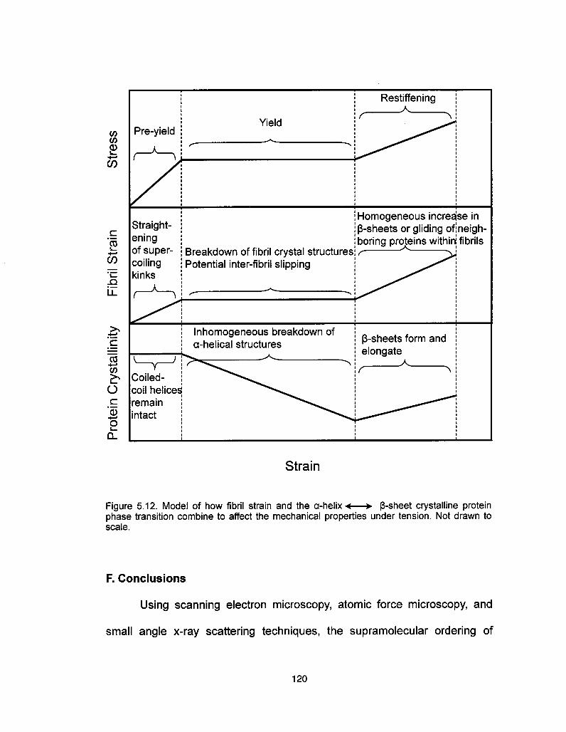
Pre-yieldYield
r
Restiffening?^1
Straight-eningof super-coilingkinks
Breakdown of fibril crystal structuresPotential inter-fibril slipping
Homogeneous increase inß-sheets or gliding ofineigh-boring proteins within; fibrils
lnhomogeneous breakdown ofa-helical structures ß-sheets form and
elongate
Coiled-coil heliceáremainintact
Strain
Figure 5.12. Model of how fibril strain and the a-helix <—? ß-sheet crystalline proteinphase transition combine to affect the mechanical properties under tension. Not drawn toscale.
F. Conclusions
Using scanning electron microscopy, atomic force microscopy, and
small angle x-ray scattering techniques, the supramolecular ordering of
120
whelk egg capsules has been observed down to the nanometer scale. This
combined with the wide angle x-ray scattering data from Chapter 4 has
elucidated the structural hierarchy of this material from the single protein
level up through the macroscopic level. When the egg capsules are put
under uniaxial tension there is a slight increase in the strain of the 10 nm
fibril component as measured by the change in D-spacing of the banding
patterns. Once the material is pulled past the yielding point, fibril strainremains constant while the coiled-coil a-helices inhomogeneously break
down. Some interfibrillar slipping may also be occurring in this region. Once
the material begins to restiffen after -45% strain, fibril strain increases again
as does ß-sheet content. Several possible models are proposed, and more
work is required to confirm which of these is occurring in this system.
G. Acknowledgements
Thanks are due to H. Gupta (Max-Planck Institute for Colloids and
Interfaces, GoIm, Germany) and A. Miserez (Materials Department, UCSB)
for providing guidance and assistance with the time-resolved SAXSsynchrotron experiments and SEM imaging respectively. S.W. wasfinancially supported by a California Sea Grant # R/MP-97B and UC BREPGREAT traineeship #2007-02. This work made use of MRL Central Facilities
supported by the MRSEC Program of the National Science Foundationunder award No. DMR05-20415.
121

/. References
1. Rapoport, H. S., & Shadwick R. E. Mechanical characterization of an unusualelastic biomaterial from the egg capsules of marine snails (Busycon spp.)Biomacromolecules 3, 42-50 (2002).
2. Rapoport, H. S., & Shadwick, R. E. Reversibly labile, sclerotization-induced elasticproperties in a keratin analog from marine snails: whelk egg capsule biopolymer(WECB). J. Exp. Biol 210, 12-26 (2007).
3. Rapoport, H. S., Biomechanics, Biochemistry, and Molecular Biology of a MolluscanScleroprotein Elastomer: Whelk Egg Capsule Biopolymer, in Marine Biology. PhDthesis. University of California San Diego (2003).
4. Miserez, ?., Wasko, S. S., Carpenter, C. R, & Waite, J. H. Non-entropic andreversible long-range deformation of an encapsulating bioelastomer. Nat Mater 8,910-916(2009).
5. Fratzl, P., & Weinkamer, R. Nature's hierarchical materials. Prog. Mater. Sci. 51,1263-1334(2007)
6. Kastelic, J. & Baer, E. Deformation in Tendon Collagen Symp. Soc. Exp. Biol 34,397-435(1980).
7. Feughelman, M. Mechanical Properties and Structure of Alpha-Keratin Fibres:Wool, Human Hair and Related Fibres, UNSW Press: Sydney, Australia, 1997.
8. J Hearle. A critical review of the structural mechanics of wool and hair fibres. Int. J.Biol. Macromol. 27, 123-138 (2000).
9. Fratzl, P., Misof, K., & Zizak, I. Fibrillar Structure and Mechanical Properties ofCollagen. J Struct. Biol. 122, 119-122 (1997).
10. Krauss, S., Fratzl, P., Seto, J., Currey, J. D., Estevez, J. A., Funari, S. S., & Gupta,H. S. Inhomogeneous fibril stretching in antler starts after macroscopic yielding:Indication fora nanoscale toughening mechanism. Bone 44, 1105-1110 (2009).
11. Gupta, H. S., Seto, J., Krauss, S., Boesecke, P., & Screen, H. R. C. In situmulti-level analysis of viscoelastic deformation mechanisms in tendon collagen. J.Struct. Biol. 169, 183-191 (2010).
12. Gupta, H .S., Seto, J., Wagermaier, W, Zaslansky, P., Boesecke, P., Fratzl, P.,Cooperative deformation of mineral and collagen in bone at the nanoscale. PNAS103, 17741-17746(2006).
13. Kreplak, L., Franbourg, A., Briki, F., Leroy, F., & Dalle, D. A New Deformation Modelof Hard a-Keratin Fibers at the Nanometer Scale: Implications for Hard a-KeratinIntermediate Filament Mechanical Properties. Biophys. J. 82, 2265-2274 (2002).
14. Chapter 4 of this dissertation.15. Creighton, T. E. Proteins: Structures and Molecular Properties, 2nd ed; W. H.
Freeman and Company: New York, NY, 1993.16. Wasko, S. S., & Waite, J. H. Structural proteins from an egg capsule with
nonentropic reversible extensibility. Submitted to: Biomacromolecules, under review(2010).
17. Hâkansson, K., Lim, N. K., Hoppe, H., & Reid, K. B. M. Crystal structure of thetrimeric a-helical coiled-coil and the three lectin domains of human lung surfactantprotein D. Structure 7, 255-264 (1999).
18. Ben-Harush, K., Wiesel, N., & Frenkiel-Krispin, D. The supramolecular organizationof the C. elegans nuclear lamin filament. J. MoI. Biol. 386, 1392-1402 (2009).
19. Fratzl. P. Collagen: Structure and Mechanics, P Fratzl Editor, Chapter 1. SpringerScience Media LLC, New York, NY, (2008).
20 Beck, K., & Brodsky, B. Supercoiled Protein Motifs: The Collagen Triple-Helix andthe a-Helical Coiled Coil. J. Struct. Biol. 122, 17-29 (1998).
122

21. Vazina, A. ?., Bras, W., Dolbnya, I. P., Korneev, V. N., Lanina, N. R, Matyushin, A.M., Sergienko, P. M., & Zabelin A. V. Peculiarities of human hair structuraldynamics. Proc. XV. Inter. Synch. Rad. Con. 543, 153-157 (2005).
22. T. I. Nikolaeva, T. I., Tiktopulo E. I., Il'yasova, E. N., & Kuznetsova, S. M. Collagentype I fibril packing in vivo and in vitro. Biofizika 52, 899-911 (2007).
23. Gupta, H. S. Collagen: Structure and Mechanics, P Rratzl Editor, Chapter 5,Springer Science Media LLC, New York, NY, (2008).
123

Chapter 6. Conclusions and future directions
A. Abstract
The biochemical composition, thermomechanical properties, and
structure-function relationship at multiple length scale orders of Busycotypus
canaliculatus egg capsules have been presented in this dissertation. Four
variants of the precursor protein family were purified from a crude extract
and thoroughly characterized. The cDNA deduced sequences were derived,and studies into protein behavior relating to structure and assembly insolution were carried out. The thermomechanical properties - specifically,
the behavior of the hydrated material in uniaxial tension at various
temperatures - were investigated and modeled. The results were applied toformulae derived from standard thermodynamic relations in order to
determine entropie and internal energy contributions to the tensile
properties. Finally structural studies were performed to examine thechanges in molecular architecture that occur as the egg capsule materialgoes through a complete strain cycle. A combination of wide and small anglex-ray scattering, Raman spectroscopy, and microscopic techniques providedstructural data that ranged from the A (single protein) level, through the
micron (bundled protein fiber) level.
124

ß. Conclusions from this Work
The family of precursor proteins that migrates near 5OkD has been
purified and characterized, and the sequence deduced from cDNA. These
proteins are highly related to each other, but are not homologous to any
other protein sequence in the collective databases. Named Busycotypus
canaliculatus capsule proteins, or Bc-cp, variants 1 through 3, the masses of
the proteins as obtained via MALDI-TOF matches very closely with the
calculated masses of the cDNA deduced sequences, suggesting that the
sequences do correspond with the proteins of interest. Further supporting
this is the fact that the amino acid composition determined experimentally
via automated ion exchange chromatography with ninhydrin detection also
matches the composition from the cDNA sequences. This composition is rich
in a-helix favoring and a-helix indifferent residues, similar to the compositionof intermediate filaments which are known a-helix structures.
Circular dichroism studies indicate that the precursor proteins are
strongly a-helical in solution, and also suggest that the helices tend to formsingle coil helices rather than coiled-coils. However, analysis of the cDNAderived primary sequence suggest that there are short regions that favor the
formation of coiled-coil trimers. This is in agreement with wide angle x-ray
scattering studies (described in detail below) which indicates coiled-coilstructures in the mature egg capsule material.
125
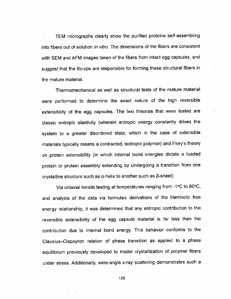
TEM micrographs clearly show the purified proteins self-assemblinginto fibers out of solution in vitro. The dimensions of the fibers are consistent
with SEM and AFM images taken of the fibers from intact egg capsules, and
suggest that the Bc-cps are responsible for forming these structural fibers inthe mature material.
Thermomechanical as well as structural tests of the mature material
were performed to determine the exact nature of the high reversibleextensibility of the egg capsules. The two theories that were tested areclassic entropie elasticity (wherein entropie energy constantly drives the
system to a greater disordered state, which in the case of extensiblematerials typically means a contracted, isotropic polymer) and Flory's theoryon protein extensibility (in which internal bond energies dictate a loadedprotein or protein assembly extending by undergoing a transition from onecrystalline structure such as a-helix to another such as ß-sheet).
Via uniaxial tensile testing at temperatures ranging from -10C to 8O0C,
and analysis of the data via formulaic derivations of the Hemholtz freeenergy relationship, it was determined that any entropie contribution to thereversible extensibility of the egg capsule material is far less than thecontribution due to internal bond energy. This behavior conforms to the
Clausius-Clapeyron relation of phase transition as applied to a phase
equilibrium previously developed to model crystallization of polymer fibersunder stress. Additionally, wide-angle x-ray scattering demonstrates such a
126

phase transition in the egg capsule material coming in the form of areversible crystalline shift from a-helix to ß-sheet. As such, Flory's paradigm
wherein protein extensibility is the result of a reversible phase transition isthe accepted explanation for extensibility in the whelk egg capsule material.
Both synchrotron x-ray and con-focal Raman microprobe analysiswith in situ tensile testing demonstrates that at the single protein level, coiled
coil a-helices dominate the pristine material and remain intact as the egg
capsules are extended throughout the stiff pre-yield region of the stressstrain curve. The a-helical component does not begin to break down until
after the yield point, when modulus decreases dramatically. The sequentialrupturing of the hydrogen bonds responsible for stabilizing the a-heìices is
hypothesized to be the primary contributing factor to this decrease instiffness. However, ß-sheet formation does not begin until the restiffeningregion is entered. When ß-sheet specific hydrogen bonds form, flexibility islost at the single protein level, which contributes to the restiffening of thematerial as a whole.
Upon relaxation, a large hysteresis loop is observed. Data suggestthat this is due to the energy required to rupture the ß-sheet stabilizing
hydrogen bonds in order to reverse the phase transition back from ß-sheetto a-helix. However, the contribution of internal friction resulting from the
sliding of hierarchical fibrils and other components of the material cannot bediscounted.
127
Whelk egg capsules, like many other biological materials, are very
hierarchical structures with multiple levels of supramolecular ordering
ranging from the A up through the micron and millimeter levels as observedfrom WAXS, SEM, AFM, SAXS, and light microscopy. These different levels
of ordering contribute to the tensile mechanical properties of the bulkmaterial in different ways. The contribution of the a-helix <-> ß-sheet
transition has already been mentioned, but there is a significant contributionfrom 10 nm diameter fibril structures as well.
Although more data analysis is required, there is clear evidenceindicating that fibrillar strain contributes to tensile behavior in the pre-yieldand restiffening regions, and multiple models for how this phenomenon islinked with mechanical properties have been presented in this dissertation.
Furthermore, in the pre-yield and restiffening regions, only a small fraction ofthe bulk strain is being converted into fibrillar strain, suggesting that there
are even more mechanisms contributing to the tensile properties of the bulk
material that have not yet been examined.
Overall, whelk egg capsule is a very unique material in terms of its
biochemistry, biophysics, structural ordering, and mechanical properties.This dissertation although providing a sizable contribution to illuminating
some of the structure-function relationships involved, has really only
scratched the surface of what is possible. A great deal of additional work will
128

be needed to more thoroughly understand this system to the point where the
synthesis of an effective mimic of the material can be explored.
C. Future Directions
Although this system does indeed show promise towards biomimetic
applications, there is clearly much more to be learned before this comes to
fruition. This biomaterial is -90% protein by dry weight. The composition of
that additional 10% has not yet been determined let alone it's structure or
function, although it is suggested to be carbohydrate1. And although fourvariants of the precursor protein family have been described, the crude
extract contains additional proteins that are, as of yet, uncharacterized2. The
proteins that have been described likely contribute to the structural fibers ofthe egg capsule material and participate in the a ß transition that has beendescribed. The uncharacterized proteins that appear on the SDS PAGE of
the crude extract, although present in lower amounts, are not similar in size,
what-so-ever, to the characterized CPs 1-3 which are all in the 5OkD range.
Rather the unknowns come in at 24kD, 37kD, and 12OkD according to their
migration patterns. A full characterization of these precursor proteins will benecessary to determine the roles that they play in the structure/function ofthe egg capsule material as a whole. One possibility for the function of theseunknowns is as matrix proteins. Hard keratins are a composite of both
129

structural fibers and amorphous matrix, both of which contribute significantly
to mechanical properties3.
Of the precursor proteins that have been characterized, the nature of
the self-assembly as well as the post translational modification steps that
guide the synthesis of the fully mature and functional material are also
unknown. Although it has been shown that CPs 1 and 2 will self-assemble invitro into fibers that are the same size as those seen in the mature material,
the specifics of this assembly including the trigger, the intermediate steps
and structures formed, the kinetics, etc, are all unknown. Further elucidating
these mechanisms could lead the way to developing synthetic self-
assembling materials.
After assembly, covalent crosslinks are formed to stabilize the
material. The nature of these crosslinks, the exact chemical structure (a
suspected lysine or other borohydride reducible derivative4) as well as thelocation within the protein sequence, is unknown. This information will be
invaluable to both confirming the individual coiled-coil protein trimer model
presented in Chapter 2, as well as determining how that model can beapplied to a complete fiber of protein trimers that are linked together. Workinto this has been started and the preliminary results will be presented later
in this chapter.
Other ideas for future work include single molecule force
spectroscopy to examine the unfolding of single proteins or of protein trimers
130

as has been performed for other coiled-coil proteins5·6; cloning of the genes
responsible for coding these proteins and making recombinant proteins that
can be used to create films, fibers, or other materials; fully characterizing
thermal properties of the egg capsule materials via such methods as
differential scanning calorimetry; determining the permeability of the
capsules to determine functionality as a biomedical encapsulant.
In the end, there are too many potential future directions to list here in
this dissertation. It seems that whereas each experiment may provide the
answer to one question, it also raises three more in turn. One of the
struggles in working on a project such as this was to learn when to finally
start to tuck the loose ends in rather than pulling on them. It will be up to the
next group of scientists working on this project to address all of theseremaining questions. There are certainly many more publications andpotential dissertations that can come from this model system.
D. Materials and Methods for Preliminary Studies
Non-viable Busycotypus canaliculatus egg capsules were purchased
from Marine Biological Laboratories (Woods Hole, MA). Capsules were cut
open and the embryonic materials drained out and were then washed
repeatedly in MiIIi-Q water with physical agitation. Afterwards the capsulematerial was ground with a stone mortar and pestle under liquid nitrogen to
131
produce a powdery particulate which was smaller than 0.5 mm in individualgrain size.
This particulate was then treated with NaBH4 (5mg/ml) in phosphate
buffer (pH 7.0) for 30 min to reduce/protect any potentially unstable lysinederived crosslinks. The reaction was terminated by the addition of several
drops of glacial acetic acid, and was then washed repeatedly with MiIIi-Qwater.
Reduced egg capsule samples were hydrolyzed in 6N HCl, in vacuo
at 1050C for 24 hours. The HCl was flash evaporated off and the hydrolysate
was washed several times each with water and methanol to remove all
traces of acid. The hydrolysate was then subjected to Bio-Gel P2 (Bio-Rad)
size exclusion chromatography on a 100 X 2 cm column eluted with 5%
acetic acid. After the void volume passed through, 100 3ml fractions were
collected for analysis.
Aliquots of fractions were flash evaporated once again, resuspended
in NaS buffer, and analyzed on a Beckman Coulter 6300 Amino Acid
Analyzer as described in the materials and methods section of Chapter 2 ofthis dissertation.
The remaining volume of the fractions was analyzed by positive ionmode TOF ESI-MS as described elsewhere7. Molecules with masses
significantly greater than single amino acids were deemed to be potentialcrosslinks and were further analyzed by MS/MS using argon gas and a
132

collision voltage of 30 V for collision-induced decomposition on the sameinstrument. Structure was deduced from decomposition-induced fragment
ion masses.
E. Preliminary results
As mentioned previously in this chapter as well as in Chapter 2 of this
dissertation, the egg capsule material adds covalent crosslinks to the
material as one of the final processing steps. Although the structure and
location of these crosslinks in the primary protein sequence is unknown,
there has been evidence dating back to the 60's that these crosslinks are
borohydride reducible compounds, suggesting the potential for lysine
derived molecules8. The goal of the experimentation listed above is to
isolate these crosslinks from the mature material as well as determine
molecular structure.
Bio-Gel P2 effectively separates biological molecules in the range of
100-1800 daltons, very much within the range of divalent and multivalent
protein crosslinks, with the larger molecules eluting first. As such, it isexpected that the crosslinks would elute in the first fractions collected from
this column whereas the single amino acids would come off later.
This proved to be the case as the amino acid analysis profile of the
first 10 or so fractions (~30ml total) collected indicated a very high proportion
of non-standard residues eluting in the slightly basic region of the curve, the
133
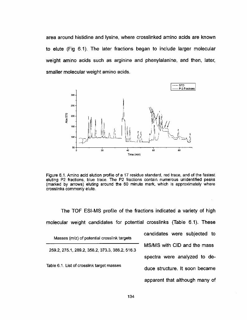
area around histidine and lysine, where crosslinked amino acids are known
to elute (Fig 6.1). The later fractions began to include larger molecular
weight amino acids such as arginine and phenylalanine, and then, later,
smaller molecular weight amino acids.
-STD- P-2 Fractions
40
Time (min)
Figure 6.1. Amino acid elution profile of a 17 residue standard, red trace, and of the fastesteluting P2 fractions, blue trace. The P2 fractions contain numerous unidentified peaks(marked by arrows) eluting around the 60 minute mark, which is approximately wherecrosslinks commonly elute.
The TOF ESI-MS profile of the fractions indicated a variety of high
molecular weight candidates for potential crosslinks (Table 6.1). These
candidates were subjected to
MS/MS with CID and the mass
spectra were analyzed to de-
duce structure. It soon became
Masses (m/z) of potential crosslink targets
259.2, 275.1, 289.2, 358.2, 373.3, 388.2, 516.3
Table 6.1 . List of crosslink target masses
apparent that although many of
134

the candidates in Table 6.1 have different masses, their fractionation
patterns are similar. Some, such as the 289.2 m/z candidate and the 516.2
m/z candidate had the not only the parent ion of the smaller molecule as a
fractionation ion in the larger molecule's spectrum , but also had the same
degradation ions (Fig 6.2). This suggests not only that the two are related,
but that the smaller molecule in it's entirety might be incorporated into a
much larger, multivalent system.
135

100
» 50
176.1
129.1
84.1
101.1
144.1
J Li,,,,] » ?
289.2
197.2
?1?·2>282275.2|323,3
313.3 341.3 516.2
100
£ 50
100 200 300
Mass (m/z)
400
84.1
144.1129.1,161.1
101.1 272.2 289.2
.........LA J, Li0 100 200 300
Mass (m/z)
400
500 600
500 600
Figure 6.2. Mass spectra of a) 516.2 dalton crosslink target and b) 289.2 dalton crosslinktarget. Not only is the 289.2 dalton parent ion present in the 516.2 dalton fractionationpatern, but the fractionation ions are similar in both suggesting a relation between the twoparent ions.
One fractionation pattern that almost all of the crosslink candidates
shared was a strong peak at 84.1 m/z and a weaker peak at 101.1 m/z. This
is indicative of an amino acid with an epsilon amine group, such as lysine.
During collision induced dissociation, when the carboxyl group is cleaved off,
the molecule preferentially cyclizes and loses an amino group. This causes
136

a distinct mass spectra pattern with a very strong peak at 84.1 m/z (the
cyclized, stable molecule) and a weaker peak at 101.1 m/z (the linear,
unstable molecule) (Fig 6.3)9. That most of the crosslink candidate
molecules have this pattern in their MS/MS spectra strongly suggests that
these molecules are lysine derivatives with an epsilon amine group.
©" NH,
^N NHC
I^C2H
84.1
©.NH3
HC
^C2H
©NH2 C2H
H9C
HO- HCI
^C2HH2C
C2H130.1
H2C
H,N
C2H
\101.1
/
Mass (m/z)
Figure 6.3. MS/MS spectra of L-Lysine. Derived from References 9 and 10
Complete analysis of one of the candidates, the 289.2 m/z peak, was
performed and the structure derived (Fig 6.4). In this model, the structure is
a divalent crosslink between lysine and citrulline, a derivative of arginine.
137

HOOC84.1
H
?,?
COOH
N^CH
®0C
H2N
289
129.1
\101.1
?,®
FH? "COOH
CH3144.1
NHp161.1
HOOC
H2N
H2N -CH =?T
=?H
COOH
NH,
T
N-CH =??2H 272.2
176.1___I
289.2
Mass (m/z)
Figure 6.4. Derived mass spectra of 289.2 dalton crosslink target. Fragmentation patternssuggest lysine-citrulline divalent crosslinks.
Although the proposed structure is consistent with the mass spectra
from the tandem MS, other questions need to be addressed before stating
that this is a definitive crosslink. First of all, in order for lysine to form
spontaneous crosslinks, it must be converted to the active aldehyde allysine
by lysyl oxidase. Preliminary studies into the presence of lysyl oxidase in
egg capsule material, although not exhaustive, has come up negative.
Similarly, arginine must be converted to citrulline enzymatically by the nitric
138

oxide synthase pathway or urea cycle. No studies have explored the
presence of those enzymes in the egg capsule materials. Furthermore, work
going into detecting free citrulline in egg capsules has indicated that there is
none. However, this may prove inconclusive since ornithine (the hydrolysis
product of citrulline) elutes very closely to lysine and the large lysine peak
may be blocking the smaller ornithine peak. Also, it could be that there is nofree citrulline in the material, and that all citrulline residues are tied up in
crosslinks. Lastly, as mentioned before, the 289.2 m/z structure appears in
its entirety in the fractionation spectra of the 516.2 m/z candidate. It is
entirely possible that the above structure is merely a small part of a
multivalent crosslink, and that the 289.2 peak is observed in the TOF ESI-
MS spectra due to unexpected degradation in the source region.
Further analysis is necessary to verify the structure of any crosslinks
in this material.
F. Acknowledgements
Thanks are due to J. Pavlovich (Chemistry Department, UCSB) for
assistance with the tandem mass spectrometry data collection and
particularly to H. Waite (BMSE Program, UCSB) for insightful analysis of
said data into meaningful structures. S.W. was financially supported by aCalifornia Sea Grant # R/MP-97B and UC BREP GREAT traineeship
#2007-02.
139

G. References
1. Rawlings, T.?., Adaptations to physical stresses in the intertidal zone: The eggcapsules of neogastropod molluscs. American Zoologist 39, 230-243 (1999).
2. Rapoport, H. S., Biomechanics, Biochemistry, and Molecular Biology of aMolluscen Scleroprotein Elastomer: Whelk Egg Capsule Biopolymer, in MarineBiology. PhD thesis. University of California San Diego (2003).
3. Feughelman, M. Mechanical Properties and Structure of Alpha-Keratin Fibres:Wool, Human Hair, and Related Fibres, UNSW Press: Sydney, Australia, 1997.
4. Price, N. R. & Hunt, S. Occurrence of Reducible Compounds in an InvertebrateProtein of Buccinum-Undatum (L). Cell. MoI. Life 32, 557- 558 (1976).
5. Brown, A. E. X., Litvinov, R. I., Discher, D. E., & Weisel, J. W Forced unfolding ofcoiled-coils in fibrinogen by single-molecule AFM. Biophysical J. 92, L39-L41(2007).
6. Lim, B. B. C, Lee, E. H., Sotomayor, M., & Schulten, K. Molecular basis of fibrinclot elasticity. Structure 16, 449-59 (2008).
7. Zhao, H., & Waite, J. H. Coating Proteins: Structure and Cross-Linking in fp-1 fromthe Green Shell Mussel Perna canaliculus. Biochemistry A4, 5915-15923 (2005)
8. Hunt, S., Carbohydrate and amino-acid composition of egg capsule of whelkBuccinum undatum L. Nature 210, 436-437 (1966).
9. Biemann, K., Seibl, J., & Gapp, F. Mass spectra of organic molecules I. ethyl estersof amino acids. J. Am. Chem. Soc. 83, 3795-3804 (1961).
10. http://www.massbank.jp
140
Top Related