


Languages
Pages
Legal


1
ASSESSMENT AND PROMOTION OF PLASTICITY AND LOCOMOTOR RECOVERY
FOLLOWING SPINAL CORD INJURY
By
SARAH ELIZABETH MONDELLO
A DISSERTATION PRESENTED TO THE GRADUATE SCHOOL OF THE UNIVERSITY OF FLORIDA IN PARTIAL FULFILLMENT
OF THE REQUIREMENTS FOR THE DEGREE OF DOCTOR OF PHILOSOPHY
UNIVERSITY OF FLORIDA
2012

2
© 2012 Sarah Elizabeth Mondello

3
To my Parents, who gave me the space and opportunity to determine my genuine goals, as well as the encouragement and resources to achieve them

4
ACKNOWLEDGMENTS
Obtaining my doctoral degree was a long and challenging journey that could not
have been completed without the support and guidance of several specific individuals.
First and foremost, I would like to thank my mentor Dr. Dena Howland, as she donated
massive amounts of her time to offer critical advice, guidance, and support through
each portion of this project. Her understanding and patience throughout the earlier
learning stages of my doctoral degree were invaluable, and I can only hope she will
continue to be a source of support in the years to come.
I can not say thank you enough to my lab peers: Wilbur O’Steen, Nicole Tester,
Stephanie Jefferson, Adele Blum, Eric Brunk, Megan Black, Kenny Maskell, Rhea
Chattopadhayay, and Valerie Tainsh. As lab manager, Wilbur helped with absolutely
every part of my project, from ordering supplies necessary for experiments, helping with
surgeries, and being an overall handy-man, to also helping maintain my sanity through
humor and kind words. I cannot imagine getting through this process without his help.
Nicole Tester, Stephanie Jefferson and Adele Blum took the time to teach me the
majority of technical skills necessary for this project and as the more senior graduate
students, never stopped offering valuable advise despite having moved on to different
laboratories. Eric Brunk, Megan Black, Kenny Maskell, Rhea Chattopadhayay, Valerie
Tainsh, and Martina Spiess were past laboratory team members that helped with
various experiments and analyses. Additionally, their humor acted as essential comedic
relief during stressful times.
My committee members in addition to Dr. Dena Howland: Dr’s. Paul Reier, Floyd
Thompson, Andrea Behrman, and Mark Lewis, offered extremely helpful guidance and
advice throughout this entire process. They have helped me understand the process of
5
research and science overall, and have pushed me to become a strong scientist. They
each deserve many thanks for their priceless time and efforts.
My family has been extremely supportive and helpful as well. My parents spent
hours of their lives listening to me vent and offering guidance whenever they could. My
older sister Donna and brother in-law Chuck always opened up their home in NY to me,
providing critical breaks and opportunities to recharge. Both my younger sister Amanda
and best friend Annie have lent strong, yet sensitive ears over the past few years,
primarily in regards to non-academic issues. Their support helped me to stay on task.
Lastly, and so importantly I would like to thank all of the priceless friends I have
made along the way outside the laboratory. My happiness, sanity, and maintained
perseverance throughout the majority of my time spent on this project was a result of
their constant understanding, caring, and thoughtful words offered throughout every
success and failure along the way.
6
TABLE OF CONTENTS page
ACKNOWLEDGMENTS .................................................................................................. 4
LIST OF FIGURES ........................................................................................................ 10
ABSTRACT ................................................................................................................... 11
1 BACKGROUND ...................................................................................................... 13
The Spinal Cord Is Inhibitory to Axonal Growth After Injury .................................... 13
The Multiple Motor Pathways that Control Locomotion ........................................... 15
Central Pattern Generator ................................................................................ 15
Propriospinal System ....................................................................................... 15
Supraspinal Motor Pathways ............................................................................ 16 Locomotor Recovery following Spinal Cord Injury .................................................. 17
Complete versus Incomplete Spinal Cord Injury ............................................... 18
The Effect of Spared Tissue on Locomotor Recovery ...................................... 20 Pathway-Specific Potential for Plasticity ................................................................. 21
Axonal Die-Back ............................................................................................... 21 Responses to Trophic Factor Application ......................................................... 22 Propriospinal Plasticity ..................................................................................... 23
Enhancing Propriospinal Plasticity ................................................................... 25 The Effects of Training on Functional Recovery after Injury ................................... 26
Cats as a Translational Model of Spinal Cord Injury ............................................... 27 The Pathology of Spinal Cord Injury ....................................................................... 28
The Glial Scar ................................................................................................... 30 The inhibitory nature of chondroitin sulfate proteoglycans ......................... 30 Chondroitin sulfate proteoglycan temporal expression patterns after
spinal cord injury ..................................................................................... 33 Chondroitin sulfate glycosaminoglycans are the primary inhibitory
component of chondroitin sulfate proteoglycans ..................................... 34 Chondroitinase ABC as a Potential Therapeutic ..................................................... 35
Chondroitinase ABC-Mediated Tract Plasticity ................................................. 36
Potential Mechanisms Underlying Chondroitinase ABC-Mediated Effects ....... 38 Determining Optimal Chondroitinase ABC Application Paradigms ................... 39
Biological stability ....................................................................................... 39
Chondroitin sulfate glycosaminoglycan turnover rate ................................. 40
Delivery period ........................................................................................... 41 The Important Role of Tract Tracing ....................................................................... 42
2 DIFFERENTIAL RECOVERY OF GAIT FEATURES DURING BASIC AND ADAPTIVE LOCOMOTION FOLLOWING SPINAL HEMISECTION IN THE CAT .. 44
Introduction ............................................................................................................. 44

7
Methods .................................................................................................................. 45
Subjects............................................................................................................ 45 Surgical Procedures ......................................................................................... 46
T10 spinal hemisection .............................................................................. 46 Behavioral Tasks, Training Paradigm, and Gait Features ................................ 46
Overall assessment of locomotor recovery ................................................ 46 Onset of recovery ....................................................................................... 47 Limb accuracy ............................................................................................ 47
Crossing speed .......................................................................................... 47 Percentage of abnormal foot positioning on rungs ..................................... 48 Step cycle duration .................................................................................... 48 Stride length ............................................................................................... 48 Footfall patterns ......................................................................................... 48
Stance percentage ..................................................................................... 49 Double support period ................................................................................ 49
Iliac crest mediolateral movement .............................................................. 49
Duration to support after toe down ............................................................. 49 Ankle flexion during yield after toe down .................................................... 49
Tissue Processing ............................................................................................ 50
Perfusions and tissue preparation .............................................................. 50 Histology: Cresyl violet and myelin staining ............................................... 50
Statistical analysis ...................................................................................... 51 Results .................................................................................................................... 51
Lesion Magnitudes ........................................................................................... 51
Recovery Onset and Effects of Injury on Crossing Speed ................................ 51 Phalangeal versus Abnormal Metatarsal Placement onto Ladder Rungs
Post-Spinal Cord Injury ................................................................................. 53 Injury Effects on Cycle Duration and Stride Length .......................................... 54
Effects of Injury on Interlimb Coordination, Stance Duration, and Double Support.......................................................................................................... 55
Mediolateral Movement .................................................................................... 58
Distal Joint Stability .......................................................................................... 59 Discussion .............................................................................................................. 60
Task-Specific Spatiotemporal Gait Changes after Injury .................................. 61 Trunk Support on Skilled Tasks after Injury ...................................................... 62 Task Specific Changes in Distal Joint Positioning after Injury .......................... 63
Conclusions ...................................................................................................... 64
3 THE EFFECT OF DIFFERENT CHONDROITINASE ABC TREATMENT DURATIONS ON LOCOMOTOR RECOVERY AND SUPRASPINAL TRACT PLASTICITY iN A FELINE MODEL OF SCI ........................................................... 74
Introduction ............................................................................................................. 74 Materials and Methods............................................................................................ 76
Subjects............................................................................................................ 76 Surgical Procedures ......................................................................................... 77
Low thoracic spinal hemisection ................................................................ 77

8
Chondroitinase ABC administration ........................................................... 77
Fluorogold spinal injections ........................................................................ 78 Behavioral Procedures ..................................................................................... 78
Behavioral tasks and training paradigm ..................................................... 78 Assessment of locomotor recovery ............................................................ 79 Horizontal ladder, peg walkway, and narrow beam assessment: Limb
accuracy ................................................................................................. 79 Tissue Processing ............................................................................................ 80
Perfusions and tissue preparation .............................................................. 80 Cresyl violet and myelin staining ................................................................ 80 Assessment of injection site ....................................................................... 81 Immunohistochemistry ............................................................................... 81 Fluorogold-labeled cell counts ................................................................... 82
Lesion ranking ............................................................................................ 82 Statistical Analysis ............................................................................................ 83
Results .................................................................................................................... 83
Lesion Magnitudes ........................................................................................... 83 Axonal Projections below the Lesion Site ......................................................... 83 Rate of Recovery .............................................................................................. 85
Ipsilateral Hindlimb Accuracy ........................................................................... 87 Horizontal ladder ........................................................................................ 87
Narrow beam ............................................................................................. 88 Peg walkway .............................................................................................. 89
The Relationship between Spared Tissue and Functional Recovery ............... 89
Onset of task recovery versus lesion size .................................................. 90 Ipsilateral hindlimb accurate targeting versus lesion size .......................... 91
Discussion .............................................................................................................. 92 Summary of Results ......................................................................................... 92
Supraspinal Connectivity below the Lesion ...................................................... 92 Locomotor Recovery and Supraspinal Plasticity .............................................. 93 Lesion Size ....................................................................................................... 96
Conclusion ........................................................................................................ 97
4 OPTIMIZATION OF FLUOROGOLD RETROGRADE TRACING ......................... 105
Introduction ........................................................................................................... 105 Materials and Methods.......................................................................................... 106
Subjects.......................................................................................................... 107
Surgical Procedures ....................................................................................... 107
Fluorogold spinal injections ...................................................................... 107 Tissue Processing: Histology and Immunohistochemistry .............................. 108
Perfusions ................................................................................................ 108
Cresyl violet and myelin staining .............................................................. 108 Fluorogold immunohistochemistry ........................................................... 109 Assessment of injection site completeness and fluorogold
autofluorescence .................................................................................. 110 Results .................................................................................................................. 110

9
Tissue Damage at the Injection Site ............................................................... 110
Fluorogold Detection Techniques ................................................................... 111 Triton Increases Tracer Travel Time .............................................................. 112
Discussion ............................................................................................................ 114
5 SUMMARY ........................................................................................................... 121
Defining Recovery ................................................................................................ 121 Advancing Chondroitinase ABC to the Clinic ........................................................ 122 Conclusions .......................................................................................................... 124
LIST OF REFERENCES ............................................................................................. 125
BIOGRAPHICAL SKETCH .......................................................................................... 149

10
LIST OF FIGURES
Figure page 2-1 Lesion magnitudes ............................................................................................. 66
2-2 Tasks, onset of recovery, and crossing speeds .................................................. 67
2-3 Phalangeal versus metatarsal placement onto ladder rungs .............................. 68
2-4 Changes in cycle duration and stride length ....................................................... 69
2-5 Change in hindlimb footfall patterns and hind-to-hindlimb coordination .............. 70
2-6 Change in hindlimb stance and double support periods ..................................... 71
2-7 Mediolateral movement of the iliac crest ............................................................ 72
2-8 Swing to stance transition ................................................................................... 73
3-1 Range of spinal hemisections ............................................................................. 99
3-2 Supraspinal connections below the lesion ........................................................ 100
3-3 Onset of basic and skilled locomotor recovery ................................................. 101
3-4 Ipsilateral hindlimb accuracy ............................................................................ 102
3-5 Spared tissue and task onset correlations ........................................................ 103
3-6 Spared tissue and accurate ipsilateral hindlimb targeting correlations ............. 104
4-1 Fluorogold injection schematic ......................................................................... 116
4-2 Effects of different fluorogold concentrations, volumes, and survival periods on tissue damage at the injection site ............................................................... 117
4-3 Anti-fluorogold immunohistochemical processing leads to greater fluorogold detection compared to native autofluorescent detection ................................... 118
4-4 Long term fluorogold detection using anti-fluorogold immunohistochemical processing ........................................................................................................ 119
4-5 Triton enhances fluorogold tracing speed ......................................................... 120

11
Abstract of Dissertation Presented to the Graduate School of the University of Florida in Partial Fulfillment of the Requirements for the Degree of Doctor of Philosophy
ASSESSMENT AND PROMOTION OF PLASTICITY AND LOCOMOTOR RECOVERY
FOLLOWING SPINAL CORD INJURY
By
Sarah Elizabeth Mondello
August 2012
Chair: Dena Ruth Howland Major: Medical Sciences – Neuroscience
Damage to the spinal cord causes sensorimotor loss that is permanent. The
resulting functional losses are debilitating, may be life threatening, and affect an
individual’s ability to be independent in the home and community. Unfortunately, there
are no effective therapeutics to reduce the functional deficits caused by spinal cord
injury (SCI). Identification of effective treatments has been complicated by the limited
regenerative capacity of the central nervous system in addition to SCI being a
multifarious problem likely to require a complex, combinatorial treatment approach.
Some individuals with incomplete SCIs may recover basic walking abilities but
continue to have difficulty with more challenging forms of locomotion including those
that require greater balance and alterations in leg trajectories. In the current studies, a
cat low thoracic hemisection model was used. This incomplete, asymmetrical injury is
similar to Brown Sequard Syndrome (BSS) described in a subpopulation of humans with
SCIs. In the first set of studies, a battery of gait features were assessed to compare
performances during a basic locomotor task (flat overground walking) and an adaptive
locomotor task (horizontal ladder crossing). Gait features critical for successful
performance of adaptive locomotion pre- and post-SCI were identified. In the second

12
study, results from previous chondroitinase abc (ch’abc) studies in the lab were
extended to determine the effects of different treatment durations on anatomical
plasticity and functional recovery. The results from this study contribute important
information relative to treatment duration for the ultimate translation of ch’abc to a
clinical setting. In the final study, retrograde tract tracing with Fluorogold (FG) was
optimized for use in a large animal model and will contribute to future assessments of
circuitry disruption and plasticity in the injured spinal cord.
Collectively, the body of work presented in this dissertation contributes to our
understanding of the anatomical and behavioral changes that occur following SCI, how
functional performance might be enhanced with a promising therapeutic treatment, and
how methods to assess anatomical plasticity can be improved to enhance future
studies.

13
CHAPTER 1 BACKGROUND
The Spinal Cord Is Inhibitory to Axonal Growth After Injury
The devastating consequences of spinal cord injury (SCI) and the permanence of
the resulting effects in humans are well documented. The earliest documentation of
SCI dates back to 1550 B.C. in the Egyptian papyrus known as the “Edwin Smith
Papyrus”. In that document it is stated that “If you examine a man with a neck
injury…and find he is without sensation in both arms and both legs, and unable to move
them…it is due to the breaking of the spinal cord caused by dislocation of the cervical
vertebra. This is a condition which cannot be treated.” Almost 400 years later, one of
the most notable neuroscientists, Ramon y Cajal wrote: “In adult centres the nerve
paths are something fixed, ended, immutable. Everything may die, nothing may be
regenerated (Cajal, 1928)”. These concepts of the impossibility of functional recovery
and the absence of axonal growth after SCI dominated the scientific community until the
1940’s, when another type of post-SCI growth was discovered. Collateral sprouting, the
growth of nerve fibers from intact axons after injury was first identified in experiments
using nerve fibers supplying the skin in rabbits (Weddell et al., 1941). Similar growth
was subsequently identified in the spinal cord by Liu and Chambers in 1958. Using a
spared root prep, they denervated several dorsal roots from one side of the cord, waited
several months, then transected the remaining roots from the chronically injured side in
addition to the matching roots from the non-injured side. Two weeks later tissue was
assessed with a neurodegeneration stain, which showed a greater number of
degenerating profiles in the chronic compared to the acute side. This was attributed to
connections between the chronic and acute sides by way of collateral sprouting (Liu and

14
Chambers, 1958; For Review See Guth, 1975). Subsequently, the concept that
regeneration of axotomized fibers does not occur was disputed by several studies which
reported that severed axons returned to active states if stimulated properly (Li and
Raisman, 1995; Kobayashi et al., 1997). For example, a study completed by Yi and
colleagues determined that damaged rubrospinal tract fibers one year after injury could
grow into a peripheral nerve graft if first treated with brain derived neurotrophic factor
(BDNF) (Kobayashi et al., 1997). These combined findings, in addition to many others,
suggested that the spinal cord has a much greater potential for axonal plasticity and
functional recovery after injury than previously believed.
The greatest functional restoration most likely will be obtained through a
combination of regeneration and collateral sprouting. Research to better understand
and enhance these types of plasticity is being actively pursued using experimental
animal models. A multitude of different approaches such as locomotor training (For
review see Battistuzzo et al., 2012), cellular replacement, introduction of matrix
substrates, as well as numerous different pharmacological interventions (For review see
Jeffery and Blakemore, 1999; Lu and Tuszynski, 2008; Boulenguez and Vinay, 2009) all
have shown some enhancement of axonal growth after injury. For this new growth to
be functionally effective after an injury, it must make functional connections. The motor
system is an extremely complex combination of many tracts each playing their own
independent, as well as complementary roles in locomotion. Understanding the function
of these pathways during locomotion in an uninjured system, as well as their
contributions to locomotor recovery after injury is important for designing approaches to
enhance locomotor recovery post-SCI.

15
The Multiple Motor Pathways that Control Locomotion
In 2004, the report of a survey taken by 681 SCI individuals ranked seven specific
functions in order of their importance for quality of life. The ability to walk ranked fourth
highest, indicating the need to determine methods for recovering locomotor function
after injury (Anderson, 2004). The underlying neural control of walking and other types
of locomotion has been well studied over the course of several centuries and provides a
strong foundation for guiding present and future investigators towards promoting
recovery of locomotor function after SCI (For review see Clarac, 2008).
Central Pattern Generator
The underlying circuitry responsible for locomotion is complex and involves all
levels of the neural axis spanning from intraspinal networks to supraspinal tracts (D.A,
1975; Eidelberg and Yu, 1981; Yu and Eidelberg, 1981). The most notable locomotor
related intraspinal systems are the central pattern generators (CPGs); a well organized
circuit of interneurons and motoneurons located in the cervical and lumbar areas of the
spinal cord. These circuits fire in specific patterns leading to alternating bilateral flexion
and extension of the arms and legs to produce stepping. This CPG activity can occur
spontaneously without descending supraspinal or sensory inputs and is considered to
be the neural basis for the basic stepping pattern. Circuitry can be modulated in
response to external stimuli through spinal interneurons allowing for some degree of
adaption to changes in the environment (For review see Grillner and Zangger, 1979;
Rossignol and Frigon, 2011).
Propriospinal System
Propriospinals (PSNs) make up a large portion of spinal interneurons and play a
significant role in locomotion. This system contributes to trunk control, inter- and intra-

16
limb integration, and modulates input from both the descending supraspinal systems
and peripheral afferents. Additionally, they synchronize motor circuitry throughout the
entire length of the spinal cord (For review see Flynn et al., 2011). The PSN system is
primarily contained within the spinal cord, linking different spinal segments together
allowing for complex movements. However, some project to supraspinal centers like
the lateral reticular nucleus (Alstermark et al., 1981b; Skinner et al., 1989), and
cerebellum (Skinner et al., 1989). Depending on their length PSN pathways are
classified as either “short”, 1-6 segments or “long”, >6 segments (Conta Steencken and
Stelzner, 2009). Thus, short PSNs modulate activity occurring relatively local to the cell
of origin and long PSNs make connections to distant regions of the spinal cord. In fact,
evidence indicates that the long PSNs may be responsible for connecting the cervical
and lumbar CPGs for quadrupedal stepping/synchrony across the forelimbs and
hindlimbs in the cat (Miller et al., 1973), and possibly the arms and legs in humans (For
review see Dietz, 2002; Huang and Ferris, 2009; Tester et al., 2012). PSN projections
can occur either in the rostral or caudal plane from their cell of origin, located in the
spinal cord gray matter (Skinner et al., 1989). This flexibility allows for the complex
circuitry that underlies the production and control of elaborate multi-segmental
movements.
Supraspinal Motor Pathways
Voluntary locomotor control is mediated by supraspinal tracts that originate in the
brain or brainstem. Primary supraspinal motor tracts include the corticospinal (CST),
rubrospinal (RuST), reticulospinal (ReST), and vestibulospinal (VST) tracts. These
tracts are separated into a medial system: ReST, VST, and a lateral system: RuST,
CST. Approximately 87.6% of the CST is located dorsolaterally while 12.4% of the CST
17
(anterior CST) travels in the ventromedial funiculi of the spinal cord (Kwon et al., 2011).
These two systems, the CST and RuST, are essential for producing fine-tuned, precise
movements like stepping over an obstacle (Mohagheghi et al., 2004), grasping food
(Alstermark et al., 1981a; Alstermark et al., 1987; Whishaw et al., 1998), and paw
placement (Batson and Amassian, 1986; For review see Drew et al., 2002). The loss of
these systems causes difficulties maneuvering through environments and completing
day-to-day activities.
The medial system is responsible for creating the necessary tone in postural
musculature allowing for upright locomotion. Additionally, the ReST has been shown to
initiate stepping (Jell et al., 1985). The VST, originating in the vestibular nuclei within the
medulla, is primarily controlled by the utricles and saccules of the vestibular system and
allows for the body’s musculature to quickly respond and recover from sudden
displacements (Markham, 1987). In this way, the VST is critical for maintaining
balance. Disruption to either of the medial or lateral systems has detrimental effects
that vary depending on the magnitude of the lesion itself.
Locomotor Recovery following Spinal Cord Injury
Complete transection of the spinal cord, which removes all supraspinal input
below the level of the lesion, results in immediate and complete loss of motor function
and reflexes below the injury. This period, known as “spinal shock”, is present in
experimental animals as well as humans. It lasts for approximately two weeks in
humans after which some reflexes begin to return (For review see Riddoch, 1917;
Eidelberg, 1981). Some investigators argue that the period of spinal shock is much
shorter on the magnitude of minutes to hours (For review see Ditunno et al., 2004).
Regardless, there is an overall period of motor depression after an injury that lasts for

18
several weeks. Several months following complete spinal transection spasticity may
occur, characterized by hypertonus, clonus, involuntary somatic reflexes, and muscle
spasms that are often extremely painful (For review see Rabchevsky and Kitzman;
Adams and Hicks, 2005). This secondary impairment as a result of SCI, is believed to
be due to aberrant persistent inward currents (PIC). In the uninjured nervous system,
PICs are depolarizing inward currents intrinsic to motoneurons that can self-sustain
firing as long as the cell remains depolarized. Cessation requires inhibitory synaptic
input, often led by supraspinal centers. PICs are controlled by the monoamines 5HT
and norepinephrine, in which their primary sources are the raphe nucleus and locus
coeruleus. Acutely after an injury, motoneuron excitability is reduced due to a loss of
supraspinal connectivity and thus, monoamines. However, over time motoneuronal
excitability returns to somewhat normal levels, and is particularly monoamine-sensitive.
Residual monoamines present in the spinal cord and vasculature are able to activate
PICs, however the remaining lack of supraspinal connectivity prevents these PICs from
being inhibited, causing spasticity (For review see ElBasiouny et al., 2009). There are
currently several treatment options available for spasticity including baclofen, tizanidine,
and botulinum neurotoxin, however they have varying degrees of efficacy (Rabchevsky
and Kitzman). Multiple animal models of spasticity are used in order to improve our
understanding of this impairment and improve treatment options (Thompson et al.,
2001).
Complete versus Incomplete Spinal Cord Injury
Although voluntary stepping does not recover after an anatomically complete SCI,
CPG based spinal stepping can be elicited as seen in humans (Dietz and Colombo,
2004), cats (Grillner and Rossignol, 1978; Eidelberg et al., 1980), rats (references),
19
dogs (Hart, 1971), and possums (Hinsey and Cutting, 1936). Incomplete SCI, like
anterior cord syndrome, central cord syndrome, posterior cord syndrome, and brown
sequard syndrome (BSS), result in substantially greater locomotor recovery due to the
sparing of some tissue (Eidelberg, 1981). Individuals with incomplete SCI typically have
notable recovery of basic locomotion like walking overground or on a treadmill, however
the presence of some common deficits remain. Often, these individuals have
decreased gait speed (Knutsson and Richards, 1979; Dietz et al., 1981; Conrad et al.,
1983; Wainberg et al., 1986; Wagenaar and Beek, 1992; Lajoie et al., 1999) and cycle
duration (Lajoie et al., 1999; Barbeau et al., 2002). Additionally, a variety of different
joint alterations have been reported such as increased hip angular excursion, and
increased knee flexion during either foot touchdown alone, or the entire step cycle. The
ankle also has been reported as either being dorsiflexed, or plantar flexed causing foot
drag (Conrad et al., 1983). Changes in muscle activation during walking also have
been shown to occur after injury. Typically, an abnormal co-activation of antagonist
muscles occurs (Fung and Barbeau, 1989, 1994) in addition to altered shape and timing
of activation patterns (Fung and Barbeau, 1989; Domingo et al., 2007).
Individuals with incomplete SCI often have greater struggles with challenging
types of locomotion. This can be illustrated by studies looking at functional recovery in
cats with a hemisection injury similar to BSS. Studies completed by the Howland
laboratory have shown that following a T10 hemisection, cats were able to recover
bipedal treadmill and crossing of a 12” wide basic overground runway well, but had
greater difficulties when performing challenging tasks like crossing of a horizontal
ladder, peg walkway, or 2” wide narrow beam (Tester and Howland, 2008; Jefferson et

20
al., 2011). The disparity between functional recovery in individuals with complete
versus incomplete SCI suggests that these two populations require different
rehabilitation strategies.
The Effect of Spared Tissue on Locomotor Recovery
The degree of recovery in individuals with incomplete SCI varies greatly across
individuals and is partially due to the different types of pathways spared by the injuries.
Animal studies in which specific cuts were made in order to axotomize select tracts both
reveal individual tract functions through the process of elimination, as well as
determines the contributions of different tracts on locomotor recovery after SCI.
Extensive damage to the medial systems often is accompanied by severe and
permanent postural control and locomotor impairments (Lawrence and Kuypers, 1968;
Brustein and Rossignol, 1998). This suggests a critical role in basic locomotion for
these systems (Lawrence and Kuypers, 1968; Brustein and Rossignol, 1998). In
contrast, studies in which injuries were isolated to the CST and RuST showed only
transient locomotor deficits and a quick recovery of overground locomotion (Laursen
and Wiesendanger, 1967; Muir and Whishaw, 2000; Kanagal and Muir, 2009) food
grasping (Blagovechtchenski et al., 2000) and other skilled finger movements (Hepp-
Reymond et al., 1970). However, components of these movements remained impaired,
indicating that these lateral systems are most important for more skillful, precision-
based movements. Damage to these lateral pathways paired with sparing of the
ventromedial cord has been associated with substantial locomotor recovery after SCI
(Windle et al., 1958; Nathan and Smith, 1973; Afelt, 1974; Eidelberg et al., 1981;
Schucht et al., 2002; Krajacic et al., 2010) indicating that alternate pathways can
contribute to functional recovery after injury.
21
Pathway-Specific Potential for Plasticity
The contributions of certain pathways to recovery after injury are largely due to
their innate responses to injury and plastic properties. These properties differ across
systems. Axonal die-back is a major response to injury that occurs in all transected
fibers (Busch et al., 2009) which affects their participation in new circuit formation after
injury; the further an axon retracts, the more distance regenerating and/or sprouting
fibers must cover to bridge the lesion site and restore function. Even if a fiber with
significant retraction sprouts onto nearby spared circuitry, that circuit itself will have
further distance to cover, increasing its chances for failure. Comparing axonal die-back
properties across several supraspinal tract systems provides insight into their individual
potential for meaningful plasticity. The application of trophic factors like BDNF and
neurotrophin 3 (NT-3) have been associated with increased axonal growth of multiple
supraspinal pathways after injury (Kobayashi et al., 1997; Hiebert et al., 2002; Kwon et
al., 2002; Plunet et al., 2002; Dolbeare and Houle, 2003). The responsiveness of
individual tracts to trophic factor application also can provide a general indication of
specific tracts’ ability to grow after injury.
Axonal Die-Back
Significant evidence indicates that the CST has limited regenerative potential. For
example, several studies looking at plasticity in multiple pathways have reported
minimal CST growth, but significant growth in the RuST (Richardson et al., 1984a;
Houle and Ye, 1999; Decherchi and Gauthier, 2000; Plunet et al., 2002), ReST, and
VST (Houle and Ye, 1999; Oudega et al., 1999). One reason CST growth is limited
after injury is likely related to its significant and prolonged axonal die-back as depicted
in several studies. Specifically, these studies found that following injury the proximal

22
ends of CST axons formed dystrophic endbulbs and retracted for several millimeters
and for up to eight weeks (Pallini et al., 1988; Oudega et al., 1999). Meanwhile, axonal
dieback only lasted for ~ four weeks post-injury in the VST, ReST and RuST tracts with
axons measuring approximately 0.5 to 1.5 mm from the lesion site (Houle and Jin,
2001). In contrast to the above-mentioned results, one study also found continued die-
back for eight weeks following axotomy of the VST, suggesting this tract may share
regenerative and sprouting difficulties similar to the CST (Oudega et al., 1999).
Responses to Trophic Factor Application
Both the RuST (Liu, 1999; Liu et al., 2002; Murray et al., 2002; Jones et al.,
2003b), and ReST (Xu et al., 1995; Menei et al., 1998) have shown enhanced post-
axotomy growth in response to trophic factor application. However, the CST and VST
have been reported to respond less notably in several studies. Specifically, two
separate groups found that while the addition of BDNF to red nucleus cell bodies
resulted in enhanced regeneration of the RuST into peripheral nerve grafts, the same
treatment to motor cortex cell bodies did not cause CST regeneration. Interestingly,
they did indicate enhanced collateral sprouting both rostral (Hiebert et al., 2002; Plunet
et al., 2002) and caudal (Plunet et al., 2002) to the injury. A similar study comparing
regeneration of the RuST, ReST, CST, and VST after application of one of three
different growth factors (ciliary neurotrophic factor (CNTF), BDNF, or neurotrophin 3
(NT-3)) found enhanced regeneration of the ReST and RuST in response to all growth
factors. However, the VST responded to only one of the growth factors, CNTF, while
the CST did not respond to any (Ye and Houle, 1997). Similar results have been found
in additional studies indicating that both the CST (Schnell et al., 1994; Tuszynski et al.,
1997; Blesch, 1999; Lu et al., 2001) and VST (Xu et al., 1995; Ye and Houle, 1997;

23
Menei et al., 1998) may be less apt for plasticity after axotomy. However, despite these
tracts underwhelming plastic responses to trophic factors, several studies utilizing
different growth promoting methods have reported increased growth. For example,
administration of leukemia inhibitory factor (Blesch, 1999), and olfactory ensheathing
cell transplants (Li et al., 1997) have both led to enhanced CST growth. Even more
interesting is the study by Bareyre and colleagues who reported spontaneous sprouting
of transected CST axons onto long PSN tracts in the cervical spinal cord following a
thoracic SCI in rats. This new circuitry was capable of bridging the lesion site and
enhancing functional recovery (Bareyre et al., 2004). Such findings indicate that CST
growth may, in fact, be occurring in regions distant from the lesion where plasticity is not
typically assessed. A similar phenomena was reported in a study by Rozensweig and
colleagues who showed substantial spontaneous CST growth in a primate model of
SCI, citing species differences as a potential reason for the lack of CST plasticity
commonly reported in rodents (Rosenzweig et al., 2010). Plasticity of the VST, though
less well studied, has shown enhancements when growing into an embryonic tissue
transplant (Ito et al., 1999). Overall, these findings indicate that the CST and VST have
the potential for plasticity, but respond less readily to certain treatments compared to
the RuSt and ReST. Alternatively, plasticity in the CST and VST may be occurring as
readily as in the ReST and RuST, but in regions distant to the lesion that are not
typically assessed.
Propriospinal Plasticity
Although plasticity of the PSN system is currently not as well studied as the
supraspinal systems, researchers are beginning to identify it as a key player in creating
novel pathways that allow for neuronal signals to bypass the lesion and restore function.

24
In a study by Courtine and colleagues, they determined that following several carefully
timed and placed incomplete lesions, spared PSN axons were independently capable of
mediating recovery of stepping without direct input from the brain (Courtine et al., 2008).
Spontaneous PSN tract plasticity also has been shown to form novel circuitry after a
high cervical injury resulting in recovery of chronic diaphragm activity (Darlot et al.,
2012). These findings indicate incredible plastic potential in this PSN system that is
capable of functional restoration. However, based on detailed investigations by the
Stelzner group the potential for plasticity appears to differ across the short and long
PSNs. In a collection of studies, they showed that long and short PSNs respond
differentially to axotomy in that there are fewer surviving short PSNs after injury
compared to long PSNs (Conta and Stelzner, 2004; Siebert et al., 2010b; Conta
Steencken et al., 2011). Furthermore, while short PSNs have an initial upregulation of
growth factor receptor gene expression, as well as immune, inflammatory, and pro-
apoptotic gene expression transiently after injury, these same genes are downregulated
in the long PSNs. This translates into more short PSN cell death compared to long.
However, surviving short PSNs have genetic profiles that return to normal by one month
(Siebert et al., 2010a; Siebert et al., 2010b) and their neuronal size does not
significantly change between two weeks and the course of the study (eight weeks).
The authors suggest that the surviving short PSNs could be a specialized population
with “sustaining collaterals” making them more resilient to axotomy (Siebert et al.,
2010a). This theory was validated in a separate study which reported significant short
PSN axonal growth across a midline injury to form functional synaptic connections with
motoneurons after SCI (Fenrich and Rose, 2009).

25
Although the above mentioned set of studies shows an initial and permanent
decrease in the number of short PSNs over the course of a 16 week study (Conta
Steencken and Stelzner, 2009), studies from my own lab have shown an initial yet
transient decrease in short PSNs followed by a significant increase by 16 weeks post-
injury (Blum, 2010). This difference in short PSN plasticity across studies may be a
result of species differences, as our study was completed in feline and theirs in rat.
However, it is most likely due to the extensive training our animals underwent and the
lack of training in their study as training increases BDNF production (Beaumont et al.,
2008) which in turn increases plasticity (Girgis et al., 2007). Additionally, the Stelzner
group used a contusion injury model, while a a hemisection model was used in ours.
Hemisections typically have been associated with greater axonal growth (Iseda et al.,
2008). These contradictory findings indicate that the PSN system has plastic potential,
however requires certain stimuli to activate this potential after injury.
Enhancing Propriospinal Plasticity
Although there are few studies that have specifically focused on the effects of
trophic factors on PSN plasticity results following Schwann cell transplants into the
lesion site showed an enhancement in long and short PSN plasticity however, the
greatest amount of plasticity was in the short PSNs (Xu et al., 1997; Takami et al., 2002;
Doperalski et al., in preparation). Additionally, one study found that the combination of
GDNF and Schwann cell seeded channels applied to the lesion site led to enhanced
plasticity of short PSNs. Collectively, the above studies suggest that although there is
substantial PSN cell death after injury, those that survive are capable of substantial
plasticity that can mediate and support recovery after SCI (Xu et al., 1995; Menei et al.,
1998).

26
The Effects of Training on Functional Recovery after Injury
Training itself has been shown to enhance plasticity and functional recovery after
injury (For review see Marsh et al., 2010). Multiple studies using training as the sole
therapeutic after injury have reported enhancements of the function being trained.
Examples of these functions include basic stepping (Harkema et al., 2011), treadmill
stepping in both humans (Thomas and Gorassini, 2005), and cats (Lovely et al., 1986;
Barbeau and Rossignol, 1987), wheel-based stepping (Beaumont et al., 2008),
staircase climbing (Singh et al., 2011), single pellet reaching, and horizontal ladder
walking in rats (Girgis et al., 2007; Starkey et al., 2011). Further assessments regarding
how training enhances functional recovery have identified training-induced
enhancements of growth factor upregulation, specifically BDNF and Anti-Growth
Associated Protein-43 (GAP-43) (Girgis et al., 2007; Beaumont et al., 2008; Ying et al.,
2008). These growth factors enhance axonal plasticity (Liu, 1999; Murray et al., 2002),
and help shape synaptic plasticity (Ying et al., 2008). Training also has been shown to
enhance motoneuronal electrophysiological properties like resting membrane potential,
spike trigger levels (Beaumont et al., 2008), and increase excitability of the CST
underlying leg muscle activity (Thomas and Gorassini, 2005).
The way training is conducted after injury has drastic affects on functional
outcomes. Task-specific training of one type of task has been shown to enhance
recovery of that task, but in some cases does not transfer to other tasks. For example,
swim training was shown to enhance swimming kinematics, but had no effect on
overground walking (Magnuson et al., 2009). Additionally, stair climbing ascent training
led to enhanced recovery of this specific task, but caused only partial improvements on
overground and grid stepping (Singh et al., 2011).

27
Task-specific training of one task also has been shown to have detrimental and/or
positive effects on the recovery of other tasks. For example, rats that were trained
specifically on single pellet reaching had significant functional improvements on this
task after injury, but were significantly worse at crossing a horizontal ladder compared
to their untrained rat counterparts (Girgis et al., 2007). Another example is one study
completed by Garcia-Alias and colleagues, who found that rats trained to walk on a grid
had significantly enhanced recovery of skilled pellet reaching, but were significantly
worse at horizontal ladder stepping compared to their untrained counterparts (Garcia-
Alias et al., 2009). The potential for task-specific training on one task to transfer to
other tasks most likely relates to the kinematic similarities across tasks as specific
neuromuscular activity patterns lead to differential muscle responses. This concept was
assessed and confirmed in a study that compared the muscle activity of spinalized cats
trained to either step on a treadmill or stand after injury. Those cats trained to stand
had increased maximum rate of shortening in their medial gastrocnemius muscles, as
well as greater muscle mass when compared to cats that were only trained to step on a
treadmill. These same cats also had an overall greater shift of muscle fiber type
towards fast fibers (Roy et al., 1999). Overall, these findings demonstrate that the
neuromuscular system is extremely sensitive to different training regimens, and that
there is a strong need to enhance our understanding of how different training paradigms
affect functional recovery after injury in order to maximize recovery.
Cats as a Translational Model of Spinal Cord Injury
Cats are considered to be a highly translational SCI model as they share
numerous similarities with humans in regards to neuro-anatomical control of movement,
gait mechanics, and interlimb coordination (For review see Vilensky, 1987; Dietz, 2002;

28
Majczynski and Slawinska, 2007). The overall organization of the large ascending and
descending tracts within the spinal cord are similar across species (Majczynski and
Slawinska, 2007; Watson, 2009). Additionally, the presence of a central pattern
generator, which underlies stepping patterning, has been confirmed in the cat (Miller et
al., 1973; Smith et al., 1983), and indirectly identified in humans (Harkema et al.;
Dimitrijevic et al., 1998; Jilge et al., 2004; Calancie, 2006). Evidence from both species
also has indicated the presence of flexible networks in the spinal cord that are
responsible for independent movement of each limb, suggesting that both species have
a “half centre” model of locomotion (For review see Brown, 1914; Prokop et al., 1995).
In regards to reflex characteristics, both cats and humans share certain reflexive
responses to afferent stimuli (Lisin et al., 1973) and also have several shared speed-
related gait changes (Vilensky, 1987). Postural control in response to perturbations also
was identified as being similar across cats and humans when humans were positioned
in a quadrupedal stance (Macpherson et al., 1989).
Despite the more extreme differences in overall body size, the average cat spinal
cord length is only 9 cm shorter than the average human spinal cord. Thus, the distance
necessary for axonal plasticity to render functional changes is similar across cats and
humans. These species similarities make the cat a valuable animal model for SCI
research, therefore the studies to be described in the experimental chapters (2-4) of this
dissertation were completed in a cat model of SCI.
The Pathology of Spinal Cord Injury
The peripheral nervous system (PNS) is substantially more conducive to axonal
growth after injury than the central nervous system (CNS), and It has been determined
that the CNS environment is responsible for this disparity (Richardson et al., 1980;

29
Benfey and Aguayo, 1982; Richardson et al., 1984a). Injury to the CNS, specifically the
spinal cord, creates an upregulation of multiple different physiological responses that
further inhibit this system’s potential for plasticity.
SCI occurs in a biphasic manner in which there is an initial mechanical injury
followed immediately by a period of ongoing damage. This period is commonly known
as secondary injury and can continue for months after the mechanical injury (Rowland
et al., 2008; Flynn et al., 2011; Kuzhandaivel et al., 2011). Secondary injury begins
immediately after insult, in which there is substantial hemorrhagic necrosis, microglial
activation (Donnelly and Popovich, 2008), and an upregulation of pro-inflammatory
cytokines like interleukin 1β, and tumor necrosis factor α (Pineau and Lacroix, 2007).
Within the next 48 hours the Blood Brain Barrier reaches its peak level of permeability
(Noble and Wrathall, 1989); neutrophils invade the lesion site, and vasogenic and
cytotoxic edema start to set in (For review see Rowland et al., 2008). Additionally,
hemorrhaging continues which leads to free radical production and a dysregulation of
Ca++ ion concentrations followed by calpain activation and mitochondrial dysfunction
(Schanne et al., 1979). Extracellular glutamate eventually reaches toxic levels at the
lesion site (Wrathall et al., 1996) and collectively these components will result in cell
death (Schanne et al., 1979). Studies indicate that neuronal death is primarily by way of
necrosis, although there have been some reports of neuronal apoptosis (Liu et al.,
1997; Beattie et al., 2000; Lu et al., 2000). Oligodendrocytes are more prone to
apoptosis than neurons (Crowe et al., 1997).
Within the next several days, multiple different inflammatory-related cell types
infiltrate the lesion site, including reactive astrocytes, monocytes, and macrophages
30
(Popovich et al., 1997; Fleming et al., 2006; Donnelly and Popovich, 2008). The axonal
death that has been occurring throughout this entire injury period leads to a breakdown
of the myelin sheaths that once surrounded them. This myelin debris acts as a strong
inhibitor to axonal growth and is more slowly removed by the immune system in the
CNS than in the PNS (Filbin, 2003). In addition to the above mentioned responses to
injury, a glial scar begins to form. This scar is one of the primary inhibitory components
of the lesion environment.
The Glial Scar
The glial scar is primarily formed by reactive astrocytes that are characterized by
many intermeshing cytoplasmic processes. Additionally, it consists of reactive
microglia, macrophages, fibroblasts, oligodendrocytes, oligodendrocyte precursor cells,
Schwann cells, and meningeal cells (Fawcett and Asher, 1999). The fibrous qualities of
this scar present as a physical barrier that is extremely difficult for axons to penetrate.
An even greater obstacle against axonal growth within the scar is the upregulation of
chondroitin sulfate proteoglycans (CSPG). Following SCI, these CSPGs are increased
at the lesion site primarily by reactive astrocytes, however reactive microglia,
macrophages, fibroblasts and oligodendrocyte precursor cells also play a role (McKeon
et al., 1991; Dou and Levine, 1994; Smith-Thomas et al., 1994; Fitch and Silver, 1997;
Fawcett and Asher, 1999; Dawson et al., 2000). These CSPGs create a strong chemical
inhibition against axonal growth (Rudge and Silver, 1990; McKeon et al., 1991).
The inhibitory nature of chondroitin sulfate proteoglycans
CSPGs are one of the largest and most abundant proteoglycan families in the
normal, uninjured nervous system. They make up a large portion of the extracellular
matrix present in the intercellular spaces between neurons and glial cells, forming a

31
tight meshwork with hyaluronate, tenascin, and link proteins (Hook et al., 1984; For
review see Kwok et al., 2008; Zimmermann and Dours-Zimmermann, 2008; Hyatt et al.,
2010). They also are an important component of the dense perineuronal nets (PNN)
that surround neurons and regulate plasticity, neuroprotection, and homeostasis (Deepa
et al., 2006). CSPGs also are important during neurodevelopment by interacting with
tenascin to help guide axons to the appropriate locations through inhibition and the
formation of inhibitory PNNs (Snow et al., 1991; Brückner et al., 2000; Pizzorusso et al.,
2002). External to the nervous system, CSPGs are present in cartilage by binding
strongly to, and stabilizing its components: Laminin, fibronectin, and collagen (Oldberg
and Ruoslahti, 1982; Snowden, 1982).
CSPGs are a large family consisting of seven different members: Brevican,
decorin, neurocan, aggrecan, versican, phosphacan and neuron-glial antigen 2 (NG2).
Each member has a different core protein with unique characteristics and multiple
attachment sites for chondroitin sulfated glycosaminoglycan sugar side chains (CS-
GAGs) (Herndon and Lander, 1990). These CS-GAG chains are sulfated repeats of
hexonate dissacharides (glucoronate or iduronate) and hexosamines (glucosamine and
galactosamine) (Silbert and Sugumaran, 2002). Sulfation occurs primarily on C-2, C-4
and C-6 and the amount of this sulfation varies greatly across core proteins, thus there
is substantial heterogeneity within this family (For review see Iozzo, 1998; Kwok et al.,
2008). In addition to the CSPGs there are several other families of proteoglycans
including Keratan Sulfate-(KSPG), and Heparan Sulfate-(HSPG). These families
consist of the same core proteins but in lieu of or in addition to CS-GAGs have keratan
or heparan sulfate chain attachments. KSPGs have recently been reported to have

32
inhibitory properties similar to CSPGs (Imagama et al., 2011; Hilton et al., 2012), while
HSPGs have been shown to promote axonal growth (Mammadov et al.; Riopelle and
Dow, 1990).
Over the past decade CSPGs have become an area of intense research in
regards to their inhibition of axonal growth following nervous system injury.
Upregulation of some CSPGs begins within hours following injury and develops over
several weeks to months (Fitch and Silver, 1997) leading to the formation of a mature
glial scar (McKeon et al., 1991; Levine, 1994; Fawcett and Asher, 1999; Haas et al.,
1999; Lemons et al., 1999; McKeon et al., 1999). Not only does this scar act as a
physical barrier to axonal growth, but also as a chemical barrier mediated by the
CSPGs.
The inhibitory properties of CSPGs have been extensively studied and confirmed
across many laboratories (Rudge and Silver, 1990; Snow et al., 1990; McKeon et al.,
1991; Dou and Levine, 1994; Milev et al., 1994; Davies et al., 1997; Fidler et al., 1999;
Hynds and Snow, 1999; Schmalfeldt et al., 2000; Becker and Becker, 2002). One of
the initial in vitro studies to investigate this idea performed a stripe assay which
determined that neurite outgrowth of chick dorsal root ganglia would grow abundantly
onto a laminin stripe, but would stop or grow along the border of a keratan sulfate
(KS)/CSPG stripe. These findings indicated that neurite outgrowth is inhibited by
KS/CSPGs (Snow et al., 1990). Extension of these results to a CNS trauma
environment was completed in a study by McKeon and colleagues. In this study, they
found that only adult, P-30 rats had CSPGs and cytotactin/tenascin (CT) present in their
gliotic scar following brain injury, while the neonates with a similar injury did not.
33
Additionally, it was only these adult rats that could not properly support axonal growth,
indicating that the presence of CSPGs and CT correlates with inhibition of axonal
growth (McKeon et al., 1991). A more detailed examination of CSPGs in vitro,
determined that CSPG inhibition is gradient-dependent with neurite growth inhibition
being greatest in the presence of higher CSPG concentrations (Snow and Letourneau,
1992). These findings indicate that the removal of these CSPGs after an SCI may
create an environment more conducive to axonal growth and act as a promising
potential therapeutic.
Chondroitin sulfate proteoglycan temporal expression patterns after spinal cord injury
The realization that CSPGs inhibit axonal growth after injury led to a much closer
examination of their upregulation pattern post-SCI. While CSPGs are considered to be
a proteoglycan “family”, the post-injury expression of each individual CSPG core protein
is unique from one another. There are six different types of CSPGs: neurocan,
brevican, versican, phosphacan, NG2, and aggrecan (For review see Kwok et al.,
2008). Currently, all studies regarding the expressions of each individual CSPG have
been completed in rats.
In an uninjured spinal cord, all CSPGs are expressed at low levels. As quickly as
24 hours post injury there is moderate upregulation of neurocan, brevican, versican
(Jones et al., 2003a) and NG2 (Jones et al., 2002; Jones et al., 2003a). Notably,
phosphacan expression significantly decreases during this period, possibly because of
an increase in proteolytic enzymes that degrade phosphacan, like plasmin (Wu et al.,
2005). Expression of aggrecan also was reported to decrease (Lemons et al., 2001;
Andrews et al., 2011). Peak expression of neurocan, brevican, versican occurs at 2

34
weeks post injury. NG2 expression peaks earlier at 1 week post-injury and remains
elevated for at least 7 weeks (Jones et al., 2002). Phosphacan expression does not
begin to increase until 4 weeks post injury after which it remains elevated for at least 8
weeks and most likely plays a large role in axonal inhibition during chronic injuries.
Expression of brevican, versican, and NG2 also were still elevated at 8 weeks post-
injury, but only in regions closely surrounding the lesion site. Neurocan expression was
reduced to basal levels by 8 weeks (Jones et al., 2002; Jones et al., 2003a) and
recovery of aggrecan expression was shown to begin at 2 weeks after hemisection
injury (Lemons et al., 2001), but remained decreased after a contusion, as determined
by a combination of western blot analysis and immunohistochemistry (Andrews et al.,
2011). Overall, these findings demonstrate considerable diversity across the CSPGs in
regards to the timing of their upregulation after injury. This is an important
consideration in regards to the timing of administration of potential therapeutics that
may act on these inhibitory components.
Chondroitin sulfate glycosaminoglycans are the primary inhibitory component of chondroitin sulfate proteoglycans
As previously described, CSPGs consist of two main components: Core protein,
and CS-GAG chains. By removing the CS-GAGs from the core proteins using the
bacterial enzyme chondroitinase abc (ch’abc), it was determined that CS-GAGs contain
the majority of this complex’s inhibitory properties (Snow et al., 1990). Ch’abc is a
bacterial enzyme purified from Proteus Vulgaris, a gram negative bacteria normally
present in the intestinal tracts of humans and other animals. It consists of two enzyme
components capable of degrading chondroitin sulfate proteoglycans: an endoeliminase,
which depolymerises CSPGs, and an exoeliminase which degrades tetra- and hexa-

35
saccharides resulting in disaccharides (For review see Crespo et al., 2007). Ch’abc
has been used extensively as an in vitro tool for understanding the role of CS-GAGs in
axonal growth inhibition. Snow and colleagues was one of the first investigators to
utilize this new tool and found that the removal of CS-GAGs with ch’abc led to
enhanced growth of chick dorsal root ganglia neurites onto KS/CS-PG substrate
suggesting that the CS-GAG chains are the primary inhibitory portion of CSPGs (Snow
et al., 1990). These studies were later confirmed by numerous others prior to advancing
the assessments of ch’abc-mediated CS-GAG removal to an in vivo setting (Snow et al.,
1990; Smith-Thomas et al., 1994; McKeon et al., 1995; Zuo et al., 1998; Chung et al.,
2000; Yu and Bellamkonda, 2001).
Chondroitinase ABC as a Potential Therapeutic
The confirmation that CS-GAG removal with ch’abc results in enhanced neurite
outgrowth in vitro sparked an interest among many investigators to determine this
enzyme’s effectiveness in vivo. Previous studies completed in the Howland laboratory
were the first to show that ch’abc applied in vivo to the injured spinal cord would lead to
CSPG cleavage (Lemons et al., 1999). Following this study, Yick and colleagues were
the first to show that ch’abc administration to the spinal cord after injury led to enhanced
axonal growth. In this study, a peripheral nerve graft implantation was paired with either
vehicle, BDNF, or ch’abc application after a T11 hemisection in rats. The vehicle, or
BDNF applications did not enhance axonal growth of Clarke’s nucleus neurons into the
graft, while ch’abc application did lead to enhanced growth (Yick et al., 2000). Further
investigations by numerous other groups also found enhanced plasticity within multiple
pathways, in addition to functional recovery. Bradbury and colleagues were the first
group to show that ch’abc administration led not only to enhanced axonal growth and a

36
restoration of post-synaptic activity caudal to the lesion, but also enhanced recovery of
multiple different functions (Bradbury et al., 2002). Following this study, a multitude of
others were completed which also reported enhanced plasticity and functional recovery
after injury (Tropea et al., 2003; Barritt et al., 2006; Houle et al., 2006; Massey et al.,
2006; Galtrey et al., 2007; Vavrek et al., 2007; Cafferty et al., 2008; Iseda et al., 2008;
Massey et al., 2008; Tester and Howland, 2008; Tom and Houlé, 2008; Garcia-Alias et
al., 2009; Lee et al., 2009; Tom et al., 2009b; Bai et al., 2010; Karimi-Abdolrezaee et al.,
2010; Jefferson et al., 2011). Each of these studies utilized different injury models and
treatment administration methods suggesting the robustness of this treatment and its
potential as a future therapeutic.
Chondroitinase ABC-Mediated Tract Plasticity
Presumably, ch’abc enhances functional recovery by enhancing axonal growth
after injury, however the exact systems underlying this recovery are currently unknown.
Multiple groups have assessed the effects of ch’abc application on the plasticity of
several different pathways. The most widely studied system has been the CST, whose
plasticity seems to be enhanced with this enzyme. Some studies, which used
anterograde tracing techniques showed increased CST growth rostral to the lesion site
(Karimi-Abdolrezaee et al., 2010) paralleling the results of the trophic factor studies
previously described. However, many studies also have found enhanced CST growth
into the lesion site (Bradbury et al., 2002), or into tissue bridges (Iseda et al., 2008)
suggesting that ch’abc has a stronger affect on enhancing CST growth compared to
trophic factors. This conclusion is strengthened by studies which found ch’abc-
mediated CST growth traveling closely around the lesion site and connecting caudal to
the injury (Barritt et al., 2006; Garcia-Alias et al., 2009). In fact, it has been shown that

37
inhibition of glycogen synthase kinase 3, a component activated by CSPGs, led to
increased CST growth. This may be related to one of the mechanisms by which ch’abc
enhances CST growth (Dill et al., 2008). Interestingly, the one study to assess CST
plasticity that utilized a retrograde tracing technique did not find any FG-labeled neurons
in the motor cortex, despite having found ch’abc mediated enhancement of plasticity in
the Rest, VST, and RuST (Bai et al., 2010). In this study, rats received a T10
transection and 12 weeks later, FG was placed into the transection site. The lack of
motor cortex labeling in conjunction with enhanced labeling in the other aforementioned
systems may be related to the CSTs tendency for extreme axonal dieback after
axotomy. Additionally, it is possible that spared CST axons are more likely to sprout
than axotomized axons are to regenerate proximal to the lesion site. As depicted by
Bareyre and colleagues, it is also possible that sprouting of axotomized fibers might be
occurring in regions distal from the lesion site (Bareyre et al., 2004).
Several studies have assessed the effects of ch’abc on plasticity of other
supraspinal systems, all of which utilized a retrograde tracing technique. These studies
reported significant ch’abc-mediated enhancement of plasticity in the ReST (Houle et
al., 2006; Vavrek et al., 2007; Bai et al., 2010) and RuST (Houle et al., 2006; Vavrek et
al., 2007; Bai et al., 2010; Jefferson et al., 2011). Additionally, the VST system also has
shown an enhancement of plasticity following ch’abc treatment in the majority of studies
(Vavrek et al., 2007; Bai et al., 2010). However, one study did not find an enhancement
within this system despite seeing enhanced plasticity in the other previously described
systems (Houle et al., 2006). These results, in conjunction with those previously
described regarding the VST, suggest that this system may not be as plastic as the

38
other brainstem derived descending pathways or, that plasticity primarily occurs at the
terminal ends in this system.
The effect of ch’abc on the plasticity of the PSN system following injury has been
minimally assessed. One study identified ch’abc-mediated plasticity within this system,
as well as other systems (ReST, RN, VST), indicating that the addition of ch’abc led to
further axonal growth into the peripheral nerve graft (Houle et al., 2006). Further
assessment of this system’s response to enzymatic digestion remains to be completed.
Potential Mechanisms Underlying Chondroitinase ABC-Mediated Effects
Within the past several years, the first CSPG receptors have been identified. The
transmembrane receptor protein tyrosine phosphatase sigma (RPTP) is one that has
been shown to inhibit axonal growth through the CS-GAG portion of the proteoglycans
(Shen et al., 2009). Several studies also have found that the disruption of the genes
encoding this receptor results in enhanced axonal growth through CSPG regions after
an SCI (Fry et al., 2009; Shen et al., 2009; Duan and Giger, 2010). In addition, both
NgR1 and NgR3, two receptors known to mediate myelin associated inhibitor (MAI)
inhibition, have been identified as binding with high affinity to the GAG moiety of CSPGs
and thus may play a role in CSPG neurite growth inhibition (Dickendesher et al., 2012).
Calcium and its interaction with epidermal growth factor receptors (EGFR) and
Protein Kinase C (PKC) also may be involved in ch’abc-mediated plasticity, though it
has yet to be directly tested. Both kinase activity of EGFR and PKC activity lead to
CSPG inhibition of neurite outgrowth with the blocking of either one of these
components causing increased neurite outgrowth. Subsequently, both EGFR
phosphorylation and PKC activity are activated by calcium which has been shown to
increase transiently in the presence of CSPGs (Snow et al., 1994; Sivasankaran et al.,

39
2004; Koprivica et al., 2005). Thus, the digestion of CS-GAGs with ch’abc may
decrease the amount of calcium within the lesion site, decreasing the activity of these
inhibitory factors. Lastly, the Rho/ROCK pathway has been affiliated with neuronal
growth cone collapse that is associated with CSPGs, though the exact connection
between the two is not well understood (Borisoff et al., 2003; Monnier et al., 2003; Duffy
et al., 2009).
Determining Optimal Chondroitinase ABC Application Paradigms
Across the full range of studies, ch’abc administration varies in a number of
different ways: Duration of treatment, the period after injury at which treatment begins,
volume, concentration, location, and delivery device. Interestingly, the majority of these
study paradigms have reported some degree of ch’abc-mediated enhanced functional
recovery and/or plasticity. One of the few examples in which ch’abc was not effective
was in a study completed by Jakeman and colleagues. In this study, a single dose of
ch’abc was administered to the lumbar cord one week after a mid-thoracic contusion
injury in mice. This treatment, paired with voluntary wheel running as training, did not
lead to enhanced functional recovery (Jakeman et al., 2010). These findings suggest
that it is important to determine the optimal administration paradigm(s) in order to
progress ch’abc to the clinic.
Biological stability
One critical factor to consider is that ch’abc is not biologically stable at body
temperature (Tester et al., 2007). Therefore, ch’abc must be administered multiple times
following injury in order to ensure continued cleavage. Since direct application to the
spinal cord is critical, many groups implant a catheter system with tubing placed within
the lesion site, as well as an injectable port implanted externally for ease of delivery

40
(Bradbury et al., 2002; Barritt et al., 2006; Houle et al., 2006; Iaci et al., 2007; Vavrek et
al., 2007; Carter et al., 2008; García-Alías et al., 2008; Garcia-Alias et al., 2009; Karimi-
Abdolrezaee et al., 2010; Carter et al., 2011). Groups utilizing this method tend to use
an osmotic mini-pump for injections in order to slowly inject solution to the cord and limit
further exacerbation of the injury site. Alternatively, multiple groups have created other
ch’abc delivery methods in order to bypass the more long-term invasive catheter
system. Examples of this include a slow-release concentrated fibrin gel for slow ch’abc
delivery (Hyatt et al., 2010), and a thermostabilized form of ch’abc using sugar trehalose
and a hydrogel microtube scaffold system (Lee et al., 2009). All effectively prolong the
activity of ch’abc in the spinal cord resulting in greater CS-GAG cleavage. Additionally,
an adeno viral tet-on ch’abc vector (Curinga et al., 2007), and mammalian cells modified
to secrete ch’abc (Muir et al., 2010; Kluppel, 2011) appear to be promising alternatives
to a multiple-injection delivery paradigm though they have yet to be tested in vivo. CS-
GAG cleavage by a lentiviral vector encoding ch’abc has been tested both in vitro and in
vivo (Jin et al., 2011). Results found both enhanced CS-GAG cleavage and neurite
outgrowth in vitro and substantial CS-GAG cleavage in vivo. Although this study did not
address in vivo axonal growth, these results suggest that vector-based ch’abc
administration have potential therapeutic implications.
Chondroitin sulfate glycosaminoglycan turnover rate
Understanding the turnover rate for CS-GAGs after ch’abc cleavage is a critical
consideration regarding treatment duration; the quicker the turnover rate, the shorter
ch’abc effects will last and the longer ch’abc needs to be delivered. Unfortunately, this
issue is only partially understood as there is no study that has focused specifically on
this issue. However, multiple papers have partially addressed it through assessment of

41
immunoreactivity of either intact CSPGs (CS56 antibody) or 2B6 sugar stubs (ch’abc
cleavage byproduct) at the lesion site after ch’abc digestion. Multiple investigators have
looked 10-14 days after a single ch’abc treatment, with the results indicating a
continued decrease in the amount of intact CSPGs present (Yick et al., 2000), as well
as the continued presence of 2B6 sugar stubs (Cafferty et al., 2008; Tom et al., 2009a;
Siebert et al., 2011). These combined findings suggest that CS-GAGs remain cleaved
and do not completely turnover within a two week period. However, by three weeks
post-injury intact CSPG reactivity appears to return back to levels comparable to a
vehicle treated animal (Hyatt et al., 2010). While these studies provide valuable clues, it
is difficult to make conclusions from these results alone. The continued presence of
2B6 sugar stubs at the lesion site two weeks after injury does not strongly confirm that
CSPG turnover has yet to occur, but instead indicates that the stubs from previous
ch’abc cleavage have yet to be cleared. Meanwhile, new CSPGs also may be present,
but not detectable with the 2B6 antibody. Determining the immunoreactivity for intact
CSPGs, as was done by Yick and colleagues, would allow for more accurate
assessment.
Delivery period
Determining the optimal delivery period is critical for progressing ch’abc to the
clinic. While the majority of ch’abc studies begin treatment the same day as injury,
there have been several investigators who have assessed delivery at more clinically
relevant, chronic time points. A study completed by Garcia Alias and colleagues was
one of the few to assess this using ch’abc alone and no additional interventions. CST
regeneration, recovery of contact placing, and stride length were all similar across the
acute rats treated at the time of injury, and the delayed rats treated at two, four, or

42
seven days post-injury. However, skilled reaching performance was significantly better
in the acutely treated group. These findings suggest that ch’abc has the potential to be
effective in chronic injuries, but that recovery of certain behaviors respond differentially
to different treatment periods (García-Alías et al., 2008). Ch’abc treatment delayed by
four weeks after an incomplete injury has shown enhanced CST axonal growth (Iseda et
al., 2008) and rescue of injured red nucleus neurons (Carter et al., 2011). However,
enhanced axonal growth was not present following a larger contusion suggesting that
ch’abe effectiveness depends on both period of delivery and lesion size (Iseda et al.,
2008). Studies combining ch’abc with either a peripheral nerve graft and glial cell-line
derived neurotrophic factor (GDNF) at eight weeks post-injury (Tom et al., 2009b), or
stem cell/progenitor cell transplants and GDNF at six weeks post-injury (Karimi-
Abdolrezaee et al., 2010) however show enhanced plasticity and recovery of both basic
(Tom et al., 2009b) and skilled locomotion (Karimi-Abdolrezaee et al., 2010) despite
having larger injuries. These results suggest that for ch’abc application to be most
effective at chronic time points and in larger lesions, a combinatorial therapy approach
is ideal.
The Important Role of Tract Tracing
Much of what we know about nervous system anatomy and plasticity after injury is
a result of tract tracing; it is a powerful technique that allows researchers to understand
both the anatomy of their tracts of interest, as well as the responses of those tracts to
insult. In regards to enhancing recovery after SCI, this technique is necessary for
understanding the circuitry changes that underlie certain functional changes in order to
identify what therapeutic and training methods are effective. The Nauta Silver
degeneration stain developed in the 1950’s, was the first widely used method of

43
anterograde tract tracing (Nauta and Gygax, 1954). This was followed by the
retrograde tracer horseradish peroxidase in the 1970’s (Nauta et al., 1975). A wave of
new tracers were then developed including but not limited to biotin dextran amines,
phaseolus vulgaris-leucoagglutinin, fast blue, and fluorogold. Each tracer comes with
its own advantages and disadvantages, and the choice regarding which one to use for a
specific study is dependent on the study paradigms and goals (For review see Lanciego
and Wouterlood, 2011). Tract tracing in larger animal models, as performed in this
work, is difficult. The tracing distance is often longer, and in some cases certain tracers
do not work properly. For example, pseudorabies virus traces beautifully in rodents
(Lane et al., 2011) and ferrets (Jian et al., 2005), but is unsuccessful in cats. Due to
the valuable nature of tract tracing, improving tracing techniques for both small and
large animal models would be beneficial for future studies in SCI research, as well as
other neuroscience field.

44
CHAPTER 2 DIFFERENTIAL RECOVERY OF GAIT FEATURES DURING BASIC AND ADAPTIVE
LOCOMOTION FOLLOWING SPINAL HEMISECTION IN THE CAT
Introduction
Although it is generally understood that human incomplete spinal cord injuries
(SCI) like Brown-Sequard Syndrome (BSS) result in substantial functional recovery, the
assessment of this recovery is typically limited to basic locomotion such as walking on a
flat, unimpeded surface at a comfortable speed (Schwab and Bartholdi, 1996; Dietz et
al., 1998; Rossignol et al., 1999). Reports in both feline incomplete SCI models
(Helgren and Goldberger, 1993; Tester and Howland, 2008; Jefferson et al., 2011) and
some patients with incomplete SCI (Barbeau et al., 2002; Capaday, 2002; Ladouceur et
al., 2003) indicate that more difficult locomotor tasks requiring adaptations of limb
trajectories and balance responses lead to poorer recovery. Gait features necessary for
the performance of these more complex locomotor tasks have not been thoroughly
examined. There is no standard battery to assess the adaptive features necessary
during locomotion to adapt to one’s environment or one’s behavioral goals after an SCI.
In the current study, spatiotemporal features, ipsilateral limb targeting and maintenance,
hind-to-hindlimb coordination, and trunk and distal limb control were determined both
before and after a low thoracic spinal hemisection in cats as they crossed a flat 30.5 cm
wide overground walkway requiring minimal alterations to the basic intraspinally-
controlled stepping pattern. These features also were determined for the same animals
as they were challenged to cross a 30.5 cm wide horizontal ladder requiring greater
supraspinal input, limb accuracy, enhanced balance, and postural stability (Bolton et al.,
2006; Beloozerova et al., 2010a). With different neural control mechanisms contributing
to successful locomotion across 1) a wide level pathway and 2) a horizontal ladder, the

45
goal of this work was to determine the necessary components of recovery based upon
specific task requirements and demands. Developing a test battery that can effectively
identify differences in recovery patterns across locomotor tasks will be valuable in
assessing new interventions and therapies promoting recovery after motor incomplete
SCI.
Methods
All procedures involving animals were performed in agreement with the NIH
guidelines for the care and use of experimental animals, which were approved by both
the Malcom Randall VA Medical Center and the University of Florida Institutional Animal
Care and Use Committees. A total of six cats, with similar lesions, were used in this
study to characterize performance and recovery of basic walking on an overground
walkway and adaptive locomotor features required for crossing of a horizontal ladder.
Subjects
Cats were purpose bred, SPF, spayed, adult, females. Spays were performed to
remove potential hormonal effects on lesion magnitude and behavioral performance
(For review see Sribnick et al., 2003; Sribnick et al., 2005). Prior to injury, all animals
were trained to perform a number of tasks as in our previous studies (Tester and
Howland, 2008). Performance of two of these tasks, 30.5 cm wide, flat runway
(overground walkway), and a 30.5 cm wide horizontal ladder were assessed for the
current study. Once cats were able to consistently perform all tasks, baseline data was
collected. Following injuries, training continued and performances were recorded
periodically across five months.

46
Surgical Procedures
T10 spinal hemisection
Detailed surgical procedures and post-op care are detailed in prior reports
(Howland et al., 1995b, a; Tester and Howland, 2008). In brief, fascia and musculature
were cleared from T10, after which a laminectomy was performed. The left half of the
spinal cord was cut using iridectomy scissors. Durafilm and gelfoam were placed over
the lesion site and muscle and skin were closed with absorbable sutures.
Behavioral Tasks, Training Paradigm, and Gait Features
Cats were conditioned to perform locomotor tasks for food rewards. Tasks ranged
from simple to challenging: Bipedal (hindlimb) stepping on a treadmill and crossing of a
30.5 cm wide overground walkway, horizontal ladder, peg walkway, and 5 cm wide
narrow beam (refer to Tester and Howland, 2008 for full description of tasks). Training
continued post-SCI, beginning the second day after injury. Specifically, cats were
trained to step bipedally (hindlimbs) on a treadmill, in addition to one of the other tasks,
which alternated equally every day for 22-35 weeks after injury. The current study
focused on performances on the basic overground runway (30.5 cm wide x 4.5m long),
and the horizontal ladder (30.5 cm wide x 4.5 m long, with rungs 2.5 cm wide and
spaced 15 cm apart) (Figure 2-1).
Overall assessment of locomotor recovery
A daily log was kept to qualitatively monitor recovery and changes in locomotor
characteristics. For quantitative assessment, animals were filmed on each of the tasks
using a 3D pan and tilt system (Peak Vicon®) at 60Hz. Baseline data was collected
prior to injury. Post injury, performances were filmed at two weeks, 4 weeks and then
monthly for at least 20 weeks. Behavioral assessments included onset of task

47
recovery, limb accuracy, paw placement on ladder rungs (phalanges versus
metatarsal), step cycle duration, stride length, footfall patterns, % of stepcycle spent in
stance (%stance), double support period, iliac crest mediolateral movement, interval
between toe down and positive support, and maximum ankle flexion at yield. These gait
features were based on the animal’s performances on the overground walkway and
horizontal ladder at pre-injury, four, and twenty weeks post-injury. Spatiotemporal
analyses were based on 10 steps taken from crossings of similar speeds and were
assessed using Motus Software (Vicon Peak®). Overground runway analysis for one of
the cats was collected at 22 weeks post injury due to technical issues with filming at 20
weeks.
Onset of recovery
Onset of recovery was defined as the number of days for cats to recover the ability
to perform three crossings independently.
Limb accuracy
The number of times each cat was able to accurately place and maintain its
ipsilateral hindlimb on a horizontal ladder rung, termed “limb accuracy”, was determined
from the three best crossings pre-injury as well as four and twenty weeks post injury.
These crossing were chosen based upon speed and best performance.
Crossing speed
Crossing speeds were calculated using markers set ~1/2 meter from the end of
each walkway and the time required to traverse this known distance (meters/second;
m/s). Exclusion of runway ends from these calculations removed acceleration and
deceleration affects which occur at the beginning and end of each runway crossing,
48
respectively. Crossings used for this assessment were those in which the cat was
crossing at its typical speed.
Percentage of abnormal foot positioning on rungs
Cats normally walk on their toes (phalanges) during crossing of the basic
overground walkway and the horizontal ladder. However, after injury cats also
positioned their ipsilateral paw abnormally on the rung, on the metatarsals. For each
animal it was determined whether they used an abnormal paw placement and if so, the
percentage of times they abnormally placed was calculated.
Step cycle duration
The number of fields for a single ipsilateral hindlimb step cycle to occur was
determined, and then converted to seconds, with each field being equivalent to 0.0166
seconds. The cycles/second was then calculated and averaged.
Stride length
Stride length was defined as the length between toe touch downs of the ipsilateral
hindlimb. These distances (cm) were determined using the transformed “R” coordinates
calculated in Peak Motus, which measures movement in the horizontal axis with respect
to the camera, located perpendicular to the walkway.
Footfall patterns
The number of frames in which the contralateral and ipsilateral hindlimbs were in
stance (limb in contact with the walking surface) and swing while walking across the
overground walkway and horizontal ladder were determined and depicted in diagram
form to determine changes in interlimb coordination.
For the horizontal ladder, each rung was assigned a number based on the first
rung stepped on. Each time a cat was in stance on a rung, that rung’s assigned number

49
was recorded onto the footfall pattern diagram. This allowed for an understanding of
both the rung skipping pattern of cats prior to and after injury, as well as how often or
infrequently cats paired hindlimbs onto the same rung.
Stance percentage
The number of fields the ipsilateral and contralateral hindlimbs were in stance
were separately determined for ten step cycles, then the total number of fields for each
of the steps for each separate hindlimb was determined. The percentage of fields that
were in stance as compared to the entire step was calculated for each hindlimb.
Double support period
The number of fields in which the right and left hindlimbs were in stance together
was determined and converted to seconds, with each field equaling 0.0166 seconds.
Iliac crest mediolateral movement
Using the transformed “X” coordinates, which measures movement occurring in
the field-of-view plane, the greatest value of the ipsilateral iliac crest in the left direction
with respect to the animal’s trunk, was subtracted from the greatest value of the same
iliac crest in the right direction for the swing and stance phases, separately.
Duration to support after toe down
The maximum ankle flexion occurring after initiation of stance (toe down) was
determined. Then, the number of fields taken to reach the point at which the limb began
to extend after this maximum flexion was determined. This was then converted to
seconds, with frames being equivalent to 0.0166 seconds.
Ankle flexion during yield after toe down
The maximum ankle flexion occurring after initiation of stance (toe down) was
determined.

50
Tissue Processing
Details of these procedures are described in our previous work (Tester and
Howland, 2008).
Perfusions and tissue preparation
At 22-35 weeks after injury cats were deeply anesthetized with an overdose of
sodium pentobarbital (>40mg/kg, i.p.). Any supplemental doses were given I.V. Heparin
(1cc;1000U/i.v.) was then administered and 20 minutes later 1cc of 1% sodium nitrite IV
given. Immediately following injection of sodium nitrite, cats were transcardially
perfused with 0.9% saline, followed by 4% paraformaldehyde in 0.1M phosphate buffer
(pH 7.4). The spinal cords were dissected, blocked, and post-fixed in 30% sucrose and
4% paraformaldehyde (pH 7.4). Lesion blocks were cut on a cryostat at 25 µms and
stored in 0.1 M Phosphate Buffered Saline (PBS; pH 7.2, saline 0.9%) at 4º Celsius.
Histology: Cresyl violet and myelin staining
Every 10th section through the lesion block was mounted onto a Superfrost/Plus
slide (Fisher Scientific©) subbed with chrom-alum and poly-L-lysine (chromium
potassium sulfate and poly-L-lysine, Sigma-Aldrich®; gelatin, Fisher Scientific©).
Sections were fume fixed on the slides with 4% paraformaldehyde. Tissue was rinsed in
water, then placed in increasing alcohol concentrations (70-100%, 6-10 mins each) and
finally, in xylene (10+ mins). Tissue then was rehydrated in decreasing alcohol
concentrations, placed in myelin dye (Eriochrome Cyanine R; Fluka™; 10 mins) followed
by dye differentiation in 1% ammonium hydroxide. After extensive rinsing, slides were
placed in 0.5% cresyl violet (cresyl violet with acetate, Sigma-Aldrich®) for three minutes
followed by differentiation with 1% glacial acetic acid in 75% alcohol. Tissue was then

51
completely dehydrated in increasing alcohol concentrations, followed by xylene (10+
mins), and coverslipped with DPX (Fluka™).
Statistical analysis
Using Analyse-It Software (Microsoft Excel®), the Kruskal-Wallis test was
bonferroni corrected in order to compare crossing speeds within tasks and across time
points. The Friedman test was used to compare the task onsets for both tasks, as well
as the pre-injury values across both tasks for crossing speed. The Mann-Whitney U
test was used to compare metatarsal placement percentage and success across the
different groups of cats. A p-value of <0.05 was set to determine significance.
Results
Lesion Magnitudes
Cresyl violet and myelin stained sections through the lesion epicenters of each
animal were used to determine the extent of tissue sparing and damage. The
representative cross-sectional drawing created for each animal from review of multiple
sections, showed that the lesions of the six cats were very similar (Figure 2-1).
Typically, complete disruption of the ipsilateral gray and white matter was seen with an
exception in one cat. Contralateral gray and white matter was completely spared with
the exception of some superficial white matter dorsally in four of the six cats. Thus,
lesion variability was minimal. The similarity in lesion magnitudes minimized the
possibility of differences in functional recovery being related to differences in spared
tracts.
Recovery Onset and Effects of Injury on Crossing Speed
Performance both prior to, and after a thoracic hemisection was compared on the
overground walkway (Figure 2-2A) and horizontal ladder (Figure 2-2B). Acutely

52
following injury, all cats showed the expected ipsilateral hindlimb flaccidity followed by
some voluntary movement within 48 hours. During this period, the contralateral
hindlimb showed good range of motion and substantial or full weight support. On
average, cats began independently crossing the overground walkway by three days
after injury (Figure 2-2C). During these crossings, the ipsilateral hindlimb began
showing partial stepping movements. Collectively, these results are consistent with our
prior works and the reports of others (Eidelberg et al., 1986; Helgren and Goldberger,
1993; Tester and Howland, 2008; Jefferson et al., 2011). The ability to independently
cross the ladder occurred significantly later than on the overground walkway (Figure 2-
2C; p=0.014, Friedman test). On average, cats began crossing the horizontal ladder
without assistance at 18 days post-SCI. This was two weeks later than on the
overground walkway. The range in initiation of horizontal ladder crossing (15-25 days)
also was much greater than seen for overground walkway (two-eight days).
Prior to injury, the typical comfortable crossing speed for all cats were significantly
faster on the overground walkway than the horizontal ladder (Figure 2-2D; p=0.014,
Friedman test) which is consistent with studies of normal cats (Beloozerova et al.,
2010a). After injury, all crossing speeds were significantly slower from pre-injury on the
overground walkway at both four (Figure 2-2D; p=0.030, Kruskal-Wallis test) and 20
weeks after injury (Figure 2-2D; p=0.003, Kruskal-Wallis test), and on the horizontal
ladder at both four (Figure 2-2D; p=006, Kruskal-Wallis test) and 20 weeks after injury
(Figure 2-2D; p=0.003, Kruskal-Wallis test). Evaluation of the absolute changes in
crossing speeds from pre-injury values was not significantly different between the
locomotor tasks at either post-injury time points (Figure 2-2E).

53
Phalangeal versus Abnormal Metatarsal Placement onto Ladder Rungs Post-Spinal Cord Injury
Pre-injury, all cats effectively placed their hindlimbs on the ladder rungs. This
ability was disrupted for the ipsilateral hindlimb following injury. Over time, the ability to
place the ipsilateral hindlimb began to recover. By 20 weeks cats showed an average
contactt rate (contact with rung) of 69.3% and an average effective placement rate
(contact and maintenance of limb position on rung) of 56.7% (Figure 2-3A). Similar to
crossing of the peg walkway described in our prior work (Jefferson et al., 2011), cats
crossed the horizontal ladder using either a three or four limb strategy. Typically, a
three limb strategy was employed early after injury when cats lacked the ability to
accurately target with their ipsilateral hindlimbs. However, as recovery continued,
animals began to incorporate their ipsilateral limbs more frequently and a four limb
strategy emerged in some animals. Half of the cats evaluated (3/6) were capable of
effectively placing and maintaining their ipsilateral hindlimb >75% of the time on the
ladder rungs by 20 weeks (75%, 100%, 100%). The other three cats were only able to
contact and maintain their ipsilateral hindlimb onto a ladder rung <40% of the time at
this chronic time point (12%, 13%, 40%). Thus, the cats fell out into two groups; Group
1 with ineffective limb placements and Group 2 with effective limb placements.
To determine potential differences that might contribute to accurate paw
placement and maintenance, frame by frame analyses of toe touch down through
weight bearing was done for both groups of cats. Prior to injury, animals stepped onto
and weight supported on ladder rungs with their phalanges (on their toes; Figure 2-3B).
After injury, two alternate patterns emerged. The first pattern, which occurred less
often, consisted of the paw not being brought forward enough and placing on the tip of a

54
single phalanx. The second, more predominant pattern was placement onto the
metatarsals (MT) (Figure 2-3C). The percent of paw placements characterized by
positioning on the metatarsals (% MT) of all placements showed a similar range in the
lower (Group 1) and higher (Group 2) paw placement performing groups at four (group
1=36-100%, group 2=23-69%) and 20 weeks (group 1=38-85%, group 2=17-95%)
(Figure 2-3D,E). However, there was a distinct difference in the two group’s abilities to
maintain this abnormal placement on the rung as the limb began to weight bear. At four
weeks, Group 2 cats were able to maintain their ipsilateral hind paws on the rungs
during a significantly greater percent of the placements with abnormal metatarsal
positioning (% MT Success) compared to Group 1 cats (Figure 2-3; p=0.05, Mann-
Whitney U test). This effect continued to the 20 week post-injury period. These results
suggest greater deficits, possibly proprioceptive, in the Group 1 cats.
To further understand crossing recovery of more highly functioning animals
characterized by effective paw placement on the horizontal ladder, the remaining
analyses focused on the Group 2 cats as they provided a sufficient number of
contiguous ipsilateral steps for evaluation. The lesion epicenters of the Group 2 cats
are shown in the second row of Figure 2-1.
Injury Effects on Cycle Duration and Stride Length
To understand what step cycle features might contribute to slower crossing
speeds, cycle duration and stride length of both the contralateral and ipsilateral
hindlimbs were assessed. Prior to injury, and consistent with normal control results
from Beloozerova and colleagues, there were no differences in cycle duration between
crossings of the overground walkway and horizontal ladder (Figure 2-4A) (Beloozerova
et al., 2010a). This may differ across species as the duration of the rat’s step cycle is

55
reported to be longer on the ladder compared to overground (Bolton et al., 2006). At
four weeks after injury, all three cats showed changes in their cycle durations (Figure 2-
4B). During overground the direction of this change (faster or slower) was mixed across
cats (Figure 2-4B). In contrast, all had slower cycle durations on the horizontal ladder.
At 20 weeks on overground walkway, cycle duration had almost returned to normal with
the absolute change in cycle duration approaching zero. The direction of even this
minimal change continued to be different across cats at this time point. In contrast on
the horizontal ladder, all cats continued to show slower cycle durations compared to
pre-injury and the average absolute change in cycle duration from pre-injury was
greater on horizontal ladder compared to on the overground walkway (Figure 2-4B).
Pre-injury, stride length was shorter on horizontal ladder compared to overground
walkway (Figure 2-4C). This was most likely a result of rung spacing on horizontal
ladder. At four weeks after SCI, some cats had shorter stride lengths, while others had
longer ones on both tasks. At 20 weeks, all cats had shorter stride lengths on
overground walkway, while on horizontal ladder there continued to be variability in the
direction of change. There was no difference in absolute change from pre-injury across
tasks (Figure 2-4D).
Effects of Injury on Interlimb Coordination, Stance Duration, and Double Support
Pre-injury, stepping patterns of the hindlimbs on the overground walkway and
horizontal ladder were similar (Figure 2-5A,B) which is consistent with reports in a
normal, uninjured system (Beloozerova et al., 2010b). Both right and left hindlimbs
alternate in a 1:1 fashion. However, after an SCI distinct differences were seen
between overground and ladder crossing patterns. On overground walkway, the 1:1
pattern primarily was preserved at both four (Figure 2-5C) and 20 weeks after injury

56
(Figure 2-5D); however this pattern was disrupted on the ladder (Figure 2-5E-J). At four
weeks the contralateral hindlimb of all three cats had a higher stepping frequency
relative to the ipsilateral hindlimb. The stance phases of both limbs were generally
increased and there was a greater overlap of the stance phases (Figure 2-5E-G). By 20
weeks, a 1:1 stepping ratio was recovered in one of the three cats (Figure 2-5H), while
the other two adopted a 2 contralateral:1 ipsilateral ratio (Figure 2-5I,J). The stance
phase of the contralateral hindlimb remained increased in all cats (Figure 2-5H-J).
Assessment of the rungs used during horizontal ladder crossing identified further
alterations in hind-to-hindlimb coordination after injury. Prior to injury, each cat placed
their hindlimbs onto every third rung, skipping over two with each swing phase.
Additionally, the ipsilateral and contralateral hindlimbs were placed on different rungs
(Figure 2-5B). After injury, this pattern was lost. At four weeks, the contralateral
hindlimb switched to a relatively consistent pattern of stepping on every second rung,
skipping over one. This pattern was seen in each of the three cats (Figure 2-5E-G). The
ipsilateral hindlimb in all three animals inconsistently made contact on every three to
five rungs, skipping over two to four rungs, but primarily missed the rungs during these
attempts (Figure 2-5E-G). Cats were most successful at accurately placing when their
ipsilateral hindlimb targeted every other rung, skipping only a single rung. Cats
frequently stepped onto the same rung with both hindlimbs which was not seen pre-
injury (Figure 2-5B).
By 20 weeks post injury, unique patterns in regards to hind-to-hindlimb
coordination and rung placements were seen in each cat. In the first example shown
(Figure 2-5H), the cat uses a 1:1 stepping ratio similar to what was seen pre-injury.

57
However, the hind-to-hindlimb coordination differed greatly from pre-injury; the
contralateral and ipsilateral hindlimbs placed onto every other rung, used the same
rungs, and typically shared a rung during part of the support phase. The contralateral
hindlimb strategy seen at four weeks continued to be used at 20 weeks post injury,
placing on every other rung, skipping one. The two other cats used a different approach
for crossing the horizontal ladder (Figure 2-5I,J). Their approach showed similarities to
the pattern seen at four weeks post injury. Between two and four rungs were skipped
by the ipsilateral hindimbs (placing onto every third to sixth rung). However, in contrast
to similar attempts made at four weeks post injury, they were successful in accurately
placing their ipsilateral hindlimb onto the rungs most of the time. The footfall pattern of
the contralateral hindlimb remained identical to the pattern exhibited at four weeks post
injury, placing onto every second rung and skipping only one rung. This footfall pattern
showed frequent sharing of a rung with the contralateral hindlimb. Collectively, these
results show that novel, yet consistent foot placement strategies and hind-to-hindlimb
coordination emerged during recovery of horizontal ladder crossing in these three cats.
Different cats used different strategies demonstrating that there are multiple ways this
task can be successfully completed after injury.
Qualitatively, the footfall pattern analysis suggested there were changes in the
support (stance) phase on both locomotor tasks. To assess this, stance as a
percentage of the step cycle and hindlimb double support periods were determined.
Prior to injury, the percent of time each hindlimb spent in stance over the course of an
entire step was similar within a task, but different across the tasks. During horizontal
ladder crossing, both hindlimbs showed increased periods of stance relative to

58
overground walkway in normal, uninjured cats (Figure 2-6A). This increase was
consistent with a greater double support period during horizontal ladder compared to
crossings of the overground walkway (Figure 2-6C).
Four weeks post-injury, the contralateral hindlimb stance period increased in all
three animals during both overground and ladder compared to pre-injury (Figure 2-6B).
This was in contrast to the ipsilateral hindlimb which showed relatively little absolute
change on either task (Figure 2-6B). The direction of that change from pre-injury was
increased in all cats in the contralateral hindlimb, but was increased in some cats and
decreased in others in the ipsilateral hindlimb. At 20 weeks post injury, this trend
continues with the absolute change in stance period from pre-injury remaining higher in
the contralateral hindlimb compared to the ipsilateral hindlimb.
Pre-injury, the period of double support was greater during ladder crossing than on
the overground runway (Figure 2-6C). Following injury, the absolute change in the
double support period of the hindlimbs showed a very small increase during overground
walking. The double support period and absolute change was much greater during
ladder crossings compared to overground walkway locomotion at both four and 20
weeks post-injury (Figure 2-6D).
Mediolateral Movement
To determine the effects of SCI on proximal stability, and how this differs across
basic and skilled tasks, the change in the range of mediolateral movement of the top of
iliac crest was assessed during both overground walkway and horizontal ladder
crossings. Prior to injury, mediolateral movement was similar across the two tasks
during both the stance and swing periods (Figure 2-7A). Movement was tightly
controlled within a ~1.5-2 cm range. Following injury, a ~25% and 50% change in the

59
range was seen during stance and swing respectively on overground walkway (Figure
2-7A). While all cats had increased mediolateral movement at 4 weeks during stance,
the change in direction from pre-injury varied across cats during swing at 4 weeks post-
injury, and both swing and stance at 20 weeks after injury. In contrast, mediolateral
movement was greater during horizontal ladder crossing compared to overground
walkway. This suggests that the cats have much greater difficulty controlling the midline
position of their hindquarters during horizontal ladder crossing post-injury in comparison
to traversing the overground walkway. Additionally, in contrast to behaviors on the
overground walkway, all cats had the same direction of change from pre-injury on
horizontal ladder at both timepoints and phases of the step cycle (Figure 2-7B).
Interestingly, while the greatest change from pre-injury occurred during the swing phase
on overground walkway, it occurred during the stance phase on horizontal ladder
(Figure 2-7B). Closer, frame-by-frame examination of this effect on horizontal ladder
showed that the majority of iliac crest movement occurred as the contralateral hindlimb
was in swing; the entire trunk would shift to the contralateral side. These findings
further suggest the importance of the contralateral hindlimb during the performance of
the horizontal ladder after injury.
Distal Joint Stability
Distal control of the limb was assessed in two ways: 1) assessment of the period
duration between toe touchdown (stance initiation) and initiation of positive support
(indicated by ankle extension) and; 2) maximum ankle flexion at yield. Prior to injury,
there was no significant difference in the period duration from toe touchdown to positive
support between the overground walkway and horizontal ladder tasks (Figure 2-8A).
After injury, some cats increased the duration to weight support and others decreased

60
this period on overground walkway, while on horizontal ladder all cats increased this
duration. The greatest average absolute changes however, were seen during
overground walkway crossing. The magnitude of the changes was greater at 20 weeks
post-injury on the overground walkway compared to the horizontal ladder (Figure 2-8B).
Prior to injury, maximum ankle flexion during yield just prior to weight support was
similar during the overground walkway and horizontal ladder tasks (Figure 2-8C). After
injury, ankle angular degrees increased in some cats while decreasing in others during
crossing of the overground walkway. In contrast, all cats showed an increase in ankle
angular degrees while crossing the horizontal ladder at both four and 20 weeks after
injury, indicating a decrease in ankle flexion at these time points. The change from pre-
injury was decreased on horizontal ladder compared to the overground walkway at 20
weeks (Figure 2-8D).
Discussion
Overall, results from this study demonstrate that there are several differences
across the performance of overground walkway and horizontal ladder. Several features
were different across these two tasks in the normal cat prior to injury. These features
include crossing speed, stride length, stance percentage, and double support period.
For some of these features, like crossing speed and double support period, SCI resulted
in an amplification of these differences across tasks. The differences seen in stride
length prior to injury remained similar after injury. Some of the greatest changes seen
after injury occurred in the contralateral hindlimb. Prior to injury, stance percentage on
horizontal ladder was longer in both hindlimbs compared to overground walkway. This
effect was lost after injury in the ipsilateral hindlimb, but magnified in the contralateral

61
hindlimb resulting in extreme asymmetry across these two limbs. This asymmetry was
amplified on the horizontal ladder task.
There also were several gait features that were similar across tasks prior to injury
but became different after injury. These features include the cycle period, footfall
patterns, mediolateral movement, and distal joint stability. After SCI, the cycle period
became decreased in all cats on horizontal ladder but had a less drastic change on the
overground walkway, with the cycle period increasing in some and decreasing in others.
Footfall patterns after injury remained similar to what they were pre-injury for
overground walkway, but changed drastically on the horizontal ladder. Mediolateral
movement became more enhanced on the horizontal ladder after injury to a much
greater extent than on the overground walkway, with a similar effect occurring for the
duration to positive support in the ankle. While maximum ankle joint flexion during yield
became less flexed on the horizontal ladder in all cats, it was differentially affected on
overground walkway, with some cats increasing in flexion and other decreasing.
Task-Specific Spatiotemporal Gait Changes after Injury
Crossing of the horizontal ladder is accomplished more slowly than on the
overground walkway. This reflects the difficulty of the tasks and the greater supraspinal
contributions. In particular, ladder requires both cortico- (Metz and Whishaw, 2002;
Beloozerova et al., 2010b) and rubrospinal input (Webb and Muir, 2003) which are
known to play a critical role in more skillful types of locomotion. Although, there were no
significant differences between the mean cycle duration across tasks, a significant
decrease in stride length was seen on ladder compared to overground, indicating that
on the horizontal ladder task, cats travel shorter distances in the same time period it
took to travel longer distances on the overground walkway. This is most likely related to

62
the spacing of the ladder rungs, as they restrict the positioning of the paws, affecting
stride length.
After injury, both crossing speed and cycle duration decreased during both tasks,
but were more pronounced on the horizontal ladder. These enhanced decreases on
horizontal ladder were accompanied by variable changes (increases and decreases)
across cats in stride length on the horizontal ladder. In contrast, all cats had a
decreased stride length on the overground walkway by 20 weeks. This decrease in
stride length on overground walkway is a well documented affect after SCI (Barbeau et
al., 1999) as well as other neural injuries (von Schroeder et al., 1995), and is typically
associated with a decrease in crossing speed (Barbeau et al., 1999). The differential
effects on speed and cycle duration across the two tasks most likely relates to the
drastic changes in footfall patterns that occur in parallel as footfall patterns have been
shown to heavily affect step length and leg propulsion (Balasubramanian et al., 2010).
The novel foot fall pattern that emerges after injury on ladder facilitates increased use of
the uninjured limb for support, both singly, and in double-support. Specifically, ladder
crossings were characterized by significantly longer periods of support relative to swing,
in addition to longer periods of double support compared to that seen during overground
walking. These features would enhance balance and postural control more greatly
during horizontal ladder than overground walkway performance (Hazime et al., 2012).
Trunk Support on Skilled Tasks after Injury
The enhanced mediolateral movement of the ipsilateral iliac crest particularly
during stance while the contralateral hindlimb is in swing, may be related to changes in
muscle activity after injury. For example, several studies have reported that active
flexion of the hip during stance is responsible for increased hip and knee flexion of the

63
other limb in swing (Patla and Prentice, 1995). Furthermore, others have reported an
increase in the activity of hip abductors and adductors activity, specifically the tensor
fascia latae and adductor longus, as a way to provide additional pelvic stability and
support of thigh acceleration during late stance and early swing of uninjured populations
while ramp or stair walking (Gottschall et al., 2011). The rectus femoris, gluteus
maximus, and contralateral obliques, which are hip and postural muscles, also have
been shown to become significantly peaked during perturbed stance (Stanek et al.,
2011). In combination, these results suggesting that the increase in mediolateral
movement to the contralateral side may be a result of contralateral hip
overcompensation. By compensating for the weakened, affected muscles of the
ipsilateral limb, the contralateral limb helps to produce appropriate gait features which
allow successful completion of the ladder task. Indeed, it has been reported that the
musculature of the contralateral side recovers substantially quicker than the ipsilateral
side after injury (Little and Halar, 1985).
Task Specific Changes in Distal Joint Positioning after Injury
Changes in ankle position after injury, particularly on the horizontal ladder, also
may be related to task-specific changes in muscle activity after injury. For example, the
lateral gastrocnemius, an ankle extensor and knee flexor, has been shown to have
increased activity during ladder in uninjured cats just prior to the beginning of stance,
and early stance (Beloozerova et al., 2010b). Additionally, the ankle plantar flexors
gastrocnemius and soleus, also have been found to provide trunk support during single
leg stance (Neptune et al., 2001) suggesting that prolonged activity of the ankle joint
immediately after toe touchdown may be representative of a heightened need to
maintain balance and postural control for target accuracy on the more challenging

64
ladder task. The lack of maximum ankle flexion as seen during overground crossings
may not necessarily suggest normalized ankle function, but instead may be a result of
increased lateral gastrocnemius, ankle extensor activity necessary for proficient ladder
performance. Alternatively, the significantly greater increase in maximum ankle flexion
seen on overground after toe touchdown may be a speed-related change as the cats
cross overground faster than ladder. The enhanced capability for cats with a high
ipsilateral hindlimb targeting percentage to maintain weight support on a ladder rung
despite abnormal metatarsal paw placement compared to those with low targeting
accuracy, may be related to some of the previously described changes in muscle
activity. Specifically, enhanced activity of the lateral gastocnemius and soleus muscles,
as described in Neptune et al., 2001, may be enhanced in these cats with higher
accuracy. Furthermore, the inability to maintain weight support after abnormal paw
placement in a subset of animals, suggests that these low accuracy animals may have
issues with proprioception (Witchalls et al., 2012).
Conclusions
Collectively, the results from this study characterize several task-specific
spatiotemporal, trunk, and distal stability features prior to injury, as well as task-specific
changes to these features after injury. These findings suggest that maximization of
functional recovery in incomplete SCI populations may require a strong focus on certain
functional components of the hip and ankle, like increasing strength of the plantarflexors
as well as the musculature of the hip, like the rectus femoris and gluteus maximus.
Subsequently, the increased use of the contralateral hindlimb during successful ladder
accuracy suggests an important facilitator role of this limb; BSS and other incomplete

65
SCI populations may strongly benefit from therapeutic strategies that focus on the
contralateral limb as much as they do the more greatly affected limb.

66
Figure 2-1. Lesion magnitudes. The amount of spared tissue at the lesion epicenter for
all six cats included in the study. Scale bar is 1mm.

67
Figure 2-2. Tasks, onset of recovery, and crossing speeds. Images of the overground walkway (A) and horizontal ladder task (B), as well as the number of days required to recover these tasks after injury (C) were assessed. Pre-injury crossing speeds (D, gray bars), post-injury crossing speeds at four and 20wks after injury (D, white bars), and the absolute change in crossing speeds from pre-injury at four and 20 weeks (E) were compared (p=<0.05). The black bars (E) indicate when all animals had decreased values from pre-injury.

68
Figure 2-3. Phalangeal versus metatarsal placement onto ladder rungs. Post-injury,
cats lose the ability to place and maintain their ipsilateral paw on the ladder rungs. Overtime, this shows some recovery (A). Normal, uninjured cats step onto ladder rungs with the phalangeal portion of their foot (B). However, after injury cats also used an abnormal placement approach, placing their foot onto the rung with the metatarsal (MT) portion of their foot (C). Cats were separated into groups based on their ability to accurately target and maintain their ipsilateral hindlimb on the rung (irrelevant of type of placement; phalangeal or metarsal). Group 1 consisted of cats able to accurately target and maintain ipsilateral hindlimb placement between 12-40% of the time by 20 weeks post injury. Group 2 consisted of those cats able to accurately target and maintain ipsilateral hindlimb placement between 75 to 100% of the time by 20 weeks post injury. The percentage of times cats used a metatarsal approach with their ipsilateral hindlimb when crossing the horizontal ladder (% MT) was compared across Group 1 and Group 2 cats at 4 (D) and 20 (E) weeks after injury. The percentage of times the ipsilateral hindlimb successfully remained on the rung despite a metatarsal placement (% MT Success) also was compared across these groups at both post injury time points (D,E). (*=≤0.05)
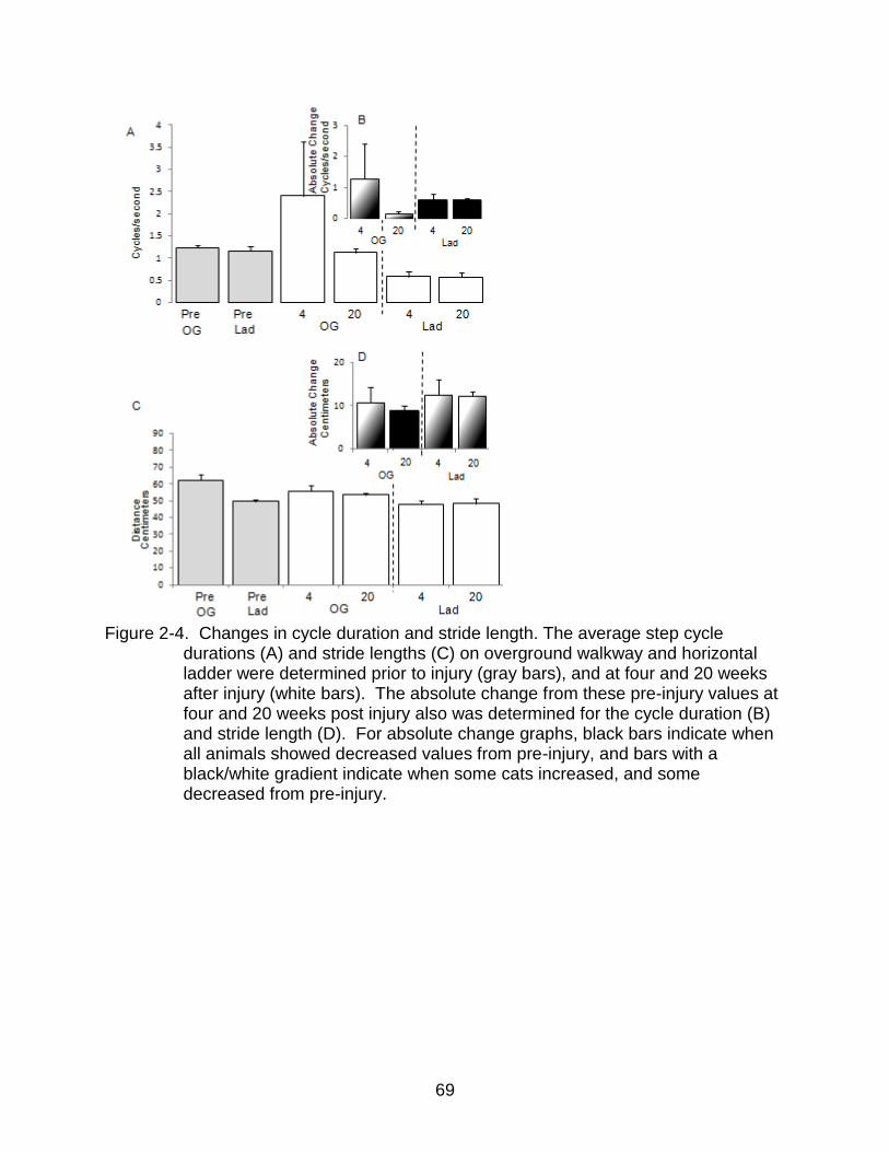
69
Figure 2-4. Changes in cycle duration and stride length. The average step cycle
durations (A) and stride lengths (C) on overground walkway and horizontal ladder were determined prior to injury (gray bars), and at four and 20 weeks after injury (white bars). The absolute change from these pre-injury values at four and 20 weeks post injury also was determined for the cycle duration (B) and stride length (D). For absolute change graphs, black bars indicate when all animals showed decreased values from pre-injury, and bars with a black/white gradient indicate when some cats increased, and some decreased from pre-injury.

70
Figure 2-5. Change in hindlimb footfall patterns and hind-to-hindlimb coordination. The
timing of left (ipsi) and right (contra) hindlimb foot placements on overground walkway are depicted for a representative cat at pre-injury (A), 4 weeks post-injury (C), and 20 weeks post-injury (D). Black rectangles indicate when a limb is in stance, and spaces between these denote swing. Footfall patterns for a representative cat were depicted for the horizontal ladder crossing at pre-injury (B). All cats performed similarly at this time point. The post-injury footfall patterns for each of the three cats on horizontal ladder were depicted for 4 weeks post injury (E,F,G) and 20 weeks post injury (H,I,J). Numbers inside the black rectangles for the horizontal ladder task indicate the rung that cats were standing on. Numbers outside of the black rectangles indicate a rung that the cat attempted to place their hindlimb onto, but failed. White rectangles indicate a stance period in which both hindlimbs were on the same rung simultaneously.

71
Figure 2-6. Change in hindlimb stance and double support periods. The percentage of time/step that the cat was in stance for both tasks (A) was determined for pre-injury on overground walkway and horizontal ladder for the ipsilateral (Ipsi) and contralateral (Contra) hindlimbs (G, gray bars). The same values at four weeks and 20 weeks post injury also were determined (white bars). The absolute change from pre-injury was depicted for the ipsilateral and contralateral hind limbs at four and 20 weeks after injury (B). For both tasks, the period at which the ipsilateral and contralateral hind limbs were in stance at the same time (double support period) were determined prior to injury (C, gray bars), and also four and 20 weeks after injury (C, white bars). The absolute change from pre-injury at four and 20 weeks also was determined (D). For graphs showing absolute change (B,D) white bars indicate when all animals had an increased change from pre-injury, and bars with a black/white gradient indicate when some cats increased, and some decreased from pre-injury.

72
Figure 2-7. Mediolateral movement of the iliac crest. The values of iliac crest’s lateral
deviations during stance and swing were compared across overground and horizontal ladder performance pre-inury (A, gray bars), four, and 20 weeks after injury (A, white bars). The absolute change from pre-injury also was determined from 4 and 20 weeks after injury (B). For graphs showing absolute change, white bars indicate when all animals had an increased change from pre-injury, and bars with a black/white gradient indicate when some cats increased, and some decreased from pre-injury.

73
Figure 2-8. Swing to stance transition. The duration to positive support at the
beginning of stance (A), and maximum ankle flexion at yield (C) were determined at pre-injury (gray bars), four, and 20 weeks after injury (white bars). The absolute change from pre-injury at four and 20 weeks after injury also was determined (B,D). For graphs describing absolute change white bars indicate when all animals had an increased change from pre-injury, and bars with a black/white gradient indicate when some cats increased, and some decreased from pre-injury.

74
CHAPTER 3 THE EFFECT OF DIFFERENT CHONDROITINASE ABC TREATMENT DURATIONS
ON LOCOMOTOR RECOVERY AND SUPRASPINAL TRACT PLASTICITY IN A FELINE MODEL OF SCI
Introduction
Many studies (Jones et al., 2002; For review see Morgenstern et al., 2002; Jones
et al., 2003a; For review see Busch and Silver, 2007; Galtrey and Fawcett, 2007),
including our own (Lemons et al., 1999; Tester and Howland, 2008) have shown that
after spinal cord injury (SCI) there is an upregulation of chondroitin sulfate
proteoglycans (CSPGs) at the lesion site. Chondroitin sulfate glycosaminoglycans (CS-
GAG), which are attached to these CSPGs, are extremely inhibitory to axonal growth
and play a primary role in preventing the axonal plasticity necessary for functional
recovery (Snow et al., 1990; Smith-Thomas et al., 1994; Zuo et al., 1998). Removal of
these CS-GAGs using the bacterial enzyme chondroitinase abc (ch’abc) following SCI
in rodent models enhances both behavioral recovery (Bradbury et al., 2002; Cafferty et
al., 2008) and axonal growth (Grimpe and Silver, 2004; Barritt et al., 2006; Houle et al.,
2006). The Howland laboratory has successfully translated use of ch’abc to a cat model
of SCI (Jefferson et al.; Tester and Howland, 2008). These studies showed enhanced
locomotor recovery and plasticity of red nucleus projections following a hemisection
lesion and 4 weeks of intralesional ch’abc administration.
A multitude of studies completed in rodent models of SCI have shown beneficial
effects using similar (Vavrek et al., 2007; Lee et al., 2009) and shorter ch’abc treatment
durations ranging from a single injection to two weeks (Bradbury et al., 2002; Barritt et
al., 2006; Cafferty et al., 2008; García-Alías et al., 2008; Iseda et al., 2008; Garcia-Alias
et al., 2009). It is currently unknown how a shortened administration period will affect

75
locomotor recovery and anatomical plasticity in a feline model of SCI and if four weeks
of treatment is necessary. Cats are a valuable translational model for ch’abc studies as
they have similar pre-dominant CS-GAG lyase products after ch’abc digestion as
humans (4S and 6S disaccharides), while the rat shows a single dominant (4S
disaccharide) lyase product (Tester and Howland, 2008). Thus, results from this study
have meaningful implications for treatment duration in regards to the advancement of
this therapeutic to a clinical setting.
The upregulated temporal expression of CSPGs after injury suggests that two
weeks of ch’abc expression may be sufficient for enhancing plasticity and functional
recovery. In rats, peak upregulation of neurocan, brevican, and versican CSPG
expression occurs at two weeks post injury, with peak neuron-glial antigen 2 (NG2)
expression occurring at one week. Both neurocan and versican production then
steadily decrease to only minimal expression by four weeks (Jones et al., 2002; Jones
et al., 2003a). It is currently unknown whether this same trend occurs in cats, but if so,
two weeks of ch’abc administration may be sufficient to enhance locomotor recovery
due to the natural decrease of a substantial proportion of CS-GAGs beginning at two
weeks. Furthermore, an unpublished finding from our previous work was greater
recovery of some locomotor features, like accurate targeting on a horizontal ladder, by
two weeks post-injury in ch’abc treated cats. This indicates that substantial plasticity
has already taken place by this two week time point and that additional ch’abc
administration may be unnecessary for continued functional improvements. In the
current study we investigate the effects of a shorter, two week period of ch’abc
76
administration on rubro-and cortico-spinal plasticity, locomotor recovery, and the
interaction of lesion size with treatment effects.
Materials and Methods
All animal procedures were conducted in accordance with the NIH guidelines for
the care and use of experimental animals and were approved by both the Malcom
Randall VA Medical Centers and University of Florida's Institutional Animal Care and
Use Committees. A total of 12 cats (6 ch’abc treated, 6 control) with similar lesion
magnitudes were used in this study to assess the effects of two weeks of ch’abc
treatment. All were trained 5x/week on 2-3 of 5 tasks daily. Some cats were used only
in the behavioral aspects of the study as they did not receive Fluorogold (FG) injections,
or their injections did not meet inclusion criteria. Inclusion criteria were as follows: 1)
FG labeling at the injection site must be in both the ipsilateral and contralateral
dorsolateral funiculi where the rubrospinal tract (RuST) and corticospinal tract (CST)
traverse 2) FG-labeled neurons in the non-axotomized red nucleus or motor cortex must
be easily detectable at 20X in order to be used for these analyses.
The lesion ranking section of this study uses a sample of convenience (laboratory
data bank) which incorporates 35 cats with varying lesion sizes. Only those that were
trained similarly to the paradigm described for the two week treatment groups were
included.
Subjects
All cats were purpose bred, SPF, spayed, adult, female cats. Spays were
performed to prevent any hormonally based behavioral changes during the study (For
review see Sribnick et al., 2003; Sribnick et al., 2005). Cats were placed into one of two
groups: two week control (N=6) or two week Ch’abc (N=6). At the beginning of the

77
study, animals were trained to perform five tasks that they were consistently trained and
assessed on throughout the study, similar to Tester and Howland, 2008: 30.5 cm
overground runway, bipedal treadmill, horizontal ladder, peg walkway and narrow beam.
Once cats were able to consistently perform the tasks, they were filmed for baseline
data collection. Following injury, cats continued to be trained and were filmed
performing each of the tasks regularly for five months. Cats then received intraspinal
FG injections caudal to the lesion site for retrograde tract tracing. Following a 13 day
survival period animals were transcardially perfused and lesion morphology, as well as
axonal plasticity, assessed.
Surgical Procedures
Low thoracic spinal hemisection
A detailed description can be found in Tester and Howland 2008. Briefly, all cats
received a left, T10 spinal hemisection created by iridectomy scissors. Following
hemisection, an injectable port body (Solomon Scientific ©, San Antonio, TX) was glued
and sutured subcutaneously to left dorsum musculature. Port tubing was positioned
over the lesion site and dura sutured closed around tubing to maintain proper
positioning within the lesion. Protease free ch’abc treatment (1U/200µL) (Seikagaku
Corporation ©, Tokyo, Japan), or vehicle (Sterile saline or Tris-HCL) were injected
immediately after port placement in order to visually confirm proper functionality of the
port system. A layer of durafilm and gelfoam were then placed over the lesion followed
by muscle and skin sutured closed in layers.
Chondroitinase ABC administration
Prior to use, ch’abc enzymatic activity was tested and confirmed with fluorophore-
assisted carbohydrate electrophoresis (FACE) as described in (Tester et al., 2007).

78
Ch’abc (0.25 U in 50 µL of saline or Tris-HCL) or vehicle (Saline or Tris-HCL)
injections began the day cats were injured and continued every other day for two
weeks. 50 µL injections were made slowly at 0.14cc/s with a syringe pump.
Fluorogold spinal injections
Between 22 and 35 weeks following injury, nine out of the twelve cats underwent a
second surgery to receive intraspinal injections of the retrograde tracer Fluorogold™
(FG) caudal to the lesion site. The details of this procedure are fully described in
Jefferson et al., 2011. In brief, 0.5% FG (Fluorochrome, LLC, CO) was mixed in sterile,
de-ionized water in an aseptic hood the morning of the procedure and kept on ice prior
to use. The lesion site was re-exposed, and regions of the spinal cord caudal to the
lesion site also were exposed for injections. The most caudal dural suture at the lesion
site was located from the previous surgery, and FG injections were made ~13mm
caudal to this suture. Four injection sites were made in a staggered formation using a
33 gauge Hamilton syringe. Each injection site consisted of two FG dumps of 0.25µL in
each, for a total of 2µLs. The exposed spinal cord was then protectively covered with
durafilm and gelfoam, the muscle and skin closed in layers. A survival period of 13
days allowed for FG to travel retrogradely to the brainstem and cortex. Post-surgical
care procedures are described in depth in previous studies (Howland et al., 1995aa;
1995bb).
Behavioral Procedures
Behavioral tasks and training paradigm
Prior to surgery, cats were conditioned to perform five tasks for a food reward. Tasks
range from simple to challenging: Bipedal treadmill (0.5m/s), 30.5 cm wide overground
runway, horizontal ladder, peg walkway, and 5 cm wide narrow beam (for detailed

79
description of tasks go to Tester and Howland 2008). Cats were trained 5x/week.
Specifically, each day consisted of training on bipedal treadmill as well as one of the
other tasks, which were alternated equally. Within 48 hours following SCI, training
continued in the same manner as previously described for 22-35 weeks after injury.
However, depending on motor function, the skilled tasks were exchanged for the 30.5
cm wide overground runway during the initial week after injury.
Assessment of locomotor recovery
For qualitative assessments a daily log was kept of each animal’s overall
performance. Additionally, the “Onset of Recovery” ie: the number of days for cats to
recover the ability to perform a task after injury, was recorded for 30.5 cm overground
runway, bipedal treadmill, horizontal ladder, peg walkway, and narrow beam. For
bipedal treadmill, this was the first day they took three consecutive, independent steps
even if at a speed slower than the standard training speed (0.5m/s). For 30.5 cm 12”
overground runway, horizontal ladder, peg walkway, and narrow beam, this referred to
the first day animals were able to make three independent crossings, consecutive or
non-consecutive. For quantitative assessment, animals were filmed on each of the
tasks using a 3D pan and tilt system (Peak Vicon®). Filming occurred once prior to
injury, as well as multiple times following SCI. For the first two months post-SCI, cats
were filmed on each task every two weeks, then once a month for the remainder of the
study.
Horizontal ladder, peg walkway, and narrow beam assessment: Limb accuracy
In addition to determining “Onset of Recovery” as previously described, cats were
also assessed for their ipsilateral hindlimb targeting accuracy. The percentage of times
cats were able to accurately place their ipsilateral hindlimb onto a rung or peg was

80
calculated for each crossing. The average percentage from the three best crossings
were computed and used for this comparison.
Tissue Processing
Perfusions and tissue preparation
The details of this procedure are described in our previous work (Tester and
Howland, 2008). In brief, at thirteen days following FG injections, cats were deeply
anesthetized with an overdose of sodium pentobarbital (>40mg/kg, i.p.). Supplemented
doses were given i.v. as needed to ensure that animals were properly sedated. Cats
also were intravenously injected with 1cc heparin (1000U/i.v.), and 20 minutes later with
1cc of 1% sodium nitrite i.v.. Immediately after, cats were transcardially perfused with
0.9% saline, followed by 4% paraformaldehyde in 0.1M phosphate buffer (pH 7.4). The
spinal cord and brain were dissected, blocked into segments, and post-fixed in 30%
sucrose and 4% paraformaldehyde. The lesion, injection site, rostral midbrain, and
medulla were cut on a cryostat at 25µm thick sections, while the motor cortex was cut at
40µm. Sections were stored in 0.1 M phosphate buffered saline (PBS; pH 7.2, saline
0.9%) at 4º Celsius until processed.
Cresyl violet and myelin staining
This procedure is described in detail in our previous work (Tester and Howland,
2008). In brief, every 10th section of the lesion site was mounted onto colorfrost slides
(Fisher Scientific©) subbed with chrom-alum and poly-L-lysine (chromium potassium
sulfate and poly-L-lysine, Sigma-Aldrich®; gelatin, Fisher Scientific©). Tissue was fume
fixed on the slides with 4% paraformaldehyde to enhance adherence. Tissue was first
dipped in water, and then dehydrated in increasing increments of alcohol (five minutes
each). After immersing in xylene (10 minutes), tissue was rehydrated in decreasing

81
increments of alcohol and placed in an aqueous myelin dye (Eriochrome Cyanine R;
Fluka™) for 10 minutes. After washing in water, differentiation took place in 1%
ammonium hydroxide (one minute). Tissue was then placed in 0.5% cresyl violet
(cresyl violet with acetate, Sigma-Aldrich®) for three minutes, and washed thoroughly in
water, 70% alcohol, and then differentiated in 95% alcohol with glacial acetic acid.
Following this, tissue was dehydrated again in increasing increments of alcohol and
then immersed in xylene. Tissue was coverslipped with DPX (Fluka™).
Assessment of injection site
Every 40th section of the injection site from animals that received FG injections
was mounted on Superfrost/Plus slides (Fisher Scientific©) and coverslipped with
Prolong Gold Antifade Reagent (Molecular Probes®) Sections were assessed using
fluorescent microscopy to ensure that the injection site reached the dorsolateral funiculi
carrying the RUST and CST for both hemispheres of the cord. Those that did not have
sufficient labeling were excluded from the tract tracing portion of the study.
Immunohistochemistry
One, 25 µm section, every 200µms through the red nucleus, and every 40th
section, every 640µms through the motor cortex were processed using the polyclonal
anti-FG antibody (1:10,000, Fluorochrome, LLC). This procedure is described in our
previous study (Jefferson et al., 2011). In brief, sections were washed in a quenching
solution (30% H2O2 and PBS) for 30 minutes, followed by 2, 10 minutes rinses in 1%
goat serum in PBS containing 0.4% Triton X-100 (1% S-PBS-T). Tissue was then
blocked in 5% S-PBS-T for one hour and then incubated in anti-FG antibody overnight
at room temperature. The next day, tissue was rinsed in 1% S-PBS-T and incubated in
5% biotinylated anti-rabbit secondary antibody made in goat (Vector Laboratories©) for 1

82
hour. After a second rinse with 1% S-PBS-T tissue was incubated in avidin, biotin,
complex (ABC) method (Vector Laboratories©). Following this incubation tissue was
rinsed with PBS, then reacted with 3,3’-Diaminobenzidine (DAB) brownish reaction
product (Sigma-Aldrich®) for 9 minutes. The DAB reaction was stopped with a PBS
rinse. Tissue was subsequently mounted onto chrom-alum and poly-L-lysine coated
slides, and fume fixed in 4% paraformaldehyde for at least one hour. Tissue was then
dipped in water and dehydrated in alcohol at increasing increments at 5 minutes each.
Tissue then was placed in xylene for 5 minutes and coverslipped with DPX (Fluka™).
Fluorogold-labeled cell counts
FG-labeled neurons of each stained section in the red nucleus, and motor cortex
were counted using a 20x microscope objective. Only those neurons with visible soma
were included in the counts. The non-axotomized cell counts were separated from the
axotomized cell counts. Additionally, the axotomized cell counts were taken as a
percentage of the non-axotomized cell counts to control for internal differences between
cats (uptake of tracer, tissue processing, etc).
Lesion ranking
The lesion epicenters from 35 cats were stained with cresyl violet and myelin dye
as described above. The control group consisted of nineteen cats, the two week ch’abc
group had seven cats, and the four week ch’abc group consisted of nine cats. Three
individuals unfamiliar with the cats, but well versed in spinal cord morphology, ranked
lesions based on the amount of total spared tissue with one being the lowest and 35
being the greatest amount of spared tissue. Individuals were asked to rank lesions that
they felt were extremely similar with the same value. Each of the three individual sets of
ranks were compared to ensure that rankings were relatively similar and that rankers

83
properly understood and performed the task. The three sets of ranks were then
averaged for each lesion and correlated against the onset of each of the five tasks, as
well as the percentage of accurate ipsilateral hindlimb targeting at four, eight, and 16
weeks post injury.
Statistical Analysis
Statistics were performed using Microsoft Excel’s Analyse-it statistical software
(Microsoft Excel®). The Kruskall Wallis test was Bonferroni corrected and used to
compare accurate ipsilateral hindlimb targeting across time points. The Mann Whitney
U test was used to assess group differences and the Spearman test was used to
correlate lesion rank and behavior.
Results
Lesion Magnitudes
The lesions of 12 cats, six control and six ch’abc, were stained with cresyl violet
and myelin dye to determine the extent of their injury. Only those with similarly
hemisected lesions were included in the part of the study assessing the effects of two
weeks of ch’abc treatment on anatomical and behavioral recovery. Figure 3-1 shows
the range of lesion sizes for the control (Figure 3-1A) and ch’abc (Figure 3-1B) groups.
In both groups there was slight sparing present within the gray matter and ventromedial
white matter. Additionally, both groups had lesions that extended into the contralateral
gray matter and ventromedial white matter. Overall, the minor variations across within
each group was equal across groups and did should not have skewed the results.
Axonal Projections below the Lesion Site
Both the RuST and CST tracts have been implicated in controlling adaptive types
of locomotion similar to the peg walkway and narrow beam (Morris et al.; Beloozerova

84
and Sirota, 1993; Lavoie and Drew, 2002; Pettersson et al., 2007). In a previous study
we found that four weeks of ch’abc delivery enhanced RuST circuitry below the level of
the lesion (Jefferson et al., 2011), however we have yet to address the effects of two
weeks delivery on this same circuitry. Additionally, the effects of ch’abc delivery on
CST plasticity after either two or four weeks of ch’abc delivery has yet to be determined
in a feline model of SCI. Four week control and ch’abc red nucleus neuronal counts
were taken from the same animals used in our previous publication, with the addition of
one, four week ch’abc cat who underwent the same treatment and behavioral training
protocols as others in that group (Jefferson et al., 2011). In that study, only the
percentages of axotomized to non-axotomized FG-labeled neurons were reported.
Here, we report absolute neuronal counts as well, due to recent findings from our lab
showing enhanced neuronal counts in both the axotomized and non-axotomized red
nuclei in control cats (Blum, 2010). This finding indicates that the non-axotomized red
nuclei/motor cortex may undergo more plastic changes than previously thought, and
should be assessed independently.
As expected, there were significantly more FG-labeled neurons in the non-
axotomized, contralateral side (Figure 3-2B,E) compared to the axotomized, ipsilateral
red nucleus (Figure 3-2A,E) in the two week control (Figure 3-2E; Mann-Whitney U test,
p=0.01), two week ch’abc (Mann-Whitney U test, p=0.05), four week control (Mann-
Whitney U test, p=0.01), and four week ch’abc group (Mann-Whitney U test, p=0.01) as
a result of the hemisection. The same was true for the motor cortex (Figure 3-2C,D,E;
Mann-Whitney U test, two week ch’abc, p=0.01; two week control, four week control,
ch’abc, p=0.05). Absolute FG-labeled neuronal counts from the non-axotomized red

85
nucleus were similar across the two and four week ch’abc groups and their control
counterparts. Absolute numbers from the axotomized side were significantly higher in
the two week ch’abc group compared to their control counterpart (Mann-Whitney U test,
p=0.03), and the four week ch’abc compared to their controls (Mann-Whitney U test,
p=0.03). Similar to what was reported in our previous work, the two week ch’abc group
had a significantly greater percentage of FG-labeled neurons in the axotomized as
compared to the non-axotomized red nucleus compared to controls (Mann-Whitney U
test, p=0.03) (Jefferson et al., 2011).
There were no significant differences in raw FG-labeled neurons or the percentage
of axotomized versus non-axotomized neuronal counts in the motor cortex between the
ch’abc and control groups, regardless of treatment duration (two or four weeks) (Figure
3-2E).
Rate of Recovery
As described in earlier studies completed by the lab, ipsilateral hindlimb motor
function was completely disrupted during the first one to two days after injury (Tester
and Howland, 2008; Jefferson et al., 2011). The contralateral hindlimb retained most of
its function during this period, however there were some animals that could not fully
weight support with this limb right away, but regained this ability within several days.
There were no significant differences between the control and ch’abc treated animals
regarding the recovery of basic locomotion. Recovery of stepping on 12” overground
runway typically began between one and three days following injury, though there were
several cats that took longer (Figure 3-3A). Typically, cats began stepping primarily
with their contralateral hindlimb, while their ipsilateral hindlimb flexed and extended in a
manner reminiscent of stepping, but with an extremely muted range of motion around

86
the joints. While the contralateral hindlimb of most cats was capable of full weight
support, the ipsilateral hindlimb could only support a small proportion of their weight.
Surprisingly, recovery of bipedal treadmill stepping, which is controlled primarily by
intraspinal circuitry, took significantly longer to recover compared to 12” overground
stepping, which involves more voluntary components (Figure 3-3A; Mann-Whitney U
test, control, p=0.01; ch’abc, p=0.004).
Recovery of the skilled locomotor tasks: horizontal ladder, peg walkway, and
narrow beam, took longer than basic locomotion as they require multiple supraspinally
controlled locomotor features, including but not limited to balance, interlimb
coordination, and accurate limb targeting (Figure 3-3B) (Whishaw et al., 1998; Metz and
Whishaw, 2002). In our previous studies, we found that four weeks of ch’abc treatment
significantly increased the rate of recovery on each skilled task (Tester and Howland,
2008). Here, we find that a shortened ch’abc delivery period does not have as robust of
an effect. Horizontal ladder performance is the easiest of the skilled tasks to perform
and was recovered significantly quicker than the peg walkway (Figure 3-3B; Mann-
Whitney, U test, control, p=0.01; ch’abc, p=0.004) and narrow beam tasks (Figure 3-3B;
Mann-Whitney U test, control, p=0.01; ch’abc, p=0.001), occurring between the 2nd and
3rd week after injury. Cats treated with 2 weeks of ch’abc treatment were able to
recover this task (horizontal ladder) significantly quicker than controls (Figure 3-3B;
Mann-Whitney U test, p=0.03).
The time frames for recovery of peg walkway and narrow beam were substantially
more variable in the ch’abc group compared to controls. In the control group, cats were
able to recover peg walkway between 16 and 55 days post injury, and narrow beam

87
between 26 and 71 days post injury (Figure 3-3C). However, in the ch’abc group there
was high variability across cats. While four out of the six cats recovered peg walkway
between 15 and 18 days after injury, two animals were never able to recover this task
(Figure 3-3D). Onset of narrow beam recovery in this two week ch’abc group also was
more variable across cats compared to the control groups, ranging from 16 to 113 days
after injury (3-3C,D). Despite these differences in variability across treatment groups,
there were no significant differences in their average rate of recovery for these two
tasks. However, the removal of these outliers led to a significant different when
comparing task onset of peg walkway, but not narrow beam (Figure 3-3E, Mann-
Whitney U test, p=0.02).
Ipsilateral Hindlimb Accuracy
Limb accuracy of the ipsilateral hindlimb is particularly difficult for animals to
recover following injury, and in fact many fail to do so on the more skilled tasks like peg
walkway. In previous studies done by the lab it was showed that four weeks of ch’abc
administration resulted in a significantly greater ability for cats to accurately target with
their ipsilateral hindlimb onto a peg compared to controls (Tester and Howland, 2008;
Jefferson et al., 2011). Here, we see that a shortened two week delivery of Ch’abc
does not significantly enhance limb accuracy over controls.
Horizontal ladder
Pre-injury, all cats were able to accurately place with their ipsilateral hindlimb onto
a ladder rung 100% of the time, however after injury all animals lost this ability for at
least several days (Figure 3-4A). By two weeks post injury, several cats had recovered
this locomotor feature, but the majority of these animals were in the ch’abc group (four
cats), while only one animal in the control group had regained ipsilateral hindlimb

88
accuracy. Hence, the ch’abc group showed a trend towards significantly greater
percentage of ipsilateral hindlimb accuracy compared to controls at two weeks after
injury (Figure 3-4A; Mann-Whitney U test, p=0.07). This ch’abc enhancement of
accuracy was transient and by four weeks the control animals were performing equal to
the ch’abc group. Both groups had substantial recovery of ipsilateral hindlimb accuracy
by eight weeks and were targeting significantly more than at two weeks post injury
(Kruskal-Wallis test, control, p=0.01; ch’abc, p=0.03). This trend continued at 16
(Kruskal-Wallis test, control, p=0.02; ch’abc, p=0.003), and 20 weeks after injury
(Kruskal-Wallis test, control, p=0.01; ch’abc, p=0.0008).
Narrow beam
Prior to injury, all cats were able to place their ipsilateral hindlimb onto the beam
100% of the time. After injury, recovery of ipsilateral hindlimb accuracy on the narrow
beam was an all-or-nothing skill (Figure 3-4B). The majority of animals in both control
and ch’abc groups struggled with recovering this feature for the first eight weeks after
injury, with none recovering by two weeks and only one becoming successful at four
weeks (ch’abc treated). Once animals did recover ipsilateral hindlimb accuracy the
majority of them had a 100% success rate. By eight weeks post injury the control group
was accurately placing their ipsilateral hindlimb significantly more than what they were
at two weeks (Figure 3-4A; Kruskal-Wallis test, p=0.04). This trend continued at 16
(Kruskal-Wallis test, p=0.005) and 20 weeks after injury (Kruskal-Wallis test, p=0.004).
Accuracy of the ch’abc group was not significantly greater than two weeks until 20
weeks after injury (Kruskal-Wallis test, p=0.05), indicating that this ch’abc group did not
perform as well as controls on this task.

89
Peg walkway
Similar to the horizontal ladder and narrow beam, all cats were able to accurately
place their ipsilateral hindlimb onto pegs 100% of the time prior to injury (Figure 3-4C).
However, for the first two weeks after injury, ipsilateral hindlimb accuracy was
completely lost. By four weeks post injury, there was one cat from the control and
ch’abc group that recovered this feature. Interestingly, the ch’abc cat was accurately
targeting substantially more than the control cat at 100% as opposed to 11%,
respectively. Over the course of the study only two other cats from each group
recovered ipsilateral hindlimb accuracy on peg walkway, suggesting the extreme
difficulty of this task. Additionally, accuracy percentage remained low in both groups,
with neither significantly improving from two weeks post injury at any time point in the
study. Only one animal within the ch’abc group was able to reach 100% accuracy,
while the other cats ranged from 7 to 23% ipsilateral hindlimb accuracy (Figure 3-4C).
The Relationship between Spared Tissue and Functional Recovery
While most of the 35 lesions obtained from the laboratory data bank were
variations on a hemisection, there were a few that were notably larger with only ~25% of
cross-sectional sparing, or notably smaller with ~75% of cross-sectional sparing.
Furthermore, the effect of ch’abc administration on the relationship between lesion size
and locomotor recovery has yet to be determined. In the current study, unbiased
individuals ranked the amount of total spared tissue present at the lesion epicenter from
all 35 cats: Control (19), two week ch’abc (seven), four week ch’abc (nine). An average
rank was determined for each cat based on the ranks assigned by the three individuals.
Lesion sizes ranged from very over-hemisected with sparing in only a quadrant of the
spinal cord crossection (Figure 3-5A), to very under-hemisected with sparing in 3/4ths of

90
the spinal cord crossection (Figure 3-5C). Those that were ranked as having a medial
amount of sparing were near perfect hemisections (Figure 3-5B). Average ranks for
each animal were correlated with onset of task recovery, as well as accurate ipsilateral
hindlimb placement on the horizontal ladder, peg walkway, and narrow beam.
Onset of task recovery versus lesion size
There was a significant correlation in the control animals between spared tissue
rank and recovery onset of overground walkway (Figure 3-5D; Spearman test,
p=0.0001), horizontal ladder (Spearman test, p=0.001), peg walkway (Spearman test,
p=004), and narrow beam (Spearman test, p=0.0001). The only lack of a relationship
was on bipedal treadmill (Figure 3-5). The two week ch’abc treated animals had a less
striking relationship in that their lesion rankings did not significantly correlate with
recovery onset of overground, or peg walkway. However, the recovery of horizontal
ladder (Spearman test, p=0.03), and narrow beam (Spearman test, p=0.0004) did
significantly correlate with spared tissue rank. The onset of overground walkway was
close to significantly correlating with a p-value of 0.06. In the four week ch’abc group
there were no significant correlations between lesion size and recovery onset on either
of the tasks except for bipedal treadmill (Spearman test, p=0.04). This lack of a
relationship between spared tissue and task onset in the four week ch’abc group only
indicates that four weeks ch’abc decreases the relationship between lesion size and
functional recovery. In other words, four weeks of ch’abc allows for animals with larger
lesions and less spared tissue to recover tasks quicker than cats with a similarly sized
lesion that were treated either with two weeks, or no ch’abc. This is validated when
looking at the individual values for each animal, using the onset of narrow beam as an
example (Figure 3-5E). Here, the onset and spared tissue rank values show that the 4

91
week ch’abc treated animals with less spared tissue were able to recover narrow beam
much quicker than the control and two week ch’abc treated cats with similarly low levels
of sparing. This effect occurs in all assessed tasks.
Ipsilateral hindlimb accurate targeting versus lesion size
The amount of spared tissue in both the control and two week ch’abc treated
groups either significantly correlated or had a p value of <0.1, with ipsilateral hindlimb
accurate targeting at four weeks post injury on horizontal ladder (Figure 3-6A;
Spearman test, control,p=0.0004; 2 week ch’abc, p=04), peg walkway (Spearman test,
control, p=0.01; 2 week ch’abc, p=0.0719), and narrow beam (Spearman test, control,
p=0.01; 2 week ch’abc, p=0.07) (Figure 3-6A). The same is true at eight weeks post
injury for horizontal ladder (Spearman test, control, p=0.0927; 2 week ch’abc, p=0.01),
peg walkway (Spearman test, two week ch’abc, p=0.0150), and narrow beam
(Spearman test, control, p=0.0004; two week ch’abc, p=0.0031). The one exception to
this was the control group at eight weeks post injury on peg walkway. In direct contrast,
there was no relationship between lesion size and accurate targeting on either of the
tasks for the four week ch’abc group. Interestingly, by 16 weeks post injury the
relationship seen between lesion size and accurate targeting was no longer present on
horizontal ladder or peg walkway in the control and two week ch’abc group, though it
was still present in narrow beam (Spearman test, control, p=0.0624; 2 week ch’abc,
p=0.01). At 16 weeks post injury there was still no relationship between the two in the
four week ch’abc group. These results further suggest the finding that ch’abc
decreases the relationship between lesion size and locomotor recovery. Similar to what
was seen in the onset of task recovery, the individual values of each animal, using
accurate ipsilateral hindlimb targeting on horizontal ladder at four weeks as an example,

92
indicates that four week ch’abc treated animals with less tissue sparing (larger lesions)
recover accurate targeting better than control and two week ch’abc treated animals with
similar amounts of spared tissue (Figure 3-6B).
Discussion
Summary of Results
In the current study we found that there was a significant enhancement in axonal
growth within the RuST, but not within the CST after both two and four weeks of ch’abc
administration. Interestingly, although two weeks of ch’abc administration led to
enhanced rubrospinal tract growth below the lesion similar to four week treated animals
(Jefferson et al., 2011), they did not share a similar enhancement of skilled locomotor
recovery. In a larger group of animals with more diverse lesion magnitudes, it was
determined that four weeks, but not two weeks of ch’abc treatment leads to enhanced
functional recovery in larger lesions.
Supraspinal Connectivity below the Lesion
Multiple ch’abc studies attribute locomotor recovery after SCI to the regeneration
and sprouting of the CST (Bradbury et al., 2002; Barritt et al., 2006; Garcia-Alias et al.,
2009). However, the current study did not find enhanced CST connectivity below the
level of the lesion based on our retrograde tracing results in both the two week ch’abc
cats, as well as the functionally enhanced four week ch’abc treated animals. These
results suggest one of two things. First, ch’abc does not enhance plasticity within this
particular pathway. In fact, multiple studies have reported a failure in significant CST
axonal growth despite seeing growth in other systems like the RuST (Richardson et al.,
1984b; Houle and Ye, 1999; Decherchi and Gauthier, 2000; Plunet et al., 2002). More
likely though, ch’abc treatment enhances CST axonal sprouting proximal to the lesion

93
site, but this sprouting does not enhance the number of motor cortex neurons with
connections below the level of the lesion. Alternatively, CST plasticity may be
occurring in more rostral regions of the spinal cord like the cervical cord, as shown in
Bareyre et al., 2004. Sprouting in those regions would not be detected by low thoracic
FG injections.
Multiple ch’abc studies utilizing anterograde tracing have reported enhanced CST
sprouting proximal to the lesion site (Bradbury et al., 2002; Barritt et al., 2006; Iseda et
al., 2008; Garcia-Alias et al., 2009; Karimi-Abdolrezaee et al., 2010). The only ch’abc
study to assess CST plasticity utilizing retrograde tracing, in addition to the current
study, had similar findings in that there was enhanced connections below the level of
the lesion in non-CST pathways (ReST, VST, and RuST) but none in the CST itself (Bai
et al., 2010). This further suggests that if CST plasticity is occurring, it most likely
occurs at the terminal ends or in more rostral segments of the spinal cord.
Locomotor Recovery and Supraspinal Plasticity
Previous studies performed by the Howland laboratory did not show a significantly
shorter recovery period for bipedal treadmill, or 12” overground runway after
hemisection and 4 weeks of ch’abc administration (Tester and Howland, 2008;
Jefferson et al., 2011). In the current study, we also see this similar lack of an effect.
This result was expected based on the aforementioned results as cats recover these
tasks within the first two weeks after injury; a period when both ch’abc treatment groups
had received the same amount of ch’abc delivery. Disparity between two week and four
week locomotor recovery was most apparent on the skilled locomotor tasks. These
tasks require much more adaptation and mimic the type of everyday tasks that

94
individuals with incomplete SCI, like Brown Sequard Syndrome, primarily struggle with
(Little and Halar, 1985; Eidelberg et al., 1986).
The recovery period for horizontal ladder, peg walkway and narrow beam were
similar across cats in the control group, with cats typically recovering horizontal ladder
first, followed closely by peg walkway, then narrow beam. All control cats recovered
these three tasks between 15 and 71 days after injury. The two week ch’abc group
recovered horizontal ladder significantly quicker than the control cats after injury,
however had substantially greater variability regarding recovery period for peg walkway
and narrow beam. On peg walkway, four cats recovered within 15 to 18 days, while two
never recovered. On narrow beam, four different cats recovered within 16 to 41 days,
while two cats, different from those who failed to recover peg walkway, did not recover
narrow beam until 111 and 113 days after injury. This extreme variability may be an
indicator of aberrant plasticity and nonfunctional circuit formation resulting from an
insufficient amount of ch’abc administration. This is further confirmed by a lack of
variability seen across the four week ch’abc treated cats (Tester and Howland, 2008;
Jefferson et al., 2011). Furthermore, results from retrograde tracing show significantly
greater RuST connections below the level of the lesion in both the two and four week
ch’abc groups compared to their control counterparts. However, the four week ch’abc
group had significantly enhanced skilled locomotor recovery while the two week ch’abc
group did not (Tester and Howland, 2008; Jefferson et al., 2011). It is possible that
while two weeks of ch’abc administration enhances axonal growth, it does not allow for
the plasticity necessary to make functionally appropriate connections capable of
producing long term beneficial effects. Neuro-developmental studies have shown that

95
experience-based plasticity is dependent on a lack of CSPGs (Pizzorusso et al., 2002;
Pizzorusso et al., 2006). After injury, the expression of some CSPGs, specifically
phosphacan, NG2, brevican, and neurocan, remain upregulated for longer than two
weeks after injury (Jones et al., 2002; Jones et al., 2003a). Additionally, aggrecan,
which initially decreases after injury, begins to recover expression at two weeks post-
injury (Lemons et al., 2001). These specific CSPGs have been shown to inhibit axonal
growth (Sango et al., 2003), especially NG2 (Dou and Levine, 1994; Fidler et al., 1999;
Jones et al., 2002; Ughrin et al., 2003), and aggrecan (Lemons et al., 2003). Although
cleaved by ch’abc for 2 weeks, their ongoing expression may continue after the
cessation of ch’abc at 2 weeks, and their presence may accumulate at the lesion site
causing inhibition of axonal growth. In addition to ongoing CSPG upregulation 2 weeks
after injury, there are multiple post-SCI inflammatory factors, such as microglia, reactive
astrocytes and oligodendrocyte precursor cells that infiltrate the lesion site quickly after
injury and continue to be present for longer than two weeks after injury (Hill et al., 2001;
Velardo et al., 2004; Donnelly and Popovich, 2008). Additionally, some of the greatest
changes to lesion morphology occur between two and four weeks after injury (Hill et al.,
2001; Velardo et al., 2004). While 2 weeks of ch’abc may be enough to enhance
plasticity, the combinatorial presence of these factors at the lesion site after cessation of
ch’abc administration at 2 weeks may prevent these new sprouts from making
appropriate connections.
Previous studies from the Howland laboratory have shown that both human and
feline CS-GAG lyase products after ch’abc digestion predominantly consist of 4S and
6S disaccharides, while digestion of rodent CS-GAGs results in primarily 4S

96
disaccharide products (Tester and Howland, 2008). This species difference suggests
that ch’abc degradation is required to cleave additional substrates in the cat versus rat
spinal cord. This may partially explain differences seen in functional recovery across
species that received ch’abc administration for the same period of time. Additionally,
due to the larger size of the cat spinal cord compared to rat, axonal regeneration and
sprouts must travel farther in order to reach appropriate connections. Combined results
from the present study, as well as those previously published by the Howland
laboratory, suggest that four weeks of ch’abc treatment results in much greater
locomotor recovery compared to two weeks of administration in a cat model of spinal
cord injury.
Lesion Size
Multiple studies have found that larger lesions significantly correlate with poorer
functional recovery and that smaller lesions are paired with greater functional recovery
(Byrnes et al.; Semler et al.; Molt et al., 1979; Norrie et al., 2005). Here, we have found
a similar result in that spared tissue significantly correlates with onset of task recovery,
and accurate targeting of the more greatly affected hindlimb on horizontal ladder, peg
walkway, and narrow beam in control and two week ch’abc treated animals. In direct
contrast, the amount of spared tissue in the four week ch’abc treated animals does not
correlate with these same locomotor features. Further investigation of individual
animals showed that this lack of a correlation is due to the fact that 4 week ch’abc
treated animals with less spared tissue had functional recovery that surpassed those in
control and 2 week ch’abc treated groups with similarly small amounts of spared tissue.
Such findings indicate that ch’abc mediates profound enhancement of functional
recovery even in large lesions, but that a minimum of four weeks of treatment is

97
necessary to produce this result. We believe the mechanism underlying this effect is
greater plasticity of multiple circuitries in four week ch’abc treated animals that is
beyond what was assessed by the tracing techniques used in the study. Although two
weeks of ch’abc treatment results in plasticity, it is not enough to produce new
functional connections that can surpass the limitations of a larger lesion.
Conclusion
The current study suggests that two weeks of ch’abc administration in a feline
model of SCI is not sufficient for enhancement of skilled locomotor recovery. While it
does lead to a significantly greater number of red nucleus neurons with axons below the
level of the lesion, this increased growth may not make functionally relevant
connections. As seen in our previous studies, four weeks of ch’abc enhances both red
nucleus plasticity, as well as skilled locomotor recovery suggesting that this longer
ch’abc administration period is necessary for functionally appropriate connections to be
made. Although we did not see increased motor cortex neuronal connectivity below the
level of the lesion using retrograde tracing for either two or four week treated groups,
other studies have found increased CST sprouting after ch’abc treatment with
anterograde tracing. These results suggest that there may be plasticity occurring at
regions rostral to the injury, or at the terminal ends that cannot be detected using
retrograde tracing. Our assessment of the spared tissue-functional recovery relationship
indicates a positive correlation with more sparing leading to greater recovery and vice
versa. The finding that four weeks, but not two weeks of ch’abc treatment mutes this
relationship by causing substantial recovery in lesions with both high and lower amounts
of spared tissue, further confirms that two weeks of ch’abc treatment is not as sufficient
of a treatment period as four weeks in feline SCI. The high translatability of felines to

98
humans suggests that a longer treatment duration should be considered for future
translationally designed, and ultimately clinical, ch’abc studies.
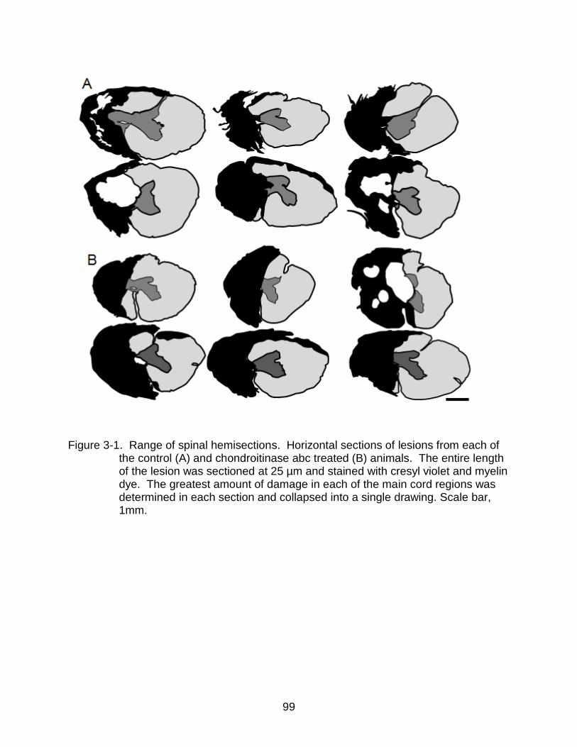
99
Figure 3-1. Range of spinal hemisections. Horizontal sections of lesions from each of
the control (A) and chondroitinase abc treated (B) animals. The entire length of the lesion was sectioned at 25 µm and stained with cresyl violet and myelin dye. The greatest amount of damage in each of the main cord regions was determined in each section and collapsed into a single drawing. Scale bar, 1mm.

100
Figure 3-2. Supraspinal connections below the lesion. The amount of fluorogold-labeled neurons were determined for the non-axotomized and axotomized side of the red nucleus and motor cortex. Additionally, the percentage of axotomized fluorogold labeled neurons as a percentage of the non-axotomized side were calculated. *,p<0.05. p=0.0628. The axotomized red nuclei (A), and motor cortex (C), had less labeling than the non-axotomized side of the red nucleus (B) and motor cortex (D), 100 µm. Average raw values, and percentage of axotomized to non-axotomized values for the two, and four week control groups, as well as the two and four week chondroitinase abc groups were reported (E).

101
Figure 3-3. Onset of basic and skilled locomotor recovery. The number of days it took for cats to recover basic locomotor tasks: Bipedal treadmill and overground walkway (A), and skilled locomotor tasks: Horizontal ladder, peg walkway, and narrow beam (B), were compared across two week control and two week ch’abc groups. (*, P=<0.05). The specific day of recovery after injury for each control (C) and chondroitinase abc cat (D) were listed for recovery of ladder, peg walkway, and narrow beam. Rows highlighted in gray indicate those cats that had extreme variability in their recovery period across the three skilled tasks after injury. Removal of these outliers within the ch’abc treated group led to significant group differences in the peg walkway, but not the narrow beam (E). Variation was assessed using standard error of the means.

102
Figure 3-4. Ipsilateral hindlimb accuracy. The percentage of times cats were able to
accurately target and place their ipsilateral hindlimb onto a rung, beam, or peg while crossing the horizontal ladder (A), narrow beam (B), or peg walkway (C) were determined periodically over a 20 week period post injury and compared between two week control and chondroitinase abc groups. (*p=<0.05), (¥p=0.0660). Standard error of the means are depicted in A-C.
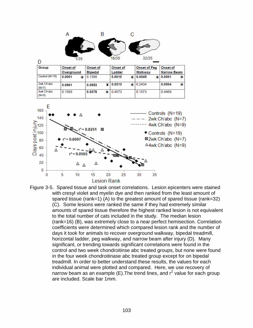
103
Figure 3-5. Spared tissue and task onset correlations. Lesion epicenters were stained
with cresyl violet and myelin dye and then ranked from the least amount of spared tissue (rank=1) (A) to the greatest amount of spared tissue (rank=32) (C). Some lesions were ranked the same if they had extremely similar amounts of spared tissue therefore the highest ranked lesion is not equivalent to the total number of cats included in the study. The median lesion (rank=16) (B), was extremely close to a near perfect hemisection. Correlation coefficients were determined which compared lesion rank and the number of days it took for animals to recover overground walkway, bipedal treadmill, horizontal ladder, peg walkway, and narrow beam after injury (D). Many significant, or trending towards significant correlations were found in the control and two week chondroitinse abc treated groups, but none were found in the four week chondroitinase abc treated group except for on bipedal treadmill. In order to better understand these results, the values for each individual animal were plotted and compared. Here, we use recovery of narrow beam as an example (E).The trend lines, and r2 value for each group are included. Scale bar 1mm.

104
Figure 3-6. Spared tissue and accurate ipsilateral hindlimb targeting correlations.
Correlation coefficients were determined which compared the lesion rank and percentage of accurate ipsilateral hindlimb targeting on horizontal ladder, peg walkway, and narrow beam (A). Similar to recovery of task onset, both the control and two week chondroitinase abc treated animals either significantly correlated, or trended towards a significant correlation with accurate targeting on all three tasks at four and eight weeks post injury. There were minimal correlations at 16 weeks post injury. In contrast, the four week chondroitinase abc treated group had no significant correlations at either of the time points. Closer analysis of this effect using the individual values of accurate targeting on ladder at four weeks as an example, shows that the four week chondroitinase abc cats with the lowest amounts of spared tissue were able to place substantially more than those animals with a similarly low amount of spared tissue (B).

105
CHAPTER 4 OPTIMIZATION OF FLUOROGOLD RETROGRADE TRACING
Introduction
The use of the retrograde tracer Fluorogold (FG) is a common technique for
determining axonal connections in multiple different experimental paradigms performed
in a variety of animal models ranging from lamprey (McClellan et al., 2006) to primate
(Gimenez-Amaya and Graybiel, 1990; Hayashi et al., 2006). Tracer is placed into axons
of interest and travels retrogradely to label the neuronal cell bodies of origin. There are
multiple benefits to using FG in addition to its compatibility with a wide range of species.
It has the rare ability to transport to the distal dendrites (Maranto, 1982; Bentivoglio and
Su, 1990; Buhl and Dann, 1990; Naumann et al., 1992) and appears to label all
neuronal cell populations (Zaborszky, 2006). Additionally, it has strong autofluorescent
properties that make it detectable without additional processing, but its signal also can
be amplified and detected using immunohistochemical (IHC) processes (Chang et al.,
1990; Naumann et al., 2000; Akhavan et al., 2006).
Despite these benefits, there are several downsides to using FG which need to be
minimized for tracer optimization. One prime example is the requirement of tissue
damage at the injection site for tracer uptake (Schmued and Fallon, 1986).
Furthermore, multiple investigators have reported that FG is cytotoxic to both
motoneurons (Garrett et al., 1991; Naumann et al., 2000) and dorsal root ganglia
neurons (Garrett et al., 1991) when left in vivo for several weeks. This neurotoxic
feature has the potential to become a significant problem in larger species who may
require longer survival periods depending upon transport distance. One way to
minimize tissue damage is by decreasing FG concentration and volume (Schmued and

106
Fallon, 1986), however this translates to decreased autofluorescence that quenches
more quickly. An anti-FG antibody is commercially available and allows for the
detection of FG using conventional immunohistochemical (IHC) techniques that lead to
a stable 3,3’-Diaminobenzidine (DAB) reaction product (Chang et al., 1990). This may
be a promising solution for FG detection when autofluorescence is not as bright due to
low FG concentration and volume, and/or permanent label is desired.
Decreasing the survival period following FG injections also will help minimize FG’s
cytotoxic effects. The detergent Triton X-100 (triton) has been implicated as a speed
enhancing supplement for tract tracing with oregon green and fluororuby dextran
amines in Possums (Fry et al., 2003). Triton is a non-ionic surfactant commonly used to
permeabilize cell membranes. Since the rate of FG uptake is directly related to its
ability to cross the cell membrane (Martin W, 1991), a mixture of FG and triton applied
to the region of interest may enhance both tracer uptake and speed of transport which
may result in minimizing cytotoxicity. Additionally, the benefits of shortening the survival
period may expand into other areas specific to a study such as financial motives and
time dependent issues. The objectives of the current study are to determine methods
for minimizing tissue damage while maximizing FG tracing speed and detection.
Materials and Methods
The following data is based on FG tracing results from 19 cats. All procedures
were conducted in accordance with NIH guidelines and were approved by both the
Malcom Randall VA Medical Center’s, and the University of Florida’s Institutional Animal
Care and Use Committees.
The injection sites of each of the four animals were processed and assessed in
order to ensure adequate spread of the tracer, especially in the dorsolateral funiculi

107
which contains the rubrospinal (RuST) and corticospinal (CST) tracts. These tracts
acted as the tracts of interest for this study as their cell bodies are located a far distance
from the injection sites and we are well experienced with tracing these systems
(Jefferson et al., 2011) (Refer to Chapter 3).
Subjects
All cats were purpose bred, SPF, adult, female cats (Liberty Vendors, Inc.).
Animals were placed into one of five different groups: 13 day FG without triton (N=12),
which was our commonly used protocol for previous tract tracing experiments (Jefferson
et al.), as well as a seven day FG without triton group (N=1), seven day FG with triton
group (N=2), three day FG without triton group (N=1), and a three day FG with triton
group (N=3).
Surgical Procedures
Fluorogold spinal injections
Triton X-100 (2.5%, Sigma-Aldrich®) was mixed with sterile saline in a sterile
setting the morning of the procedure. Fluorogold™ (0.5%, Fluorochrome, LLC) was then
either mixed in sterile saline or the 2.5% Triton X-100 solution and kept on ice prior to
use. The spinal cord segment T11 was exposed and injections were made in four
injection sites placed in a staggered formation using a 33 gauge Hamilton syringe.
Each injection site consisted of three FG injections of 0.25µL in each, for a total of 3µLs
(Figure 4-1). The exposed spinal cord was then protectively covered with durafilm and
gelfoam and the muscle and skin closed in layers. Our previous studies describe post-
surgical procedures in detail (Howland et al., 1995b, a)

108
Tissue Processing: Histology and Immunohistochemistry
Perfusions
The details of this procedure are described in our previous work (Tester and
Howland, 2008). In brief, at three, seven, or 13 days following FG injections, cats were
deeply anesthetized with an overdose of sodium pentobarbital (>40mg/kg, I.P) and
supplemented i.v. as needed to ensure that animals were properly sedated. Cats also
were injected with heparin (i.v.;1000 U) and then 20 minutes later injected with sodium
nitrite (I.V.;1%, 1cc). Immediately after, cats were then transcardially perfused with
0.9% saline followed by 4% paraformaldehyde in 0.1M phosphate buffer (pH 7.4). The
injection sites were blocked and sectioned at 25 µm, while the red nuclei and motor
cortices were blocked and sectioned into 50 µm sections.
Cresyl violet and myelin staining
A full description is described in our previous work (Tester and Howland, 2008). In
brief, every 12th section of the injection site was mounted onto Colorfrost slides (Fisher
Scientific©) subbed with chrom-alum and poly-L-lysine (chromium potassium sulfate and
poly-L-lysine, Fisher Scientific©) and fume fixed with paraformaldehyde. Tissue was first
dipped in water, and then in alcohols at increasing concentrations for dehydration for
five minutes. After dipping in xylene, tissue was then rehydrated in decreasing
concentrations of alcohol and placed in myelin dye for 10 minutes. After washing in
water, differentiation then took place in 1% ammonium hydroxide (Sigma-Aldrich©) for
one minute. Tissue was then placed in 0.5% cresyl violet (Sigma-Aldrich©) for 3
minutes and washed thoroughly in water, then 70% alcohol, then differentiated in 95%
alcohol with glacial acetic acid. Following this, tissue continued to be dehydrated in

109
increasing increments of alcohol for five minutes, and then placed in xylene. Tissue
was then coverslipped with DPX (Fluka™).
Fluorogold immunohistochemistry
A full description of this procedure can be found in Jefferson et al., 2011. In brief,
multiple sections of the injection sites and red nuclei were processed using the
monoclonal anti-FG antibody (1:60,000, 1:10,000, respectively) (Fluorochrome, LLC).
Sections were washed in a quenching solution (30% H202 and phosphate buffered
saline (PBS)) for 30 minutes, followed by two 10 minute rinses in 1% goat serum in PBS
with 0.4% triton X-100 (1% S-PBS-T). After this, tissue was blocked in 5% S-PBS-T for
one hour. Tissue was then incubated in anti-FG antibody overnight at room
temperature. The following day, tissue was rinsed in 1% S-PBS-T and incubated in 5%
biotinylated anti-rabbit secondary antibody made in goat for 1 hour (Vector
Laboratories©). Following another rinse with 1% S-PBS-T, tissue was incubated for two
hours in ABC Reagent (Vector Laboratories©). Tissue was then rinsed with PBS and
reacted with 3,3’-Diaminobenzidine for 9 minutes resulting in a brownish reaction
product. The DAB reaction was stopped with a PBS rinse once the reaction product
was the appropriate darkness. Tissue was then mounted onto chrom-alum and poly-L-
lysine coated slides and fume fixed in 4% paraformaldehyde for ~ one hour. Lastly,
tissue was dehydrated in alcohols at increasing concentrations for 5 minutes, placed in
xylene for five minutes, and coverslipped with DPX (Fluka™). The sections that were
stained for FG immunoreactivity one and nine years ago underwent the same
procedures as described above. These slides were stored at room temperature in hard
cover slide boxes.

110
Assessment of injection site completeness and fluorogold autofluorescence
Every 12th section through the injection sites were mounted onto Superfrost/Plus
slides (Fisher Scientific©) and were coverslipped using prolong gold anti-fade mounting
media (Molecular Probes®). Sections were assessed using the UV cube with
fluorescent microscopy. Red nucleus and motor cortex sections adjacent to those
known to have heavy FG labeling based on results from the anti-FG IHC analysis
described above, were processed in a similar manner in order to compare
autofluoroscent and IHC based FG detection.
Results
Tissue Damage at the Injection Site
It has been previously reported that FG causes tissue necrosis at the injection site
and that this damage is necessary for FG uptake and subsequent retrograde tracing
(Schmued and Fallon, 1986; Divac and Mogensen, 1990). However, the extent of this
necrosis, as well as its sensitivity to FG volume and concentration, have yet to be
described. Here we compare the injection sites for animals that received FG injections
of different concentrations, volumes, and had different survival periods after FG
injections. Additionally, the effects of triton mixed in the FG solution were assessed.
Cats that underwent a three (Figure 4-2C) or seven day (Figure 4-2E) survival period
after injections of the same FG concentration (0.5%) and volume (3µL) had similar
amounts of necrosis at the injection site. Cats that both had a 13 day survival period
after receiving FG injections of either 0.5% (Figure 4-2A) or 2.5% (Figure 4-2B) had
different amounts of tissue damage at the injection site, with the 2.5% FG cat having a
substantially greater amount of damage. Thus, a small increase in FG concentration
had drastic effects on the tissue necrosis. Increasing the volume of FG injections to the

111
spinal cord by just 1µL also resulted in greater damage at the injection site. Specifically,
cats that received 2µLs of FG (0.5%) had some damage at the injection site but had no
large regions of tissue loss (Figure 4-2A). In contrast, cats that received 3µLs of FG of
the same concentration (0.5%) had several large regions of tissue loss (Figure 4-2C,E).
Surprisingly, the addition of triton to the FG injections (0.5%, 3µL, three or seven day
survival period; Figure 4-2D,F), which enhances cell membrane permeability, did not
visibly increase the amount of tissue damage at the injection sites in cats with a similar
survival period, FG concentration, and volume (Figure 4-2C,E).
Fluorogold Detection Techniques
FG’s autofluorescent properties decrease following repeated exposure.
Depending on the tracing parameters, autofluorescence also may not be as bright and
easily detectable prior to any exposure. In the present study, FG autofluorescence in
the motor cortex was extremely light in the cat following an injection of 3µLs of 0.5%
FG, and a survival period of three days (Figure 4-3A). The addition of triton to the FG
injections of a similar volume, concentration, and survival period did not enhance
autofluorescence (Figure 4-3B). Autofluorescent FG detection also was limited in the
red nucleus of the same cats despite being closer to the injection site (Figure 4-3C).
Comparisons of FG detection at the injection site using autofluorescent detection versus
anti-FG IHC detection showed a surprising amount of disparity across these two
detection methods. Autofluorescent detection of FG at the injection site, the most FG-
concentrated region of the nervous system, appeared to be strong (Figure 4-3E).
However, when compared to an adjacent section with anti-FG IHC processing (Figure
4-3F), it was apparent that there was a substantial portion of FG not detected with

112
autofluorescence alone. The IHC processed tissue has substantially more detectable
FG.
Additional benefits to using anti-FG IHC processing are that the DAB reaction
products, and therefore FG detection, remain strong for years following the initial
processing. Images were taken of sections immediately after processing (Figure 4-4A),
one (Figure 4-4B), and nine years (Figure 4-4C) after processing. There is minimal to
no fading at either of these time points.
Triton Increases Tracer Travel Time
Our past studies in the cat determined that a 13 day survival period for FG tracing
from T12 to the brainstem, specifically red nucleus (Jefferson et al., 2011) as well as the
motor cortex (refer to chapter 3) resulted in adequate neuronal cell body labeling.
However, depending on the study being performed, a 13 day survival period may be an
unrealistic or inconvenient period of time. For example, the attempt to assess the
nervous system immediately following a specific event would be skewed by this 13 day
survival period. Here, we assess the use of triton as a method for decreasing survival
time. The amount of FG-labeled neurons in the red nucleus and motor cortex as
detected using anti-FG IHC, were compared across cats that received FG-only or FG
with triton after a three or seven day survival period. As used in our previous studies
(Jefferson et al., 2011; Doperalski et al., in preparation) a 13 day survival period
following injections of FG-only led to substantial FG-labeling in the red nucleus (Figure
4-5A) and motor cortex (Figure 4-5F). Labeling was typically darker in the red nucleus
compared to the motor cortex due to the greater proximity to the injection site. A
survival period of either three (Figure 4-5D) or seven days (Figure 4-5B) without triton
led to some labeling of the red nucleus though was insufficient. However, those that

113
received FG injections mixed with triton and a survival period of three or seven days
(Figure 4-5C) had more labeled neurons. Specifically, the FG-only cat (Figure 4-5B)
had multiple dark FG-labeled neurons but were missing the more lightly labeled neurons
as seen in the FG-triton animals (Figure 4-5C). A similar effect was seen in the three
day FG-triton injected tissue which also had additional neurons labeled that were
typically of a lighter shade (Figure 4-5E).
Group differences between FG neuronal labeling were more extreme in the motor
cortex, which is ~2 centimeters further from the injection site compared to the red
nucleus. While both the seven (Figure 4-5G) and three day (Figure 4-5I) FG-only
animals had minimal labeling of just a few neurons in this region, the seven (Figure 4-
5H) and three day FG-triton animals (Figure 4-5J) had robust labeling of many neurons.
A seven day survival period (Figure 4-5H) led to labeling very similar to what was seen
in the FG-only, 13 day survival period cats (Figure 4-5F), where-as a three day survival
period (FG-triton; Figure 4-5J) led to labeling that was not as dark. However, this three
day labeling was readily detectable for neuronal counting.
Overall, the results from this study show that FG is necrotic at the injection site
and should be used sparingly to prevent extensive damage. Although a minimized FG
concentration or volume may lead to underwhelming autofluorescence that is
insufficient for precise FG detection, anti-FG IHC is capable of amplifying the FG signal
to permit easy detection. Furthermore, the mixing of triton with FG injections can
enhance tracer speed thus decreasing the necessary survival time for successful
tracing. This shortened tracing time has the potential to benefit studies that are time
dependent and/or have other rationales that require shorter tracing periods.

114
Discussion
Consistent with a few previous studies, this study shows extensive tissue damage
at the injection site following FG injections (Schmued and Fallon, 1986; Divac and
Mogensen, 1990). This study also showed that a slight change in concentration or
volume greatly affected the amount of tissue damage at the injection site. A similar
finding was found by Schmued and Fallon, 1986 who compared tissue damage
following a larger range of concentrations (~10% versus ~3%) in the rat. In addition to
tissue damage at the injection site, other studies have reported cytotoxic damage to the
targeted cell bodies after FG tracing (Garrett et al., 1991; Naumann et al., 2000). It is
possible that these small alterations in FG concentration and volume also may result in
less damage to the cell bodies.
Although using small concentrations and volumes of FG injections minimizes
tissue damage at the injection site and possibly the targeted cell bodies, we also
showed that it is accompanied by decreased FG autofluorescence that is insufficient for
neuronal counts or other quantitative assessments. While this lack of autofluorescence
suggests that an insufficient amount of FG was present at the region of interest, we
demonstrated that through anti-FG IHC amplification there is in fact abundant FG
labeling. Similar results have been reported in Akhaven et al., 2006 who showed that
anti-FG IHC detection led to the detection of FG in cells that no longer displayed FG-
based autofluorescence. Furthermore, by comparing labeling of an adjacent section
that received anti-FG IHC processing we demonstrated that in regions displaying bright
autofluorescence, like the injection site itself, some FG was not being detected by
autofluorescence. These findings suggest that IHC leads to optimal detection of FG
compared to autofluorescence.
115
In large animal models like the cat, tracing often requires a relatively long survival
period due to the longer distances being traced. We have shown that this survival
period can be significantly decreased by mixing triton with the FG injections. This is
most likely due to the increase in cell permeability as it has been reported that tracers
taken up by pH trapping, like FG, are done so with greater efficiency if the cell
membrane is more permeable (Martin W, 1991). Surprisingly, despite causing
increased cell membrane permeability, the addition of triton to FG injections did not
visibly enhance damage at the injection site.
There are multiple benefits to shortening FG tracer survival period. For example,
decreasing the length of time FG is present in the nervous system will in turn decrease
the amount of biochemical changes that are occurring as a response to FG’s neurotoxic
properties. Thus, the regions of the nervous system being investigated will be more
similar to how they were prior to tracer injection. Fewer restrictions on the length of time
needed for tracing also will benefit those studies that have certain time limitations or
restrictions.
Collectively, the current study shows that by decreasing the concentration and
volume of FG injections, as well as enhancing cell membrane permeability with triton,
the amount of tissue damage at the injection site can be minimized and the tracing
speed optimized. Furthermore, although these decreases in FG concentration, volume,
and survival times often lead to insufficient autofluorescence, the use of anti-FG IHC
processing can be used to achieve robust FG detection.

116
Figure 4-1. Fluorogold injection schematic. At T11 four injection sites were staggered over the span of approximately 2mm. At each injection site, three fluorogold deposits of 0.25 µL were made for a total of 3 µL.

117
Figure 4-2. Effects of different fluorogold concentrations, volumes, and survival periods on tissue damage at the injection site. Representative sections from the injection site with the greatest amount of damage were compared across the different fluorogold injection conditions: A) 13 day survival time, 2µLs of 0.5% FG, B) 13 day survival time, 2µL of 2.5% fluorogold, C) three day survival time with 3µLs of 0.5% FG injections, E) seven day survival time with 3µL of 0.5% fluorogold injections. The effects of triton mixed with fluorogold on tissue damage also were compared in animals that received 3µL of 0.5% triton and either a three (D) or seven day survival period (F) Scale bar is 1mm.

118
Figure 4-3. Anti-fluorogold immunohistochemical processing leads to greater fluorogold
detection compared to native autofluorescent detection. Autofluorescent fluorogold (FG) detection in the motor cortex was weak following 3µL injections of 0.5% FG and a three day survival period (A). The amount of fluorescence was the same in an animal receiving the same injection parameters but with the addition of triton (B), 200µm. Autofluorescent detection was similar in the red nucleus of this same animal (C) but anti-FG IHC processing using anti-FG and DAB of an adjacent section enhanced FG detection (D), 100µm. This same effect was seen in regards to detecting the aerial spread of FG at the injection site with autofluorescence (E), and anti-FG IHC (F), 1mm.

119
Figure 4-4. Long term fluorogold detection using anti-fluorogold immunohistochemical processing. A newly stained section of red nucleus containing immunoreactive fluorogold-labeled neurons (A) is compared to similar sections that were processed one (B), and nine years ago (C), 100µm.
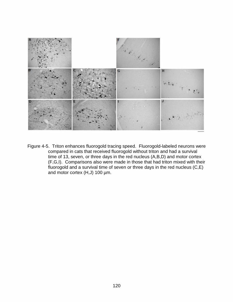
120
Figure 4-5. Triton enhances fluorogold tracing speed. Fluorogold-labeled neurons were
compared in cats that received fluorogold without triton and had a survival time of 13, seven, or three days in the red nucleus (A,B,D) and motor cortex (F,G,I). Comparisons also were made in those that had triton mixed with their fluorogold and a survival time of seven or three days in the red nucleus (C,E) and motor cortex (H,J) 100 µm.

121
CHAPTER 5 SUMMARY
Defining Recovery
There is great debate amongst scientists and clinicians regarding the definition of
“recovery” relative to functional changes associated with central nervous system (CNS)
injury or disease. Some maintain that “recovery” has not been truly achieved unless the
original functional dynamics are completely restored. Other opinions view any form of
clinical improvement as being equivalent to “recovery” (Levin et al., 2009). Thus, it is
critical for investigators to clearly define the definition of recovery for their specific study,
as was done in Chapters 2 and 3. However, the definition employed in these studies
remains controversial as they allowed for compensatory strategies supporting
successful movement patterns that differed from pre-injury. The decision to use a
definition of recovery that allows for compensatory strategies as opposed to the strict
definition of recovery described in Levin and colleagues, is primarily due to the
extensive changes in axonal circuitry that are known to occur by way of injury-induced
sprouting and regeneration (Bareyre et al., 2004; Courtine et al., 2009; Blum, 2010).
This novel circuitry, which underlies locomotor performance, will most likely be
accompanied by novel movement patterns that differ from pre-injury. A more strict
definition of recovery may be difficult to establish and not particularly applicable to
individuals with neurological injuries. Using this line of thought “compensation” or “the
use of new motor patterns resulting from the adaptation of remaining motor elements or
substitution (Levin et al., 2009)” may be synonymous with “recovery” as new circuitry
most likely will strongly incorporate and require enhanced use of spared fibers.

122
Advancing Chondroitinase ABC to the Clinic
As chondroitinase abc (ch’abc) continues heading towards a clinical direction the
definitions for recovery in subsequent studies, as well as the functions being assessed
need to be well defined and deliberately chosen. As reported in Chapter 3, models of
partial spinal cord injury (SCI), similar in many respects to Brown-Sequard Syndrome
(BSS), showed the greatest ch’abc-mediated functional effects on adaptive tasks like
the peg walkway and were not as readily detected on more simple tasks like the
overground walkway. However, ch’abc-mediated effects may be more noticeable on
simple tasks in larger injury models, such as contusions. Furthermore, animals
frequently employed a variety of compensatory strategies for successful completion of
the more adaptive tasks that differed across ch’abc and control cats (Jefferson et al.,
2011). Thus, not only is it important to carefully choose the functional assay used for
the model of injury being assessed but, it also is critical to assess performance of a
variety of different gait features in order to properly understand the effects of ch’abc on
functional improvements after SCI. For future clinical assessments, the development of
a test battery for adaptive locomotor features for the human SCI population would be
beneficial and allow for quick assessment of adaptive functions as well as their
improvements over time, as reported in our previous ch’abc studies (Tester and
Howland, 2008; Jefferson et al., 2011).
Another major translational issue is whether the magnitude of therapeutic efficacy
seen in an animal study will translate to humans. Therapeutic effects seen in small
rodent models of SCI indicate promising treatment potential, but do not necessarily
mean the treatment will be as effective in humans. Moving these treatments to larger
animal models provides greater insight into the therapeutic potential of treatments.

123
Results from Chapter 3 demonstrate this issue. Despite there being multiple reports of
two weeks of ch’abc administration or less leading to significant functional recovery in
rodent SCI models, this treatment duration was not effective in our larger cat model of
SCI, but four weeks administration resulted in significant functional benefits. It is
possible that due to the larger size of humans, four weeks of ch’abc may not be
sufficient for humans, but that six to eight weeks of treatment may be more optimal. It
would be interesting to determine how a six week period of ch’abc administration will
affect functional recovery and if greater recovery can be achieved with this longer
treatment duration or, if too much ch’abc administration could be detrimental in some
way. In addition to duration, optimal ch’abc concentration and treatment window also
are important to assess. Although several studies have begun to look at treatment
windows, the majority have used a single injection paradigm. This single dose
approach, as determined in Chapter 3, is most likely not an optimal treatment duration
(Yick et al., 2003; Massey et al., 2006; Iseda et al., 2008). Furthermore, determining
the best mode of ch’abc administration is critical. Currently the most common mode of
delivery is via intrathecal administration. However this mode has been reported to cause
increased scarring and compression to the spinal cord (Jones and Tuszynski, 2001) and
is not particularly feasible in humans with acute SCI. Several studies have begun to
identify alternative modes of delivery such as a thermostabilized ch’abc hydrogel (Lee et
al., 2009), slow ch’abc-release fibrin gel (Hyatt et al., 2010), and ch’abc lentiviral vectors
(Jin et al., 2011). However, assessments of these delivery modes are limited, and it is
unclear how effective they will be. The need to continue to explore clinically applicable

124
delivery approaches is one of the more critical variables for the future translation of this
therapeutic.
Conclusions
As research continues to allow for a better understanding of the physiological
responses and plastic potential of the central nervous system after injury, the
development of a treatment paradigm capable of significantly enhancing functional
recovery after injury appears more plausible than previously thought to be. An effective
treatment paradigm will most likely require a multifaceted approach and, as depicted in
Chapters 2 and 3, the therapeutic agent ch’abc and an increased focus on optimizing
training/rehabilitation for simple as well as adaptive locomotor functions will be
important components of this effective treatment paradigm. The optimization of tract
tracing in both small and large pre-clinical animal models will aid in this treatment’s
development by allowing for an understanding of the treatment-mediated circuitry
changes that underlie the functional changes.
125
LIST OF REFERENCES
Adams MM, Hicks AL (2005) Spasticity after spinal cord injury. Spinal Cord 43:577-586.
Afelt Z (1974) Functional significance of ventral descending tracts of the spinal cord in the cat. Acta Neurobiol Exp (Wars) 34:393-407.
Akhavan M, Hoang TX, Havton LA (2006) Improved detection of fluorogold-labeled neurons in long-term studies. Journal of Neuroscience Methods 152:156-162.
Alstermark B, Lundberg A, Norrsell U, Sybirska E (1981a) Integration in descending motor pathways controlling the forelimb in the cat. 9. Differential behavioural defects after spinal cord lesions interrupting defined pathways from higher centres to motoneurones. Exp Brain Res 42:299-318.
Alstermark B, Lindstrom S, Lundberg A, Sybirska E (1981b) Integration in descending motor pathways controlling the forelimb in the cat. 8. Ascending projection to the lateral reticular nucleus from C3-C4 propriospinal also projecting to forelimb motoneurones. Exp Brain Res 42:282-298.
Alstermark B, Lundberg A, Pettersson LG, Tantisira B, Walkowska M (1987) Motor recovery after serial spinal cord lesions of defined descending pathways in cats. Neurosci Res 5:68-73.
Anderson KD (2004) Targeting recovery: priorities of the spinal cord-injured population. J Neurotrauma 21:1371-1383.
Andrews EM, Richards RJ, Yin FQ, Viapiano MS, Jakeman LB (2011) Alterations in chondroitin sulfate proteoglycan expression occur both at and far from the site of spinal contusion injury. Experimental Neurology.
Bai F, Peng H, Etlinger JD, Zeman RJ (2010) Partial functional recovery after complete spinal cord transection by combined chondroitinase and clenbuterol treatment. Pflugers Arch 460:657-666.
Balasubramanian CK, Neptune RR, Kautz SA (2010) Foot placement in a body reference frame during walking and its relationship to hemiparetic walking performance. Clinical Biomechanics 25:483-490.
Barbeau H, Rossignol S (1987) Recovery of locomotion after chronic spinalization in the adult cat. Brain Research 412:84-95.
Barbeau H, Fung J, Leroux A, Ladouceur M (2002) A review of the adaptability and recovery of locomotion after spinal cord injury. Prog Brain Res 137:9-25.
Barbeau H, Ladouceur M, Norman KE, Pepin A, Leroux A (1999) Walking after spinal cord injury: evaluation, treatment, and functional recovery. Arch Phys Med Rehabil 80:225-235.

126
Bareyre FM, Kerschensteiner M, Raineteau O, Mettenleiter TC, Weinmann O, Schwab ME (2004) The injured spinal cord spontaneously forms a new intraspinal circuit in adult rats. Nat Neurosci 7:269-277.
Barritt AW, Davies M, Marchand F, Hartley R, Grist J, Yip P, McMahon SB, Bradbury EJ (2006) Chondroitinase ABC Promotes Sprouting of Intact and Injured Spinal Systems after Spinal Cord Injury. The Journal of Neuroscience 26:10856-10867.
Batson DE, Amassian VE (1986) A dynamic role of rubral neurons in contact placing by the adult cat. Journal of Neurophysiology 56:835-856.
Battistuzzo CR, Callister RJ, Callister R, Galea M (2012) A Systematic Review of Exercise Training to Promote Locomotor Recovery in Animal Models of Spinal Cord Injury. J Neurotrauma.
Beattie MS, Farooqui AA, Bresnahan JC (2000) Review of current evidence for apoptosis after spinal cord injury. J Neurotrauma 17:915-925.
Beaumont E, Kaloustian S, Rousseau G, Cormery B (2008) Training improves the electrophysiological properties of lumbar neurons and locomotion after thoracic spinal cord injury in rats. Neuroscience Research 62:147-154.
Becker CG, Becker T (2002) Repellent Guidance of Regenerating Optic Axons by Chondroitin Sulfate Glycosaminoglycans in Zebrafish. The Journal of Neuroscience 22:842-853.
Beloozerova IN, Sirota MG (1993) The role of the motor cortex in the control of accuracy of locomotor movements in the cat. The Journal of Physiology 461:1-25.
Beloozerova IN, Farrell BJ, Sirota MG, Prilutsky BI (2010a) Differences in Movement Mechanics, Electromyographic, and Motor Cortex Activity Between Accurate and Nonaccurate Stepping. Journal of Neurophysiology 103:2285-2300.
Beloozerova IN, Farrell BJ, Sirota MG, Prilutsky BI (2010b) Differences in movement mechanics, electromyographic, and motor cortex activity between accurate and nonaccurate stepping. J Neurophysiol 103:2285-2300.
Benfey M, Aguayo AJ (1982) Extensive elongation of axons from rat brain into peripheral nerve grafts. Nature 296:150-152.
Bentivoglio M, Su HS (1990) Photoconversion of fluorescent retrograde tracers. Neurosci Lett 113:127-133.
Blagovechtchenski E, Pettersson LG, Perfiliev S, Krasnochokova E, Lundberg A (2000) Control of digits via C3–C4 propriospinal neurones in cats; recovery after lesions. Neuroscience Research 38:103-108.

127
Blesch A (1999) Leukemia inhibitory factor augments neurotrophin expression and corticospinal axon growth after adult CNS injury. The Journal of Neuroscience 19:3556-3566.
Blum AE (2010) Plasticity of the central nervous system and functional recovery following spinal cord injury. In: Neuroscience, p 118. Gainesville: University of Florida.
Bolton DAE, Tse ADY, Ballermann M, Misiaszek JE, Fouad K (2006) Task specific adaptations in rat locomotion: Runway versus horizontal ladder. Behavioural Brain Research 168:272-279.
Borisoff JF, Chan CCM, Hiebert GW, Oschipok L, Robertson GS, Zamboni R, Steeves JD, Tetzlaff W (2003) Suppression of Rho-kinase activity promotes axonal growth on inhibitory CNS substrates. Molecular and Cellular Neuroscience 22:405-416.
Boulenguez P, Vinay L (2009) Strategies to restore motor functions after spinal cord injury. Curr Opin Neurobiol 19:587-600.
Bradbury EJ, Moon LDF, Popat RJ, King VR, Bennett GS, Patel PN, Fawcett JW, McMahon SB (2002) Chondroitinase ABC promotes functional recovery after spinal cord injury. Nature 416:636-640.
Brown TG (1914) On the nature of the fundamental activity of the nervous centres; together with an analysis of the conditioning of rhythmic activity in progression, and a theory of the evolution of function in the nervous system. The Journal of Physiology 48:18-46.
Brückner G, Grosche J, Schmidt S, Härtig W, Margolis RU, Delpech B, Seidenbecher CI, Czaniera R, Schachner M (2000) Postnatal development of perineuronal nets in wild-type mice and in a mutant deficient in tenascin-R. The Journal of Comparative Neurology 428:616-629.
Brustein E, Rossignol S (1998) Recovery of Locomotion After Ventral and Ventrolateral Spinal Lesions in the Cat. I. Deficits and Adaptive Mechanisms. Journal of neurophysiology 80:1245-1267.
Buhl EH, Dann JF (1990) Basal dendrites are a regular feature of hippocampal granule cells in flying fox hippocampus. Neurosci Lett 116:263-268.
Busch SA, Silver J (2007) The role of extracellular matrix in CNS regeneration. Current Opinion in Neurobiology 17:120-127.
Busch SA, Horn KP, Silver DJ, Silver J (2009) Overcoming Macrophage-Mediated Axonal Dieback Following CNS Injury. The Journal of Neuroscience 29:9967-9976.

128
Byrnes KR, Fricke ST, Faden AI Neuropathological differences between rats and mice after spinal cord injury. Journal of Magnetic Resonance Imaging 32:836-846.
Cafferty WBJ, Bradbury EJ, Lidierth M, Jones M, Duffy PJ, Pezet S, McMahon SB (2008) Chondroitinase ABC-Mediated Plasticity of Spinal Sensory Function. The Journal of Neuroscience 28:11998-12009.
Cajal Ry (1928) Degeneration and regeneration of the central nervous system.
Calancie B (2006) Spinal myoclonus after spinal cord injury. J Spinal Cord Med 29:413-424.
Capaday C (2002) The special nature of human walking and its neural control. Trends Neurosci 25:370-376.
Carter LM, McMahon SB, Bradbury EJ (2011) Delayed treatment with Chondroitinase ABC reverses chronic atrophy of rubrospinal neurons following spinal cord injury. Exp Neurol 228:149-156.
Carter LM, Starkey ML, Akrimi SF, Davies M, McMahon SB, Bradbury EJ (2008) The yellow fluorescent protein (YFP-H) mouse reveals neuroprotection as a novel mechanism underlying chondroitinase ABC-mediated repair after spinal cord injury. J Neurosci 28:14107-14120.
Chang HT, Kuo H, Whittaker JA, Cooper NG (1990) Light and electron microscopic analysis of projection neurons retrogradely labeled with Fluoro-Gold: notes on the application of antibodies to Fluoro-Gold. J Neurosci Methods 35:31-37.
Chung KY, Taylor JS, Shum DK, Chan SO (2000) Axon routing at the optic chiasm after enzymatic removal of chondroitin sulfate in mouse embryos. Development 127:2673-2683.
Clarac Fo (2008) Some historical reflections on the neural control of locomotion. Brain Research Reviews 57:13-21.
Conrad B, Benecke R, Carnehl J, Hohne J, Meinck HM (1983) Pathophysiological aspects of human locomotion. Adv Neurol 39:717-726.
Conta AC, Stelzner DJ (2004) Differential vulnerability of propriospinal tract neurons to spinal cord contusion injury. J Comp Neurol 479:347-359.
Conta Steencken AC, Stelzner DJ (2009) Loss of propriospinal neurons after spinal contusion injury as assessed by retrograde labeling. Neuroscience 170:971-980.
Conta Steencken AC, Smirnov I, Stelzner DJ (2011) Cell survival or cell death: differential vulnerability of long descending and thoracic propriospinal neurons to low thoracic axotomy in the adult rat. Neuroscience 194:359-371.

129
Courtine G, Song B, Roy RR, Zhong H, Herrmann JE, Ao Y, Qi J, Edgerton VR, Sofroniew MV (2008) Recovery of supraspinal control of stepping via indirect propriospinal relay connections after spinal cord injury. Nat Med 14:69-74.
Courtine G, Gerasimenko Y, van den Brand R, Yew A, Musienko P, Zhong H, Song B, Ao Y, Ichiyama RM, Lavrov I, Roy RR, Sofroniew MV, Edgerton VR (2009) Transformation of nonfunctional spinal circuits into functional states after the loss of brain input. Nat Neurosci 12:1333-1342.
Crespo D, Asher RA, Lin R, Rhodes KE, Fawcett JW (2007) How does chondroitinase promote functional recovery in the damaged CNS? Experimental Neurology 206:159-171.
Crowe MJ, Bresnahan JC, Shuman SL, Masters JN, Beattie MS (1997) Apoptosis and delayed degeneration after spinal cord injury in rats and monkeys. Nat Med 3:73-76.
Curinga GM, Snow DM, Mashburn C, Kohler K, Thobaben R, Caggiano AO, Smith GM (2007) Mammalian-produced chondroitinase AC mitigates axon inhibition by chondroitin sulfate proteoglycans. Journal of Neurochemistry 102:275-288.
Darlot F, Cayetanot F, Gauthier P, Matarazzo V, Kastner A (2012) Extensive respiratory plasticity after cervical spinal cord injury in rats: Axonal sprouting and rerouting of ventrolateral bulbospinal pathways. Exp Neurol 236:88-102.
Davies SJA, Fitch MT, Memberg SP, Hall AK, Raisman G, Silver J (1997) Regeneration of adult axons in white matter tracts of the central nervous system. Nature 390:680-683.
Dawson MRL, Levine JM, Reynolds R (2000) NG2-expressing cells in the central nervous system: Are they oligodendroglial progenitors? Journal of Neuroscience Research 61:471-479.
Decherchi P, Gauthier P (2000) Regrowth of acute and chronic injured spinal pathways within supra-lesional post-traumatic nerve grafts. Neuroscience 101:197-210.
Deepa SS, Carulli D, Galtrey C, Rhodes K, Fukuda J, Mikami T, Sugahara K, Fawcett JW (2006) Composition of Perineuronal Net Extracellular Matrix in Rat Brain. Journal of Biological Chemistry 281:17789-17800.
Dickendesher TL, Baldwin KT, Mironova YA, Koriyama Y, Raiker SJ, Askew KL, Wood A, Geoffroy CG, Zheng B, Liepmann CD, Katagiri Y, Benowitz LI, Geller HM, Giger RJ (2012) NgR1 and NgR3 are receptors for chondroitin sulfate proteoglycans. Nat Neurosci advance online publication.
Dietz V (2002) Do human bipeds use quadrupedal coordination? Trends in Neurosciences 25:462-467.

130
Dietz V, Colombo G (2004) Recovery from spinal cord injury--underlying mechanisms and efficacy of rehabilitation. Acta Neurochir Suppl 89:95-100.
Dietz V, Wirz M, Curt A, Colombo G (1998) Locomotor pattern in paraplegic patients: training effects and recovery of spinal cord function. Spinal Cord 36:380-390.
Dill J, Wang H, Zhou F, Li S (2008) Inactivation of Glycogen Synthase Kinase 3 Promotes Axonal Growth and Recovery in the CNS. The Journal of Neuroscience 28:8914-8928.
Dimitrijevic MR, Gerasimenko Y, Pinter MM (1998) Evidence for a Spinal Central Pattern Generator in Humans. Annals of the New York Academy of Sciences 860:360-376.
Ditunno JF, Little JW, Tessler A, Burns AS (2004) Spinal shock revisited: a four-phase model. Spinal Cord 42:383-395.
Divac I, Mogensen J (1990) Long-term retrograde labelling of neurons. Brain Research 524:339-341.
Dolbeare D, Houle JD (2003) Restriction of Axonal Retraction and Promotion of Axonal Regeneration by Chronically Injured Neurons after Intraspinal Treatment with Glial Cell Line-Derived Neurotrophic Factor (GDNF). Journal of Neurotrauma 20:1251-1261.
Domingo A, Sawicki G, Ferris D (2007) Kinematics and muscle activity of individuals with incomplete spinal cord injury during treadmill stepping with and without manual assistance. Journal of NeuroEngineering and Rehabilitation 4:32.
Donnelly DJ, Popovich PG (2008) Inflammation and its role in neuroprotection, axonal regeneration and functional recovery after spinal cord injury. Exp Neurol 209:378-388.
Dou CL, Levine JM (1994) Inhibition of neurite growth by the NG2 chondroitin sulfate proteoglycan. The Journal of Neuroscience 14:7616-7628.
Drew T, Jiang W, Widajewicz W (2002) Contributions of the motor cortex to the control of the hindlimbs during locomotion in the cat. Brain Research Reviews 40:178-191.
Duan Y, Giger RJ (2010) A New Role for RPTP{sigma} in Spinal Cord Injury: Signaling Chondroitin Sulfate Proteoglycan Inhibition. Sci Signal 3:pe6-.
Duffy P, Schmandke A, Sigworth J, Narumiya S, Cafferty WB, Strittmatter SM (2009) Rho-associated kinase II (ROCKII) limits axonal growth after trauma within the adult mouse spinal cord. J Neurosci 29:15266-15276.

131
Eidelberg E (1981) Consequences of spinal cord lesions upon motor function, with special reference to locomotor activity. Progress in Neurobiology 17:185-202.
Eidelberg E, Nguyen LH, Deza LD (1986) Recovery of locomotor function after hemisection of the spinal cord in cats. Brain Research Bulletin 16:507-515.
Eidelberg E, Story JL, Meyer BL, Nystel J (1980) Stepping by chronic spinal cats. Experimental Brain Research 40:241-246.
Eidelberg E, Story JL, Walden JG, Meyer BL (1981) Anatomical correlates of return of locomotor function after partial spinal cord lesions in cats. Experimental Brain Research 42:81-88.
ElBasiouny SM, Schuster JE, Heckman CJ (2009) Persistent inward currents in spinal motoneurons: Important for normal function but potentially harmful after spinal cord injury and in amyotrophic lateral sclerosis. Clinical neurophysiology : official journal of the International Federation of Clinical Neurophysiology 121:1669-1679.
Fawcett JW, Asher RA (1999) The glial scar and central nervous system repair. Brain Research Bulletin 49:377-391.
Fenrich KK, Rose PK (2009) Spinal interneuron axons spontaneously regenerate after spinal cord injury in the adult feline. J Neurosci 29:12145-12158.
Fidler PS, Schuette K, Asher RA, Dobbertin A, Thornton SR, Calle-Patino Y, Muir E, Levine JM, Geller HM, Rogers JH, Faissner A, Fawcett JW (1999) Comparing Astrocytic Cell Lines that Are Inhibitory or Permissive for Axon Growth: the Major Axon-Inhibitory Proteoglycan Is NG2. The Journal of Neuroscience 19:8778-8788.
Filbin MT (2003) Myelin-associated inhibitors of axonal regeneration in the adult mammalian CNS. Nat Rev Neurosci 4:703-713.
Fitch MT, Silver J (1997) Glial cell extracellular matrix: boundaries for axon growth in development and regeneration. Cell and Tissue Research 290:379-384.
Fleming JC, Norenberg MD, Ramsay DA, Dekaban GA, Marcillo AE, Saenz AD, Pasquale-Styles M, Dietrich WD, Weaver LC (2006) The cellular inflammatory response in human spinal cords after injury. Brain 129:3249-3269.
Flynn JR, Graham BA, Galea MP, Callister RJ (2011) The role of propriospinal interneurons in recovery from spinal cord injury. Neuropharmacology 60:809-822.
Fry EJ, Stolp HB, Lane MA, Dziegielewska KM, Saunders NR (2003) Regeneration of supraspinal axons after complete transection of the thoracic spinal cord in neonatal opossums (Monodelphis domestica). The Journal of Comparative Neurology 466:422-444.

132
Fry EJ, Chagnon MJ, Lopez-Vales R, Tremblay ML, David S (2009) Corticospinal tract regeneration after spinal cord injury in receptor protein tyrosine phosphatase sigma deficient mice. Glia 58:423-433.
Fung J, Barbeau H (1989) A dynamic EMG profile index to quantify muscular activation disorder in spastic paretic gait. Electroencephalogr Clin Neurophysiol 73:233-244.
Galtrey CM, Fawcett JW (2007) The role of chondroitin sulfate proteoglycans in regeneration and plasticity in the central nervous system. Brain Research Reviews 54:1-18.
Galtrey CM, Asher RA, Nothias F, Fawcett JW (2007) Promoting plasticity in the spinal cord with chondroitinase improves functional recovery after peripheral nerve repair. Brain 130:926-939.
Garcia-Alias G, Barkhuysen S, Buckle M, Fawcett JW (2009) Chondroitinase ABC treatment opens a window of opportunity for task-specific rehabilitation. Nat Neurosci 12:1145-1151.
García-Alías G, Lin R, Akrimi SF, Story D, Bradbury EJ, Fawcett JW (2008) Therapeutic time window for the application of chondroitinase ABC after spinal cord injury. Experimental Neurology 210:331-338.
Garrett WT, McBride RL, Williams JK, Jr., Feringa ER (1991) Fluoro-Gold's toxicity makes it inferior to True Blue for long-term studies of dorsal root ganglion neurons and motoneurons. Neurosci Lett 128:137-139.
Gimenez-Amaya JM, Graybiel AM (1990) Compartmental origins of the striatopallidal projection in the primate. Neuroscience 34:111-126.
Girgis J, Merrett D, Kirkland S, Metz GAS, Verge V, Fouad K (2007) Reaching training in rats with spinal cord injury promotes plasticity and task specific recovery. Brain 130:2993-3003.
Gottschall JS, Okita N, Sheehan RC (2011) Muscle activity patterns of the tensor fascia latae and adductor longus for ramp and stair walking. Journal of Electromyography and Kinesiology 22:67-73.
Grillner S, Rossignol S (1978) On the initiation of the swing phase of locomotion in chronic spinal cats. Brain Res 146:269-277.
Grillner S, Zangger P (1979) On the central generation of locomotion in the low spinal cat. Experimental Brain Research 34:241-261.
Grimpe B, Silver J (2004) A Novel DNA Enzyme Reduces Glycosaminoglycan Chains in the Glial Scar and Allows Microtransplanted Dorsal Root Ganglia Axons to Regenerate beyond Lesions in the Spinal Cord. The Journal of Neuroscience 24:1393-1397.

133
Guth L (1975) History of central nervous system regeneration research. Exp Neurol 48:3-15.
Haas CA, Rauch U, Thon N, Merten T, Deller T (1999) Entorhinal Cortex Lesion in Adult Rats Induces the Expression of the Neuronal Chondroitin Sulfate Proteoglycan Neurocan in Reactive Astrocytes. The Journal of Neuroscience 19:9953-9963.
Harkema S, Gerasimenko Y, Hodes J, Burdick J, Angeli C, Chen Y, Ferreira C, Willhite A, Rejc E, Grossman RG, Edgerton VR Effect of epidural stimulation of the lumbosacral spinal cord on voluntary movement, standing, and assisted stepping after motor complete paraplegia: a case study. Lancet 377:1938-1947.
Harkema SJ, Schmidt-Read M, Lorenz D, Edgerton VR, Behrman AL (2011) Balance and Ambulation Improvements in Individuals With Chronic Incomplete Spinal Cord Injury Using Locomotor Training–Based Rehabilitation. Archives of Physical Medicine and Rehabilitation.
Hart BL (1971) Facilitation by strychnine of reflex walking in spinal dogs. Physiology & Behavior 6:627-628.
Hayashi J, Takagi Y, Fukuda H, Imazato T, Nishimura M, Fujimoto M, Takahashi J, Hashimoto N, Nozaki K (2006) Primate embryonic stem cell-derived neuronal progenitors transplanted into ischemic brain. J Cereb Blood Flow Metab 26:906-914.
Hazime FA, Allard P, Ide MR, Siqueira CM, Amorim CF, Tanaka C (2012) Postural control under visual and proprioceptive perturbations during double and single limb stances: insights for balance training. J Bodyw Mov Ther 16:224-229.
Helgren ME, Goldberger ME (1993) The recovery of postural reflexes and locomotion following low thoracic hemisection in adult cats involves compensation by undamaged primary afferent pathways. Exp Neurol 123:17-34.
Hepp-Reymond MC, Wiesendanger M, Brunnert A, Mackel R, Unger C, Wespi J (1970) Effects of unilateral pyramidotomy on conditioned finger movement in monkey (Macaca irus). Brain Res 24:544.
Herndon ME, Lander AD (1990) A diverse set of developmentally regulated proteoglycans is expressed in the rat central nervous system. Neuron 4:949-961.
Hiebert GW, Khodarahmi K, McGraw J, Steeves JD, Tetzlaff W (2002) Brain-derived neurotrophic factor applied to the motor cortex promotes sprouting of corticospinal fibers but not regeneration into a peripheral nerve transplant. Journal of Neuroscience Research 69:160-168.
Hill CE, Beattie MS, Bresnahan JC (2001) Degeneration and Sprouting of Identified Descending Supraspinal Axons after Contusive Spinal Cord Injury in the Rat. Experimental Neurology 171:153-169.

134
Hilton BJ, Lang BT, Cregg JM (2012) Keratan Sulfate Proteoglycans in Plasticity and Recovery after Spinal Cord Injury. The Journal of Neuroscience 32:4331-4333.
Hinsey JC, Cutting CC (1936) Reflexes in the spinal opossum. The Journal of Comparative Neurology 64:375-387.
Hook M, Kjellen L, Johansson S (1984) Cell-surface glycosaminoglycans. Annu Rev Biochem 53:847-869.
Houle JD, Ye JH (1999) Survival of chronically-injured neurons can be prolonged by treatment with neurotrophic factors. Neuroscience 94:929-936.
Houle JD, Jin Y (2001) Chronically injured supraspinal neurons exhibit only modest axonal dieback in response to a cervical hemisection lesion. Exp Neurol 169:208-217.
Houle JD, Tom VJ, Mayes D, Wagoner G, Phillips N, Silver J (2006) Combining an Autologous Peripheral Nervous System "Bridge" and Matrix Modification by Chondroitinase Allows Robust, Functional Regeneration beyond a Hemisection Lesion of the Adult Rat Spinal Cord. The Journal of Neuroscience 26:7405-7415.
Howland DR, Bregman BS, Tessler A, Goldberger ME (1995a) Development of Locomotor Behavior in the Spinal Kitten. Experimental Neurology 135:108-122.
Howland DR, Bregman BS, Tessler A, Goldberger ME (1995b) Transplants Enhance Locomotion in Neonatal Kittens Whose Spinal Cords Are Transected: A Behavioral and Anatomical Study. Experimental Neurology 135:123-145.
Huang HJ, Ferris DP (2009) Upper and lower limb muscle activation is bidirectionally and ipsilaterally coupled. Med Sci Sports Exerc 41:1778-1789.
Hyatt AJT, Wang D, Kwok JC, Fawcett JW, Martin KR (2010) Controlled release of chondroitinase ABC from fibrin gel reduces the level of inhibitory glycosaminoglycan chains in lesioned spinal cord. Journal of Controlled Release 147:24-29.
Hynds DL, Snow DM (1999) Neurite Outgrowth Inhibition by Chondroitin Sulfate Proteoglycan: Stalling/Stopping Exceeds Turning in Human Neuroblastoma Growth Cones. Experimental Neurology 160:244-255.
Iaci JF, Vecchione AM, Zimber MP, Caggiano AO (2007) Chondroitin Sulfate Proteoglycans in Spinal Cord Contusion Injury and the Effects of Chondroitinase Treatment. Journal of Neurotrauma 24:1743-1760.
Imagama S, Sakamoto K, Tauchi R, Shinjo R, Ohgomori T, Ito Z, Zhang H, Nishida Y, Asami N, Takeshita S, Sugiura N, Watanabe H, Yamashita T, Ishiguro N, Matsuyama Y, Kadomatsu K (2011) Keratan Sulfate Restricts Neural Plasticity after Spinal Cord Injury. The Journal of Neuroscience 31:17091-17102.

135
Iozzo RV (1998) Matrix proteoglycans: from molecular design to cellular function. Annu Rev Biochem 67:609-652.
Iseda T, Okuda T, Kane-Goldsmith N, Mathew M, Ahmed S, Chang Y-W, Young W, Grumet M (2008) Single, High-Dose Intraspinal Injection of Chondroitinase Reduces Glycosaminoglycans in Injured Spinal Cord and Promotes Corticospinal Axonal Regrowth after Hemisection but Not Contusion. Journal of Neurotrauma 25:334-349.
Ito J, Murata M, Kawaguchi S (1999) Regeneration of the lateral vestibulospinal tract in adult rats by transplants of embryonic brain tissue. Neuroscience Letters 259:67-70.
Jakeman LB, Hoschouer EL, Basso DM (2010) Injured mice at the gym: Review, results and considerations for combining chondroitinase and locomotor exercise to enhance recovery after spinal cord injury. Brain Res Bull 84:317-326.
Jefferson SC, Tester NJ, Howland DR Chondroitinase ABC Promotes Recovery of Adaptive Limb Movements and Enhances Axonal Growth Caudal to a Spinal Hemisection. The Journal of Neuroscience 31:5710-5720.
Jefferson SC, Tester NJ, Howland DR (2011) Chondroitinase ABC Promotes Recovery of Adaptive Limb Movements and Enhances Axonal Growth Caudal to a Spinal Hemisection. The Journal of Neuroscience 31:5710-5720.
Jeffery ND, Blakemore WF (1999) Spinal cord injury in small animals 2. Current and future options for therapy. Vet Rec 145:183-190.
Jell RM, Elliott C, Jordan LM (1985) Initiation of locomotion from the mesencephalic locomotor region: effects of selective brainstem lesions. Brain Res 328:121-128.
Jian BJ, Acernese AW, Lorenzo J, Card JP, Yates BJ (2005) Afferent pathways to the region of the vestibular nuclei that participates in cardiovascular and respiratory control. Brain Research 1044:241-250.
Jilge B, Minassian K, Rattay F, Pinter MM, Gerstenbrand F, Binder H, Dimitrijevic MR (2004) Initiating extension of the lower limbs in subjects with complete spinal cord injury by epidural lumbar cord stimulation. Exp Brain Res 154:308-326.
Jin Y, Ketschek A, Jiang Z, Smith G, Fischer I (2011) Chondroitinase activity can be transduced by a lentiviral vector in vitro and in vivo. Journal of Neuroscience Methods 199:208-213.
Jones LL, Tuszynski MH (2001) Chronic intrathecal infusions after spinal cord injury cause scarring and compression. Microsc Res Tech 54:317-324.

136
Jones LL, Margolis RU, Tuszynski MH (2003a) The chondroitin sulfate proteoglycans neurocan, brevican, phosphacan, and versican are differentially regulated following spinal cord injury. Exp Neurol 182:399-411.
Jones LL, Sajed D, Tuszynski MH (2003b) Axonal Regeneration through Regions of Chondroitin Sulfate Proteoglycan Deposition after Spinal Cord Injury: A Balance of Permissiveness and Inhibition. The Journal of Neuroscience 23:9276-9288.
Jones LL, Yamaguchi Y, Stallcup WB, Tuszynski MH (2002) NG2 is a major chondroitin sulfate proteoglycan produced after spinal cord injury and is expressed by macrophages and oligodendrocyte progenitors. J Neurosci 22:2792-2803.
Kanagal SG, Muir GD (2009) Task-dependent compensation after pyramidal tract and dorsolateral spinal lesions in rats. Experimental Neurology 216:193-206.
Karimi-Abdolrezaee S, Eftekharpour E, Wang J, Schut D, Fehlings MG (2010) Synergistic effects of transplanted adult neural stem/progenitor cells, chondroitinase, and growth factors promote functional repair and plasticity of the chronically injured spinal cord. J Neurosci 30:1657-1676.
Kluppel M (2011) Efficient secretion of biologically active Chondroitinase ABC from mammalian cells in the absence of an N-terminal signal peptide. Mol Cell Biochem.
Kobayashi NR, Fan D-P, Giehl KM, Bedard AM, Wiegand SJ, Tetzlaff W (1997) BDNF and NT-4/5 Prevent Atrophy of Rat Rubrospinal Neurons after Cervical Axotomy, Stimulate GAP-43 and Tα1-Tubulin mRNA Expression, and Promote Axonal Regeneration. The Journal of Neuroscience 17:9583-9595.
Koprivica V, Cho K-S, Park JB, Yiu G, Atwal J, Gore B, Kim JA, Lin E, Tessier-Lavigne M, Chen DF, He Z (2005) EGFR Activation Mediates Inhibition of Axon Regeneration by Myelin and Chondroitin Sulfate Proteoglycans. Science 310:106-110.
Krajacic A, Weishaupt N, Girgis J, Tetzlaff W, Fouad K (2010) Training-induced plasticity in rats with cervical spinal cord injury: Effects and side effects. Behavioural Brain Research 214:323-331.
Kuzhandaivel A, Nistri A, Mazzone GLn, Mladinic M (2011) Molecular mechanisms underlying cell death in spinal networks in relation to locomotor activity after acute injury in vitro. Frontiers in Cellular Neuroscience 5.
Kwok JC, Afshari F, Garcia-Alias G, Fawcett JW (2008) Proteoglycans in the central nervous system: plasticity, regeneration and their stimulation with chondroitinase ABC. Restor Neurol Neurosci 26:131-145.

137
Kwon BK, Liu J, Messerer C, Kobayashi NR, McGraw J, Oschipok L, Tetzlaff W (2002) Survival and regeneration of rubrospinal neurons 1 year after spinal cord injury. Proceedings of the National Academy of Sciences 99:3246-3251.
Kwon HG, Lee DG, Son SM, Byun WM, Hong CP, Lee DH, Kim S, Jang SH (2011) Identification of the anterior corticospinal tract in the human brain using diffusion tensor imaging. Neurosci Lett 505:238-241.
Ladouceur M, Barbeau H, McFadyen BJ (2003) Kinematic Adaptations of Spinal Cord-Injured Subjects during Obstructed Walking. Neurorehabilitation and Neural Repair 17:25-31.
Lanciego JL, Wouterlood FG (2011) A half century of experimental neuroanatomical tracing. Journal of Chemical Neuroanatomy 42:157-183.
Lane MA, Lee K-Z, Salazar K, O'Steen BE, Bloom DC, Fuller DD, Reier PJ (2011) Respiratory function following bilateral mid-cervical contusion injury in the adult rat. Experimental Neurology 235:197-210.
Laursen AM, Wiesendanger M (1967) The effect of pyramidal lesions on response latency in cats. Brain Research 5:207-220.
Lavoie S, Drew T (2002) Discharge Characteristics of Neurons in the Red Nucleus During Voluntary Gait Modifications: A Comparison with the Motor Cortex. Journal of Neurophysiology 88:1791-1814.
Lawrence DG, Kuypers HGJM (1968) THE FUNCTIONAL ORGANIZATION OF THE MOTOR SYSTEM IN THE MONKEY. Brain 91:1-14.
Lee H, McKeon RJ, Bellamkonda RV (2009) Sustained delivery of thermostabilized chABC enhances axonal sprouting and functional recovery after spinal cord injury. Proc Natl Acad Sci U S A 107:3340-3345.
Lemons ML, Howland DR, Anderson DK (1999) Chondroitin Sulfate Proteoglycan Immunoreactivity Increases Following Spinal Cord Injury and Transplantation. Experimental Neurology 160:51-65.
Lemons ML, Sandy JD, Anderson DK, Howland DR (2001) Intact Aggrecan and Fragments Generated by Both Aggrecanse and Metalloproteinase-Like Activities Are Present in the Developing and Adult Rat Spinal Cord and Their Relative Abundance Is Altered by Injury. The Journal of Neuroscience 21:4772-4781.
Lemons ML, Sandy JD, Anderson DK, Howland DR (2003) Intact aggrecan and chondroitin sulfate-depleted aggrecan core glycoprotein inhibit axon growth in the adult rat spinal cord. Experimental Neurology 184:981-990.

138
Levin MF, Kleim JA, Wolf SL (2009) What Do Motor "Recovery" and "Compensation" Mean in Patients Following Stroke? Neurorehabilitation and Neural Repair 23:313-319.
Levine JM (1994) Increased expression of the NG2 chondroitin-sulfate proteoglycan after brain injury. The Journal of Neuroscience 14:4716-4730.
Li Y, Raisman G (1995) Sprouts from Cut Corticospinal Axons Persist in the Presence of Astrocytic Scarring in Long-Term Lesions of the Adult Rat Spinal Cord. Experimental Neurology 134:102-111.
Li Y, Field PM, Raisman G (1997) Repair of Adult Rat Corticospinal Tract by Transplants of Olfactory Ensheathing Cells. Science 277:2000-2002.
Lisin VV, Frankstein SI, Rechtmann MB (1973) The influence of locomotion on flexor reflex of the hind limb in cat and man. Experimental Neurology 38:180-183.
Little JW, Halar E (1985) Temporal course of motor recovery after Brown-Sequard spinal cord injuries. Paraplegia 23:39-46.
Liu CN, Chambers WW (1958) Intraspinal sprouting of dorsal root axons; development of new collaterals and preterminals following partial denervation of the spinal cord in the cat. AMA Arch Neurol Psychiatry 79:46-61.
Liu XZ, Xu XM, Hu R, Du C, Zhang SX, McDonald JW, Dong HX, Wu YJ, Fan GS, Jacquin MF, Hsu CY, Choi DW (1997) Neuronal and Glial Apoptosis after Traumatic Spinal Cord Injury. The Journal of Neuroscience 17:5395-5406.
Liu Y (1999) Transplants of fibroblasts genetically modified to express BDNF promote regeneration of adult rat rubrospinal axons and recovery of forelimb function. The Journal of Neuroscience 19:4370-4387.
Liu Y, Himes BT, Murray M, Tessler A, Fischer I (2002) Grafts of BDNF-Producing Fibroblasts Rescue Axotomized Rubrospinal Neurons and Prevent Their Atrophy. Experimental Neurology 178:150-164.
Lovely RG, Gregor RJ, Roy RR, Edgerton VR (1986) Effects of training on the recovery of full-weight-bearing stepping in the adult spinal cat. Exp Neurol 92:421-435.
Lu J, Ashwell KW, Waite P (2000) Advances in secondary spinal cord injury: role of apoptosis. Spine (Phila Pa 1976) 25:1859-1866.
Lu P, Tuszynski MH (2008) Growth factors and combinatorial therapies for CNS regeneration. Exp Neurol 209:313-320.
Lu P, Blesch A, Tuszynski MH (2001) Neurotrophism without neurotropism: BDNF promotes survival but not growth of lesioned corticospinal neurons. The Journal of Comparative Neurology 436:456-470.

139
Macpherson JM, Horak FB, Dunbar DC, Dow RS (1989) Stance dependence of automatic postural adjustments in humans. Exp Brain Res 78:557-566.
Magnuson DSK, Smith RR, Brown EH, Enzmann G, Angeli C, Quesada PM, Burke D (2009) Swimming as a Model of Task-Specific Locomotor Retraining After Spinal Cord Injury in the Rat. Neurorehabilitation and Neural Repair 23:535-545.
Majczynski H, Slawinska U (2007) Locomotor recovery after thoracic spinal cord lesions in cats, rats and humans. Acta Neurobiol Exp (Wars) 67:235-257.
Mammadov B, Mammadov R, Guler MO, Tekinay AB Cooperative effect of heparan sulfate and laminin mimetic peptide nanofibers on the promotion of neurite outgrowth. Acta Biomaterialia 8:2077-2086.
Maranto AR (1982) Neuronal mapping: a photooxidation reaction makes Lucifer yellow useful for electron microscopy. Science 217:953-955.
Markham CH (1987) Vestibular control of muscular tone and posture. Can J Neurol Sci 14:493-496.
Marsh BC, Astill SL, Utley A, Ichiyama RM (2010) Movement rehabilitation after spinal cord injuries: Emerging concepts and future directions. Brain Research Bulletin 84:327-336.
Martin W W (1991) Fluro-Gold: composition, and mechanism of uptake. Brain Research 553:135-148.
Massey JM, Hubscher CH, Wagoner MR, Decker JA, Amps J, Silver J, Onifer SM (2006) Chondroitinase ABC Digestion of the Perineuronal Net Promotes Functional Collateral Sprouting in the Cuneate Nucleus after Cervical Spinal Cord Injury. The Journal of Neuroscience 26:4406-4414.
Massey JM, Amps J, Viapiano MS, Matthews RT, Wagoner MR, Whitaker CM, Alilain W, Yonkof AL, Khalyfa A, Cooper NGF, Silver J, Onifer SM (2008) Increased chondroitin sulfate proteoglycan expression in denervated brainstem targets following spinal cord injury creates a barrier to axonal regeneration overcome by chondroitinase ABC and neurotrophin-3. Experimental Neurology 209:426-445.
McClellan AD, Zhang L, Palmer R (2006) Fluorogold labeling of descending brain neurons in larval lamprey does not cause cell death. Neuroscience Letters 401:119-124.
McKeon RJ, Höke A, Silver J (1995) Injury-Induced Proteoglycans Inhibit the Potential for Laminin-Mediated Axon Growth on Astrocytic Scars. Experimental Neurology 136:32-43.

140
McKeon RJ, Jurynec MJ, Buck CR (1999) The Chondroitin Sulfate Proteoglycans Neurocan and Phosphacan Are Expressed by Reactive Astrocytes in the Chronic CNS Glial Scar. The Journal of Neuroscience 19:10778-10788.
McKeon RJ, Schreiber RC, Rudge JS, Silver J (1991) Reduction of neurite outgrowth in a model of glial scarring following CNS injury is correlated with the expression of inhibitory molecules on reactive astrocytes. The Journal of Neuroscience 11:3398-3411.
Menei P, Montero-Menei C, Whittemore SR, Bunge RP, Bunge MB (1998) Schwann cells genetically modified to secrete human BDNF promote enhanced axonal regrowth across transected adult rat spinal cord. European Journal of Neuroscience 10:607-621.
Metz GA, Whishaw IQ (2002) Cortical and subcortical lesions impair skilled walking in the ladder rung walking test: a new task to evaluate fore- and hindlimb stepping, placing, and co-ordination. Journal of Neuroscience Methods 115:169-179.
Milev P, Friedlander DR, Sakurai T, Karthikeyan L, Flad M, Margolis ReK, Grumet M, Margolis RU (1994) Interactions of the Chondroitin Sulfate Proteoglycan Phosphacan, the Extracellular Domain of a Receptor-Type Protein Tyrosine Phosphatase, with Neurons, Glia, and Neural Cell Adhesion Molecules. The Journal of Cell Biology 127:1703-1715.
Miller S, Reitsma DJ, van der Meche FG (1973) Functional organization of long ascending propriospinal pathways linking lumbo-sacral and cervical segments in the cat. Brain Res 62:169-188.
Mohagheghi AA, Moraes R, Patla AE (2004) The effects of distant and on-line visual information on the control of approach phase and step over an obstacle during locomotion. Exp Brain Res 155:459-468.
Molt JT, Nelson LR, Poulos DA, Bourke RS (1979) Analysis and measurement of some sources of variability in experimental spinal cord trauma. Journal of Neurosurgery 50:784-791.
Monnier PP, Sierra A, Schwab JM, Henke-Fahle S, Mueller BK (2003) The Rho/ROCK pathway mediates neurite growth-inhibitory activity associated with the chondroitin sulfate proteoglycans of the CNS glial scar. Molecular and Cellular Neuroscience 22:319-330.
Morgenstern DA, Asher RA, Fawcett JW (2002) Chondroitin sulphate proteoglycans in the CNS injury response. Prog Brain Res 137:313-332.
Morris Re, Tosolini AP, Goldstein JD, Whishaw IQ Impaired Arpeggio Movement in Skilled Reaching by Rubrospinal Tract Lesions in the Rat: A Behavioral/Anatomical Fractionation. Journal of Neurotrauma.

141
Muir EM, Fyfe I, Gardiner S, Li L, Warren P, Fawcett JW, Keynes RJ, Rogers JH (2010) Modification of N-glycosylation sites allows secretion of bacterial chondroitinase ABC from mammalian cells. J Biotechnol 145:103-110.
Muir GD, Whishaw IQ (2000) Red nucleus lesions impair overground locomotion in rats: a kinetic analysis. European Journal of Neuroscience 12:1113-1122.
Murray M, Kim D, Liu Y, Tobias C, Tessler A, Fischer I (2002) Transplantation of genetically modified cells contributes to repair and recovery from spinal injury. Brain Research Reviews 40:292-300.
Nathan PW, Smith MC (1973) EFFECTS OF TWO UNILATERAL CORDOTOMIES ON THE MOTILITY OF THE LOWER LIMBS. Brain 96:471-494.
Naumann T, Linke R, Frotscher M (1992) Fine structure of rat septohippocampal neurons: I. Identification of septohippocampal projection neurons by retrograde tracing combined with electron microscopic immunocytochemistry and intracellular staining. J Comp Neurol 325:207-218.
Naumann T, Hartig W, Frotscher M (2000) Retrograde tracing with Fluoro-Gold: different methods of tracer detection at the ultrastructural level and neurodegenerative changes of back-filled neurons in long-term studies. J Neurosci Methods 103:11-21.
Nauta HJW, Kaiserman-Abramof IR, Lasek RJ (1975) Electron microscopic observations of horseradish peroxidase transported from the caudoputamen to the substantia nigra in the rat: Possible involvement of the agranular reticulum. Brain Research 85:373-384.
Nauta WJ, Gygax PA (1954) Silver impregnation of degenerating axons in the central nervous system: a modified technic. Stain Technol 29:91-93.
Neptune RR, Kautz SA, Zajac FE (2001) Contributions of the individual ankle plantar flexors to support, forward progression and swing initiation during walking. Journal of Biomechanics 34:1387-1398.
Noble LJ, Wrathall JR (1989) Distribution and time course of protein extravasation in the rat spinal cord after contusive injury. Brain Research 482:57-66.
Norrie BA, Nevett-Duchcherer JM, Gorassini MA (2005) Reduced Functional Recovery by Delaying Motor Training After Spinal Cord Injury. Journal of Neurophysiology 94:255-264.
Oldberg A, Ruoslahti E (1982) Interactions between chondroitin sulfate proteoglycan, fibronectin, and collagen. J Biol Chem 257:4859-4863.
142
Oudega M, Vargas CG, Weber AB, Kleitman N, Bunge MB (1999) Long-term effects of methylprednisolone following transection of adult rat spinal cord. European Journal of Neuroscience 11:2453-2464.
Pallini R, Fernandez E, Sbriccoli A (1988) Retrograde degeneration of corticospinal axons following transection of the spinal cord in rats. Journal of Neurosurgery 68:124-128.
Patla AE, Prentice SD (1995) The role of active forces and intersegmental dynamics in the control of limb trajectory over obstacles during locomotion in humans. Exp Brain Res 106:499-504.
Pettersson LG, Alstermark B, Blagovechtchenski E, Isa T, Sasaski S (2007) Skilled digit movements in feline and primate – recovery after selective spinal cord lesions. Acta Physiologica 189:141-154.
Pineau I, Lacroix S (2007) Proinflammatory cytokine synthesis in the injured mouse spinal cord: multiphasic expression pattern and identification of the cell types involved. J Comp Neurol 500:267-285.
Pizzorusso T, Medini P, Berardi N, Chierzi S, Fawcett JW, Maffei L (2002) Reactivation of Ocular Dominance Plasticity in the Adult Visual Cortex. Science 298:1248-1251.
Pizzorusso T, Medini P, Landi S, Baldini S, Berardi N, Maffei L (2006) Structural and functional recovery from early monocular deprivation in adult rats. Proceedings of the National Academy of Sciences 103:8517-8522.
Plunet W, Kwon BK, Tetzlaff W (2002) Promoting axonal regeneration in the central nervous system by enhancing the cell body response to axotomy. Journal of Neuroscience Research 68:1-6.
Popovich PG, Wei P, Stokes BT (1997) Cellular inflammatory response after spinal cord injury in Sprague-Dawley and Lewis rats. J Comp Neurol 377:443-464.
Prokop T, Berger W, Zijlstra W, Dietz V (1995) Adaptational and learning processes during human split-belt locomotion: interaction between central mechanisms and afferent input. Exp Brain Res 106:449-456.
Rabchevsky A, Kitzman P Latest Approaches for the Treatment of Spasticity and Autonomic Dysreflexia in Chronic Spinal Cord Injury. Neurotherapeutics 8:274-282.
Richardson PM, McGuinness UM, Aguayo AJ (1980) Axons from CNS neurons regenerate into PNS grafts. Nature 284:264-265.
Richardson PM, Issa VM, Aguayo AJ (1984a) Regeneration of long spinal axons in the rat. J Neurocytol 13:165-182.

143
Richardson PM, Issa VMK, Aguayo AJ (1984b) Regeneration of long spinal axons in the rat. Journal of Neurocytology 13:165-182.
Riddoch G (1917) The reflex function of the completely divided spinal cord in man, compared with those associated with less severe lesions. Brain 40:264-402.
Riopelle RJ, Dow KE (1990) Functional interactions of neuronal heparan sulphate proteoglycans with laminin. Brain Research 525:92-100.
Rosenzweig ES, Courtine G, Jindrich DL, Brock JH, Ferguson AR, Strand SC, Nout YS, Roy RR, Miller DM, Beattie MS, Havton LA, Bresnahan JC, Edgerton VR, Tuszynski MH (2010) Extensive spontaneous plasticity of corticospinal projections after primate spinal cord injury. Nat Neurosci 13:1505-1510.
Rossignol S, Frigon A (2011) Recovery of Locomotion After Spinal Cord Injury: Some Facts and Mechanisms. Annual Review of Neuroscience 34:413-440.
Rossignol S, Drew T, Brustein E, Jiang W (1999) Locomotor performance and adaptation after partial or complete spinal cord lesions in the cat. Prog Brain Res 123:349-365.
Rowland JW, Hawryluk GW, Kwon B, Fehlings MG (2008) Current status of acute spinal cord injury pathophysiology and emerging therapies: promise on the horizon. Neurosurg Focus 25:E2.
Roy RR, Talmadge RJ, Hodgson JA, Oishi Y, Baldwin KM, Edgerton VR (1999) Differential response of fast hindlimb extensor and flexor muscles to exercise in adult spinalized cats. Muscle Nerve 22:230-241.
Rudge JS, Silver J (1990) Inhibition of neurite outgrowth on astroglial scars in vitro. The Journal of Neuroscience 10:3594-3603.
Sango K, Oohira A, Ajiki K, Tokashiki A, Horie M, Kawano H (2003) Phosphacan and neurocan are repulsive substrata for adhesion and neurite extension of adult rat dorsal root ganglion neurons in vitro. Experimental Neurology 182:1-11.
Schanne FA, Kane AB, Young EE, Farber JL (1979) Calcium dependence of toxic cell death: a final common pathway. Science 206:700-702.
Schmalfeldt M, Bandtlow CE, Dours-Zimmermann MT, Winterhalter KH, Zimmermann DR (2000) Brain derived versican V2 is a potent inhibitor of axonal growth. J Cell Sci 113:807-816.
Schmued LC, Fallon JH (1986) Fluoro-gold: a new fluorescent retrograde axonal tracer with numerous unique properties. Brain Research 377:147-154.

144
Schnell L, Schneider R, Kolbeck R, Barde Y-A, Schwab ME (1994) Neurotrophin-3 enhances sprouting of corticospinal tract during development and after adult spinal cord lesion. Nature 367:170-173.
Schucht P, Raineteau O, Schwab ME, Fouad K (2002) Anatomical correlates of locomotor recovery following dorsal and ventral lesions of the rat spinal cord. Exp Neurol 176:143-153.
Schwab ME, Bartholdi D (1996) Degeneration and regeneration of axons in the lesioned spinal cord. Physiological Reviews 76:319-370.
Semler J, Wellmann K, Wirth F, Stein G, Angelova S, Ashrafi M, Schempf G, Ankerne J, Ozsoy O, Ozsoy U, Schönau E, Angelov DN, Irintchev A Objective Measures of Motor Dysfunction after Compression Spinal Cord Injury in Adult Rats: Correlations with Locomotor Rating Scores. Journal of Neurotrauma 28:1247-1258.
Shen Y, Tenney AP, Busch SA, Horn KP, Cuascut FX, Liu K, He Z, Silver J, Flanagan JG (2009) PTPsigma is a receptor for chondroitin sulfate proteoglycan, an inhibitor of neural regeneration. Science 326:592-596.
Siebert JR, Middelton FA, Stelzner DJ (2010a) Intrinsic response of thoracic propriospinal neurons to axotomy. BMC Neurosci 11:69.
Siebert JR, Middleton FA, Stelzner DJ (2010b) Long descending cervical propriospinal neurons differ from thoracic propriospinal neurons in response to low thoracic spinal injury. BMC Neurosci 11:148.
Siebert JR, Stelzner DJ, Osterhout DJ (2011) Chondroitinase treatment following spinal contusion injury increases migration of oligodendrocyte progenitor cells. Experimental Neurology In Press, Uncorrected Proof.
Silbert JE, Sugumaran G (2002) Biosynthesis of chondroitin/dermatan sulfate. IUBMB Life 54:177-186.
Singh A, Murray M, Houle JD (2011) A Training Paradigm to Enhance Motor Recovery in Contused Rats. Neurorehabilitation and Neural Repair 25:24-34.
Sivasankaran R, Pei J, Wang KC, Zhang YP, Shields CB, Xu X-M, He Z (2004) PKC mediates inhibitory effects of myelin and chondroitin sulfate proteoglycans on axonal regeneration. Nat Neurosci 7:261-268.
Skinner RD, Nelson R, Griebel M, Garcia-Rill E (1989) Ascending projections of long descending propriospinal tract (LDPT) neurons. Brain Res Bull 22:253-258.
Smith-Thomas LC, Fok-Seang J, Stevens J, Du JS, Muir E, Faissner A, Geller HM, Rogers JH, Fawcett JW (1994) An inhibitor of neurite outgrowth produced by astrocytes. J Cell Sci 107:1687-1695.

145
Smith JL, Edgerton VR, Eldred E, Zernicke RF (1983) The chronic spinalized cat: a model for neuromuscular plasticity. Birth Defects Orig Artic Ser 19:357-373.
Snow DM, Letourneau PC (1992) Neurite outgrowth on a step gradient of chondroitin sulfate proteoglycan (CS-PG). J Neurobiol 23:322-336.
Snow DM, Watanabe M, Letourneau PC, Silver J (1991) A chondroitin sulfate proteoglycan may influence the direction of retinal ganglion cell outgrowth. Development 113:1473-1485.
Snow DM, Lemmon V, Carrino DA, Caplan AI, Silver J (1990) Sulfated proteoglycans in astroglial barriers inhibit neurite outgrowth in vitro. Experimental Neurology 109:111-130.
Snow DM, Atkinson PB, Hassinger TD, Letourneau PC, Kater SB (1994) Chondroitin Sulfate Proteoglycan Elevates Cytoplasmic Calcium in DRG Neurons. Developmental Biology 166:87-100.
Snowden JM (1982) The stabilization of in vivo assembled collagen fibrils by proteoglycans/glycosaminoglycans. Biochim Biophys Acta 703:21-25.
Sribnick EA, Wingrave JM, Matzelle DD, Ray SK, Banik NL (2003) Estrogen as a Neuroprotective Agent in the Treatment of Spinal Cord Injury. Annals of the New York Academy of Sciences 993:125-133.
Sribnick EA, Wingrave JM, Matzelle DD, Wilford GG, Ray SK, Banik NL (2005) Estrogen attenuated markers of inflammation and decreased lesion volume in acute spinal cord injury in rats. Journal of Neuroscience Research 82:283-293.
Stanek JM, McLoda TA, Csiszer VJ, Hansen AJ Hip- and trunk-muscle activation patterns during perturbed gait. J Sport Rehabil 20:287-295.
Stanek JM, McLoda TA, Csiszer VJ, Hansen AJ (2011) Hip- and trunk-muscle activation patterns during perturbed gait. J Sport Rehabil 20:287-295.
Starkey ML, Bleul C, Maier IC, Schwab ME (2011) Rehabilitative training following unilateral pyramidotomy in adult rats improves forelimb function in a non-task-specific way. Experimental Neurology 232:81-89.
Takami T, Oudega M, Bates ML, Wood PM, Kleitman N, Bunge MB (2002) Schwann cell but not olfactory ensheathing glia transplants improve hindlimb locomotor performance in the moderately contused adult rat thoracic spinal cord. J Neurosci 22:6670-6681.
Tester NJ, Howland DR (2008) Chondroitinase ABC improves basic and skilled locomotion in spinal cord injured cats. Exp Neurol 209:483-496.

146
Tester NJ, Plaas AH, Howland DR (2007) Effect of body temperature on chondroitinase ABC's ability to cleave chondroitin sulfate glycosaminoglycans. Journal of Neuroscience Research 85:1110-1118.
Tester NJ, Barbeau H, Howland DR, Cantrell A, Behrman AL (2012) Arm and leg coordination during treadmill walking in individuals with motor incomplete spinal cord injury: A preliminary study. Gait & Posture 36:49-55.
Thomas SL, Gorassini MA (2005) Increases in Corticospinal Tract Function by Treadmill Training After Incomplete Spinal Cord Injury. Journal of Neurophysiology 94:2844-2855.
Thompson FJ, Parmer R, Reier PJ, Wang DC, Bose P (2001) Scientific Basis of Spasticity: Insights from a Laboratory Model. Journal of Child Neurology 16:2-9.
Tom VJ, Houlé JD (2008) Intraspinal microinjection of chondroitinase ABC following injury promotes axonal regeneration out of a peripheral nerve graft bridge. Experimental Neurology 211:315-319.
Tom VJ, Kadakia R, Santi L, Houle JD (2009a) Administration of chondroitinase ABC rostral or caudal to a spinal cord injury site promotes anatomical but not functional plasticity.(Report). Journal of Neurotrauma 26:2323(2311).
Tom VJ, Sandrow-Feinberg HR, Miller K, Santi L, Connors T, Lemay MA, Houle JD (2009b) Combining peripheral nerve grafts and chondroitinase promotes functional axonal regeneration in the chronically injured spinal cord. J Neurosci 29:14881-14890.
Tropea D, Caleo M, Maffei L (2003) Synergistic Effects of Brain-Derived Neurotrophic Factor and Chondroitinase ABC on Retinal Fiber Sprouting after Denervation of the Superior Colliculus in Adult Rats. The Journal of Neuroscience 23:7034-7044.
Tuszynski MH, Murai K, Blesch A, Grill R, Miller I (1997) Functional characterization of NGF-secreting cell grafts to the acutely injured spinal cord. Cell Transplant 6:361-368.
Ughrin YM, Chen ZJ, Levine JM (2003) Multiple Regions of the NG2 Proteoglycan Inhibit Neurite Growth and Induce Growth Cone Collapse. The Journal of Neuroscience 23:175-186.
Vavrek R, Pearse DD, Fouad K (2007) Neuronal Populations Capable of Regeneration following a Combined Treatment in Rats with Spinal Cord Transection. Journal of Neurotrauma 24:1667-1673.
Velardo MJ, Burger C, Williams PR, Baker HV, López MC, Mareci TH, White TE, Muzyczka N, Reier PJ (2004) Patterns of Gene Expression Reveal a Temporally Orchestrated Wound Healing Response in the Injured Spinal Cord. The Journal of Neuroscience 24:8562-8576.

147
Vilensky JA (1987) Locomotor behavior and control in human and non-human primates: Comparisons with cats and dogs. Neuroscience & Biobehavioral Reviews 11:263-274.
von Schroeder HP, Coutts RD, Lyden PD, Billings E, Jr., Nickel VL (1995) Gait parameters following stroke: a practical assessment. J Rehabil Res Dev 32:25-31.
Watson CP, G; Kayalioglu, G (2009) The Spinal Cord: A Christopher and Dane Reeve Foundation Text and Atlas. In: (Ltd. E, ed). London, UK; Burlington, MA; San Diego, CA: Academic Press.
Webb AA, Muir GD (2003) Unilateral dorsal column and rubrospinal tract injuries affect overground locomotion in the unrestrained rat. European Journal of Neuroscience 18:412-422.
Weddell G, Guttmann L, Gutmann E (1941) The Local Extension of Nerve Fibres into Denervated Areas of Skin. J Neurol Psychiatry 4:206-225.
Whishaw IQ, Gorny B, Sarna J (1998) Paw and limb use in skilled and spontaneous reaching after pyramidal tract, red nucleus and combined lesions in the rat: behavioral and anatomical dissociations. Behav Brain Res 93:167-183.
Windle WF, Smart JO, Beers JJ (1958) Residual function after subtotal spinal cord transection in adult cats. Neurology 8:518-521.
Witchalls JB, Newman P, Waddington G, Adams R, Blanch P (2012) Functional performance deficits associated with ligamentous instability at the ankle. Journal of Science and Medicine in Sport.
Wrathall JR, Teng YD, Choiniere D (1996) Amelioration of functional deficits from spinal cord trauma with systemically administered NBQX, an antagonist of non-N-methyl-D-aspartate receptors. Exp Neurol 137:119-126.
Wu Y, Wu J, Lee DY, Yee A, Cao L, Zhang Y, Kiani C, Yang BB (2005) Versican protects cells from oxidative stress-induced apoptosis. Matrix Biol 24:3-13.
Xu XM, Guenard V, Kleitman N, Bunge MB (1995) Axonal regeneration into Schwann cell-seeded guidance channels grafted into transected adult rat spinal cord. J Comp Neurol 351:145-160.
Xu XM, Chen A, Guenard V, Kleitman N, Bunge MB (1997) Bridging Schwann cell transplants promote axonal regeneration from both the rostral and caudal stumps of transected adult rat spinal cord. J Neurocytol 26:1-16.
Ye J-H, Houle JD (1997) Treatment of the Chronically Injured Spinal Cord with Neurotrophic Factors Can Promote Axonal Regeneration from Supraspinal Neurons. Experimental Neurology 143:70-81.

148
Yick L-W, Cheung P-T, So K-F, Wu W (2003) Axonal regeneration of Clarke's neurons beyond the spinal cord injury scar after treatment with chondroitinase ABC. Experimental Neurology 182:160-168.
Yick LW, Wu W, So KF, Yip HK, Shum DK (2000) Chondroitinase ABC promotes axonal regeneration of Clarke's neurons after spinal cord injury. Neuroreport 11:1063-1067.
Ying Z, Roy RR, Zhong H, Zdunowski S, Edgerton VR, Gomez-Pinilla F (2008) BDNF exercise interactions in the recovery of symmetrical stepping after a cervical hemisection in rats. Neuroscience 155:1070-1078.
Yu X, Bellamkonda RV (2001) Dorsal root ganglia neurite extension is inhibited by mechanical and chondroitin sulfate-rich interfaces. Journal of Neuroscience Research 66:303-310.
Zaborszky LW, Floris G.; Lanciego, Jose, Luis (2006) Neuroanatomical Tract Tracing 3: Molecules, Neurons, and Systems. In: Springer.
Zimmermann DR, Dours-Zimmermann MT (2008) Extracellular matrix of the central nervous system: from neglect to challenge. Histochem Cell Biol 130:635-653.
Zuo J, Neubauer D, Dyess K, Ferguson TA, Muir D (1998) Degradation of Chondroitin Sulfate Proteoglycan Enhances the Neurite-Promoting Potential of Spinal Cord Tissue. Experimental Neurology 154:654-662.

149
BIOGRAPHICAL SKETCH
Sarah Elizabeth Mondello was born the 2nd of three daughters during the summer
of 1983, in Austin, Texas. Two years later she moved with her family to Niskayuna, NY
where she spent the rest of her childhood years. Through ’99-’02 Sarah attended
Niskayuna High School where she grew particularly interested in clarinet performance.
After graduating from high school in 2002 she attended McGill University as a music
performance major and studied with the clarinetist Alain Desgagne of the Montreal
Symphony Orchestra. In addition to the multiple music courses she took during her first
year of study, Sarah also took several psychology courses and became increasingly
more interested in that field of study. She switched her major to psychology just prior to
beginning her second year of undergraduate studies.
For her remaining time at McGill, Sarah became particularly interested in the
neuroscience components of her psychology studies. She spent two summers as an
intern at the Biosciences division of General Electric where she was first introduced to
research and studied Alzheimer’s Disease. This particular experience led her to choose
a career path towards neuroscience research. Following graduation from McGill in
2006, Sarah immediately began working on a neuroscience Ph.D. at the University of
Florida, Interdisciplinary Program in Biomedical Sciences. She joined the lab of Dena
Howland in 2007 and began her studies on plasticity and functional recovery following
spinal cord injury. In August 2012 Sarah successfully defended her research and
obtained her doctoral degree.
Top Related