


Languages
Pages
Legal

ARVO 2013 Annual Meeting Abstracts
©2013, Copyright by the Association for Research in Vision and Ophthalmology, Inc., all rights reserved. Go to iovs.org to access the version of record. For permission to reproduce any abstract, contact the ARVO Office at [email protected].
141 Retinal Prosthesis II Sunday, May 05, 2013 1:00 PM-2:45 PM
Exhibit Hall Poster Session
Program #/Board # Range: 1024-1068/C0001-C0045
Organizing Section: Retina
Program Number: 1024 Poster Board Number: C0001
Presentation Time: 1:00 PM - 2:45 PM
Electrically elicited visually evoked potentials (eVEPs) in Argus®
II prosthesis wearers H Christiaan Stronks1, 2, Gislin Dagnelie2, Michael P. Barry3. 1Computer Vision, NICTA CRL, Canberra, ACT, Australia; 2Ophthalmology, Johns Hopkins University, Baltimore, MD; 3Biomedical Engineering, Johns Hopkins University, Baltimore, MD.
Purpose: To investigate whether electrically elicited visually evoked
potentials (eVEPs) can be used to establish input-output
characteristics and predict subjective threshold of electrical
stimulation in Argus® II retinal prosthesis wearers.
Methods: We recorded eVEPs in three subjects while systematically
varying stimulus level. Subjects provided feedback by rating the
brightness and size of the perceived flashes of light (‘phosphenes’).
Input-output functions were generated using eVEP amplitude and
latency based of the first two positive peaks (P1 and P2). Correlation
was determined using linear regression, followed by an F-test on the
slope. eVEP thresholds were defined as the amplitude equaling 4
times the standard deviation (4×SD) of the eVEP waveform. We also
investigated the effect of stimulating different retinal locations, the
maximal feasible pulse rate, and adaptation (‘fading’).
Results: P1 and P2 amplitudes significantly increased as a function of
subjective percept in all three subjects (linear regression and F-test,
P< 0.05). Only 1 out of 3 subjects showed a significant decrease of
eVEP latency with stimulus level and rating (P< 0.05). P2 amplitude
yielded accurate predictions of subjective threshold in all three
subjects (table 1). Stimulating macular electrodes resulted in higher
eVEP amplitudes and shorter latencies compared to more peripheral
electrodes (RM ANOVA and Tukey’s post hoc test, P< 0.05), while
subjective ratings were not different (P> 0.05). At pulse rates above 2/3 Hz, eVEP waveforms became distorted and amplitudes declined.
Subjective phosphene brightness decreased over time, which was
reflected in P1 amplitude (linear regression and F-test, P< 0.01), but
not in P2 amplitude (P> 0.05).
Conclusions: The eVEP P2 amplitude is a robust measure for
generating input-output relationships and is a fairly accurate predictor
of subjective threshold. Pulse rates of up to 2/3 Hz can be used for
eVEP recordings. Retinal location affects eVEP amplitudes and
latencies irrespective of subjective percept, which has to be taken into
account when using the eVEP clinically. We envision that eVEPs
may become a diagnostic and monitoring tool that can find important
use as an objective measure for rehabilitation purposes.
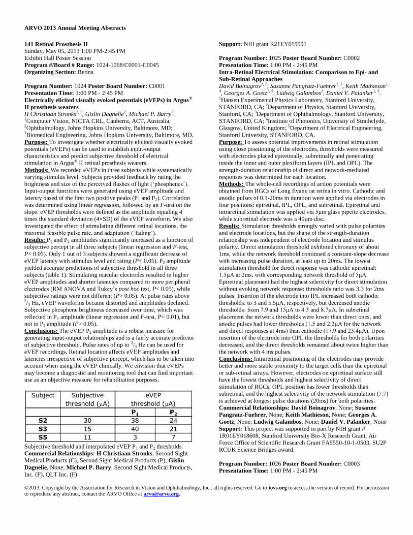
Subjective threshold and interpolated eVEP P1 and P2 thresholds.
Commercial Relationships: H Christiaan Stronks, Second Sight
Medical Products (C), Second Sight Medical Products (P); Gislin
Dagnelie, None; Michael P. Barry, Second Sight Medical Products,
Inc. (F), QLT Inc. (F)
Support: NIH grant R21EY019991
Program Number: 1025 Poster Board Number: C0002
Presentation Time: 1:00 PM - 2:45 PM
Intra-Retinal Electrical Stimulation: Comparison to Epi- and
Sub-Retinal Approaches David Boinagrov1, 2, Susanne Pangratz-Fuehrer1, 3, Keith Mathieson1,
4, Georges A. Goetz1, 5, Ludwig Galambos1, Daniel V. Palanker1, 3. 1Hansen Experimental Physics Laboratory, Stanford University,
STANFORD, CA; 2Department of Physics, Stanford University,
Stanford, CA; 3Department of Ophthalmology, Stanford University,
STANFORD, CA; 4Institute of Photonics, University of Strathclyde,
Glasgow, United Kingdom; 5Department of Electrical Engineering,
Stanford University, STANFORD, CA.
Purpose: To assess potential improvements in retinal stimulation
using close positioning of the electrodes, thresholds were measured
with electrodes placed epiretinally, subretinally and penetrating
inside the inner and outer plexiform layers (IPL and OPL). The
strength-duration relationship of direct and network-mediated
responses was determined for each location.
Methods: The whole-cell recordings of action potentials were
obtained from RGCs of Long Evans rat retina in vitro. Cathodic and
anodic pulses of 0.1-20ms in duration were applied via electrodes in
four positions: epiretinal, IPL, OPL, and subretinal. Epiretinal and
intraretinal stimulation was applied via 5μm glass pipette electrodes,
while subretinal electrode was a 40μm disc.
Results: Stimulation thresholds strongly varied with pulse polarities
and electrode locations, but the shape of the strength-duration
relationship was independent of electrode location and stimulus
polarity. Direct stimulation threshold exhibited chronaxy of about
1ms, while the network threshold continued a constant-slope decrease
with increasing pulse duration, at least up to 20ms. The lowest
stimulation threshold for direct response was cathodic epiretinal:
1.5μA at 2ms, with corresponding network threshold of 5μA.
Epiretinal placement had the highest selectivity for direct stimulation
without evoking network response: thresholds ratio was 3.3 for 2ms
pulses. Insertion of the electrode into IPL increased both cathodic
thresholds: to 3 and 5.5μA, respectively, but decreased anodic
thresholds: from 7.9 and 15μA to 4.3 and 8.7μA. In subretinal
placement the network thresholds were lower than direct ones, and
anodic pulses had lower thresholds (1.5 and 2.2μA for the network
and direct responses at 4ms) than cathodic (17.9 and 23.4μA). Upon
insertion of the electrode into OPL the thresholds for both polarities
decreased, and the direct thresholds remained about twice higher than
the network with 4 ms pulses.
Conclusions: Intraretinal positioning of the electrodes may provide
better and more stable proximity to the target cells than the epiretinal
or sub-retinal arrays. However, electrodes on epiretinal surface still
have the lowest thresholds and highest selectivity of direct
stimulation of RGCs. OPL position has lower thresholds than
subretinal, and the highest selectivity of the network stimulation (7.7)
is achieved at longest pulse durations (20ms) for both polarities.
Commercial Relationships: David Boinagrov, None; Susanne
Pangratz-Fuehrer, None; Keith Mathieson, None; Georges A.
Goetz, None; Ludwig Galambos, None; Daniel V. Palanker, None
Support: This project was supported in part by NIH grant #
1R01EY018608, Stanford University Bio-X Research Grant, Air
Force Office of Scientific Research Grant FA9550-10-1-0503, SU2P
RCUK Science Bridges award.
Program Number: 1026 Poster Board Number: C0003
Presentation Time: 1:00 PM - 2:45 PM

ARVO 2013 Annual Meeting Abstracts
©2013, Copyright by the Association for Research in Vision and Ophthalmology, Inc., all rights reserved. Go to iovs.org to access the version of record. For permission to reproduce any abstract, contact the ARVO Office at [email protected].
Comparison of Electrically Evoked Retinal Ganglion Cell (RGC)
Responses by Square Pulse and Triangle Pulse in rd1 mice Yongsook Goo1, 2, Kun No Ahn1, 2, Seol A Jae1, 2, Joo Yun Kim1, 2. 1Physiology, Chungbuk National Univ Med School, Cheongju,
Republic of Korea; 2Nano Artificial Vision Research Center, Seoul
National University Hospital, Seoul, Republic of Korea.
Purpose: Retinal prosthesis has been developed for the patients with
retinitis pigmentosa (RP) and age related macular degeneration
(AMD), and is regarded as the most feasible method to restore vision.
Extracting optimal electrical stimulation parameters for the prosthesis
is one of the most important elements for the success of retinal
prosthesis. Here, we used charge balanced biphasic current pulse and
we compared efficiency of three different pulse shapes evoking RGC
responses in rd1 mice.
Methods: The well-known animal model for RP, rd1 (Pde6brd1)
mice at postnatal 8 weeks were used (n=9). From the ex-vivo retinal
preparation, retinal waveforms were recorded with 8 × 8 MEA.
Biphasic current pulses in the form of cathodic phase-1st and anodic
phase-2nd were applied once per second (1 Hz, x50). We tested 3
different pulse shapes with same charge; 1) biphasic square pulse (I:
intensity, D: duration), 2) biphasic triangle pulse with intensity
doubled (2xI, D) to satisfy same charge requirement with square
pulse, 3) biphasic triangle pulse with duration doubled (I, 2xD) to
satisfy same charge requirement with square pulse. For intensity
modulation, duration of the pulse was fixed to 500 μs and the
intensities of the pulse were modulated from 5 to 40 μA. For duration
modulation, intensity of the pulse was fixed to 30 μA and the
durations of the pulse were modulated from 60 to 500 μs. The
electrically-evoked RGC spikes was defined as positive when the
number of RGC spikes for 400 ms after stimulus was 1.3 times
higher than that for 400 ms before stimulus in post-stimulus time
histogram.
Results: RGC responses were well modulated both with square pulse
and triangle pulse by varying the intensity and duration of the pulse.
Amplitude modulation shows that RGC response is preferentially
activated by biphasic triangle pulse with duration doubled especially
with 5 μA (p<0.001) and 10 μA (p<0.01). Duration modulation
shows that RGC response is preferentially activated by biphasic
triangle pulse with duration doubled especially with 60 and 100 μs
(p<0.001) and 200 μs (p<0.05).
Conclusions: Biphasic triangle pulse with duration doubled is always
efficient than square pulse and biphasic triangle pulse with intensity
doubled both in amplitude modulation and duration modulation.
1) Square Pulse 2) Triangle Pulse with Intensity Doubled and 3)
Triangle Pulse with Duration Doubled
Commercial Relationships: Yongsook Goo, None; Kun No Ahn,
None; Seol A Jae, None; Joo Yun Kim, None
Support: Korean MEST grant (2010-0020852 )
Program Number: 1027 Poster Board Number: C0004
Presentation Time: 1:00 PM - 2:45 PM
The Feasibility of Explanting a Suprachoroidal Electrode Array
in a Feline Model Ronald T. Leung1, 2, David A. Nayagam1, 2, Richard A. Williams2, 3,
Penelope J. Allen4, 5, Cesar M. Salinas-La Rosa3, Chi D. Luu4, 5,
Lauren N. Ayton4, 5, Meri Basa3, Robert K. Shepherd1, 2, Chris E.
Williams1, 2. 1Bionics Institute, East Melbourne, VIC, Australia; 2Faculty of Medicine, Dentistry and Health Sciences, The University
of Melbourne, Parkville, VIC, Australia; 3Department of Anatomical
Pathology, St. Vincent’s Hospital Melbourne, Fitzroy, VIC,
Australia; 4Royal Victorian Eye & Ear Hospital, East Melbourne,
VIC, Australia; 5Centre for Eye Research Australia, East Melbourne,
VIC, Australia.
Purpose: To determine whether chronically implanted
suprachoroidal electrode arrays can be safely explanted in a feline
model.
Methods: Six healthy subjects were unilaterally implanted with
suprachoroidal electrode arrays which were surgically explanted after
one month. Fundus photography and optical coherence tomography
(OCT) was performed pre- and post- operatively. The subjects were
overdosed two months post-explantation and the eyes were prepared
for histological study.
Results: All the arrays were explanted with no intra-operative
complications. OCT and fundus photography showed that the
tapetum and retina was disrupted near the tip of the implant within
two weeks of implantation and that explantation did not result in
further disruption. Staphylomas were observed in five subjects upon
macroscopic dissection. There was no change in the thickness of the
choroid, photoreceptor layer or inner retina compared to the
contralateral unimplanted control eye following array implantation or
explantation. Histological results showed that the morphology of the
retina was well preserved except for focal disruption of the tapetum
and photoreceptors near the optic disk which corresponded to the
same damaged regions observed in fundus photographs and OCT.
There was a minor foreign body response with mild episcleral acute

ARVO 2013 Annual Meeting Abstracts
©2013, Copyright by the Association for Research in Vision and Ophthalmology, Inc., all rights reserved. Go to iovs.org to access the version of record. For permission to reproduce any abstract, contact the ARVO Office at [email protected].
inflammation and mild chronic inflammation in the suprachoroidal
space.
Conclusions: The feasibility of explanting a suprachoroidal retinal
prosthesis has been demonstrated. This procedure can be safely
performed provided that there is good management of the scleral
wound. There was minimal damage to the globe and surrounding
tissues. These findings have important implications for
suprachoroidal array explantation in the clinical setting.
Commercial Relationships: Ronald T. Leung, None; David A.
Nayagam, None; Richard A. Williams, None; Penelope J. Allen,
Bionic Vision Australia (P); Cesar M. Salinas-La Rosa, None; Chi
D. Luu, None; Lauren N. Ayton, None; Meri Basa, None; Robert
K. Shepherd, None; Chris E. Williams, Bionic Vision Australia (P)
Support: The Bionics Institute and the Centre for Eye Research
Australia acknowledge the support they receive from the Victorian
Government through its Operational Infrastructure Support Program.
This research was supported by the Australian Research Council
(ARC) through its Special Research Initiative (SRI) in Bionic Vision
Science and Technology grant to Bionic Vision Australia (BVA) and
the Harold Mitchell Foundation. The Bionics Institute would also like
to acknowledge support from the Bertalli Family Trust.
Program Number: 1028 Poster Board Number: C0005
Presentation Time: 1:00 PM - 2:45 PM
Development of a surgical procedure for implanting a wide view
electrode array in the suprachoroidal space Jonathan Yeoh1, 3, Alexia Saunders2, David A. Nayagam2, 4, Chris E.
Williams2, 4, Mark McCombe1, 3, Burns Owen2, Joel Villalobos2,
Michelle McPhedran2, Robert Briggs1, 2, Penelope J. Allen1, 3. 1The
Royal Victorian Eye and Ear Hospital, Melbourne, VIC, Australia; 2Bionics Institute, Melbourne, VIC, Australia; 3Centre for Eye
Research Australia, Melbourne, VIC, Australia; 4Department of
Anatomical Pathology, The University of Melbourne, Melbourne,
VIC, Australia.
Purpose: The goal of this study was to develop the surgical
procedure for implanting a suprachoroidal retinal prosthesis in
patients. The preclinical implant was adapted to conform to human
orbital anatomy.
Methods: A surgical technique for suprachoroidal implantation of a
conformable electrode array (19 mm × 8 mm) and lead was
developed. Human cadavers (n = 5) were used to adapt the approach
which was previously used in a pre-clinical model . A method of
tunnelling the array forward from behind the pinna towards the orbit
was used, with a custom trocar. First, an anteriorly based C-shape
flap was created behind the pinna to position the percutaneous
pedestal. Next a lateral canthotomy was made. An orbitotomy was
drilled in the frontal process of the zygomatic bone, 10 mm below the
orbital suture line. The lead was anchored inside this notch with a
compression fit and by a conformable patch sutured onto the sclera.
The lateral rectus muscle was detached and a 9 mm full scleral
thickness incision was made 1-4 mm behind the muscle insertion
point, according to eye size. A pocket was created in the
suprachoroidal space. The moulded array was inserted into the
suprachoroidal space . The incision was closed and scleral anchor
point sutured.
Results: Dissection of the cadaver eyes confirmed that the retinal
prostheses were reliably positioned in the suprachoroidal space and
beneath the macula. A conformable anchor point sutured to the sclera
stabilised the transscleral lead exit. This anchor point was angled
accordingly to ensure a simple fit of the intraorbital lead with
minimal strain. Safe tunnelling was achieved with the use of a
tunnelling trocar beneath the temporalis facia. The 150 mm lead
reliably reached the pedestal behind the pinna.
Conclusions: A surgical approach for suprachoroidal prosthetic
implantation was developed for successful anatomical placement,
mechanical stability and safe tunnelling, to be used in subsequent
patient testing. Electrode impedance measurements and
psychophysics testing in patients have shown the retina to be
functional after uncomplicated surgery and several months of
implantation.
Commercial Relationships: Jonathan Yeoh, None; Alexia
Saunders, None; David A. Nayagam, None; Chris E. Williams,
Bionic Vision Australia (P); Mark McCombe, Bionic Vision
Australia (P); Burns Owen, Bionics Institute (P); Joel Villalobos,
The Bionics Institute of Australia (P); Michelle McPhedran, None;
Robert Briggs, Cochlear (C); Penelope J. Allen, Bionic Vision
Australia (P)
Program Number: 1029 Poster Board Number: C0006
Presentation Time: 1:00 PM - 2:45 PM
In Vivo Electrical Stimulation of a Retinal Prosthesis Containing
Conductive Diamond Electrodes Mohit N. Shivdasani1, David J. Garrett2, David A. Nayagam1, Joel
Villalobos1, Penelope J. Allen3, 4, Alexia Saunders1, Michelle
McPhedran1, Ceara McGowan1, Hamish Meffin5, 6, Robert K.
Shepherd1. 1Bionics Institute, East Melbourne, VIC, Australia; 2School of Physics, The University of Melbourne, Melbourne, VIC,
Australia; 3Centre for Eye Research Australia, East Melbourne, VIC,
Australia; 4Royal Victorian Eye & Ear Hospital, East Melbourne,
VIC, Australia; 5Victoria Research Laboratory, National ICT
Australia, Melbourne, VIC, Australia; 6Electrical & Electronic
Engineering, The University of Melbourne, Melbourne, VIC,
Australia.
Purpose: Nitrogen incorporated ultrananocrystalline diamond (N-
UNCD) has recently been shown to be a promising material as a
stimulating electrode. The aim of this study was to assess if N-UNCD
electrodes are capable of stimulating retinal ganglion cells (RGCs) in
vivo, at stimulus intensities considered safe for this material.
Methods: Hermetic arrays containing 120x120 µm N-UNCD
electrodes were fabricated on a polycrystalline diamond substrate and
embedded in a silicone carrier. A pars plana vitrectomy was
performed in five normally-sighted anesthetized cats. Implants were
inserted through a 5mm incision and fixed epiretinally using a
titanium tack. Impedances were measured before implantation and in
vivo. Multiunit responses to monopolar stimulation with biphasic
current pulses (500 µs per phase), were recorded from the visual
cortex using 60-electrode penetrating arrays (Blackrock
Microsystems, UT). Eyes were enucleated, fixed and examined
histologically for trauma to tissue surrounding the implant.
Results: Impedances in vivo (26.6±2 kΩ, Mean±SEM) did not differ
(p>0.05) to those measured in vitro (26.4±2.3 kΩ). Epiretinal
stimulation led to robust activation of the visual cortex with reliable
thresholds measured from stimulating 30 of 42 electrodes. Best
thresholds ranged between 29.5-442.6 µC/cm2. Cathodic first pulses
(54.9±2.8 nC) resulted in significantly lower (paired t-test, p<0.001)
thresholds than anodic first pulses (73±5.2 nC). Histological analysis
revealed focal damage to the retina surrounding the tacked edge of
the implant. The retina beneath the diamond array remained attached.
The extent of damage attributed to tacking alone versus the combined
pressure from the tack and silicone implant body was unclear.
Conclusions: For all but three electrodes, charge densities required to
evoke cortical responses were well within the previously established
electrochemical safe limit for diamond (300 µC/cm2). N-UNCD
electrodes were successfully used to acutely stimulate RGCs via an
epiretinal approach with some electrodes requiring very low
intensities to activate the visual cortex. Variability in impedances and
ARVO 2013 Annual Meeting Abstracts
©2013, Copyright by the Association for Research in Vision and Ophthalmology, Inc., all rights reserved. Go to iovs.org to access the version of record. For permission to reproduce any abstract, contact the ARVO Office at [email protected].
thresholds, along with histological analyses, suggest that further
optimization of the implant shape and tack insertion procedure is
required to consistently obtain low thresholds and minimize damage.
In addition, performance of these electrodes needs to be evaluated in
chronic studies.
Commercial Relationships: Mohit N. Shivdasani, None; David J.
Garrett, None; David A. Nayagam, None; Joel Villalobos, The
Bionics Institute of Australia (P); Penelope J. Allen, Bionic Vision
Australia (P); Alexia Saunders, None; Michelle McPhedran, None;
Ceara McGowan, None; Hamish Meffin, NICTA (P); Robert K.
Shepherd, None
Support: This work was supported by the Australian Research
Council through its Special Research Initiative in Bionic Vision
Science and Technology awarded to Bionic Vision Australia. The
Bionics Institute wishes to acknowledge the support it receives from
the Victorian Government through its Operational Infrastructure
Program.
Program Number: 1030 Poster Board Number: C0007
Presentation Time: 1:00 PM - 2:45 PM
A Suprachoroidal Retinal Prosthesis with a Flexible Lead is
Reliable for Patient Testing Joel Villalobos1, Penelope J. Allen2, 3, Chi D. Luu2, 3, Lauren N.
Ayton2, 3, Jonathan Yeoh2, 3, David A. Nayagam1, Nicholas L. Opie2, 3,
Mohit N. Shivdasani1, Robert K. Shepherd1, 4, Chris E. Williams1, 4. 1Bionics Institute, East Melbourne, VIC, Australia; 2Centre for Eye
Research Australia, East Melbourne, VIC, Australia; 3Royal
Victorian Eye and Ear Hospital, East Melbourne, VIC, Australia; 4Medical Bionics Department, University of Melbourne, East
Melbourne, VIC, Australia.
Purpose: A flexible intraorbital lead was developed to minimise risk
and simplify implantation of a retinal prosthesis. The microwire
based prosthesis was tested in a preclinical model, then optimised in
cadavers and tested in a clinical pilot study.
Methods: Initially, a suprachoroidal (SC) electrode array was
developed with a transscleral lead of 14-22 platinum microwires in
silicone. It was implanted in cats (n = 16) for 3 months. The lead was
sutured on the sclera with a silicone patch and tunnelled under
conjunctiva to a patch on the orbital rim. It followed either a straight
path (3 implants) of 12 mm; or a curved path (13 implants; Fig. 1) of
16 mm with 1 mm strain relief cones. Histological assessment was
performed on the tissue around the lead.
The lead routing was then fitted to a human orbit and tested in
cadavers. This lead, with strain relief cones, was 34 mm to the lateral
orbit where it was fitted inside a channel. Lead durability was then
tested in a mechanical model of a skull and eye moving to 25° of
abduction/adduction. A 24-channel electrode array with optimised
lead and a percutaneous connector (on the parietal bone) was then
implanted in a clinical pilot. The lead location was monitored with X-
ray and CT imaging.
Results: The SC implant and orbital lead were well tolerated in all 16
cat eyes, with no conjunctival or skin erosion around the eye. From
197 individually wired electrodes with a curved lead, 177 were
connected following 3 months of implantation; which contrasted with
24 connected out of 36 implanted using a straight lead (Chi-square P
< 0.001). Implant and lead were stable in all but one case where the
implant’s anterior end eroded the sclera. The typical tissue response
around the moving lead was a thick granulomatous fibrous capsule.
The leads in mechanical durability testing have undergone 65 million
eye movements with no wire breakages. In humans, the implant with
intraorbital lead was stable during the initial 6 months of implantation
(Fig. 2). All the electrodes remained connected.
Conclusions: The SC retinal implant with flexible lead allowed for
minimal surgical manipulation of the eye and distal placement of
larger components. The strain-relieved lead was reliable during
chronic implantation in a preclinical model and in a clinical pilot.
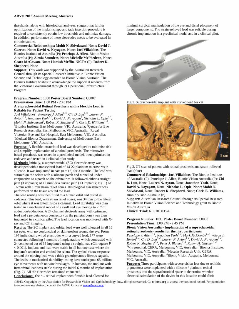
Fig 1. Suprachoroidal implant with curved lead for cat
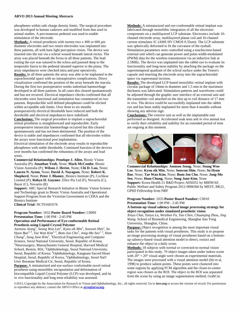
Fig 2. CT scan of patient with retinal prosthesis and strain-relieved
lead (blue)
Commercial Relationships: Joel Villalobos, The Bionics Institute
of Australia (P); Penelope J. Allen, Bionic Vision Australia (P); Chi
D. Luu, None; Lauren N. Ayton, None; Jonathan Yeoh, None;
David A. Nayagam, None; Nicholas L. Opie, None; Mohit N.
Shivdasani, None; Robert K. Shepherd, None; Chris E. Williams,
Bionic Vision Australia (P)
Support: Australian Research Council through its Special Research
Initiative in Bionic Vision Science and Technology grant to Bionic
Vision Australia
Clinical Trial: NCT01603576
Program Number: 1031 Poster Board Number: C0008
Presentation Time: 1:00 PM - 2:45 PM
Bionic Vision Australia - Implantation of a suprachoroidal
retinal prosthesis- results for the first participants Penelope J. Allen1, 4, Jonathan Yeoh1, 4, Mark McCombe1, 4, Wilson J.
Heriot1, 4, Chi D. Luu3, 4, Lauren N. Ayton3, 4, David A. Nayagam2, 4,
Robert K. Shepherd2, 4, Peter J. Blamey2, 4, Robyn H. Guymer3, 4. 1Vitreoretinal, CERA, Melbourne, VIC, Australia; 2Bionics Institute,
Melbourne, VIC, Australia; 3Macular Research Unit, CERA,
Melbourne, VIC, Australia; 4Bionic Vision Australia, Melbourne,
VIC, Australia.
Purpose: Three participants with severe vision loss due to retinitis
pigmentosa were implanted with a silicone / platinum retinal
prosthesis into the suprachoroidal space to determine whether
electrical stimulation of the device in this location could elicit
ARVO 2013 Annual Meeting Abstracts
©2013, Copyright by the Association for Research in Vision and Ophthalmology, Inc., all rights reserved. Go to iovs.org to access the version of record. For permission to reproduce any abstract, contact the ARVO Office at [email protected].
phosphenes within safe charge density limits. The surgical procedure
was developed in human cadavers and modified from that used in
animal studies. A percutaneous pedestal was used to enable
stimulation of the electrode.
Methods: A retinal prosthesis with twenty-two x 400 or 600 μm
diameter electrodes and two return electrodes was implanted into
three patients, all with bare light perception vision. The device was
inserted into the eye via a scleral wound beneath lateral rectus. The
array was placed beneath the fovea in all three patients. The lead
exiting the eye was sutured to the sclera and passsed deep to the
temporalis fascia to the pedestal situated superior to the pinna. The
array impedances were checked intra and post operatively.
Results: In all three patients the array was able to be implanted in the
suprachoroidal space with no intraoperative complications. Direct
visualization confirmed the position of the array beneath the macula.
During the first two postoperative weeks subretinal haemorrhage
developed in all three patients. In all cases this cleared spontaneously
and has not recurred. Electrical impedance checks confirmed that all
electrodes were functional and stimulation has commenced on two
patients. Reproducible well defined phosphenes could be elicited
within acceptable safe limits. Over three to six months
postoperatively electrical thresholds have reduced and both the
thresholds and electrical impedances have stabilised.
Conclusions: The surgical procedure to implant a suprachoroidal
retinal prosthesis is straightforward and reproducible. Early
postoperative intraocular haemorrhage occurred but this cleared
spontaneously and has not been detrimental. The position of the
device is stable and impedances confirmed that all electrodes within
the arrays were functional post implantation.
Electrical stimulation of the electrode array results in reproducible
phosphenes with stable thresholds. Continued function of the devices
over months has confirmed the robustness of the arrays and lead
wires.
Commercial Relationships: Penelope J. Allen, Bionic Vision
Australia (P); Jonathan Yeoh, None; Mark McCombe, Bionic
Vision Australia (P); Wilson J. Heriot, None; Chi D. Luu, None;
Lauren N. Ayton, None; David A. Nayagam, None; Robert K.
Shepherd, None; Peter J. Blamey, Bionics Institute (P), Cochlear
Limited (P); Robyn H. Guymer, Ellex Pty Ltd (F), Novartis (C),
Bayer (C), Novartis (R)
Support: ARC Special Research Initiative in Bionic Vision Science
and Technology grant to Bionic Vision Australia and Operational
Support Program from the Victorian Government to CERA and the
Bionics Institute
Clinical Trial: NCT01603576
Program Number: 1032 Poster Board Number: C0009
Presentation Time: 1:00 PM - 2:45 PM
Operation and Performance of Eye-conformable Retinal
Prosthesis using Liquid Crystal Polymer Joonsoo Jeong1, Seung Woo Lee2, Kyou sik Min1, Soowon Shin1, So
Hyun Bae3, 4, Tae Wan Kim3, 5, Bum-Joo Cho3, Jong-Mo Seo1, 3, Hum
Chung3, Sung June Kim1. 1Electrical Engineering and Computer
Science, Seoul National University, Seoul, Republic of Korea; 2Neurosurgery, Massachusetts General Hospital, Harvard Medical
School, Boston, MA; 3Ophthalmology, Seoul National University,
Seoul, Republic of Korea; 4Ophthalmology, Kangnam Sacred Heart
Hospital, Seoul, Republic of Korea; 5Ophthalmology, Seoul Nat'l
Univ Boramae Medical Ctr, Seoul, Republic of Korea.
Purpose: A miniaturized and eye-surface conformable novel retinal
prosthesis using monolithic encapsulation and deformation of
biocompatible Liquid Crystal Polymer (LCP) was developed, and its
in vivo functionality and long term reliability was assessed.
Methods: A miniaturized and eye-conformable retinal implant was
fabricated through monolithic integration of all the electronic
components on a multilayered LCP substrate. Electronics include 16-
channel electrode array, multilayered planar coil and 16-channel
current stimulator IC (AMS HV CMOS 0.35um). The LCP substrate
was spherically deformed to fit the curvature of the eyeball.
Stimulation parameters were controlled using a touchscreen-based
external unit which can generate power and pulse-width-modulated
(PWM) data for the wireless transmission via an inductive link at
2.5MHz. The device was implanted into the rabbit eye to evaluate its
functionality and long-term stability by attaching the package on the
superotemporal quadrant of the scleral surface under the Tenon's
capsule and inserting the electrode array into the suprachoroidal
space via superonasal incision.
Results: The developed LCP-based monolithic retinal implant with
circular package of 14mm in diameter and 1.5 mm in the maximum
thickness was fabricated. Stimulation patterns and waveforms could
be adjusted through the graphic user interface of an external unit and
the transmitter coil attached on the skin up to coil distance of 17 mm
in vivo. The device could be successfully implanted into the rabbit
eye and has been stably implanted for more than 4 months without
showing any adverse sign.
Conclusions: The exterior unit as well as the implantable unit
performed as designed. Accelerated soak tests and in vivo animal test
to verify their reliability and stability for an extended period of time
are ongoing at this moment.
Commercial Relationships: Joonsoo Jeong, None; Seung Woo
Lee, None; Kyou sik Min, None; Soowon Shin, None; So Hyun
Bae, None; Tae Wan Kim, None; Bum-Joo Cho, None; Jong-Mo
Seo, None; Hum Chung, None; Sung June Kim, None
Support: Korea Health 21 R&D Project A050251 by MIHWAF,
Public Welfare and Safety Program 2012-0006566 by MEST, BK21,
GPhD Fellowship from NRF
Program Number: 1033 Poster Board Number: C0010
Presentation Time: 1:00 PM - 2:45 PM
A bottom-up visual saliency-based image processing strategy for
object recognition under simulated prosthetic vision Xinyu Chai, Yanyu Lu, Weizhen Fu, Yao Chen, Chuanqing Zhou, Jing
Wang. School of Biomedical Engineering, Shanghai Jiao Tong
University, Shanghai, China.
Purpose: Object recognition is among the most important visual
tasks for the patients with visual prostheses. This study is to propose
an image processing strategy of visual prostheses based on a bottom-
up saliency-based visual attention model to detect, extract and
enhance the object in a daily scene.
Methods: 18 subjects with normal or corrected-to-normal vision
participated in this study. 70 object images taken under indoor scene
with 20° × 20° visual angle were chosen as experimental materials.
The images were processed with a visual attention model (Itti et al,
1998) to produce salient points. These points were clustered into
some regions by applying FCM algorithm and the closet-to-center
region was chosen as the ROI. The object in the ROI was separated
from the scene by using an image segmentation method, GrabCut.

ARVO 2013 Annual Meeting Abstracts
©2013, Copyright by the Association for Research in Vision and Ophthalmology, Inc., all rights reserved. Go to iovs.org to access the version of record. For permission to reproduce any abstract, contact the ARVO Office at [email protected].
We adopted two foreground/background contrast-enhancing
strategies to present images under simulated prosthetic vision
compared with a directly pixelization strategy (DP): the foreground
with 8 gray levels while the background with 4 lower gray levels (8-4
separated pixelization, 8-4 SP) and the foreground with 8 gray levels
while the background with edge extracted information (Background
edge extraction, BEE).
Results: Subjects achieved above chance (1/70 = 1.43%) recognition
accuracy (26.41 ± 8.83%) under DP condition. The two
foreground/background separated strategies, 8-4 SP (41.73 ± 7.29%)
and BEE (44.44 ± 7.70%), significantly increased the object
recognition accuracy compared with DP (P < 0.05); however, there
was no significant difference between these two strategies (P > 0.05).
70 objects were classified into 3 categories (perfect, good and bad )
according to Jaccard Coefficient (JC) which evaluated the
effectiveness of the object extraction from background. As JC
(segmentation quality) increased, the object recognition accuracy
significantly increased using 8-4 SP and BEE strategies (P < 0.05),
while there was no significant difference among the categories using
DP strategy.
Conclusions: The results showed the foreground/background
contrast-enhancing strategies based on a saliency-based visual
attention model can significantly improve the recognition accuracy of
objects under daily scenes. We hope our study on image processing
strategies will be helpful to the future design and development of
visual prosthesis to restore functional vision for the blinds.
Commercial Relationships: Xinyu Chai, None; Yanyu Lu, None;
Weizhen Fu, None; Yao Chen, None; Chuanqing Zhou, None;
Jing Wang, None
Support: The National Basic Research Program of China (973
Program, 2011CB7075003/2); The National Natural Science
Foundation of China (61273368, 91120304)
Program Number: 1034 Poster Board Number: C0011
Presentation Time: 1:00 PM - 2:45 PM
Immunohistochemical and electrophysiological analysis of rat
retinas after subretinal implantation of photovoltaic arrays Jacob G. Light1, 2, James W. Fransen3, Alice Adkins2, Gobinda
Pangeni4, James Loudin5, 6, Keith Mathieson5, 7, Daniel V. Palanker5,
6, Maureen A. McCall3, 4, Machelle T. Pardue1, 2. 1Ophthalmology,
Emory University, Atlanta, GA; 2Rehab Center of Excellence,
Atlanta VA Medical Center, Atlanta, GA; 3Anatomical Sciences &
Neurobiology, University of Louisville, Louisville, KY; 4Ophthalmology & Visual Sciences, University of Louisville,
Louisville, KY; 5Hansen Experimental Physics Laboratory, Stanford
University, Stanford, CA; 6Ophthalmology, The Byers Eye Institute
at Stanford University, Palo Alto, CA; 7Santa Cruz Institute for
Particle Physics, University of California, Santa Cruz, Santa Cruz,
CA.
Purpose: Network-mediated retinal stimulation by subretinal
prostheses may depend on a functional retinal interface. Excessive
glial scarring or the loss of inner retinal neurons at the implant site
could reduce or eliminate implant-evoked signaling. In this study,
immunohistochemical evaluation of the inner retina was compared to
electrophysiological recordings of implant-evoked responses in the
superior colliculus (SC) to assess signal transmission and retinal
health after implantation of photovoltaic arrays (PVAs) in the
transgenic S334ter-3 rat model of retinitis pigmentosa.
Methods: Silicon-based PVAs were implanted monocularly into the
subretinal space of S334ter-3 rats at 7 or 12 weeks of age. In an intact
animal preparation at 18 to 27 weeks post-implantation, arrays were
stimulated with IR light (905 nm) at locations on the implant surface
and evoked responses were mapped across the dorsal surface of the
SC. Immediately after, animals were sacrificed and eyes were
enucleated, fixed, and processed for immunohistochemistry. Retinal
sections were labeled with primary antibodies against ChAT, PKCα,
and GFAP with fluorescent secondary antibodies and visualized with
confocal microscopy. The morphology of cells in regions
immediately contacting the PVA was compared with areas adjacent
to and distant from the implant site.
Results: In most implanted animals, PVA-evoked responses were
recorded in the SC. PKCα and ChAT labeling showed preservation of
rod bipolar cells and cholinergic amacrine cells, respectively, in and
around the implant site. In fully subretinal implantations, amacrine
cell lamination patterns were well-maintained in the IPL. Similarly,
rod bipolar cells were robustly labeled, and their axon terminal
locations were consistent across the retina. GFAP labeling was
widespread in all retinal areas, indicating a general glial reaction, but
showed little or no additional glial scarring at the implant site.
Conclusions: PVA-evoked responses in the SC indicate that the
implanted degenerate retina is still able to pass signals to higher
visual structures. That rod bipolar and cholinergic amacrine cells
exhibit well-preserved morphology indicates that they should be able
to contribute to retinal signal transmission. Glial reactivity is uniform
across the retina and is unlikely to interfere with signaling.
Commercial Relationships: Jacob G. Light, None; James W.
Fransen, None; Alice Adkins, None; Gobinda Pangeni, None;
James Loudin, None; Keith Mathieson, None; Daniel V. Palanker,
None; Maureen A. McCall, None; Machelle T. Pardue, None
Support: NIH NEI EY018608; NIH P30 EY006360; Department of
Veterans Affairs; Departmental Award from Research to Prevent
Blindness (Emory and UoL)
Program Number: 1035 Poster Board Number: C0012
Presentation Time: 1:00 PM - 2:45 PM
Physiological Response of RD Mouse Retinal Ganglion Cells to
Electrical Stimulation Alice Cho1, Alapakkam P. Sampath1, 3, Mark S. Humayun1, 2, James
D. Weiland1, 2. 1Biomedical Engineering, University of Southern
California, Los Angeles, CA; 2Ophthalmology, University of
Southern California, Los Angeles, CA; 3Physiology and Biophysics,
University of Southern California, Los Angeles, CA.
Purpose: The aim of this study was to assess how the intrinsic
properties of retinal ganglion cells (RGC) in a mouse model of retinal
degeneration affect thresholds to electrical stimulation.
Methods: The animal model used for this study was the rd10 mouse,
aged 6-10 weeks. The rd10 mutation is caused by a missense
mutation on the gene encoding the PDE-β subunit and results in
almost complete loss of photoreceptors by the end of two months.
Spontaneous baseline activity and spikes elicited by external
stimulation were recorded from RGCs using whole-cell patch clamp.
The stimulating electrode was a 75 μm diameter Pt-Ir disk positioned
approximately 50 μm above and 50 μm laterally from the targeted
RGC; the ground electrode was positioned behind the retina on the
photoreceptor side. Charge-balanced biphasic square pulses
(cathodic-phase first, 500 μs/phase) were delivered at 10 Hz
frequency.
Results: For each ganglion cell, measurements for threshold, soma
diameter, resting membrane potential, spontaneous firing rate, and
presence/absence of rebound excitation were recorded. Since a subset
of rd10 RGCs exhibited a high rate of spontaneous activity that was
not observed in wildtype mice, cells were classified into two groups -
low rate (spontaneous rate < 10 Hz) and high rate (spontaneous rate ≥
10 Hz) - based on their baseline activity. RGCs with high
spontaneous rates had significantly lower electrical thresholds than
cells with lower spontaneous rates (μlow = 46.66 μA, μhigh = 27.67 μA,

ARVO 2013 Annual Meeting Abstracts
©2013, Copyright by the Association for Research in Vision and Ophthalmology, Inc., all rights reserved. Go to iovs.org to access the version of record. For permission to reproduce any abstract, contact the ARVO Office at [email protected].
p = 0.0423); the relationships between threshold/resting membrane
potential and spontaneous rate were not significant. RGCs were also
functionally classified into ON or OFF cells based on their ability to
exhibit rebound excitation (D.J. Margolis, 2007). OFF ganglion cells
had higher spontaneous firing rates than ON ganglion cells (μOFF =
4.93 Hz, μON = 0.238 Hz, p = 0.0114) but their threshold and resting
potential measurements were not statistically different.
Conclusions: OFF ganglion cells appear to exhibit higher rates of
spontaneous activity compared to ON ganglion cells and RGCs with
higher spontaneous rates had lower electrical thresholds than cells
with lower rates. These results suggest that intrinsic spontaneous
activity may increase sensitivity to extracellular stimulation and may
enable OFF RGCs to be selectively stimulated at lower thresholds
than ON RGCs due to their elevated spontaneous firing rates.
Commercial Relationships: Alice Cho, None; Alapakkam P.
Sampath, None; Mark S. Humayun, Bausch & Lomb (F), Bausch
& Lomb (C), Bausch & Lomb (P), Bausch & Lomb (R), Bausch &
Lomb (S), Alcon (C), Alcon (R), Iridex (P), Iridex (R), Replenish (I),
Replenish (C), Replenish (R), Replenish (S), Second Sight (F),
Second Sight (I), Second Sight (C), Second Sight (P), Second Sight
(R), Second Sight (S), Regenerative Patch Technologies (I),
Regenerative Patch Technologies (C); James D. Weiland, Second
Sight Medical Products, Inc. (F)
Support: NSF EEC-0310723; Research to Prevent Blindness, W.M.
Keck Foundation
Program Number: 1036 Poster Board Number: C0013
Presentation Time: 1:00 PM - 2:45 PM
Re-alignment and explantation of subretinal prostheses: surgical
aspects and proteomic analyses Florian Gekeler1, Helmut G. Sachs2, Veronique Kitiratschky1,
Katarina Stingl1, Udo Greppmaier3, Eberhart Zrenner1, Karl-Ulrich
Bartz-Schmidt1, Marius Ueffing1, Sascha Dammeier1. 1Centre for
Ophthalmology, Tuebingen Univ Eye Hosp, Tuebingen, Germany; 2Ophthalmology, Staedtisches Klinikum Friedrichstadt, Dresden,
Germany; 3Retina Implant AG, Reutlingen, Germany.
Purpose: Active subretinal implants are highly complex, constantly
improved devices requiring sophisticated surgical procedures. Under
certain circumstances corrections, such as realignments,
explantations, and reimplantations become necessary. Intraocular
tissue reactions to devices, e.g. from scleral, retinal or RPE cells,
have been described on a histologic (Gekeler, ARVO 2010) but not
on a proteomic level.
Methods: 34 patients have been implanted with a subretinal MPDA;
2 patients underwent re-alignment (avg. 1.5), 10 patients after the
pilot trial had explantation (avg. 9.5), and 1 re-implantation surgery
(2.5, all in months). 3 removed implants were analyzed using liquid
chromatography coupled to electrospray Orbitrap mass spectrometry
(nHPLC-MS/MS) following limited proteolysis of the proteins that
covered the surfaces.
Results: Longer intervals since implantation resulted in more
scarring around the polyimide (PI) foil on the sclera but no adhesion
was found; implants were retrieved in 2-3 pieces, unharmed for
technical analyses. PI foils and MPDAs were pulled out without
silicone oil exchange. One patient (with initial subretinal
hemorrhage) suffered from retinal detachment after explantation
requiring oil exchange. Retinas over the MPDA remained unaltered
in funduscopy and SD-OCT. In case of the first explant, proteomic
analysis exhibited more than 900 protein identifications over the
MPDA and 364 on the section of the implant on the sclera. Besides a
huge overlap of common housekeeping, e.g. GAP-dehydrogenase and
aldolase, and plasma proteins like serotransferrin a set of eye-, and/or
retina-specific proteins like alpha-crystallin, beta arrestin and retinol-
converting enzymes were identified from the chip section. All
sections of the explanted device showed slight coverage by
immunoreactive proteins like immunglobulins lambda and J,
integrins alpha and beta, and by complement factors.
Conclusions: Explantation and reimplantation surgery is feasible in
subretinal implants as tissue reactions in the subretinal space are
minimal. Proteomic analyses of explanted subretinal MPDAs
revealed common protein sets on the surface as reported for other
types of implants, e.g. silicone devices, reflecting both, minor
irritation of the surrounding tissue and the body’s tendency to coat
implants with an extracellular matrix. Proteomics can be valuable to
improve biocompatibility tests in retinal implants.
Commercial Relationships: Florian Gekeler, Retina Implant AG
(F), Okuvision GmbH (F), Retina Implant AG (C), Retina Implant
AG (P); Helmut G. Sachs, Retina Implant AG (R), Retina Implant
AG (C); Veronique Kitiratschky, None; Katarina Stingl, Retina
Implant AG (F); Udo Greppmaier, Retina Implant AG (E),
Okuvision GmbH (E); Eberhart Zrenner, Retina Implant AG (F),
Retina Implant AG (I), Retina Implant AG (C), Retina Implant AG
(P), QLT Inc (C), Servier, Paris (C), Steinbeis GmbH&CoKG,
Stuttgart (I), Steinbeis GmbH&CoKG, Stuttgart (C), Neurotech, USA
(C), Pfizer, USA (C); Karl-Ulrich Bartz-Schmidt, Retina Implant
(P); Marius Ueffing, None; Sascha Dammeier, None
Support: Retina Implant AG, Reutlingen, Germany
Clinical Trial: NCT00515814, NCT01024803, NCT01497379
Program Number: 1037 Poster Board Number: C0014
Presentation Time: 1:00 PM - 2:45 PM
Long-term Reliability of Argus® II Retinal Implants David Zhou, Alexander Istomin, Amy Hines, Alina Agazaryan,
Charles A. Byers, James Little, Brian V. Mech, Robert J. Greenberg.
Second Sight Medical Products, Inc, Sylmar, CA.
Purpose: Retinal prostheses are susceptible to damage by body fluids
over time. Long-term reliability of retinal prostheses requires
hermetic packaging to protect the electronic circuitry of the implant
from the harsh environment of the human body and a robust high-
density electrode array for safe chronic stimulation. In addition to
lifetime testing under normal use conditions, accelerated lifetime
testing has been widely used to predict the implants’ life and to better
understand their failure modes.
Methods: Lifetime testing of the Argus II implant has been
conducted at the component, subsystem and final device levels.
Long-term stability of the implants is assessed in vitro through active
soak tests under constant pulse stimulation. The implants are tested in
buffered saline at body temperature, or elevated temperatures for
accelerated tests. The implants are attached to the silicone eye model
to simulate the actual implanted condition. In some tests, a motor
moves the entire eye model to simulate micromotion of a human eye.
The device functionality, visual appearance, and material changes are
monitored through the course of the lifetime test.
Results: Electrode material lifetime has reached 7 years in real-time
testing under constant pulse stimulation and predicted equivalent of
over 50 years of use in accelerated testing. Thin-film polymer
electrode array insulation has reached 7 years in real-time testing and
an equivalent lifetime of over 26 years in accelerated testing.
Finished implants have reached more than 10 year lifetime in
accelerated testing. These bench-test results are supported by clinical
trial data: 30 subjects have been implanted an average of 4.2 years
(range 3.3 to 5.5, excluding one explant at 18 months). Cumulatively,
this represents 125 patient-years with only one device failure at 4
years post-implant.
Conclusions: The Argus II Retinal Implant has been tested at the
component and system levels for long-term reliability. Thin-film

ARVO 2013 Annual Meeting Abstracts
©2013, Copyright by the Association for Research in Vision and Ophthalmology, Inc., all rights reserved. Go to iovs.org to access the version of record. For permission to reproduce any abstract, contact the ARVO Office at [email protected].
electrode arrays withstood aggressive constant pulse stimulation and
provided long-term safe stimulation without corrosion or material
degradation. The hermetic package demonstrated the functional
lifetime of the implant by preventing the moisture level accumulated
inside the device. Lifetime tests support long-term reliability of
Argus II Retinal Implants. Such long-term reliability is of paramount
concern with respect to regulatory approval, and clinical utility,
safety, and adoption.
Commercial Relationships: David Zhou, Second Sight Medical
Products (E); Alexander Istomin, Second Sight Medical Products
(E); Amy Hines, Second Sight Medical Products (E); Alina
Agazaryan, Second Sight Medical Products, Inc (E); Charles A.
Byers, None; James Little, Second Sight Medical Products Inc (I),
Second Sight Medical Products Inc (E), Second Sight Medical
Products Inc (P); Brian V. Mech, Second Sight (E), Second Sight (I),
Second Sight (P); Robert J. Greenberg, Second Sight Medical
Products, Inc. (I), Second Sight Medical Products, Inc. (E), Second
Sight Medical Products, Inc. (P), Second Sight Medical Products, Inc.
(S)
Support: NIH grant EY012893
Program Number: 1038 Poster Board Number: C0015
Presentation Time: 1:00 PM - 2:45 PM
Morphological comparisons of flat and 3-dimensional subretinal
photovoltaic arrays in rat and pig models of retinitis pigmentosa Alice Adkins1, Wei Wang2, Henry J. Kaplan2, Douglas Emery2, Juan
P. Fernandez de Castro2, Sang-Joon Lee2, 3, Philip Huie4, 5, Daniel V.
Palanker4, 5, Maureen A. McCall2, 6, Machelle T. Pardue1, 7. 1Research R&D Service, Atlanta VAMC, Decatur, GA; 2Ophthalmology & Visual Sciences, University of Louisville,
Louisville, KY; 3Ophthalmology, Kosin University,College of
Medicine, Busan, Democratic People's Republic of Korea; 4Ophthalmology, Stanford University, Stanford, CA; 5Hansen
Experimental Physics LaboratorySciences & Neurobiology, Stanford
University, Stanford, CA; 6Anatomical Sciences & Neurobiology,
University of Louisville, louisville, KY; 7Ophthalmology, Emory
University, Decatur, GA.
Purpose: Retinal prosthetics are designed to restore vision to patients
with photoreceptor degeneration. Theoretically, close proximity of
inner retinal cells to stimulating electrodes is needed for their optimal
activation. We evaluated the morphology of retinas with subretinal
inactive polymer arrays (IPAs) containing gaps between the pixels or
pillar electrodes. In addition, we compared IPAs to active silicon
photovoltaic arrays (PVAs) with only gaps.
Methods: IPAs or PVAs were implanted into the subretinal space of
P23H-1 rats (8-11 wks of age) and P23H-1 transgenic and wildtype
(WT) pigs (6-14 wks age). Implanted IPAs were either: (1) flat
devices with 70 or 140 micron pixels separated by 5 or 10 micron
gaps or (2) vertical pillar devices of various densities but without
gaps. PVAs had a similar flat design. SD-OCT images visualized the
placement of the implant 2 weeks after implantation and
subsequently at regular biweekly intervals to monitor changes in
retinal morphology. At the end of the experiment, rat (8 and 16 wks
post-implantation) and pig eyes (2, 4 and 8 wks post-implantation)
were enucleated and fixed. The tissue under the implant was
examined in histological sections for migration of inner retinal cells
through the implant gaps and any other morphological changes and
compared to tissue near and distal to the implant.
Results: IPAs with 10 micron gaps showed significant migration of
inner retinal cells to the RPE-side of the implant compared to 5
micron gap devices (36% vs 15%, p<0.001). Similar to IPAs, PVAs
with 5 micron gaps showed little migration in rats or pigs. Surgical
implantation of pillar devices was significantly more difficult than
flat devices and created greater tissue trauma and glial reactions. In
38% of the cases, the ends of some pillars protruded through the rat
retina over time.
Conclusions: The retinal response to PVAs or IPAs is similar. Ten
micron gaps created significant inner retinal cell migration while 5
micron gaps allow close proximity of the inner retina to the device
without excessive migration through the gaps. Flat devices with gaps
are much easier to implant, they produced less trauma than the pillar
arrays, and results in better inner retinal proximity.
Commercial Relationships: Alice Adkins, None; Wei Wang,
None; Henry J. Kaplan, None; Douglas Emery, None; Juan P.
Fernandez de Castro, None; Sang-Joon Lee, None; Philip Huie,
None; Daniel V. Palanker, None; Maureen A. McCall, None;
Machelle T. Pardue, None
Support: Departmental grant from Research to Prevent Blindness
(Emory and UofL), NIH P30 EY006360, Department of Veterans
Affairs; NIH NEI EY018608
Program Number: 1039 Poster Board Number: C0016
Presentation Time: 1:00 PM - 2:45 PM
Combining holographic stimulation with cellular resolution
imaging in the rodent eye Shy Shoham1, Adi Schejter1, Limor Tsur1, Inna Reutsky-Gefen2, 1,
Nairouz Farah1. 1Biomedical Engineering, Technion, IIT, Haifa,
Israel; 2Medical Engineering, Ruppin Academic Center, Emek Hefer,
Israel.
Purpose: Optical retinal prostheses for patients with outer-retinal
degenerative diseases could interface directly with surviving retinal
neurons in order to mimic the normal input obtained from
photoreceptors in healthy retinas. Recently, we introduced an
artificial photo-stimulation technique based on the projection of
holographic patterns with high spatio-temporal resolution onto
optogentic probes, to selectively control large retinal neuronal
populations in the isolated retina. Here, we explore the ability to
target single optogenetic-expressing retinal ganglion cells (RGCs)
with holographic patterns at a cellular resolution, in-vivo.
Methods: For image-guided neuronal targeting, we constructed a
system that combines precise spatiotemporal holographic photo-
stimulation with high resolution fundus imaging. The system is also
integrated with a multiphoton microscope to enable functional
imaging of the responses to artificial stimulation.
Results: The system was utilized to acquire both brightfield and
fluorescence fundus images of mice and rats in-vivo, enabling the
identification of single fluorescent RGCs for stimulation.
Holographic patterns were projected onto the rodents’ retinas and
imaged. The stimulation spot diameter is sufficient for cellular
targeting using patterned photo-stimulation.
Conclusions: Our system enables single-cell resolved patterned
holographic photo-stimulation of RGCs in-vivo which will enable the
further development of a novel optical retinal prosthesis.
Commercial Relationships: Shy Shoham, None; Adi Schejter,
None; Limor Tsur, None; Inna Reutsky-Gefen, None; Nairouz
Farah, None
Support: European Research Council (grant #211055)
Program Number: 1040 Poster Board Number: C0017
Presentation Time: 1:00 PM - 2:45 PM
Argus® II Retinal Prosthesis System safety profile in post-
market patients Robert J. Greenberg1, Peter Walter3, Albert J. Augustin6, Bernd
Kirchhof4, Lyndon daCruz2, Hannah Schimitzek3, Gernot Roessler3,
Stanislao Rizzo5. 1Second Sight Medical Products, Inc, Sylmar, CA; 2The John Saunders Suite, Moorfields Eye Hospital, London, United
ARVO 2013 Annual Meeting Abstracts
©2013, Copyright by the Association for Research in Vision and Ophthalmology, Inc., all rights reserved. Go to iovs.org to access the version of record. For permission to reproduce any abstract, contact the ARVO Office at [email protected].
Kingdom; 3Department of Ophthalmology, RWTH Aachen
University, Aachen, Germany; 4Dept. of Ophthalmology, University
Clinic Koeln, Koeln, Germany; 5Eye Surgery Dept., Azienda
Ospedaliera Universitaria Pisana, Pisa, Italy; 6Dept. of
Ophthalmology, City Hospital of Karlsruhe, Karlsruhe, Germany.
Purpose: The Argus II Retinal Prosthesis System (Argus II) is the
first and only artificial retina approved for market use (2011 CE mark
in Europe, US FDA approval pending) and has since been implanted
in 16 severely vision impaired retinitis pigmentosa patients in 5
surgical centers in Italy, Germany and the UK. A post-market
surveillance study has been initiated with the purpose to evaluate the
safety profile of the approved device during commercial use
compared to that observed in the investigational clinical study.
Methods: Safety data has been collected from the day of surgery to
early December 2012, covering on average 6.2 months (median 6.6
months, range from 2 days to 13.3 months; 8.2 patient years) of
exposure. Vigilance requirements have ensured complete capture of
all safety related events in all implanted patients, independently from
their participation in the post market study. The demographic
distribution is: 6 female and 10 male patients, average age 50.8 years
(range 31.0 - 74.0 years); 9 OD and 7 OS implanted eyes.
Results: No surgical procedure or device-related serious adverse
event has occurred to date.Ten patients experienced no surgery- or
device-related adverse events. The other six patients experienced 7
surgery related non-serious adverse events, which occurred between
the day of surgery up to 1 month post operatively: two instances of
IOP elevation, and one each of nausea, vomiting, fainting,
conjunctival irritation, and retinal tear (occurred during surgery when
a retinal membrane was peeled prior to placing the implant). The first
5 events resolved between the same day and 9 days with medical
treatment; the last two are ongoing. One patient also experienced
device induced headache that is mitigated by adjustment of the device
settings.
Conclusions: The first group of Argus II patients using the
commercially available device demonstrates a safety profile that is at
6 months post implantation markedly better than that observed in the
developmental phase of Argus II.
Commercial Relationships: Robert J. Greenberg, Second Sight
Medical Products, Inc. (I), Second Sight Medical Products, Inc. (E),
Second Sight Medical Products, Inc. (P), Second Sight Medical
Products, Inc. (S); Peter Walter, Novartis (R), Bayer (R), Second
Sight (R), Bayer (F), Novartis (F); Albert J. Augustin, 2nd sight (F),
Alcon (C), Allergan (C), Novartis (F); Bernd Kirchhof, None;
Lyndon daCruz, Second Sight Medical products Inc. (R); Hannah
Schimitzek, None; Gernot Roessler, None; Stanislao Rizzo, None
Program Number: 1041 Poster Board Number: C0018
Presentation Time: 1:00 PM - 2:45 PM
Simulating current focusing and steering in penetrative optic
nerve stimulation: a computational model Qiushi Ren1, Menghui Li1, Yan Yan2, Liming Li2. 1Biomedical
Engineering, Peking University, Beijing, China; 2Biomedical
Engineering, Shanghai Jiao Tong University, Shanghai, China.
Purpose: A visual prosthesis based on penetrative optic nerve (ON)
stimulation is a potentail way for vision recovery. The fact that an
enormous number of RGC axons of various diameters are tightly
packed in mammalian optic nerve makes it difficult to implant more
numbers of stimulating electrodes. Here we presented a
computational model to simulate and evaluate two widely used field
shaping strategies, current steering and current focusing, in
penetrative stimulation of rabbit ON.
Methods: Finite element models with were established to compute
the 3D electric potential distribution generated with different
stimulating parameters under 2 and 3-electrode configurations. Then
the external electric potential was fed to a large number of
randomized multi-compartment cable models to calculate the
membrane potential of each ON fibre to predict whether AP could be
elicited in each sampled position. Finally a statistical process was
conducted to quantify the recruitment region and three indicators
were statistically derived to evaluate effects of current steering and
current focusing.
Results: The shifting rate (SR) value of the 2-electrode configuration
showed consistent correlation to the steering coefficient, whereas no
such significant correlation could be detected in that of the 3-
electrode configuration. For the 3-electrode configuration, large
values of compensation coefficient resulted in a large recruitment
area in the (ON) under arbitrary stimulus amplitude, which was
contrary to the intention of current focusing. Both configurations
demonstrated rather localized neural fibre recruitment compared with
the surface ON stimulation.
Conclusions: The simulation results show that the 2-electrode
configuration excels in current steering whereas the 3-electrode
configuration performs poorly in both current steering and focusing.
The localized recruitment of both configurations implies that current
focusing might be unnecessary in penetrative optic nerve stimulation.
Commercial Relationships: Qiushi Ren, None; Menghui Li, None;
Yan Yan, None; Liming Li, None
Support: The National Basic Research Program of China (973
Program, 2011CB707502), the National Natural Science Foundation
of China (61171174, 60971102).
Program Number: 1042 Poster Board Number: C0019
Presentation Time: 1:00 PM - 2:45 PM
Retinal Prostheses: Functional Use of Monocular Depth
Perception in the Low Resolution Limit Noelle R. Stiles1, Ben P. McIntosh2, Armand R. Tanguay3, Mark S.
Humayun4. 1Computation and Neural Systems Program, California
Institute of Technology, Pasadena, CA; 2Electrical Engineering-
Electrophysics, University of Southern California, Los Angeles, CA; 3Electrical Engineering-Electrophysics, Biomedical Engineering, and
Ophthalmology, University of Southern California, Los Angeles, CA; 4Ophthalmology, Cell and Neurobiology, and Biomedical
Engineering, University of Southern California, Los Angeles, CA.
Purpose: Monocular depth perception (using cues such as
perspective and relative size) persists to very low resolution in both
static and dynamic images (at video frame rates), with potential
implications for limited resolution retinal prostheses that are currently
implanted in only one eye. An image depth-rating task previously
demonstrated a significant improvement in depth perceived by
Gaussian filtering of pixellated images that exhibit false depth cues
(N=11). The depth-rating task also showed that depth could be
perceived even at very low resolutions (e.g., 8 x 10 pixels or
electrodes) when dynamic depth cues (such as motion parallax) were
present. This study investigates the degree to which image
representation and resolution affects the performance of functional
tasks. Furthermore, the role of foveation in improving the
performance of such tasks at low resolution with restricted field-of-
view was studied. Foveation may improve the functionality of retinal
prostheses that currently use a head-mounted camera without eye-
tracking capability, and can be implemented either with an
intraocular camera or with eye-tracking and a wide field-of-view
scene camera.
Methods: A functional reaching task with a head-mounted display
(HMD) was used to determine monocular depth perception
capabilities at low resolution with different image representations.
Images were acquired from a head-mounted wide-field-of-view

ARVO 2013 Annual Meeting Abstracts
©2013, Copyright by the Association for Research in Vision and Ophthalmology, Inc., all rights reserved. Go to iovs.org to access the version of record. For permission to reproduce any abstract, contact the ARVO Office at [email protected].
camera, processed to low resolution (pixellation and blur or
pixellation alone), and then displayed in real time within the HMD.
In the eye-pointed mode, the subject’s gaze directed the subregion of
the wide-field-of-view (FOV) image displayed to the user; head
position alone determined the subregion of the image displayed in the
head-pointed mode.
Results: Subjects (N=3) were able to reach and grasp a bottle while
avoiding obstacles with pixellated and blurred images in the eye-
pointed mode faster than with pixellated images in either mode. The
task with 32 x 32 pixels, Gaussian post-pixellation blurred and eye-
pointed took on average 5.3 seconds, as compared with 2.4 seconds
on average with normal vision.
Conclusions: Results show that appropriate presentation of images as
well as the implementation of foveation in retinal prostheses improve
the efficiency of depth task performance. The functional task also
affirms that subjects with a retinal prosthesis may be able to perceive
depth monocularly.
Commercial Relationships: Noelle R. Stiles, Patent (not licensed)
(P); Ben P. McIntosh, None; Armand R. Tanguay, University of
Southern California (P); Mark S. Humayun, Bausch & Lomb (F),
Bausch & Lomb (C), Bausch & Lomb (P), Bausch & Lomb (R),
Bausch & Lomb (S), Alcon (C), Alcon (R), Iridex (P), Iridex (R),
Replenish (I), Replenish (C), Replenish (R), Replenish (S), Second
Sight (F), Second Sight (I), Second Sight (C), Second Sight (P),
Second Sight (R), Second Sight (S), Regenerative Patch
Technologies (I), Regenerative Patch Technologies (C)
Support: National Science Foundation, National Science Foundation
Graduate Research Fellowship
Program Number: 1043 Poster Board Number: C0020
Presentation Time: 1:00 PM - 2:45 PM
Relative efficiency of voltage vs. current pulses for retinal
stimulation Navya S. Davuluri1, Mark S. Humayun2, 1, James D. Weiland2, 1. 1Biomedical Engineering, University of Southern California, Los
Angeles, CA; 2Ophthalmology, Doheny Eye Institute, Los Angeles,
CA.
Purpose: This in-vivo study is aimed at comparing supra-threshold
responses of rat retinal ganglion cells (RGC) to rectangular current
controlled and rectangular voltage controlled pulses. This is
motivated by the need to determine efficient stimulation strategies for
an electronic retinal prosthesis.
Methods: In 10 Long-Evans rats, we inserted a Pt/Ir electrode (D=
75 μm) in the left eye and exposed the right SC. The left retina was
stimulated with current controlled and voltage controlled pulses of
varying pulse width and amplitude. A large needle electrode distant
from the eye was used as a current return. While stimulating the
retina, evoked potentials were recorded from the superior colliculus
(SC), a midbrain structure in the visual pathway. The pulse widths
were 0.3 ms, 0.5 ms, 1 ms and 2 ms. For each pulse width, 4
amplitudes between 10 and 60 nC were applied in both current and
voltage mode.
Results: Power in the evoked potentials recorded from the SC was
used to determine stimulation efficiency. In general, whether current
controlled or voltage controlled pulses are more efficient depends on
the stimulus pulse width. For 0.3 ms pulse width, at every charge
level both current controlled and voltage controlled pulses were
equally efficient (student t-test, p > 0.05). For 0.5 ms pulse width,
voltage controlled pulses generated evoked potentials with
significantly higher power than current controlled pulses (student t-
test, p < 0.05). For 1 ms case, current controlled pulses were
significantly efficient than voltage controlled pulses for the lower
charge levels (student t-test, p < 0.05). And for 2 ms pulse width,
current controlled pulses were significantly efficient than voltage
controlled pulses for all charge levels (student t-test p < 0.05).
Conclusions: The efficiency of retinal stimulation depends on the
pulse width and the stimulation mode. Other factors that may affect
stimulation efficiency include electrode size and electrode material.
Commercial Relationships: Navya S. Davuluri, None; Mark S.
Humayun, Bausch & Lomb (F), Bausch & Lomb (C), Bausch &
Lomb (P), Bausch & Lomb (R), Bausch & Lomb (S), Alcon (C),
Alcon (R), Iridex (P), Iridex (R), Replenish (I), Replenish (C),
Replenish (R), Replenish (S), Second Sight (F), Second Sight (I),
Second Sight (C), Second Sight (P), Second Sight (R), Second Sight
(S), Regenerative Patch Technologies (I), Regenerative Patch
Technologies (C); James D. Weiland, Second Sight Medical
Products, Inc. (F)
Support: Research to Prevent Blindness, W.M. Keck Foundation;
NSF EEC-0310723;
Program Number: 1044 Poster Board Number: C0021
Presentation Time: 1:00 PM - 2:45 PM
Psychophysics of a suprachoroidal retinal prosthesis Peter J. Blamey1, 2, Nicholas C. Sinclair1, Kyle Slater1, 2, Hugh J.
McDermott1, 2, Thushara Perera1, Peter N. Dimitrov3, Mary
Varsamidis3, Lauren N. Ayton3, Robyn H. Guymer3, Chi D. Luu3. 1Bionics Institute, East Melbourne, VIC, Australia; 2Medical Bionics,
The University of Melbourne, Melbourne, VIC, Australia; 3Centre for
Eye Research Australia, Melbourne, VIC, Australia.
Purpose: The hypothesis was tested that electrodes implanted in the
suprachoroidal space would produce phosphenes suitable for the
representation of visual information in patients with profound vision
loss.
Methods: Two patients with retinitis pigmentosa were implanted
with a suprachoroidal electrode array having 17 600μ and three 400μ
platinum electrodes and several return configurations. Pre-
operatively, both patients had bare light perception and could not
recognise shapes. A specially designed stimulator and psychophysics
test setup were used to measure electrode impedance, threshold,
dynamic range, perceived shape, size, position, and intensity of
phosphenes produced by stimulating one electrode at a time. These
data were included in phosphene maps suitable for encoding images
or creating complex shapes by stimulating multiple electrodes in an
interleaved fashion.
Results: Electrode impedances were around 20 kΩ for stimulation
with biphasic current pulses with durations 500μs per phase and
500μs interphase gap. All electrodes produced phosphenes .
Thresholds were lowest (down to 50 nC per phase) when the anodic
phase preceded the cathodic phase of the pulse, the monopolar
electrode configuration was used, and pulse rates of 200 to 500 pps
were used. Dynamic ranges were limited by the maximum electric
charge density per phase. Phosphenes varied from quite complex
(including dark and light regions) for electrodes close to the fovea,
and became simpler for more peripheral electrodes (such as grey
cloudy convex shapes). Phosphene shape, size , and position did not
vary much between electrode configurations (monopolar, common
ground, hexagonal) or with pulse parameters. Pulse rates of 220
Perceived position of phosphenes in space varied with head position
and eye gaze direction. Phosphenes tended to become larger and/or
more intense as charge per phase was increased. Several phosphene
maps using monopolar and common ground electrode configurations
were constructed from the data and used to create small sets of
recognisable stimuli representing numerals or letters of the alphabet.
It was found that the order of stimulation of electrodes in an
interleaved pattern affected the combined percepts.
Conclusions: A suprachoroidal retinal prosthesis with relatively

ARVO 2013 Annual Meeting Abstracts
©2013, Copyright by the Association for Research in Vision and Ophthalmology, Inc., all rights reserved. Go to iovs.org to access the version of record. For permission to reproduce any abstract, contact the ARVO Office at [email protected].
large electrodes produces distinct phosphenes when stimulated in
monopolar or common ground electrode configurations, and these
phosphenes can be used to "paint" distinctive shapes.
Commercial Relationships: Peter J. Blamey, Bionics Institute (P),
Cochlear Limited (P); Nicholas C. Sinclair, None; Kyle Slater,
None; Hugh J. McDermott, None; Thushara Perera, None; Peter
N. Dimitrov, None; Mary Varsamidis, None; Lauren N. Ayton,
None; Robyn H. Guymer, Novartis Advisory board (C), Bayer
Advisory Board (C), Novartis (R); Chi D. Luu, None
Support: Australian Research Council Linkage Grant SR1000005
Clinical Trial: NCT01603576
Program Number: 1045 Poster Board Number: C0022
Presentation Time: 1:00 PM - 2:45 PM
Determination of Electrode Proximity to the Retinal Surface
from the Stimulus Pulse Waveform Joseph Majdi1, Saugandhika Minnikanti2, Anant Agrawal1, Nathalia
Peixoto2, Ethan D. Cohen1. 1Division of Physics, Office of Science
and Engineering Laboratories, Center for Devices and Radiological
Health, FDA, Silver Spring, MD; 2Electrical & Computer
Engineering Department, George Mason University, Fairfax, VA.
Purpose: To determine the proximity of an stimulus electrode to the
retinal surface for effective activation of the underlying local retinal
neurons by epiretinal prostheses.
Methods: Using a superfused rabbit eyecup preparation, we
advanced a thick fluopolymer insulated (1.6mm) Pt electrode
(0.25mm diameter, cut at a 37° angle) parallel to the retinal surface,
while applying 0.95msec biphasic 10µA pulses and recorded the
resulting voltage waveform. Using a micromanipulator, we advanced
the electrode from 1000µm towards the retinal surface, while
simultaneously monitoring the electrode’s charging characteristics
and position using Fourier domain optical coherence tomography. A
series of proximity experiments were performed by applying 10mV
rms sine waves from frequencies of 1Hz to 100kHz in order to
evaluate changes in the electrode impedance and phase angle.
Results: We found that the voltage step at pulse onset increased as
the electrode approached the retinal surface. The series resistance of
electrical stimulation rose indirectly proportional to the electrode-
retina proximity by 3300±680Ω with the 50% midpoint averaging
32µm from the surface and the charging characteristics fit closely to
exponential decay for greater distances. The resistance increased an
additional 1300±640Ω as the electrode depressed the retina 50µm
against the choroid. A similar impedance relation was seen for retinal
proximity with impedance spectroscopy. At 100kHz, the impedance
modulus increased 4.6±0.9 fold (n=6) at the retinal surface compared
to a distance of 1000µm. Interestingly, at 10kHz we observed a 25°
difference in phase between the 1000µm and 0µm electrode
configurations. This difference was significant and observed in all
experiments we performed (n=6). These results point to signature
impedance spectral features (modulus and phase) for proximity of
electrode to the retinal surface.
Conclusions: We found several different methods to measure retinal
proximity from an epiretinal stimulus electrode. An inverse
relationship was found between the voltage step on the stimulus pulse
and its proximity to the retinal surface and there were also differences
using impedance spectroscopy.
Commercial Relationships: Joseph Majdi, None; Saugandhika
Minnikanti, None; Anant Agrawal, None; Nathalia Peixoto, None;
Ethan D. Cohen, None
Support: Critial Path Initiative (FDA)
Program Number: 1046 Poster Board Number: C0023
Presentation Time: 1:00 PM - 2:45 PM
Evaluation of safety of porous surface electrodes for STS Retinal
prosthesis Hiroyuki Kanda1, Takeshi Morimoto1, Yasuo Terasawa2, Yukari
Nakano2, Kohji Nishida3, Takashi Fujikado1. 1Applied Visual
Science, Osaka University Graduate School of Medicine, Suita,
Japan; 2Vision Institute, NIDEK Co., Ltd., Gamagori, Japan; 3Ophthalmology, Osaka University Graduate School of Medicine,
Suita, Japan.
Purpose: We have reported that our originally developed retinal
prostheses with suprachoroidal-transretinal stimulation (STS)
effectively elicited phosphene with current less than 1.0 mA in
patients with advanced retinitis pigmentosa (Fujikado, IOVS 2010).
Now we have developed a novel porous stimulating electrode (porous
electrode) for retinal prostheses for STS system (Terasawa, ARVO
2012). The porous electrode has a large surface area introducing
porous surface of the platinum bulk electrode with a femtosecond
laser irradiation. To verify their safety, we investigated the threshold
current for retinal damage by a 48-h continuous-stimulation test.
Methods: Ten eyes of 10 pigmented rabbits were used in this study.
We created a scleral pocket at the posterior pole of the eye and
implanted the stimulating electrode. The stimulating electrode
comprised a single-channel porous electrode (diameter, 0.5 mm;
height, 0.3 mm). The rabbits were divided into four groups and
stimulated with electrical currents of different intensities: 1.0 mA (n
= 2), 1.5 mA (n = 3), 2.0 mA (n = 3), and 3.0 mA (n = 2). Continuous
biphasic pulses of 0.5 ms/phase were applied at a frequency of 20 Hz
for 48 h under general anesthesia. Fundus photography, fluorescein
angiography (FA), and optical coherence tomography (OCT) were
performed before and after applying the electrical currents. After the
experiment was concluded, the eyes were enucleated and the retinas
were stained with hematoxylin and eosin, and examined by light
microscopy.
Results: In the 1.0 mA and 1.5 mA groups, changes were not
observed on fundus photography, FA, OCT, or histological
examination. Leakage of fluorescent dye was observed in one of
three cases in the 2.0 mA group and in all cases in the 3.0 mA group.
Probit analysis of the relationship between retinal pigment epithelium
(RPE) damage and stimulus intensity suggested that 2.1 mA led to an
RPE damage incidence rate of 50%.
Conclusions: These results indicate that the threshold current to
induce retinal damage is greater than the current to elicit phosphene
in STS. Therefore, the porous electrodes are feasible as stimulating
electrodes for STS retinal prostheses.
Commercial Relationships: Hiroyuki Kanda, Nidek Co., Ltd. (P);
Takeshi Morimoto, None; Yasuo Terasawa, Nidek Co.,Ltd. (E);
Yukari Nakano, NIDEK CO.,LTD. (E); Kohji Nishida, Alcon (C),
Alcon (F), HOYA (F), Senju (F), Pfizer (F), Santen (F), Osaka
University (P); Takashi Fujikado, Nidek (P)
Support: None in the Support
Program Number: 1047 Poster Board Number: C0024
Presentation Time: 1:00 PM - 2:45 PM
A monolithic diamond microelectrode array fabricated for a high
acuity retinal prosthesis Kumaravelu Ganesan1, 2, David J. Garrett1, 2, Mohit N. Shivdasani3,
David A. Nayagam3, 4, Joel Villalobos3, Hamish Meffin5, Kate E.
Fox1, 2, Samantha G. Lichter1, 2, Robert K. Shepherd3. 1Physics, The
University of Melbourne, Parkville, VIC, Australia; 2Bionic Vision
Australia, Melbourne, VIC, Australia; 3The Bionics Institute, East
Melbourne, VIC, Australia; 4Department of Pathology, The
University of Melbourne, Fitzroy, VIC, Australia; 5NICTA,
Department of Electrical and Electronic Engineering, The University
of Melbourne, Melbourne, VIC, Australia.

ARVO 2013 Annual Meeting Abstracts
©2013, Copyright by the Association for Research in Vision and Ophthalmology, Inc., all rights reserved. Go to iovs.org to access the version of record. For permission to reproduce any abstract, contact the ARVO Office at [email protected].
Purpose: To fabricate and test a biocompatible, chemically inert,
hermetically sealed, near-conformable microelectrode array
comprised of two kinds of diamond materials
Methods: Feedthroughs were made on insulating polycrystalline
diamond (PCD) substrate using laser milling and electrically
conducting nitrogen incorporated ultrananocrystalline diamond (N-
UNCD) was grown on top of it using chemical vapour deposition.
Finally, the N-UNCD electrodes were electrically isolated by laser
milling. An optical 3D profile image of the flat part of a 16x16
electrode array is shown in Fig.1. The electrode array was tapered in
order to reduce the space between the electrode and the retina with
approximate radius of 12 mm as shown in the Fig. 2. In order to test
the stimulation efficacy, diamond electrodes were tested using acute
in vivo preclinical models. The hermeticity of the array was tested
using a helium leak tester by attaching the electrode array by a
Viton® O-ring.
Results: A high density hermetic diamond microelectrode array was
fabricated and implanted in vivo with a PDMS carrier and the average
minimum charge density required to produce cortical activity was
151 µC cm-2. The in vivo electrode impedances were between 2.5-
76kΩ and stable over the course of the acute testing phase. The
helium leak rate was found lower than the detection limit of the
helium mass spectrometer (10-11 mbar L s-1) for approximately 30s
and thereafter helium is found to leak through the O-ring.
Conclusions: The average threshold charge density exhibited by the
diamond arrays presented here is well within the safe charge injection
limit. The hermeticity of the high density electrode array means that
the array can also function as encapsulation for implantable
microelectronics. The use of a single material for construction of the
array eliminates any potential materials mismatches that could lead to
device failure. The results are evidence that diamond shows potential
for both active and passive components of an implantable neural
prosthesis.
Fig. 1: A 3-D optical profile image of the 16x16 diamond micro
electrode array.
Fig.2: A schematic of the near-conformable diamond electrode array
and the high acuity implant. (Not to scale). 1. Retina, 2. N-UNCD
electrode. 3. Electrical feedthrough. 4. Indium contact bump.
5.Diamond Encapsulation. 6. PCD substrate. 7. microprocessor chip.
Commercial Relationships: Kumaravelu Ganesan, None; David J.
Garrett, None; Mohit N. Shivdasani, None; David A. Nayagam,
None; Joel Villalobos, The Bionics Institute of Australia (P);
Hamish Meffin, NICTA (P); Kate E. Fox, University of Melbourne
(P); Samantha G. Lichter, National Information and
Communication Technology Australia (NICTA) (F), Bionic Vision
Australia (E), Bionic Vision Australia (P); Robert K. Shepherd,
None
Support: Australian Research Council (ARC) through its Special
Research Initiative (SRI) in Bionic Vision Science and Technology
Program Number: 1048 Poster Board Number: C0025
Presentation Time: 1:00 PM - 2:45 PM
New Advancements to High-Density Packaging Technology for
the Boston Retinal Prosthesis Douglas B. Shire1, 2, Thomas Salzer3, William K. Jones4, Ali
Karbasi4, Oscar D. Mendoza5, Shawn K. Kelly5, John L. Wyatt5,
Joseph F. Rizzo6. 1Research - Vision, VA Healthcare System, Boston,
MA; 2NanoScale Science and Technology Facility, Cornell
University, Ithaca, NY; 3Proprietor, Hermetric, Inc., Bedford, MA; 4Advanced Materials Engineering Research Institute, Florida
International University, Miami, FL; 5Electrical Engineering,
Massachusetts Institute of Technology, Cambridge, MA; 6Neuro-
Ophthalmology, Massachusetts Eye and Ear Infirmary, Boston, MA.
Purpose: This work is related to the efforts of the Boston Retinal
Implant Project to develop a sub-retinal prosthesis to restore vision to
the blind. We present new high density packaging advancements for
the Boston prosthesis, and bonding processes for the exterior of the
packages.
Methods: Co-fired ceramic structures with signal feedthroughs
formed from a platinum-rhodium alloy were fabricated to mate with
hermetic packages for the retinal implant; feedthrough discs were
gold brazed to the miniature titanium housings. In subsequent
assembly steps, flexible electrode arrays with electroplated gold
contact pads were joined to the feedthrough assemblies by thermo-
compression bonding. A custom fixture for making temporary
contact to the bonded prostheses for testing purposes was designed
and built, which allowed the bonded devices to be wirelessly tested
before affixing the protective headers.
Results: Tests of the retinal implant assemblies' quality were
performed by helium leak testing and bond pull tests. In the Figures,
the co-fired ceramic discs and the bonded prosthesis assemblies are
shown. When thermo-compression bonds to the prosthesis' iridium
oxide electrode arrays were performed at 150 degrees C had an
average shear strength of more than 50 grams force, indicating good
quality bonding. Room temperature bonds were markedly weaker.
Helium leak rates better than 1.0x10E-09 standard cc He / second
were measured. The completed assemblies were extensively tested
wirelessly in vitro and can monitor individual electrode voltage
waveforms via on-chip analog to digital converters.
Conclusions: Improved feedthrough fabrication and bonding
methods to miniature implantable titanium retinal prosthesis
assemblies have been demonstrated, and completed retinal implants
having 256+ channels were successfully tested wirelessly. These
results pave the way for clinical testing of the Boston retinal
prosthesis in the near future.

ARVO 2013 Annual Meeting Abstracts
©2013, Copyright by the Association for Research in Vision and Ophthalmology, Inc., all rights reserved. Go to iovs.org to access the version of record. For permission to reproduce any abstract, contact the ARVO Office at [email protected].
A high-density co-fired ceramic disc for the Boston Retinal
Prosthesis, prior to hermetic sealing and bonding of the electrode
array.
Flexible electrode Array for the high-density Boston Retinal
Prosthesis bonded to the exterior of the ceramic feedthrough disc,
prior to attachment of the protective header.
Commercial Relationships: Douglas B. Shire, VA CIVR (P);
Thomas Salzer, Hermetric, Inc. (C); William K. Jones, None; Ali
Karbasi, None; Oscar D. Mendoza, None; Shawn K. Kelly, Bionic
Eye Technologies (P); John L. Wyatt, Bionic Eye, Ltd. (P); Joseph
F. Rizzo, Bionic Eye Technology (I), Bionic Eye Technology (P)
Support: CNF Facility; VA Merit Review Grant; NIH Grant
2R01EY016674; NSF Grant IIS -1117093; Massachusetts Lions Eye
Research Fund
Program Number: 1049 Poster Board Number: C0026
Presentation Time: 1:00 PM - 2:45 PM
Evaluating Lanczos2 Image Filtering for Visual Acuity In
Simulated Prosthetic Vision Paulette Lieby1, 2, Adele F. Scott1, 2, Nick M. Barnes1, 2, Ashley
Stacey1, 2, Lauren N. Ayton4, Janine G. Walker1, 3. 1Computer Vision
Research Group, NICTA, Canberra, ACT, Australia; 2College of
Engineering and Computer Science, Australian National University,
Canberra, ACT, Australia; 3Centre for Mental Health Research,
Australian National University, Canberra, ACT, Australia; 4Centre
for Eye Research, Melbourne, VIC, Australia.
Purpose: Optimising visual acuity is an important challenge in
prosthetic vision. With a small pupil size, the eye optics can result in
low-pass filtering of the retinal image at the Nyquist frequency with
respect to the photoreceptor density so that aliasing is not apparent.
The Lanczos2 filter offers a better compromise than other image
filters in reducing aliasing while retaining image sharpness. We
evaluated the performance of Lanczos2 image filtering for visual
acuity in simulated prosthetic vision using a validated clinical
measure.
Methods: 26 normally-sighted (20/20) adult participants each
completed on average 22 trials of the Freiburg Visual Acuity Test
(FrACT) during one experimental session. The test material consisted
of a black background with a white Landolt C whose gap orientation
was presented randomly in any of eight directions. The participants
had to determine the gap orientation using a keypad. Visual acuity
was automatically computed by the FrACT program. Before
presentation on a computer monitor, the test material was processed
to simulate prosthetic vision using 98 phosphenes rendered over eight
levels of brightness. Interphosphene spacing was 1.79 of visual
field. A Lanczos2 filter was applied to the test material image prior to
sampling. Five cut-off frequencies, fc=a*fN, a=2.0, 1.33, 1.0, 0.80,
0.66, fN= Nyquist frequency, and a no-filtering case were used.
Participants were masked to the presentation order of the six filtering
conditions which were randomly allocated and equally represented in
the session. Institutional Ethics Board approval was obtained.
Results: The highest mean VA scores were obtained for the cut-off
frequencies of 1.33*fN (n=96, mean logMAR 1.655) and fN (n=96,
mean logMAR 1.644) which were not significantly different
(p=.985). However both were significantly higher than the mean VA
scores obtained for the three other cut-off frequencies and no-filtering
(p<.001). The lowest mean VA score was obtained for no-filtering
(n=97, mean logMAR 1.816). All the mean VA scores obtained were
significantly higher than the highest score predicted by the Nyquist
theorem (i.e., 2.031 logMAR) at this phosphene spacing.
Conclusions: Lanczos2 filtering with cut-off at or just above the
Nyquist frequency may be used as an effective approach for
enhancing visual acuity under conditions of limited resolution and
dynamic range characterising current prosthetic vision devices.
Commercial Relationships: Paulette Lieby, None; Adele F. Scott,
None; Nick M. Barnes, None; Ashley Stacey, None; Lauren N.
Ayton, None; Janine G. Walker, None
Support: Bionic Vision Australia (ARC SRI on Bionic Vision
Science and Technology), NICTA (DBCDE, Australian Govt, and
ARC), NH&MRC Capacity Building Grant #418020
Program Number: 1050 Poster Board Number: C0027
Presentation Time: 1:00 PM - 2:45 PM
Responses evoked by monopolar and bipolar photovoltaic arrays
in the S334ter-3 rat model of retinitis pigmentosa James W. Fransen1, Gobinda Pangeni2, Benjamin M. James2, James
Loudin4, Keith Mathieson7, Theodore I. Kamins6, James Harris6,
Daniel V. Palanker4, 5, Machelle T. Pardue3, Maureen A. McCall1, 2. 1Anatomical Sciences and Neurobiology, University of Louisville,
Louisville, KY; 2Ophthalmology, University of Louisville,
Louisville, KY; 3Research R&D Service, Atlanta VA Medical
Center, Decatur, GA; 4Hansen Experimental Physics Laboratory,
Stanford University, Stanford, CA; 5Ophthalmology, Stanford
University, Stanford, CA; 6Electrical Engineering, Stanford
University, Stanford, CA; 7Institute of Photonics, University of
Strathclyde, Glasgow, United Kingdom.
Purpose: Evaluate and compare monopolar and bipolar photovoltaic
array (PVA) designs and three pixel sizes as prosthetic replacements
for degenerate photoreceptors. Specifically, we compared thresholds
and spatial aspects of evoked responses between monopolar (mPVA)
and bipolar (bPVA) PVAs implanted in the transgenic S334ter-3 rat

ARVO 2013 Annual Meeting Abstracts
©2013, Copyright by the Association for Research in Vision and Ophthalmology, Inc., all rights reserved. Go to iovs.org to access the version of record. For permission to reproduce any abstract, contact the ARVO Office at [email protected].
model of retinitis pigmentosa (RP).
Methods: PVAs were implanted monocularly and subretinal
placement confirmed using OCT. Rats were implanted at two
different ages (35 & 75 days old) and assessed at two time ranges
post-surgery (50-90 & 120-150 days). A scanning IR laser (905 nm)
stimulated the PVA while multicellular responses were recorded in
the superior colliculus (SC). PVA-evoked responses were
characterized by varying the stimulus duration (0.25-20ms), intensity
(1.3-21 mW/mm2) and spot diameter (1mm or 0.5mm). BPVAs had
three different pixel sizes: 70, 140 and 280 µm. Localization of
activation was assessed by stimulating different regions of the PVA
while recording at one SC location with robust PVA-evoked
responses. We determined if PVAs stimulated the inner retina or
ganglion cells (RGCs) directly using an intravitreal injection of
synaptic blockers.
Results: Evoked responses were present in 19/20 (95%) rats with
bPVAs and in 24/32 (75%) with mPVAs. Thresholds for PVA-
evoked responses are at least two orders of magnitude below ocular
safety limits. Monopolar PVA-evoked thresholds were similar
regardless of implantation age or duration. MPVAs and bPVAs with
medium or large pixels had similar thresholds, while bPVAs with
small pixels had significantly higher thresholds, except with 0.25 and
0.5ms pulses. Responses recorded at a single SC site while varying
the stimulation region of a bPVA showed 95% (22/23) of sites with at
least one quadrant having a significantly different response. In
contrast, only 17% (3/17) of mPVA sites showed any difference. In
the presence of synaptic blockers, spontaneous activity and PVA-
evoked responses were eliminated.
Conclusions: The current required to induce PVA-evoked responses
is below established safety limits and only pixel size influences
threshold. Spatial localization of activation is significantly better in
bPVAs than in mPVAs. The current generated by the PVAs
stimulates the existing circuitry in the INL and not RGCs directly.
The bPVA design represents a significant improvement over the
older mPVA designs.
Commercial Relationships: James W. Fransen, None; Gobinda
Pangeni, None; Benjamin M. James, None; James Loudin, None;
Keith Mathieson, None; Theodore I. Kamins, None; James
Harris, None; Daniel V. Palanker, None; Machelle T. Pardue,
None; Maureen A. McCall, None
Support: NIH Grant EY018608 and an unrestriced Research to
Prevent Blindness Grant
Program Number: 1051 Poster Board Number: C0028
Presentation Time: 1:00 PM - 2:45 PM
MRI Brain Scan in Argus II Retinal Prosthesis Patients Yvonne H. Luo2, 3, Indran Davagnanam4, Lyndon daCruz1, 2. 1Vitreoretinal Service, Moorfields Eye Hospital, London, United
Kingdom; 2National Institute of Health Research, Biomedical
Research Centre, Moorfields Eye Hospital, London, United
Kingdom; 3Institute of Ophthalmology, University College London,
London, United Kingdom; 4Brain Repair & Rehabilitation Unit,
Institute of Neurology, University College London, London, United
Kingdom.
Purpose: To report the safety and feasibility of MRI brain scanning
on 2 blind retinitis pigmentosa patients fitted with Argus II retinal
implant for prosthetic vision. To our knowledge, these are the first
two patients ever to undergo MRI brain scan in the presence of Argus
II device. Being commercially available in Europe and with recent
FDA recommendation for marketing approval, it is foreseeable that
clinicians will encounter more and more patients with Argus II
implants in the near future.
Methods: Two Argus II retinal prosthesis patients underwent MRI
brain scans at 1.5 Tesla field strength for investigations of unrelated
medical conditions. Changes in implant position are assessed by
comparing color fundus photographs and OCT scans of the implant
taken before and after MRI. Changes in implant function are assessed
by comparing the impedances and thresholds of the 60 electrodes.
Artifacts produced by the implant on MRI image are estimated. The
subjects have been followed-up for 3 months and 15 months
respectively and ongoing to report any adverse event.
Results: Visual and quantitative comparison of the fundus
photographs and OCT images respectively did not show any
positional change of the implant in both patients. The frequency
distributions of impedance readings for both implants are comparable
before and after MRI indicating no significant change in retinal
contact. Some deterioration in electrode thresholds are noted for both
patients, but these are no greater than the general trend of threshold
deterioration with time observed due to disease progression. The
implant produces local moderate artifacts of up to 29mm x 37mm x
40mm (AP x TR x SI) which preclude visualization of intra-orbital
contents immediately adjacent to the implant, but interpretation of
other orbital and retro-orbital structures are possible. There is no
adverse event e.g. implant dislocation, retinal detachment during the
follow-up.
Conclusions: MRI brain scans have been performed on 2 patients
fitted with Argus II retinal prosthesis to date with no adverse effect
on patient safety and implant function. The implant produces a
moderate size artifact on MRI to preclude visualization of intra-
orbital contents, but interpretations of adjacent structures are
reasonable.
Commercial Relationships: Yvonne H. Luo, None; Indran
Davagnanam, None; Lyndon daCruz, Second Sight Medical
products Inc. (R)
Program Number: 1052 Poster Board Number: C0029
Presentation Time: 1:00 PM - 2:45 PM
Developing a Rehabilitation Curriculum for Argus® II System
Users Jessy D. Dorn1, Duane Geruschat2, 3, Fatima Anaflous1, Robert J.
Greenberg1. 1Second Sight Medical Products Inc, Sylmar, CA; 2Salus
University, Elkins Park, PA; 3Johns Hopkins University, Baltimore,
MD.
Purpose: The purpose of this work was to develop an instructional
curriculum for Argus II Retinal Prosthesis System patients who have
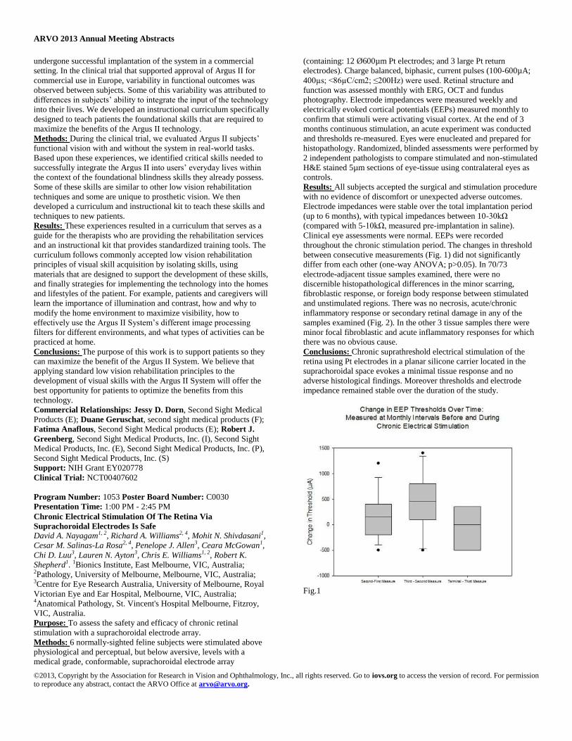
ARVO 2013 Annual Meeting Abstracts
©2013, Copyright by the Association for Research in Vision and Ophthalmology, Inc., all rights reserved. Go to iovs.org to access the version of record. For permission to reproduce any abstract, contact the ARVO Office at [email protected].
undergone successful implantation of the system in a commercial
setting. In the clinical trial that supported approval of Argus II for
commercial use in Europe, variability in functional outcomes was
observed between subjects. Some of this variability was attributed to
differences in subjects’ ability to integrate the input of the technology
into their lives. We developed an instructional curriculum specifically
designed to teach patients the foundational skills that are required to
maximize the benefits of the Argus II technology.
Methods: During the clinical trial, we evaluated Argus II subjects’
functional vision with and without the system in real-world tasks.
Based upon these experiences, we identified critical skills needed to
successfully integrate the Argus II into users’ everyday lives within
the context of the foundational blindness skills they already possess.
Some of these skills are similar to other low vision rehabilitation
techniques and some are unique to prosthetic vision. We then
developed a curriculum and instructional kit to teach these skills and
techniques to new patients.
Results: These experiences resulted in a curriculum that serves as a
guide for the therapists who are providing the rehabilitation services
and an instructional kit that provides standardized training tools. The
curriculum follows commonly accepted low vision rehabilitation
principles of visual skill acquisition by isolating skills, using
materials that are designed to support the development of these skills,
and finally strategies for implementing the technology into the homes
and lifestyles of the patient. For example, patients and caregivers will
learn the importance of illumination and contrast, how and why to
modify the home environment to maximize visibility, how to
effectively use the Argus II System’s different image processing
filters for different environments, and what types of activities can be
practiced at home.
Conclusions: The purpose of this work is to support patients so they
can maximize the benefit of the Argus II System. We believe that
applying standard low vision rehabilitation principles to the
development of visual skills with the Argus II System will offer the
best opportunity for patients to optimize the benefits from this
technology.
Commercial Relationships: Jessy D. Dorn, Second Sight Medical
Products (E); Duane Geruschat, second sight medical products (F);
Fatima Anaflous, Second Sight Medical products (E); Robert J.
Greenberg, Second Sight Medical Products, Inc. (I), Second Sight
Medical Products, Inc. (E), Second Sight Medical Products, Inc. (P),
Second Sight Medical Products, Inc. (S)
Support: NIH Grant EY020778
Clinical Trial: NCT00407602
Program Number: 1053 Poster Board Number: C0030
Presentation Time: 1:00 PM - 2:45 PM
Chronic Electrical Stimulation Of The Retina Via
Suprachoroidal Electrodes Is Safe David A. Nayagam1, 2, Richard A. Williams2, 4, Mohit N. Shivdasani1,
Cesar M. Salinas-La Rosa2, 4, Penelope J. Allen3, Ceara McGowan1,
Chi D. Luu3, Lauren N. Ayton3, Chris E. Williams1, 2, Robert K.
Shepherd1. 1Bionics Institute, East Melbourne, VIC, Australia; 2Pathology, University of Melbourne, Melbourne, VIC, Australia; 3Centre for Eye Research Australia, University of Melbourne, Royal
Victorian Eye and Ear Hospital, Melbourne, VIC, Australia; 4Anatomical Pathology, St. Vincent's Hospital Melbourne, Fitzroy,
VIC, Australia.
Purpose: To assess the safety and efficacy of chronic retinal
stimulation with a suprachoroidal electrode array.
Methods: 6 normally-sighted feline subjects were stimulated above
physiological and perceptual, but below aversive, levels with a
medical grade, conformable, suprachoroidal electrode array
(containing: 12 Ø600µm Pt electrodes; and 3 large Pt return
electrodes). Charge balanced, biphasic, current pulses (100-600µA;
400µs; <86µC/cm2; ≤200Hz) were used. Retinal structure and
function was assessed monthly with ERG, OCT and fundus
photography. Electrode impedances were measured weekly and
electrically evoked cortical potentials (EEPs) measured monthly to
confirm that stimuli were activating visual cortex. At the end of 3
months continuous stimulation, an acute experiment was conducted
and thresholds re-measured. Eyes were enucleated and prepared for
histopathology. Randomized, blinded assessments were performed by
2 independent pathologists to compare stimulated and non-stimulated
H&E stained 5µm sections of eye-tissue using contralateral eyes as
controls.
Results: All subjects accepted the surgical and stimulation procedure
with no evidence of discomfort or unexpected adverse outcomes.
Electrode impedances were stable over the total implantation period
(up to 6 months), with typical impedances between 10-30kΩ
(compared with 5-10kΩ, measured pre-implantation in saline).
Clinical eye assessments were normal. EEPs were recorded
throughout the chronic stimulation period. The changes in threshold
between consecutive measurements (Fig. 1) did not significantly
differ from each other (one-way ANOVA; p>0.05). In 70/73
electrode-adjacent tissue samples examined, there were no
discernible histopathological differences in the minor scarring,
fibroblastic response, or foreign body response between stimulated
and unstimulated regions. There was no necrosis, acute/chronic
inflammatory response or secondary retinal damage in any of the
samples examined (Fig. 2). In the other 3 tissue samples there were
minor focal fibroblastic and acute inflammatory responses for which
there was no obvious cause.
Conclusions: Chronic suprathreshold electrical stimulation of the
retina using Pt electrodes in a planar silicone carrier located in the
suprachoroidal space evokes a minimal tissue response and no
adverse histological findings. Moreover thresholds and electrode
impedance remained stable over the duration of the study.
Fig.1

ARVO 2013 Annual Meeting Abstracts
©2013, Copyright by the Association for Research in Vision and Ophthalmology, Inc., all rights reserved. Go to iovs.org to access the version of record. For permission to reproduce any abstract, contact the ARVO Office at [email protected].
Fig.2
Commercial Relationships: David A. Nayagam, None; Richard A.
Williams, None; Mohit N. Shivdasani, None; Cesar M. Salinas-La
Rosa, None; Penelope J. Allen, Bionic Vision Australia (P); Ceara
McGowan, None; Chi D. Luu, None; Lauren N. Ayton, None;
Chris E. Williams, Bionic Vision Australia (P); Robert K.
Shepherd, None
Support: The Bionics Institute and the Center for Eye Research
Australia acknowledge the support they receive from the Victorian
Government through its Operational Infrastructure Support Program.
This research was supported by the Australian Research Council
(ARC) through its Special Research Initiative (SRI) in Bionic Vision
Science and Technology grant to Bionic Vision Australia (BVA).
Program Number: 1054 Poster Board Number: C0031
Presentation Time: 1:00 PM - 2:45 PM
In vivo chronological observation of electrochemical properties of
porous electrodes with chronic suprachoroidal-transretinal
stimulation Yasuo Terasawa1, 2, Hiroyuki Tashiro4, Yukari Nakano1, Koji
Osawa1, Motoki Ozawa3. 1Vision Institute, Nidek Co., Ltd.,
Gamagori, Japan; 2Graduate School of Materials Science, Nara
Institute of Science & Technology, Ikoma, Japan; 3Nidek Co., Ltd.,
Gamagori, Japan; 4Department of Health Sciences, Kyushu
University, Fukuoka, Japan.
Purpose: Stabilizing electrochemical properties of stimulating
electrodes in vivo for long term is a key issue for visual prostheses.
We previously proposed stimulating electrodes with porous surface
[Terasawa et al., Invest Ophthalmol Vis Sci 2012;53: E-Abstract
5538]. The purpose of this study is to confirm the stability of
electrochemical properties of stimulating electrodes with chronic
suprachoroidal-transretinal stimulation in vivo.
Methods: An electrode array with two porous electrodes was
developed for this study. The diameter and height of an electrode
were 0.5mm and 0.3mm respectively. The electrode array was
inserted into a scleral pocket of a rabbit eye and sutured onto the
sclera. Charge-balanced biphasic pulses were applied to one of two
electrodes on the array for eight hour per day for one month.
Electrode impedance spectra were obtained periodically. In addition,
charge injection capacities (CIC) of the electrodes were determined
both in saline and in vivo. All in vivo experiments were conducted in
accordance with the Association for Research in Vision and
Ophthalmology statement for the use of animals in ophthalmic and
vision research, and institutional guidelines for the care and use of
laboratory animals.
Results: Data analysis using equivalent circuit fitting suggested that
double-layer capacitances in vivo were two orders of magnitude
lower than that in vitro. No significant changes were observed in
impedance spectra during chronic electrical stimulation. However
increase of electrode impedance was observed at sixth day after
cessation of stimulation. Double-layer capacitances decreased by half
after cessation of stimulation.
The CIC decreased from 225μC/cm2 to 78μC/cm2 after one month
stimulation. Inter pulse potential (electrode potential before onset of
each pulses) decreased from 0.217V (vs. Ag/AgCl) to 0.005V after
stimulation. The decrease of inter pulse potential moved electrode
potential closer to the lower limit of water window, which resulted in
the decrease of the CIC of stimulating electrodes.
Conclusions: Impedance spectra in vivo kept unchanged during one-
month chronic stimulation. The increase of impedance after cessation
of stimulation suggested that electrical stimulation significantly
affects the electrochemical properties of stimulating electrodes.
Studies with longer period of stimulation are ongoing.
Commercial Relationships: Yasuo Terasawa, Nidek Co.,Ltd. (E);
Hiroyuki Tashiro, Nidek Co., Ltd. (F); Yukari Nakano, NIDEK
CO.,LTD. (E); Koji Osawa, NIDEK (E); Motoki Ozawa, Nidek Co.,
Ltd. (E)
Program Number: 1055 Poster Board Number: C0032
Presentation Time: 1:00 PM - 2:45 PM
Comparison of Ankyrin-G Labeling of Axon Initial Segments on
Ganglion Cells in Normal and Degenerate Rat Retina Steven T. Walston1, Eun-Jin Lee1, Mark S. Humayun1, 2, James D.
Weiland1, 2. 1Biomedical Engineering, University of Southern
Caliifornia, Los Angeles, CA; 2Ophthalmology, University of
Southern Caliifornia, Los Angeles, CA.
Purpose: To compare the position and length of the axon initial
segment (AIS) on retinal ganglion cells (RGCs) as it relates to
activation thresholds in normal and degenerate retina.
Methods: Immunohistochemistry was used to label the AIS and the
corresponding RGCs. Studies were conducted on normal Long-
Evans, and heterozygous S334ter-line-3 rat retina. Antibodies against
ankryin-G, calretinin, and Brn3 were used to label the AIS, RGC
somas and axons, and confirm RGC identify, respectively. The length
of the AIS and its distance from the soma was acquired using
fluorescence microscopy and measured using ImageJ.
Results: RGCs and their corresponding AIS are identified in the
ganglion cell and nerve fiber layer with fluorescence microscopy.
The average length of the AIS is 1.8μm shorter in degenerate retina
when compared to normal (p=0.022, Student t-test). The mean AIS
length is 21.1 ± 7.17μm (mean ± std) (n=173) in normal retina and
19.3 ± 6.09μm (n=122) in degenerate retina. The distance between
the soma and proximal edge of the AIS along the axon is not
significantly different between normal and degenerate retina
(p=0.183). The mean position of the AIS is 13.4 ± 8.97μm (n=173) in
normal retina and 12.1 ± 7.21μm (n=122) in degenerate retina.
Conclusions: Based on population statistics, the average length of
the AIS is statistically shorter in degenerate retina than in normal
retina. Although there is significant overlap of the data, this decrease
in length may, in theory, increase the threshold at which the neuron
fires action potentials. Further study is necessary to determine if there
are differences within individual ganglion cell subtypes for normal
and degenerate retina.

ARVO 2013 Annual Meeting Abstracts
©2013, Copyright by the Association for Research in Vision and Ophthalmology, Inc., all rights reserved. Go to iovs.org to access the version of record. For permission to reproduce any abstract, contact the ARVO Office at [email protected].
Figure 1. Histogram plots of A AIS length and B distance from soma
to proximal edge of AIS on RGCs of normal Long-Evans (black) and
S334ter-line-3 retina (blue).
Commercial Relationships: Steven T. Walston, None; Eun-Jin
Lee, None; Mark S. Humayun, Bausch & Lomb (F), Bausch &
Lomb (C), Bausch & Lomb (P), Bausch & Lomb (R), Bausch &
Lomb (S), Alcon (C), Alcon (R), Iridex (P), Iridex (R), Replenish (I),
Replenish (C), Replenish (R), Replenish (S), Second Sight (F),
Second Sight (I), Second Sight (C), Second Sight (P), Second Sight
(R), Second Sight (S), Regenerative Patch Technologies (I),
Regenerative Patch Technologies (C); James D. Weiland, Second
Sight Medical Products, Inc. (F)
Support: Research Innovation Fund from USC; Research to Prevent
Blindness; W.M. Keck Foundation; NSF EEC-0310723
Program Number: 1056 Poster Board Number: C0033
Presentation Time: 1:00 PM - 2:45 PM
An Optimal Power Coil Configuration to Minimize Surgical Cut
for Intraocular Retinal Prostheses Yu Zhao1, Charles DeBoer1, Mandheerej S. Nandra1, James D.
Weiland2, Mark S. Humayun2, Yu-Chong Tai1. 1Electrical
Engineering, California Institute of Technology, Pasadena, CA; 2Doheny Eye Institute, Los Angeles, CA.
Purpose: Power coils used in current telemetries powering the
epiretinal, subretinal and suprachoroidal prosthetic devices, such as
microfabricated planar coil with metal electroplating and wire-wound
coil, are not suitable/ideal for intraocular implantation as they are
usually thick, rigid and large. For intraocular implantation, we
propose a foil coil as an optimized configuration. It demonstrates not
only better electrical performance, but also superior mechanical
flexibility and bendability allowing the surgeon to implant through a
minimal incision.
Methods: The lens capsule is a possible intraocular implantation site
for the telemetry coils, which also sets the size and mass constraints.
This implantation is similar to the common intraocular lens
implantation. While making the best use of the coil winding window,
it is still necessary to make the coil flexible for minimally invasive
surgical implantation. Flexural stiffness, i.e., resistance to bending,
equals to the product of the Young’s modulus E and second moment
of area I reflecting the material stiffness and its geometrical cross
section, respectively. Given same constructing metal, second moment
of area varies with cross-sectional size and geometry (Fig. 1). The
long-span high-aspect ratio foil strip can achieve smallest I and
therefore the most flexibility. The strip was fabricated using a
parylene-metal-parylene sandwich structure. The thermally deposited
metal is thin (0.25 μ m) and patterned to form a 1-mm-wide stripe to
fit into lens capsule. After the foil was dry-peeled off from the silicon
substrate and manually wound into a circular coil, the original width
of the encapsulated metal foil becomes the equivalent thickness of the
coil.
Results: To demonstrate the surgical feasibility, the coil was
implanted into the lens capsule of a porcine eye (Fig. 2). The coil was
compressed and inserted into a porcine lens capsule through a
capsulorhexis. After implantation the forceps were withdrawn and the
coil regained its circular shape (dotted circle in the middle panel). By
observing the implanted coil after removal of the iris, the full
recovery of the coil shape was achieved in vitro.
Conclusions: Foil coil configuration has been theoretically proven
and experimentally demonstrated to be more bendable, thus less
invasive during implantation. The resulting smaller surgical incision
has the potential to reduce post operative recovery time.
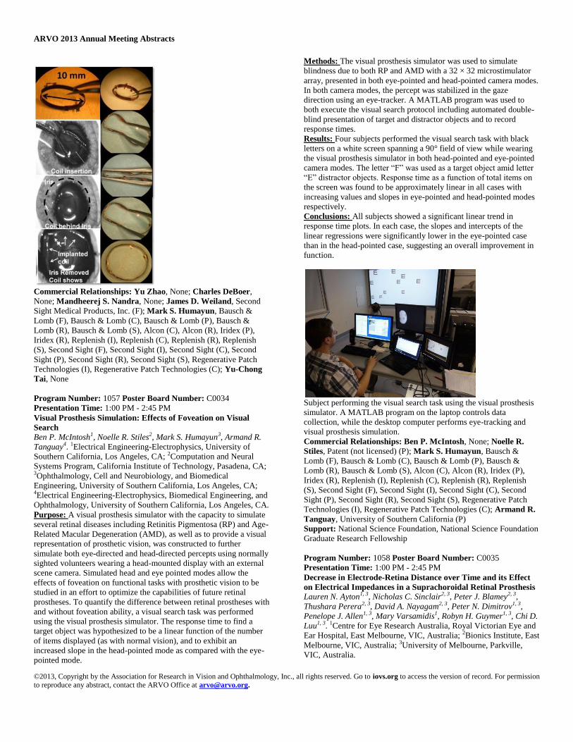
ARVO 2013 Annual Meeting Abstracts
©2013, Copyright by the Association for Research in Vision and Ophthalmology, Inc., all rights reserved. Go to iovs.org to access the version of record. For permission to reproduce any abstract, contact the ARVO Office at [email protected].
Commercial Relationships: Yu Zhao, None; Charles DeBoer,
None; Mandheerej S. Nandra, None; James D. Weiland, Second
Sight Medical Products, Inc. (F); Mark S. Humayun, Bausch &
Lomb (F), Bausch & Lomb (C), Bausch & Lomb (P), Bausch &
Lomb (R), Bausch & Lomb (S), Alcon (C), Alcon (R), Iridex (P),
Iridex (R), Replenish (I), Replenish (C), Replenish (R), Replenish
(S), Second Sight (F), Second Sight (I), Second Sight (C), Second
Sight (P), Second Sight (R), Second Sight (S), Regenerative Patch
Technologies (I), Regenerative Patch Technologies (C); Yu-Chong
Tai, None
Program Number: 1057 Poster Board Number: C0034
Presentation Time: 1:00 PM - 2:45 PM
Visual Prosthesis Simulation: Effects of Foveation on Visual
Search Ben P. McIntosh1, Noelle R. Stiles2, Mark S. Humayun3, Armand R.
Tanguay4. 1Electrical Engineering-Electrophysics, University of
Southern California, Los Angeles, CA; 2Computation and Neural
Systems Program, California Institute of Technology, Pasadena, CA; 3Ophthalmology, Cell and Neurobiology, and Biomedical
Engineering, University of Southern California, Los Angeles, CA; 4Electrical Engineering-Electrophysics, Biomedical Engineering, and
Ophthalmology, University of Southern California, Los Angeles, CA.
Purpose: A visual prosthesis simulator with the capacity to simulate
several retinal diseases including Retinitis Pigmentosa (RP) and Age-
Related Macular Degeneration (AMD), as well as to provide a visual
representation of prosthetic vision, was constructed to further
simulate both eye-directed and head-directed percepts using normally
sighted volunteers wearing a head-mounted display with an external
scene camera. Simulated head and eye pointed modes allow the
effects of foveation on functional tasks with prosthetic vision to be
studied in an effort to optimize the capabilities of future retinal
prostheses. To quantify the difference between retinal prostheses with
and without foveation ability, a visual search task was performed
using the visual prosthesis simulator. The response time to find a
target object was hypothesized to be a linear function of the number
of items displayed (as with normal vision), and to exhibit an
increased slope in the head-pointed mode as compared with the eye-
pointed mode.
Methods: The visual prosthesis simulator was used to simulate
blindness due to both RP and AMD with a 32 × 32 microstimulator
array, presented in both eye-pointed and head-pointed camera modes.
In both camera modes, the percept was stabilized in the gaze
direction using an eye-tracker. A MATLAB program was used to
both execute the visual search protocol including automated double-
blind presentation of target and distractor objects and to record
response times.
Results: Four subjects performed the visual search task with black
letters on a white screen spanning a 90° field of view while wearing
the visual prosthesis simulator in both head-pointed and eye-pointed
camera modes. The letter “F” was used as a target object amid letter
“E” distractor objects. Response time as a function of total items on
the screen was found to be approximately linear in all cases with
increasing values and slopes in eye-pointed and head-pointed modes
respectively.
Conclusions: All subjects showed a significant linear trend in
response time plots. In each case, the slopes and intercepts of the
linear regressions were significantly lower in the eye-pointed case
than in the head-pointed case, suggesting an overall improvement in
function.
Subject performing the visual search task using the visual prosthesis
simulator. A MATLAB program on the laptop controls data
collection, while the desktop computer performs eye-tracking and
visual prosthesis simulation.
Commercial Relationships: Ben P. McIntosh, None; Noelle R.
Stiles, Patent (not licensed) (P); Mark S. Humayun, Bausch &
Lomb (F), Bausch & Lomb (C), Bausch & Lomb (P), Bausch &
Lomb (R), Bausch & Lomb (S), Alcon (C), Alcon (R), Iridex (P),
Iridex (R), Replenish (I), Replenish (C), Replenish (R), Replenish
(S), Second Sight (F), Second Sight (I), Second Sight (C), Second
Sight (P), Second Sight (R), Second Sight (S), Regenerative Patch
Technologies (I), Regenerative Patch Technologies (C); Armand R.
Tanguay, University of Southern California (P)
Support: National Science Foundation, National Science Foundation
Graduate Research Fellowship
Program Number: 1058 Poster Board Number: C0035
Presentation Time: 1:00 PM - 2:45 PM
Decrease in Electrode-Retina Distance over Time and its Effect
on Electrical Impedances in a Suprachoroidal Retinal Prosthesis Lauren N. Ayton1, 3, Nicholas C. Sinclair2, 3, Peter J. Blamey2, 3,
Thushara Perera2, 3, David A. Nayagam2, 3, Peter N. Dimitrov1, 3,
Penelope J. Allen1, 3, Mary Varsamidis1, Robyn H. Guymer1, 3, Chi D.
Luu1, 3. 1Centre for Eye Research Australia, Royal Victorian Eye and
Ear Hospital, East Melbourne, VIC, Australia; 2Bionics Institute, East
Melbourne, VIC, Australia; 3University of Melbourne, Parkville,
VIC, Australia.

ARVO 2013 Annual Meeting Abstracts
©2013, Copyright by the Association for Research in Vision and Ophthalmology, Inc., all rights reserved. Go to iovs.org to access the version of record. For permission to reproduce any abstract, contact the ARVO Office at [email protected].
Purpose: The initial patient tests of a suprachoroidal retinal
prosthesis prototype have shown success in both the surgical
procedure and initial psychophysics testing. In this study, we
investigated the relationship between electrode-retina separation and
electrical impedances in a patient with an implanted suprachoroidal
device.
Methods: Three patients with end stage retinitis pigmentosa (light
perception vision) were implanted with a suprachoroidal retinal
prosthesis prototype, which consisted of 24 electrodes. Optical
coherence tomography scans were taken weekly, and high quality
images were obtained from one patient (52yo female) with steady
fixation. From these scans the array position, distance between
electrode and retina, and retinal thickness above the electrodes were
measured. Scans were taken both horizontally and vertically through
all rows and columns of electrodes. Electrical impedances were
measured weekly using a 500µs phase width and 20µs interphase gap
biphasic pulse. The results of 50 pulses delivered at 500Hz were
averaged.
Results: The average electrode-retina separation decreased gradually
with time from an average of 422µm at the initial stimulation (55
days postop) to 362µm at 6 months postop (p=0.02). Average retinal
thickness did not change significantly over this time (186µm to
184µm, p=0.71). The electrical impedances increased over this same
time period, from an initial average impedance of 17.75kΩ to
20.08kΩ at 6 months postop (p<0.005). The decrease in electrode-
retina distance was significantly correlated with the increased
impedances (r=-0.59, p<0.005).
Conclusions: Following suprachoroidal retinal prosthesis
implantation, the electrode-retina distance gradually decreases with
time. The improved proximity of electrode to retinal tissue correlated
with increased electrical impedances in this patient.
Commercial Relationships: Lauren N. Ayton, None; Nicholas C.
Sinclair, None; Peter J. Blamey, Bionics Institute (P), Cochlear
Limited (P); Thushara Perera, None; David A. Nayagam, None;
Peter N. Dimitrov, None; Penelope J. Allen, Bionic Vision
Australia (P); Mary Varsamidis, None; Robyn H. Guymer, Ellex
Pty Ltd (F), Novartis (C), Bayer (C), Novartis (R); Chi D. Luu, None
Support: This research was supported by the Australian Research
Council (ARC) through its Special Research Initiative (SRI) in
Bionic Vision Science and Technology grant to Bionic Vision
Australia (BVA).
Clinical Trial: NCT01603576
Program Number: 1059 Poster Board Number: C0036
Presentation Time: 1:00 PM - 2:45 PM
Analysis of retinal and cortical response to electrical stimulation
by Subretinal implant in rodent model Frederic Matonti1, 2, Sebastien Roux2, Virginie Donnadieu1, 2, Henri
Lorach3, Olivier Marre3, Serge A. Picaud3, Frederic Chavane2. 1Ophthalmology, Hopital Nord, Marseille, France; 2Institut de
Neurosciences de la Timone, Aix Marseille Université, Marseille,
France; 3Institut de la Vision, Université Pierre et Marie Curie, Paris,
France.
Purpose: This research aims to develop and improve the use of
retinal prosthesis in animal model. For that purpose, we measured
retinal and cortical response to direct subretinal electrical stimulation
to understand how the patterns of stimuli can be adapted to improve
stimulation to get closer to the response evoked by natural visual
stimuli.
Methods: In a rat model, a comparative analysis of the functional
impact of similar stimulation of a subretinal implant is done at two
levels, (i) in the retina in vitro by multi electrodes array and (ii) in
vivo in the primary visual cortex by optical imaging recordings.
Optical imaging permits a functional mapping of the cortex, using
light reflection and absorption changes depending on the rate of
blood oxygenation. Diverse parameters were investigated: stimulus
shape and polarity, intensity, size and location.
Results: At the cortical level, we have quantified the size, position
and intensity of the point-spread function in response to the various
electrical stimulations and compared them to those generated by
calibrated light stimuli. The point-spread function was much larger
for electrical stimulations compared to visual stimulation. We are
currently performing retinal recordings of ganglion cells, while the
prosthesis is on the photoreceptor side. We will compare the size of
ganglion cell receptive fields to the size of the electric field activation
to see if a difference in the size of the region activated by the two
stimulations in the retina could explain the difference observed at the
cortical level.
The ability to evaluate in vitro within the retina and in vivo the
cortical responses induced by the prosthesis allowed us refining the
patterns of electrical stimulation to get closer to a natural visual
activation.
Conclusions: These results offer interesting prospect for improving
the design of prostheses as well as their patterns of stimulation for a
medical application
Commercial Relationships: Frederic Matonti, Allergan (C), Alcon
(R), Théa (R), Topcon (R); Sebastien Roux, None; Virginie
Donnadieu, None; Henri Lorach, None; Olivier Marre, None;
Serge A. Picaud, UPMC (P), Pixium Vision (C); Frederic Chavane,
None
Support: Bourse de recherche de la SFO
Program Number: 1060 Poster Board Number: C0037
Presentation Time: 1:00 PM - 2:45 PM
Spatial resolution of the Penetrative Optic Nerve Visual
Prosthesis estimated by epidural cortical recording Yao Chen, Yiliang Lu, Yan Yan, Liming Li, Xinyu Chai. Biomedical
Engineering, Shanghai Jiao Tong University, Shanghai, China.
Purpose: A visual prosthesis based on penetrating stimulation
electrodes within the optic nerve (ON) is a potential way to restore
partial functional vision for the blind patients. Although a rough
visuotopographic relationship was found between each of the four
contacts of the electrode implanted just anterior to the chiasm in the
ON by Veraart et al., it is still unknown the topography and spatial
resolution of penetrative iON prosthesis implanted close to ON head.
In this study, we investigated retinotopic organization of the ON
stimulation and its spatial resolution in in vivo cat experiments.
Methods: A five-electrode array was inserted perpendicular to the
ON axis or a single electrode was advanced at different depths within
the ON ~1-2 mm behind the eyeball in 13 cats. Cortical responses
were recorded by a 5 × 6 epidural electrode array. A sparse noise
method was used to map electrode position in ON and the visual
cortex to establish ON and cortical visuotopic maps. Then we
compared the visual field positions of ON stimulation sites and their
elicited greatest cortical response (M-Channels) sites. Finally, we
estimated the spatial resolution of the penetrative ON prosthesis by
calculating the difference in M-channel visual field positions
corresponding to neighboring ON sites with an inter-electrode
distance of 150 μm.
Results: Electrical stimulation with penetrating ON electrodes
inserted just posterior to eyeball could elicit cortical responses with
visuotopographical correspondence in cats. The electrical stimulation
through electrodes within the temporal ON can elicit cortical
responses corresponding to the central visual field. According to the
increment of penetrating depth, the corresponding elicited cortical
responses shift from lower to central visual field. About 2° to 3°

ARVO 2013 Annual Meeting Abstracts
©2013, Copyright by the Association for Research in Vision and Ophthalmology, Inc., all rights reserved. Go to iovs.org to access the version of record. For permission to reproduce any abstract, contact the ARVO Office at [email protected].
spatial resolution within a limited visuotopic representation in the
cortex could be obtained by this approach.
Conclusions: Visuotopic electrical stimulation with relatively fine
spatial resolution could be accomplished by penetrative electrodes
implanted at multi-sites and different depths within ON close to the
optic nerve head. This study also provided useful experimental data
for the design of electrode density and distribution of penetrating ON
electrodes for a visual prosthesis.
Commercial Relationships: Yao Chen, None; Yiliang Lu, None;
Yan Yan, None; Liming Li, None; Xinyu Chai, None
Support: The National Basic Research Program of China (973
Program, 2011CB707502), the National Natural Science Foundation
of China (31070981, 61171174, 91120304).
Program Number: 1061 Poster Board Number: C0038
Presentation Time: 1:00 PM - 2:45 PM
Development of large multielectrode arrays for epiretinal
stimulation - feasibility of implantation procedures
Gernot Roessler1, Thomas Laube3, Florian Waschkowski2, Anne C.
Rieck1, Claudia Brockmann3, Wilfried Mokwa2, Peter Walter1. 1Dept
of Ophthalmology, RWTH Aachen University, Aachen, Germany; 2Dept of Materials in Electrical Engineering, RWTH Aachen
University, Aachen, Germany; 3Dept of Ophthalmology, University
of Duisburg-Essen, Essen, Germany.
Purpose: To demonstrate the feasibility of implantation surgery of
Very Large Retinal Stimulation arrays (VLARS) to provide artificial
vision with enlarged visual fields.
Methods: Polyimide foils with a star-shaped pattern and a diameter
of 12.5 mm have been fabricated for implantation. Each foil carries
244 gold electrodes divided into 63 electrodes for central and 181
electrodes for peripheral stimulation of the retinal surface. Openings
for tack fixation were embedded paracentrally and into each arm of
the star-shaped electrode array. Implantation was performed in pigs’
eyes including lens removal, vitrectomy and the implantation via
corneal incision.
Results: Following lens removal by phacoemulsification and three-
port vitrectomy, a decaline bubble was installed into the vitreous
cavity. Via a corneal incision the folded stimulator was inserted into
the eyeball, pushed through a posterior capsulotomy and positioned
onto the decaline bubble. While the stimulator showed a good overall
contact to the retinal surface after removal of the decaline bubble,
implantation was finished by fixation using retinal tacks.
Conclusions: Our experiments demonstrated the feasibility of
implantation surgery of large electrode arrays. Long term
implantation for biocompatibility testing as well as stimulation
experiments for the generation of cortical activation in animal
experiments will follow.
Commercial Relationships: Gernot Roessler, None; Thomas
Laube, None; Florian Waschkowski, None; Anne C. Rieck, None;
Claudia Brockmann, None; Wilfried Mokwa, None; Peter Walter,
Novartis (R), Bayer (R), Second Sight (R), Bayer (F), Novartis (F)
Support: Jackstaedt Foundation
Program Number: 1062 Poster Board Number: C0039
Presentation Time: 1:00 PM - 2:45 PM
In vivo evaluation of the liquid crystal polymer-based retinal
prosthesis by optical coherence tomography Hum Chung1, Bum-Joo Cho1, Hee Sun Park1, So Hyun Bae1, 2, Tae
Wan Kim1, 3, Jong-Mo Seo1, 4, Hyeong Gon Yu1, Joonsoo Jeong4,
Soowon Shin4, Sung June Kim4. 1Ophthalmology, Seoul National
University, Seoul, Republic of Korea; 2Ophthalmology, Kangnam
Sacred Heart Hospital, Seoul, Republic of Korea; 3Ophthalmology,
Seoul Nat'l Univ Boramae Medical Ctr, Seoul, Republic of Korea;
4Electrical Engineering, Seoul National University, Seoul, Republic
of Korea.
Purpose: Liquid crystal polymer (LCP) - based, monolithic retinal
prosthesis was developed, and in vivo biocompatibility and
biostability were evaluated in the rabbit eye by using optical coherent
tomography (OCT).
Methods: LCP-based gold electrode array was fabricated and the
electrical retinal stimulator was integrated into the LCP package to
form a monolithic prosthesis system. After making scleral tunnel in
the rabbit eye, electrode part was implanted suprachoroidally or
subretinally, and the stimulator package was positioned and fixed
over the sclera, in the sub-tenon space. Regular post-operative
evaluation was done by OCT, fundus photography and
electroretinography (ERG).
Results: OCT showed the chorioretinal structure over the electrodes
in detail. However, LCP was opaque to the light, thus there were
shadows under the electrodes. This made the choroidal evaluation
difficult in the eye with subretinal LCP electrode. Localized tenting
or stretching of the retina or choroid over the electrode was well
visualized by OCT, and this could not be identified easily on the
fundus photograph. Some structural changes of the retina over the
LCP electrodes were found in 7 out of 14 eyes, and these might be
due to the intraoperative surgical events, not due to the toxicity of the
LCP itself. ERG waveform was correlated with the change on the
OCT finding.
Conclusions: LCP retinal prosthesis showed good in vivo
biocompatibility and biostability. OCT was useful for the evaluation
of anatomical and histological outcome of LCP retinal prosthesis
implantation and the physical characteristics of the LCP implants.
Commercial Relationships: Hum Chung, None; Bum-Joo Cho,
None; Hee Sun Park, None; So Hyun Bae, None; Tae Wan Kim,
None; Jong-Mo Seo, None; Hyeong Gon Yu, None; Joonsoo Jeong,
None; Soowon Shin, None; Sung June Kim, None
Support: Korea Health 21 R&D Project A050251 by MIHWAF,
Technology Innovation Program 10033634 by MKE, Public Welfare
and Safety Program 2012-0006566 by MEST
Program Number: 1063 Poster Board Number: C0040
Presentation Time: 1:00 PM - 2:45 PM
Optimal electrode conditions for surprachoroidal-transretinal
stimulation system to elicit electrically-evoked potentials Kentaro Nishida2, 1, Hirokazu Sakaguchi1, Takashi Fujikado1,
Motohiro Kamei1, Kohji Nishida1. 1Osaka University Graduate
School of Medicine, Suita, Japan; 2Sakai City Hospital, Sakai, Japan.
Purpose: To determine the effects of the height and the surface area
of the stimulating electrode on the electrical evoked potentials (EEPs)
elicited by suprachoroidal-transretinal stimulation (STS) in rabbit
eye.
Methods: A scleral pocket (3x5 mm) was created just over the visual
streak in anesthetized rabbits (weight, 2.2-2.5 kg). The STS
stimulating electrode system was implanted into the pocket. We
varied the height of the stimulating electrode (300µm or 500µm) and
the surface of the electrode (smooth or porous, Figure). EEPs were
then elicited by each type of electrode conditions. Three sessions
were repeated for each group.
Results: For the height of the stimulating electrode, the implicit
times of the EEPs were 31.6±0.5 ms (300µm) and 31.9±0.8 ms
(500µm), respectively, and the amplitudes were 78.2±9.5 µV
(300µm) and 70.0±5.1 µV(500µm) respectively. These differences
were not significant (P=0.588; P=0.261). The surface area of the
stimulating electrode has no significant effect on the implicit times
and the amplitudes of the EEPs (P>0.05).
Conclusions: The height and the surface area of the stimulating

ARVO 2013 Annual Meeting Abstracts
©2013, Copyright by the Association for Research in Vision and Ophthalmology, Inc., all rights reserved. Go to iovs.org to access the version of record. For permission to reproduce any abstract, contact the ARVO Office at [email protected].
electrode do not significantly affect the implicit times and amplitudes
of the EEPs elicited by the STS system.
Commercial Relationships: Kentaro Nishida, None; Hirokazu
Sakaguchi, HOYA Corporation (R); Takashi Fujikado, Nidek (P);
Motohiro Kamei, None; Kohji Nishida, Alcon (C), Alcon (F),
HOYA (F), Senju (F), Pfizer (F), Santen (F), Osaka University (P)
Program Number: 1064 Poster Board Number: C0041
Presentation Time: 1:00 PM - 2:45 PM
Reducing the Number of Stimulators and Electrical Tracks in
High-Resolution Visual Prostheses Milan Djilas1, Ryad Benosman1, Philippe Bergonzo2, Christoph
Posch1, Jose A. Sahel1, 3, Serge A. Picaud1, 3. 1Institut de la Vision,
INSERM/UPMC Univ Paris 6/CNRS/CHNO des XV-XX, Paris,
France; 2LIST, CEA, Saclay, France; 3Fondation Ophtalmologique
Adolphe de Rothschild, Paris, France.
Purpose: Retinal diseases such as RP and AMD cause photoreceptor
degeneration while other cell layers partially remain. Retinal
prostheses restore useful vision by activating these cells. The NxN
electrode matrix and stimulators are coupled with N2 electrical tracks.
With the increase of pixel resolution, this inter-connection becomes
difficult to fabricate. The foil also becomes more rigid, making the
implantation surgery more challenging. Here we propose an
alternative electrode design and connectivity layout that significantly
reduces the required number of metal tracks and stimulators.
Methods: The proposed layout is shown on Figure 1. To stimulate a
particular pixel, segments from one row, one column and two
diagonals containing that pixel are activated simultaneously. To
excite only the tissue at the target pixel, the stimulation intensity of
each active segment is kept below threshold, whereas the intensity of
simultaneous activation of four active segments at the target pixel is
above threshold.
Results: The proposed layout requires a total of 6(N-1) electrical
tracks and only 4 stimulators (1 for row, 1 for column, and 2 for
diagonals) regardless of implant resolution. One additional track is
needed for a common counter-electrode. Finite-element method
simulations clearly confirm significantly higher stimulation intensity
at the target pixel compared to other pixels stimulated with 4x lower
intensity. This range should be sufficient to activate only the former.
Conclusions: The proposed strategy requires 6N-5 tracks and 4
stimulators, compared to N2+1 tracks and N2 stimulators in the
standard one-track-per-electrode layout. The downside of such an
approach is that only a single pixel at a time can be activated. This
would however not be a limiting factor if asynchronous image
acquisition is implemented. Future work includes experimental
validation of the proposed strategy.
Figure 1 A 5x5 electrode layout with 4 stimulating electrodes per
pixel. Segments designated with the same number are electrically
connected. Simultaneous electrical stimulation through segments
from one column (segments 7), one row (segments 2), and two
diagonals (segments 14 for \ and 19 for /) produces maximal
stimulation in the target pixel located in the intersection of the four
lines (thick outline). A metalized grid surrounding the electrodes
serves as the counter-electrode.
Commercial Relationships: Milan Djilas, None; Ryad Benosman,
None; Philippe Bergonzo, None; Christoph Posch, None; Jose A.
Sahel, UPMC/Essilor (P), Second Sight (F); Serge A. Picaud,
UPMC (P), Pixium Vision (C)
Support: NEUROCARE (FP7-NMP-2011-SMALL-5)
Program Number: 1065 Poster Board Number: C0042
Presentation Time: 1:00 PM - 2:45 PM
Design of the electrode and the system package for the retina
prosthesis based on liquid crystal polymer Jong-Mo Seo1, 2, Bum-Joo Cho1, So Hyun Bae1, 3, Tae Wan Kim1, 4,
Hyo Joo Shin1, Joonsoo Jeong2, Soowon Shin2, Dong-il "Dan" Cho2,
Sung June Kim2, Hum Chung1. 1Ophthalmology, Seoul National
University, Seoul, Republic of Korea; 2Electrical Engineering, Seoul
National University, Seoul, Republic of Korea; 3Ophthalmology,
Kangnam Sacred Heart Hospital, Seoul, Republic of Korea; 4Ophthalmology, Seoul Nat'l Univ Boramae Medical Ctr, Seoul,
Republic of Korea.
Purpose: To develop a retina prosthesis which can be implanted in
the orbit, the stimulator and the electrode were integrated into one
system. Whole system was fabricated with liquid crystal polymer
(LCP) in multi-layered structure, and their shape and the size were
investigated.
Methods: The 16-channel current stimulator integrated circuit and

ARVO 2013 Annual Meeting Abstracts
©2013, Copyright by the Association for Research in Vision and Ophthalmology, Inc., all rights reserved. Go to iovs.org to access the version of record. For permission to reproduce any abstract, contact the ARVO Office at [email protected].
multi-layered coil were implemented onto the LCP film by
semiconductor fabrication technology, and stacked to form a tiny
package. A semi-flat, ovoid LCP capsule was designed and applied
onto the system to establish the hermetic sealing. The electrode was
directly connected to the stimulator package to make an integrated
system. Whole system was implanted into the rabbit eye, and the the
shape, size, and the topological relationship between the electrode
and the stimulator package were evaluated.
Results: The electrode with the maximum diameter of 3 mm could
be safely implanted into the subretinal or suprachoroidal space
through scleral tunnel.Meticulous hemostasis of the choroid with
high-frequency electrocauterization increased the success rate of the
operation. Sharp angulation between the stimulator package and the
electrode shaft could help stabilizing the electrode tip under the retina
during surgery. The size of the system package was the major issue
for the stable and safe implantation of the whole system. The
flexibility of the LCP electrode and the leverage effect of the
electrode shaft affected the surgical outcome.
Conclusions: The flexibility and the edge sharpness of the electrode
were important for decreasing the tissue damage during implant
surgery. The size and the weight of the external part also affected the
stability of the electode by leverage effect. Optimized design
according to the size and the shape of the eyeball is necessary for the
successful implantation of the mololithic retina prosthesis.
Commercial Relationships: Jong-Mo Seo, None; Bum-Joo Cho,
None; So Hyun Bae, None; Tae Wan Kim, None; Hyo Joo Shin,
None; Joonsoo Jeong, None; Soowon Shin, None; Dong-il "Dan"
Cho, None; Sung June Kim, None; Hum Chung, None
Support: Korea Health 21 R&D Project A050251 by MIHWAF,
Technology Innovation Program 10033634 by MKE, Public Welfare
and Safety Program 2012-0006566 by MEST
Program Number: 1066 Poster Board Number: C0043
Presentation Time: 1:00 PM - 2:45 PM
Suprachoroidal Electrical Stimulation by an MEMS-based
Flexible Microelectrode array Xiaohong Sui, Yan Yan, Jingjing Sun, Liming Li, Qiushi Ren, Xinyu
Chai. Shanghai Jiao Tong University, Shanghai, China.
Purpose: To explore the spatial characteristics of indirect stimulation
of the retina by MEMS based flexible microelectrode arrays and to
evaluate the spatial characteristics of suprachoroidal stimulation for
visual prosthesis.
Methods: Based on MEMS techniques, a 16 microelectrode array in
4 × 4 arrangement was fabricated using single metal layer, and a
wide-view electrode array with 60 stimulating sites was also
fabricated with dual metal layers. Arrays were implanted into the
suprachoroidal space of rabbits for in-vivo test. For the 4 × 4
electrode array, diagonal “L” shapes stimulation and two square
shapes- small square using the central four electrodes and large
square using the peripheral 12 electrodes-stimulation were applied.
Row stimulations were applied with the 60 one. The electrical evoked
responses (EEPs) from the primary visual cortex were recorded by a
silver-ball electrode array. The amplitudes, maximum locations and
spatial spreads of the cortical activities were analyzed.
Results: Cortical responses could be consistently evoked by
suprachoroidal stimulation using both types of MEMS
microelectrode array. In two diagonal “L” shapes stimulations,
maximum cortical responses locations and spatial spreads were
similar. While in square stimulations, maximum EEPs locations were
at the same place but the responses amplitudes increased and the
spatial spread expanded in the large square shape stimulation. Rows
stimulation in 60 electrode array showed that horizontally orientated
cortical responses could be evoked, which were consisted
visuotopically with the row-electrodes horizontal position on the
retina. The amplitudes of EEPs increased with increasing stimulus
intensity, but the maximum EEPs positions remained unchanged.
Differentiated maximum EEPs positions showed up in different row
stimulations. The spatial spreads varied with a relatively large
responded cortical area resulted in stimulation by rows located under
the visual streak.
Conclusions: Preliminary pattern differentiated cortical responses
could be evoked by simultaneously stimulation of multiple selected
electrodes using the suprachoroidal MEMS based array. Results
indicated that this approach may be feasible to potentially restore
functional vision for the blinds.
Commercial Relationships: Xiaohong Sui, None; Yan Yan, None;
Jingjing Sun, None; Liming Li, None; Qiushi Ren, None; Xinyu
Chai, None
Support: NBRPC(Nos.2011CB707502/2011CB707503/2011CB707505)
Program Number: 1067 Poster Board Number: C0044
Presentation Time: 1:00 PM - 2:45 PM
A Center-Surround-Selectable, High-Acuity, Large-Field-of-
View, Flexible Retinal Prosthesis Long-Sheng Fan1, Chia-He Chung1, Frank Yang1, Eunice Liu1,
Jasmine Lin1, Zung-Hua Yang1, Grace Teng1, Chang-Hao Yang3,
Kea-Tiong Tang2. 1Institute of NEMS, National Tsing Hua
University, Hsinchu, Taiwan; 2Institute of Electronics, National Tsing
Hua University Hospital, Hsinchu, Taiwan; 3Opthalmology, National
Taiwan University Hospital, Taipei, Taiwan.
Purpose: We previously reported a 4,096-pixel flexible retinal
prosthesis generating bi-phasic electrical stimulations 30 μm in pitch
to enable the possibility of high visual acuity (VA) and cover a field
of view (FOV) around 10o. The pixel electrical output is a function
of the light intensity on that pixel. However, retinal photo sensors
influence retinal neurons across hundreds of micrometers, and the
retinal networks exhibit a center-surround receptive field through
lateral inhibition. Various stages of retina degeneracy may also need
certain artificial restoration of such neural function through
electronics. We target to implement a 16,328-pixel prosthesis that is
selectable to have an electrical output consistent with the center-
surround receptive field, and the chip covers up to 20o FOV with 30
μm pixel pitch, and conform to the surface of a 25 mm eyeball.
Methods: Since the ratio of the number of photo sensors to that of
ganglion cells is low near the fovea, the center-surround
characteristics is expected to be the key feature of the coding in this
region. The new retinal prosthesis chip includes the option that the
pixel electrical output from the pixel signal processing circuitry can
be selected to be a function of the light intensities on neighboring
pixels to a range of the typical receptive field in additional to the light
intensity on that pixel. The primary weight matrix is a Laplacian
Gaussian.
Results: We have implemented a flexible 16,328-element retina chip
30 μm thick using a 180nm, flexible CIS technology. The electrodes
are 10 μm in size and photo sensors 30 μm in pitch. The retina chip
with a large FOV is 6 mm in size including multiplexing electronics
for pixel characterizations and perfusion openings, and formed into a
spherical-shaped patch during the packaging process. We have
previously shown that the identical 10 μm-sized electrode arrays are
capable of inducing RGC spiking in vitro, and details are under
characterization.
Conclusions: This study demonstrates the feasibility to integrate a
high-acuity, large-field-of-view, flexible retinal prosthesis in a
spherical-shaped patch with the selectable feature to have an
electrical output characteristic consistent with the center-surround

ARVO 2013 Annual Meeting Abstracts
©2013, Copyright by the Association for Research in Vision and Ophthalmology, Inc., all rights reserved. Go to iovs.org to access the version of record. For permission to reproduce any abstract, contact the ARVO Office at [email protected].
receptive field using a 180 nm mixed-signal CMOS Image Sensor
technology. Detailed characteristics of this retinal chip will be
reported.
Commercial Relationships: Long-Sheng Fan, National Tsing-Hua
University (P); Chia-He Chung, None; Frank Yang, None; Eunice
Liu, None; Jasmine Lin, None; Zung-Hua Yang, None; Grace
Teng, None; Chang-Hao Yang, None; Kea-Tiong Tang, None
Support: NTHU
Program Number: 1068 Poster Board Number: C0045
Presentation Time: 1:00 PM - 2:45 PM
Design of a Compact Wide-Field-of-View Camera for Retinal
Prostheses Furkan E. Sahin1, Ben P. McIntosh1, Patrick J. Nasiatka1, James D.
Weiland2, Mark S. Humayun3, Armand R. Tanguay4. 1Department of
Electrical Engineering-Electrophysics, University of Southern
California, Los Angeles, CA; 2Departments of Biomedical
Engineering, and Ophthalmology, University of Southern California,
Los Angeles, CA; 3Departments of Ophthalmology, Cell and
Neurobiology, and Biomedical Engineering, University of Southern
California, Los Angeles, CA; 4Departments of Electrical
Engineering-Electrophysics, Biomedical Engineering, and
Ophthalmology, University of Southern California, Los Angeles, CA.
Purpose: To design and implement an ultraminiature, head-mounted,
wide-field-of-view extraocular camera that can be used in
conjunction with an ultraminiature eye-tracking camera to restore
natural foveation capabilities to patients implanted with intraocular
retinal prostheses.
Methods: In the proposed approach, a wide-field-of-view image of
the outside world is captured with the wide-field-of-view camera.
The eye-tracking camera in conjunction with appropriate software
algorithms extracts the direction of gaze, and the corresponding
subregion of the full camera field of view is exported to the
microstimulator array of the retinal prosthesis.
Custom compact multi-element 90° and 120° wide-field-of-view
lenses were first designed. Based on these designs, less optimal but
commercially available miniature lenses were chosen with matching
parameters, and the optical system design reoptimized.
The wide-field-of view extraocular camera can easily fit on a pair of
eyeglasses in such a way as to be relatively unobtrusive yet add
minimal weight and volume.
Results: A compact, low mass, wide-field-of-view optical system has
been designed and successfully implemented. This lens system has
four commercially available spherical lens elements. The lens system
provides a diagonal field of view of 100°, and fits into a housing with
dimensions of only 8 mm diameter and 8 mm overall length. This
lens design was coupled with a commercially available wide dynamic
range image sensor (OV10633) from OmniVision Technologies and
test images have been successfully captured.
Any wide field of view optical system generates a degree of
unwanted image (typically barrel) distortion that may cause adverse
complications with either direct stimulation of the visual system or
the implementation of subsequent image processing algorithms. To
this end, an image dewarping algorithm capable of removing this
distortion in real time has been developed that has the additional
feature of greatly reducing the associated chromatic aberration in the
image.
Conclusions: A very compact wide field of view camera was
designed to be used in conjunction with an eye-tracking camera to
restore natural foveation for retinal prosthesis patients.
Test image of common grocery items captured with the designed and
implemented wide-field-of-view camera.
Same image, but following software dewarping with chromatic
aberration correction to achieve a rectilinear, chromatically corrected
image.
Commercial Relationships: Furkan E. Sahin, None; Ben P.
McIntosh, None; Patrick J. Nasiatka, None; James D. Weiland,
Second Sight Medical Products, Inc. (F); Mark S. Humayun,
Bausch & Lomb (F), Bausch & Lomb (C), Bausch & Lomb (P),
Bausch & Lomb (R), Bausch & Lomb (S), Alcon (C), Alcon (R),
Iridex (P), Iridex (R), Replenish (I), Replenish (C), Replenish (R),
Replenish (S), Second Sight (F), Second Sight (I), Second Sight (C),
Second Sight (P), Second Sight (R), Second Sight (S), Regenerative
Patch Technologies (I), Regenerative Patch Technologies (C);
Armand R. Tanguay, University of Southern California (P)
Support: National Science Foundation
Top Related