


Languages
Pages
Legal

Malignant Mesothelioma Audit
April 2014Dr J King
Dr K Syred

90% mesotheliomas are linked to asbestos exposure
May be eligible for compensation 3 yr survival rate 8% Subtype related to prognosis and different
treatment options◦ Epithelioid 1yr survival 52%◦ Sarcomatoid 1 yr survival 13%
Background

Correlation of tissue diagnosis with clinical and radiology
IHC:◦ Panel 1: TTF-1, CEA, calretinin, CK5, D240, WT-1,
MOC31◦ Panel 2: MNF116, CD15, CK7, CK20
Diagnosis
Ihc Reactive Malignant
EMA -/+ + Strong membranous
Desmin + -
p53 - +
Identify practice within our department◦ Typing ◦ Assess what ihc has been performed◦ Determine any improvements in diagnosis◦ Report quality
Aim

Reviewed mesothelioma reports in the last 9 months (1/1/14 – 30/9/14)
Identify further work done Possibly devise an improved panel of
markers make diagnosis more efficient and cost saving
Objectives

Standards and criteria

‘6.4 Pathological Histological type should be stated (epithelioid,
biphasic, sarcomatoid (desmoplastic variant if present). Given the need for ancillary investigations to make the diagnosis, the immunohistochemistry panel used should be documented, this being at least two ‘mesothelium-associated’ markers and two ‘epithelium-associated’ markers for epithelioid and biphasic tumours (discussed in section 5). For sarcomatoid variants, due to the wide differential diagnosis, the full repertoire of antibodies used should be listed.’
RCPath Guidelines

‘For pleural mesothelioma-like tumours with an epithelial component, it is recommended that immunolabelling for both calretinin and TTF-1 is routinely carried out.’
‘Guidelines for the diagnosis and treatment of malignant pleural mesothelioma’ recommendation 6 states:
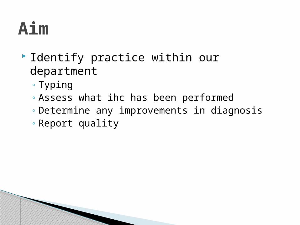
‘As with biopsies, cytological findings should be correlated with the clinical and imaging findings to establish whether the available cytological material is sufficient to render a specific diagnosis or a clinically relevant differential diagnosis. If a pleural cytology specimen is positive or suspicious for malignancy, and there is no other specimen, then material should undergo the same ancillary investigations as for biopsies in terms of the differential diagnosis, which ideally is via a cell pellet for histology as this allows preservation of residual material. Identification of an epithelial phenotype will allow a definitive diagnosis of metastatic carcinoma. Identification of a mesothelial phenotype will allow further management decisions in terms of a definitive diagnosis of mesothelioma or further sampling, dependent on the clinical scenario’
RCPath
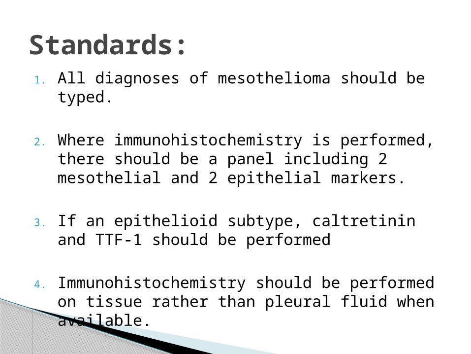
1. All diagnoses of mesothelioma should be typed.
2. Where immunohistochemistry is performed, there should be a panel including 2 mesothelial and 2 epithelial markers.
3. If an epithelioid subtype, caltretinin and TTF-1 should be performed
4. Immunohistochemistry should be performed on tissue rather than pleural fluid when available.
Standards:
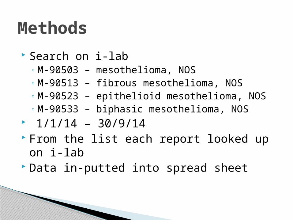
Search on i-lab◦ M-90503 – mesothelioma, NOS◦ M-90513 – fibrous mesothelioma, NOS◦ M-90523 – epithelioid mesothelioma, NOS◦ M-90533 – biphasic mesothelioma, NOS
1/1/14 – 30/9/14 From the list each report looked up on i-lab Data in-putted into spread sheet
Methods
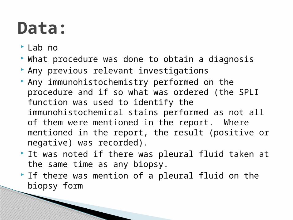
Lab no What procedure was done to obtain a diagnosis Any previous relevant investigations Any immunohistochemistry performed on the
procedure and if so what was ordered (the SPLI function was used to identify the immunohistochemical stains performed as not all of them were mentioned in the report. Where mentioned in the report, the result (positive or negative) was recorded).
It was noted if there was pleural fluid taken at the same time as any biopsy.
If there was mention of a pleural fluid on the biopsy form
Data:
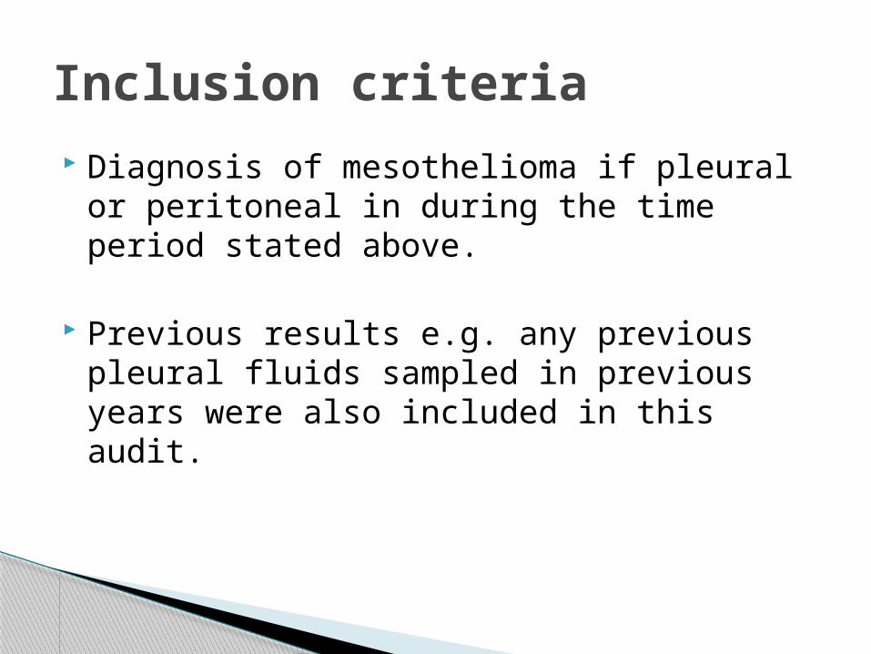
Diagnosis of mesothelioma if pleural or peritoneal in during the time period stated above.
Previous results e.g. any previous pleural fluids sampled in previous years were also included in this audit.
Inclusion criteria
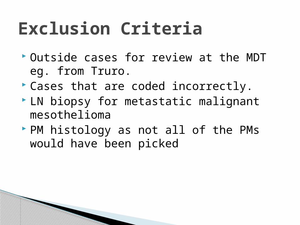
Outside cases for review at the MDT eg. from Truro.
Cases that are coded incorrectly. LN biopsy for metastatic malignant
mesothelioma PM histology as not all of the PMs would
have been picked
Exclusion Criteria

Total 31 biopsies performed◦ 3 excluded
1outside case2 adenocarcinomas coded incorrectly
Remaining 28◦ 26 pleural bx - 20 had cytology at the same time◦ 2 omental bx◦ 27 had ihc
1 did not as it was being performed on the fluid◦ 1 had ihc on both bx and fluid
Results

Accompanying cytology◦ 8/20 (36%) malignant or suspicious of malignancy◦ 12/20 no malignant cells seen◦ 6 cytology forms mentioned bx taken
7 cases bx only taken◦ 1 recurrent case with prev diagnosis 3 yrs ago◦ 1 had 2 previous pleural fluids – blood only and nmcs◦ 5 did not have any previous procedure
2 cases diagnosed on pleural fluid alone
Results
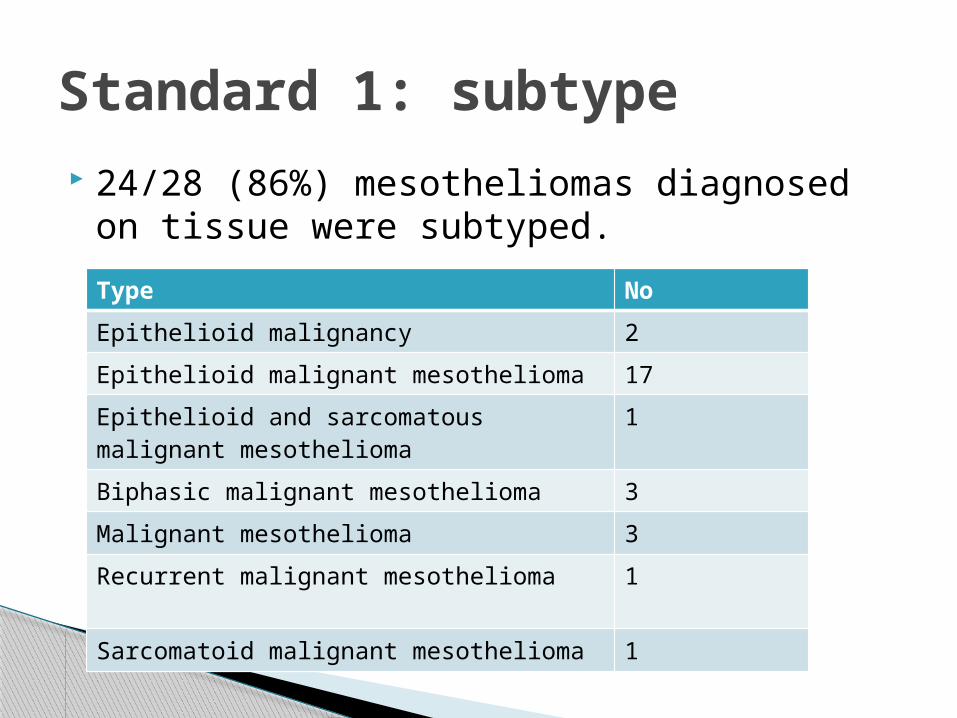
24/28 (86%) mesotheliomas diagnosed on tissue were subtyped.
Standard 1: subtype
Type No
Epithelioid malignancy 2
Epithelioid malignant mesothelioma 17
Epithelioid and sarcomatous malignant mesothelioma
1
Biphasic malignant mesothelioma 3
Malignant mesothelioma 3
Recurrent malignant mesothelioma 1
Sarcomatoid malignant mesothelioma 1
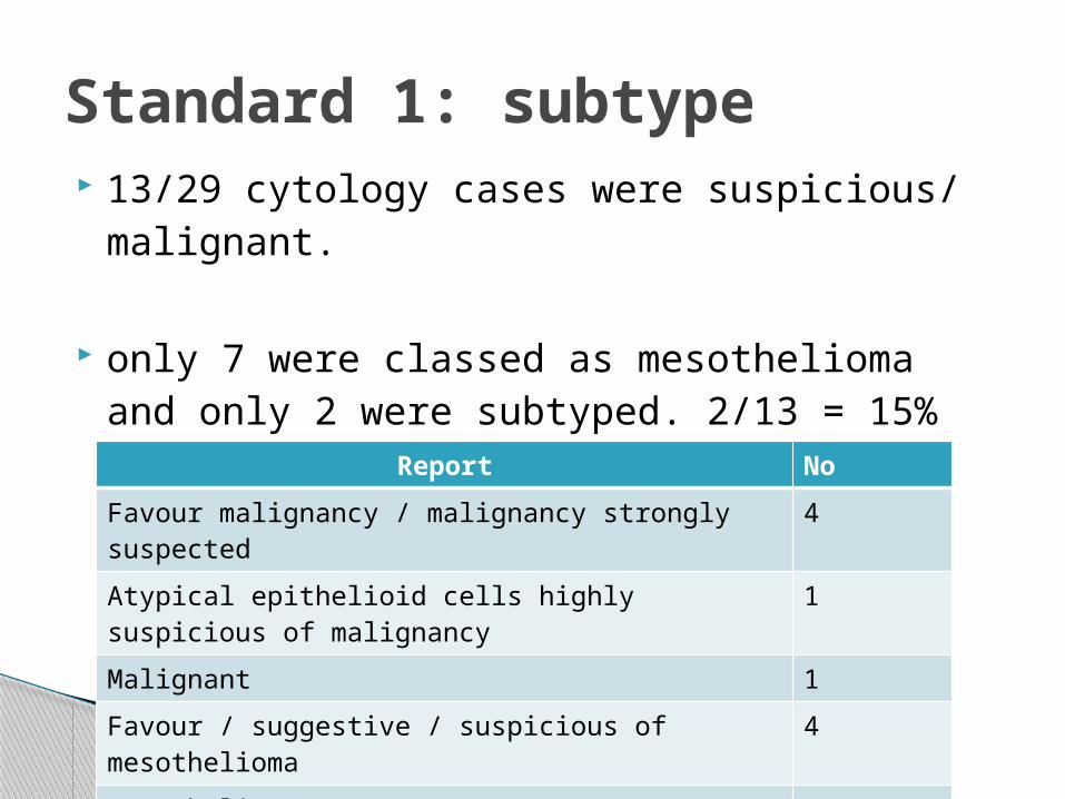
13/29 cytology cases were suspicious/ malignant.
only 7 were classed as mesothelioma and only 2 were subtyped. 2/13 = 15%
Standard 1: subtype
Report No
Favour malignancy / malignancy strongly suspected
4
Atypical epithelioid cells highly suspicious of malignancy
1
Malignant 1
Favour / suggestive / suspicious of mesothelioma 4
Mesothelioma 1
Epithelioid meosthelioma 2
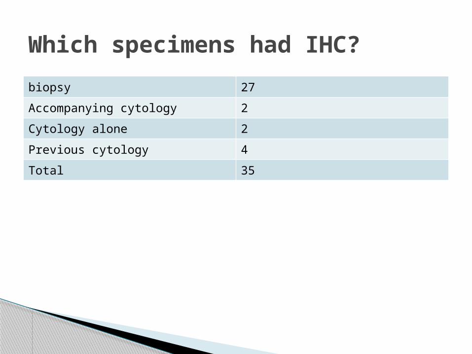
biopsy 27
Accompanying cytology 2
Cytology alone 2
Previous cytology 4
Total 35
Which specimens had IHC?

39 different immuno-stains were ordered Total of 286 ihc orders Average = 8 per case Mucin stains = 10
Ae1/3
CK19
vim
entin CK5
EMA
MNF116
calre
tinin
WT-
1CK7
D240
p53
MOC31
BerEp
4
desm
inCEA
Cam5.
2
pank
erat
in
thro
mbo
mod
ulin
p63
TTF-
1CK20
CDX 2 ER PSA
Hepar
1CD31
CD34s1
00DPA
S
ABDPAS
0
10
20
30
40
50
60
70
80
90
100
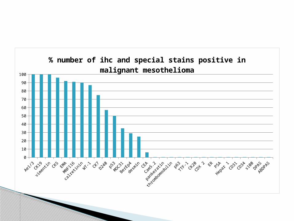
% number of ihc and special stains positive in ma-lignant mesothelioma
Cam5.
2
pank
erat
in
thro
mbo
mod
ulin
p63
TTF-
1CK20
CDX 2 ER PSA
Hepar
1CD31
CD34s1
00DPA
S
ABDPAS
CEA
desm
in
BerEp
4
MOC31 p53D24
0CK7
WT-
1
calre
tinin
MNF116
EMA
CK5
Ae1/3
CK19
vim
entin
0
10
20
30
40
50
60
70
80
90
100
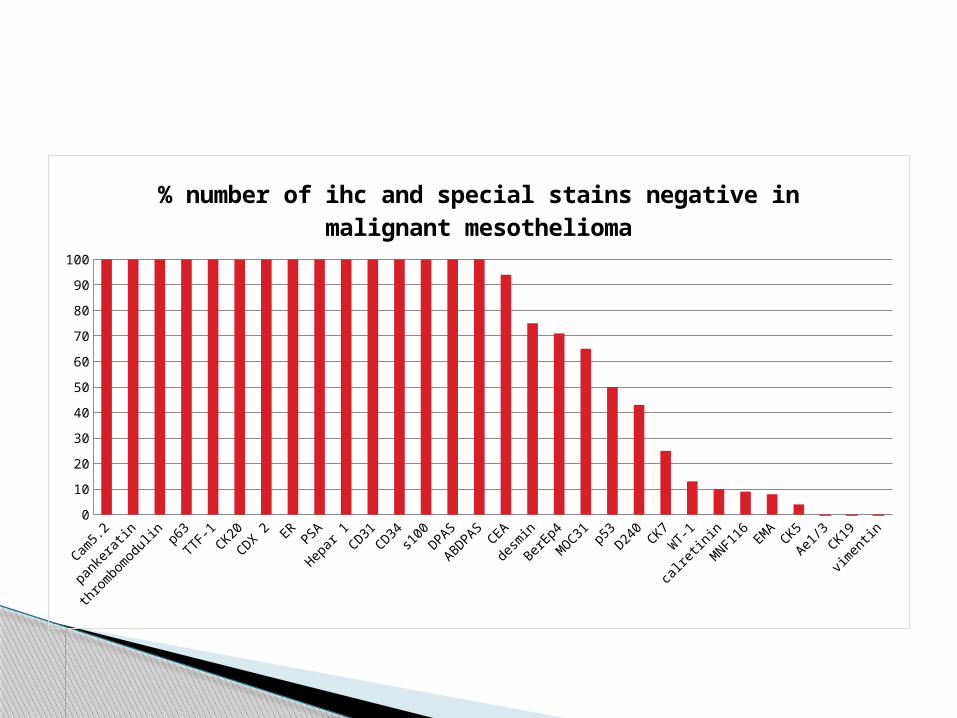
% number of ihc and special stains negative in ma-lignant mesothelioma
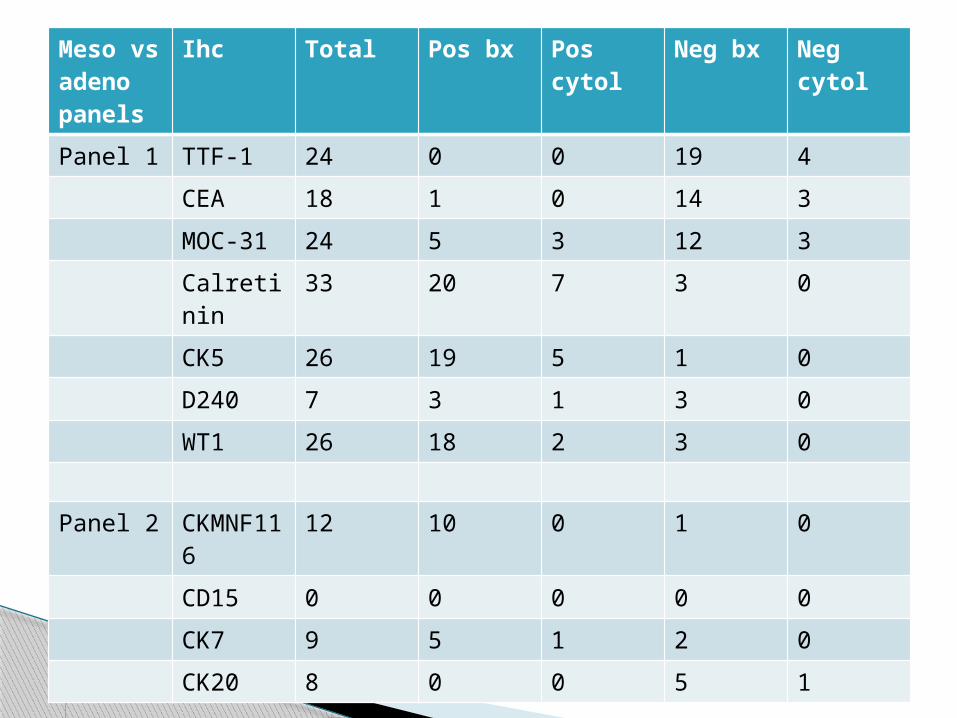
Meso vs adeno panels
Ihc Total Pos bx Pos cytol
Neg bx Neg cytol
Panel 1 TTF-1 24 0 0 19 4
CEA 18 1 0 14 3
MOC-31 24 5 3 12 3
Calretinin
33 20 7 3 0
CK5 26 19 5 1 0
D240 7 3 1 3 0
WT1 26 18 2 3 0
Panel 2 CKMNF116
12 10 0 1 0
CD15 0 0 0 0 0
CK7 9 5 1 2 0
CK20 8 0 0 5 1

Mesothelial markers % positive
CK5 96% (27/28)
Calretinin 90% (29/32)
WT-1 88% (22/25)
Top mesothelial and epithelial markers
Epithelial markers % positive
CEA 6% (1/18)
BerEp4 29% (7/24)
MOC31 35% (8/23)
TTF1 0% (0/23)

30/35 (86%) had 2 epithelial and 2 meso markers
Remaining 5:◦ One had reactive panel◦ 4 had both an epithelial and mesothelial marker,
some with 2 of one but not 2 of both
Standard 2: where ihc performed, there should be 2 epithelial and 2 mesothelial markers
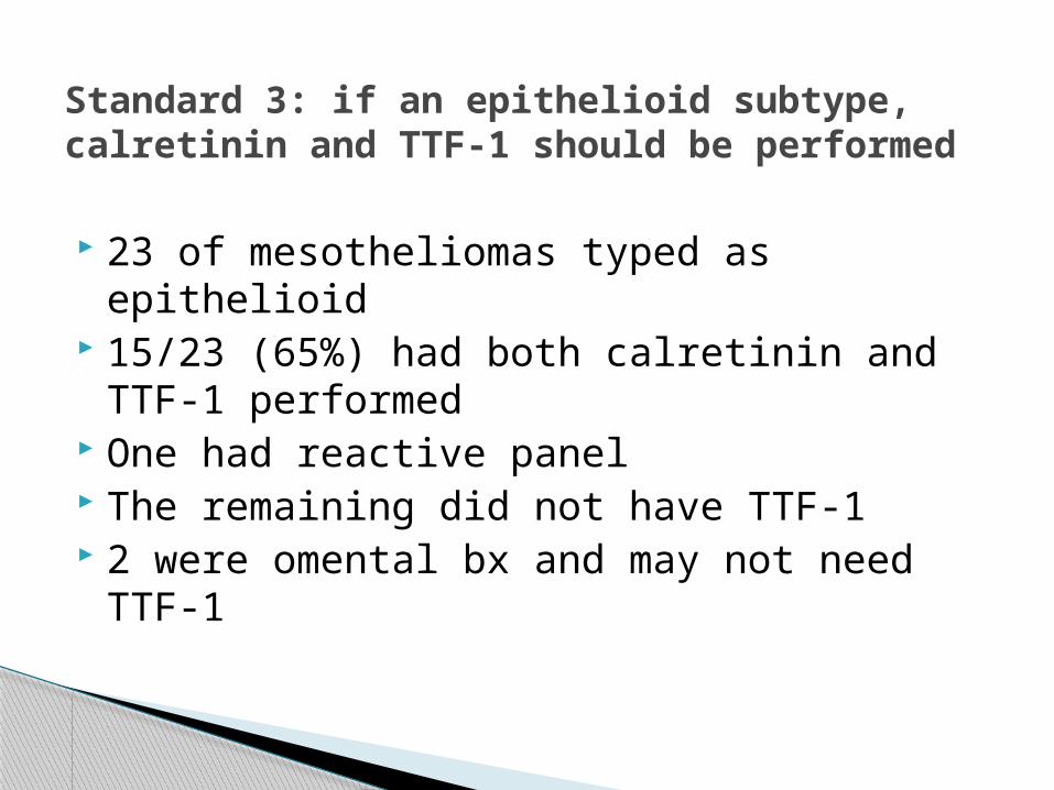
23 of mesotheliomas typed as epithelioid 15/23 (65%) had both calretinin and TTF-1
performed One had reactive panel The remaining did not have TTF-1 2 were omental bx and may not need TTF-1
Standard 3: if an epithelioid subtype, calretinin and TTF-1 should be performed

Ihc performed on 27 of the biopsies 1 did not have ihc as it was requested on
the cytology (commented on in the report) Only 1 patient had ihc on both biopsy and
cytology
Standard 4: ihc should be performed on tissue rather than pleural fluid when available
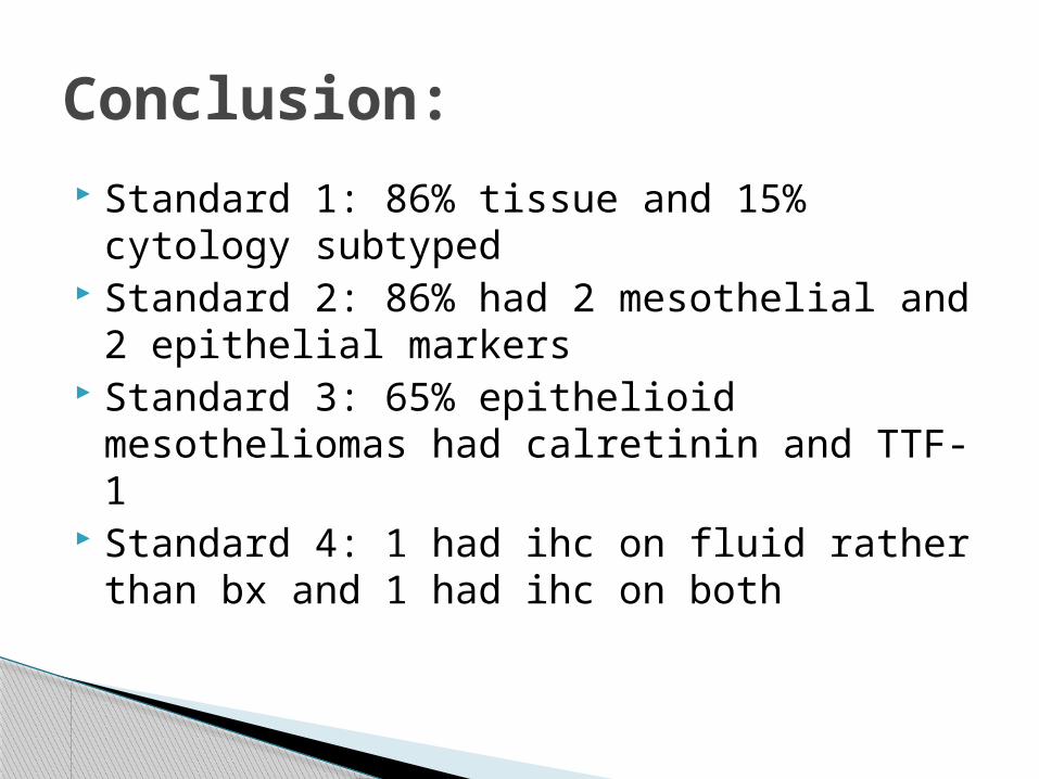
Standard 1: 86% tissue and 15% cytology subtyped
Standard 2: 86% had 2 mesothelial and 2 epithelial markers
Standard 3: 65% epithelioid mesotheliomas had calretinin and TTF-1
Standard 4: 1 had ihc on fluid rather than bx and 1 had ihc on both
Conclusion:
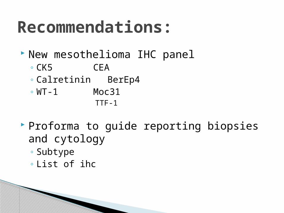
New mesothelioma IHC panel◦ CK5 CEA◦ Calretinin BerEp4◦ WT-1 Moc31
TTF-1
Proforma to guide reporting biopsies and cytology◦ Subtype◦ List of ihc
Recommendations:
http://www.hscic.gov.uk/media/15038/Mesothelioma-audit/pdf/EMBARGOEDTO120914_NLCA_Meso_Report_final.pdf
Standards and datasets for reporting cancers. The dataset for the histological reporting of mesothelioma. RCPath guidelines
References:
Top Related