


Languages
Pages
Legal

UNIVERSIDADE DE LISBOA
FACULDADE DE CIÊNCIAS
DEPARTAMENTO DE BIOLOGIA VEGETAL
Biogeography of Xanthophyllomyces dendrorhous,
a yeast with biotechnological potential – population
mapping and host associations
Márcia Cristina David Palma
MESTRADO EM MICROBIOLOGIA APLICADA
2011
UNIVERSIDADE DE LISBOA
FACULDADE DE CIÊNCIAS
DEPARTAMENTO DE BIOLOGIA VEGETAL
Biogeography of Xanthophyllomyces dendrorhous,
a yeast with biotechnological potential – population
mapping and host associations
Dissertação orientada por Prof. Dr. José Paulo Sampaio (CREM, FCT-UNL)
e Profª. Drª. Margarida Barata (FCUL)
Márcia Cristina David Palma
MESTRADO EM MICROBIOLOGIA APLICADA
2011
Biogeography of Xanthophyllomyces dendrorhous,
a yeast with biotechnological potential – population
mapping and host associations
Márcia Cristina David Palma
MASTER THESIS
2011
This thesis was fully performed at CREM (Centro de Recursos Microbiológicos),
Departamento de Ciências da Vida of Universidade Nova de Lisboa under the direct
supervision of Prof. Dr. José Paulo Sampaio.
Prof. Dr. Margarida Barata was the internal designated supervisor in the scope of the Master
in Applied Microbiology of the Faculty of Sciences of the University of Lisbon.

I
To my father, the best of teachers
II
Acknowledgments
I wish to express my gratitude to my supervisor, Prof. Dr. José Paulo Sampaio first, for
allowing me to integrate his team as a research fellow and for giving me the opportunity to
learn and grow as a scientist. Secondly, I would like to thank him for accepting me as a
master student, for giving me time to write this thesis and for all the scientific knowledge
passed on to me during this period.
I would like to convey my acknowledgement to the host laboratory CREM (Centro de
Recursos Microbiológicos) and FCT-UNL, for giving me the necessary conditions in order to
perform my work. I also thank FCT (Fundação para a Ciência e Tecnologia) for providing
financial support to the project in which this work was included (PTDC/BIA-
BDE/73566/2006).
A special thanks to Dr. Diego Libkind for all the strains that he conveyed for this study,
for all the information regarding the genome of X. dendrorhous and for all his collaboration in
so many aspects of this work.
I would like to thank Prof. Dr. Rogério Tenreiro and Prof. Dr. Margarida Barata for their
help, answering promptly to all my questions during the time of preparation of this thesis.
I would like to thank all my lab colleagues whose knowledge, encouragement and cheer
kept me up when science got me down, especially Marco Coelho, Dr. Elisabete Valério,
Carla Gonçalves and Jorge Santos.
To my best friend, my love and my life, thank you for being all this and more.
My parents deserve special mention for their limitless support and for all the things that
they taught me. Words fail me to express my never ending gratitude and love for my mother
whose support has allowed me to pursue my dream of becoming a scientist. No words are
necessary to thank my father for wherever he may be, he knows that my inquisitive spirit was
born from his own and my unwearyingly willpower and work ethics is but a fragment of his.
III
Resumo
Xanthophyllomyces dendrorhous é uma levedura basidiomiceta cuja grande relevância
biotecnológica advém do facto de ser a única espécie cujo principal pigmento produzido é
astaxantina. Este carotenoide possui várias atividades biológicas relevantes e inúmeras
aplicações industriais, sendo o segundo carotenoide mais rentável do mercado. Apesar do
interesse nesta levedura devido às suas aplicações biotecnológicas, ainda são limitados os
conhecimentos sobre a sua biologia e biogeografia.
X. dendrorhous foi isolada pela primeira vez durante a década de 60 do século passado
a partir de exsudados de árvores de folha caduca em diversas localizações no Hemisfério
Norte, tendo-se considerado durante muito tempo restrita a estes habitats e latitudes.
Inicialmente descrita como uma levedura assexuada designada Phaffia rhodozyma, em
1995 o seu estado sexuado, Xanthophyllomyces dendrorhous, foi observado e descrito pela
primeira vez. O seu ciclo sexuado é caracterizado pela conjugação entre a célula-mãe e a
gémula, após a qual ocorre a formação de um holobasídio aéreo que subsequentemente dá
origem a basidiósporos na sua extremidade apical. Este estado sexuado é despoletado pela
presença de polióis no meio de cultura. Taxonomicamente esta levedura encontra-se
classificada na ordem Cystofilobasidiales, classe Tremellomycetes e subfilo
Agaricomycotina. A descoberta de um novo habitat de X. dendrorhous em 2007 na
Argentina (Patagónia), veio mostrar que esta levedura possui uma distribuição geográfica
mais diversificada do que aquela considerada inicialmente. Nas florestas da Patagónia, X.
dendrorhous encontra-se associada aos corpos frutíferos de um fungo ascomiceta, parasita
obrigatório de árvores Nothofagus, denominado Cyttaria. À semelhança dos exsudados de
árvores, os corpos frutíferos de Cyttaria são ricos em açúcares simples, estando presentes
nos troncos de Nothofagus uma vez por ano. Após a descoberta de novas populações de X.
dendrorhous no Hemisfério Sul foi realizado um estudo filogenético da região ITS (internal
transcribed spacer) do rDNA das estirpes de X. dendrorhous e das diferentes árvores
hospedeiras tendo-se verificado concordância entre ambas as filogenias. Este facto levou à
formulação da hipótese de que a variabilidade intraespecífica observada em X. dendrorhous,
poderia dever-se ao facto de cada linhagem se encontrar associada a um hospedeiro
diferente. Após a descoberta de X. dendrorhous no Hemisfério Sul, a exploração de outras
regiões geográficas onde, tanto Nothofagus como Cyttaria estão presentes, foi levada a
cabo em 2009 por elementos do laboratório de acolhimento. As amostras biológicas
recolhidas na Austrália e Nova Zelândia resultaram na obtenção de novos isolados de
Xanthophyllomyces.
Face ao número crescente de isolados de Xanthophyllomyces existentes, o presente
trabalho teve como principal objetivo o estudo das relações filogenéticas entre as diferentes
IV
linhagens existentes assim como, a exploração de possíveis relações entre a variabilidade
genética encontrada em Xanthophyllomyces e os hospedeiros aos quais esta levedura se
encontra associada. De forma a atingir este objetivo, duas abordagens distintas foram
exploradas: (i) amplificação, sequenciação e estudo filogenético de sete fragmentos de
genes de Xanthophyllomyces e (ii) a cariotipagem de isolados representativos das diferentes
linhagens encontradas. Os dados obtidos foram analisados sob o ponto de vista da
associação aos hospedeiros e localização geográfica dos isolados. Os resultados
mostraram a existência de quatro populações distintas em X. dendrorhous. A estrutura
populacional encontrada sugere que as populações naturais se propagam preferencialmente
de forma clonal. Cada uma das diferentes populações apresenta-se associada a diferentes
plantas hospedeiras, sendo esse o fator que mais parece influenciar a estrutura
populacional. No entanto verificou-se também alguma diferenciação geográfica apesar da
sua influência parecer menos relevante. Na filogenia concatenada dos sete fragmentos de
genes estudados foi observado que cada grupo filogenético principal agrupa estirpes
isoladas a partir de um mesmo hospedeiro, sendo igualmente possível verificar a existência
de subdivisões dentro destes mesmos grupos, que estão por sua vez associadas à origem
geográfica dos isolados. Este facto foi corroborado pelas networks filogenéticas construídas
no software Splistree4 e pela análise da estrutura populacional usando uma abordagem
bayesiana implementada no software Structure. A determinação dos cariótipos de estirpes
representativas de cada uma das populações encontradas em X. dendrorhous revelou
padrões únicos de cromossomas para cada estirpe. O número de cromossomas das onze
estirpes estudadas varia entre seis e 11, sendo o seu tamanho entre 0,8 Mb e 3,5 Mb. Não
foi observada uma relação entre a estrutura populacional de X. dendrorhous e os cariótipos
observados para as diferentes estirpes.
Verificou-se que um dos isolados em estudo, possui três genes heterozigóticos. Os
alelos de cada gene foram separados por clonagem e posteriormente sequenciados, tendo-
se verificado a existência de dois alelos distintos para cada gene heterozigótico. A análise
filogenética permitiu observar que, para os três genes heterozigóticos, os alelos encontrados
pertencem a duas populações distintas originárias respectivamente da Australásia (em
associação com Nothofagus) e Japão (em associação com Betula). Este facto sugere a
existência de contacto entre as duas populações.
Diversos estudos biogeográficos têm sido realizados sobre os géneros Nothofagus,
Betula e Cornus. Enquanto que a biogeografia de Nothofagus parece ter sido moldada por
fenómenos como a fragmentação do super continente Gondwana e eventos de dispersão a
longas distâncias, as histórias biogeográficas de Betula e Cornus parecem ter sido
influenciadas por migrações intercontinentais através das pontes terrestres de Bering e do
Oceano Atlântico durante o período geológico Terciário. A análise da atual distribuição de X.
V
dendrorhous levando em consideração as histórias biogeográfias dos seus hospedeiros
parece sugerir uma correlação entre ambas. É possível que a distribuição de X. dendrorhous
no Hemisfério Sul esteja relacionada com a biogeografia de Nothofagus, assim como a sua
distribuição no hemisfério Norte parece ser coerente com as histórias biogeográficas de
Betula e Cornus. A coexistência de Nothofagaceae, Fagaceae e Betulaceae na região do
arquipélago indo-australiano poderá ter permitido um salto de hospedeiro por parte de X.
dendrorhous que assim colonizou outras áreas no Hemisfério Norte.
Entre os isolados recolhidos na Australásia foram encontradas duas putativas novas
espécies de Xanthophyllomyces. É possível observar que a divergência destas novas
espécies relativamente a X. dendrorhous se verifica ao nível molecular tanto na região ITS
como nas sete regiões genómicas estudadas. As duas espécies, Xanthophyllomyces sp. I e
Xanthophyllomyces sp. II apresentam o mesmo estado sexuado que X. dendrorhous, assim
como produção de astaxantina. O presente estudo constitui o primeiro registo da existência
de mais do que uma espécie no género Xanthophyllomyces. Com este estudo iniciou-se a
exploração da recém descoberta biodiversidade existente na Australásia, identificando-a
como um possível hotspot de diversidade e possivelmente como ponto de origem do género
Xanthophyllomyces.
Palavras chave: Xanthophyllomyces; Biogeografia; Associação a hospedeiros; Espécies novas;
Diversidade.
VI
Abstract
The basidiomycetous yeast Xanthophyllomyces dendrorhous has biotechnological
relevance due to the production of astaxanthin, a carotenoid with remarkable biological
properties. First isolated during the 1960s from exudates of deciduous trees throughout the
Northern Hemisphere, this yeast was for long considered to be restricted to these habitats
and latitudes. The discovery of a new Xanthophyllomyces habitat in the Southern
Hemisphere in 2007 brought a better understanding of the geographic distribution and
ecology of this yeast. In Patagonian forests, X. dendrorhous was discovered to be associated
with the fruiting bodies of Cyttaria, an ascomycetous parasite of Nothofagus trees. Given that
the Cyttaria-Nothofagus host system is also present in Oceania, sampling of selected
localities in Australasia was carried out in 2009 yielding a considerable number of new
isolates of Xanthophyllomyces.
Benefiting from the growing collection of Xanthophyllomyces strains available, that
encompasses now novel Australasian lineages, the major intend of this work was to
elucidate the phylogenetic relationships between the different lineages, and to further explore
the genetic variation within Xanthophyllomyces. To achieve this goal a representative group
of isolates was studied using multilocus sequence analysis and karyotyping assays within an
ecological and geographic framework.
The results showed that X. dendrorhous is composed by four well structured
populations, which suggests that natural populations of this yeast are mostly clonal. The
existence of an isolate bearing alleles from two distinct populations suggests contact
between populations from Australasia and Japan. Xanthophyllomyces dendrorhous
populations have different ecological associations and appear to have diverged first
according to host tree and secondly by geography. Different karyotypes were presented by
each of the tested strains and no correlation with the revealed population structure was
observed. The Australasian population harbors two putative new species that appear to be
endemic to the area. This study represents the first record of the existence of more than one
species in the Xanthophyllomyces genus. The extant distribution of the X. dendrorhous in the
Southern Hemisphere may be related with the biogeographic history of Nothofagus trees,
while its Northern Hemisphere distribution appears to be coherent with the biogeographic
histories of Betula and Cornus trees.
Keywords: Xanthophyllomyces; Biogeography; Host association; Novel species; Diversity.
VII
Table of contents
Page
Figure Index VIII
Table Index IX
Symbol and Abbreviation List X
_________________________________________________________________________________
1. Introduction 1
2. Material and Methods 5
2.1. Studied strains 5
2.2. DNA extraction, MLSA primers and PCR conditions 6
2.3. Cloning assays 6
2.4. Phylogenetic analysis 7
2.5. Phylogenetic Networks analysis 7
2.6. Analysis of X. dendrorhous population structure by a quantitative clustering
method 8
2.7. Karyotyping assays 8
3. Results and discussion 11
3.1. Xanthophyllomyces multilocus phylogeny. 11
3.2. Xanthophyllomyces novel species. 14
3.3. Exploring the population structure of X. dendrorhous. 16
3.4. X. dendrorhous karyotype. 20
3.5. X. dendrorhous population structure, host system and geographical distribution. 22
4. Final considerations and future perspectives 27
5. References 28
6. Supplementary data 32
VIII
Figure Index
Page
Figure 1 - Xanthophyllomyces dendrorhous sexual state. 2
Figure 2 - Presently known habitats for Xanthophyllomyces. 3
Figure 3 - Nothofagus world distribution. 3
Figure 4 - Xanthophyllomyces multilocus phylogeny and distribution. 12
Figure 5 - ITS phylogeny of Xanthophyllomyces and Cystofilobasidium. 15
Figure 6 - Splits network of Xanthophyllomyces. 17
Figure 7 - Ancestry of X. dendrorhous estimated with Structure software. 19
Figure 8 - X. dendrorhous karyotypes. 21
Figure 9 - X. dendrorhous population structure and host tree phylogeny. 23
Figure 10 - Xanthophyllomyces extant distribution and hypothetical routes for the colonization
of the Northern Hemisphere. 25
________________________________________________________________________________
Figure S1 - Individual phylogenies of Xanthophyllomyces by ML method. 33
Figure S2 - Multilocus phylogeny of Xanthophyllomyces by ML method. 34
Figure S3 - Sexual state of Xanthophyllomyces strains from Australasia. 35
Figure S4 - X. dendrorhous SN and RN. 35
IX
Table Index
Page
Table 1 - List of strains used in the study and relevant information regarding them. 5
Table 2 - Summary of the PFGE running parameters used. 9
________________________________________________________________________________
Table S1 - Sequences used for the construction of the primers for the multilocus sequence
analysis. 32
Table S2 - List genes and genetic regions used in this study and relevant information
regarding them. 32
Table S3 - composition of the multilocus sequence alignment 33
X
Symbol and abbreviations list
AIC − Akaike information criterion
ATCC - American Type Culture Collection
bp − Base pairs
CBS − Centraalbureau voor Schimmelcultures, Netherlands
CHEF − Contour-clamped homogeneous electric field
CLP − Chromosome length polymorphism
CN − Chromosome number
CRUB - Centro Regional Universitario Bariloche
Cy. − Cystofilobasidium
E. coli − Escherichia coli
ITS − Internal transcribed spacer
k − Population
MCMC − Markov Chain Monte Carlo
ML − Maximum likelihood
MLSA − Multilocus sequence analysis
MP − Maximum parsimony
MYA − Million years ago
PALM − Phylogenetic reconstruction by automated likelihood model selector
PCR − Polymerase chain reaction
PFGE − Pulse field gel electrophoreses
rDNA − Ribosomal deoxyribonucleic acid
RN − Reticulate network
SDS − Sodium dodecyl sulfate
SN − Splits network
Tris − 2-amino-2-hydroxymethyl-propane-1,3-diol
w/v − Weight / Volume
Δk − Delta k (determined by the rate of change in the log probability of data between
successive k values)
1
1. Introduction
Xanthophyllomyces dendrorhous (asexual state Phaffia rhodozyma) is the only
basidiomycetous yeast that produces astaxanthin, a high-value carotenoid, as its main
pigment (Johnson 2003). Astaxanthin has remarkable antioxidant, neuroprotective, anti-
inflammatory and antitumor properties (Naguib 2000, Kurihara et al. 2002, Frengova and
Beshkova 2009, Lee et al. 2010) and is the second most important carotenoid in the market
(Schmidt et al. 2010). The ability to ferment sugars, a rare attribute for basidiomycetous
yeasts, makes X. dendrorhous a promising candidate for a cost-competitive biotechnological
production of astaxanthin (Rodriguez-Saiz et al. 2010).
This yeast was first isolated by Herman Phaff in 1967 from exudates of deciduous trees
in Japan and Alaska (Miller et al. 1976, Golubev 1995) and was described as an asexual
species, Phaffia rhodozyma, in 1976 (Miller et al. 1976). On the following years, other
isolates were obtained also from exudates of deciduous trees in Finland, Germany and
Russia (Golubev 1995, Weber et al. 2006). Through phylogenetic analysis using rDNA
sequences, it was observed that the species of genus Cystofilobasidium were the closest
relatives of Phaffia rhodozyma. The molecular phylogenies placed this yeast in the order
Cystofilobasidiales, class Tremellomycetes and subphylum Agaricomycotina (Scorzetti et al.
2002). In 1995 the sexual state was observed for the first time after inoculation of strains in
an agar medium containing polyols. The sexual state was described as Xanthophyllomyces
dendrorhous and is characterized by the conjugation between cell and bud (pedogamic
sexual process), after which a slender cylindrical holobasidium forms. Basidiospores, usually
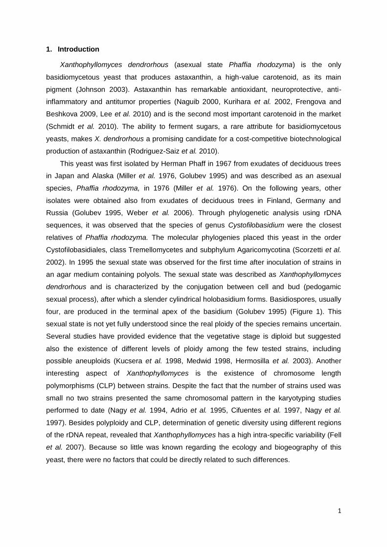
four, are produced in the terminal apex of the basidium (Golubev 1995) (Figure 1). This
sexual state is not yet fully understood since the real ploidy of the species remains uncertain.
Several studies have provided evidence that the vegetative stage is diploid but suggested
also the existence of different levels of ploidy among the few tested strains, including
possible aneuploids (Kucsera et al. 1998, Medwid 1998, Hermosilla et al. 2003). Another
interesting aspect of Xanthophyllomyces is the existence of chromosome length
polymorphisms (CLP) between strains. Despite the fact that the number of strains used was
small no two strains presented the same chromosomal pattern in the karyotyping studies
performed to date (Nagy et al. 1994, Adrio et al. 1995, Cifuentes et al. 1997, Nagy et al.
1997). Besides polyploidy and CLP, determination of genetic diversity using different regions
of the rDNA repeat, revealed that Xanthophyllomyces has a high intra-specific variability (Fell
et al. 2007). Because so little was known regarding the ecology and biogeography of this
yeast, there were no factors that could be directly related to such differences.
2
Figure 1 – Xanthophyllomyces dendrorhous sexual state. (a) Scanning electron microscopy photos showing
two conjugated cells [1] originating a non-septate basidium [2]; (b) aerial basidium [2] with apical basidiospores
[3]; (c) detail of the basidium apex [2] and basidiospores [3]. Image adapted from the website of the American
Society for Microbiology, http://202.195.144.50/ASM/106-Introduce.htm.
Since all Xanthophyllomyces isolates were obtained from tree exudates belonging to
genera Betula, Alnus, Fagus and Cornus it was assumed that the distribution of X.
dendrorhous was limited to these host trees. Moreover, considering the biogeography of the
plants (Chen et al. 1999, Chen and Li 2004, Manchester et al. 2007), it was assumed that
Xanthophyllomyces was restricted to the Northern Hemisphere (Golubev 1995, Fell and Blatt
1999). However, the discovery of a new Xanthophyllomyces habitat in the Southern
Hemisphere brought novel insights regarding the geographic distribution and ecology of this
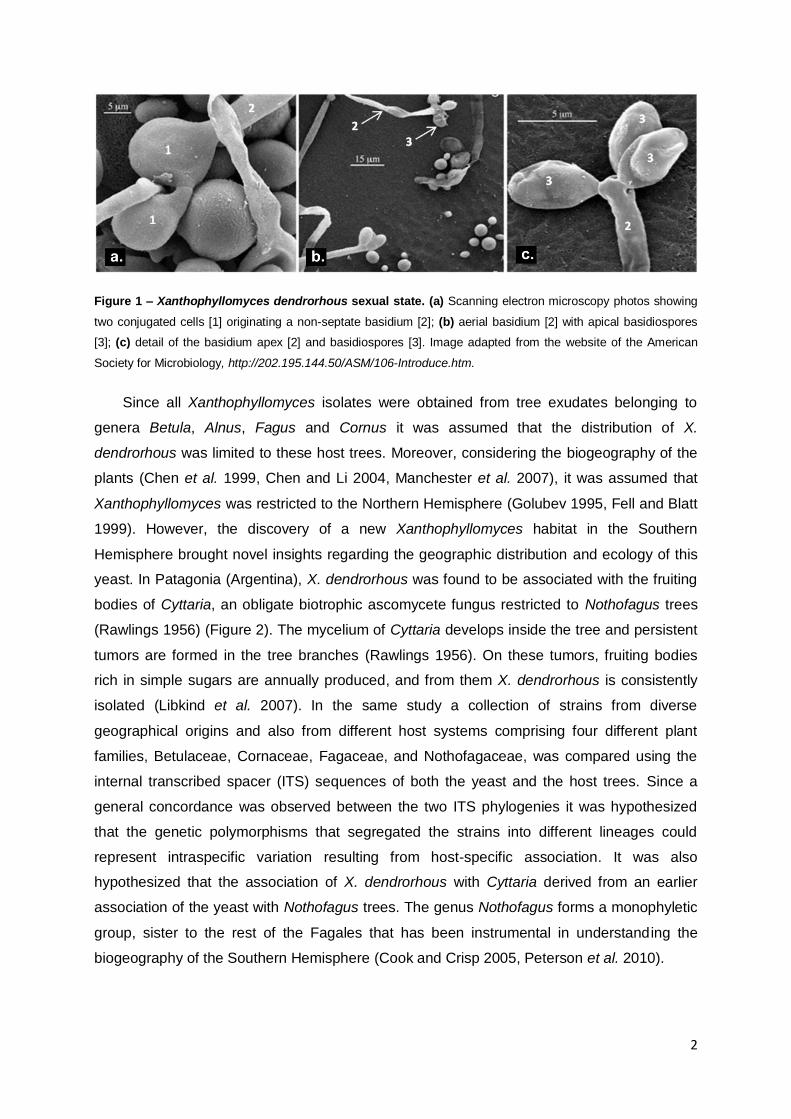
yeast. In Patagonia (Argentina), X. dendrorhous was found to be associated with the fruiting
bodies of Cyttaria, an obligate biotrophic ascomycete fungus restricted to Nothofagus trees
(Rawlings 1956) (Figure 2). The mycelium of Cyttaria develops inside the tree and persistent
tumors are formed in the tree branches (Rawlings 1956). On these tumors, fruiting bodies
rich in simple sugars are annually produced, and from them X. dendrorhous is consistently
isolated (Libkind et al. 2007). In the same study a collection of strains from diverse
geographical origins and also from different host systems comprising four different plant
families, Betulaceae, Cornaceae, Fagaceae, and Nothofagaceae, was compared using the
internal transcribed spacer (ITS) sequences of both the yeast and the host trees. Since a
general concordance was observed between the two ITS phylogenies it was hypothesized
that the genetic polymorphisms that segregated the strains into different lineages could
represent intraspecific variation resulting from host-specific association. It was also
hypothesized that the association of X. dendrorhous with Cyttaria derived from an earlier
association of the yeast with Nothofagus trees. The genus Nothofagus forms a monophyletic
group, sister to the rest of the Fagales that has been instrumental in understanding the
biogeography of the Southern Hemisphere (Cook and Crisp 2005, Peterson et al. 2010).
3
Figure 2 – Presently known habitats for Xanthophyllomyces. (a) Betula tree trunk with an orange colored
exudate in North America (enlarged detail on the right); (b) branch of Nothofagus cunninghamii with Cyttaria
gunnii fruiting bodies (Tasmania).
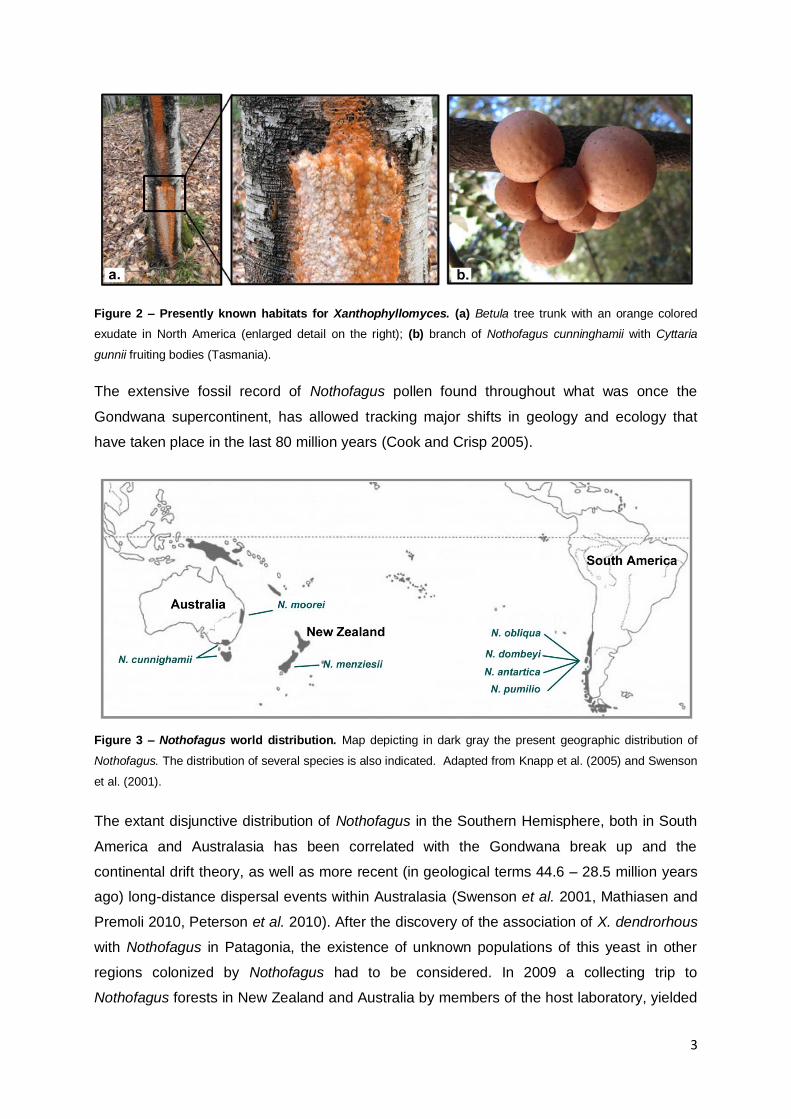
The extensive fossil record of Nothofagus pollen found throughout what was once the
Gondwana supercontinent, has allowed tracking major shifts in geology and ecology that
have taken place in the last 80 million years (Cook and Crisp 2005).
Figure 3 – Nothofagus world distribution. Map depicting in dark gray the present geographic distribution of
Nothofagus. The distribution of several species is also indicated. Adapted from Knapp et al. (2005) and Swenson
et al. (2001).
The extant disjunctive distribution of Nothofagus in the Southern Hemisphere, both in South
America and Australasia has been correlated with the Gondwana break up and the
continental drift theory, as well as more recent (in geological terms 44.6 – 28.5 million years
ago) long-distance dispersal events within Australasia (Swenson et al. 2001, Mathiasen and
Premoli 2010, Peterson et al. 2010). After the discovery of the association of X. dendrorhous
with Nothofagus in Patagonia, the existence of unknown populations of this yeast in other
regions colonized by Nothofagus had to be considered. In 2009 a collecting trip to
Nothofagus forests in New Zealand and Australia by members of the host laboratory, yielded
4
a considerable number of new Xanthophyllomyces isolates confirming the existence of
additional populations associated with Nothofagus trees. As the number of
Xanthophyllomyces strains increased, also did the possibilities of better understanding the
biology, ecology and world distribution of this microorganism.
With the increase of sequence information available, a broad application of phylogenetic
methods has taken hold of fungal taxonomy and of biogeography studies. The multilocus
sequence analysis (MLSA) or complete genome comparisons are being widely applied in
detriment of the single genetic marker approach, given the extensively grater information
yielded (Taylor et al. 1999). These techniques allow not only the recognition of species
(Taylor et al. 2000) but also the study of distinct populations among a species (Taylor and
Fisher, 2003). The statistical phylogeographic approach connects phylogeography
methodologies and classical population genetics methods, providing insights on the
evolutionary history of organisms (Knowles, 2009). Bioinformatics tools available today allow
the inference of parameters such as recombination, population structure and to assign
individuals to a population based on their ancestry composition from MLSA datasets
(Crandall et al. 2000, Posada and Crandall 2001, Excoffier and Heckel 2006, Huson and
Bryant 2006, Falush et al. 2007, Perez-Losada et al. 2007, Chen et al. 2009)
Taking advantage of the growing collection of Xanthophyllomyces strains that
encompasses novel Australasian lineages, the major intend of this work was to shed light on
the phylogenetic relationships between the different lineages, and to explore in more detail
the genetic variation previously observed. To achieve this goal a representative group of
isolates was investigated by MLSA and karyotype analysis within an ecological and
geographic framework.
5
2. Material and methods
2.1. Studied strains
Forty strains from nine different geographical localizations and two distinct host systems
were studied (Table 1).
Table 1 – List of strains used in the study and relevant information regarding them.
Species Strain Locality Substrate
X. dendrorhous
CBS 5905 Kyoto, Japan Exudate of Fagus crenata
ATCC 24230 Japan Exudate of Betula tauschii
ATCC 24229 Hiroshima, Japan Exudate of Cornus brachypoda
CBS 5908 Kiso, Japan Exudate of Alnus japonica
ATCC 24201 Hiroshima, Japan Exudate of Cornus brachypoda
ATCC 24261 Yamagata, Japan Exudate of Betula maximowicziana
GY13L04 Taiwan Soil sample near Stauntonia purpurea
ATCC 24228 Kenai Lake, Alaska, United States of America Exudate of Betula papyritosa
NRRL Y-17432 Wisconsin, United States of America Exudate of Betula populifolia
NRRL Y-17433 Illinois, United States of America Exudate of Betula populifolia
NRRL Y-17434 Illinois, United States of America Exudate of Betula populifolia
NRRL Y-27348 Illinois, United States of America Exudate of Betula populifolia
CBS 7918T Moscow, Russia Exudate of Betula verrucosa
CBS 6938 Finland Exudate of Betula sp.
KBP 2604 Novgorod, Russia Exudate of Betula sp.
KBP 2607 Komi Republic, Russia Exudate of Betula sp.
CRUB 0853 Gutierrez Lake coast, Patagonia, Argentina C. hariotii on N.dombeyi
CRUB 1149 Ilón Lake, Patagonia, Argentina Water sample near C. hariotii on N. pumilio
CRUB 1490 Gutierrez Lake coast, Patagonia, Argentina C.hariotii on N.dombeyi
CRUB 1613 Suisse colony, Patagonia, Argentina C.hariotii on N.dombeyi
CRUB 1614 Llao-llao Forest, Patagonia, Argentina C. hariotii on N.dombeyi
CRUB 1617 Tronador area, Patagonia, Argentina C. hariotii on N.antartica
CRUB 1620 Chile border, Patagonia, Argentina C. hariotii on N.dombeyi
CRUB 1621 Tronador area, Patagonia, Argentina C. hariotii on N. antarctica
CRUB 1151 Ilón Lake, Patagonia, Argentina Water sample near C. hariotii on N. pumilio
CRUB 1492 Gutierrez Lake coast, Patagonia, Argentina Stroma of C. hariotii on N. dombeyi
CRUB 1917 Hua-Hum, Patagonia, Argentina C.espinosae on N. obliqua
CRUB 1896 South America Leaves of N. pumilio
ZP 869 (Australia ) Tasmania C.gunnii on N.cunninghamii
ZP 874 (Australia ) Tasmania C. gunnii on N.cunninghamii
ZP 922 Haast Pass, New Zealand C.nigra on N.menziesii
ZP 888 Lewis Pass, New Zealand C. gunnii on N.menziesii
Xanthophyllomyces
sp. I
ZP 955 Lamington Park, Queensland, Australia Leaves of N. mooreii
ZP 928 Lamington Park, Queensland, Australia Leaves of N. mooreii
ZP 938 Lamington Park, Queensland, Australia Leaves of N. mooreii
ZP 909 Haast Pass, New Zealand C.nigra on N. menziesii
ZP 884 Lewis Pass, New Zealand C.nigra on N.menziesii
Xanthophyllomyces
sp. II
ZP 863 Mount Field National Park, Tasmania,
Australia C.gunnii on N. cunninghamii
ZP 878 Mount Field National Park, Tasmania,
Australia C. gunnii on N.cunninghamii
ZP 875 Mount Field National Park, Tasmania,
Australia C.gunnii on N. cunninghamii
6
All the strains from Australasia (referred to as ZP) were isolated (Libkind et al. 2007) during
the research project in which the present study is included. Reference cultures were obtained
from the following culture collections: Centraalbureau voor Schimmelcultures, Netherlands
(CBS); Department of Soil Biology, Moscow State University (KBP); American Type Culture
Collection (ATCC), Agricultural Research Service Culture Collection (also referred to as
NRRL Collection) and from Centro Regional Universitario Bariloche (CRUB) in Argentina.
Strain GY13L04 was provided by Dr. Ching-Fu Lee of the National Hsinchu University of
Education in Taiwan.
2.2. DNA extraction, MLSA primers and PCR conditions
Genomic DNA of all strains studied was obtained as previously described (Gonçalves et
al. 2011) and used to amplify different genomic regions. Primers for the MLSA were
designed using sequences publicly available on GenBank (Table S1). The ITS region of the
rDNA comprising the ITS1, 5.8S gene and the ITS2 region was amplified and sequenced
using universal primers (Sampaio et al. 2001) for all Xanthophyllomyces isolates from
Australasia and Taiwan (Table S2). For MLSA a total of seven nuclear genes with distinct
biological functions were selected and fragments were amplified by PCR using the conditions
presented in Table S2. The evaluation of the chemical properties of each of the pair of
primers was performed with software Primer Premier 5.0 (PREMIER Biosoft International,
Palo Alto, California) and also with AutoDimer v1 software (Vallone and Butler 2004). After
the PCR reaction, all products were purified either with NucleoSpin® kit from Macherey-
Nagel Company or the GFXTM PCR DNA and Gel Band Purification Kit from GE Healthcare
Company according to the manufacturers protocols. All PCR products were sequenced by
STAB VIDA. All sequences presented in this study will became available on Genbank once
the results of this thesis are published in a scientific paper.
2.3. Cloning assays
The detection of heterozygous sequences of CRTS, IDI and EPH1 genes in CBS 5905,
made necessary the separation of the different haplotypes through cloning. The genes
fragments were amplified and the products of the PCR reaction were purified as previously
mentioned. Those products were then inserted into the vector pJET1/blunt and the cloning
protocol of the CloneJET™ PCR Cloning Kit from Fermentas® was carried out. Competent
DH5-α E. coli cells were prepared according to previously described method (Sambrook J.
1989) and then used for cloning assays. Due to specific characteristics of the cloning vector
used, only transformed cells are able to grow and no additional screening method was
7
necessary. Colony PCR was performed using gene-specific primers (Table S2) in the
obtained clones and the resulting products were sequenced as previously mentioned.
2.4. Phylogenetic analysis
All sequences produced in this study were aligned using ClustalW Multiple alignment
tool (Thompson et al. 1994), present in the BioEdit software (Hall 1999). The MLSA
phylogenetic analysis was performed using two different inference methods: Maximum
Parsimony (MP) and Maximum Likelihood (ML). All MP phylogenies were performed in
MEGA5 software (Tamura et al. 2011) using 1000 replicates (bootstrap analysis) for each
phylogeny. The ML phylogenetic trees were performed in PALM server (phylogenetic
reconstruction by automated likelihood model selector), which performs automated evolution
model selection (Chen et al. 2009). The best fitting model was determined by Akaike
Information Criterion (AIC) (Akaike 1987) for all alignments (Table S3). The nucleotide
alignments were submitted in PHYLIP format (Felsenstein 1981), the number of substitution
rate category was four (Chen et al. 2009). Individual phylogenetic trees were constructed for
each of the seven partially sequenced genes (CRTS; CRTI; IDI; L41; INV; EPH1 and GDHA)
using both MP and ML. The concatenated alignment was composed by the seven genes
fragments and 41 sequences (CBS 5905 is represented by two sequences consisting of the
two different haplotypes resolved by cloning). The multilocus alignment was constituted by a
total of 3187 positions (1287 informative positions). ML phylogenies are presented as
supplementary data.
The phylogenetic analysis of Xanthophyllomyces ITS sequences was performed using
sequences produced in this study together with sequences already available in GenBank.
Moreover, 17 representative sequences of Cystofilobasidium available in GenBank were
included in the ITS phylogenetic analysis, having the resulting alignment a total of 45
sequences and 369 positions. The phylogenetic analysis of the ITS region of several X.
dendrorhous host trees was also performed. For both ITS phylogenies, the MP method was
employed as detailed before and in these two phylogenies the complete deletion option was
used.
2.5. Phylogenetic Networks analysis
The multilocus alignment was also used to construct phylogenetic networks. This
method allows a more accurate representation of reticulate evolutionary events and also to
evaluate the strength of the phylogenetic signal of a dataset. Two different types of networks
were constructed: (i) Splits Networks (SN) using the Neighbor-net model, and (ii) Reticulate
networks (RN) using the Recombination Network model (Huson and Bryant 2006). In a SN
8
the length of an edge is proportional to the weight of the associated split analogously to the
length of a branch in a phylogenetic tree. A set of incompatible splits is represented in a
network using bands of parallel edges. In a RN the internal nodes represent ancestral
species and nodes with more than one parental group correspond to reticulated events.
Additionally the Phi-test was also performed. This test uses the pairwise homoplasy index,
Phi statistic, to detect refined incompatibility indicating recombination (Bruen et al. 2006).
Both the construction of networks and the Phi-test were conducted in SplitsTree4 software
(Huson and Bryant 2006).
2.6. Analysis of X. dendrorhous population structure by a quantitative clustering
method
In order to investigate the population structure of X. dendrorhous, the multi-locus
sequence dataset was analyzed in Structure software (Falush et al. 2007, Hubisz et al.
2009). This model-based clustering method (Pritchard et al. 2000) consists on a Bayesian
approach using Markov Chain Monte Carlo (MCMC) (Hastings 1970). It detects the
underlying genetic population structure among a set of individuals genotyped at multiple
markers and also computes the proportion of the genetic information of an individual
originating from each inferred population using a quantitative clustering method. The main
characteristics of the computation process were a burning length of 100.000 and a MCMC
after burning of 1.000.000. The ancestry model used was admixture and the number of
populations (K) tested was from 1 up to 10 with 20 runs per each K value. The assumed
ploidy of the genome was diploid. The results obtained by Structure were treated according
to a mathematical method developed in order to perform a more formal analysis of the output
(Evanno et al. 2005). This method uses an ad hoc statistic (ΔK) based on the rate of change
in the log probability of data between successive K values thus allowing accurately detecting
the uppermost hierarchical level of population structure in a dataset.
2.7. Karyotyping assays
With the aim of characterizing the chromosomal profile of strains representative of each
of the X. dendrorhous lineages, an optimized protocol for PFGE-CHEF based on previous
works was developed (Nagy et al. 1994, Adrio et al. 1995, Santopietro et al. 1995). Briefly, a
pre-inoculum of each strain was prepared in a complete medium [Yeast extract, 0.3% (w/v);
peptone, 0.5% (w/v); malt extract, 0.3% (w/v); glucose, 1.0% (w/v)], and incubated at 20ºC
and 150 rpm for 24 h. A portion of that culture was then used to inoculate (DO600 nm= 0.2) 100
ml of new liquid culture medium. After 24 h, cells were collected by centrifugation, washed
with sterile water and quantified on the hemocytometer. A pellet containing 4x10⁸ cells was

9
resuspended in 8 mL of a pre-treatment solution (0.1 M β-mercaptoethanol, 0.01 M EDTA,
0.1 M Tris-HCl, pH 7.4) and incubated for 30 minutes at 25°C, followed by a washing step
and gentle centrifugation. The pellet was subsequently resuspended in an enzyme solution
containing 10 mg/ml of lysing enzymes from Trichoderma harzianum from Sigma prepared
with 1.2 M sorbitol in sodium citrate buffer. Incubation was done in a 50 ml Erlenmeyer at
25°C and 30 rpm. Because the incubation time is strain-specific, periodic microscopic
observations were done and the cells were only collected when at least 90% had formed
protoplasts. The protoplasts were washed with the osmotic stabilizer solution (1.2 M sorbitol)
and then added to 1 ml of low melting point agarose [1.3% (w/v)] also prepared with the
osmotic stabilizer solution. Using Bio-Rad plug molds, protoplast plugs are prepared and
subsequently treated with Proteinase K enzyme solution (1 mg/ml Proteinase K from Sigma;
0.01 M Tris-HCl; 1% SDS; 0.5 M EDTA) for 24 h at 50°C. The plugs were then washed with
0.05 M EDTA and stored at 4ºC until used. For strains that did not form protoplasts
conveniently an extra step was introduced, consisting in the addition of MnCl2 to the liquid
culture medium 12 h after inoculation (to a final concentration of 4.0 mM), which resulted in a
weakening of the cell wall (Pera et al. 1999). The preparation of protoplasts in sufficient
quantity to obtain distinguishable bands without degraded genetic material represents one
limiting step of this technique. Saccharomyces cerevisiae CBS 8803 was used as a standard
for the PFGE-CHEF assays, and its plugs were prepared with the same protocol but the
incubation time with the enzyme solution was only 20 minutes. Two different sets of
conditions were used for the PFGE-CHEF assays. First a shorter preliminary assay, in order
to access the absence/presence of degraded genetic material. If good quality plugs were
obtained, another PFGE run would be done under adequate conditions for the separation of
the X. dendrorhous chromosomes (Table 2). All PFGE-CHEF assays were performed in a
CHEF-DR III system from Bio-Rad.
Table 2 – Summary of the PFGE running parameters used.
Parameters Preliminary assay Final assay
Agarose 1% Mega Base Agarose
prepared in 0.5X TBE
(45 mM Tris-borate: 1 mM EDTA, pH 8.0)
0.9% Mega Base Agarose
prepared in 0.5X TBE
(45 mM Tris-borate: 1 mM EDTA, pH 8.0)
Buffer 0.5X TBE 0.5X TBE
Temperature (ºC) 14 14
Voltage (V/cm) 6 2.7
Pulse (s) 50-90 200-1100
Run time (h) 24 72
Angle(º) 120 120
10
The gel was stained with an ethidium bromide solution (0.5 µg/ml) for 30 min and then
unstained for 30 min in sterile water. Images of the gel were collected with a GelDoc (Bio-
Rad) image acquisition system and analyzed in Gelcompar v4.1 from Applied Maths.
11
3. Results and discussion
3.1. Xanthophyllomyces multilocus phylogeny.
With the aim of assessing the phylogenetic relationships between the different
Xanthophyllomyces lineages a MLSA approach was devised using seven different genomic
regions encoding proteins involved in several metabolic processes (Table S2). The
biotechnological interest in the production of astaxanthin has driven the sequencing and
characterization of the genes associated with its biosynthesis. Three of those genes were
chosen for MLSA due to the fact that each of them had been sequenced for more than one
strain, enabling the selection of regions comprising a larger number of single nucleotide
polymorphisms (SNP). Unfortunately for genes not related to astaxanthin biosynthesis only
one sequence was usually available therefore, the four other genes were chosen for the
multi-locus scheme based on the information of one single sequence each. The expanded
collection of isolates studied made it necessary to construct several sets of primers for four of
the seven genes (Table S2). Previously, it was determined that CRTI and CRTS genes are
not in the same chromosome (Niklitschek et al. 2008) but regarding the rest of the 5 genes
used for the MLSA no such information is available. However the genes chosen codify
proteins involved in very distinct roles in the yeast metabolism, lowering the probability of
being genetically linked. Moreover, unpublished data regarding the genome draft of X.
dendrorhous type strain (CBS 7918T) indicated that all seven genes are present in separate
contigs, being the smallest ~3.0 kb long (personal communication by Dr. Diego Libkind).
Although this does not prove that they are genetically unlinked, it points in favor of this
hypothesis.
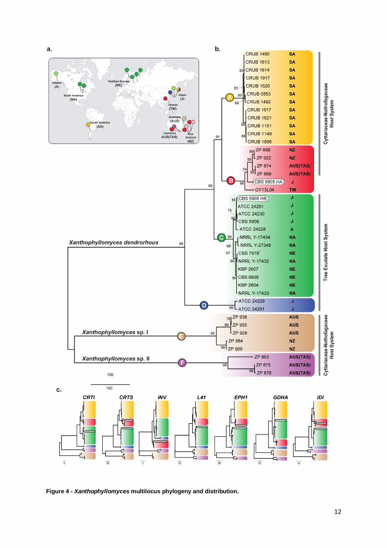
Each of the seven genes was amplified for the collection of 40 strains and the resulting
alignments were used to construct MP (Figure 4c) and ML (Figure S1) based phylogenies.
The individual phylogenies yielded similar results in both methods and were found to be
generally concordant. The multilocus alignment of the seven gene fragments was used to
construct a phylogeny using MP (Figure 4b) and ML (Figure S2). The concatenated dataset
increased the resolution of the analysis in comparison to the individual ones. On every
individual phylogeny a total of six major clades were identified whatever the inference
method used and the same number of clades was resolved in the multilocus phylogeny
(Figure 4b). Before this study, a phylogenetic analysis using the ITS region resolved
Xanthophyllomyces into 3 major clades/lineages (Libkind et al. 2007) that in the multilocus
phylogeny (Figure 4b) would partially correspond to clade A, C and D. The phylogeny also
had a more divergent branch corresponding to strain CBS 5905 that was phylogenetically
isolated from the other lineages (Libkind et al. 2007).
12
Figure 4 - Xanthophyllomyces multilocus phylogeny and distribution.

13
Figure 4 - Xanthophyllomyces multilocus phylogeny and distribution. (a) World map pinpointing the
geographical origins of the strains belonging to each of the main phylogenetic clades presented in the multilocus
phylogeny. Colored pins correspond to the phylogenetic clade to which the isolate belongs. (b) MP multilocus
phylogeny identifying six major clades shown in distinct colors and represented with the letters A - F. Next to each
strain the geographical origin is indicated and also the host system from which it was isolated. Bootstraps values
from 1000 replicates are shown next to the branches. There were a total of 3187 positions in the final dataset
(1287 informative positions). The scale is in the units of the number of changes over the whole sequence. Strain
CBS 5905 presents two different haplotypes (HA and HB) that are highlighted by two white rounded boxes. (c)
Individual MP phylogenies of the seven genes used in MLSA are depicted. Clades are presented in the same
colors has shown above. Each phylogeny is identified by the abbreviated gene names. Full information regarding
the genes is present in Table S2.
In the multilocus phylogeny the clades A, C and D include not only the strains used in the
study by Libkind et al. (Libkind et al. 2007), but also additional isolates comprising a more
diverse sampling both in geography and host trees. From the six resolved clades, three
represent novel Xanthophyllomyces clades (B, E and F), comprising strains from Australasia
and also including one strain (GY13L04) from Taiwan. Within the cluster formed by clades A,
B, C and D small differences in the topology exist between the individual gene phylogenies,
coherent with what is expected under the Phylogenetic Species Recognition model proposed
by Taylor and co-authors (Taylor et al. 2000). In this case, the species cluster of X.
dendrorhous encompasses four different lineages (Figure 4b) where clade B represents a
novel clade. Inside each of the four clades of X. dendrorhous, subgroups of strains are also
resolved and well supported by high bootstrap values. For example, in clade A two
subgroups exist and in clade C, whereas all the strains from Japan group together with the
strain from Alaska, the rest of the strains cluster separately (strains from NA and NE) (Figure
4a and b).The clades E and F always appear in a most divergent position in relation to the
other 4 clades. The fact that the concatenated phylogeny, which has a greater resolution
power than any of the individual ones, is not able to present one of the referred clades as
being the most divergent is probably related to the fact that an appropriate outgroup does not
exist for this multilocus phylogeny.
Regarding strain CBS 5905 it was found that three of the seven gene fragments
sequenced were heterozygotic (Figure 4c; genes CRTS, EPH1 and IDI). The haplotypes
were separated by cloning and then sequenced. It was found that each heterozygotic gene
presented two different haplotypes that always grouped in clades B and C in the respective
gene phylogeny. The other 4 genes were found to be homozygotic for strain CBS 5905 and
presented haplotypes that grouped either in clade B (INV gene) or in clade C (CRTI, L41 and
GDHA genes) (Figure 4c), which further confirms the results obtained for the heterozygotic
genes. These results seem to indicate that this strain is a result of a cross between members
of two lineages represented by clades B and C, which originate respectively from
14
Australasia/Taiwan and Japan. This suggests that a contact area between lineages B and C
might have existed allowing gene flow between them.
Concerning the host system, it is possible to observe that the strains from the Cyttaria -
Nothofagus host system are dispersed along the phylogeny in opposition with the isolates
collected from tree exudates that are confined to clades C and D (Figure 4b).
3.2. Xanthophyllomyces novel species.
The recently found Australasian populations of Xanthophyllomyces encompass the
largest genetic diversity ever observed in this genus. The Australasian genotypes are divided
into three distinct lineages, one (clade B) somewhat similar to X. dendrorhous found in
Southern America and two others (clades E and F) that are distinct from any other population
ever reported (Figure 4b). Although the isolates from the latter lineages present the same life
cycle as X. dendrorhous (Figure S3) and also produce astaxanthin (personal communication
by Dr. Diego Libkind), they have a considerable genetic divergence in comparison to all
known populations of X. dendrorhous (Figure 4b and Figure 5). The relevant divergence
existing between clade E and F and the remaining clades suggests that these novel
Australasian clades might in fact represent two new distinct species within the genus
Xanthophyllomyces (Figure 4b). The separation of Xanthophyllomyces into three species is
not only supported by the multilocus phylogeny but also by the ITS phylogeny (Figure 5). The
ITS phylogeny presented in Figure 5 includes the genera Xanthophyllomyces and
Cystofilobasidium and allowed comparing the genetic divergence of the different species
within both genera. The degree of genetic divergence observed in the ITS sequences within
X. dendrorhous species is somewhat similar to that observed within Cy. capitatum or Cy.
macerans (Figure 5). However, the number of different genotypes included in X. dendrorhous
appears to be higher than that found in each Cystofilobasidium species, which seem to be
much more homogenous. The two putative novel species of Xanthophyllomyces also exhibit
genetic diversity. Within clades, E and F, two different lineages appear to exist both in ITS
(Figure 5) and in the multilocus phylogeny (Figure 4b). The study of more strains from these
two new species might elucidate the biogeographic reasons that are accountable for this
apparent lineage differentiation within each species. On clade E, strains ZP 938 and ZP 928
were collected from leafs of N. mooreii in Australia, while strains ZP 909 and ZP 884 were
collected from fruiting bodies of C. nigra on N. menziesii in New Zealand (Table 1). Therefore
geography and host are different and might justify the observed genetic diversity of
Xanthophyllomyces sp. I.
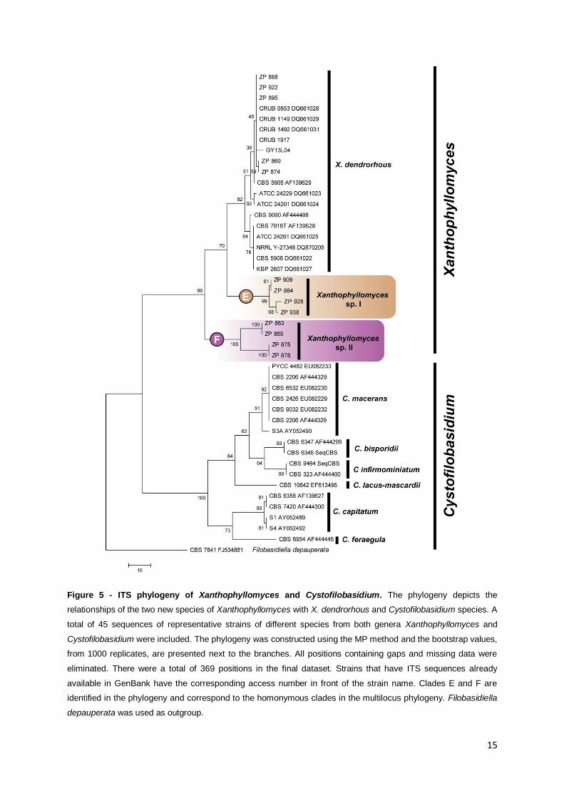
15
Figure 5 - ITS phylogeny of Xanthophyllomyces and Cystofilobasidium. The phylogeny depicts the
relationships of the two new species of Xanthophyllomyces with X. dendrorhous and Cystofilobasidium species. A
total of 45 sequences of representative strains of different species from both genera Xanthophyllomyces and
Cystofilobasidium were included. The phylogeny was constructed using the MP method and the bootstrap values,
from 1000 replicates, are presented next to the branches. All positions containing gaps and missing data were
eliminated. There were a total of 369 positions in the final dataset. Strains that have ITS sequences already
available in GenBank have the corresponding access number in front of the strain name. Clades E and F are
identified in the phylogeny and correspond to the homonymous clades in the multilocus phylogeny. Filobasidiella
depauperata was used as outgroup.
16
On the other hand in clade F all Xanthophyllomyces sp. II isolates share the same
geographical origin and host system (C. gunnii on N. cunninghamii of Tasmania) but present
clearly distinguishable genotypes. Although studied strains belonging to this new species are
few, it seems that the factors influencing their population structure may differ from those
influencing the population structure of X. dendrorhous. While X. dendrorhous has a vast
geographic distribution (Figure 4c, orange, green, red and blue pins), these new putative
species appear to be endemic to Australasia. Whereas X. dendrorhous apparently adapted
to different host trees as new geographic areas where colonized and therefore formed
different lineages, the Australasian species remained in the same host (clade E and F) and in
the same geographical area (Clade F). Both, sympatric speciation or a phenomenon of
secondary contact may be related to the formation or present existence of the different
species encountered in Tasmanian samples (X. dendrorhous and Xanthophyllomyces sp. II).
A more extensive study of isolates of the new species and of the Australasian habitats is
needed in order to draw further insights on their biogeography and evolutionary history.
3.3. Exploring the population structure of X. dendrorhous.
The multilocus sequence analysis provided new information regarding the phylogenetic
relationships of the different lineages that had previously been identified in X. dendrorhous
by sequencing the ITS region. In light of the data provided by the MLSA, X. dendrorhous
seems to be composed by four different lineages represented in the multilocus phylogeny by
four distinct clades (A, B, C and D in Figure 4b). The assessment of evolutionary
relationships using the phylogenetic tree model is done under the assumption that the
evolution process is dominated by two types of events, mutation and speciation (Huson and
Scornavacca 2011). Under more complex models of evolution that might involve events as
complex as horizontal gene transfer, hybridization and recombination, phylogenetic trees
may not provide an adequate representation of the phylogenetic history of the organism.
Phylogenetic networks comprise more realistic models which are able to clearly represent
events such as recombination (Huson 1998, Huson and Bryant 2006). The utilization of
phylogenetic networks also allows determining if the dataset under analysis has a strong
phylogenetic signal or not. In order to assess the strength of the phylogenetic signal (Figure
6) and if reticulate events as recombination played a relevant role in the evolution of this
species (Figure S4), phylogenetic networks were constructed using the multilocus dataset.
Two types of networks were tested: (i) Splits Networks (SN), which constitutes a
combinatorial generalization of phylogenetic trees therefore enabling the visualization of
incompatibilities within and between datasets (Huson and Scornavacca 2011) and (ii)
Reticulate networks (RN) that provide a explicit representation of the evolutionary history in
17
the presence of reticulate events such as recombination (Huson and Bryant 2006, Huson
and Scornavacca 2011).
Figure 6 - Splits network of Xanthophyllomyces. (a) Neighbor-net network constructed with the multilocus
dataset, depicting all Xanthophyllomyces strains except CBS 5905 (39 strains). The main edges of the network
correspond to the clade of the multilocus phylogeny of Figure 4b. (b) Neighbor joining tree constructed with the
multilocus dataset, depicting all Xanthophyllomyces strains except CBS 5905 (39 strains). (c) Phi-test results
between and within populations. Analysis was performed using the multilocus dataset with all X. dendrorhous
strains (d) Neighbor-net network depicting all 40 strains of Xanthophyllomyces.
In order to determine if the phylogenetic signals obtained from the seven different genomic
regions were strong and coherent, individual alignments of each genetic marker were
introduce and concatenated in SplitsTree4 to build Neighbor-net networks (a category of SN)
that implicitly represents such differences. Three different multilocus sub datasets were
18
tested in network analysis: (i) one comprising all Xanthophyllomyces species (40 strains)
(Figure 6d); (ii) another comprising all Xanthophyllomyces strains except CBS 5905 (39
strains) (Figure 6a); (iii) and finally one compose only by the strains belonging to X.
dendrorhous but without CBS 5905 (31 strains) (Figure S4). The Neighbor-net network on
Figure 6a presents a group of incompatible splits regarding the relation of clade D with the
other 3 clades that compose X. dendrorhous. This, and the small topological differences
observed when comparing the individual phylogenetic trees (Figures 4c and 5) suggests
more clearly that the limit of the species X. dendrorhous lies in the transition from
concordance among branches to incongruity among branches observed within clades A to D
(Taylor et al. 2000). On Figure 6d the network differs slightly from the tree-like form (Figure
6b) because it includes the two haplotypes of the CBS 5905 heterozygotic genes. The
network clearly shows that alleles in CBS 5905 most likely derived from representatives of
clade B and from a subgroup of Japanese strains in clade C. The remaining strains present a
position in the network similar to that in the phylogenetic tree. A RN was constructed for the
X. dendrorhous multilocus dataset but did not yield any additional information regarding the
occurrence of reticulate events since the obtained topology overlapped that of the SN (Figure
S4). Because all SN have generally a tree-like form (Figure 6a, 6b and 6d), it is possible to
assess that reticulate events were not dominant in the evolution of X. dendrorhous and also
that the phylogenetic signal in the multilocus dataset is strong (Wagele and Mayer 2007).
The pairwise homoplasy index (Phi) test (Bruen et al. 2006) was applied in order to
determine if signs of recombination within and between the four distinct populations in X.
dendrorhous could be detected. This test detects refined incompatibility in sequence data
(Bruen et al. 2006) assuming the infinite sites model of evolution (Nei 1973) in which the
detection of incompatibility for a pair of sites indicates recombination. The test was
performed in a two by two comparison as represented in Figure 6c using the multilocus
dataset with all the strains from X. dendrorhous. Since at least three sequences are
necessary to perform the test, it was not possible to perform Phi-test within clade D.
Recombination was detected only between clades B and C. When the same test was
performed using the multilocus dataset composed by all X. dendrorhous strains with the
exception of CBS 5905 strain, no statistically significant values were retrieved from the
analysis, therefore no recombination was detected. Recombination was already
demonstrated in the laboratory between strains CBS 6932 and CBS 5908 (both belonging to
clade C) by crossing auxotrophic mutants and analyzing of the progeny (Kucsera et al.
1998). However no recombination was detected by Phi-test within that clade. This might
imply that although the strains are able to cross and undergo meiosis in the laboratory, in
natural populations of X. dendrorhous inbreeding appears to be dominant. The association to
19
very specific ecological niches and the peculiar mating process between parental cell and
bud might also be contributing to the well structured populations detected in X. dendrorhous.
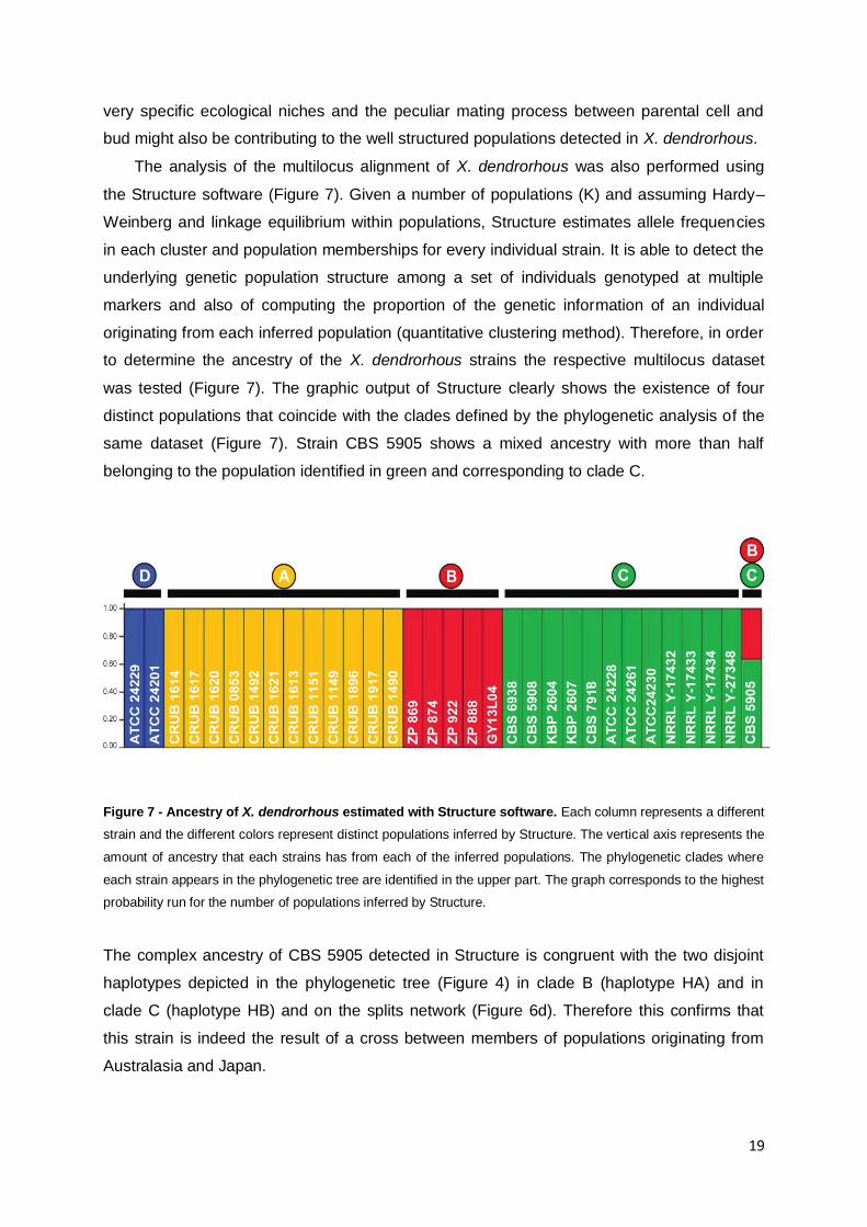
The analysis of the multilocus alignment of X. dendrorhous was also performed using
the Structure software (Figure 7). Given a number of populations (K) and assuming Hardy–
Weinberg and linkage equilibrium within populations, Structure estimates allele frequencies
in each cluster and population memberships for every individual strain. It is able to detect the
underlying genetic population structure among a set of individuals genotyped at multiple
markers and also of computing the proportion of the genetic information of an individual
originating from each inferred population (quantitative clustering method). Therefore, in order
to determine the ancestry of the X. dendrorhous strains the respective multilocus dataset
was tested (Figure 7). The graphic output of Structure clearly shows the existence of four
distinct populations that coincide with the clades defined by the phylogenetic analysis of the
same dataset (Figure 7). Strain CBS 5905 shows a mixed ancestry with more than half
belonging to the population identified in green and corresponding to clade C.
Figure 7 - Ancestry of X. dendrorhous estimated with Structure software. Each column represents a different
strain and the different colors represent distinct populations inferred by Structure. The vertical axis represents the
amount of ancestry that each strains has from each of the inferred populations. The phylogenetic clades where
each strain appears in the phylogenetic tree are identified in the upper part. The graph corresponds to the highest
probability run for the number of populations inferred by Structure.
The complex ancestry of CBS 5905 detected in Structure is congruent with the two disjoint
haplotypes depicted in the phylogenetic tree (Figure 4) in clade B (haplotype HA) and in
clade C (haplotype HB) and on the splits network (Figure 6d). Therefore this confirms that
this strain is indeed the result of a cross between members of populations originating from
Australasia and Japan.
20
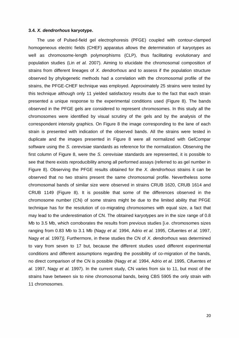
3.4. X. dendrorhous karyotype.
The use of Pulsed-field gel electrophoresis (PFGE) coupled with contour-clamped
homogeneous electric fields (CHEF) apparatus allows the determination of karyotypes as
well as chromosome-length polymorphisms (CLP), thus facilitating evolutionary and
population studies (Lin et al. 2007). Aiming to elucidate the chromosomal composition of
strains from different lineages of X. dendrorhous and to assess if the population structure
observed by phylogenetic methods had a correlation with the chromosomal profile of the
strains, the PFGE-CHEF technique was employed. Approximately 25 strains were tested by
this technique although only 11 yielded satisfactory results due to the fact that each strain
presented a unique response to the experimental conditions used (Figure 8). The bands
observed in the PFGE gels are considered to represent chromosomes. In this study all the
chromosomes were identified by visual scrutiny of the gels and by the analysis of the
correspondent intensity graphics. On Figure 8 the image corresponding to the lane of each
strain is presented with indication of the observed bands. All the strains were tested in
duplicate and the images presented in Figure 8 were all normalized with GelCompar
software using the S. cerevisiae standards as reference for the normalization. Observing the
first column of Figure 8, were the S. cerevisiae standards are represented, it is possible to
see that there exists reproducibility among all performed assays (referred to as gel number in
Figure 8). Observing the PFGE results obtained for the X. dendrorhous strains it can be
observed that no two strains present the same chromosomal profile. Nevertheless some
chromosomal bands of similar size were observed in strains CRUB 1620, CRUB 1614 and
CRUB 1149 (Figure 8). It is possible that some of the differences observed in the
chromosome number (CN) of some strains might be due to the limited ability that PFGE
technique has for the resolution of co-migrating chromosomes with equal size, a fact that
may lead to the underestimation of CN. The obtained karyotypes are in the size range of 0.8
Mb to 3.5 Mb, which corroborates the results from previous studies [i.e. chromosomes sizes
ranging from 0.83 Mb to 3.1 Mb (Nagy et al. 1994, Adrio et al. 1995, Cifuentes et al. 1997,
Nagy et al. 1997)]. Furthermore, in these studies the CN of X. dendrorhous was determined
to vary from seven to 17 but, because the different studies used different experimental
conditions and different assumptions regarding the possibility of co-migration of the bands,
no direct comparison of the CN is possible (Nagy et al. 1994, Adrio et al. 1995, Cifuentes et
al. 1997, Nagy et al. 1997). In the current study, CN varies from six to 11, but most of the
strains have between six to nine chromosomal bands, being CBS 5905 the only strain with
11 chromosomes.
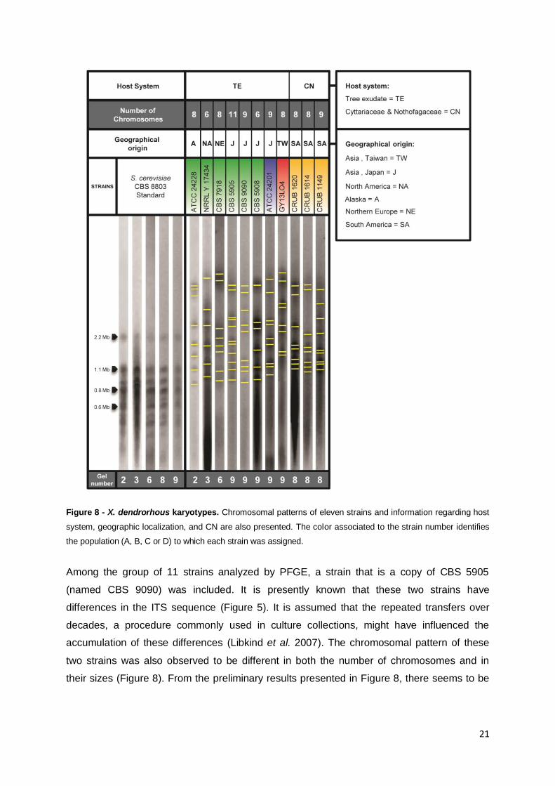
21
Figure 8 - X. dendrorhous karyotypes. Chromosomal patterns of eleven strains and information regarding host
system, geographic localization, and CN are also presented. The color associated to the strain number identifies
the population (A, B, C or D) to which each strain was assigned.
Among the group of 11 strains analyzed by PFGE, a strain that is a copy of CBS 5905
(named CBS 9090) was included. It is presently known that these two strains have
differences in the ITS sequence (Figure 5). It is assumed that the repeated transfers over
decades, a procedure commonly used in culture collections, might have influenced the
accumulation of these differences (Libkind et al. 2007). The chromosomal pattern of these
two strains was also observed to be different in both the number of chromosomes and in
their sizes (Figure 8). From the preliminary results presented in Figure 8, there seems to be
22
no direct relation between the chromosomal profiles and the population structure of X.
dendrorhous.
Although X. dendrorhous karyotypes show a considerable variability in chromosomal
patterns, this species does not represent a unique situation among Fungi, since many other
species present CLP olan P re - rt n et al. 2002, Giraud et al. 2008).The existence
of intraspecific CLP can arise mainly by rearrangements of linked groups of genes or gains
and losses of non coding sequences (Zolan 1995, Poláková et al. 2009). It was shown for the
basidiomycetous Coprinopsis cinerea (Coprinus cinereus) that two strains differing in the
length of most of their chromosomes are interfertile, and that new CLP appear after meiosis
(Zolan 1995). This means that the progeny of such cross presents a variety of karyotypes,
indicating that the changes in length observed in the chromosomes represent gains and
losses of nonessential sequences (Zolan 1995). A similar situation has been observed when
crossing strains of X. dendrorhous. The progeny showed not only composed karyotypes from
both parental strains, but also novel CLP what could indicate that the genomic regions
altered in the karyotypes of the progeny might not be essential (Kucsera et al. 1998).
Nevertheless in order to assess the importance of the CLP observed in X. dendrorhous, an
understanding of how the genome is arranged is necessary (Zolan 1995).
3.5. X. dendrorhous population structure, host system and geographical
distribution.
In the present study X. dendrorhous was found to have a well defined population
structure with four distinct populations. Considering both host and geographic origin, it seems
that the populations that colonize the same host tree are more similar among themselves
than those sharing the same geographical origin but a different host tree (Figure 4). The ITS
phylogenies of yeast and host trees were compared by Libkind et al. in 2007 and a
correlation between both was proposed. In light of the broader sampling of
Xanthophyllomyces isolates presently available, this analysis was updated with greater
detail. In Figure 9a the ITS phylogeny of the host trees shows that all the strains isolated
from Nothofagus are present either in clade A or clade B of the multilocus phylogeny (Figure
9b). Moreover, strains isolated from Betula grouped in clade C and those isolated from
Cornus grouped in clade D (of the multilocus phylogeny) (Figure 9). Two strains, CBS 5905
and GY13L04, present a unique situation regarding the yeast-host association. Strain CBS
5905 was isolated from an exudate of Fagus crenata and due to its mixed ancestry presents
two different haplotypes, one in clade B and another in clade C of the multilocus phylogeny
(Figure 9b). Strain GY13L04 was collected in Taiwan from Stauntonia purpurea (Figure 9a).
Unfortunately the lack of more isolates from the same host tree or same geographic location
23
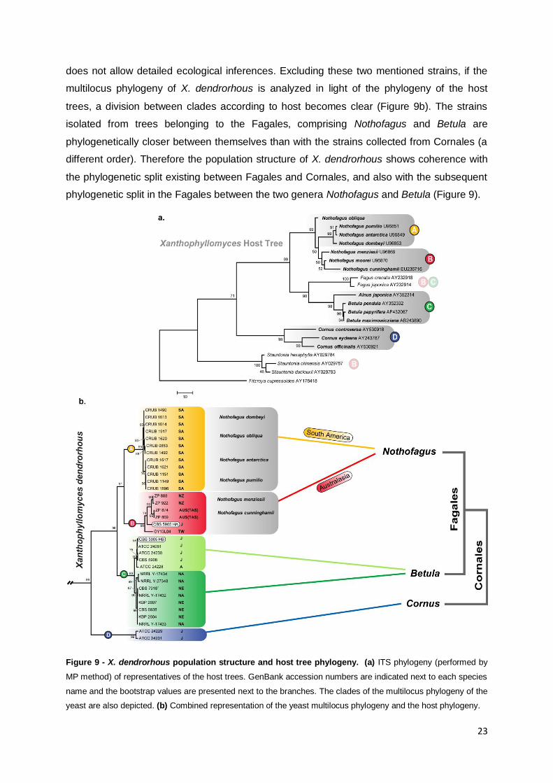
does not allow detailed ecological inferences. Excluding these two mentioned strains, if the
multilocus phylogeny of X. dendrorhous is analyzed in light of the phylogeny of the host
trees, a division between clades according to host becomes clear (Figure 9b). The strains
isolated from trees belonging to the Fagales, comprising Nothofagus and Betula are
phylogenetically closer between themselves than with the strains collected from Cornales (a
different order). Therefore the population structure of X. dendrorhous shows coherence with
the phylogenetic split existing between Fagales and Cornales, and also with the subsequent
phylogenetic split in the Fagales between the two genera Nothofagus and Betula (Figure 9).
Figure 9 - X. dendrorhous population structure and host tree phylogeny. (a) ITS phylogeny (performed by
MP method) of representatives of the host trees. GenBank accession numbers are indicated next to each species
name and the bootstrap values are presented next to the branches. The clades of the multilocus phylogeny of the
yeast are also depicted. (b) Combined representation of the yeast multilocus phylogeny and the host phylogeny.

24
Isolates from Japan, sharing same geography but distinct hosts, grouped in distinct clades
and isolates from North America and Northern Europe, with distinct geography but same host
end up grouping on the same clade (Figure 9b).
On the other hand, additional evolutionary factors, such as geographic origin, appear to
shape X. dendrorhous population structure. Within the strains isolated from Nothofagus,
those originating from South America grouped together (clade A) and are separated from
those originating from Australasia (clade B) (Figure 9b). In clade C, an existing subdivision
also appears to be related with geographic origin. The isolates from Japan always grouped
together with the isolate from Alaska, while those from NA and NE clustered separately but in
the same clade, as can be observed in clade C represented by two different shades of green
(Figure 9b).
X. dendrorhous was found to be present not only in the Northern Hemisphere but also in
the Southern one, namely Patagonia and now more recently in New Zealand and Tasmania
(Figure 9). The existence of X. dendrorhous in both Australasia and South America might
suggest that just as Nothofagus trees were disseminated though the southern part of
Gondwana so was X. dendrorhous. This might therefore present a possible explanation for
the presence of X. dendrorhous in both geographic locations (grey arrow on Figure 10).
Nevertheless the fact that populations from clades A and B are phylogenetic closer among
themselves seems contradicting to the vicariance scenario implied by the fragmentation of
Gondwana. The biogeographic history of Nothofagus and of its obligate parasite, Cyttaria are
considered to be a reflection of the break up of Gondwana but also of transoceanic long
dispersal events (Peterson et al. 2010). It is therefore possible that the extant distribution of
X. dendrorhous in the Southern Hemisphere may be related with the same factors affecting
Nothofagus biogeography. In order to test the different hypotheses regarding the events that
might have led to the present X. dendrorhous distribution in the Southern Hemisphere
(vicariance or dispersal), assessment of parameters such as the divergence time between
clades A and B and comparison of those dates with the break up process of Gondwana
would be necessary (Crisp et al. 2011). Unfortunately such analyses in yeasts are
challenging because of the absence of fossil records for the correct calibration of
phylogenies, and thus permitting only indirect methods of calibration (Crisp et al. 2011).
The fact that similar genotypes of X. dendrorhous are present in Australia, New Zealand and
Asia (Taiwan and Japan) and the mixed ancestry of CBS 5905 may suggest that an area of
contact might have existed between Australasia and Asia (Figure 10, arrow 1). The
biogeographic history of Southeast Asia and the West Pacific (Turner et al. 2001) is
considered to comprise two general patterns: (i) a pattern of Southeast Asian elements,
perhaps of Laurasian origin, expanding into Australian areas, and (ii) a reverse pattern for
Australian elements, perhaps of Gondwanan origin. Both patterns would make the mentioned
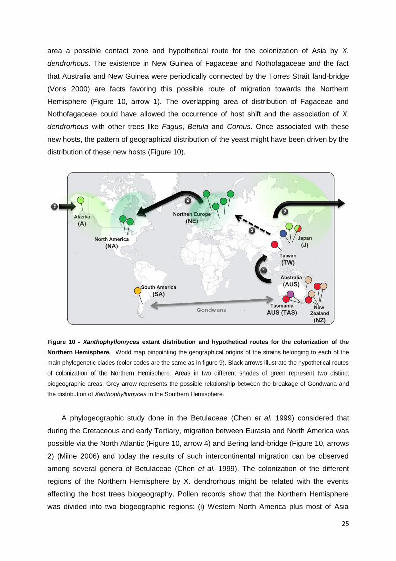
25
area a possible contact zone and hypothetical route for the colonization of Asia by X.
dendrorhous. The existence in New Guinea of Fagaceae and Nothofagaceae and the fact
that Australia and New Guinea were periodically connected by the Torres Strait land-bridge
(Voris 2000) are facts favoring this possible route of migration towards the Northern
Hemisphere (Figure 10, arrow 1). The overlapping area of distribution of Fagaceae and
Nothofagaceae could have allowed the occurrence of host shift and the association of X.
dendrorhous with other trees like Fagus, Betula and Cornus. Once associated with these
new hosts, the pattern of geographical distribution of the yeast might have been driven by the
distribution of these new hosts (Figure 10).
Figure 10 - Xanthophyllomyces extant distribution and hypothetical routes for the colonization of the
Northern Hemisphere. World map pinpointing the geographical origins of the strains belonging to each of the
main phylogenetic clades (color codes are the same as in figure 9). Black arrows illustrate the hypothetical routes
of colonization of the Northern Hemisphere. Areas in two different shades of green represent two distinct
biogeographic areas. Grey arrow represents the possible relationship between the breakage of Gondwana and
the distribution of Xanthophyllomyces in the Southern Hemisphere.
A phylogeographic study done in the Betulaceae (Chen et al. 1999) considered that
during the Cretaceous and early Tertiary, migration between Eurasia and North America was
possible via the North Atlantic (Figure 10, arrow 4) and Bering land-bridge (Figure 10, arrows
2) (Milne 2006) and today the results of such intercontinental migration can be observed
among several genera of Betulaceae (Chen et al. 1999). The colonization of the different
regions of the Northern Hemisphere by X. dendrorhous might be related with the events
affecting the host trees biogeography. Pollen records show that the Northern Hemisphere
was divided into two biogeographic regions: (i) Western North America plus most of Asia
26
(Figure 10, light green area) and (ii) Eastern North America plus Europe (Figure 10, dark
green area) (Milne 2006). Possible evidence of these two biogeographic areas appear to
exist within clade C, were a separation between strains of Asia from the Occidental ones is
noticeable (Figure 9, clade C, division represented by two shades of green). The knowledge
of the historical evolution of the tree hosts enables the formulation of hypotheses regarding
the history of colonization of the Northern Hemisphere by X. dendrorhous. Nevertheless, one
must consider that a set of results can be consistent with a set of alternative explanations
(Crisp et al. 2011). Therefore in order to fully explore the present hypothesis, a large
sampling would be necessary from the vast area between Australasia and Asia and further
study would be necessary in order to test the different scenarios that could be responsible for
the present distribution of X. dendrorhous.
27
4. Final considerations and future perspectives
X. dendrorhous is composed by four different populations, presenting a well defined
population structure that suggests that natural populations of X. dendrorhous are mostly
clonal and in nature inbreeding supersedes outcrossing. Strain CBS 5905 shows a mixed
ancestry and may represent an event of contact between two distinct populations of X.
dendrorhous. The different populations of X. dendrorhous have different ecological
associations that appear to have diverged first according to host tree and secondly by
geographic origin. The association between yeast and the host system Cyttaria–Nothofagus
appears to be older than the association of the yeast with the tree exudates host system.
Furthermore, the association of X. dendrorhous with Cyttaria appears to have derived from
an earlier association of the yeast with Nothofagus trees, due to the consistent isolation of
strains from Nothofagus leaves. Different karyotypes were presented by each of the tested X.
dendrorhous strains and no correlation with the revealed population structure was observed.
The existence of X. dendrorhous in both Australasia and South America may indicate that
the extant distribution of the species in the Southern Hemisphere is a relic of the Gondwana
break up process. The results obtained in this work suggest that Southeast Asia and
Australasia are a Xanthophyllomyces diversity hotspot from which different lineages have
radiated. Moreover, Australasia harbors two putative new species that appear to be endemic
to the area, representing this study the first record of the existence of more than one species
in the Xanthophyllomyces genus. The outstanding diversity found in Australasia, with three
distinct Xanthophyllomyces species and the fact that the Northern Hemisphere isolates show
greater genetic homogeneity suggests that Australasia might represent the place of origin of
the Xanthophyllomyces genus. The extant distribution of the X. dendrorhous appears to be
coherent with the biogeographic histories of the trees colonized by the yeast. Although in
need of testing, a hypothetical route of colonization of the Northern Hemisphere by X.
dendrorhous was drawn from the biogeographic processes undergone by the trees colonized
and by the extant genetic distribution observed in the population structure of the yeast.
The sampling of areas where known hosts exist, like New Guinea, could provide
valuable information regarding the existence (or not) of more strains with mixed ancestry that
could prove contact and cross between elements of different populations and also test the
hypothetical route of colonization of the Northern Hemisphere by X. dendrorhous. The
availability of the complete genome sequence of X. dendrorhous would certainly be a
valuable tool in understanding the CLP observed among strains and also for the
biotechnological research concerning astaxanthin production.
28
5. References
Adrio JL, López M, Casqueiro J, Fernández C and Veiga M (1995) Electrophoretic karyotype of the
astaxanthin-producing yeast Phaffia rhodozyma. Cur. Genet. 27, 447-450.
Akaike H (1987) Factor analysis and AIC. Psychometrika 52, 317-332.
Bruen TC, Philippe H and Bryant D (2006) A Simple and Robust Statistical Test for Detecting the
Presence of Recombination. Genetics 172, 2665-2681.
Chen S-H, Su S-Y, Lo C-Z, Chen K-H, Huang T-J, Kuo B-H and Lin C-Y (2009) PALM: A Paralleled
and Integrated Framework for Phylogenetic Inference with Automatic Likelihood Model
Selectors. PLoS ONE 4, e8116.
Chen Z-D, Manchester SR and Sun H-Y (1999) Phylogeny and evolution of the Betulaceae as inferred
from DNA sequences, morphology, and paleobotany. Am. J. of Bot. 86, 1168-1181.
Chen ZD and Li J (2004) Phylogenetics and biogeography of Alnus (Betulaceae) inferred from
sequences of nuclear ribosomal DNA ITS region. Int. J. Plant Sci. 165, 325-335.
Cifuentes V, Hermosilla G, Martinez C, Leon R, Pincheira G and Jimenez A (1997) Genetics and
electrophoretic karyotyping of wild-type and astaxanthin mutant strains of Phaffia rhodozyma
A. Van Leeuw. 72, 111-117.
Cook LG and Crisp MD (2005) Not so ancient: the extant crown group of Nothofagus represents a
post-Gondwanan radiation. Proc. Biol. Sci. 272, 2535-2544.
Crandall K, Fetzner J, Jara C and Buckup L (2000) On the phylogenetic positioning of the South
American freshwater crayfish genera (Decapoda: Parastacidae). J. Crust. Biol. 20, 530 - 540.
Crisp MD, Trewick SA and Cook LG (2011) Hypothesis testing in biogeography. Trends Ecol. & Evol.
26, 66-72.
Evanno G, Regnaut S and Goudet J (2005) Detecting the number of clusters of individuals using the
software STRUCTURE: a simulation study. Mol. Ecol. 14, 2611-20.
Excoffier L and Heckel G (2006) Computer programs for population genetics data analysis: a survival
guide. Nat. Rev. Genet. 7, 745-758.
Falush D, Stephens M and Pritchard JK (2007) Inference of population structure using multilocus
genotype data: dominant markers and null alleles. Mol. Ecol. Notes 7, 574-578.
Fell JW and Blatt GM (1999) Separation of strains of the yeasts Xanthophyllomyces dendrorhous
and Phaffia rhodozyma based on rDNA IGS and ITS sequence analysis. J. Ind. Microbiol.
Biotechnol. 23, 677-681.
Fell JW, Scorzetti G, Statzell-Tallman A and Boundy-Mills K (2007) Molecular diversity and
intragenomic variability in the yeast genus Xanthophyllomyces : the origin of Phaffia
rhodozyma ? FEMS Yeast Res. 7, 1399-1408.
Felsenstein J (1981) Evolutionary trees from DNA sequences: a maximum likelihood approach. J. Mol.
Evol. 17, 368-76.
Frengova GI and Beshkova DM (2009) Carotenoids from Rhodotorula and Phaffia : yeasts of
biotechnological importance. J. Ind. Microbiol. Biotechnol. 36, 163-180.
29
Giraud T, Refregier G, Le GM, de Vienne DM and Hood ME (2008) Speciation in fungi. Fungal.Genet.
Biol. 45, 791-802.
Golubev WI (1995) Perfect state of Rhodomyces dendrorhous ( Phaffia rhodozyma ). Yeast 11, 101-
110.
Gonçalves P, Valério E, Correia C, de Almeida JMGCF and Sampaio JP (2011) Evidence for
Divergent Evolution of Growth Temperature Preference in Sympatric Saccharomyces Species.
PLoS ONE 6, e20739.
Hall T (1999) BioEdit: a user-friendly biological sequence alignment editor and analysis program for
Windows 95/98/NT. Nucl. Acids. Symp. Ser 41, 95 - 98.
Hastings WK (1970) Monte Carlo sampling methods using Markov chains and their applications.
Biometrika 57, 97-109.
Hermosilla G, Martinez C, Retamales P, Leon R and Cifuentes V (2003) Genetic determination of
ploidy level in Xanthophyllomyces dendrorhous. A. Van Leeuw. 84, 279-287.
Hubisz MJ, Falush D, Stephens M and Pritchard JK (2009) Inferring weak population structure with the
assistance of sample group information. Mol. Ecol. Resour. 9, 1322-32.
Huson DH (1998) SplitsTree: analyzing and visualizing evolutionary data. Bioinformatics 14, 68-73.
Huson DH and Bryant D (2006) Application of phylogenetic networks in evolutionary studies. Mol. Biol.
Evol. 23, 254-267.
Huson DH and Scornavacca C (2011) A Survey of Combinatorial Methods for Phylogenetic Networks.
Genome Biol. Evol. 3, 23-35.
Johnson EA (2003) Phaffia rhodozyma: colorful odyssey. Int. Microbiol. 6, 169-174.
Kucsera J, Pfeiffer I and Ferenczy L (1998) Homothallic life cycle in the diploid red yeast
Xanthophyllomyces dendrorhous ( Phaffia rhodozyma ). A. Van Leeuw. 73, 163-168.
Kurihara H, Koda H, Asami S, Kiso Y and Tanaka T (2002) Contribution of the antioxidative property
of astaxanthin to its protective effect on the promotion of cancer metastasis in mice treated
with restraint stress. Life Sci. Vol. 70.
Lee DH, Kim CS and Lee YJ (2010) Astaxanthin protects against MPTP/MPP+-induced mitochondrial
dysfunction and ROS production in vivo and in vitro. Food Chem.Toxicol.49,271- 80
Libkind D, Ruffini A, van BM, Alves L and Sampaio JP (2007) Biogeography, host specificity, and
molecular phylogeny of the basidiomycetous yeast Phaffia rhodozyma and its sexual form,
Xanthophyllomyces dendrorhous Appl. Environ. Microbiol. 73, 1120-1125.
Lin CY, Chen YC, Lo HJ, Chen KW and Li SY (2007) Assessment of Candida glabrata strain
relatedness by pulsed-field gel electrophoresis and multilocus sequence typing. J. Clin.
Microbiol. 45, 2452-9.
Manchester SR, Xiang Q-Y and Xiang Q-P (2007) Curtisia (Cornales) from the Eocene of Europe and
its phytogeographical significance. Bot. J. Linn. Soc. 155, 127-134.
Mathiasen P and Premoli AC (2010) Out in the cold: genetic variation of Nothofagus pumilio
(Nothofagaceae) provides evidence for latitudinally distinct evolutionary histories in austral
South America. Mol. Ecol. 19, 371-385.
Medwid RD (1998) Phaffia rhodozyma is polyploid. J. Ind. Microbiol. Biot. 21, 228-232.
30
Miller MW, Yoneyama MINO and Soneda MASA (1976) Phaffia, a New Yeast Genus in the
Deuteromycotina (Blastomycetes). Int. J. Syst. Bacteriol. 26, 286-291.
Milne R (2006) Northern Hemisphere Plant Disjunctions: A Window on Tertiary Land Bridges and
Climate Change? Ann. Bot. 98, 465-472.
Naguib YM (2000) Antioxidant activities of astaxanthin and related carotenoids. J. Agric. Food Chem.
48, 1150-1154.
Nagy Á, Garamszegi N, Vágvölgyi C and Ferenczy L (1994) Electrophoretic karyotypes of Phaffia
rhodozyma strains. FEMS Microbiol. Lett. 123, 315-318.
Nagy Á, Palágyi Z, Ferenczy L and Vágvölgyi C (1997) Radiation-induced chromosomal
rearrangement as an aid to analysis of the genetic constitution of Phaffia rhodozyma. FEMS
Microbiol. Lett. 152, 249-254.
Nei M (1973) Analysis of gene diversity in subdivided populations. Proc. Natl. Acad. Sci. USA 70,
3321-3.
Niklitschek M, Alcaino J, Barahona S, Sepulveda D, Lozano C, Carmona M, Marcoleta A, Martinez C,
Lodato P, Baeza M, et al. (2008) Genomic organization of the structural genes controlling the
astaxanthin biosynthesis pathway of Xanthophyllomyces dendrorhous. Biol. Res. 41, 93-108.
Pera LM Ru instein L aigor M , de Figueroa LaIC and Callieri DA (1999) Influence of manganese
on cell morphology protoplasts formation and β-d-glucosidase activity in Phaffia rhodozyma.
FEMS Microbiol. Lett. 171, 155-160.
Perez-Losada M, Porter ML, Tazi L and Crandall KA (2007) New methods for inferring population
dynamics from microbial sequences. Infect. Genet. Evol. 7, 24-43.
P re - rt n JE, Querol A, Puig S and Barrio E (2002) Molecular Characterization of a Chromosomal
Rearrangement Involved in the Adaptive Evolution of Yeast Strains. Genome Res. 12, 1533-
1539.
Peterson KR, Pfister DH and Bell CD (2010) Cophylogeny and biogeography of the fungal parasite
Cyttaria and its host Nothofagus, southern beech. Mycologia 102, 1417-25.
Poláková S, Blume C, Zárate JÁ, Mentel M, Jørck-Ram erg Stenderup J and Piškur J 200 )
Formation of new chromosomes as a virulence mechanism in yeast Candida glabrata. Proc.
Nat. Acad. .Sci. 106, 2688-2693.
Posada D and Crandall KA (2001) Intraspecific gene genealogies: trees grafting into networks. Trends
Ecol. Evol. 16, 37-45.
Pritchard JK, Stephens M and Donnelly P (2000) Inference of population structure using multilocus
genotype data. Genetics 155, 945-59.
Rawlings GB (1956) Australasian Cyttariaceae. Trans. & Proc. Roy. Soc. New Zealand. 84, 19-28.
Rodriguez-Saiz M, de la Fuente JL and Barredo JL (2010) Xanthophyllomyces dendrorhous for the
industrial production of astaxanthin. Appl. Microbiol. Biotechnol. 88, 645-658.
Sambrook J. FEF, Maniatis T. 1989. Molecular cloning: a Laboratory Manual. New York: Cold Spring
Harbor Laboratory Press.
31
Sampaio JP, Gadanho M, Santos S, Duarte FL, Pais C, Fonseca A and Fell JW (2001) Polyphasic
taxonomy of the basidiomycetous yeast genus Rhodosporidium: Rhodosporidium
kratochvilovae and related anamorphic species. Int. J. Syst. Evol. Microbiol. 51, 687-97.
Santopietro LMD, Spencer JFT, Spencer DM and Siñeriz F (1995) Formation of protoplasts from
Phaffia rhodozyma. Biot. Tech. 9, 339-344.
Schmidt I, Schewe H, Gassel S, Jin C, Buckingham J, Humbelin M, Sandmann G and Schrader J
(2010) Biotechnological production of astaxanthin with Phaffia rhodozyma /
Xanthophyllomyces dendrorhous. Appl.Microbiol.Biotechnol. 89 (3), 555-571
Scorzetti G, Fell JW, Fonseca A and Statzell-Tallman A (2002) Systematics of basidiomycetous
yeasts: a comparison of large subunit D1/D2 and internal transcribed spacer rDNA regions.
FEMS Yeast Res. 2, 495-517.
Swenson U, Backlund A, McLoughlin S and Hill RS (2001) Nothofagus Biogeography Revisited with
Special Emphasis on the Enigmatic Distribution of Subgenus Brassospora in New Caledonia.
Cladistics 17, 28-47.
Tamura K, Peterson D, Peterson N, Stecher G, Nei M and Kumar S (2011) MEGA5: Molecular
Evolutionary Genetics Analysis using maximum likelihood, evolutionary distance, and
maximum parsimony methods. Mol. Biol. Evol.,[Epub ahead of print].
Taylor J, Jacobson D and Fisher M (1999) The evolution of THE EVOLUTION OF ASEXUAL FUNGI:
Reproduction, Speciation and Classification. Annu. Rev. Phytopathol. 37, 197-246.
Taylor JW, Jacobson DJ, Kroken S, Kasuga T, Geiser DM, Hibbett DS and Fisher MC (2000)
Phylogenetic species recognition and species concepts in fungi. Fungal. Genet. Biol. 31, 21-
32.
Thompson J, Higgins D and Gibson T (1994) CLUSTAL W: improving the sensitivity of progressive
multiple sequence alignment through sequence weighting, position-specific gap penalties and
weight matrix choice. Nucl. Acids Res. 22, 4673 - 4680.
Turner, Hovenkamp and Van W (2001) Biogeography of Southeast Asia and the West Pacific. J.
Biogeo. 28, 217-230.
Vallone PM and Butler JM (2004) AutoDimer: a screening tool for primer-dimer and hairpin structures.
Biotechniques 37, 226-31.
Voris HK (2000) Maps of Pleistocene sea levels in Southeast Asia: shorelines, river systems and time
durations. J. Biogeo. 27, 1153-1167.
Wagele J and Mayer C (2007) Visualizing differences in phylogenetic information content of
alignments and distinction of three classes of long-branch effects. BMC Evol. Biol. 7, 147.
Weber RWS, Davoli P and Anke H (2006) A microbial consortium involving the astaxanthin producer
Xanthophyllomyces dendrorhous on freshly cut birch stumps in Germany. Mycologist 20, 57-
61.
Zolan ME (1995) Chromosome-length polymorphism in fungi. Microbiol Rev 59, 686-98.
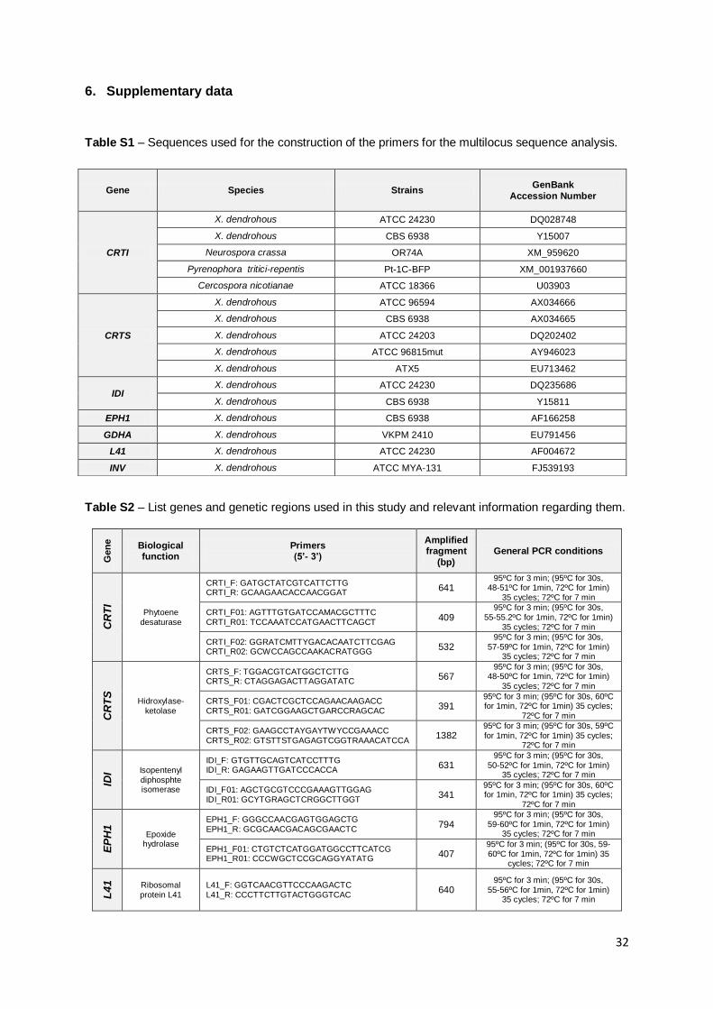
32
6. Supplementary data
Table S1 – Sequences used for the construction of the primers for the multilocus sequence analysis.
Table S2 – List genes and genetic regions used in this study and relevant information regarding them.
Gen
e
Biological function
Primers (5’- 3’)
Amplified fragment
(bp)
General PCR conditions
CR
TI
Phytoene desaturase
CRTI_F: GATGCTATCGTCATTCTTG CRTI_R: GCAAGAACACCAACGGAT
641 95ºC for 3 min; (95ºC for 30s,
48-51ºC for 1min, 72ºC for 1min) 35 cycles; 72ºC for 7 min
CRTI_F01: AGTTTGTGATCCAMACGCTTTC CRTI_R01: TCCAAATCCATGAACTTCAGCT
409 95ºC for 3 min; (95ºC for 30s,
55-55.2ºC for 1min, 72ºC for 1min) 35 cycles; 72ºC for 7 min
CRTI_F02: GGRATCMTTYGACACAATCTTCGAG CRTI_R02: GCWCCAGCCAAKACRATGGG
532 95ºC for 3 min; (95ºC for 30s,
57-59ºC for 1min, 72ºC for 1min) 35 cycles; 72ºC for 7 min
CR
TS
Hidroxylase-ketolase
CRTS_F: TGGACGTCATGGCTCTTG CRTS_R: CTAGGAGACTTAGGATATC
567 95ºC for 3 min; (95ºC for 30s,
48-50ºC for 1min, 72ºC for 1min) 35 cycles; 72ºC for 7 min
CRTS_F01: CGACTCGCTCCAGAACAAGACC CRTS_R01: GATCGGAAGCTGARCCRAGCAC
391 95ºC for 3 min; (95ºC for 30s, 60ºC for 1min, 72ºC for 1min) 35 cycles;
72ºC for 7 min
CRTS_F02: GAAGCCTAYGAYTWYCCGAAACC
CRTS_R02: GTSTTSTGAGAGTCGGTRAAACATCCA 1382
95ºC for 3 min; (95ºC for 30s, 59ºC for 1min, 72ºC for 1min) 35 cycles;
72ºC for 7 min
IDI Isopentenyl
diphosphte isomerase
IDI_F: GTGTTGCAGTCATCCTTTG IDI_R: GAGAAGTTGATCCCACCA
631 95ºC for 3 min; (95ºC for 30s,
50-52ºC for 1min, 72ºC for 1min) 35 cycles; 72ºC for 7 min
IDI_F01: AGCTGCGTCCCGAAAGTTGGAG IDI_R01: GCYTGRAGCTCRGGCTTGGT
341 95ºC for 3 min; (95ºC for 30s, 60ºC for 1min, 72ºC for 1min) 35 cycles;
72ºC for 7 min
EP
H1
Epoxide hydrolase
EPH1_F: GGGCCAACGAGTGGAGCTG EPH1_R: GCGCAACGACAGCGAACTC
794 95ºC for 3 min; (95ºC for 30s,
59-60ºC for 1min, 72ºC for 1min) 35 cycles; 72ºC for 7 min
EPH1_F01: CTGTCTCATGGATGGCCTTCATCG EPH1_R01: CCCWGCTCCGCAGGYATATG
407 95ºC for 3 min; (95ºC for 30s, 59-60ºC for 1min, 72ºC for 1min) 35
cycles; 72ºC for 7 min
L41
Ribosomal protein L41
L41_F: GGTCAACGTTCCCAAGACTC L41_R: CCCTTCTTGTACTGGGTCAC
640 95ºC for 3 min; (95ºC for 30s,
55-56ºC for 1min, 72ºC for 1min) 35 cycles; 72ºC for 7 min
Gene Species Strains GenBank
Accession Number
CRTI
X. dendrohous ATCC 24230 DQ028748
X. dendrohous CBS 6938 Y15007
Neurospora crassa OR74A XM_959620
Pyrenophora tritici-repentis Pt-1C-BFP XM_001937660
Cercospora nicotianae ATCC 18366 U03903
CRTS
X. dendrohous ATCC 96594 AX034666
X. dendrohous CBS 6938 AX034665
X. dendrohous ATCC 24203 DQ202402
X. dendrohous ATCC 96815mut AY946023
X. dendrohous ATX5 EU713462
IDI X. dendrohous ATCC 24230 DQ235686
X. dendrohous CBS 6938 Y15811
EPH1 X. dendrohous CBS 6938 AF166258
GDHA X. dendrohous VKPM 2410 EU791456
L41 X. dendrohous ATCC 24230 AF004672
INV X. dendrohous ATCC MYA-131 FJ539193
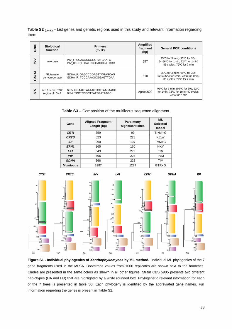
33
Table S2 (cont.) – List genes and genetic regions used in this study and relevant information regarding
them. G
en
e
Biological
function
Primers
(5’- 3’)
Amplified
fragment (bp)
General PCR conditions
INV
Invertase INV_F: CCACGCCGGGTATCAATC INV_R: CCTTGATCTCGACGGATCCC
557 95ºC for 3 min; (95ºC for 30s,
54-56ºC for 1min, 72ºC for 1min) 35 cycles; 72ºC for 7 min
GD
HA
Glutamate dehydrogenase
GDHA_F: GAGCCCGAGTTCGAGCAG GDHA_R: TCCCAAAGCGGAGTTGAA
610 95ºC for 3 min; (95ºC for 30s,
52-53.5ºC for 1min, 72ºC for 1min)
35 cycles; 72ºC for 7 min
ITS
ITS1, 5.8S, ITS2 region of rDNA
ITS5: GGAAGTAAAAGTCGTAACAAGG ITS4: TCCTCCGCTTATTGATATGC
Aprox.600 95ºC for 5 min; (95ºC for 30s, 52ºC for 1min, 72ºC for 1min) 40 cycles;
72ºC for 7 min
Table S3 – Composition of the multilocus sequence alignment.
Gene Aligned Fragment
Length (bp)
Parsimony
significant sites
ML
Selected
model
CRTI 359 99 TrNef+G
CRTS 523 223 K81uf
IDI 290 107 TVM+G
EPH1 365 160 HKY
L41 543 273 TrN
INV 506 225 TVM
GDHA 568 226 TIM
Multilocus 3187 1287 GTR+G
Figure S1 - Individual phylogenies of Xanthophyllomyces by ML method. Individual ML phylogenies of the 7
gene fragments used in the MLSA. Bootstraps values from 1000 replicates are shown next to the branches.
Clades are presented in the same colors as shown in all other figures. Strain CBS 5905 presents two different
haplotypes (HA and HB) that are highlighted by a white rounded box. Phylogenetic relevant information for each
of the 7 trees is presented in table S3. Each phylogeny is identified by the abbreviated gene names. Full
information regarding the genes is present in Table S2.
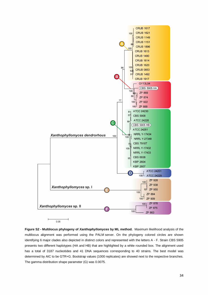
34
Figure S2 - Multilocus phylogeny of Xanthophyllomyces by ML method. Maximum likelihood analysis of the
multilocus alignment was performed using the PALM server. On the phylogeny colored circles are shown
identifying 6 major clades also depicted in distinct colors and represented with the letters A - F. Strain CBS 5905
presents two different haplotypes (HA and HB) that are highlighted by a white rounded box. The alignment used
has a total of 3187 nucleotides and 41 DNA sequences corresponding to 40 strains. The best model was
determined by AIC to be GTR+G. Bootstrap values (1000 replicates) are showed next to the respective branches.
The gamma distribution shape parameter (G) was 0.0075.
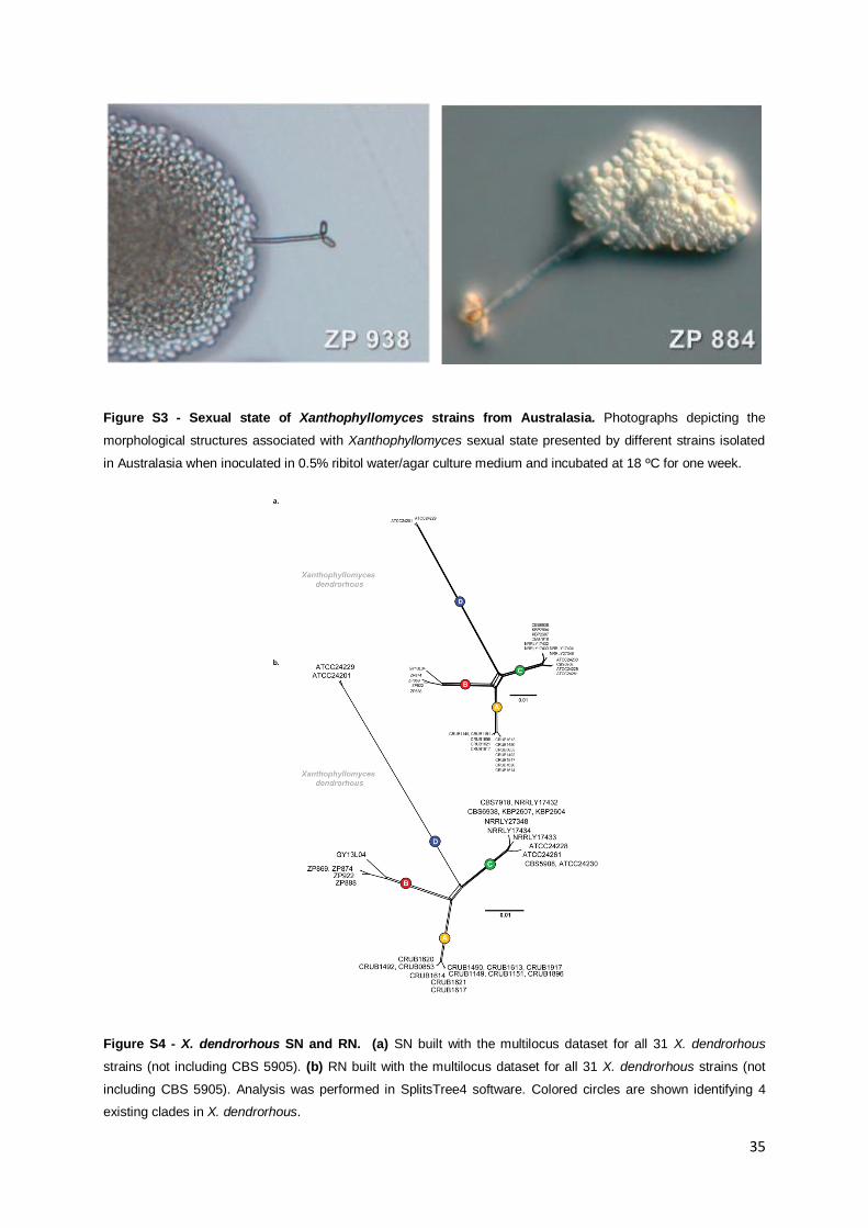
35
Figure S3 - Sexual state of Xanthophyllomyces strains from Australasia. Photographs depicting the
morphological structures associated with Xanthophyllomyces sexual state presented by different strains isolated
in Australasia when inoculated in 0.5% ribitol water/agar culture medium and incubated at 18 ºC for one week.
Figure S4 - X. dendrorhous SN and RN. (a) SN built with the multilocus dataset for all 31 X. dendrorhous
strains (not including CBS 5905). (b) RN built with the multilocus dataset for all 31 X. dendrorhous strains (not
including CBS 5905). Analysis was performed in SplitsTree4 software. Colored circles are shown identifying 4
existing clades in X. dendrorhous.
Top Related