


Languages
Pages
Legal

Accepted Manuscript
Title: A systematic review of the neurophysiology ofmindfulness on EEG oscillations
Author: Tim Lomas Itai Ivtzan Cynthia H.Y. Fu
PII: S0149-7634(15)00251-1DOI: http://dx.doi.org/doi:10.1016/j.neubiorev.2015.09.018Reference: NBR 2278
To appear in:
Received date: 15-5-2015Revised date: 23-9-2015Accepted date: 30-9-2015
Please cite this article as: Lomas, T., Ivtzan, I., Fu, C.H.Y.,A systematic review of theneurophysiology of mindfulness on EEG oscillations, Neuroscience and BiobehavioralReviews (2015), http://dx.doi.org/10.1016/j.neubiorev.2015.09.018
This is a PDF file of an unedited manuscript that has been accepted for publication.As a service to our customers we are providing this early version of the manuscript.The manuscript will undergo copyediting, typesetting, and review of the resulting proofbefore it is published in its final form. Please note that during the production processerrors may be discovered which could affect the content, and all legal disclaimers thatapply to the journal pertain.
Page 1 of 53
Accep
ted M
anus
cript
We conducted a systematic review of EEG studies of mindfulness meditation
We examined power differentials between mindfulness and a control state
Mindfulness was associated with enhanced alpha and theta power
No consistent patterns were observed in terms of beta, delta and gamma
Elevated alpha and theta may signify a state of relaxed alertness
*Highlights (for review)
Page 2 of 53
Accep
ted M
anus
cript
1 2 3 4 5 6 7 8 9 10 11 12 13 14 15 16 17 18 19 20 21 22 23 24 25 26 27 28 29 30 31 32 33 34 35 36 37 38 39 40 41 42 43 44 45 46 47 48 49 50 51 52 53 54 55 56 57 58 59 60 61 62 63 64 65
Running title: The neurophysiology of mindfulness
1
Title A systematic review of the neurophysiology of mindfulness on EEG oscillations
Authors Tim Lomas 1, Itai Ivtzan 1, Cynthia H.Y. Fu 1,2
Affiliations: 1. School of Psychology, University of East London, London, UK
2. Centre for Affective Disorders, Institute of Psychiatry, Psychology and Neuroscience,
King’s College London, London, UK
Author for Correspondence: Dr. Tim Lomas, School of Psychology, University of East London, Arthur Edwards Building,
Water Lane, London, E15 4LZ, United Kingdom
Tel: +44 (0)20 8223 4465 Fax: +44 (0)20 8223 4937 Email: [email protected]
*ManuscriptClick here to view linked References

Page 3 of 53
Accep
ted M
anus
cript
1 2 3 4 5 6 7 8 9 10 11 12 13 14 15 16 17 18 19 20 21 22 23 24 25 26 27 28 29 30 31 32 33 34 35 36 37 38 39 40 41 42 43 44 45 46 47 48 49 50 51 52 53 54 55 56 57 58 59 60 61 62 63 64 65
Running title: The neurophysiology of mindfulness
2
Abstract:
Mindfulness meditation has been purported as a beneficial practice for wellbeing. It would
be expected that the neurophysiology of mindfulness would reflect this impact on wellbeing.
However, investigations of the effect of mindfulness have generated mixed reports of
increases, decreases, as well as no differences in EEG oscillations in comparison with a
resting state and a variety of tasks. We have performed systematic review of EEG studies of
mindfulness meditation in order to determine any common effects and to identify factors
which may impact on the effects. Databases were reviewed from 1966 to August 2015.
Eligibility criteria included empirical quantitative analyses of mindfulness meditation practice
and EEG measurements acquired in relation to practice. A total of 56 papers met the
eligibility criteria and were included in the systematic review, consisting of a total 1,715
subjects: 1,358 healthy individuals and 357 individuals with psychiatric diagnoses. Studies
were principally examined for power outcomes in each bandwidth, in particular the power
differentials between mindfulness and the control state, as well as outcomes relating to
hemispheric asymmetry and event-related potentials. The systematic review revealed that
mindfulness was most commonly associated with enhanced alpha and theta power as
compared to an eyes closed resting state, although such outcomes were not uniformly
reported. No consistent patterns were observed with respect to beta, delta and gamma
bandwidths. In summary, mindfulness is associated with increased alpha and theta power in
both healthy individuals and in patient groups. This co-presence of elevated alpha and theta
may signify a state of relaxed alertness which is conducive to mental health.
Keywords: mindfulness; meditation; neurophysiology; EEG; systematic review.
Page 4 of 53
Accep
ted M
anus
cript
1 2 3 4 5 6 7 8 9 10 11 12 13 14 15 16 17 18 19 20 21 22 23 24 25 26 27 28 29 30 31 32 33 34 35 36 37 38 39 40 41 42 43 44 45 46 47 48 49 50 51 52 53 54 55 56 57 58 59 60 61 62 63 64 65
Running title: The neurophysiology of mindfulness
3
Introduction
Meditation refers to a diverse range of mental activities which share a common focus on the
regulation of attention and awareness (Cahn and Polich, 2006) in order to improve voluntary
control of mental processes which is purported to foster general wellbeing (Walsh and
Shapiro, 2006). Most world cultures have developed their own forms of meditation; for
example, Christianity has a long tradition of contemplative prayer (Egan, 1978). Much of the
recent scientific interest in meditation has centred on mindfulness meditation, which is a
practice that is believed to have originated with Buddhism around the fifth millennium B.C.
although its roots may stretch back further to the third millennium B.C. in Hindu culture
(Cousins, 1996).
The most common forms of meditation may be conceptualized as involving either focused
attention or an open-monitoring form of processes (Lutz et al., 2008). Focused attention
practices may be operationalized into their respective attention networks (Posner and
Petersen, 1990; Mirsky et al., 1991): sustained attention (e.g. towards a target, such as the
breath), executive attention (e.g. preventing one’s focus from ‘wandering’), attention
switching (e.g. disengaging from distractions), selective attention and attention re-orienting
(e.g. redirecting focus back to the breath), and working memory (Lutz et al., 2008; Vago and
Silbersweig, 2012). Open-monitoring refers to a broader receptive awareness or capacity to
detect events within an unrestricted awareness without a specific focus (Raffone and
Srinivasan, 2010), which can include a process of ‘meta-awareness’ (i.e., awareness of
awareness, in which practitioners are able to reflect on the process of consciousness itself).
Mindfulness has been described as the awareness that arises through purposeful attention
on the present moment with nonjudgmental experience (Kabat-Zinn, 2003). While
mindfulness has been commonly viewed as an example of open-monitoring, it has been
proposed to involve an admixture of focused attention and open-monitoring (Lutz et al.,
2008; Vago and Silbersweig, 2012) as most mindfulness practices begin with a period of
Page 5 of 53
Accep
ted M
anus
cript
1 2 3 4 5 6 7 8 9 10 11 12 13 14 15 16 17 18 19 20 21 22 23 24 25 26 27 28 29 30 31 32 33 34 35 36 37 38 39 40 41 42 43 44 45 46 47 48 49 50 51 52 53 54 55 56 57 58 59 60 61 62 63 64 65
Running title: The neurophysiology of mindfulness
4
focused attention on a target, such as the breath, in order to focus awareness, followed by
the more receptive state of open-monitoring (Cahn and Polich, 2006). In Vago and
Silbersweig’smodel (2012), the practice of mindfulness leads to three overarching self-
related capacities: meta self-awareness, self-regulation, and self-transcendence. These are
subserved by numerous subcomponent cognitive components, including motivation (which is
crucial in terms of people practicing meditation in the first place), attention regulation (via the
development of attention modalities), and de-centring (an ability, defined below, that arises
from enhanced attention regulation, and which facilitates self-awareness and
transcendence). It is further proposed that these three overarching capacities modulate
‘self-specifying and narrative self-networks’ through an integrative fronto-parietal control
network.
Mindfulness has been applied as a clinical intervention based on the notion that it is a
method for training attention and awareness. By developing the ability to observe one’s
thoughts and feelings, practitioners learn how to perceive them as temporary, objective
events in the mind as opposed to reflections of the self that are necessarily true, which has
been termed as the ability to “decentre” (Fresco et al., 2007). As a clinical intervention, it
involves the process to engage with negative experiences, such as pain or dysphoric
emotions, with more dispassion and less reactivity (Shapiro et al., 2005). Mindfulness was
initially applied as an intervention for chronic pain with Kabat-Zinn’s (1982) Mindfulness-
Based Stress Reduction (MBSR) program. The MBSR program has since been applied in
the treatment for number of conditions, including cancer (Ledesma & Kumano, 2009) and
migraine (Schmidt et al., 2010), and adapted as a treatment to prevent relapse in depression
(Mindfulness-Based Cognitive Therapy; Segal et al., 2002) and for the treatment of
substance abuse (Mindfulness-Based Relapse Prevention; Bowen et al., 2014, Mindfulness-
Oriented Recovery Enhancement; Garland et al., 2014).
The effectiveness of mindfulness has been assessed by measures for depression and
quality of life (Hofmann et al., 2010). As mindfulness may be considered to be a method of
Page 6 of 53
Accep
ted M
anus
cript
1 2 3 4 5 6 7 8 9 10 11 12 13 14 15 16 17 18 19 20 21 22 23 24 25 26 27 28 29 30 31 32 33 34 35 36 37 38 39 40 41 42 43 44 45 46 47 48 49 50 51 52 53 54 55 56 57 58 59 60 61 62 63 64 65
Running title: The neurophysiology of mindfulness
5
attention training and emotion regulation, we would expect that the corresponding
neurophysiological states should be observable. Electroencephalography (EEG) is a non-
invasive technique that analyzes spatiotemporal aspects of underlying brain activity, which
provides a measure of the large-scale synchronization of neural networks (Cacioppo et al.,
2007). Patterns of EEG activity to particular meditative states have been investigated. A
commonly reported feature of meditation has been theta and alpha event-related
synchronization (Fell et al., 2010), which are regarded as markers of internally-directed
attention processing (Shaw, 1996). Such synchronization has been observed across
different meditation practices, including mindfulness, as well as practices such as
transcendental meditation, which involves focused attention upon an internally-voiced
mantra. However, different types of meditation practice have been associated with unique
frequency patterns, reflecting the form of attention (Dunn et al., 1999). For example,
mindfulness has been associated with increase alpha power while focused attention has
been associated with increased gamma activity and idiosyncratic meditation with decreased
alpha and beta (Hinterberger et al., 2014).
Additionally, Event-Related Potentials (ERP) provide a measure of large number of time-
locked experimental trials, enabling the analysis of sensory, perceptual, and cognitive
processing (Light et al., 2010). Such studies involve the precision analysis of populations of
neuronal transients directly manifested via a stimulus/event, which is frequently a stimulus
connected to an attention-based task (e.g., listening to an auditory signal) (Schoenberg and
Speckens, 2014). The high temporal resolution of this approach, involving millisecond
precision, allows the investigation of early information processing stages and subsequent
transitions to higher-level cognitive operations. ERP studies have been used to corroborate
the idea of mindfulness as a system of attention training. For example, van Leeuwen et al.
(2012) examined the impact of mindfulness practice on hierarchical stimulus processing and
attentional selection, focusing on differences in early components of the evoked visual
response (e.g., P1 and N1 components) in meditators versus matched controls. Meditators

Page 7 of 53
Accep
ted M
anus
cript
1 2 3 4 5 6 7 8 9 10 11 12 13 14 15 16 17 18 19 20 21 22 23 24 25 26 27 28 29 30 31 32 33 34 35 36 37 38 39 40 41 42 43 44 45 46 47 48 49 50 51 52 53 54 55 56 57 58 59 60 61 62 63 64 65
Running title: The neurophysiology of mindfulness
6
exhibited faster attentional disengagement from a dominant global presentation in order to
focus in on specific stimuli, suggesting that meditation enhances speed of attention
allocation and relocation, thus increasing the depth of information processing.
In the present review, we have focused on mindfulness meditation. We have examined
factors which appear to impact upon EEG measures including the experience of the
meditator, being a novice or relative expert, as experience has been reported to accentuate
amplitude differences between meditation and the resting state (Hinterberger et al., 2014)
while the converse has also been observed (Cahn et al., 2010). An additional factor
includes the location of the brain activity. For example, increased alpha during mindfulness
has been localized to frontal regions (Takahashi et al., 2005) but has also been observed
increases in posterior regions (Lagopoulos et al., 2009; Cahn et al., 2010). Furthermore,
EEG analysis of meditation may be affected by whether the control task is a resting state or
a cognitive task as increased theta amplitude during meditation has been observed in
comparison to a resting state baseline, but was comparable in amplitude to an executive
attention task, which may be further modulated by the experience of the meditator (Lomas et
al., 2014).
We sought to perform a systematic review of patterns of electrophysiological activity
associated mindfulness in order to examine the impact on neurophysiology as assessed by
EEG bandwidth activation and other measures, including hemispheric asymmetry or event-
related potential, and the functional significance of these activities. If mindfulness is
expected to impact on functioning attentional networks as well as open-monitoring, then we
would expect to observe distinct neural features associated with its practice. We also
expected that the experience of the meditator, type of control task, and location of the EEG
oscillation would moderate the impact of mindfulness on neurophysiology.
Methods
Page 8 of 53
Accep
ted M
anus
cript
1 2 3 4 5 6 7 8 9 10 11 12 13 14 15 16 17 18 19 20 21 22 23 24 25 26 27 28 29 30 31 32 33 34 35 36 37 38 39 40 41 42 43 44 45 46 47 48 49 50 51 52 53 54 55 56 57 58 59 60 61 62 63 64 65
Running title: The neurophysiology of mindfulness
7
The literature search was conducted using the MEDLINE and Scopus electronic databases
with the criteria: “EEG” (AND) “mindfulness OR meditation”, in all fields in MEDLINE and
limited to article title, abstract, and keywords in Scopus, with the dates: from 1966 to 1st
August 2015. The participants, interventions, comparisons, outcomes and study design
(PICOS) characteristics, the key criteria were interventions: mindfulness meditation or
functional equivalent; participants: adults; and outcomes: EEG analysis. Studies were
required to be published, or a manuscript in press, and to be in English. The review was
conducted according to the Preferred Reporting Items for Systematic Reviews and Meta-
Analyses (PRISMA) guidelines (Moher et al., 2009). The review protocol was registered with
the International Prospective Register of Systematic Reviews (PROSPERO) database on
15th September 2014. Registration number: CRD42014013766
(http://www.crd.york.ac.uk/PROSPERO).
Inclusion criteria were: 1) mindfulness meditation practice or functional equivalent, such as
Vipassana or Zen meditation; 2) EEG measurements acquired in relation to mindfulness
meditation practice (whether assessment during the practice itself or connected to its
practice, e.g., pre- and post-intervention); 3) quantitative analysis supported by appropriate
statistical methodology; and exclusion criteria; and 4) adult sample; and exclusion criteria: 1)
theoretical articles or commentaries without statistical analyses.
The following variables were extracted from each paper: experimental protocol (control
condition, meditation condition, and/or experimental task), experience of participants (novice
or expert), sample features (clinical or non-clinical), outcomes for each individual bandwidth
(alpha, beta, theta, delta, and gamma), hemispheric asymmetry, and any event-related
potential outcomes.
The primary summary measures were differences in levels of power in each of the
bandwidths. Neural activity generates electrical potentials which can be analyzed in terms of
parameters of amplitude, frequency, coherence and synchrony. Amplitude, or power, which
Page 9 of 53
Accep
ted M
anus
cript
1 2 3 4 5 6 7 8 9 10 11 12 13 14 15 16 17 18 19 20 21 22 23 24 25 26 27 28 29 30 31 32 33 34 35 36 37 38 39 40 41 42 43 44 45 46 47 48 49 50 51 52 53 54 55 56 57 58 59 60 61 62 63 64 65
Running title: The neurophysiology of mindfulness
8
is the square of the amplitude, reflects the magnitude of the electrical signal, representing
the level of synchronized activity in the underlying tissue, i.e. neurons discharging
simultaneously. Frequency is the number of oscillatory cycles per second and is divided into
the following bandwidths: Delta (1-4 Hz); Theta (4-8 Hz); Alpha (8-13 Hz); Beta (13–30 Hz);
and Gamma (36-44 Hz) (Cacioppo et al., 2007). EEG connectivity is the functional
integration of spatially distributed neural populations which can be assessed in terms of
synchrony, the degree of leading or lagging in the relationship between signals from
electrode pairs, and coherence, the stability of that phase relationship.
The primary summary variable was principally the difference in power between a meditation
condition and a resting state condition. Secondary power differentials included longitudinal
pre- and post- differences, such as, in meditation and/or resting state and/or task conditions
before and after an intervention. If applicable, outcomes relating to coherence, synchrony,
asymmetry and event-related potentials were also noted.
Of note, there was considerable diversity in how the experience of the participant was
defined. In terms of years meditating, the range for which papers rated participants as being
‘experienced’ varied from 1 year (Kasamatsu and Hirai, 1966) to 9 years (Lagopoulos et al.,
2009). Likewise, in terms of hours meditating, the range for which papers rated participants
as being ‘experienced’ varied from 40 hours (Hinterberger et al., 2011) to 1740 hours
(Berkovich-Ohana et al., 2012). In the present systematic review, we have applied the
lowest of these cutoffs, such that an ‘experienced’ (i.e., non-novice) meditator was
considered to have been meditating for longer than 1 year or have completed more than 40
hours of meditation.
The Quality Assessment Tool for Quantitative Studies (QATQS; National Collaborating
Centre for Methods and Tools, 2008) was used to assess the quality of the studies. QATQS
assesses methodological rigor in six areas: (a) selection bias; (b) design; (c) confounders;
(d) blinding; (e) data collection method; and (f) withdrawals and drop-outs. Each area is
Page 10 of 53
Accep
ted M
anus
cript
1 2 3 4 5 6 7 8 9 10 11 12 13 14 15 16 17 18 19 20 21 22 23 24 25 26 27 28 29 30 31 32 33 34 35 36 37 38 39 40 41 42 43 44 45 46 47 48 49 50 51 52 53 54 55 56 57 58 59 60 61 62 63 64 65
Running title: The neurophysiology of mindfulness
9
assessed on a quality score of 1 to 3 (1 = strong; 2 = moderate; 3 = weak). Scores for each
area were collated, and a global score was assigned to each study. If there are no weak
ratings, the study is given a score of 1 (judged as strong); one weak rating leads to a score
of 2 (moderate); and two or more weak ratings generates a score of 3 (weak)
(Supplementary Materials). QATQS scoring was conducted (II) and checked independently
(TL). Any discrepancies were resolved by discussion with agreement reached in all cases.
The first authors of each paper were contacted for additional information as needed (Amihai
and Kozhevnikov, 2014; Arita, 2012; Cahn et al., 2010; Cahn et al., 2013; Hinterberger et al.,
2011; Hinterberger et al., Walach, 2014; Howells et al., 2012; Huang and Lo, 2009;
Lagopoulos et al., 2009; Lehmann et al., 2012; Lo et al., 2003; Milz et al., 2014; Murata et
al., 2004; Saggar et al., 2012; Stinson and Arthur, 2013; Tang et al., 2009; Xue et al., 2014).
Data were extracted (TL) and reviewed (II) with guidance and review (CF).
Results
Search results
Following removal of duplicate citations, 284 potentially relevant papers were identified (302
articles from Scopus, 291 articles from MEDLINE, and 12 from the reference lists of articles).
From the abstract review, 120 papers were excluded. From the full text reviews of 164
papers, 108 papers were excluded. Thus, a total of 56 papers were included in the
systematic analysis. Ten of these papers were identified as reporting on overlapping
samples: (Berkovich-Ohana et al., 2012; Berkovich-Ohana et al., 2013); (Cahn et al., 2010;
Cahn et al., 2013); (Slagter et al., 2007; Slagter et al., 2009); (Hinterberger et al., 2011;
Hinterberger et al., 2014); (Schoenberg and Speckens, 2014; Schoenberg and Speckens,
2015). As such, the 56 papers included in the systematic analysis represented results from
51 independent participant samples (n = 1,715 subjects; age range = 19-72 years) (Figure
1). 46 papers focused on healthy participants, representing results from 42 independent
Page 11 of 53
Accep
ted M
anus
cript
1 2 3 4 5 6 7 8 9 10 11 12 13 14 15 16 17 18 19 20 21 22 23 24 25 26 27 28 29 30 31 32 33 34 35 36 37 38 39 40 41 42 43 44 45 46 47 48 49 50 51 52 53 54 55 56 57 58 59 60 61 62 63 64 65
Running title: The neurophysiology of mindfulness
10
samples (n = 1,358 subjects; age range = 18-72 years)(Table 1), and 10 papers included
participants with a psychiatric disorder, representing results from 9 independent samples (n
= 357 subjects; age range = 22-64 years): 3 studies on depressed patients in remission (n =
157), 1 study of patients with suicidal depression (n = 22),1 study involving patients
diagnosed with major depressive disorder, reported across 2 papers (Schoenberg and
Speckens, 2014, 2015) (n = 51), 1 study of patients with bipolar disorder (n = 21), 1 study of
patients with chronic pain (n = 27), 1 study of patients with chronic pain with risk of opioid
abuse (n = 29), and 1 study of patients with attention-deficit/hyperactivity disorder (ADHD) (n
= 50) (Table 2).
The findings fall into two main types: (a) studies examining the effects of mindfulness in
comparison with a resting state; and (b) studies examining longitudinal changes in EEG
patterns relating to practicing mindfulness (Table 3, Supplementary Tables 3-9).
Effects of mindfulness on neurophysiology
Twenty-one studies examined the alpha bandwidth, reporting greater amplitude during
mindfulness in comparison with an eyes-closed resting state (n = 12), lower amplitude (n =
1), and no significant differences (n = 3) (Table 3). Most of the studies involved experienced
meditators; novice participants were involved in4 of the reports of greater amplitude and 1 of
the reports of no significant differences. Coherence was examined in 2 papers, with mixed
results, and more complex analyses in another 2 papers.
The beta bandwidth was examined in 12 studies which compared mindfulness with a resting
state, reporting greater amplitude during mindfulness (n = 3; including n = 1 with novice
meditators), lower amplitude (n = 1), and no significant differences (n = 5, including n = 2
novices). Coherence (n = 1, with no difference found), asynchrony (n = 1, finding higher
synchrony with meditation) and more complex analyses (n = 1) were also examined.
The theta bandwidth was examined in 19 studies, reporting greater amplitude during
mindfulness (n = 11; including n = 3 with novices), lower amplitude (n = 3; including n = 2
Page 12 of 53
Accep
ted M
anus
cript
1 2 3 4 5 6 7 8 9 10 11 12 13 14 15 16 17 18 19 20 21 22 23 24 25 26 27 28 29 30 31 32 33 34 35 36 37 38 39 40 41 42 43 44 45 46 47 48 49 50 51 52 53 54 55 56 57 58 59 60 61 62 63 64 65
Running title: The neurophysiology of mindfulness
11
with novices), and no significant differences (n = 2; n = 1 with novices). Coherence (n = 2,
with no difference found), and more complex analyses (n = 1) were also examined.
The delta bandwidth was examined in 5 studies, reporting greater amplitude during
mindfulness (n = 1, with novices, limited to frontal regions) as well as no significant
differences (n =3; n = 1 with novices). More complex analyses (n = 1) were also examined.
The gamma bandwidth was examined in 7 studies, reporting greater amplitude during
mindfulness (n = 3) and no significant differences (n = 2; n = 1 with novices). Gamma
amplitude during mindfulness also correlated with train mindfulness and years of practice (n
= 1). Coherence (n = 1, with no difference found) and asymmetry (n = 3, finding greater left-
sided activation (n = 2) and no differences (n = 1)) were also examined.
Event-related potentials were examined in 15 studies, with mindfulness found to have an
impact on attention processing measures including P300 (n = 5; n = 2 on P3b specifically),
Late Positive Potential (n = 2), Feedback Related Negativity (n = 1), Error Related Negativity
(n = 1), Readiness Potential (n = 1), pain-evoked ERPs (n = 2), Late Contingent Negative
Variation (n = 1), and a Go/NoGo task (n = 2).
Longitudinal neurophysiological changes associated with mindfulness practice
In healthy individuals, learning mindfulness was associated with decreased alpha amplitude
(n = 2 studies), increased (n = 1) as well as decreased (n = 1) theta amplitude, and changes
in asymmetry with an increase relative left-sided activation (n = 1).
In participants with chronic pain, a course of mindfulness was associated with a decrease in
beta amplitude (n = 1). In patients with depression and suicidal ideation, a relative increase
in left-sided activation following mindfulness training was observed, while the inverse pattern
with a relative decrease in left-sided activation was reported in patients in remission from
depression.
Page 13 of 53
Accep
ted M
anus
cript
1 2 3 4 5 6 7 8 9 10 11 12 13 14 15 16 17 18 19 20 21 22 23 24 25 26 27 28 29 30 31 32 33 34 35 36 37 38 39 40 41 42 43 44 45 46 47 48 49 50 51 52 53 54 55 56 57 58 59 60 61 62 63 64 65
Running title: The neurophysiology of mindfulness
12
Discussion
The main finding to emerge from the systematic review is an increase in alpha power
associated with mindfulness relative to a resting state. Additional effects have been reported
in the oscillation bandwidths, including a majority trend towards increased theta power
during meditation compared to resting state. The patterns of increased alpha and theta
amplitude associated with meditation were observed in both experienced and novice
meditators. Clinical studies of mindfulness-based interventions revealed a shift towards
greater relative left-sided activation which may be associated with increased positive affect.
However, these findings have been mixed with reports of increases, decreases as well as no
differences, particularly in other bandwidths, but also in alpha and theta bandwidths.
Alpha synchronization has been regarded as one of the ‘signatures’ of meditation as it has
been consistently observed across a range of different meditation practices relatively
independent of both technique and degree of practice (Fell et al., 2010). In the present
review, increased alpha synchronization during meditation as compared to a resting state
was reported 65% of papers that analyzed this outcome (12 out of 18), all of which involved
healthy participants, including both novice (Lo et al., 2003; Milz et al., 2014; Takahashiet al.,
2005; Yu et al., 2011) and experienced meditators (Ahani et al., 2014; Arita, 2012; Cahn et
al., 2013; Dunn et al., 1999; Hinterberger et al., 2014; Huang and Lo, 2009; Kasamatsu and
Hirai, 1966; Lagopoulos et al., 2009). Most of the studies had examined participants during
mindfulness in comparison to a resting state with eyes closed with a few exceptions (ex.
Takahashiet al., 2005). However, the findings have not been wholly consistent as a few
studies found no differences with mindfulness in novice (Kubota et al., 2001) or experienced
(Cahn et al., 2010; Lehmann et al., 2012) participants, as well as decreased alpha power
during mindfulness (Amihai and Kozhevnikov, 2014). It is of note that none of the studies
involving clinical populations had analyzed or reported findings on alpha power.
Comparisons of mindfulness with performance on attention tasks reported no differences in
alpha power with eyes closed while attending to auditory clicks (Becker and Shapiro, 1981);
Page 14 of 53
Accep
ted M
anus
cript
1 2 3 4 5 6 7 8 9 10 11 12 13 14 15 16 17 18 19 20 21 22 23 24 25 26 27 28 29 30 31 32 33 34 35 36 37 38 39 40 41 42 43 44 45 46 47 48 49 50 51 52 53 54 55 56 57 58 59 60 61 62 63 64 65
Running title: The neurophysiology of mindfulness
13
with a time production task (Berkovich-Ohana et al., 2013); and with an eyes-open session
watching a video about neurofeedback (Stinson and Arthur, 2013), although Ren et al.
(2011) found lower levels of alpha compared to a problem-solving task.
The functional significance of alpha has been much debated. Alpha synchronization has
been understood as reflecting the ‘de-activation’ of cortical areas as a signifier of the brain
‘idling’ since it occurs during relaxed eyes closed wakefulness (Shaw, 1996; Pfurtscheller et
al., 1996). The increase in alpha synchronization with mindfulness as compared to an eyes
closed rest may indicate even greater levels of synchronization associated with mindfulness.
According to the ‘brain idling’ hypothesis, the effect suggests that meditation generates
greater cortical de-activation than during an eyes closed resting state. However, Shaw
(1996) proposes that there is a paradoxical response which distinguishes between ‘outer-
directed’ and ‘inner-directed’ attention. While ‘outer-directed’ attention is associated with
alpha desynchronisation, ‘inner-directed’ attention, which is also referred to as ‘intention,’ is
associated with increases in alpha power. In support, tasks requiring memory (Jensen et al.,
2002) and imagination (Cooper et al., 2006) lead to increases in alpha power. Mindfulness
improves the training and development of various attention networks (sustained, executive,
executive, selective, and re-orienting) in terms of its focused-attention aspects and
awareness in terms of its open-monitoring aspects (Lutz et al., 2008; Vago and Silbersweig,
2012). As such, it is possible to infer that increased alpha power associated with
mindfulness is evidence that alpha synchronization is indeed a signifier of increased
processing in these various attention modalities (e.g., as per Vago and Silbersweig’s (2012)
model) with respect to internally generated stimuli.
With regards to beta oscillations, of the 12 studies which compared beta activity in
meditation with eyes closed rest in healthy individuals, only 3 studies reported that beta
amplitude was higher in meditation, involving experienced meditators (Ahani et al., 2014;
Cahn et al., 2013) and novices (Dunn et al., 1999). Five studies found no significant
differences in experienced practitioners (Cahn et al., 2010; Lagopoulos et al., 2009;
Page 15 of 53
Accep
ted M
anus
cript
1 2 3 4 5 6 7 8 9 10 11 12 13 14 15 16 17 18 19 20 21 22 23 24 25 26 27 28 29 30 31 32 33 34 35 36 37 38 39 40 41 42 43 44 45 46 47 48 49 50 51 52 53 54 55 56 57 58 59 60 61 62 63 64 65
Running title: The neurophysiology of mindfulness
14
Lehmann et al., 2012) and in novices (Milz et al., 2014; Yu et al., 2011), while one study
observed lower beta amplitude in meditation in experienced practitioners (Amihai and
Kozhevnikov, 2014), and 5 studies found no significant differences (Cahn et al., 2010;
Lagopoulos et al., 2009; Lehmann et al., 2012; Milz et al., 2014; Yu et al., 2011). A
comparison of mindfulness with task performance, an eyes open session watching a video
about neurofeedback, reported lower amplitude in meditation relative to the task (Stinson
and Arthur, 2013). Only one paper reported on beta power in clinical populations, observing
pre-post longitudinal decreases in beta power during the resting state which was linked to
the practice of mindfulness (Howells et al., 2012).
Interpretations of the significance of beta are mixed because it has been proposed to reflect
a reduction in cortical activity as it is associated with barbiturates and benzodiazepines use
(Herning et al., 1994), but beta activity has also been attenuated with increasing cognitive
task demands (Ray and Cole, 1985) while around 20% of patients with deficit hyperactivity
disorder exhibit ‘excessive’ beta activity, which is associated with elevated behavioural
problems (Clarke et al., 2001).
Increased theta power has been considered to be another key feature of meditation
(Josipovic, 2010; Fell et al., 2010). This pattern was to some extent borne out in the present
review and was observed in both novice and experienced meditators, although there did
appear to be a slight weighting towards this effect being more prevalent in experienced
practitioners. Of the 19 studies that that compared theta activity in meditation with eyes
closed rest, a majority (n = 11) reported that theta power was higher in mindfulness,
including 8 with experienced practitioners (Ahani et al., 2014; Arita, 2012; Cahn et al., 2010;
Chan et al., 2008; Kasamatsu and Hirai, 1966; Lagopoulos et al., 2009; Lomas et al., 2014),
but only 2 with novices (Kubota et al., 2001; Takahashiet al., 2005), plus also Tanaka et al.
(2014), who found this effect with both novice and experienced practitioners. Against this, 3
studies reported that theta was lower during mindfulness compared to eyes-closed rest, 2 of
which involved novices (Dunn et al., 1999; Yu et al., 2011) and 1 involving experienced

Page 16 of 53
Accep
ted M
anus
cript
1 2 3 4 5 6 7 8 9 10 11 12 13 14 15 16 17 18 19 20 21 22 23 24 25 26 27 28 29 30 31 32 33 34 35 36 37 38 39 40 41 42 43 44 45 46 47 48 49 50 51 52 53 54 55 56 57 58 59 60 61 62 63 64 65
Running title: The neurophysiology of mindfulness
15
practitioners (Huang and Lo, 2009). Moreover, 2 studies found no significant differences in
experienced (Amihai and Kozhevnikov, 2014) and novice practitioners (Milz et al., 2014). An
additional 2 longitudinal studies also observed pre-post decreases in theta power during the
resting state which was linked to the practice of mindfulness (Saggar et al., 2012; Tang et
al., 2009). Only one paper reported on theta power in clinical populations, observing pre-
post longitudinal increases in theta power (during the resting state) linked to the practice of
mindfulness (Howells et al., 2012).
The presence of theta along with alpha synchronization during mindfulness lends support to
the hypothesis that increased alpha power during signifies internalized attention rather than
the brain ‘idling’ because theta synchronization is widely viewed as a marker of executive
functioning. Theta activity has been linked to various types of cognitive activity, including
switching and orienting attention (Dietl et al., 1999), processing of new information
(Grunwald et al., 1999), and memory in episodic encoding and retrieval (Klimesch et al.,
1997), and theta power increases as task demands increase (Klimesch et al., 1997). Taken
together, the finding suggests that mindfulness constitutes a state of enhanced internally-
directed attention. Theta oscillations during wakefulness generally occur maximally in the
frontal-midline regions of the brain, particularly in the prefrontal cortex (Asada et al., 1999)
and may be localized to the anterior cingulate cortex (Onton et al., 2005), in contrast to theta
activity during REM sleep, which is generated mainly by the hippocampus (Cantero et al.,
2003). These regions are centrally involved in the executive control of attention, as well as
other higher-level cognitive activities such as volition and planning (Posner and Dehaene,
1994; Miller and Cohen, 2001), and have been proposed as central to the development of
attention and awareness in meditation (Newberg and Iversen, 2003).
This interpretation is strengthened by the differences observed between experienced and
novice practitioners, in which the former were more reliably found across the studies to
exhibit higher levels of theta activation during meditation in comparison to a resting state,
suggesting that enhanced theta activation during meditation is to some extent a function of
Page 17 of 53
Accep
ted M
anus
cript
1 2 3 4 5 6 7 8 9 10 11 12 13 14 15 16 17 18 19 20 21 22 23 24 25 26 27 28 29 30 31 32 33 34 35 36 37 38 39 40 41 42 43 44 45 46 47 48 49 50 51 52 53 54 55 56 57 58 59 60 61 62 63 64 65
Running title: The neurophysiology of mindfulness
16
training and practice in meditation by learning to maintain an inner-directed attention.
Furthermore, it has been suggested that the co-presence of theta and alpha in mindfulness
indicates a state of ‘relaxed alertness’ (Britton et al., 2014), which is corroborated by
qualitative self-reports of their experiences in mindfulness (Cahn and Polich, 2006).
Fewer studies have reported delta and gamma activity with mixed findings and have all been
limited to healthy individuals. Slow wave delta band activity is more commonly associated
with sleep, particularly during deep non-REM stages (Hofle et al., 1997). It has been
suggested though that an increase in delta activity during wakefulness reflects attention to
internal processing during the performance of cognitive tasks, such as difficult arithmetical
calculation tasks (Harmony et al., 1996). The reports of delta activity have generally found
no differences (Lagopoulos et al., 2009; Amihai and Kozhevnikov, 2014; Milz et al., 2014),
although reduced (Dunn et al., 1999) as well as increased amplitudes, which were localized
to frontal regions (Cahn et al., 2010), have been described in comparison to an eyes closed
resting state in novice (Dunn et al., 1999; Milz et al., 2014) and experienced (Lagopoulos et
al., 2009; Cahn et al., 2010; Amihai and Kozhevnikov, 2014) meditators. Stinson and Arthur
(2013) also found lower amplitude during meditation compared a control task of watching a
neurofeedback video.
Gamma synchronization is purported to reflect activity in the default mode network
(Berkovich-Ohana et al., 2012) which refers to the self-referential and reflective thoughts that
occur in the absence of requirements to respond to external stimuli (Buckner, Andrews-
Hanna, and Schacter, 2008). With mindfulness, gamma power has been reported as
increased (Berkovich-Ohana et al., 2012; Cahn et al., 2010; Lehmann et al., 2012) as well as
showing no differences (Amihai and Kozhevnikov, 2014; Milz et al., 2014) in comparison with
an eyes closed resting state. Of interest, increased gamma activity was observed in
experienced meditators (Berkovich-Ohana et al., 2012; Cahn et al., 2010; Lehmann et al.,
2012), although no differences were also found in both experienced (Amihai and
Kozhevnikov, 2014) and novice (Milz et al., 2014) meditators. In comparison with a control

Page 18 of 53
Accep
ted M
anus
cript
1 2 3 4 5 6 7 8 9 10 11 12 13 14 15 16 17 18 19 20 21 22 23 24 25 26 27 28 29 30 31 32 33 34 35 36 37 38 39 40 41 42 43 44 45 46 47 48 49 50 51 52 53 54 55 56 57 58 59 60 61 62 63 64 65
Running title: The neurophysiology of mindfulness
17
task, lower amplitude was reported during mindfulness as compared to a neurofeedback
video task (Stinson and Arthur, 2013). In addition, studying experienced Zen meditators,
Hauswald et al. (2015) found that gamma power during meditation correlated both with
levels of trait mindfulness and years of meditation practice. Ferrarelli et al. (2013) also
reported a correlation between meditation experience and gamma power during non-REM
sleep, but Berkovich-Ohana et al. (2012) found no difference in coherence between
meditation and rest. Gamma oscillations have also been implicated in theories of
consciousness, in which the fast rhythmic synchronization of neural discharges provide the
necessary spatial and temporal links to bind processing across different brain areas, thereby
integrating disparate experiential qualia into a coherent state of moment-to-moment
awareness (Singer, 1993; Tallon-Baudry and Bertrand, 1999). Increased gamma power
during mindfulness thus might indicate a more unified and coherent mental state.
In addition to analysis of specific bandwidths, patterns of asymmetric brain activation have
been examined in which left prefrontal activity has been associated with positive affect and
‘approach-related’ behavior, and right prefrontal activity with negative affect and ‘withdrawal-
related’ behavior (Davidson, 1992). If mindfulness is associated with enhanced subjective
wellbeing, then its practice should be linked to greater left prefrontal activity. Such an
asymmetry has been observed during mindfulness in experienced meditators relative to an
eyes closed resting state (Amihai and Kozhevnikov, 2014; Chan et al., 2008). Following
mindfulness training, similar changes have been reported in novice participants who were
healthy volunteers (Davidson et al., 2003) as well as with a history of suicidal ideation
(Barnhofer et al., 2007). In novice participants with a history of depression, there have been
reports of no differences (Milz et al., 2014), increased (Barnhofer et al., 2010) and
decreased (Keune et al., 2011) left-sided activation.
Using event-related potentials, reduced P3b in response to distractor stimuli (Slagter et al.,
2007) and faster attentional disengagement from a dominant global presentation in order to
focus in on specific stimuli (van Leeuwen et al., 2012) was observed in experienced
Page 19 of 53
Accep
ted M
anus
cript
1 2 3 4 5 6 7 8 9 10 11 12 13 14 15 16 17 18 19 20 21 22 23 24 25 26 27 28 29 30 31 32 33 34 35 36 37 38 39 40 41 42 43 44 45 46 47 48 49 50 51 52 53 54 55 56 57 58 59 60 61 62 63 64 65
Running title: The neurophysiology of mindfulness
18
meditators. Likewise, Delgado et al. (2013) found that experienced Vipassana meditators
demonstrated larger P3b amplitudes to a target tone after meditation than before meditation,
findings, which are interpreted as reflecting increased attentional engagement following
meditation, given that P3b is interpreted as reflecting allocation of attentional resources to
incoming stimulation to facilitate information processing, thus corroborating the notion of
mindfulness as a system of attention training. Moreover, anticipatory and pain-evoked ERPs
to acute pain were reduced in participants who received mindfulness training but not in
controls (Brown and Jones, 2013). Sobolewski et al. (2011) explored the impact of
meditation practice on late positive potential (LPP), the amplitude of which tends to be
greater in ERPs evoked by emotionally arousing images, particularly ones that are
negatively valenced. While control participants with no meditation experience showed an
increase in LPP amplitude in response to negative stimuli, no such increases were observed
in meditators, suggesting that the latter were less affected by negative emotional load than
control participants; in contrast, both groups responded equally to positively-valenced
stimuli. Teper and Inzlicht (2014) explored participants’ neuroaffective reaction to rewarding,
aversive and neutral feedback, as gauged by feedback-related negativity (FRN), a brain
response that differentiates positive from negative feedback, reporting that trait levels of
mindfulness in novice meditators predicted less differentiation of reward from neutral
feedback. Lakey et al. (2011) explored the impact of brief mindfulness training on
performance of a P300-based brain-compute interface task. Compared to non-meditating
control participants, the experimental subjects produced significantly larger P300 amplitudes
and were also more accurate at the task which was understood as suggesting that the
experimental participants were better able to harness present-moment attentional resources.
Working with patients with ADHD, Schoenberg et al. (2014) explored the impact of
Mindfulness-Based Cognitive Therapy (MBCT) on error processing (ERN, Pe), conflict
monitoring (NoGo-N2), and inhibitory control (NoGo-P3) in relation to a continuous
performance task (CPT-X). Compared to matched controls, MBCT was linked to increased
Page 20 of 53
Accep
ted M
anus
cript
1 2 3 4 5 6 7 8 9 10 11 12 13 14 15 16 17 18 19 20 21 22 23 24 25 26 27 28 29 30 31 32 33 34 35 36 37 38 39 40 41 42 43 44 45 46 47 48 49 50 51 52 53 54 55 56 57 58 59 60 61 62 63 64 65
Running title: The neurophysiology of mindfulness
19
Pe and NoGo-P3 amplitudes, which coincided with reduced ‘hyperactivity/impulsivity’ and
‘inattention’ symptomatology. In a trial involving patients currently diagnosed with major
depressive disorder, Schoenberg and Speckens (2014) found that an MBCT intervention
had a modulating effect on evoked FM-theta power during a Go/NoGo task: enhanced
event-related synchronization (ERS) in the late temporal window was observed pre-to-post
for the experimental group, with the reverse pattern found in control participants. It was
suggested that these findings were reflective of optimized allocation of attentional resources
as a result of the intervention. Moreover, these modulated ERS dynamics were also found to
correlate with ameliorated depressive and rumination symptoms in the MBCT group.
Studying patients with chronic pain at risk of opioid abuse, Garland et al. (2015) found that a
Mindfulness-Oriented Recovery Enhancement intervention was able to enhance natural
reward processing. In particular, the intervention was associated with increases in LPP in
response to natural reward stimuli relative to neutral stimuli, which also correlated with
reduced opioid craving from pre- to post-treatment. Jo et al. (2014) explored the Readiness
Potential correlates of the intentional binding effect, and found that early neural activity
correlates with the participants’ reports of initiating a voluntary action; however, there were
no differences between experienced Zen meditators and matched controls in this regard.
A significant limitation of the present systematic review has been the variability of the
measures which were acquired and reported such that a meta-analysis was not feasible for
any of the measures because there were no more than 3 studies which used the same
measure at the same site. The quality was assessed for each of the studies using the
Quality Assessment Tool for Quantitative Studies (National Collaborating Centre for Methods
and Tools, 2008), revealing considerable variation. Clinical studies were generally of higher
quality as they tended to keep track of withdrawal and attrition rates and used standardized
meditation protocols. Furthermore, a key issue was limited reporting on participants’ prior
level of meditation experience. Some studies reported this in terms of years, some in terms
of total number of hours, and a few omitted to specify this. Moreover, there was variation in
Page 21 of 53
Accep
ted M
anus
cript
1 2 3 4 5 6 7 8 9 10 11 12 13 14 15 16 17 18 19 20 21 22 23 24 25 26 27 28 29 30 31 32 33 34 35 36 37 38 39 40 41 42 43 44 45 46 47 48 49 50 51 52 53 54 55 56 57 58 59 60 61 62 63 64 65
Running title: The neurophysiology of mindfulness
20
the criteria for which studies rated participants as ‘experienced’; in terms of years, this
ranged from 1 year (Kasamatsu and Hirai, 1966) to 9 years (Lagopoulos et al., 2009), while
in terms of hours this ranged from 40 hours (Hinterberger et al., 2011) to 1740 hours
(Berkovich-Ohana et al., 2012). We applied the lowest of these cutoffs such that an
‘experienced’ (i.e., non-novice) meditator was considered to have been meditating for longer
than 1 year or have completed more than 40 hours of meditation. Arguably hours would be
a better metric than years since it better reflects a person’s general amount of practice;
however, it is recommended that future studies report both hours and years which would
provide some indication of the ‘intensity’ of participants’ practice. Another issue was key
poor and/or inconsistent reporting on the nature of participants’ meditation practice.
Although all the studies included in the review featured mindfulness specifically (or a
functional equivalent), even this is a somewhat generic label, with nuances and differences
among practices that can be classified as such mindfulness prior level of meditation
experience. Many studies had not described in detail the form and type of mindfulness
practice engaged in by participants.
In conclusion, the burgeoning literature on EEG investigations of mindfulness is beginning to
highlight some consistent trends, most notably with respect to increased amplitude in the
alpha and theta bandwidths. The co-presence of elevated alpha and theta waves may
reflect a state of ‘relaxed alertness as alpha and theta can both be interpreted as signifiers of
increased attention with alpha specifically representing internalized attention and both have
also been identified as indexing states of relaxation. Further work will be needed to explore
the nuances of brain states associated with mindfulness, particularly with respect to the
other bandwidths and measures such as ERP and asymmetry, to elucidate the differences
between mindfulness and other meditation practices, and to further explore the impact of
factors such as degree of meditation practice.
Page 22 of 53
Accep
ted M
anus
cript
1 2 3 4 5 6 7 8 9 10 11 12 13 14 15 16 17 18 19 20 21 22 23 24 25 26 27 28 29 30 31 32 33 34 35 36 37 38 39 40 41 42 43 44 45 46 47 48 49 50 51 52 53 54 55 56 57 58 59 60 61 62 63 64 65
Running title: The neurophysiology of mindfulness
21
References
Aftanas, L. I., & Golocheikine, S. A. (2001). Human anterior and frontal midline theta and
lower alpha reflect emotionally positive state and internalized attention: High-resolution EEG
investigation of meditation. Neuroscience Letters, 310(1), 57-60. doi:
http://dx.doi.org/10.1016/S0304-3940(01)02094-8
Ahani, A., Wahbeh, H., Nezamfar, H., Miller, M., Erdogmus, D., & Oken, B. (2014).
Quantitative change of EEG and respiration signals during mindfulness meditation. Journal
of NeuroEngineering and Rehabilitation, 11(87), 10.1186.
Amihai, I., & Kozhevnikov, M. (2014). Arousal vs. relaxation: A comparison of the
neurophysiological and cognitive correlates of Vajrayana and Theravada meditative
practices. PLoS One. doi: 10.1371/journal.pone.0102990
Arita, H. (2012). Anterior prefrontal cortex and serotonergic system activation during Zen
meditation practice induces negative mood improvement and increased alpha band in EEG.
Rinsho Shinkeigaku (Clinical Neurology), 52(11), 1279-1280. doi:
10.5692/clinicalneurol.52.1279
Asada, H., Fukuda, Y., Tsunoda, S., Yamaguchi, M., & Tonoike, M. (1999). Frontal midline
theta rhythms reflect alternative activation of prefrontal cortex and anterior cingulate cortex in
humans. Neuroscience Letters, 274(1), 29-32. doi: 10.1016/S0304-3940(99)00679-5
Barnhofer, T., Chittka, T., Nightingale, H., Visser, C., & Crane, C. (2010). State effects of two
forms of meditation on prefrontal EEG asymmetry in previously depressed individuals.
Mindfulness, 1(1), 21-27. doi: 10.1007/s12671-010-0004-7
Barnhofer, T., Duggan, D., Crane, C., Hepburn, S., Fennell, M. J., & Williams, J. M. (2007).
Effects of meditation on frontal alpha-asymmetry in previously suicidal individuals.
Neuroreport, 18(7), 709-712. doi: 10.1097/WNR.0b013e3280d943cd
Becker, D. E., & Shapiro, D. (1981). Physiological responses to clicks during Zen, Yoga, and
TM meditation. Psychophysiology, 18(6), 694-699. doi: 10.1111/j.1469-8986.1981.tb01846.x
Page 23 of 53
Accep
ted M
anus
cript
1 2 3 4 5 6 7 8 9 10 11 12 13 14 15 16 17 18 19 20 21 22 23 24 25 26 27 28 29 30 31 32 33 34 35 36 37 38 39 40 41 42 43 44 45 46 47 48 49 50 51 52 53 54 55 56 57 58 59 60 61 62 63 64 65
Running title: The neurophysiology of mindfulness
22
Berkovich-Ohana, A., Glicksohn, J., & Goldstein, A. (2012). Mindfulness-induced changes in
gamma band activity – Implications for the default mode network, self-reference and
attention. Clinical Neurophysiology, 123(4), 700-710. doi:
http://dx.doi.org/10.1016/j.clinph.2011.07.048
Berkovich-Ohana, A., Glicksohn, J., & Goldstein, A. (2013). Studying the default mode and
its mindfulness-induced changes using EEG functional connectivity. Social Cognitive and
Affective Neuroscience. doi: 10.1093/scan/nst153
Bostanov, V., Keune, P. M., Kotchoubey, B., & Hautzinger, M. (2012). Event-related brain
potentials reflect increased concentration ability after mindfulness-based cognitive therapy
for depression: a randomized clinical trial. Psychiatry Research, 199(3), 174-180. doi:
10.1016/j.psychres.2012.05.031
Bowen, S., Witkiewitz, K., Clifasefi, S. L., Grow, J., Chawla, N., Hsu, S. H., . . . Larimer, M.
E. (2014). Relative efficacy of mindfulness-based relapse prevention, standard relapse
prevention, and treatment as usual for substance use disorders: A randomized clinical trial.
JAMA Psychiatry, 71(5), 547-556. doi: 10.1001/jamapsychiatry.2013.4546
Brown, C. A., & Jones, A. K. (2010). Meditation experience predicts less negative appraisal
of pain: Electrophysiological evidence for the involvement of anticipatory neural responses.
Pain, 150(3), 428-438. doi: 10.1016/j.pain.2010.04.017
Brown, C. A., & Jones, A. K. (2013). Psychobiological correlates of improved mental health
in patients with musculoskeletal pain after a mindfulness-based pain management program.
Clinical Journal of Pain, 29(3), 233-244. doi: 10.1097/AJP.0b013e31824c5d9f
Britton, W. B., Lindahl, J. R., Cahn, B. R., Davis, J. H., & Goldman, R. E. (2014). Awakening
is not a metaphor: The effects of Buddhist meditation practices on basic wakefulness.
Annals of the New York Academy of Sciences, 1307(1), 64-81. doi: 10.1111/nyas.12279
Buckner, R. L., Andrews-Hanna, J. R., & Schacter, D. L. (2008). The brain's default network.
Annals of the New York Academy of Sciences, 1124(1), 1-38. doi: 10.1196/annals.1440.011
Cacioppo, J. T., Tassinary, L. G., & Berntson, G. G. (2007). Handbook of Psychophysiology
(3rd ed.). Cambridge: Cambridge University Press.
Page 24 of 53
Accep
ted M
anus
cript
1 2 3 4 5 6 7 8 9 10 11 12 13 14 15 16 17 18 19 20 21 22 23 24 25 26 27 28 29 30 31 32 33 34 35 36 37 38 39 40 41 42 43 44 45 46 47 48 49 50 51 52 53 54 55 56 57 58 59 60 61 62 63 64 65
Running title: The neurophysiology of mindfulness
23
Cahn, B. R., Delorme, A., & Polich, J. (2010). Occipital gamma activation during Vipassana
meditation. Cognitive Processing, 11(1), 39-56. doi: 10.1007/s10339-009-0352-1
Cahn, B. R., Delorme, A., & Polich, J. (2013). Event-related delta, theta, alpha and gamma
correlates to auditory oddball processing during Vipassana meditation. Social, Cognitive and
Affective Neuroscience, 8(1), 100-111. doi: 10.1093/scan/nss060
Cahn, B. R., & Polich, J. (2006). Meditation states and traits: EEG, ERP, and neuroimaging
studies. Psychological Bulletin, 132(2), 180-211. doi: 10.1037/0033-2909.132.2.180
Cantero, J. L., Atienza, M., Stickgold, R., Kahana, M. J., Madsen, J. R., & Kocsis, B. (2003).
Sleep-dependent theta oscillations in the human hippocampus and neocortex. The Journal
of Neuroscience, 23(34), 10897-10903.
Chalmers, D. J. (1995). Facing up to the problem of consciousness. Journal of
Consciousness Studies, 2, 200-219.
Chambers, R., Lo, B., & Allen, N. (2008). The impact of intensive mindfulness training on
attentional control, cognitive style, and affect. Cognitive Therapy and Research, 32(3), 303-
322.10.1007/s10608-007-9119-0
Chan, A. S., Han, Y. M., & Cheung, M. C. (2008). Electroencephalographic (EEG)
measurements of mindfulness-based Triarchic body-pathway relaxation technique: A pilot
study. Applied Psychophysiology and Biofeedback, 33(1), 39-47. doi: 10.1007/s10484-008-
9050-5
Chiesa, A., Calati, R., & Serretti, A. (2011). Does mindfulness training improve cognitive
abilities? A systematic review of neuropsychological findings. Clinical Psychology Review,
31(3), 449-464. doi: 10.1016/j.cpr.2010.11.003
Clarke, A. R., Barry, R. J., McCarthy, R., & Selikowitz, M. (2001). Excess beta activity in
children with attention-deficit/hyperactivity disorder: An atypical electrophysiological group.
Psychiatry Research, 103(2), 205-218. doi: 10.1016/S0165-1781(01)00277-3
Cooper, N. R., Burgess, A. P., Croft, R. J., & Gruzelier, J. H. (2006). Investigating evoked
and induced electroencephalogram activity in task-related alpha power increases during an
internally directed attention task. Neuroreport, 17(2), 205-208.

Page 25 of 53
Accep
ted M
anus
cript
1 2 3 4 5 6 7 8 9 10 11 12 13 14 15 16 17 18 19 20 21 22 23 24 25 26 27 28 29 30 31 32 33 34 35 36 37 38 39 40 41 42 43 44 45 46 47 48 49 50 51 52 53 54 55 56 57 58 59 60 61 62 63 64 65
Running title: The neurophysiology of mindfulness
24
Cousins, L. S. (1996). The dating of the historical Buddha: A review article. Journal of the
Royal Asiatic Society (Third Series), 6(1), 57-63. doi: doi:10.1017/S1356186300014760
Davidson, R. J. (1992). Anterior cerebral asymmetry and the nature of emotion. Brain and
Cognition, 20(1), 125-151. doi: 10.1016/0278-2626(92)90065-T
Davidson, R. J., Kabat-Zinn, J., Schumacher, J., Rosenkranz, M., Muller, D., Santorelli, S.
F., . . . Sheridan, J. F. (2003). Alterations in brain and immune function produced by
mindfulness meditation. Psychosomatic Medicine, 65(4), 564-570. doi:
10.1097/01.psy.0000077505.67574.e3
Delgado-Pastor, L. C., Perakakis, P., Subramanya, P., Telles, S., & Vila, J. (2013).
Mindfulness (Vipassana) meditation: Effects on P3b event-related potential and heart rate
variability. International Journal of Psychophysiology, 90(2), 207-214. doi:
10.1016/j.ijpsycho.2013.07.006
Dietl, T., Dirlich, G., Vogl, L., Lechner, C., & Strian, F. (1999). Orienting response and frontal
midline theta activity: A somatosensory spectral perturbation study. Clinical
Neurophysiology, 110(7), 1204-1209. doi: http://dx.doi.org/10.1016/S1388-2457(99)00057-7
Dunn, B. R., Hartigan, J. A., & Mikulas, W. L. (1999). Concentration and mindfulness
meditations: Unique forms of consciousness? Applied Psychophysiology and Biofeedback,
24(3), 147-165. doi: 10.1023/A:1023498629385
Egan, H. D. (1978). Christian apophatic and kataphatic mysticisms. Theological Studies,
39(3), 399-426. doi: 10.1177/004056397803900301
Fell, J. (2004). Identifying neural correlates of consciousness: The state space approach.
Consciousness and Cognition, 13(4), 709-729. doi: 10.1016/j.concog.2004.07.001
Fell, J., Axmacher, N., & Haupt, S. (2010). From alpha to gamma: Electrophysiological
correlates of meditation-related states of consciousness. Medical Hypotheses, 75(2), 218-
224. doi: http://dx.doi.org/10.1016/j.mehy.2010.02.025
Ferrarelli, F., Smith, R., Dentico, D., Riedner, B. A., Zennig, C., Benca, R. M., . . . Tononi, G.
(2013). Experienced mindfulness meditators exhibit higher parietal-occipital EEG gamma
activity during NREM sleep. PLoS One, 8(8), e73417. doi: 10.1371/journal.pone.0073417
Page 26 of 53
Accep
ted M
anus
cript
1 2 3 4 5 6 7 8 9 10 11 12 13 14 15 16 17 18 19 20 21 22 23 24 25 26 27 28 29 30 31 32 33 34 35 36 37 38 39 40 41 42 43 44 45 46 47 48 49 50 51 52 53 54 55 56 57 58 59 60 61 62 63 64 65
Running title: The neurophysiology of mindfulness
25
Fresco, D. M., Moore, M. T., van Dulmen, M. H. M., Segal, Z. V., Ma, S. H., Teasdale, J. D.,
& Williams, J. M. G. (2007). Initial psychometric properties of the experiences questionnaire:
Validation of a self-report measure of decentering. Behavior Therapy, 38(3), 234-246. doi:
10.1016/j.beth.2006.08.003
Garland, E., Froeliger, B., & Howard, M. (2015). Neurophysiological evidence for
remediation of reward processing deficits in chronic pain and opioid misuse following
treatment with Mindfulness-Oriented Recovery Enhancement: exploratory ERP findings from
a pilot RCT. Journal of Behavioral Medicine, 38(2), 327-336. doi: 10.1007/s10865-014-9607-
0
Garland, E. L., Manusov, E. G., Froeliger, B., Kelly, A., Williams, J. M., & Howard, M. O.
(2014). Mindfulness-oriented recovery enhancement for chronic pain and prescription opioid
misuse: Results from an early-stage randomized controlled trial. Journal of Consulting and
Clinical Psychology, 82(3), 448-459. doi: 10.1037/a0035798
Grunwald, M., Weiss, T., Krause, W., Beyer, L., Rost, R., Gutberlet, I., & Gertz, H.-J. (1999).
Power of theta waves in the EEG of human subjects increases during recall of haptic
information. Neuroscience Letters, 260(3), 189-192. doi: http://dx.doi.org/10.1016/S0304-
3940(98)00990-2
Harmony, T., Fernández, T., Silva, J., Bernal, J., Díaz-Comas, L., Reyes, A., . . . Rodríguez,
M. (1996). EEG delta activity: An indicator of attention to internal processing during
performance of mental tasks. International Journal of Psychophysiology, 24(1–2), 161-171.
doi: http://dx.doi.org/10.1016/S0167-8760(96)00053-0
Hauswald, A., Übelacker, T., Leske, S., & Weisz, N. (2015). What it means to be Zen:
Marked modulations of local and interareal synchronization during open monitoring
meditation. Neuroimage, 108, 265-273. doi: 10.1016/j.neuroimage.2014.12.065
Herning, R. I., Glover, B. J., Koeppl, B., Phillips, R. L., & London, E. D. (1994). Cocaine-
induced increases in EEG alpha and beta activity: evidence for reduced cortical processing.
Neuropsychopharmacology, 11(1), 1-9. doi: 10.1038/npp.1994.30
Page 27 of 53
Accep
ted M
anus
cript
1 2 3 4 5 6 7 8 9 10 11 12 13 14 15 16 17 18 19 20 21 22 23 24 25 26 27 28 29 30 31 32 33 34 35 36 37 38 39 40 41 42 43 44 45 46 47 48 49 50 51 52 53 54 55 56 57 58 59 60 61 62 63 64 65
Running title: The neurophysiology of mindfulness
26
Hinterberger, T., Kamei, T., & Walach, H. (2011). Psychophysiological classification and
staging of mental states during meditative practice. Biomedizinische Technik (Biomedical
Engineering), 56(6), 341-350. doi: 10.1515/bmt.2011.021
Hinterberger, T., Schmidt, S., Kamei, T., & Walach, H. (2014). Decreased
electrophysiological activity represents the conscious state of emptiness in meditation.
Frontiers in Psychology, 5, 99. doi: 10.3389/fpsyg.2014.00099
Hofle, N., Paus, T., Reutens, D., Fiset, P., Gotman, J., Evans, A. C., & Jones, B. E. (1997).
Regional cerebral blood flow changes as a function of delta and spindle activity during slow
wave sleep in humans. The Journal of Neuroscience, 17(12), 4800-4808.
Hofmann, S. G., Sawyer, A. T., Witt, A. A., & Oh, D. (2010). The effect of mindfulness-based
therapy on anxiety and depression: A meta-analytic review. Journal of Consulting and
Clinical Psychology, 78(2), 169-183. doi: 10.1037/a0018555
Howells, F. M., Ives-Deliperi, V. L., Horn, N. R., & Stein, D. J. (2012). Mindfulness based
cognitive therapy improves frontal control in bipolar disorder: a pilot EEG study. BMC
Psychiatry, 12, 15. doi: 10.1186/1471-244x-12-15
Huang, H. Y., & Lo, P. C. (2009). EEG dynamics of experienced Zen meditation practitioners
probed by complexity index and spectral measure. Journal of Medical Engineering &
Technology, 33(4), 314-321. doi: 10.1080/03091900802602677
Jensen, O., Gelfand, J., Kounios, J., & Lisman, J. E. (2002). Oscillations in the alpha band
(9–12 hz) increase with memory load during retention in a short-term memory task. Cerebral
Cortex, 12(8), 877-882. doi: doi:10.1093/cercor/12.8.87
Jha, A. P., Krompinger, J., &Baime, M. J. (2007). Mindfulness training modifies subsystems
of attention. Cognitive, Affective, & Behavioral Neuroscience, 7(2), 109-119. doi:
10.3758/CABN.7.2.109
Jo, H.-G., Wittmann, M., Hinterberger, T., & Schmidt, S. (2014). The readiness potential
reflects intentional binding. Frontiers in Human Neuroscience, 8, 421. doi:
10.3389/fnhum.2014.00421

Page 28 of 53
Accep
ted M
anus
cript
1 2 3 4 5 6 7 8 9 10 11 12 13 14 15 16 17 18 19 20 21 22 23 24 25 26 27 28 29 30 31 32 33 34 35 36 37 38 39 40 41 42 43 44 45 46 47 48 49 50 51 52 53 54 55 56 57 58 59 60 61 62 63 64 65
Running title: The neurophysiology of mindfulness
27
Josipovic, Z. (2010). Duality and nonduality in meditation research. Consciousness and
Cognition, 19(4), 1119-1121. doi: http://dx.doi.org/10.1016/j.concog.2010.03.016
Kabat-Zinn, J. (1982). An outpatient program in behavioral medicine for chronic pain patients
based on the practice of mindfulness meditation: Theoretical considerations and preliminary
results. General Hospital Psychiatry, 4(1), 33-47. doi: http://dx.doi.org/10.1016/0163-
8343(82)90026-3
Kabat-Zinn, J. (2003). Mindfulness-based interventions in context: Past, present, and future.
Clinical Psychology: Science and Practice, 10(2), 144-156. doi: 10.1093/clipsy.bpg016
Kasamatsu, A., & Hirai, T. (1966). Electroencephalogram study on the Zen meditation
(Zazen). Psychiatry and Clinical Neurosciences, 20(4), 315-336. doi: 10.1111/j.1440-
1819.1966.tb02646.x
Kerr, C. E., Jones, S. R., Wan, Q., Pritchett, D. L., Wasserman, R. H., Wexler, A., . . . Moore,
C. I. (2011). Effects of mindfulness meditation training on anticipatory alpha modulation in
primary somatosensory cortex. Brain Research Bulletin, 85(3-4), 96-103. doi:
10.1016/j.brainresbull.2011.03.026
Keune, P. M., Bostanov, V., Hautzinger, M., & Kotchoubey, B. (2011). Mindfulness-based
cognitive therapy (MBCT), cognitive style, and the temporal dynamics of frontal EEG alpha
asymmetry in recurrently depressed patients. Biological Psychology, 88(2-3), 243-252. doi:
10.1016/j.biopsycho.2011.08.008
Keune, P. M., Bostanov, V., Hautzinger, M., & Kotchoubey, B. (2013). Approaching
dysphoric mood: State-effects of mindfulness meditation on frontal brain asymmetry.
Biological Psychology, 93(1), 105-113. doi: 10.1016/j.biopsycho.2013.01.016
Klimesch, W., Doppelmayr, M., Schimke, H., & Ripper, B. (1997). Theta synchronization and
alpha desynchronization in a memory task. Psychophysiology, 34(2), 169-176. doi:
10.1111/j.1469-8986.1997.tb02128.x
Kubota, Y., Sato, W., Toichi, M., Murai, T., Okada, T., Hayashi, A., & Sengoku, A. (2001).
Frontal midline theta rhythm is correlated with cardiac autonomic activities during the
Page 29 of 53
Accep
ted M
anus
cript
1 2 3 4 5 6 7 8 9 10 11 12 13 14 15 16 17 18 19 20 21 22 23 24 25 26 27 28 29 30 31 32 33 34 35 36 37 38 39 40 41 42 43 44 45 46 47 48 49 50 51 52 53 54 55 56 57 58 59 60 61 62 63 64 65
Running title: The neurophysiology of mindfulness
28
performance of an attention demanding meditation procedure. Cognitive Brain Research,
11(2), 281-287. doi: 10.1016/S0926-6410(00)00086-0
Lagopoulos, J., Xu, J., Rasmussen, I., Vik, A., Malhi, G. S., Eliassen, C. F., . . . Ellingsen, O.
(2009). Increased theta and alpha EEG activity during nondirective meditation. Journal of
Alternative and Complementary Medicine, 15(11), 1187-1192. doi: 10.1089/acm.2009.0113
Lakey, C. E., Berry, D. R., & Sellers, E. W. (2011). Manipulating attention via mindfulness
induction improves P300-based brain-computer interface performance. Journal of Neural
Engineering, 8(2), 025019. doi: 10.1088/1741-2560/8/2/025019
Ledesma, D., & Kumano, H. (2009). Mindfulness-based stress reduction and cancer: A
meta-analysis. Psycho-Oncology, 18(6), 571-579. doi: doi:10.1002/pon.1400
Lehmann, D., Faber, P. L., Achermann, P., Jeanmonod, D., Gianotti, L. R., & Pizzagalli, D.
(2001). Brain sources of EEG gamma frequency during volitionally meditation-induced,
altered states of consciousness, and experience of the self. Psychiatry Research, 108(2),
111-121. doi: 10.1016/S0925-4927(01)00116-0
Lehmann, D., Faber, P. L., Tei, S., Pascual-Marqui, R. D., Milz, P., & Kochi, K. (2012).
Reduced functional connectivity between cortical sources in five meditation traditions
detected with lagged coherence using EEG tomography. Neuroimage, 60(2), 1574-1586.
doi: 10.1016/j.neuroimage.2012.01.042
Light, G. A., Williams, L. E., Minow, F., Sprock, J., Rissling, A., Sharp, R., . . . Braff, D. L.
(2010). Electroencephalography (EEG) and Event-Related Potentials (ERP’s) with human
participants. Current Protocols in Neuroscience, CHAPTER, Unit-6.2524. doi:
10.1002/0471142301.ns0625s52
Lo, P. C., & Chang, C. H. (2013). Spatially Nonlinear Interdependence of Alpha-Oscillatory
Neural Networks under Chan Meditation. Evidence Based Complementary and Alternative
Medicine, 2013, 360371. doi: 10.1155/2013/360371
Lo, P. C., Huang, M. L., & Chang, K. M. (2003). EEG alpha blocking correlated with
perception of inner light during zen meditation. American Journal of Chinese Medicine,
31(4), 629-642. doi: 10.1142/s0192415x03001272
Page 30 of 53
Accep
ted M
anus
cript
1 2 3 4 5 6 7 8 9 10 11 12 13 14 15 16 17 18 19 20 21 22 23 24 25 26 27 28 29 30 31 32 33 34 35 36 37 38 39 40 41 42 43 44 45 46 47 48 49 50 51 52 53 54 55 56 57 58 59 60 61 62 63 64 65
Running title: The neurophysiology of mindfulness
29
Lomas, T., Edginton, T., Cartwright, T., & Ridge, D. (2014). Men developing emotional
intelligence through meditation? Combining narrative, cognitive, and electroencephalography
(EEG) evidence. Psychology of Men and Masculinity, 15(2), 213-224. doi:
10.1037/a0032191
Lutz, A., Slagter, H. A., Dunne, J. D., & Davidson, R. J. (2008). Attention regulation and
monitoring in meditation. Trends in Cognitive Sciences, 12(4), 163-169. doi:
10.1016/j.tics.2008.01.005
Manna, A., Raffone, A., Perrucci, M. G., Nardo, D., Ferretti, A., Tartaro, A., . . . Romani, G.
L. (2010). Neural correlates of focused attention and cognitive monitoring in meditation.
Brain Research Bulletin, 82(1-2), 46-56. doi: 10.1016/j.brainresbull.2010.03.001
Miller, E. K., & Cohen, J. D. (2001). An integrative theory of prefrontal cortex function.
Annual Review of Neuroscience, 24, 167-202. doi: 10.1146/annurev.neuro.24.1.167
Milz, P., Faber, P. L., Lehmann, D., Kochi, K., & Pascual-Marqui, R. D. (2014). sLORETA
intracortical lagged coherence during breath counting in meditation-naïve participants.
Frontiers in Human Neuroscience, 8(303). doi: 10.3389/fnhum.2014.00303
Mirsky, A., Anthony, B., Duncan, C., Ahearn, M., & Kellam, S. (1991). Analysis of the
elements of attention: A neuropsychological approach. Neuropsychology Review, 2(2), 109-
145. doi: 10.1007/BF01109051
Moher, D., Liberati, A., Tetzlaff, J., & Altman, D. G. (2009). Preferred Reporting Items for
Systematic Reviews and Meta-Analyses: The PRISMA Statement. PLoS Medicine, 6(7),
e1000097. doi: doi:10.1371/journal.pmed.1000097
Moore, A., Gruber, T., Derose, J., & Malinowski, P. (2012). Regular, brief mindfulness
meditation practice improves electrophysiological markers of attentional control. Frontiers in
Human Neuroscience, 6, 18. doi: 10.3389/fnhum.2012.00018
Murata, T., Takahashi, T., Hamada, T., Omori, M., Kosaka, H., Yoshida, H., & Wada, Y.
(2004). Individual trait anxiety levels characterizing the properties of zen meditation.
Neuropsychobiology, 50(2), 189-194. doi: 10.1159/000079113

Page 31 of 53
Accep
ted M
anus
cript
1 2 3 4 5 6 7 8 9 10 11 12 13 14 15 16 17 18 19 20 21 22 23 24 25 26 27 28 29 30 31 32 33 34 35 36 37 38 39 40 41 42 43 44 45 46 47 48 49 50 51 52 53 54 55 56 57 58 59 60 61 62 63 64 65
Running title: The neurophysiology of mindfulness
30
National Collaborating Centre for Methods and Tools (2008). Quality Assessment Tool for
Quantiative Studies (QATQS). Hamilton, ON: McMaster University.
Newberg, A. B., & Iversen, J. (2003). The neural basis of the complex mental task of
meditation: neurotransmitter and neurochemical considerations. Medical Hypotheses, 61(2),
282-291. doi: http://dx.doi.org/10.1016/S0306-9877(03)00175-0
Onton, J., Delorme, A., & Makeig, S. (2005). Frontal midline EEG dynamics during working
memory. Neuroimage, 27(2), 341-356. doi: 10.1016/j.neuroimage.2005.04.014
Pasquini, H. A., Tanaka, G. K., Basile, L. F. H., Velasques, B., Lozano, M. D., & Ribeiro, P.
(2015). Electrophysiological correlates of long-term Soto Zen meditation. BioMed Research
International. doi: 10.1155/2015/598496
Pfurtscheller, G., Stancák, A., & Neuper, C. (1996). Event-related synchronization (ERS) in
the alpha band — an electrophysiological correlate of cortical idling: A review. International
Journal of Psychophysiology, 24(1-2), 39-46. doi: 10.1016/S0167-8760(96)00066-9
Posner, M. I., & Dehaene, S. (1994). Attentional networks. Trends in Neurosciences, 17(2),
75-79. doi: 10.1016/0166-2236(94)90078-7
Posner, M. I., & Petersen, S. E. (1990). The attention system of the human brain. Annual
Review of Neuroscience, 13(1), 25-42. doi: doi:10.1146/annurev.ne.13.030190.000325
Raffone, A., & Srinivasan, N. (2010). The exploration of meditation in the neuroscience of
attention and consciousness. Cognitive Processing, 11(1), 1-7. doi: 10.1007/s10339-009-
0354-z
Rangaswamy, M., Porjesz, B., Chorlian, D. B., Wang, K., Jones, K. A., Kuperman, S., . . .
Reich, T. (2004). Resting EEG in offspring of male alcoholics: beta frequencies. International
Journal of Psychophysiology, 51(3), 239-251. doi: 10.1016/j.ijpsycho.2003.09.003
Ray, W. J., & Cole, H. W. (1985). EEG alpha activity reflects attentional demands, and beta
activity reflects emotional and cognitive processes. Science, 228(4700), 750-752. doi:
10.1126/science.3992243
Page 32 of 53
Accep
ted M
anus
cript
1 2 3 4 5 6 7 8 9 10 11 12 13 14 15 16 17 18 19 20 21 22 23 24 25 26 27 28 29 30 31 32 33 34 35 36 37 38 39 40 41 42 43 44 45 46 47 48 49 50 51 52 53 54 55 56 57 58 59 60 61 62 63 64 65
Running title: The neurophysiology of mindfulness
31
Ren, J., Huang, Z., Luo, J., Wei, G., Ying, X., Ding, Z., . . . Luo, F. (2011). Meditation
promotes insightful problem-solving by keeping people in a mindful and alert conscious
state. Sci China Life Sci, 54(10), 961-965. doi: 10.1007/s11427-011-4233-3
Sadaghiani, S., Scheeringa, R., Lehongre, K., Morillon, B., Giraud, A.-L., D'Esposito, M., &
Kleinschmidt, A. (2012). Alpha-band phase synchrony is related to activity in the fronto-
parietal adaptive control network. The Journal of Neuroscience, 32(41), 14305-14310.
Saggar, M., King, B. G., Zanesco, A. P., Maclean, K. A., Aichele, S. R., Jacobs, T. L., . . .
Saron, C. D. (2012). Intensive training induces longitudinal changes in meditation state-
related EEG oscillatory activity. Front Hum Neurosci, 6, 256. doi: 10.3389/fnhum.2012.00256
Schmidt, S., Simshäuser, K., Aickin, M., Lüking, M., Schultz, C., & Kaube, H. (2010).
Mindfulness-based stress reduction is an effective intervention for patients suffering from
migraine—Results from a controlled trial. European Journal of Integrative Medicine, 2(4),
196-196. doi: 10.1016/j.eujim.2010.09.052
Schoenberg, P. L. A., Hepark, S., Kan, C. C., Barendregt, H. P., Buitelaar, J. K., &Speckens,
A. E. M. (2014). Effects of mindfulness-based cognitive therapy on neurophysiological
correlates of performance monitoring in adult attention-deficit/hyperactivity disorder. Clinical
Neurophysiology, 125(7), 1407-1416. doi: http://dx.doi.org/10.1016/j.clinph.2013.11.031
Schoenberg, P. L., &Speckens, A. E. (2014). Modulation of induced frontocentral theta (Fm-
θ) event-related (de-) synchronisation dynamics following mindfulness-based cognitive
therapy in Major Depressive Disorder. Cognitive Neurodynamics, 8(5), 373-388. doi:
10.1007/s11571-014-9294-0
Schoenberg, P. L., &Speckens, A. E. (2015). Multi-dimensional modulations of α and γ
cortical dynamics following mindfulness-based cognitive therapy in Major Depressive
Disorder. Cognitive Neurodynamics, 9(1), 13-29. doi: 10.1007/s11571-014-9308-y
Segal, Z. V., Williams, J. M. G., & Teasdale, J. D. (2002). Mindfulness-Based Cognitive
Therapy for Depression: A New Approach to Preventing Relapse. New York: Guilford Press.
Page 33 of 53
Accep
ted M
anus
cript
1 2 3 4 5 6 7 8 9 10 11 12 13 14 15 16 17 18 19 20 21 22 23 24 25 26 27 28 29 30 31 32 33 34 35 36 37 38 39 40 41 42 43 44 45 46 47 48 49 50 51 52 53 54 55 56 57 58 59 60 61 62 63 64 65
Running title: The neurophysiology of mindfulness
32
Shapiro, S. L., Astin, J. A., Bishop, S. R., & Cordova, M. (2005). Mindfulness-based stress
reduction for health care professionals: Results from a randomized trial. International Journal
of Stress Management, 12(2), 164-176. doi: 10.1037/1072-5245.12.2.164
Shaw, J. C. (1996). Intention as a component of the alpha-rhythm response to mental
activity. International Journal of Psychophysiology, 24(1–2), 7-23. doi:
http://dx.doi.org/10.1016/S0167-8760(96)00052-9
Singer, W. (1993). Synchronization of cortical activity and its putative role in information
processing and learning. Annual Review of Physiology, 55(1), 349-374. doi:
10.1146/annurev.ph.55.030193.002025
Slagter, H. A., Lutz, A., Greischar, L. L., Francis, A. D., Nieuwenhuis, S., Davis, J. M., &
Davidson, R. J. (2007). Mental training affects distribution of limited brain resources. PLoS
Biology, 5(6), e138. doi: 10.1371/journal.pbio.0050138
Slagter, H. A., Lutz, A., Greischar, L. L., Nieuwenhuis, S., & Davidson, R. J. (2009). Theta
phase synchrony and conscious target perception: impact of intensive mental training.
Journal of Cognitive Neuroscience, 21(8), 1536-1549. doi: 10.1162/jocn.2009.21125
Sobolewski, A., Holt, E., Kublik, E., & Wrobel, A. (2011). Impact of meditation on emotional
processing - A visual ERP study. Neuroscience Research, 71(1), 44-48. doi:
10.1016/j.neures.2011.06.002
Stinson, B., & Arthur, D. (2013). A novel EEG for alpha brain state training,
neurobiofeedback and behavior change. Complementary Therapies in Clinical Practice,
19(3), 114-118. doi: 10.1016/j.ctcp.2013.03.003
Takahashi, T., Murata, T., Hamada, T., Omori, M., Kosaka, H., Kikuchi, M., . . . Wada, Y.
(2005). Changes in EEG and autonomic nervous activity during meditation and their
association with personality traits. International Journal of Psychophysiology, 55(2), 199-207.
doi: 10.1016/j.ijpsycho.2004.07.004
Tallon-Baudry, C., & Bertrand, O. (1999). Oscillatory gamma activity in humans and its role
in object representation. Trends in Cognitive Sciences, 3(4), 151-162. doi: 10.1016/S1364-
6613(99)01299-1
Page 34 of 53
Accep
ted M
anus
cript
1 2 3 4 5 6 7 8 9 10 11 12 13 14 15 16 17 18 19 20 21 22 23 24 25 26 27 28 29 30 31 32 33 34 35 36 37 38 39 40 41 42 43 44 45 46 47 48 49 50 51 52 53 54 55 56 57 58 59 60 61 62 63 64 65
Running title: The neurophysiology of mindfulness
33
Tanaka, G. K., Peressutti, C., Teixeira, S., Cagy, M., Piedade, R., Nardi, A. E., . .
.Velasques, B. (2014). Lower trait frontal theta activity in mindfulness meditators. Arquivos
de Neuro-Psiquiatria, 72(9), 687-693. doi: http://dx.doi.org/10.1590/0004-282X20140133
Tang, Y. Y., Ma, Y., Fan, Y., Feng, H., Wang, J., Feng, S., . . . Fan, M. (2009). Central and
autonomic nervous system interaction is altered by short-term meditation. Proceedings of
the National Academy of Sciences of the United States of America, 106(22), 8865-8870. doi:
10.1073/pnas.0904031106
Teper, R., & Inzlicht, M. (2013). Meditation, mindfulness and executive control: The
importance of emotional acceptance and brain-based performance monitoring. Social
Cognitive and Affective Neuroscience, 8(1), 85-92. doi: 10.1093/scan/nss045
Teper, R., & Inzlicht, M. (2014). Mindful acceptance dampens neuroaffective reactions to
external and rewarding performance feedback. Emotion, 14(1), 105-114. doi:
10.1037/a0034296
Vago, D. R., &Silbersweig, D. A. (2012). Self-awareness, self-regulation, and self-
transcendence (S-ART): a framework for understanding the neurobiological mechanisms of
mindfulness. Frontiers in Human Neuroscience, 6, 296. doi: 10.3389/fnhum.2012.00296
van Leeuwen, S., Singer, W., & Melloni, L. (2012). Meditation increases the depth of
information processing and improves the allocation of attention in space. Front Hum
Neurosci, 6, 133. doi: 10.3389/fnhum.2012.00133
Walsh, R., & Shapiro, S. L. (2006). The meeting of meditative disciplines and western
psychology: A mutually enriching dialogue. American Psychologist, 61(3), 227-239. doi:
10.1037/0003-066X.61.3.227
Williams, J. D., & Gruzelier, J. H. (2001). Differentiation of hypnosis and relaxation by
analysis of narrow band theta and alpha frequencies. International Journal of Clinical and
Experimental Hypnosis, 49(3), 185-206. doi: 10.1080/00207140108410070
Xue, S.-W., Tang, Y.-Y., Tang, R., & Posner, M. I. (2014). Short-term meditation induces
changes in brain resting EEG theta networks. Brain and Cognition, 87, 1-6. doi:
10.1016/j.bandc.2014.02.008
Page 35 of 53
Accep
ted M
anus
cript
1 2 3 4 5 6 7 8 9 10 11 12 13 14 15 16 17 18 19 20 21 22 23 24 25 26 27 28 29 30 31 32 33 34 35 36 37 38 39 40 41 42 43 44 45 46 47 48 49 50 51 52 53 54 55 56 57 58 59 60 61 62 63 64 65
Running title: The neurophysiology of mindfulness
34
Yu, X., Fumoto, M., Nakatani, Y., Sekiyama, T., Kikuchi, H., Seki, Y., . . . Arita, H. (2011).
Activation of the anterior prefrontal cortex and serotonergic system is associated with
improvements in mood and EEG changes induced by Zen meditation practice in novices.
International Journal of Psychophysiology, 80(2), 103-111. doi:
10.1016/j.ijpsycho.2011.02.004

Page 36 of 53
Accep
ted M
anus
cript
1 2 3 4 5 6 7 8 9 10 11 12 13 14 15 16 17 18 19 20 21 22 23 24 25 26 27 28 29 30 31 32 33 34 35 36 37 38 39 40 41 42 43 44 45 46 47 48 49 50 51 52 53 54 55 56 57 58 59 60 61 62 63 64 65
Running title: The neurophysiology of mindfulness
35
Figure Legends
Figure 1.The Preferred Reporting Items for Systematic Reviews and Meta-Analyses (PRISMA) Flow
Diagram
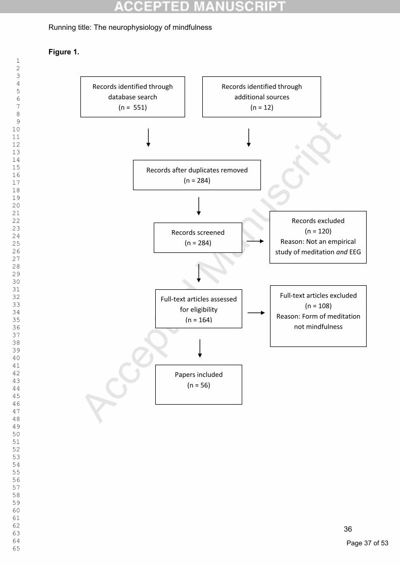
Page 37 of 53
Accep
ted M
anus
cript
1 2 3 4 5 6 7 8 9 10 11 12 13 14 15 16 17 18 19 20 21 22 23 24 25 26 27 28 29 30 31 32 33 34 35 36 37 38 39 40 41 42 43 44 45 46 47 48 49 50 51 52 53 54 55 56 57 58 59 60 61 62 63 64 65
Running title: The neurophysiology of mindfulness
36
Figure 1.
Records identified through
database search
(n = 551)
Records identified through
additional sources
(n = 12)
Records after duplicates removed
(n = 284)
Records screened
(n = 284)
Records excluded
(n = 120)
Reason: Not an empirical
study of meditation and EEG
Full-text articles assessed
for eligibility
(n = 164)
Full-text articles excluded
(n = 108)
Reason: Form of meditation
not mindfulness
Papers included
(n = 56)
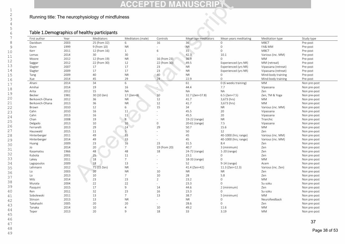
Page 38 of 53
Accep
ted M
anus
cript 1
2 3 4 5 6 7 8 9 10 11 12 13 14 15 16 17 18 19 20 21 22 23 24 25 26 27 28 29 30 31 32 33 34 35 36 37 38 39 40 41 42 43 44 45 46 47 48 49
Running title: The neurophysiology of mindfulness
37
Table 1.Demographics of healthy participants First author Year Meditators Meditators (male) Controls Mean age meditators Mean years meditating Meditation type Study type
Davidson 2003 25 (from 32) 6 16 36 0 MBCT Pre-post Dunn 1999 9 (from 10) NR - NR 0 FA& MM Pre-post Kerr 2011 12 (from 16) 1 6 31 0 MBCT Pre-post Lomas 2014 30 30 - 42.3 10.1 Various (inc. MM) Pre-post Moore 2012 12 (from 19) NR 16 (from 23) 36.9 0 MM Pre-post Saggar 2012 22 (from 30) 12 22 (from 30) 49.5 Experienced (yrs NR) MM (retreat) Pre-post Slagter 2007 17 17 23 NR Experienced (yrs NR) Vipassana (retreat) Pre-post Slagter 2009 17 17 23 NR Experienced (yrs NR) Vipassana (retreat) Pre-post Tang 2009 40 NR 40 NR 0 Mind-body training Pre-post Xue 2014 45 29 24 22.9 0 Mind-body training Pre-post
Ahani 2014 34 6 - 61 0 (6 weeks training) MM Non pre-post
Amihai 2014 19 16 - 44.4 7.7 Vipassana Non pre-post Arita 2012 15 NA - NA NA Zen Non pre-post Becker 1981 30 (10 Zen) 17 (Zen=8) 10 32.7 (Zen=37.8) 6.5 (Zen=7.5) Zen, TM & Yoga Non pre-post Berkovich-Ohana 2012 36 NR 12 41.7 3,673 (hrs) MM Non pre-post Berkovich-Ohana 2013 36 NR 12 41.7 3,673 (hrs) MM Non pre-post Brown 2010 12 6 15 34 NR Various (inc. MM) Non pre-post Cahn 2010 16 11 - 45.5 20 Vipassana Non pre-post Cahn 2013 16 11 - 45.5 20 Vipassana Non pre-post Chan 2008 19 8 - 19-22 (range) NR Triarchic Non pre-post Delgado 2013 10 10 0 20-61 (range) 7.5 Vipassana Non pre-post Ferrarelli 2013 29 14 29 50.7 15.6 MM Non pre-post Hauswald 2015 11 5 - 50 12 Zen Non pre-post Hinterberger 2011 49 33 - 45 40-1000 (hrs; range) Various (inc. MM) Non pre-post Hinterberger 2014 49 33 - 45 40-1000 (hrs; range) Various (inc. MM) Non pre-post Huang 2009 23 16 23 31.5 8.4 Zen Non pre-post Jo 2014 20 7 19 (from 20) 40.7 3 (minimum) Zen Non pre-post Kasamatsu 1966 48 48 18 24-72 (range) 1-20 (range) Zen Non pre-post Kubota 2001 25 11 - 23.1 0 Zen Non pre-post Lakey 2011 18 7 - 18-33 (range) 0 MM Non pre-post Lagopoulos 2009 18 13 - 52 9-14 (range) Acem Non pre-post Lehmann 2012 71 (15 Zen) NR - 41.4 (Zen=42) 11.3 (Zen=12.3) Various (inc. Zen) Non pre-post Lo 2003 20 NR 10 NR NR Zen Non pre-post Lo 2013 10 7 10 28 5.8 Zen Non pre-post Milz 2014 23 23 2 23.2 0 MM Non pre-post Murata 2004 22 22 - 23.3 0 Su-soku Non pre-post Pasquini 2015 17 9 14 44.6 2 (minimum) Zen Non pre-post Ren 2011 32 23 16 23.3 0 Su-soku Non pre-post Sobolewski 2011 13 7 13 38.7 5 (minimum) MM Non pre-post Stinson 2013 13 NR - NR 0 Neurofeedback Non pre-post Takahashi 2005 20 20 - 28.6 0 Zen Non pre-post Tanaka 2014 10 4 10 49.2 11.6 MM Non pre-post Teper 2013 20 9 18 33 3.19 MM Non pre-post
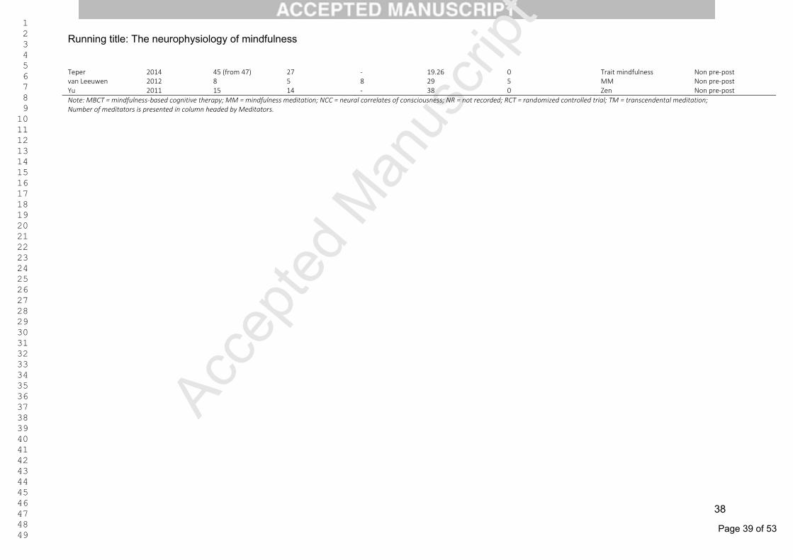
Page 39 of 53
Accep
ted M
anus
cript 1
2 3 4 5 6 7 8 9 10 11 12 13 14 15 16 17 18 19 20 21 22 23 24 25 26 27 28 29 30 31 32 33 34 35 36 37 38 39 40 41 42 43 44 45 46 47 48 49
Running title: The neurophysiology of mindfulness
38
Teper 2014 45 (from 47) 27 - 19.26 0 Trait mindfulness Non pre-post van Leeuwen 2012 8 5 8 29 5 MM Non pre-post Yu 2011 15 14 - 38 0 Zen Non pre-post
Note: MBCT = mindfulness-based cognitive therapy; MM = mindfulness meditation; NCC = neural correlates of consciousness; NR = not recorded; RCT = randomized controlled trial; TM = transcendental meditation; Number of meditators is presented in column headed by Meditators.

Page 40 of 53
Accep
ted M
anus
cript 1
2 3 4 5 6 7 8 9 10 11 12 13 14 15 16 17 18 19 20 21 22 23 24 25 26 27 28 29 30 31 32 33 34 35 36 37 38 39 40 41 42 43 44 45 46 47 48 49
Running title: The neurophysiology of mindfulness
39
Table 2.Demographics of participants with a clinical history First author Year n meditators n males
meditators n controls Mean age
meditators Meditation type Psychiatric Disorder
Barnhofer 2007 10 (from 16) 5 12 (from 18) 48 MBCT Suicidal depression Barnhofer 2010 8 1 8 31.6 MM Previously depressed Bostanov 2012 32 (from 45) 9 32 (from 46) 50.9 MBCT Depressed (remission) Brown 2013 12 NA 15 NA MM pain manage. Chronic pain Garland 2015 11 NA 18 NA MORE Chronic pain Howells 2012 12 2 9 37 MBCT Bipolar disorder Keune 2013 40 (from 53) 10 37 (from 50) 48.9 MBCT Depressed (remission) Schoenberg (et al.) 2014 26 (from 32) NA 24 (from 29) NA MBCT ADHD Schoenberg (& S.) 2014 26 6 25 47.8 MBCT Depression (current) Schoenberg (& S.) 2015 26 6 25 47.8 MBCT Depression (current)
Note: MBCT = mindfulness-based cognitive therapy; MM = mindfulness meditation; MORE = mindfulness-oriented recovery enhancement. All studies featured pre-post designs, and all except Howells were RCTs. All subjects participating had no previous experience with meditation.
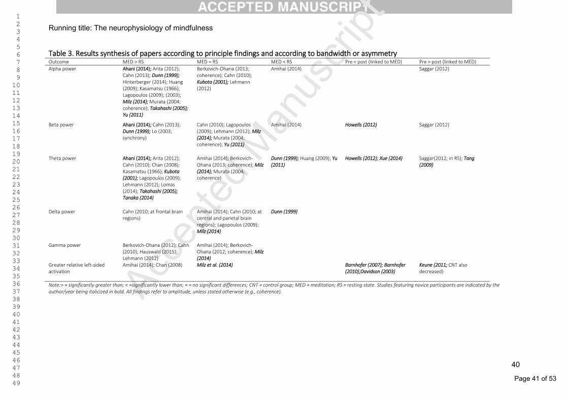
Page 41 of 53
Accep
ted M
anus
cript 1
2 3 4 5 6 7 8 9 10 11 12 13 14 15 16 17 18 19 20 21 22 23 24 25 26 27 28 29 30 31 32 33 34 35 36 37 38 39 40 41 42 43 44 45 46 47 48 49
Running title: The neurophysiology of mindfulness
40
Table 3. Results synthesis of papers according to principle findings and according to bandwidth or asymmetry Outcome MED > RS MED = RS MED < RS Pre < post (linked to MED) Pre > post (linked to MED)
Alpha power Ahani (2014); Arita (2012); Cahn (2013); Dunn (1999); Hinterberger (2014); Huang (2009); Kasamatsu (1966); Lagopoulos (2009); (2003); Milz (2014); Murata (2004; coherence); Takahashi (2005); Yu (2011)
Berkovich-Ohana (2013; coherence); Cahn (2010); Kubota (2001); Lehmann (2012)
Amihai (2014) Saggar (2012)
Beta power Ahani (2014); Cahn (2013); Dunn (1999); Lo (2003; synchrony)
Cahn (2010); Lagopoulos (2009); Lehmann (2012); Milz (2014); Murata (2004; coherence); Yu (2011)
Amihai (2014) Howells (2012) Saggar (2012)
Theta power Ahani (2014); Arita (2012); Cahn (2010); Chan (2008); Kasamatsu (1966); Kubota (2001); Lagopoulos (2009); Lehmann (2012); Lomas (2014); Takahashi (2005); Tanaka (2014)
Amihai (2014); Berkovich-Ohana (2013; coherence); Milz (2014); Murata (2004; coherence)
Dunn (1999); Huang (2009); Yu (2011)
Howells (2012); Xue (2014) Saggar(2012; in RS); Tang (2009)
Delta power Cahn (2010; at frontal brain regions)
Amihai (2014); Cahn (2010; at central and parietal brain regions); Lagopoulos (2009); Milz (2014)
Dunn (1999)
Gamma power Berkovich-Ohana (2012); Cahn (2010); Hauswald (2015); Lehmann (2012)
Amihai (2014); Berkovich-Ohana (2012; coherence); Milz (2014)
Greater relative left-sided activation
Amihai (2014); Chan (2008) Milz et al. (2014) Barnhofer (2007); Barnhofer (2010);Davidson (2003)
Keune (2011; CNT also decreased)
Note:> = significantly greater than; < =significantly lower than; = = no significant differences; CNT = control group; MED = meditation; RS = resting state. Studies featuring novice participants are indicated by the author/year being italicized in bold. All findings refer to amplitude, unless stated otherwise (e.g., coherence).
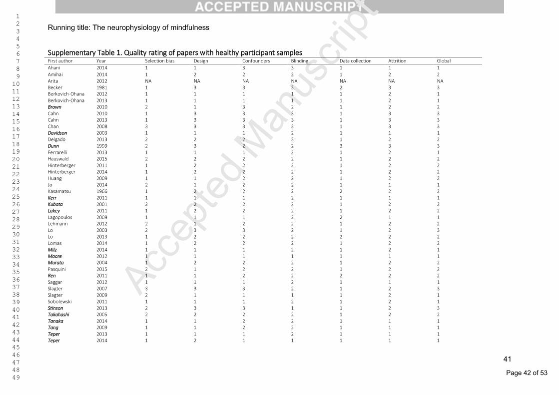
Page 42 of 53
Accep
ted M
anus
cript 1
2 3 4 5 6 7 8 9 10 11 12 13 14 15 16 17 18 19 20 21 22 23 24 25 26 27 28 29 30 31 32 33 34 35 36 37 38 39 40 41 42 43 44 45 46 47 48 49
Running title: The neurophysiology of mindfulness
41
Supplementary Table 1. Quality rating of papers with healthy participant samples First author Year Selection bias Design Confounders Blinding Data collection Attrition Global
Ahani 2014 1 1 3 3 1 1 1
Amihai 2014 1 2 2 2 1 2 2 Arita 2012 NA NA NA NA NA NA NA Becker 1981 1 3 3 3 2 3 3 Berkovich-Ohana 2012 1 1 1 1 1 2 1 Berkovich-Ohana 2013 1 1 1 1 1 2 1 Brown 2010 2 1 3 2 1 2 2 Cahn 2010 1 3 3 3 1 3 3 Cahn 2013 1 3 3 3 1 3 3 Chan 2008 3 3 3 3 1 3 3 Davidson 2003 1 1 1 2 1 1 1 Delgado 2013 2 2 2 3 1 2 2 Dunn 1999 2 3 2 2 3 3 3 Ferrarelli 2013 1 1 1 2 1 2 1 Hauswald 2015 2 2 2 2 1 2 2 Hinterberger 2011 1 2 2 2 1 2 2 Hinterberger 2014 1 2 2 2 1 2 2 Huang 2009 1 1 2 2 1 2 2 Jo 2014 2 1 2 2 1 1 1 Kasamatsu 1966 1 2 2 2 2 2 2 Kerr 2011 1 1 1 2 1 1 1 Kubota 2001 2 2 2 2 1 2 2 Lakey 2011 1 2 2 2 1 2 2 Lagopoulos 2009 1 1 1 2 2 1 1 Lehmann 2012 2 1 2 2 1 2 2 Lo 2003 2 3 3 2 1 2 3 Lo 2013 1 2 2 2 2 2 2 Lomas 2014 1 2 2 2 1 2 2 Milz 2014 1 1 1 2 1 2 1 Moore 2012 1 1 1 1 1 1 1 Murata 2004 1 2 2 2 1 2 2 Pasquini 2015 2 1 2 2 1 2 2 Ren 2011 1 1 2 2 2 2 2 Saggar 2012 1 1 1 2 1 1 1 Slagter 2007 3 3 3 2 1 2 3 Slagter 2009 2 1 1 1 1 2 1 Sobolewski 2011 1 1 1 2 1 2 1 Stinson 2013 2 3 3 1 1 2 3 Takahashi 2005 2 2 2 2 1 2 2 Tanaka 2014 1 1 2 2 1 1 1 Tang 2009 1 1 2 2 1 1 1 Teper 2013 1 1 1 2 1 1 1 Teper 2014 1 2 1 1 1 1 1

Page 43 of 53
Accep
ted M
anus
cript 1
2 3 4 5 6 7 8 9 10 11 12 13 14 15 16 17 18 19 20 21 22 23 24 25 26 27 28 29 30 31 32 33 34 35 36 37 38 39 40 41 42 43 44 45 46 47 48 49
Running title: The neurophysiology of mindfulness
42
van Leeuwen 2012 1 1 1 2 1 2 1 Xue 2014 1 1 2 2 1 2 2 Yu 2011 2 3 2 3 2 2 3
Note: NA = full pdf not available. Studies featuring novice participants are indicated by the author being italicized in bold.
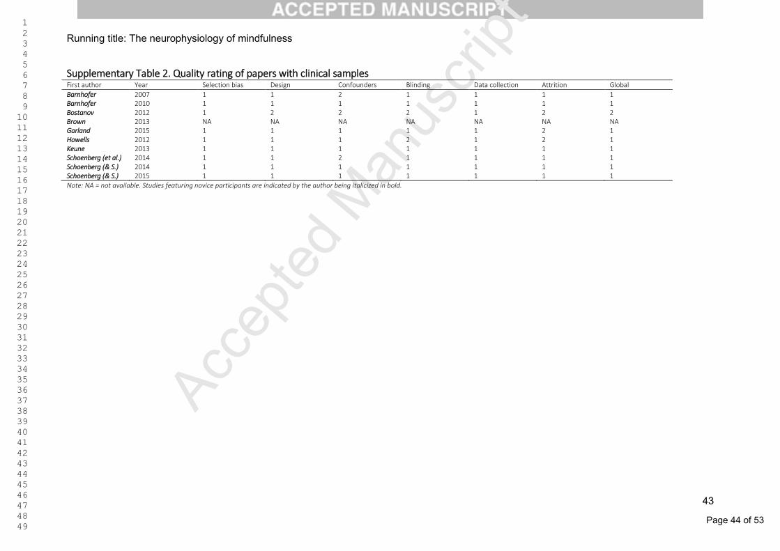
Page 44 of 53
Accep
ted M
anus
cript 1
2 3 4 5 6 7 8 9 10 11 12 13 14 15 16 17 18 19 20 21 22 23 24 25 26 27 28 29 30 31 32 33 34 35 36 37 38 39 40 41 42 43 44 45 46 47 48 49
Running title: The neurophysiology of mindfulness
43
Supplementary Table 2. Quality rating of papers with clinical samples First author Year Selection bias Design Confounders Blinding Data collection Attrition Global
Barnhofer 2007 1 1 2 1 1 1 1 Barnhofer 2010 1 1 1 1 1 1 1 Bostanov 2012 1 2 2 2 1 2 2 Brown 2013 NA NA NA NA NA NA NA Garland 2015 1 1 1 1 1 2 1 Howells 2012 1 1 1 2 1 2 1 Keune 2013 1 1 1 1 1 1 1 Schoenberg (et al.) 2014 1 1 2 1 1 1 1 Schoenberg (& S.) 2014 1 1 1 1 1 1 1 Schoenberg (& S.) 2015 1 1 1 1 1 1 1
Note: NA = not available. Studies featuring novice participants are indicated by the author being italicized in bold.
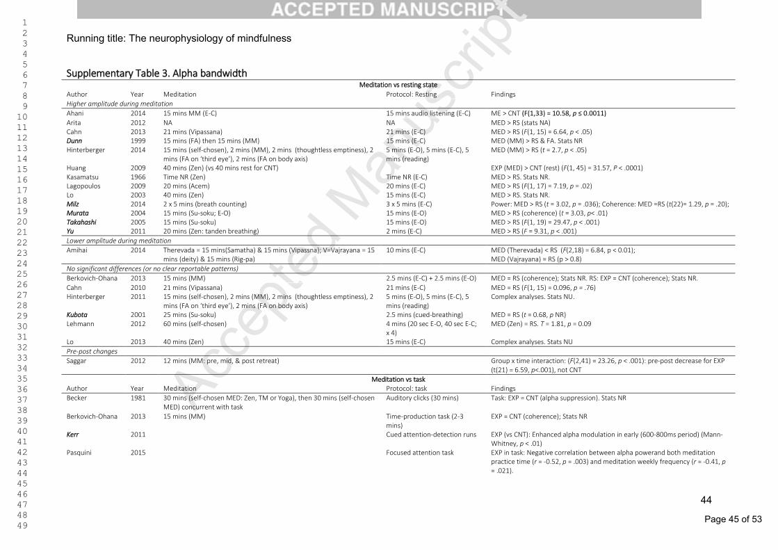
Page 45 of 53
Accep
ted M
anus
cript 1
2 3 4 5 6 7 8 9 10 11 12 13 14 15 16 17 18 19 20 21 22 23 24 25 26 27 28 29 30 31 32 33 34 35 36 37 38 39 40 41 42 43 44 45 46 47 48 49
Running title: The neurophysiology of mindfulness
44
Supplementary Table 3. Alpha bandwidth Meditation vs resting state
Author Year Meditation Protocol: Resting Findings
Higher amplitude during meditation
Ahani 2014 15 mins MM (E-C) 15 mins audio listening (E-C) ME > CNT (F(1,33) = 10.58, p ≤ 0.0011)
Arita 2012 NA NA MED > RS (stats NA) Cahn 2013 21 mins (Vipassana) 21 mins (E-C) MED > RS (F(1, 15) = 6.64, p < .05) Dunn 1999 15 mins (FA) then 15 mins (MM) 15 mins (E-C) MED (MM) > RS & FA. Stats NR Hinterberger 2014 15 mins (self-chosen), 2 mins (MM), 2 mins (thoughtless emptiness), 2
mins (FA on ‘third eye’), 2 mins (FA on body axis) 5 mins (E-O), 5 mins (E-C), 5 mins (reading)
MED (MM) > RS (t = 2.7, p < .05)
Huang 2009 40 mins (Zen) (vs 40 mins rest for CNT) EXP (MED) > CNT (rest) (F(1, 45) = 31.57, P < .0001) Kasamatsu 1966 Time NR (Zen) Time NR (E-C) MED > RS. Stats NR. Lagopoulos 2009 20 mins (Acem) 20 mins (E-C) MED > RS (F(1, 17) = 7.19, p = .02) Lo 2003 40 mins (Zen) 15 mins (E-C) MED > RS. Stats NR. Milz 2014 2 x 5 mins (breath counting) 3 x 5 mins (E-C) Power: MED > RS (t = 3.02, p = .036); Coherence: MED =RS (t(22)= 1.29, p = .20); Murata 2004 15 mins (Su-soku; E-O) 15 mins (E-O) MED > RS (coherence) (t = 3.03, p< .01) Takahashi 2005 15 mins (Su-soku) 15 mins (E-O) MED > RS (F(1, 19) = 29.47, p < .001) Yu 2011 20 mins (Zen: tanden breathing) 2 mins (E-C) MED > RS (F = 9.31, p < .001)
Lower amplitude during meditation
Amihai 2014 Therevada = 15 mins(Samatha) & 15 mins (Vipassna); V=Vajrayana = 15 mins (deity) & 15 mins (Rig-pa)
10 mins (E-C) MED (Therevada) < RS (F(2,18) = 6.84, p < 0.01); MED (Vajrayana) = RS (p > 0.8)
No significant differences (or no clear reportable patterns)
Berkovich-Ohana 2013 15 mins (MM) 2.5 mins (E-C) + 2.5 mins (E-O) MED = RS (coherence); Stats NR. RS: EXP = CNT (coherence); Stats NR.
Cahn 2010 21 mins (Vipassana) 21 mins (E-C) MED = RS (F(1, 15) = 0.096, p = .76) Hinterberger 2011 15 mins (self-chosen), 2 mins (MM), 2 mins (thoughtless emptiness), 2
mins (FA on ‘third eye’), 2 mins (FA on body axis) 5 mins (E-O), 5 mins (E-C), 5 mins (reading)
Complex analyses. Stats NU.
Kubota 2001 25 mins (Su-soku) 2.5 mins (cued-breathing) MED = RS (t = 0.68, p NR) Lehmann
2012 60 mins (self-chosen) 4 mins (20 sec E-O, 40 sec E-C; x 4)
MED (Zen) = RS. T = 1.81, p = 0.09
Lo 2013 40 mins (Zen) 15 mins (E-C) Complex analyses. Stats NU
Pre-post changes
Saggar
2012 12 mins (MM: pre, mid, & post retreat) Group x time interaction: (F(2,41) = 23.26, p < .001): pre-post decrease for EXP (t(21) = 6.59, p<.001), not CNT
Meditation vs task Author Year Meditation Protocol: task Findings
Becker 1981 30 mins (self-chosen MED: Zen, TM or Yoga), then 30 mins (self-chosen MED) concurrent with task
Auditory clicks (30 mins) Task: EXP = CNT (alpha suppression). Stats NR
Berkovich-Ohana 2013 15 mins (MM) Time-production task (2-3 mins)
EXP = CNT (coherence); Stats NR
Kerr 2011 Cued attention-detection runs EXP (vs CNT): Enhanced alpha modulation in early (600-800ms period) (Mann-Whitney, p < .01)
Pasquini
2015 Focused attention task EXP in task: Negative correlation between alpha powerand both meditation practice time (r = -0.52, p = .003) and meditation weekly frequency (r = -0.41, p = .021).

Page 46 of 53
Accep
ted M
anus
cript 1
2 3 4 5 6 7 8 9 10 11 12 13 14 15 16 17 18 19 20 21 22 23 24 25 26 27 28 29 30 31 32 33 34 35 36 37 38 39 40 41 42 43 44 45 46 47 48 49
Running title: The neurophysiology of mindfulness
45
Ren 2011 Time NR (Su-soku) Problem solving (Time NR) EXP (MED) < CNT (cognitive task) (F(2, 45) = 4.14, p = .05) Schoenberg (& S.)
2015 Go/NoGo task Task: pre-post power increase for negative stimuli (t(24) = 2.58, p = .02) for CNT only (EXT: no significant increase).
Stinson
2013 Time NR (relaxation – 'Alpha brain state exercise') Neurofeedback video (Time NR) MED = task. Stats NR.
Note: > = significantly greater than; < =significantly lower than; = = no significant differences; CNT = control group; E-C = eyes-closed; E-O =eyes-open; EXP =experimental group; FA= focussed-attention (concentrative) meditation; (from …) = initial number of participants in a pre-post study; MED = meditation; MM = Mindfulness meditation; NA = not available; NR = not reported; NU = not usable (here); NCC = neural correlates of consciousness (i.e., EEG measurement during MED vs RS); ROI = region of interest; RS = resting state. All bandwidth outcomes pertain to power, unless otherwise stated in parentheses (e.g., coherence). Most entries are comparing the experimental group (EXT; i.e., meditators) under different conditions (e.g., RS vs MED): significantly higher power levels during meditation are written as MED > RS; significantly lower levels as MED < RS; and no significant differences as MED = RS. Some entries are comparing two groups (i.e., EXP vs CNT) on a particular condition (e.g., RS): this will be indicated as RS: EXT >/=/< CNT. Studies featuring novice participants are indicated by the author being italicized in bold.
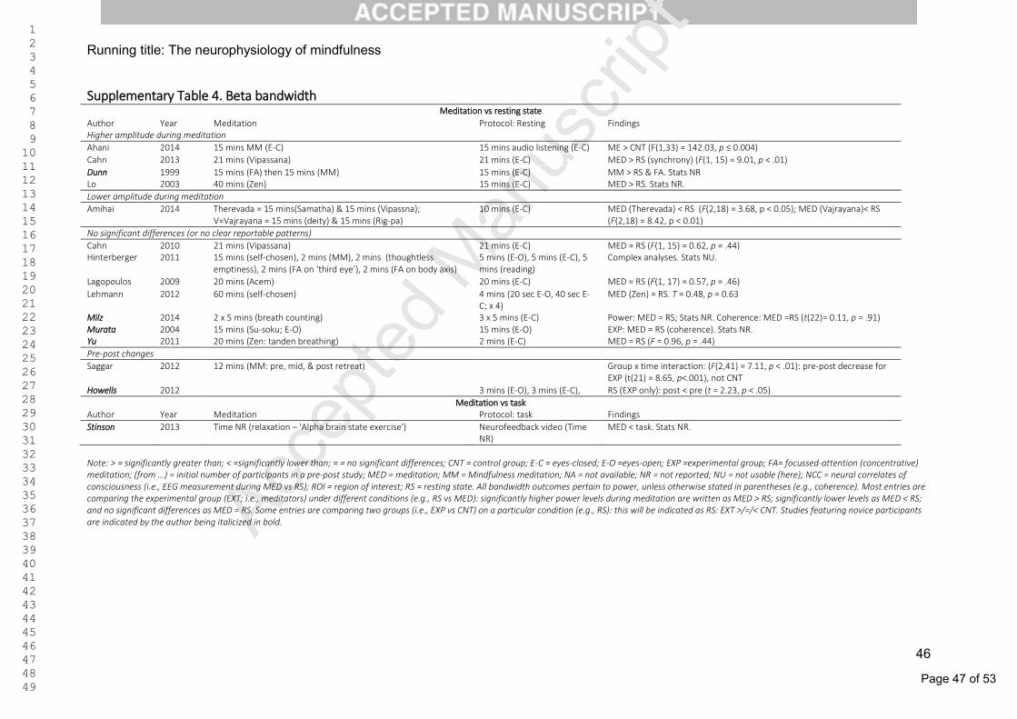
Page 47 of 53
Accep
ted M
anus
cript 1
2 3 4 5 6 7 8 9 10 11 12 13 14 15 16 17 18 19 20 21 22 23 24 25 26 27 28 29 30 31 32 33 34 35 36 37 38 39 40 41 42 43 44 45 46 47 48 49
Running title: The neurophysiology of mindfulness
46
Supplementary Table 4. Beta bandwidth Meditation vs resting state
Author Year Meditation Protocol: Resting Findings Higher amplitude during meditation
Ahani 2014 15 mins MM (E-C) 15 mins audio listening (E-C) ME > CNT (F(1,33) = 142.03, p ≤ 0.004)
Cahn 2013 21 mins (Vipassana) 21 mins (E-C) MED > RS (synchrony) (F(1, 15) = 9.01, p < .01)
Dunn 1999 15 mins (FA) then 15 mins (MM) 15 mins (E-C) MM > RS & FA. Stats NR Lo 2003 40 mins (Zen) 15 mins (E-C) MED > RS. Stats NR.
Lower amplitude during meditation
Amihai 2014 Therevada = 15 mins(Samatha) & 15 mins (Vipassna); V=Vajrayana = 15 mins (deity) & 15 mins (Rig-pa)
10 mins (E-C) MED (Therevada) < RS (F(2,18) = 3.68, p < 0.05); MED (Vajrayana)< RS (F(2,18) = 8.42, p < 0.01)
No significant differences (or no clear reportable patterns)
Cahn 2010 21 mins (Vipassana) 21 mins (E-C) MED = RS (F(1, 15) = 0.62, p = .44) Hinterberger 2011 15 mins (self-chosen), 2 mins (MM), 2 mins (thoughtless
emptiness), 2 mins (FA on ‘third eye’), 2 mins (FA on body axis) 5 mins (E-O), 5 mins (E-C), 5 mins (reading)
Complex analyses. Stats NU.
Lagopoulos 2009 20 mins (Acem) 20 mins (E-C) MED = RS (F(1, 17) = 0.57, p = .46)
Lehmann
2012
60 mins (self-chosen) 4 mins (20 sec E-O, 40 sec E-C; x 4)
MED (Zen) = RS. T = 0.48, p = 0.63
Milz 2014 2 x 5 mins (breath counting) 3 x 5 mins (E-C) Power: MED = RS; Stats NR. Coherence: MED =RS (t(22)= 0.11, p = .91) Murata 2004 15 mins (Su-soku; E-O) 15 mins (E-O) EXP: MED = RS (coherence). Stats NR. Yu 2011 20 mins (Zen: tanden breathing) 2 mins (E-C) MED = RS (F = 0.96, p = .44)
Pre-post changes
Saggar 2012
12 mins (MM: pre, mid, & post retreat) Group x time interaction: (F(2,41) = 7.11, p < .01): pre-post decrease for EXP (t(21) = 8.65, p<.001), not CNT
Howells 2012 3 mins (E-O), 3 mins (E-C), RS (EXP only): post < pre (t = 2.23, p < .05)
Meditation vs task Author Year Meditation Protocol: task Findings
Stinson 2013
Time NR (relaxation – 'Alpha brain state exercise') Neurofeedback video (Time NR)
MED < task. Stats NR.
Note: > = significantly greater than; < =significantly lower than; = = no significant differences; CNT = control group; E-C = eyes-closed; E-O =eyes-open; EXP =experimental group; FA= focussed-attention (concentrative) meditation; (from …) = initial number of participants in a pre-post study; MED = meditation; MM = Mindfulness meditation; NA = not available; NR = not reported; NU = not usable (here); NCC = neural correlates of consciousness (i.e., EEG measurement during MED vs RS); ROI = region of interest; RS = resting state. All bandwidth outcomes pertain to power, unless otherwise stated in parentheses (e.g., coherence). Most entries are comparing the experimental group (EXT; i.e., meditators) under different conditions (e.g., RS vs MED): significantly higher power levels during meditation are written as MED > RS; significantly lower levels as MED < RS; and no significant differences as MED = RS. Some entries are comparing two groups (i.e., EXP vs CNT) on a particular condition (e.g., RS): this will be indicated as RS: EXT >/=/< CNT. Studies featuring novice participants are indicated by the author being italicized in bold.
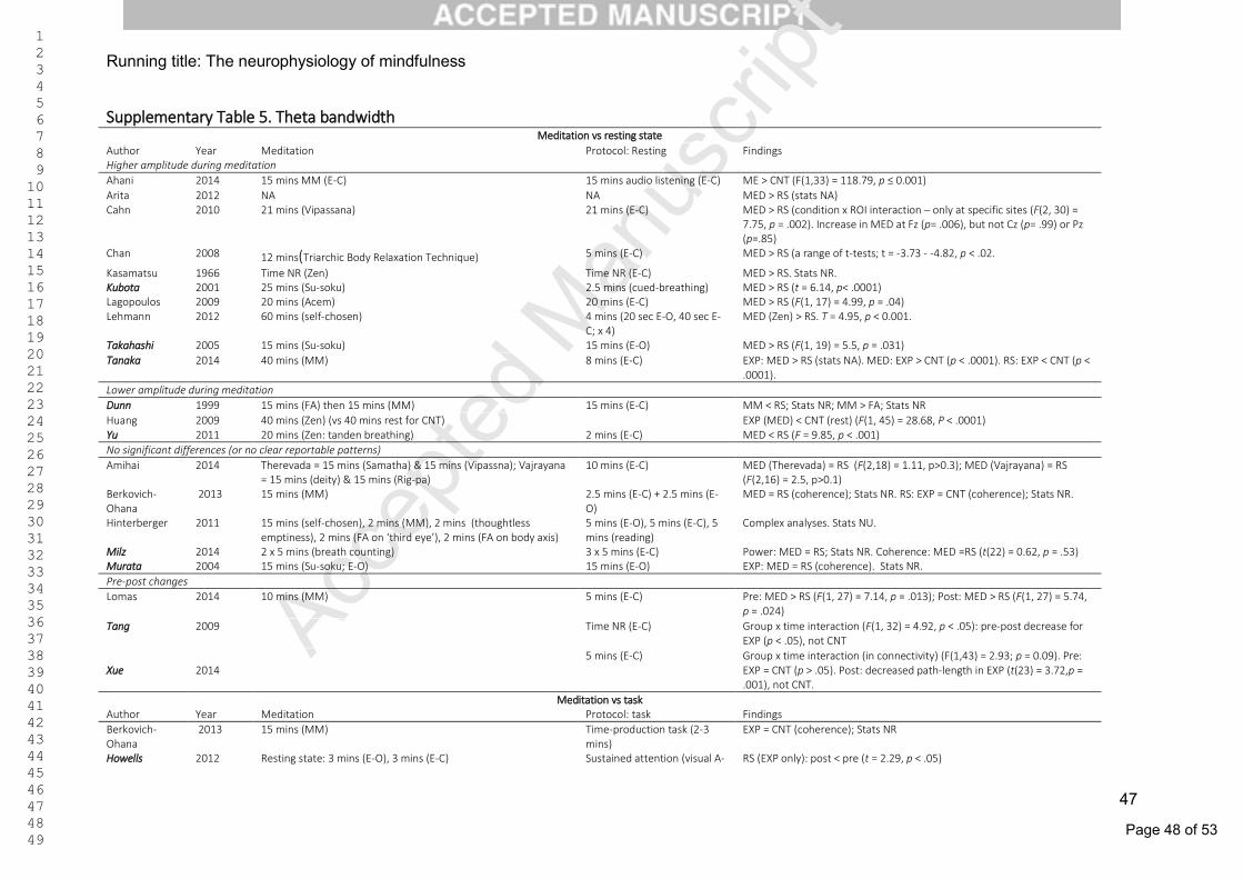
Page 48 of 53
Accep
ted M
anus
cript 1
2 3 4 5 6 7 8 9 10 11 12 13 14 15 16 17 18 19 20 21 22 23 24 25 26 27 28 29 30 31 32 33 34 35 36 37 38 39 40 41 42 43 44 45 46 47 48 49
Running title: The neurophysiology of mindfulness
47
Supplementary Table 5. Theta bandwidth Meditation vs resting state
Author Year Meditation Protocol: Resting Findings Higher amplitude during meditation
Ahani 2014 15 mins MM (E-C) 15 mins audio listening (E-C) ME > CNT (F(1,33) = 118.79, p ≤ 0.001)
Arita 2012 NA NA MED > RS (stats NA) Cahn 2010
21 mins (Vipassana) 21 mins (E-C) MED > RS (condition x ROI interaction – only at specific sites (F(2, 30) =
7.75, p = .002). Increase in MED at Fz (p= .006), but not Cz (p= .99) or Pz (p=.85)
Chan 2008 12 mins(Triarchic Body Relaxation Technique) 5 mins (E-C) MED > RS (a range of t-tests; t = -3.73 - -4.82, p < .02.
Kasamatsu 1966 Time NR (Zen) Time NR (E-C) MED > RS. Stats NR. Kubota 2001 25 mins (Su-soku) 2.5 mins (cued-breathing) MED > RS (t = 6.14, p< .0001) Lagopoulos 2009 20 mins (Acem) 20 mins (E-C) MED > RS (F(1, 17) = 4.99, p = .04) Lehmann 2012
60 mins (self-chosen) 4 mins (20 sec E-O, 40 sec E-
C; x 4) MED (Zen) > RS. T = 4.95, p < 0.001.
Takahashi 2005 15 mins (Su-soku) 15 mins (E-O) MED > RS (F(1, 19) = 5.5, p = .031)
Tanaka
2014
40 mins (MM) 8 mins (E-C) EXP: MED > RS (stats NA). MED: EXP > CNT (p < .0001). RS: EXP < CNT (p < .0001).
Lower amplitude during meditation
Dunn 1999 15 mins (FA) then 15 mins (MM) 15 mins (E-C) MM < RS; Stats NR; MM > FA; Stats NR
Huang 2009 40 mins (Zen) (vs 40 mins rest for CNT) EXP (MED) < CNT (rest) (F(1, 45) = 28.68, P < .0001) Yu 2011 20 mins (Zen: tanden breathing) 2 mins (E-C) MED < RS (F = 9.85, p < .001)
No significant differences (or no clear reportable patterns)
Amihai 2014 Therevada = 15 mins (Samatha) & 15 mins (Vipassna); Vajrayana = 15 mins (deity) & 15 mins (Rig-pa)
10 mins (E-C) MED (Therevada) = RS (F(2,18) = 1.11, p>0.3); MED (Vajrayana) = RS (F(2,16) = 2.5, p>0.1)
Berkovich-Ohana
2013 15 mins (MM) 2.5 mins (E-C) + 2.5 mins (E-O)
MED = RS (coherence); Stats NR. RS: EXP = CNT (coherence); Stats NR.
Hinterberger 2011 15 mins (self-chosen), 2 mins (MM), 2 mins (thoughtless emptiness), 2 mins (FA on ‘third eye’), 2 mins (FA on body axis)
5 mins (E-O), 5 mins (E-C), 5 mins (reading)
Complex analyses. Stats NU.
Milz 2014 2 x 5 mins (breath counting) 3 x 5 mins (E-C) Power: MED = RS; Stats NR. Coherence: MED =RS (t(22) = 0.62, p = .53) Murata 2004 15 mins (Su-soku; E-O) 15 mins (E-O) EXP: MED = RS (coherence). Stats NR.
Pre-post changes
Lomas 2014
10 mins (MM)
5 mins (E-C) Pre: MED > RS (F(1, 27) = 7.14, p = .013); Post: MED > RS (F(1, 27) = 5.74, p = .024)
Tang
2009
Time NR (E-C) Group x time interaction (F(1, 32) = 4.92, p < .05): pre-post decrease for EXP (p < .05), not CNT
Xue
2014
5 mins (E-C) Group x time interaction (in connectivity) (F(1,43) = 2.93; p = 0.09). Pre: EXP = CNT (p > .05). Post: decreased path-length in EXP (t(23) = 3.72,p = .001), not CNT.
Meditation vs task Author Year Meditation Protocol: task Findings
Berkovich-Ohana
2013 15 mins (MM) Time-production task (2-3 mins)
EXP = CNT (coherence); Stats NR
Howells 2012 Resting state: 3 mins (E-O), 3 mins (E-C) Sustained attention (visual A- RS (EXP only): post < pre (t = 2.29, p < .05)
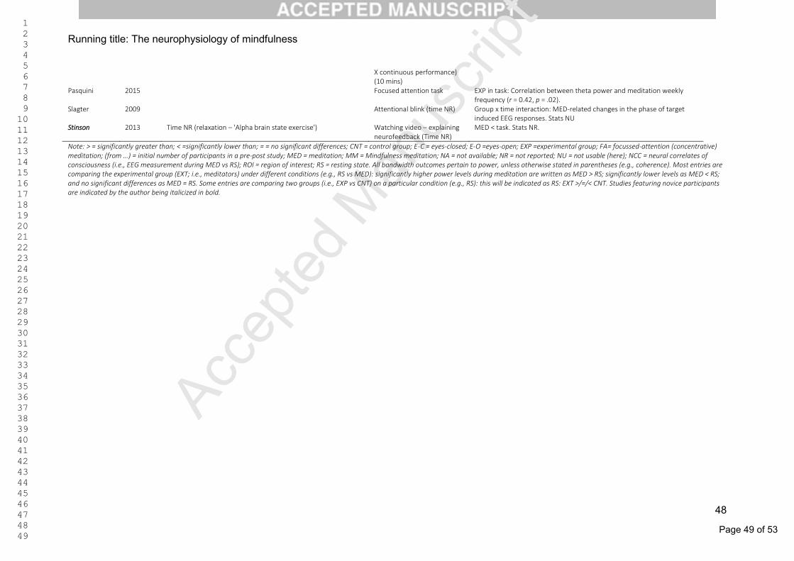
Page 49 of 53
Accep
ted M
anus
cript 1
2 3 4 5 6 7 8 9 10 11 12 13 14 15 16 17 18 19 20 21 22 23 24 25 26 27 28 29 30 31 32 33 34 35 36 37 38 39 40 41 42 43 44 45 46 47 48 49
Running title: The neurophysiology of mindfulness
48
X continuous performance) (10 mins)
Pasquini
2015 Focused attention task EXP in task: Correlation between theta power and meditation weekly frequency (r = 0.42, p = .02).
Slagter 2009
Attentional blink (time NR) Group x time interaction: MED-related changes in the phase of target induced EEG responses. Stats NU
Stinson 2013
Time NR (relaxation – 'Alpha brain state exercise') Watching video – explaining neurofeedback (Time NR)
MED < task. Stats NR.
Note: > = significantly greater than; < =significantly lower than; = = no significant differences; CNT = control group; E-C = eyes-closed; E-O =eyes-open; EXP =experimental group; FA= focussed-attention (concentrative) meditation; (from …) = initial number of participants in a pre-post study; MED = meditation; MM = Mindfulness meditation; NA = not available; NR = not reported; NU = not usable (here); NCC = neural correlates of consciousness (i.e., EEG measurement during MED vs RS); ROI = region of interest; RS = resting state. All bandwidth outcomes pertain to power, unless otherwise stated in parentheses (e.g., coherence). Most entries are comparing the experimental group (EXT; i.e., meditators) under different conditions (e.g., RS vs MED): significantly higher power levels during meditation are written as MED > RS; significantly lower levels as MED < RS; and no significant differences as MED = RS. Some entries are comparing two groups (i.e., EXP vs CNT) on a particular condition (e.g., RS): this will be indicated as RS: EXT >/=/< CNT. Studies featuring novice participants are indicated by the author being italicized in bold.
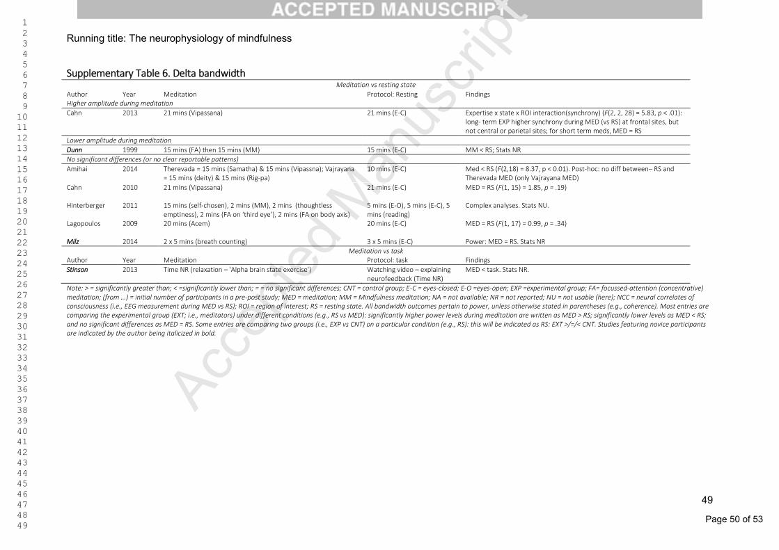
Page 50 of 53
Accep
ted M
anus
cript 1
2 3 4 5 6 7 8 9 10 11 12 13 14 15 16 17 18 19 20 21 22 23 24 25 26 27 28 29 30 31 32 33 34 35 36 37 38 39 40 41 42 43 44 45 46 47 48 49
Running title: The neurophysiology of mindfulness
49
Supplementary Table 6. Delta bandwidth Meditation vs resting state
Author Year Meditation Protocol: Resting Findings Higher amplitude during meditation
Cahn 2013 21 mins (Vipassana) 21 mins (E-C)
Expertise x state x ROI interaction(synchrony) (F(2, 2, 28) = 5.83, p < .01): long- term EXP higher synchrony during MED (vs RS) at frontal sites, but not central or parietal sites; for short term meds, MED = RS
Lower amplitude during meditation
Dunn 1999 15 mins (FA) then 15 mins (MM) 15 mins (E-C) MM < RS; Stats NR
No significant differences (or no clear reportable patterns)
Amihai 2014 Therevada = 15 mins (Samatha) & 15 mins (Vipassna); Vajrayana = 15 mins (deity) & 15 mins (Rig-pa)
10 mins (E-C) Med < RS (F(2,18) = 8.37, p < 0.01). Post-hoc: no diff between– RS and Therevada MED (only Vajrayana MED)
Cahn 2010 21 mins (Vipassana) 21 mins (E-C) MED = RS (F(1, 15) = 1.85, p = .19)
Hinterberger 2011 15 mins (self-chosen), 2 mins (MM), 2 mins (thoughtless emptiness), 2 mins (FA on ‘third eye’), 2 mins (FA on body axis)
5 mins (E-O), 5 mins (E-C), 5 mins (reading)
Complex analyses. Stats NU.
Lagopoulos
2009 20 mins (Acem) 20 mins (E-C) MED = RS (F(1, 17) = 0.99, p = .34)
Milz 2014 2 x 5 mins (breath counting) 3 x 5 mins (E-C) Power: MED = RS. Stats NR
Meditation vs task Author Year Meditation Protocol: task Findings
Stinson
2013 Time NR (relaxation – 'Alpha brain state exercise') Watching video – explaining neurofeedback (Time NR)
MED < task. Stats NR.
Note: > = significantly greater than; < =significantly lower than; = = no significant differences; CNT = control group; E-C = eyes-closed; E-O =eyes-open; EXP =experimental group; FA= focussed-attention (concentrative) meditation; (from …) = initial number of participants in a pre-post study; MED = meditation; MM = Mindfulness meditation; NA = not available; NR = not reported; NU = not usable (here); NCC = neural correlates of consciousness (i.e., EEG measurement during MED vs RS); ROI = region of interest; RS = resting state. All bandwidth outcomes pertain to power, unless otherwise stated in parentheses (e.g., coherence). Most entries are comparing the experimental group (EXT; i.e., meditators) under different conditions (e.g., RS vs MED): significantly higher power levels during meditation are written as MED > RS; significantly lower levels as MED < RS; and no significant differences as MED = RS. Some entries are comparing two groups (i.e., EXP vs CNT) on a particular condition (e.g., RS): this will be indicated as RS: EXT >/=/< CNT. Studies featuring novice participants are indicated by the author being italicized in bold.
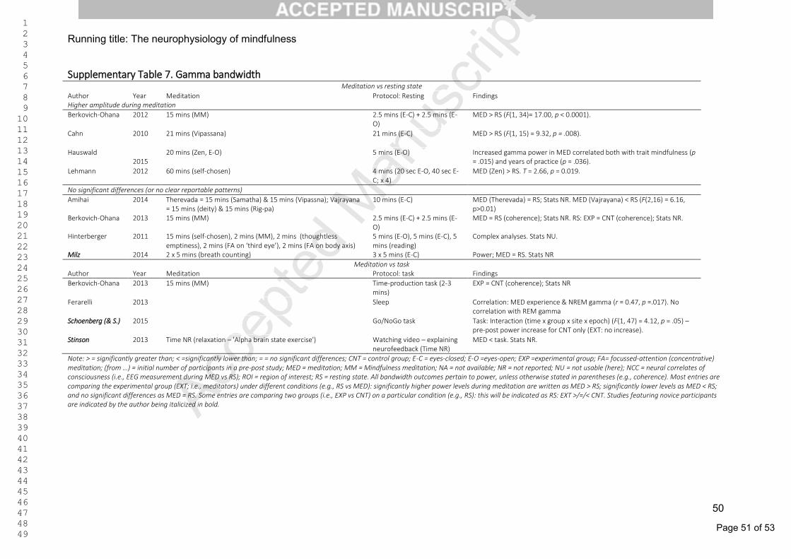
Page 51 of 53
Accep
ted M
anus
cript 1
2 3 4 5 6 7 8 9 10 11 12 13 14 15 16 17 18 19 20 21 22 23 24 25 26 27 28 29 30 31 32 33 34 35 36 37 38 39 40 41 42 43 44 45 46 47 48 49
Running title: The neurophysiology of mindfulness
50
Supplementary Table 7. Gamma bandwidth Meditation vs resting state
Author Year Meditation Protocol: Resting Findings Higher amplitude during meditation
Berkovich-Ohana 2012 15 mins (MM) 2.5 mins (E-C) + 2.5 mins (E-O)
MED > RS (F(1, 34)= 17.00, p < 0.0001).
Cahn 2010
21 mins (Vipassana) 21 mins (E-C) MED > RS (F(1, 15) = 9.32, p = .008).
Hauswald 2015
20 mins (Zen, E-O) 5 mins (E-O) Increased gamma power in MED correlated both with trait mindfulness (p = .015) and years of practice (p = .036).
Lehmann 2012
60 mins (self-chosen) 4 mins (20 sec E-O, 40 sec E-C; x 4)
MED (Zen) > RS. T = 2.66, p = 0.019.
No significant differences (or no clear reportable patterns)
Amihai 2014 Therevada = 15 mins (Samatha) & 15 mins (Vipassna); Vajrayana = 15 mins (deity) & 15 mins (Rig-pa)
10 mins (E-C) MED (Therevada) = RS; Stats NR. MED (Vajrayana) < RS (F(2,16) = 6.16, p>0.01)
Berkovich-Ohana 2013 15 mins (MM) 2.5 mins (E-C) + 2.5 mins (E-O)
MED = RS (coherence); Stats NR. RS: EXP = CNT (coherence); Stats NR.
Hinterberger 2011 15 mins (self-chosen), 2 mins (MM), 2 mins (thoughtless emptiness), 2 mins (FA on ‘third eye’), 2 mins (FA on body axis)
5 mins (E-O), 5 mins (E-C), 5 mins (reading)
Complex analyses. Stats NU.
Milz 2014 2 x 5 mins (breath counting) 3 x 5 mins (E-C) Power; MED = RS. Stats NR
Meditation vs task Author Year Meditation Protocol: task Findings
Berkovich-Ohana 2013 15 mins (MM) Time-production task (2-3 mins)
EXP = CNT (coherence); Stats NR
Ferarelli 2013 Sleep Correlation: MED experience & NREM gamma (r = 0.47, p =.017). No correlation with REM gamma
Schoenberg (& S.)
2015 Go/NoGo task Task: Interaction (time x group x site x epoch) (F(1, 47) = 4.12, p = .05) – pre-post power increase for CNT only (EXT: no increase).
Stinson
2013 Time NR (relaxation – 'Alpha brain state exercise') Watching video – explaining neurofeedback (Time NR)
MED < task. Stats NR.
Note: > = significantly greater than; < =significantly lower than; = = no significant differences; CNT = control group; E-C = eyes-closed; E-O =eyes-open; EXP =experimental group; FA= focussed-attention (concentrative) meditation; (from …) = initial number of participants in a pre-post study; MED = meditation; MM = Mindfulness meditation; NA = not available; NR = not reported; NU = not usable (here); NCC = neural correlates of consciousness (i.e., EEG measurement during MED vs RS); ROI = region of interest; RS = resting state. All bandwidth outcomes pertain to power, unless otherwise stated in parentheses (e.g., coherence). Most entries are comparing the experimental group (EXT; i.e., meditators) under different conditions (e.g., RS vs MED): significantly higher power levels during meditation are written as MED > RS; significantly lower levels as MED < RS; and no significant differences as MED = RS. Some entries are comparing two groups (i.e., EXP vs CNT) on a particular condition (e.g., RS): this will be indicated as RS: EXT >/=/< CNT. Studies featuring novice participants are indicated by the author being italicized in bold.

Page 52 of 53
Accep
ted M
anus
cript 1
2 3 4 5 6 7 8 9 10 11 12 13 14 15 16 17 18 19 20 21 22 23 24 25 26 27 28 29 30 31 32 33 34 35 36 37 38 39 40 41 42 43 44 45 46 47 48 49
Running title: The neurophysiology of mindfulness
51
Supplementary Table 8. Asymmetry Findings Meditation vs resting state
Author Year Meditation Protocol Findings Increase in relative left-frontal activation linked to meditation
Amihai 2014 Therevada = 15 mins (Samatha) & 15 mins (Vipassna); Vajrayana = 15 mins (deity) & 15 mins (Rig-pa)
10 mins resting state (E-C) MED (Therevada): Condition x location interaction (F(4,36) = 3.09, p<0.05). MED (Vipassana) > RS at left location (p<0.05), but not right or center (p>0.2)
Barnhofer 2007 8 x 1min (4 = E-O, 4 = E-C) 8 x 1min (4 = E-O, 4 = E-C) Group x time interaction (F(2, 19) = 3.7, p = .044): pre-post decreases in relative left prefrontal asymmetry for CNT (p = .003), but not EXP (p = .918)
Barnhofer
2010 EXP = 15 mins (MM); CNT = 15 mins (LKM) 2 mins (E-C): EEG assessed pre and post MED
Pre-post increase (EXP & CNT) in relative left prefrontal activation (F(1, 13) = 5.06, p = .04)
Chan
2008 Triarchic Body Relaxation Technique (12 mins) 5 mins (E-C) 5 mins resting state (E-C MED > RS (left-sided activation; F(1, 18) = 5.42, p = .032)
Davidson 2003 8 x 1min (4 = E-O, 4 = E-C) Writing about experiences (EEG recorded 1 min before & 3 mins after)
Group x time interaction in RS (F(1, 37) = 5.14, p < .05): pre-post increase in relative left- activation for EXP (not CNT)
Decrease in relative left-frontal activation linked to meditation
Keune 2011 Sad mood induction (sad music, and neg. experience recall). Time NR.
Pre-post decrease (EXP & CNT) in relative left prefrontal activation (F(4, 64) = 3.38, p < .05)
No change in relative left-frontal activation linked to meditation
Milz 2014 2 x 5 mins (breath counting) 3 x 5 mins resting state (E-C) Power: MED = RS. Stats NR
Note: > = significantly greater than; < =significantly lower than; = = no significant differences; CNT = control group; E-C = eyes-closed; E-O =eyes-open; EXP =experimental group; FA= focussed-attention (concentrative) meditation; (from …) = initial number of participants in a pre-post study; MED = meditation; MM = Mindfulness meditation; NA = not available; NR = not reported; NU = not usable (here); NCC = neural correlates of consciousness (i.e., EEG measurement during MED vs RS); ROI = region of interest; RS = resting state. All bandwidth outcomes pertain to power, unless otherwise stated in parentheses (e.g., coherence). Most entries are comparing the experimental group (EXT; i.e., meditators) under different conditions (e.g., RS vs MED): significantly higher power levels during meditation are written as MED > RS; significantly lower levels as MED < RS; and no significant differences as MED = RS. Some entries are comparing two groups (i.e., EXP vs CNT) on a particular condition (e.g., RS): this will be indicated as RS: EXT >/=/< CNT. Studies featuring novice participants are indicated by the author being italicized in bold.
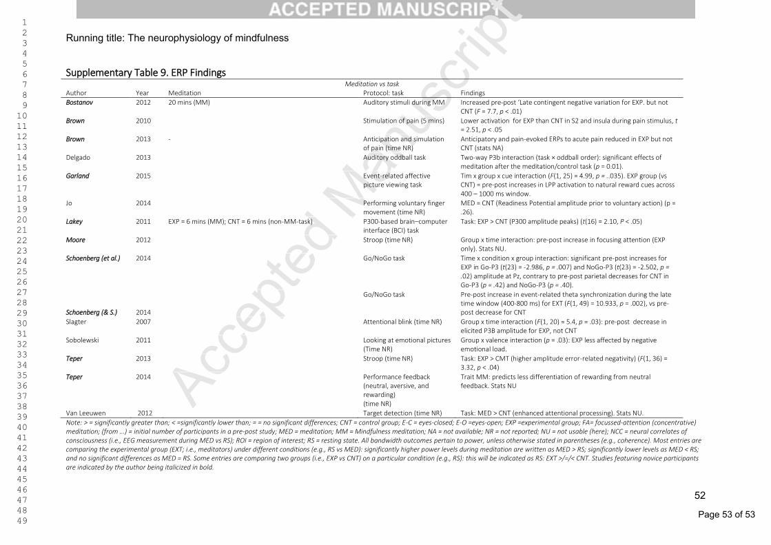
Page 53 of 53
Accep
ted M
anus
cript 1
2 3 4 5 6 7 8 9 10 11 12 13 14 15 16 17 18 19 20 21 22 23 24 25 26 27 28 29 30 31 32 33 34 35 36 37 38 39 40 41 42 43 44 45 46 47 48 49
Running title: The neurophysiology of mindfulness
52
Supplementary Table 9. ERP Findings Meditation vs task
Author Year Meditation Protocol: task Findings
Bostanov 2012 20 mins (MM) Auditory stimuli during MM Increased pre-post ‘Late contingent negative variation for EXP. but not CNT (F = 7.7, p < .01)
Brown 2010 Stimulation of pain (5 mins) Lower activation for EXP than CNT in S2 and insula during pain stimulus, t = 2.51, p < .05
Brown
2013 - Anticipation and simulation of pain (time NR)
Anticipatory and pain-evoked ERPs to acute pain reduced in EXP but not CNT (stats NA)
Delgado
2013 Auditory oddball task Two-way P3b interaction (task × oddball order): significant effects of meditation after the meditation/control task (p = 0.01).
Garland
2015 Event-related affective picture viewing task
Tim x group x cue interaction (F(1, 25) = 4.99, p = ..035). EXP group (vs CNT) = pre-post increases in LPP activation to natural reward cues across 400 – 1000 ms window.
Jo
2014 Performing voluntary finger movement (time NR)
MED = CNT (Readiness Potential amplitude prior to voluntary action) (p = .26).
Lakey
2011 EXP = 6 mins (MM); CNT = 6 mins (non-MM-task) P300-based brain–computer interface (BCI) task
Task: EXP > CNT (P300 amplitude peaks) (t(16) = 2.10, P < .05)
Moore
2012
Stroop (time NR) Group x time interaction: pre-post increase in focusing attention (EXP only). Stats NU.
Schoenberg (et al.) 2014 Go/NoGo task Time x condition x group interaction: significant pre-post increases for EXP in Go-P3 (t(23) = -2.986, p = .007) and NoGo-P3 (t(23) = -2.502, p = .02) amplitude at Pz, contrary to pre-post parietal decreases for CNT in Go-P3 (p = .42) and NoGo-P3 (p = .40).
Schoenberg (& S.) 2014
Go/NoGo task Pre-post increase in event-related theta synchronization during the late time window (400-800 ms) for EXT (F(1, 49) = 10.933, p = .002), vs pre-post decrease for CNT
Slagter
2007
Attentional blink (time NR) Group x time interaction (F(1, 20) = 5.4, p = .03): pre-post decrease in elicited P3B amplitude for EXP, not CNT
Sobolewski
2011
Looking at emotional pictures (Time NR)
Group x valence interaction (p = .03): EXP less affected by negative emotional load.
Teper
2013
Stroop (time NR) Task: EXP > CMT (higher amplitude error-related negativity) (F(1, 36) = 3.32, p < .04)
Teper
2014
Performance feedback (neutral, aversive, and rewarding) (time NR)
Trait MM: predicts less differentiation of rewarding from neutral feedback. Stats NU
Van Leeuwen 2012 Target detection (time NR) Task: MED > CNT (enhanced attentional processing). Stats NU.
Note: > = significantly greater than; < =significantly lower than; = = no significant differences; CNT = control group; E-C = eyes-closed; E-O =eyes-open; EXP =experimental group; FA= focussed-attention (concentrative) meditation; (from …) = initial number of participants in a pre-post study; MED = meditation; MM = Mindfulness meditation; NA = not available; NR = not reported; NU = not usable (here); NCC = neural correlates of consciousness (i.e., EEG measurement during MED vs RS); ROI = region of interest; RS = resting state. All bandwidth outcomes pertain to power, unless otherwise stated in parentheses (e.g., coherence). Most entries are comparing the experimental group (EXT; i.e., meditators) under different conditions (e.g., RS vs MED): significantly higher power levels during meditation are written as MED > RS; significantly lower levels as MED < RS; and no significant differences as MED = RS. Some entries are comparing two groups (i.e., EXP vs CNT) on a particular condition (e.g., RS): this will be indicated as RS: EXT >/=/< CNT. Studies featuring novice participants are indicated by the author being italicized in bold.
Top Related