


Languages
Pages
Legal

This is an archived version of a publication, provided by Chris Sampson.Please cite appropriately.
A randomised controlled trial of laser scanning
and casting for the construction of ankle foot
orthoses
Andrew Roberts∗ Johanna WalesChristopher James Sampson Heather Smith Peter Jones
Marilyn James
November 25, 2014
Publication type:
Peer-reviewed journal article
This version:
This is a post-print version of the manuscript, meaning that it has been peer-reviewed.
Published version:
Roberts, A., Wales, J., Smith, H., Sampson, C. J., Jones, P., & James, M.(2014). A randomised controlled trial of laser scanning and casting for theconstruction of ankle–foot orthoses. Prosthetics and orthotics international,0309364614550263. DOI: 10.1177/0309364614550263
Copyright:
This work is licensed under the Creative Commons Attribution 3.0 Unported Li-cense. To view a copy of this license, visit http://creativecommons.org/licenses/by/3.0/or send a letter to Creative Commons, 444 Castro Street, Suite 900, MountainView, California, 94041, USA.
∗Correspondence to: A Roberts, ORLAU, Robert Jones & Agnes Hunt Orthopaedic Hos-pital NHS Foundation Trust, Oswestry, SY10 7AG, UK [email protected]

1
TITLE
A randomised controlled trial of laser scanning and casting for the construction of ankle foot
orthoses.

2
ABSTRACT
Study Design: Randomised controlled trial with blinding of orthotists and patients to the
construction technique used. Background: Three-dimensional laser scanning has been used
for patient measurement for cranial helmets and spinal braces. Ankle foot orthoses are
commonly prescribed for children with orthopaedic conditions. This trial sought to compare
ankle foot orthoses produced by laser scanning or traditional plaster casting. Objectives:
Assessment of the effectiveness and efficiency of using laser scanning to produce AFOs.
Methods: A randomised double blind trial comparing fabrication of AFOs from casts or laser
scans. Results: The time spent in the rectification and moulding of scanned AFOs was around
50% less than for cast AFOs. A non-significant increase of 9 days was seen in the time to
delivery to the patient for LSCAD/CAM. There was a higher incidence of problems with the
scan-based AFOs at delivery of the device, but no difference in how long the AFOs lasted.
Costs associated with laser scanning were not significantly different from traditional methods of
AFO manufacture. Conclusions: Compared with conventional casting techniques laser scan
based AFO manufacture neither significantly improved the quality of the final product nor
delivered a useful saving in time.
(word count 193)
CLINICAL RELEVANCE
Ankle foot orthoses (AFOs) are a common requirement for chronic neurological conditions
during childhood. Improved efficiency of provision of AFOs would benefit children and families
by reducing the delay in provision of devices and would benefit the health service by making
best use of valuable orthotist time.
(word count 47)

3
BACKGROUND
Orthoses are devices that are applied externally in order to modify structural and functional
attributes of the neuromuscular and musculoskeletal systems, for example in cerebral palsy.
The prevalence of cerebral palsy in the UK has been estimated as 2 per 1000 live births for birth
years 1986-19961.
AFOs have considerable support in the literature in the management of cerebral palsy2.
Children who are managed in AFOs often require intervention and care from multiple clinicians
and agencies so efficient orthotic provision is important in reducing the logistical demands on
their families.
The traditional method of producing an AFO involves taking a negative limb cast from the
patient and then filling the negative cast with plaster to make a positive cast. The orthosis is
then formed by moulding polypropylene sheets over the positive cast. This method can be
replaced by laser scanning with computer-aided design and manufacture (LSCAD/CAM), where
a positive of the limb is milled in foam directly from the digital image. A recent systematic review
suggests that foot orthotics designed from 3D scans are at least comparable with those made
through traditional methods, although none of the studies reviewed were randomised controlled
trials and further research is required to confirm this3. One study suggested that plaster casting
may be preferable to laser scanning when it is important to capture the forefoot-to-rearfoot
relationship4.
We were able to identify four studies comparing plaster casting with LSCAD/CAM technology in
the fabrication of spinal orthoses for scoliosis5-8
. Radiological improvement of the scoliotic
curves was assessed in three of these studies, and LSCAD/CAM was reported to be as
effective as the traditional method. The LSCAD/CAM technique was also found to increase
4
productivity in both of the studies which measured this outcome. None of these studies
measured health economic outcomes.
In the NHS, high capital costs and a lack of orthotists with time to learn new technology are
likely reasons why LSCAD/CAM technology has not been introduced. If it proved successful,
LSCAD/CAM could enable a more efficient process for the provision of a large number of
bespoke orthotic devices.
Two hypotheses are explored in this paper. Firstly, that there is a difference in the length of time
taken to deliver of a well-fitting AFOs produced by means of LSCAD/CAM compared to AFOs
produced by traditional plaster casting. Secondly, that there is a difference in patient benefit in
terms of functioning and satisfaction with AFOs produced by means of LSCAD/CAM compared
to AFOs produced by traditional plaster casting. In addition the cost-effectiveness of AFOs
produced by means of LSCAD/CAM versus traditional plaster casting was estimated using a
societal perspective.
METHODS
Design of Study
A double blind randomised (1:1) controlled trial design in which patients had their AFO(s)
fabricated either using LSCAD/CAM technology or by a traditional plaster method.
A positive opinion for the study was received from the xxxxxxx Local Research Ethics
Committee on 15th
August 2007 (ref: 07/Q2604/85).
Inclusion and Exclusion Criteria
All patients up to 18 years of age referred for rigid and hinged AFOs at the Trust were
candidates for inclusion. Young adults up to the age of 21 years were also included if they were
5
still receiving services for children (for example, because they were in education). Exclusion of
the patient occurred if informed consent was not obtained; the patient was considered by the
orthotist to be un-splintable; a Saltiel style device had been specified or if communication was
not possible with the subject or their parent/carer.
Setting
The study took place in the xxxxxxx Unit (xxxxx) and Orthotics Departments at the xxxxxxxxxxx
NHS Foundation Trust between February 2008 and August 2010. Eligible subjects were
contacted by the study nurse who provided an explanation of the study. Those subjects who
assented to participate had demographic data and details about diagnosis and condition
collected.
Interventions
All subjects had both a casting and a scan of each limb requiring an AFO, in the same session,
and the order in which the two methods were performed was determined according to a
computer-generated randomisation list. The allocation sequence was concealed from both the
researcher and the participant in sequentially numbered, sealed envelopes. The study orthotists
were coached on how to minimise bias in their interaction with the subject (for example,
participants were to receive the same level of encouragement for each method). After
experiencing both methods subjects were asked to state whether they would prefer to have
casting or scanning next time. The time taken and consumables for each method and session
were measured and recorded by the attending clinician. The subjects or their parents/carers
completed a questionnaire about travel and associated costs. The subject was given an
appointment to return two weeks later to receive their AFO(s). Both the LSCAD/CAM digital

6
image (scan) and the cast were rectified by an orthotic technician and length of time for each
method was recorded. Our usual Standard Operating Procedure was applied to both the cast
and scan where 3mm is applied to malleoli/metatarsals and navicular when requested on the
specification sheet. A 1% increase in all dimensions was added to the scans to allow for ease of
doffing and donning as well as allowing for growth.
A second randomisation was then performed to determine whether the AFO was manufactured
from the cast or from the scan. Allocation to a manufacturing method was achieved through a
central telephone randomisation system provided by the University of York Trials Centre, and
was stratified by age (less than 6 years and 6-21 years), Gillette Functional Walking score (1-5
and 6-10)9
and required number of AFOs (unilateral or bilateral).
Laser scans were obtained with a Fastscan (Polhemus, 40 Hercules Drv, PO Box 560,
Colchester, VT 05446, USA.) and rectified using Rodin4D software (Rodin4D Parc Le Biogalien
- Bâtiment B, 27 Allée Charles Darwin, 33600 Pessac, France). Image files were passed to an
orthotic milling company (Relief Orthotics, Unit 34B, Viking Road, Airdrie, Scotland ML6 9SE)
who milled a positive of the limb in foam that was returned to the Trust for manufacture of the
polypropylene device. Where cast manufacture was employed, a plaster male form was cast,
rectified and then draped in the usual fashion. The length of time for the moulding of the
polypropylene AFO was recorded. There were no differences in the manufacturing process
between the two groups once the draping of the polypropylene sheet and curing had been
performed.
Sample size Calculations
The sample size was based on data which suggest the total time taken in the moulding and
rectification process is reduced by approximately 60% when using LSCAD/CAM compared to

7
casting6. The proposed sample size of 150 patients would enable the detection of a proportional
reduction in time taken in the moulding and rectification of a cast of 30% with 90% power using
a two-sided test at p=0.05.
Outcome Measures
1. The primary outcome measure was the length of time taken (in minutes) in the moulding and
rectification process.
2. Secondary outcome measures included: (a) Length of time (in minutes) spent with subjects to
cast and scan limbs. (b) Length of time (in minutes) to fit the AFO(s). (c) Number of days taken
from initial scanning/casting of a subject to the completion of an AFO (or AFOs) that met fitting
specifications. Any additional time taken for rescanning or remaking was included. (d) The
length of life of the AFO in days.
Patient-focused outcome measures included the “Satisfaction with Device” and “Satisfaction
with Service” questionnaires, which were developed in the US as part of the Orthotics and
Prosthetics Users’ Survey (OPUS)10
. Both questionnaires use a four-point Likert scale ranging
from Strongly Agree (highest level of satisfaction) to Strongly Disagree (lowest level of
satisfaction). Satisfaction with Device was measured with nine items (e.g. skin
abrasions/irritations; whether the orthosis was pain-free to wear; whether it looked good and
whether it was easy to put on) and ten items measured Satisfaction with Service (e.g. amount of
time waiting for services; whether the orthotist was responsive to concerns; and whether staff
co-ordinated services with doctors and therapists). The questions were administered by post at
3, 6, and 12 months follow-up. Non-responders were followed up by telephone if possible. In
most cases parents/carers acted as proxies by answering the questions on behalf of their child.

8
Statistical Analysis
All planned analyses were by intention-to-treat. Differences in the primary and secondary
outcome measures were analysed using the independent t-test for continuous data and the
logrank tests for time to event data. Data from the patient-focused satisfaction questions, using
the Likert scale, were analysed individually using the Mann Whitney U test, proportions using
chi squared tests and time to event data using the logrank test. All tests were carried out using
a 5% level of significance and no adjustment was made for multiple testing and missing values
were not imputed.
Economic Analysis
In addition to the above, an economic analysis was completed of LSCAD/CAM compared with
traditional casting. The work undertook a cost-effectiveness analysis using the time to functional
AFO as the primary outcome, and sought to assess costs using a societal perspective. An
individual resource collection tool was developed through pilot work with parent-representatives
and designed to fully capture all the elements. In addition to intervention costs and health care
resource use, this included the cost of patient travel to the hospital and productivity losses
associated with time off work due to treatment and care. The time horizon was 12 months; as
such, costs and outcomes were not discounted.
Resource use was collected by various means and in most cases national unit costs were
assigned. Resource use relating to the scanning and casting sessions and the manufacture of a
device were recorded by the attending orthotist. This included data on the time spent carrying
out specific tasks, staff attending, the use of consumables and the duration of group sessions.
Similarly, these data were collected for fittings and adjustments.

9
Follow-up data were collected from patients at 3, 6, 9 and 12 months. These included
medication, time off work, service use and associated travel costs. Published unit costs from
NHS Staff Earnings Estimates11
, Personal Social Services Research Unit12
and British National
Formulary13
were applied as appropriate. All costings were made at 2011 prices.
An incremental cost-effectiveness ratio (ICER) – the cost per day saved of LSCAD/CAM
compared with traditional casting – was estimated and bootstrapping was used to estimate
confidence intervals for the ICER. Results are based on a complete case analysis.
RESULTS
Subjects
During the study the provision of orthotic services at the Trust was reorganised following a
“sustainable services review” and the study orthotists left the Trust. At this point, 92 (69% of
total) subjects had been recruited into the study. New study orthotists were less experienced in
laser scanning and had less time to devote to the study so the ability to recruit patients into the
trial became more difficult. Despite an extension to the study duration; widening of the inclusion
criteria from 18 years to 21 years of age (provided the subject was in education); and a move to
bring more AFO moulding ‘in house’ in xxxxxxxx, the sample size target of 150 subjects was not
met. One hundred and fifty one individuals were approached and 134 subjects were recruited
into the study (two participants were inadvertently randomised twice; in both cases the first
allocation was used). The flow of subjects and numbers randomised into each group, including
those that received the planned intervention, are presented in Figure 1. The majority of subjects
were affected by cerebral palsy with no statistically significant differences between groups
(Table 1). Ninety-six (72%) patients required bilateral AFOs while assistive devices were used
by 42% of the patients.
10
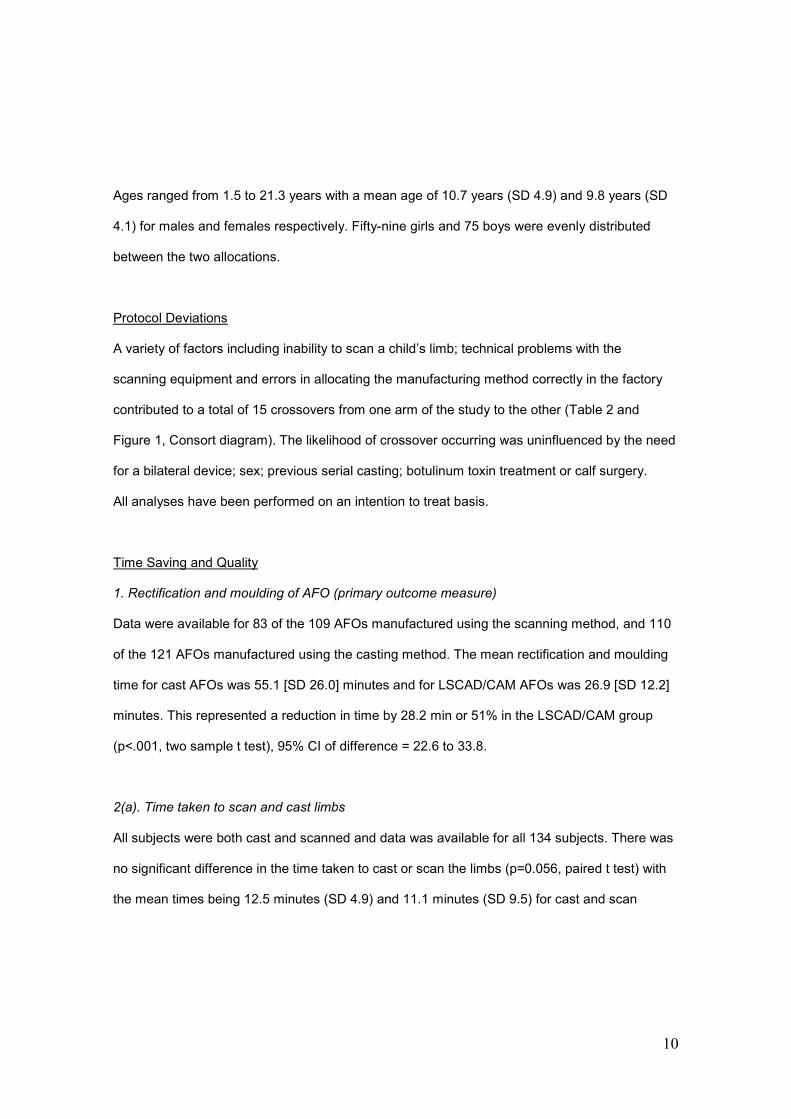
Ages ranged from 1.5 to 21.3 years with a mean age of 10.7 years (SD 4.9) and 9.8 years (SD
4.1) for males and females respectively. Fifty-nine girls and 75 boys were evenly distributed
between the two allocations.
Protocol Deviations
A variety of factors including inability to scan a child’s limb; technical problems with the
scanning equipment and errors in allocating the manufacturing method correctly in the factory
contributed to a total of 15 crossovers from one arm of the study to the other (Table 2 and
Figure 1, Consort diagram). The likelihood of crossover occurring was uninfluenced by the need
for a bilateral device; sex; previous serial casting; botulinum toxin treatment or calf surgery.
All analyses have been performed on an intention to treat basis.
Time Saving and Quality
1. Rectification and moulding of AFO (primary outcome measure)
Data were available for 83 of the 109 AFOs manufactured using the scanning method, and 110
of the 121 AFOs manufactured using the casting method. The mean rectification and moulding
time for cast AFOs was 55.1 [SD 26.0] minutes and for LSCAD/CAM AFOs was 26.9 [SD 12.2]
minutes. This represented a reduction in time by 28.2 min or 51% in the LSCAD/CAM group
(p<.001, two sample t test), 95% CI of difference = 22.6 to 33.8.
2(a). Time taken to scan and cast limbs
All subjects were both cast and scanned and data was available for all 134 subjects. There was
no significant difference in the time taken to cast or scan the limbs (p=0.056, paired t test) with
the mean times being 12.5 minutes (SD 4.9) and 11.1 minutes (SD 9.5) for cast and scan

11
respectively, and a mean difference of 1.33 [SD 10.07], 95% CI of paired difference = -0.4 to
3.1.
Evaluation of average casting time throughout the study revealed no training effect or trend in
casting time. However, a striking difference was seen over time in the time taken to scan as a
result of unplanned staff turnover (Figure 2). If we only include scan times for the two orthotists
who had received more training and were already familiar with the technique prior to the start of
recruitment, a statistically significant difference (p<.001, two sample t-test) was then seen with
average times 13.1 (SD 4.4) minutes for casting and 8.9 minutes (SD 2.9) for scans.
2(b) Supply and fit of AFO(s)
At the second visit the AFO(s) were provided to the patient. A significant proportion (75%) of
patients guessed their actual method of fabrication correctly (p<.001, Chi sq test). There were
no significant differences in delays in supply between the two methods (p=0.711, logrank test).
A significantly higher proportion of scan-based AFOs failed to meet the specification stipulated
by the scanning orthotist, see Table 3 (p=0.004, Chi Sq test ). Remaking was required in 17% of
scan-based AFOs and 3% of cast-based AFOs (p<.001, Chi Sq test). Whilst none of the cast
patients required recasting, 4 scanned patients required a rescan, 1 scanned patient was
subsequently cast, and 8 scanned patients required an adaptation of the original scan.
Although there was an increased incidence of problems with the scan-based AFOs, the time
taken to fit the device was not significantly different between the groups.
2(c) Time to Deliver Device
Data were available for 68 of the subjects allocated to the cast group and 62 subjects allocated
to the scanned group. No significant effects were seen (p=.12 logrank test) of allocated group

12
on the time taken from casting/scanning to delivery of a well-fitting AFO/AFOs. The mean (SD)
number of days was 46.9 (45.3) and 37.9 (31.9) for scan and cast respectively with 95% CI for
the difference (scan – cast) of -4.5 to 22.5. The time from receipt of the order in the
manufacturing unit to the date the order was completed was close in the two groups (13 days ±6
in the cast group, 15 days ±8.2 in the scanned group).
2(d). AFO Length of Life
The comparison of length of AFO life was carried out using Kaplan-Meier survival plots (Figure
3) and a logrank test. This is due to the fact that times to failure are only recorded up to one
year and hence those surviving beyond one year are censored. 29 of the 70 subjects allocated
to casting and 29 of the 64 subjects allocated to scanning returned to clinic for a new AFO
within the first year, and around half of these were as a result of growth (see Table 4). No
significant difference was seen in the length of life of AFOs manufactured using the casting or
scanning method (p=.57, logrank test).
Patient Preference / Satisfaction
After experiencing both casting and scanning (in random order) 70% of the patients said they
preferred being scanned to having the limbs cast in plaster.
A Mann Whitney U test to evaluate differences in responses to the four-point Likert scale
(Strongly Agree – Strongly Disagree) found no significant differences between the allocated
groups when applied individually to the nine “Satisfaction with Device” questions and the 10
“Satisfaction with Service” questions at each time point (3, 6, & 12 months follow-up). “Agree”
was the predominant category selected by respondents for all questions thus indicating a
reasonable level of satisfaction with the AFOs and services.
13
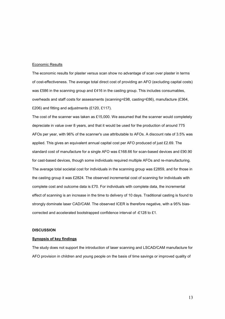
Economic Results
The economic results for plaster versus scan show no advantage of scan over plaster in terms
of cost-effectiveness. The average total direct cost of providing an AFO (excluding capital costs)
was £586 in the scanning group and £416 in the casting group. This includes consumables,
overheads and staff costs for assessments (scanning=£98, casting=£86), manufacture (£364,
£206) and fitting and adjustments (£120, £117).
The cost of the scanner was taken as £15,000. We assumed that the scanner would completely
depreciate in value over 8 years, and that it would be used for the production of around 775
AFOs per year, with 96% of the scanner's use attributable to AFOs. A discount rate of 3.5% was
applied. This gives an equivalent annual capital cost per AFO produced of just £2.69. The
standard cost of manufacture for a single AFO was £168.66 for scan-based devices and £90.90
for cast-based devices, though some individuals required multiple AFOs and re-manufacturing.
The average total societal cost for individuals in the scanning group was £2859, and for those in
the casting group it was £2824. The observed incremental cost of scanning for individuals with
complete cost and outcome data is £70. For individuals with complete data, the incremental
effect of scanning is an increase in the time to delivery of 10 days. Traditional casting is found to
strongly dominate laser CAD/CAM. The observed ICER is therefore negative, with a 95% bias-
corrected and accelerated bootstrapped confidence interval of -£128 to £1.
DISCUSSION
Synopsis of key findings
The study does not support the introduction of laser scanning and LSCAD/CAM manufacture for
AFO provision in children and young people on the basis of time savings or improved quality of

14
device. Despite a reduction of around 50% in the time spent in the rectification and moulding
processes, this benefit was not transferred to the patient where an average increase in the time
to delivery of the devices of 9 days was seen for LSCAD/CAM. This delay was probably due to
the proportion of devices failing to meet the required specification being considerably higher
after manufacture by LSCAD/CAM than after plaster-casting, and thus requiring a re-make and
lengthening the time to delivery. However, there is potential for improvement in the design of
AFOs manufactured by LSCAD/CAM, and this study has highlighted the importance of training
for orthotists in this technique.
Pros and Cons of LSCAD/CAM compared to plaster casting
Anecdotally several patients reported that they believed their AFO was produced by a scan
because it fitted so well. Conversely, excessively intimate fits, particularly around the malleoli,
make donning difficult and spasticity or calf tightness complicates the process. An intimate fit
also presents problems with the longevity of the device in the presence of growth. During the
study a policy was adopted of enlarging the scan by 1% in all dimensions; this was found to give
a closeness of fit comparable to a conventionally fabricated device to give room for growth.
There is clear potential for improvements in the design of AFOs manufactured by LSCAD/CAM.
The inability of the orthotist to correctly position the patient during the scanning process makes
the correction of postural deformity totally dependent on the rectification process whereas in the
process of casting the foot can be held in a corrected position rendering the rectification process
less complex and critical.
Re-making is a costly process. Given that 17% of the scanned patients required a re-scan, re-
cast or adaptation of the original scan, versus 3% of the plastered patients, plastering is again

15
the favourable option. Where a re-make is required the hospital bears the staff, capital and
consumable costs of this, whilst the families incur increased travel costs and productivity losses.
In addition, there is a large difference in the manufacturing costs between AFOs made by
scanning and by casting, with scanning being over £70 more expensive.
Being able to store the scan for enlargement in response to growth does offer a potential
advantage in terms of a reduction in return visits for families if a previous scan can be stretched
and an AFO fabricated. However, given the increased frequency of rescans and defects seen in
this study, the cost of such a strategy would be currently unacceptable and the time saving for
families would be reduced.
Reduction in cost due to time-savings in rectification and moulding may be negated by an
increase in costs due to problems at delivery of device (e.g. re-scanning).
Technical difficulties with the hardware and software led to occasional failures of scanning,
particularly early on in the study when the technology was relatively new. Whilst the trained
orthotists were able to scan in a consistently short period of time, the locums and new staff
members frequently had prolonged setup times and were uncomfortable with the process. A
study by Telfer et al. demonstrated that only the more experienced CAD operator was able to
achieve excellent inter- and intra-operator reliability14
. However, in this study the imperfections
in the resulting AFOs were seen equally in scans performed by “trained” orthotists as well as
those not previously trained in the use of the technology. The consistent times for casting
across all orthotists and throughout the study demonstrated the ubiquitous nature of the manual
skill of casting instilled in trainee orthotists in the United Kingdom.
Limitations of the study
16
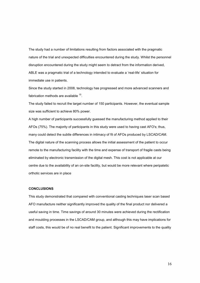
The study had a number of limitations resulting from factors associated with the pragmatic
nature of the trial and unexpected difficulties encountered during the study. Whilst the personnel
disruption encountered during the study might seem to detract from the information derived,
ABLE was a pragmatic trial of a technology intended to evaluate a ‘real-life’ situation for
immediate use in patients.
Since the study started in 2008, technology has progressed and more advanced scanners and
fabrication methods are available15
.
The study failed to recruit the target number of 150 participants. However, the eventual sample
size was sufficient to achieve 80% power.
A high number of participants successfully guessed the manufacturing method applied to their
AFOs (75%). The majority of participants in this study were used to having cast AFO's; thus,
many could detect the subtle differences in intimacy of fit of AFOs produced by LSCAD/CAM.
The digital nature of the scanning process allows the initial assessment of the patient to occur
remote to the manufacturing facility with the time and expense of transport of fragile casts being
eliminated by electronic transmission of the digital mesh. This cost is not applicable at our
centre due to the availability of an on-site facility, but would be more relevant where peripatetic
orthotic services are in place
CONCLUSIONS
This study demonstrated that compared with conventional casting techniques laser scan based
AFO manufacture neither significantly improved the quality of the final product nor delivered a
useful saving in time. Time savings of around 30 minutes were achieved during the rectification
and moulding processes in the LSCAD/CAM group, and although this may have implications for
staff costs, this would be of no real benefit to the patient. Significant improvements to the quality

17
of devices manufactured using the LSCAD/CAM method would be required before any overall
benefit would be seen.
ACKNOWLEDGEMENTS
All authors contributed equally in the preparation of this manuscript.
xxxxxxxxx coordinated subject recruitment and undertook the data collection and collation.
xxxxxxxxx and xxxxxxxxxxxx managed the orthotic workshop and implemented the
randomisation provided by the York Trials Unit. The orthotists taking part in the trial included
xxxxxxxx, xxxxxxxxxxxxx, xxxxxxxxxxxxxx, xxxxxxxxxxx, xxxxxxxxxxx, xxxxxxxxxxx and
xxxxxxxxxxx. xxxxxx staff, xxxxxxxxxxxx and xxxxxxxxxxx, offered xxxxxxx facilities to support
the study.
DECLARATION OF CONFLICTING INTERESTS
The Authors declare that there is no conflict of interest.
FUNDING ACKNOWLEDGEMENT
This work was supported by the National Institute for Health Research reference number PB-
PG-0706-10626.
(word count 4101)

18
REFERENCES
1. Surman G, Bonellie S, Chalmers J, Colver A, Dolk H, Hemming K, King A, Kurinczuk JJ,
Parkes J, Platt MJ. UKCP: a collaborative network of cerebral palsy registers in the United
Kingdom. J Public Health (Oxf), 2006;28:148-156.
2. Figueiredo E M; Ferreira GB, Maia Moreira RC; Kirkwood R N; Fetters L. Efficacy of Ankle-
Foot Orthoses on Gait of Children with Cerebral Palsy: Systematic Review of Literature.
Pediatric Physical Therapy, 2008; 20(3):207-223
3. Telfer S, Woodburn J. The use of 3D surface scanning for the measurement and assessment
of the human foot. J Foot Ankle Res 2010;3:19. [Published online 2010 September 5. doi:
10.1186/1757-1146-3-19]
4. Laughton C, McClay Davis I, Williams DS. A comparison of four methods of obtaining a
negative impression of the foot. J Am Podiatr Med Assoc. 2002;92(5):261-8
5. Cottalorda J, Kohler R, Garin C, Genevois P, Lecante C, Berge B. Orthoses for mild scoliosis:
a prospective study comparing traditional plaster mold manufacturing with fast, noncontact, 3-
dimensional acquisition. Spine 2005;30:399-405.
6. Wong MS, Cheng JCY, Lo KH. A comparison of treatment effectiveness between the
CAD/CAM method and the manual method for managing adolescent idiopathic scoliosis.
Prosthet Orthot Int, 2005;29:105-111
19
7. Wong MS, Cheng JCY, Wong MW, So SF. A work study of the CAD/CAM metod and
conventional manual method in the fabrication of spinal orthoses for patients with adolescent
idiopathic scoliosis. Prosthet Orthot Int. 2005;29(1):93-104
8. Wong MS, Cheng CY, Ng BK, Lam TP, Chiu SW. A comparison of the clinical effectiveness
of spinal orthoses manufactured using the conventional method and CAD/CAM method in the
management of AIS. Stud Health Technol Inform. 2006:123:225-32
9. Novacheck TF, Stout JL, Tervo R. Reliability and validity of the Gillette Functional
Assessment Questionnaire as an outcome measure in children with walking disabilities. J
Pediatr Orthop, 2000;20:75-81.
10. Heinemann AW, Bode RK, O'Reilly C. Development and measurement properties of the
Orthotics and Prosthetics Users' Survey (OPUS): a comprehensive set of clinical outcome
instruments. Prosthet Orthot Int. 2003;27(3):191-206.
11. Department of Health. NHS Staff Earnings Estimates, July to September 2011. Available
from: http://data.gov.uk/dataset/nhs-staff-earnings
12. Curtis, L. Unit Costs of Health and Social Care 2011. Personal Social Services Research
Unit. Available from : http://www.pssru.ac.uk/project-pages/unit-costs/2011/index.php#sections

20
13. Joint Formulary Committee. British National Formulary. 62nd
ed. London: BMJ Group and
Pharmaceutical Press; 2011.
14. Telfer S, Gibson KS, Hennessy K, Steultjens MP, Woodburn J. Computer-aided design of
customized foot orthoses: reproducibility and effect of method used to obtain foot shape. Arch
Phys Med Rehabil. 2012;93(5):863-70. [doi: 10.1016/j.apmr.2011.12.019]
15. Mavroidis C, Ranky RG, Sivak ML, Patritti BL, DiPisa J, Caddle A, Gilhooly K, Govoni L,
Sivak S, Lancia M, Drillio R, Bonato P. Patient specific ankle-foot orthoses using rapid
prototyping. J Neuroeng Rehabil. 2011;8:1. [Published online 2011 January 12. doi:
10.1186/1743-0003-8-1]
21
FIGURES
Figure 1. Consolidated Standards of Reporting Trials (CONSORT) flow chart.[insert figure 1, pdf]
Figure 2. Trends in first visit scan time during study. indicates the point during the study whenthe orthotists experienced in the use of the scanner left the Trust and were replaced withorthotists inexperienced in the use of the scanner.[insert figure 2]
Figure 3. Kaplan-Meier plot of time to failure (days) of the device for the two groups. Grey line =AFOs manufactured by casting method, black line = AFOs manufactured using LSCAD/CAM[insert figure 3]
22
TABLES
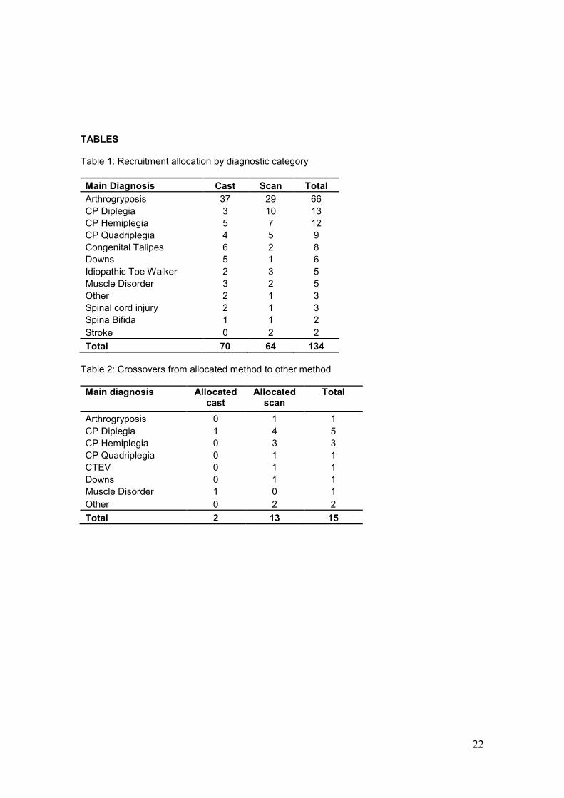
Table 1: Recruitment allocation by diagnostic category
Main Diagnosis Cast Scan Total
Arthrogryposis 37 29 66
CP Diplegia 3 10 13
CP Hemiplegia 5 7 12
CP Quadriplegia 4 5 9
Congenital Talipes 6 2 8
Downs 5 1 6
Idiopathic Toe Walker 2 3 5
Muscle Disorder 3 2 5
Other 2 1 3
Spinal cord injury 2 1 3
Spina Bifida 1 1 2
Stroke 0 2 2
Total 70 64 134
Table 2: Crossovers from allocated method to other method
Main diagnosis Allocatedcast
Allocatedscan
Total
Arthrogryposis 0 1 1
CP Diplegia 1 4 5
CP Hemiplegia 0 3 3
CP Quadriplegia 0 1 1
CTEV 0 1 1
Downs 0 1 1
Muscle Disorder 1 0 1
Other 0 2 2
Total 2 13 15

23
Table 3: Incidence of problems at delivery of first device
Problem Cast Scan
Number of subjects/Total number of
subjects for which datais available for
% Number of subjects/Total number of subjects
for which data isavailable for
%
Remake needed 2/69 3 11/63 17
Recast / Rescan 0/69 0 5/63 8
Lack of intimate fit 7/69 10 13/62 21
Difficulty donning 3/69 4 10/62 16
Poor quality finish 1/69 1 5/62 8
Trim lines incorrect 8/68 12 15/62 24
Footplate incorrect 40/69 58 47/63 75
Height incorrect 5/69 7 6/62 10
Pitch incorrect 0/69 0 5/62 8
Adjustments needed 34/69 49 37/62 60
Table 4: Reasons for replacement of first AFO
Reason for Replacement Cast Scan
Outgrown 18 13
Needs different design 4 6
Poor fit 4 6
Too uncomfortable 1 2
Broken 1 1
Worn out 1 0
Not made to specification 0 1
Total 29 29

24

25
Figure 2:
0 900

26
Figure 3
0.000
0.250
0.500
0.750
1.000
0 50 100 150 200 250 300 350 400
surv
Cum
ula
tive
pro
ba
bili
tyo
fsurv
ivalo
fdevic
e
Time (days)
_______ scan group
_______ cast group
Top Related