


Languages
Pages
Legal

© 2013 Pearson Education, Inc.
Cartilage: Basic Structure, Types and Locations
• Skeletal cartilage– Water lends resiliency– Contains no blood vessels or nerves– Perichondrium surrounds
• Dense connective tissue girdle– Contains blood vessels for nutrient delivery– Resists outward expansion

© 2013 Pearson Education, Inc.
Skeletal Cartilages
• All contain chondrocytes in lacunae and extracellular matrix
• Three types– Hyaline cartilage
• Provides support, flexibility, and resilience• Collagen fibers only; most abundant type• Articular, costal, respiratory, nasal cartilage
– Elastic cartilage• Similar to hyaline cartilage, but contains elastic fibers• External ear and epiglottis
– Fibrocartilage• Thick collagen fibers—has great tensile strength• Menisci of knee; vertebral discs

© 2013 Pearson Education, Inc.
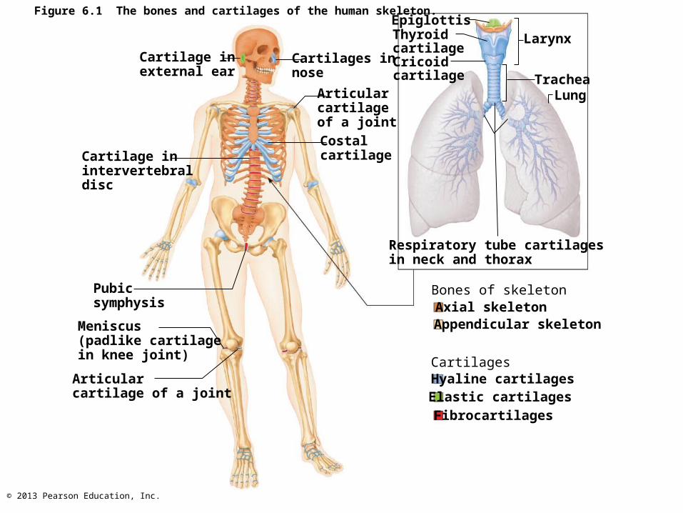
Figure 6.1 The bones and cartilages of the human skeleton.
Cartilage inexternal ear
Cartilage inintervertebraldisc
Pubicsymphysis
Meniscus(padlike cartilagein knee joint)
Articularcartilage of a joint
Costalcartilage
Articularcartilageof a joint
Cartilages innose
EpiglottisThyroidcartilageCricoidcartilage
Larynx
TracheaLung
Bones of skeletonAxial skeletonAppendicular skeleton
Hyaline cartilagesElastic cartilagesFibrocartilages
Cartilages
Respiratory tube cartilagesin neck and thorax

© 2013 Pearson Education, Inc.
Growth of Cartilage
• Appositional growth– Cells secrete matrix against external face of
existing cartilage
• Interstitial growth– Chondrocytes divide and secrete new matrix,
expanding cartilage from within
• Calcification of cartilage– Occurs during normal bone growth
• Youth and old age
– Hardens, but cacified cartilage is not bone
© 2013 Pearson Education, Inc.
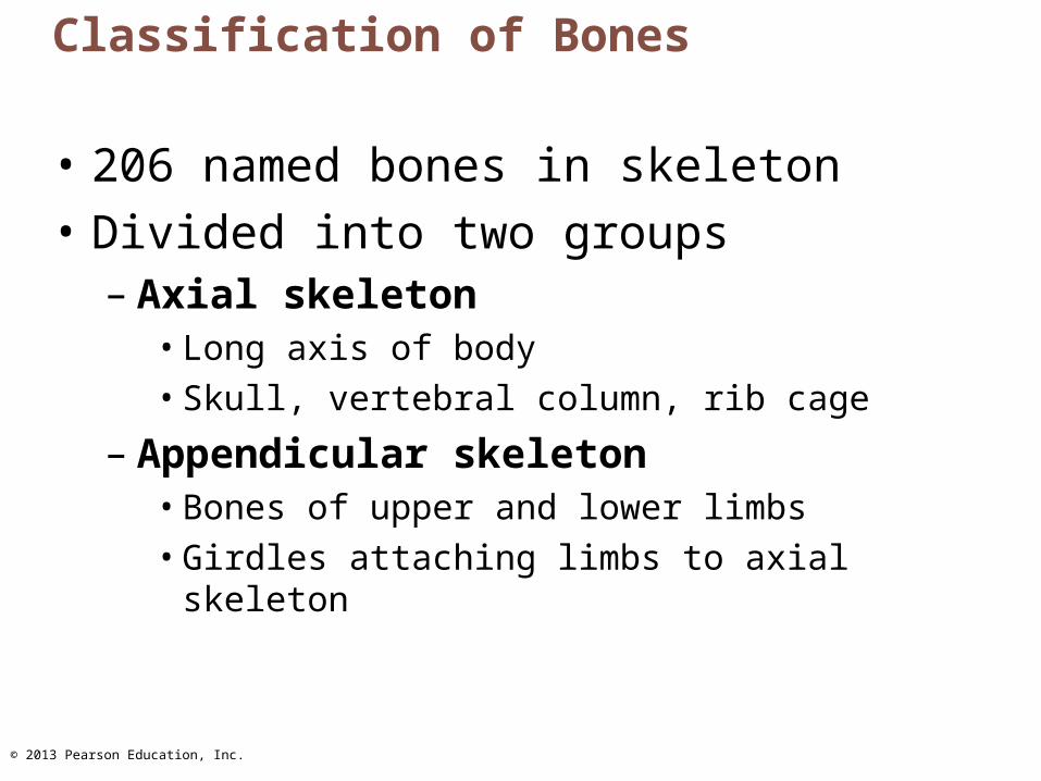
Classification of Bones
• 206 named bones in skeleton
• Divided into two groups– Axial skeleton
• Long axis of body• Skull, vertebral column, rib cage
– Appendicular skeleton• Bones of upper and lower limbs• Girdles attaching limbs to axial skeleton
© 2013 Pearson Education, Inc.
Figure 6.1 The bones and cartilages of the human skeleton.
Cartilage inexternal ear
Cartilage inintervertebraldisc
Pubicsymphysis
Meniscus(padlike cartilagein knee joint)
Articularcartilage of a joint
Costalcartilage
Articularcartilageof a joint
Cartilages innose
EpiglottisThyroidcartilageCricoidcartilage
Larynx
TracheaLung
Bones of skeletonAxial skeletonAppendicular skeleton
Hyaline cartilagesElastic cartilagesFibrocartilages
Cartilages
Respiratory tube cartilagesin neck and thorax
© 2013 Pearson Education, Inc.
Classification of Bones by Shape
• Long bones
• Short bones
• Flat bones
• Irregular bones

© 2013 Pearson Education, Inc.
Classification of Bones by Shape
• Long bones– Longer than they are wide– Limb, wrist, ankle bones
• Short bones– Cube-shaped bones (in wrist and ankle)– Sesamoid bones (within tendons, e.g., Patella)– Vary in size and number in different individuals
• Flat bones– Thin, flat, slightly curved– Sternum, scapulae, ribs, most skull bones
• Irregular bones– Complicated shapes– Vertebrae, coxal bones

© 2013 Pearson Education, Inc.
Figure 6.2 Classification of bones on the basis of shape.
Long bone(humerus)
Flat bone (sternum)
Irregular bone (vertebra),right lateral view Short bone (talus)
© 2013 Pearson Education, Inc.
Functions of Bones
• Seven important functions– Support– Protection– Movement– Mineral and growth factor storage– Blood cell formation– Triglyceride (fat) storage– Hormone production

© 2013 Pearson Education, Inc.
Functions of Bones
• Support– For body and soft organs
• Protection– For brain, spinal cord, and vital organs
• Movement– Levers for muscle action
© 2013 Pearson Education, Inc.
Functions of Bones
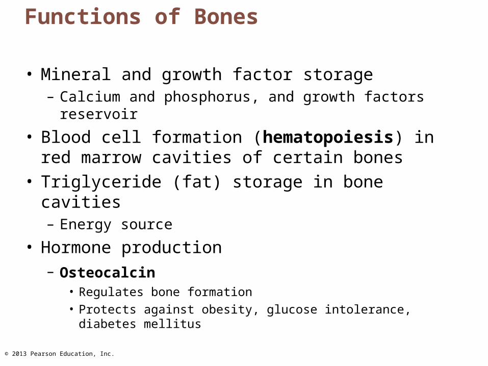
• Mineral and growth factor storage– Calcium and phosphorus, and growth factors
reservoir
• Blood cell formation (hematopoiesis) in red marrow cavities of certain bones
• Triglyceride (fat) storage in bone cavities– Energy source
• Hormone production– Osteocalcin
• Regulates bone formation• Protects against obesity, glucose intolerance, diabetes
mellitus
© 2013 Pearson Education, Inc.
Bones
• Are organs– Contain different types of tissues
• Bone (osseous) tissue, nervous tissue, cartilage, fibrous connective tissue, muscle and epithelial cells in its blood vessels
• Three levels of structure– Gross anatomy– Microscopic– Chemical
© 2013 Pearson Education, Inc.
Gross Anatomy
• Bone textures – Compact and spongy bone
• Compact– Dense outer layer; smooth and solid
• Spongy (cancellous or trabecular)– Honeycomb of flat pieces of bone deep to
compact called trabeculae
© 2013 Pearson Education, Inc.
Structure of Short, Irregular, and Flat Bones
• Thin plates of spongy bone covered by compact bone
• Plates sandwiched between connective tissue membranes– Periosteum (outer layer) and endosteum
• No shaft or epiphyses
• Bone marrow throughout spongy bone; no marrow cavity
• Hyaline cartilage covers articular surfaces
© 2013 Pearson Education, Inc.
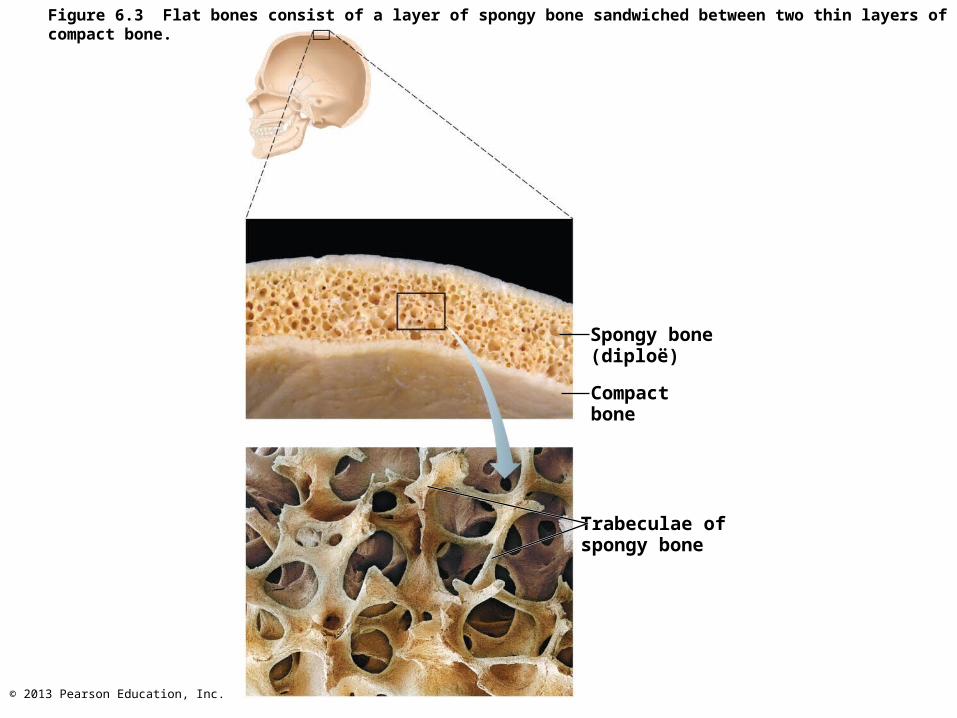
Figure 6.3 Flat bones consist of a layer of spongy bone sandwiched between two thin layers of compact bone.
Spongy bone(diploë)
Compact bone
Trabeculae ofspongy bone

© 2013 Pearson Education, Inc.
Structure of Typical Long Bone
• Diaphysis– Tubular shaft forms long axis– Compact bone surrounding medullary cavity
• Epiphyses– Bone ends– External compact bone; internal spongy bone– Articular cartilage covers articular surfaces– Between is epiphyseal line
• Remnant of childhood bone growth at epiphyseal plate

© 2013 Pearson Education, Inc.
Figure 6.4a The structure of a long bone (humerus of arm).Articularcartilage
Spongy bone
Epiphysealline
Periosteum
Compact bone
Medullarycavity (linedby endosteum)
Proximalepiphysis
Diaphysis
Distalepiphysis

© 2013 Pearson Education, Inc.
Figure 6.4b The structure of a long bone (humerus of arm).
Compact bone
Endosteum
Articularcartilage
Spongy bone
© 2013 Pearson Education, Inc.
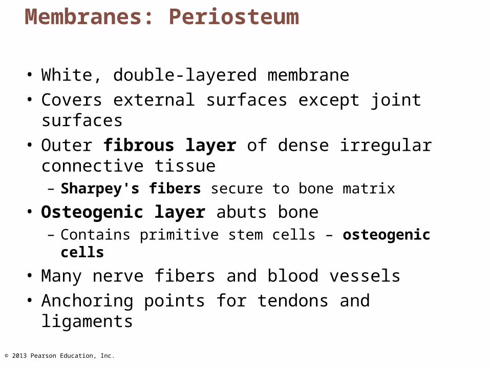
Membranes: Periosteum
• White, double-layered membrane• Covers external surfaces except joint surfaces• Outer fibrous layer of dense irregular
connective tissue– Sharpey's fibers secure to bone matrix
• Osteogenic layer abuts bone – Contains primitive stem cells – osteogenic cells
• Many nerve fibers and blood vessels• Anchoring points for tendons and ligaments

© 2013 Pearson Education, Inc.
Membranes: Endosteum
• Delicate connective tissue membrane covering internal bone surface
• Covers trabeculae of spongy bone
• Lines canals that pass through compact bone
• Contains osteogenic cells that can differentiate into other bone cells
© 2013 Pearson Education, Inc.
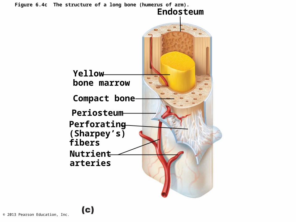
Figure 6.4c The structure of a long bone (humerus of arm).
Endosteum
Yellowbone marrow
Compact bone
PeriosteumPerforating(Sharpey’s)fibersNutrientarteries
© 2013 Pearson Education, Inc.
Hematopoietic Tissue in Bones
• Red marrow– Found within trabecular cavities of spongy
bone and diploë of flat bones (e.g., Sternum)– In medullary cavities and spongy bone of
newborns– Adult long bones have little red marrow
• Heads of femur and humerus only
– Red marrow in diploë and some irregular bones is most active
– Yellow marrow can convert to red, if necessary

© 2013 Pearson Education, Inc.
Bone Markings
• Sites of muscle, ligament, and tendon attachment on external surfaces
• Joint surfaces
• Conduits for blood vessels and nerves
• Projections
• Depressions
• Openings
© 2013 Pearson Education, Inc.
Bone Markings
• Projections– Most indicate stresses created by muscle pull
or joint modifications• Depressions and openings • Usually allow nerves and blood vessels to pass
© 2013 Pearson Education, Inc.
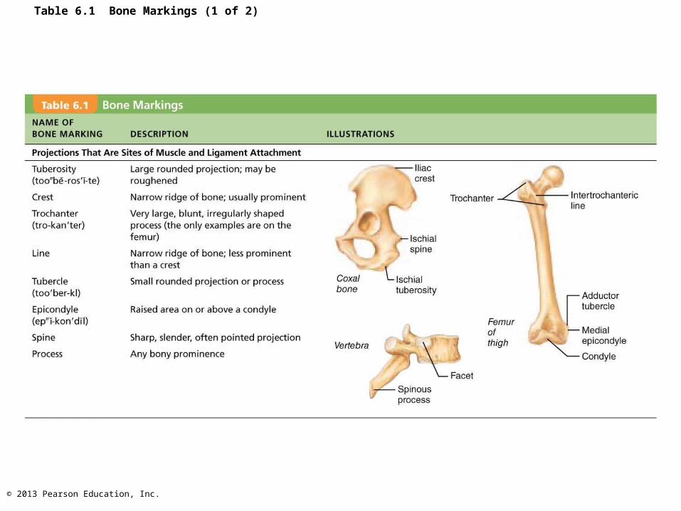
Table 6.1 Bone Markings (1 of 2)

© 2013 Pearson Education, Inc.
Table 6.1 Bone Markings (2 of 2)

© 2013 Pearson Education, Inc.
Microscopic Anatomy of Bone: Cells of Bone Tissue
• Five major cell types
• Each specialized form of same basic cell type– Osteogenic cells– Osteoblasts– Osteocytes– Bone lining cells– Osteoclasts
© 2013 Pearson Education, Inc.
Osteogenic Cells
• Also called osteoprogenitor cells– Mitotically active stem cells in periosteum and
endosteum– When stimulated differentiate into osteoblasts
or bone lining cells• Some persist as osteogenic cells
© 2013 Pearson Education, Inc.
Osteoblasts
• Bone-forming cells
• Secrete unmineralized bone matrix or osteoid– Includes collagen and calcium-binding
proteins• Collagen = 90% of bone protein
• Actively mitotic

© 2013 Pearson Education, Inc.
Figure 6.5a–b Comparison of different types of bone cells.
Osteogenic cell
Stem cell
Osteoblast
Matrix-synthesizingcell responsible for
bone growth

© 2013 Pearson Education, Inc.
Osteocytes
• Mature bone cells in lacunae
• Monitor and maintain bone matrix
• Act as stress or strain sensors– Respond to and communicate mechanical
stimuli to osteoblasts and osteoclasts (cells that destroy bone) so bone remodeling can occur

© 2013 Pearson Education, Inc.
Bone Lining Cells
• Flat cells on bone surfaces believed to help maintain matrix
• On external bone surface called periosteal cells
• Lining internal surfaces called endosteal cells
© 2013 Pearson Education, Inc.
Osteoclasts
• Derived from hematopoietic stem cells that become macrophages
• Giant, multinucleate cells for bone resorption
• When active rest in resorption bay and have ruffled border– Ruffled border increases surface area for
enzyme degradation of bone and seals off area from surrounding matrix
© 2013 Pearson Education, Inc.
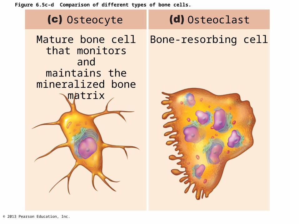
Figure 6.5c–d Comparison of different types of bone cells.
Osteocyte
Mature bone cellthat monitors and
maintains themineralized bone
matrix
Osteoclast
Bone-resorbing cell

© 2013 Pearson Education, Inc.
Microscopic Anatomy of Bone: Compact Bone
• Also called lamellar bone
• Osteon or haversian system– Structural unit of compact bone– Elongated cylinder parallel to long axis of
bone– Hollow tubes of bone matrix called lamellae
• Collagen fibers in adjacent rings run in different directions
– Withstands stress – resist twisting
© 2013 Pearson Education, Inc.
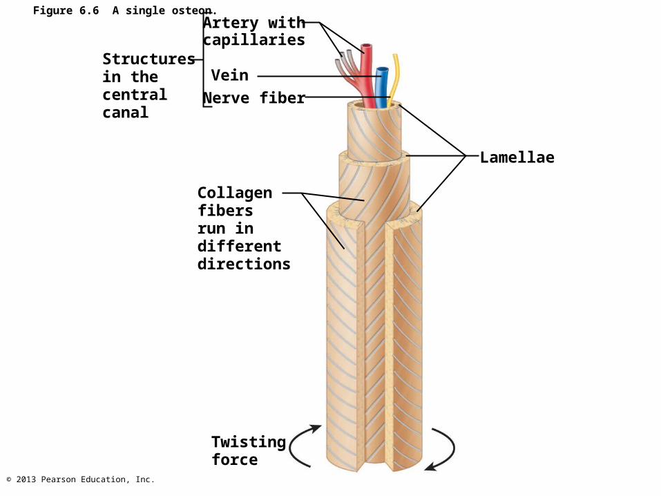
Figure 6.6 A single osteon.
Structuresin thecentralcanal
Artery withcapillaries
Vein
Nerve fiber
Collagenfibersrun indifferentdirections
Twistingforce
Lamellae

© 2013 Pearson Education, Inc.
Microscopic Anatomy of Bone: Compact Bone
• Canals and canaliculi– Central (haversian) canal runs through core of
osteon• Contains blood vessels and nerve fibers
• Perforating (volkmann's) canals– Canals lined with endosteum at right angles to central canal– Connect blood vessels and nerves of periosteum, medullary
cavity, and central canal
• Lacunae—small cavities that contain osteocytes• Canaliculi—hairlike canals that connect lacunae to each
other and central canal
© 2013 Pearson Education, Inc.
Canaliculi
• When matrix hardens and cells are trapped the canaliculi form– Allow communication– Permit nutrients and wastes to be relayed
from one osteocyte to another throughout osteon
© 2013 Pearson Education, Inc.
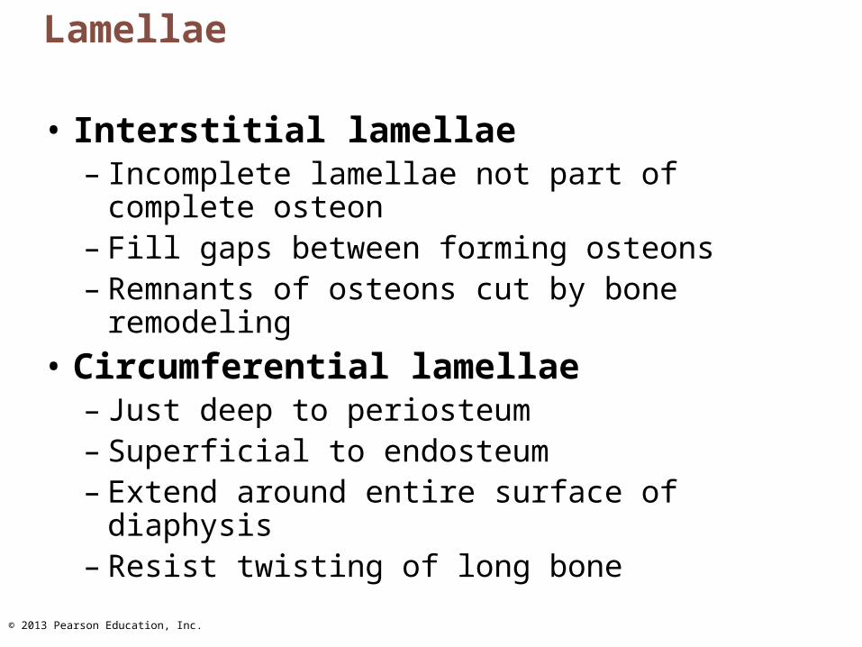
Lamellae
• Interstitial lamellae– Incomplete lamellae not part of complete
osteon– Fill gaps between forming osteons– Remnants of osteons cut by bone remodeling
• Circumferential lamellae– Just deep to periosteum– Superficial to endosteum– Extend around entire surface of diaphysis– Resist twisting of long bone
© 2013 Pearson Education, Inc.
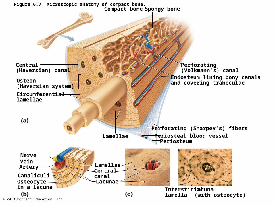
Figure 6.7 Microscopic anatomy of compact bone.Compact bone Spongy bone
Central(Haversian) canal
Osteon(Haversian system)
Circumferentiallamellae
Perforating (Volkmann’s) canal
Endosteum lining bony canalsand covering trabeculae
Perforating (Sharpey’s) fibers
Periosteal blood vesselPeriosteum
Lamellae
NerveVeinArtery
CanaliculiOsteocytein a lacuna
LamellaeCentralcanalLacunae
Interstitiallamella
Lacuna(with osteocyte)

© 2013 Pearson Education, Inc.
Microscopic Anatomy of Bone:Spongy Bone
• Appears poorly organized
• Trabeculae– Align along lines of stress to help resist it– No osteons– Contain irregularly arranged lamellae and
osteocytes interconnected by canaliculi– Capillaries in endosteum supply nutrients
© 2013 Pearson Education, Inc.
Chemical Composition of Bone: Organic Components• Includes cells and osteoid
– Osteogenic cells, osteoblasts, osteocytes, bone- lining cells, and osteoclasts
– Osteoid—1/3 of organic bone matrix secreted by osteoblasts• Made of ground substance (proteoglycans and
glycoproteins)• Collagen fibers• Contributes to structure; provides tensile strength and
flexibility
• Resilience of bone due to sacrificial bonds in or between collagen molecules– Stretch and break easily on impact to dissipate energy and
prevent fracture– If no addition trauma, bonds re-form

© 2013 Pearson Education, Inc.
Chemical Composition of Bone: Inorganic Components
• Hydroxyapatites (mineral salts)– 65% of bone by mass– Mainly of tiny calcium phosphate crystals in
and around collagen fibers– Responsible for hardness and resistance to
compression
© 2013 Pearson Education, Inc.
Bone
• Half as strong as steel in resisting compression
• As strong as steel in resisting tension
• Last long after death because of mineral composition– Reveal information about ancient people– Can display growth arrest lines
• Horizontal lines on bones• Proof of illness - when bones stop growing so
nutrients can help fight disease
Top Related