


Languages
Pages
Legal

© 2013 Pearson Education, Inc.
Appendicular Skeleton
• Bones of limbs and their girdles– Pectoral girdle
• Attaches upper limbs to body trunk
– Pelvic girdle • Attaches lower limbs to body trunk
© 2013 Pearson Education, Inc.
• Clavicles and scapulae– Attach upper limbs to axial skeleton – Provide attachment sites for muscles that
move upper limbs
A&P Flix™: Movement of the Pectoral Girdle
Pectoral Girdle (Shoulder Girdle)
PLAYPLAY
© 2013 Pearson Education, Inc.
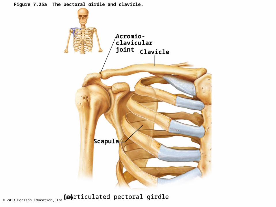
Figure 7.25a The pectoral girdle and clavicle.
Acromio-clavicularjoint Clavicle
Scapula
Articulated pectoral girdle
© 2013 Pearson Education, Inc.
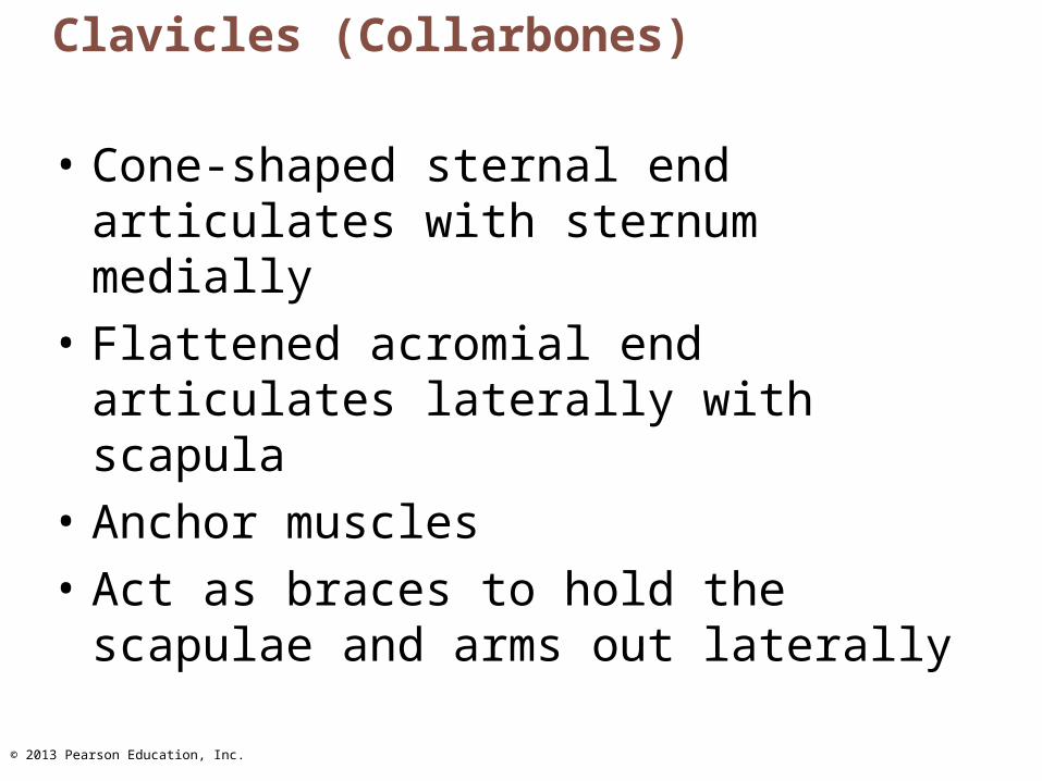
Clavicles (Collarbones)
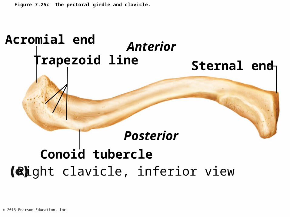
• Cone-shaped sternal end articulates with sternum medially
• Flattened acromial end articulates laterally with scapula
• Anchor muscles
• Act as braces to hold the scapulae and arms out laterally
© 2013 Pearson Education, Inc.
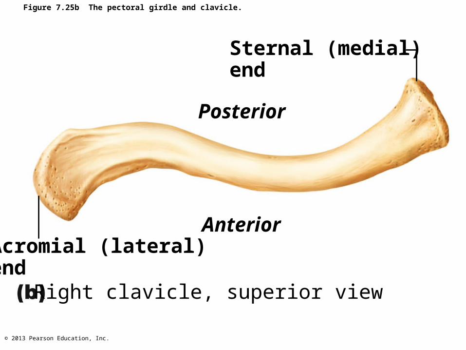
Figure 7.25b The pectoral girdle and clavicle.
Sternal (medial)end
Posterior
AnteriorAcromial (lateral)end
Right clavicle, superior view
© 2013 Pearson Education, Inc.
Figure 7.25c The pectoral girdle and clavicle.
Acromial end
Trapezoid lineAnterior
Sternal end
Conoid tubercle
Posterior
Right clavicle, inferior view

© 2013 Pearson Education, Inc.
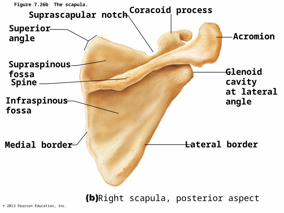
Scapulae (Shoulder Blades)
• On dorsal surface of rib cage, between ribs 2 and 7
• Flat and triangular, with three borders and three angles
• Several large fossae named according to location

© 2013 Pearson Education, Inc.
Figure 7.26a The scapula.
AcromionSuprascapular notch
Superior border
Superiorangle
Subscapularfossa
Medial border
Glenoidcavity
Coracoidprocess
Lateral border
Inferior angleRight scapula, anterior aspect
© 2013 Pearson Education, Inc.
Figure 7.26b The scapula.
Superiorangle
Supraspinousfossa
Infraspinousfossa
Medial border
Suprascapular notch
Glenoidcavityat lateralangle
Lateral border
Acromion
Coracoid process
Spine
Right scapula, posterior aspect

© 2013 Pearson Education, Inc.
Figure 7.26c The scapula.
Supraspinousfossa
Infraspinousfossa
Posterior Anterior
Subscapularfossa
Acromion
Supraspinous fossa
Supraglenoidtubercle
Coracoidprocess
Glenoidcavity
Spine
Infraspinousfossa
Infraglenoidtubercle
Subscapularfossa
Inferior angleRight scapula, lateral aspect

© 2013 Pearson Education, Inc.
The Upper Limb
• 30 bones form skeletal framework of each upper limb– Arm
• Humerus
– Forearm• Radius and ulna
– Hand• 8 carpal bones in the wrist• 5 metacarpal bones in the palm• 14 phalanges in the fingers
© 2013 Pearson Education, Inc.
Humerus
• Largest, longest bone of upper limb
• Articulates superiorly with glenoid cavity of scapula
• Articulates inferiorly with radius and ulna
© 2013 Pearson Education, Inc.
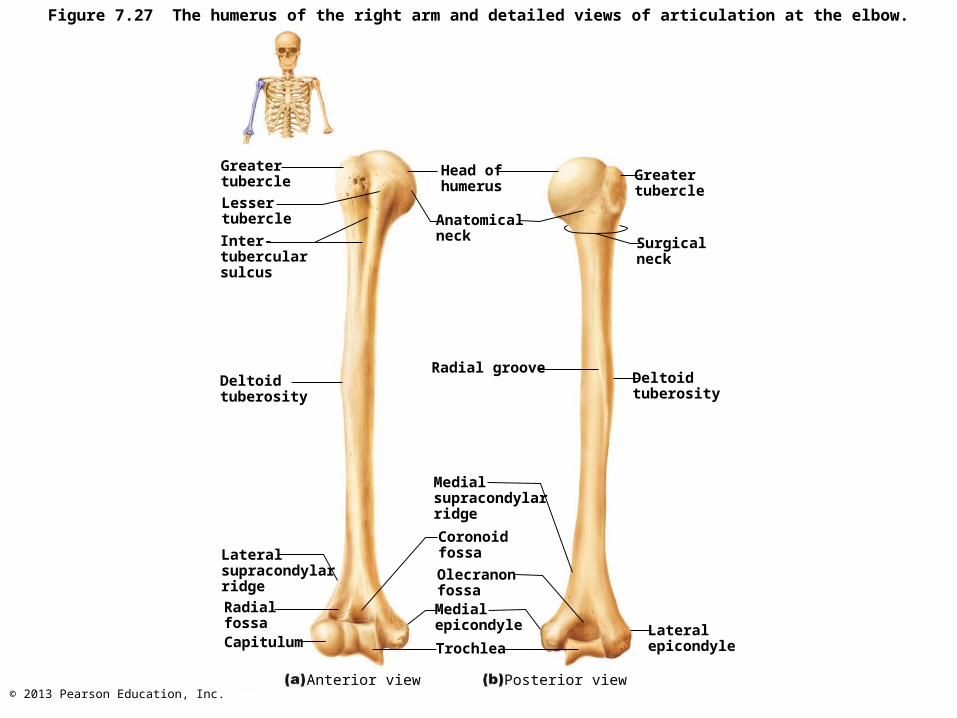
Figure 7.27 The humerus of the right arm and detailed views of articulation at the elbow.
Greatertubercle
Lessertubercle
Deltoid tuberosity
Lateralsupracondylarridge
Inter-tubercularsulcus
RadialfossaCapitulum
Head ofhumerus
Anatomicalneck
Radial groove
Medialsupracondylarridge
Coronoidfossa
OlecranonfossaMedialepicondyle
Trochlea
Greatertubercle
Surgicalneck
Deltoidtuberosity
Lateralepicondyle
Anterior view Posterior view
© 2013 Pearson Education, Inc.
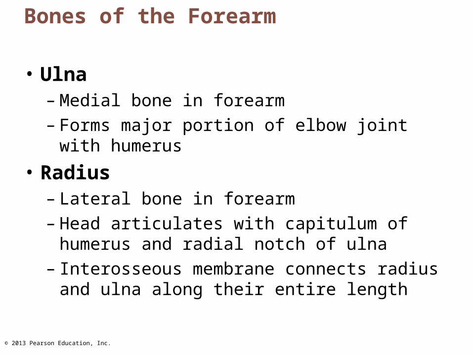
Bones of the Forearm
• Ulna– Medial bone in forearm – Forms major portion of elbow joint with
humerus
• Radius– Lateral bone in forearm– Head articulates with capitulum of humerus
and radial notch of ulna– Interosseous membrane connects radius and
ulna along their entire length

© 2013 Pearson Education, Inc.
Radialnotch ofthe ulna
HeadNeck
Radialtuberosity
Olecranon
Trochlearnotch
Coronoid process
Proximalradioulnarjoint
Interosseousmembrane
Ulna
RadiusUlnar notchof the radius
Head of ulna
Ulnar styloid processDistalradioulnarjoint
Radial styloidprocess
Anterior view Posterior view
Radial styloidprocess
Radius
Neck ofradius
Head ofradius
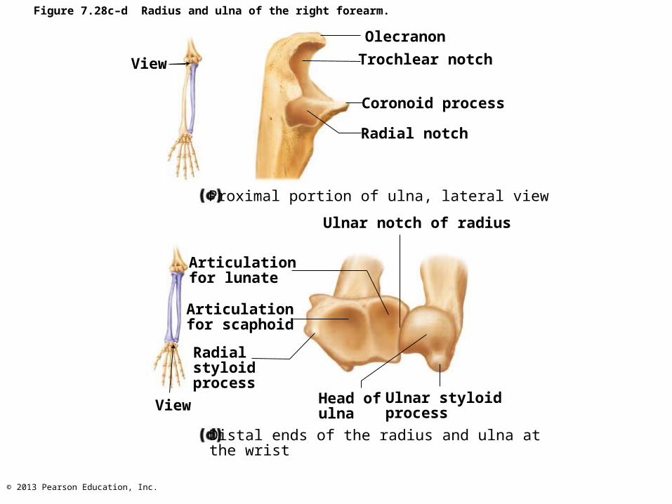
Figure 7.28a–b Radius and ulna of the right forearm.
© 2013 Pearson Education, Inc.
View
Olecranon
Trochlear notch
Coronoid process
Radial notch
Proximal portion of ulna, lateral view
Ulnar notch of radius
Articulationfor lunate
Articulationfor scaphoid
Radialstyloidprocess
Head ofulna
Ulnar styloidprocessView
Distal ends of the radius and ulna atthe wrist
Figure 7.28c–d Radius and ulna of the right forearm.

© 2013 Pearson Education, Inc.
Humerus
Capitulum
Head ofradius
RadialtuberosityRadius
Coronoid fossa
Medialepicondyle
Trochlea
Coronoidprocess of ulnaRadial notchUlna
Humerus
Olecranon
Medialepicondyle
Ulna
Olecranonfossa
Lateralepicondyle
HeadNeck
Radius
Posterior view of extended elbow
Anterior view at the elbow region
Figure 7.27c–d The humerus of the right arm and detailed views of articulation at the elbow.
© 2013 Pearson Education, Inc.
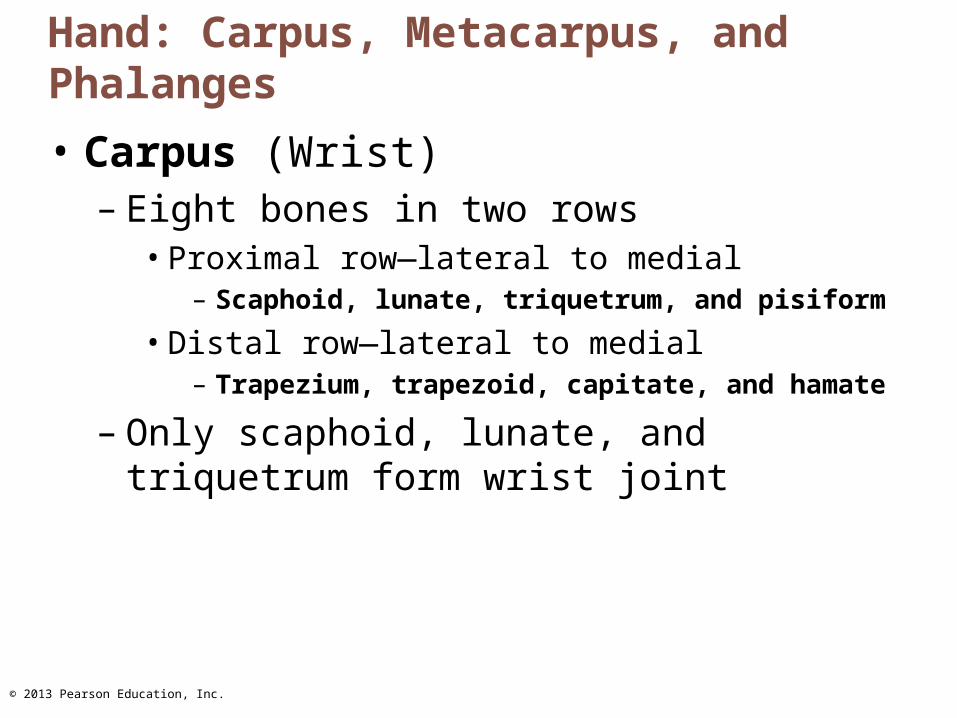
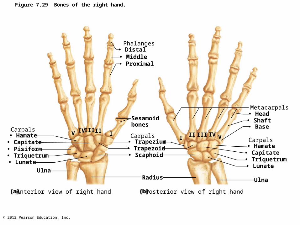
Hand: Carpus, Metacarpus, and Phalanges
• Carpus (Wrist)– Eight bones in two rows
• Proximal row—lateral to medial– Scaphoid, lunate, triquetrum, and pisiform
• Distal row—lateral to medial– Trapezium, trapezoid, capitate, and hamate
– Only scaphoid, lunate, and triquetrum form wrist joint
© 2013 Pearson Education, Inc.
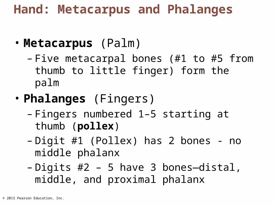
Hand: Metacarpus and Phalanges
• Metacarpus (Palm)– Five metacarpal bones (#1 to #5 from thumb
to little finger) form the palm
• Phalanges (Fingers)– Fingers numbered 1–5 starting at thumb
(pollex)– Digit #1 (Pollex) has 2 bones - no middle
phalanx– Digits #2 – 5 have 3 bones—distal, middle,
and proximal phalanx
© 2013 Pearson Education, Inc.
Figure 7.29 Bones of the right hand.
Phalanges• Distal• Middle• Proximal
Carpals• Hamate• Capitate• Pisiform• Triquetrum
Carpals
• Lunate
Ulna
Sesamoidbones
• Trapezium• Trapezoid• Scaphoid
Radius
Metacarpals• Head• Shaft• Base
Carpals• Hamate• Capitate• Triquetrum• Lunate
Ulna
Anterior view of right hand Posterior view of right hand
V IV III IIVIVIIIIII
I
© 2013 Pearson Education, Inc.
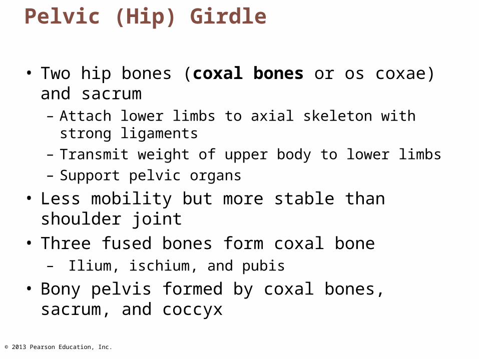
Pelvic (Hip) Girdle
• Two hip bones (coxal bones or os coxae) and sacrum– Attach lower limbs to axial skeleton with strong
ligaments– Transmit weight of upper body to lower limbs– Support pelvic organs
• Less mobility but more stable than shoulder joint• Three fused bones form coxal bone
– Ilium, ischium, and pubis
• Bony pelvis formed by coxal bones, sacrum, and coccyx
© 2013 Pearson Education, Inc.
Animation: Rotatable Pelvis
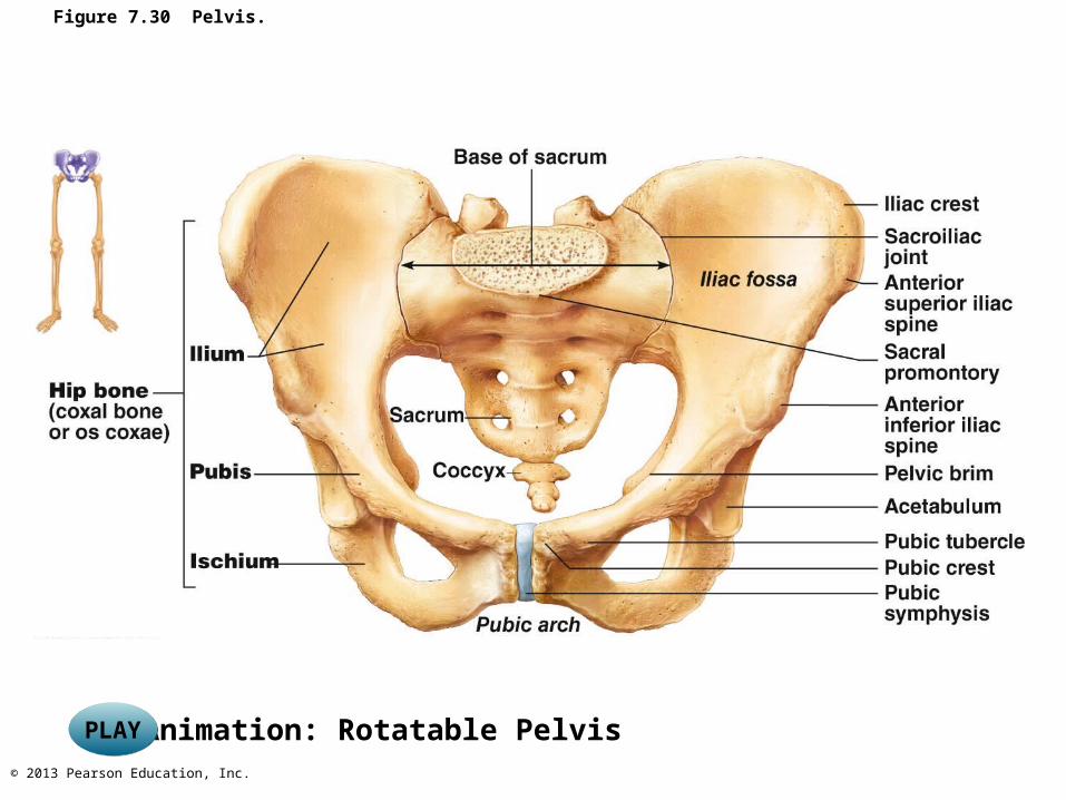
Figure 7.30 Pelvis.
PLAYPLAY
© 2013 Pearson Education, Inc.
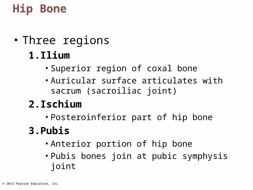
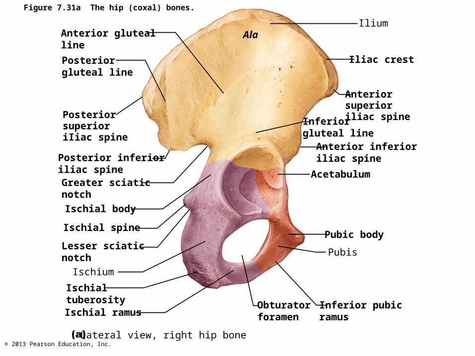
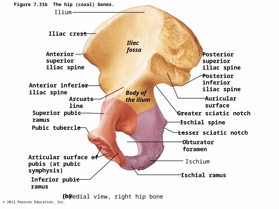
Hip Bone
• Three regions1. Ilium
• Superior region of coxal bone• Auricular surface articulates with sacrum
(sacroiliac joint)
2. Ischium• Posteroinferior part of hip bone
3. Pubis• Anterior portion of hip bone• Pubis bones join at pubic symphysis joint
© 2013 Pearson Education, Inc.
Figure 7.31a The hip (coxal) bones.
Anterior glutealline
Posterior gluteal line
PosteriorsuperioriIiac spine
Posterior inferioriliac spineGreater sciaticnotch
Ischial body
Ischial spine
Lesser sciatic notch
Ischium
IschialtuberosityIschial ramus
Lateral view, right hip bone
AlaIlium
Iliac crest
Anterior superioriliac spineInferior
gluteal lineAnterior inferioriliac spine
Acetabulum
Pubic body
Pubis
Inferior pubicramus
Obturatorforamen

© 2013 Pearson Education, Inc.
Figure 7.31c The hip (coxal) bones.
Anteriorgluteal line
Posteriorgluteal line
Posteriorsuperioriliac spine
Posteriorinferioriliac spine
Greatersciatic notch
Ischial bodyIschial spine
Lessersciatic notch
Ischium
Ischialtuberosity
Lateral view, right hip bone
Ischial ramus Obturatorforamen
Ilium
Anterior superioriliac spine
Anterior inferioriliac spine
Inferior glutealline
Acetabulum
Pubic body
Pubic tubercle
Inferior pubicramus
© 2013 Pearson Education, Inc.
Figure 7.31b The hip (coxal) bones.
Ilium
Iliac crest
Anterior superioriliac spine
Anterior inferioriliac spine
Arcuateline
Superior pubicramusPubic tubercle
Articular surface ofpubis (at pubic symphysis)
Inferior pubicramus
Medial view, right hip bone
Iliacfossa
Body ofthe ilium
Posteriorsuperioriliac spine
Posteriorinferioriliac spine
Auricularsurface
Greater sciatic notch
Ischial spine
Lesser sciatic notch
Obturatorforamen
Ischium
Ischial ramus

© 2013 Pearson Education, Inc.
Figure 7.31d The hip (coxal) bones.
Medial view, right hip bone
Articular surface of pubis(at pubic symphysis)
Inferior pubicramus
Ischialramus
Obturatorforamen
Ischium
Lessersciatic notch
Ischial spine
Greatersciatic notch
Posterior inferioriliac spine
Posterior superioriliac spine
Auricularsurface
Iliacfossa
Ilium
Anteriorsuperioriliac spine
Anteriorinferioriliac spine
Superiorpubicramus
Pubictubercle
Arcuateline
© 2013 Pearson Education, Inc.
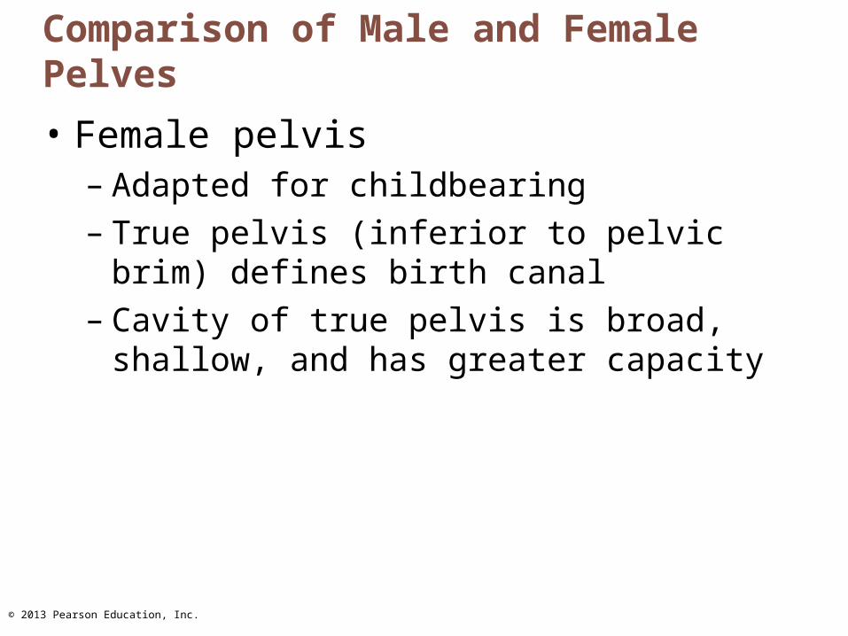
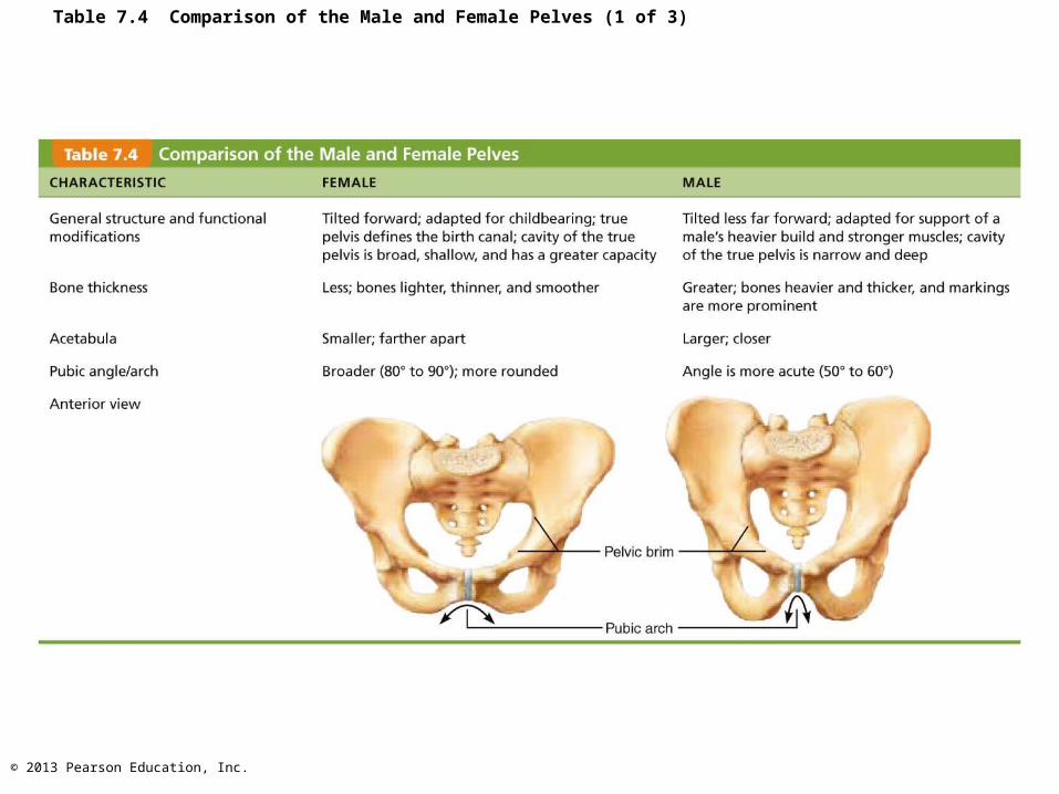
Comparison of Male and Female Pelves
• Female pelvis– Adapted for childbearing– True pelvis (inferior to pelvic brim) defines
birth canal– Cavity of true pelvis is broad, shallow, and
has greater capacity
© 2013 Pearson Education, Inc.
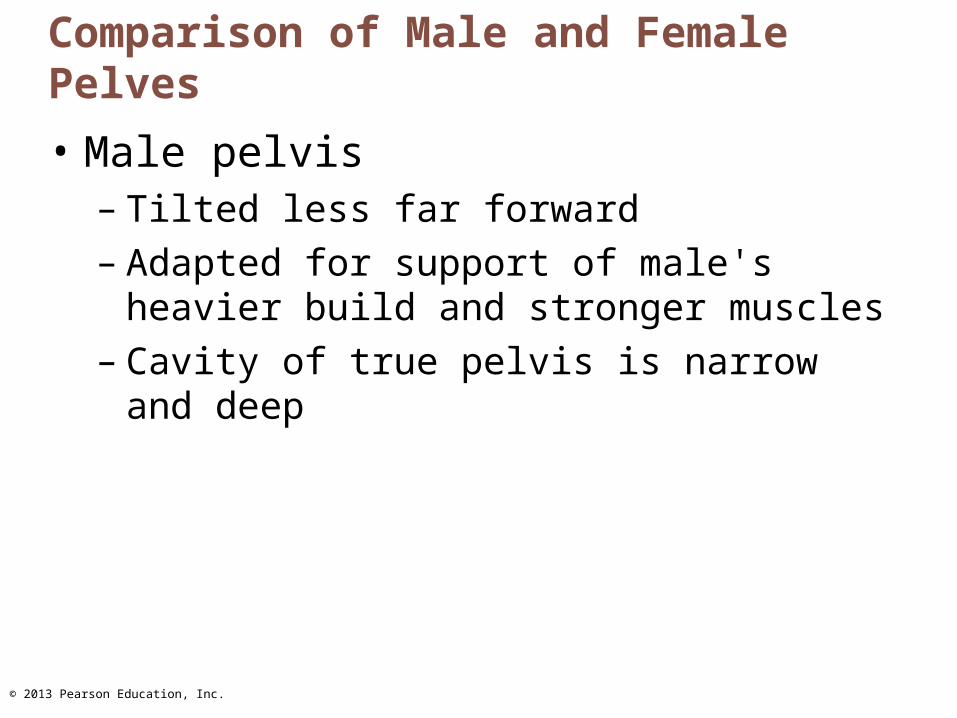
Comparison of Male and Female Pelves
• Male pelvis– Tilted less far forward– Adapted for support of male's heavier build
and stronger muscles– Cavity of true pelvis is narrow and deep
© 2013 Pearson Education, Inc.
Table 7.4 Comparison of the Male and Female Pelves (1 of 3)
© 2013 Pearson Education, Inc.
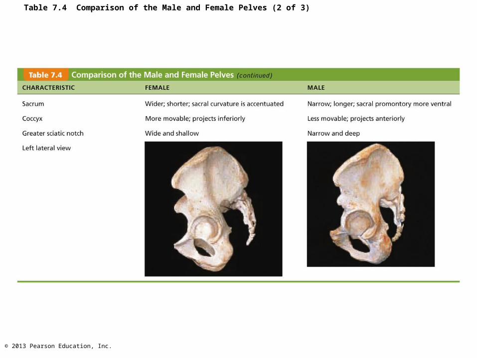
Table 7.4 Comparison of the Male and Female Pelves (2 of 3)
© 2013 Pearson Education, Inc.
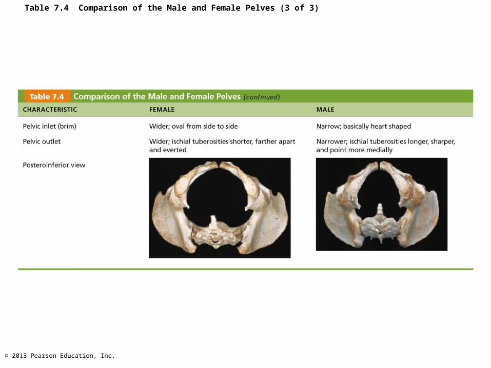
Table 7.4 Comparison of the Male and Female Pelves (3 of 3)
© 2013 Pearson Education, Inc.
The Lower Limb
• Carries entire weight of erect body
• Subjected to exceptional forces if jump or run
• Three segments of lower limb– Thigh– Leg– Foot

© 2013 Pearson Education, Inc.
Bones Of The Thigh
• Femur– Largest and strongest bone in the body– Length ~ ¼ of person's height– Articulates proximally with acetabulum of hip
and distally with tibia and patella
• Patella– Sesamoid bone in quadriceps tendon
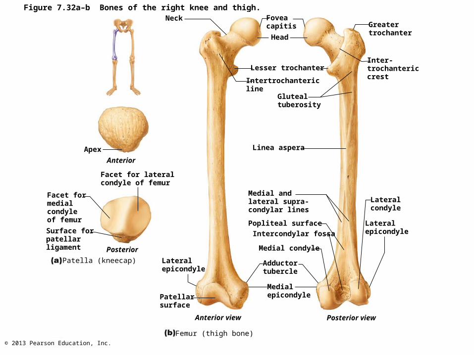
© 2013 Pearson Education, Inc.
Figure 7.32a–b Bones of the right knee and thigh.Neck
Apex
Anterior
Facet for lateralcondyle of femur
Facet formedialcondyleof femur
Surface forpatellarligament Posterior
Patella (kneecap) Lateralepicondyle
Patellarsurface
Anterior view
Foveacapitis
Head
Lesser trochanter
Intertrochantericline
Glutealtuberosity
Linea aspera
Medial andlateral supra-condylar lines
Popliteal surface
Intercondylar fossa
Medial condyle
Adductortubercle
Medialepicondyle
Greatertrochanter
Inter-trochantericcrest
Lateralcondyle
Lateralepicondyle
Posterior view
Femur (thigh bone)
© 2013 Pearson Education, Inc.
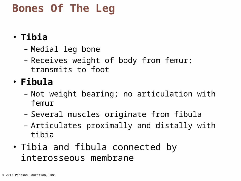
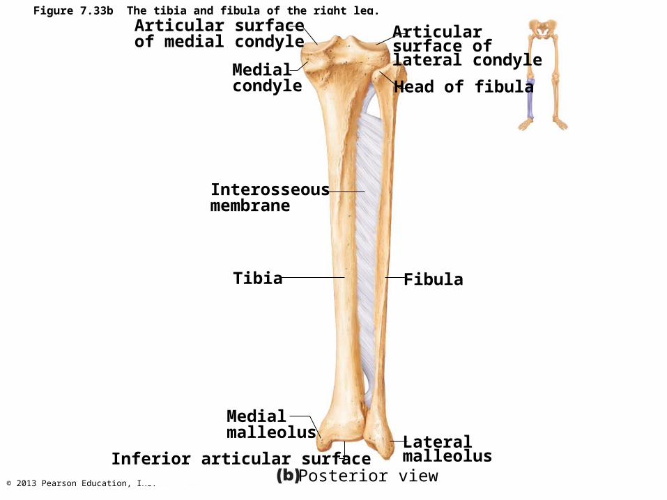
Bones Of The Leg
• Tibia– Medial leg bone– Receives weight of body from femur; transmits to foot
• Fibula– Not weight bearing; no articulation with femur– Several muscles originate from fibula– Articulates proximally and distally with tibia
• Tibia and fibula connected by interosseous membrane
© 2013 Pearson Education, Inc.
Figure 7.33a The tibia and fibula of the right leg.
Interosseousmembrane
IntercondylareminenceLateralcondyleHeadSuperiortibiofibularjoint
Fibula
InferiortibiofibularjointLateralmalleolus
Anterior view
Medialmalleolus
Inferior articular surface
Tibia
Anteriorborder
Tibialtuberosity
Medial condyle
© 2013 Pearson Education, Inc.
Figure 7.33b The tibia and fibula of the right leg.
Interosseousmembrane
Posterior view
Medialmalleolus
Inferior articular surface
Tibia
Medialcondyle
Articular surface of medial condyle
Articularsurface oflateral condyle
Head of fibula
Fibula
Lateralmalleolus

© 2013 Pearson Education, Inc.
LateralcondyleFibulaarticulateshere
Line for soleus muscle
Posterior view,proximal tibia
Anterior view,proximal tibia
Lateralcondyle
Tibialtuberosity
Figure 7.33c–d The tibia and fibula of the right leg.
© 2013 Pearson Education, Inc.
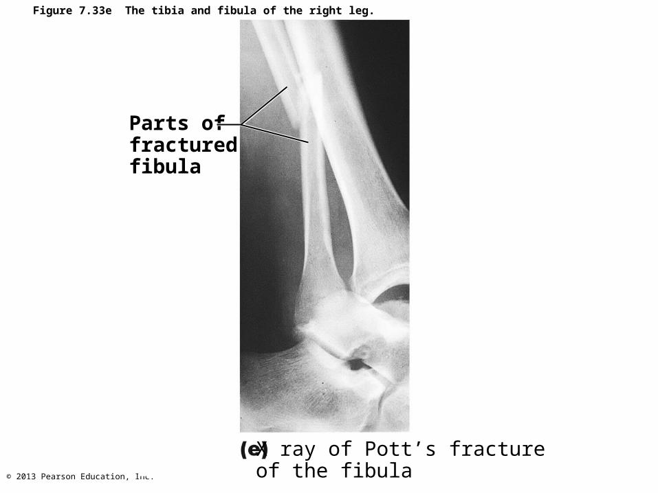
Parts offracturedfibula
X ray of Pott’s fractureof the fibula
Figure 7.33e The tibia and fibula of the right leg.
© 2013 Pearson Education, Inc.
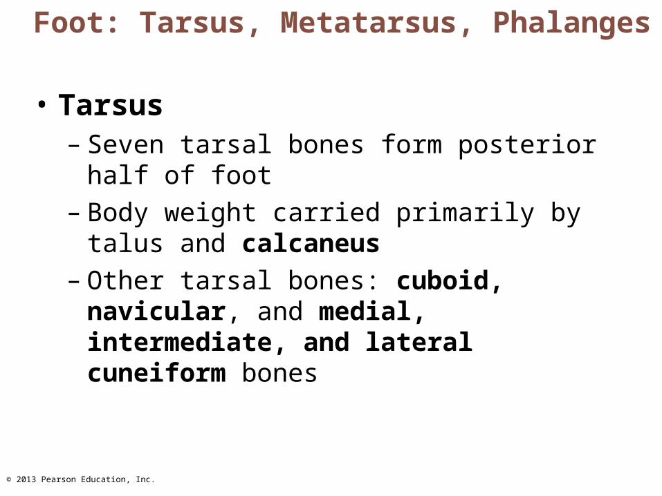
Foot: Tarsus, Metatarsus, Phalanges
• Tarsus– Seven tarsal bones form posterior half of foot– Body weight carried primarily by talus and
calcaneus– Other tarsal bones: cuboid, navicular, and
medial, intermediate, and lateral cuneiform bones
© 2013 Pearson Education, Inc.
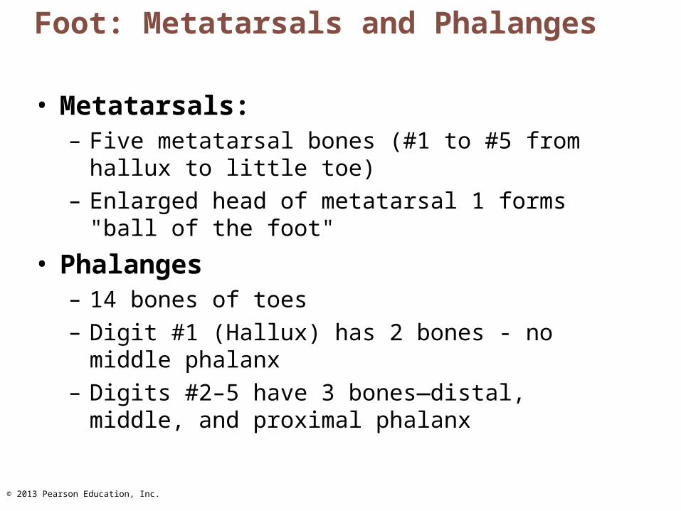
Foot: Metatarsals and Phalanges
• Metatarsals:– Five metatarsal bones (#1 to #5 from hallux to little
toe) – Enlarged head of metatarsal 1 forms "ball of the foot"
• Phalanges– 14 bones of toes– Digit #1 (Hallux) has 2 bones - no middle phalanx– Digits #2–5 have 3 bones—distal, middle, and
proximal phalanx
© 2013 Pearson Education, Inc.
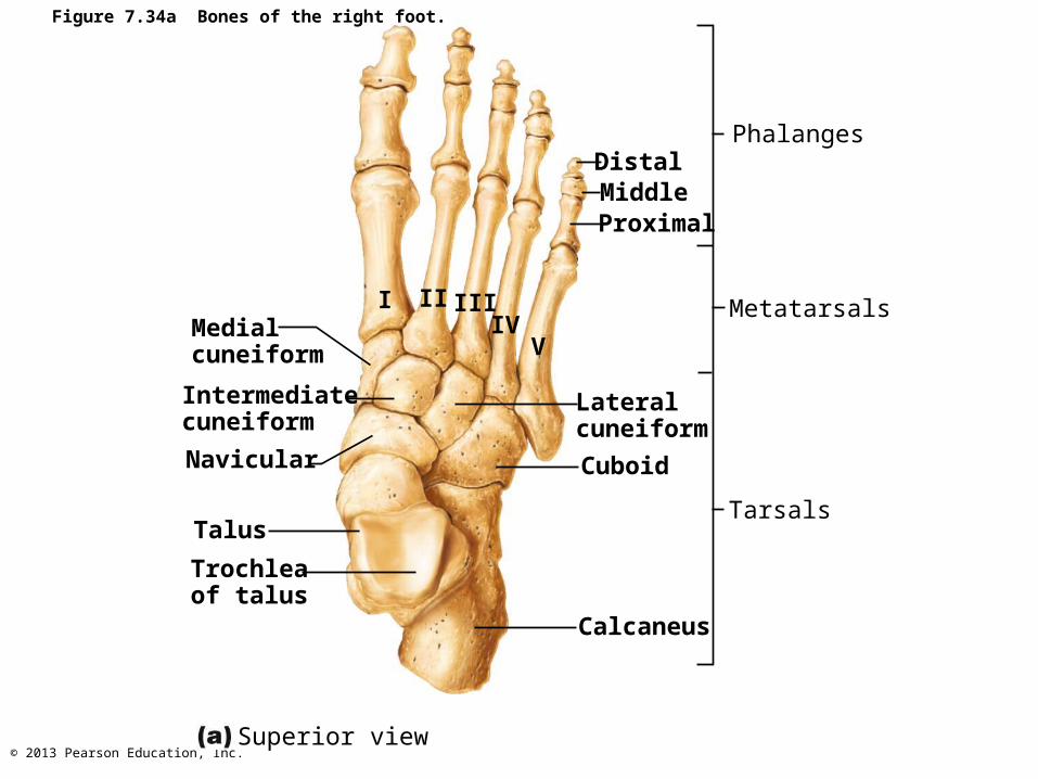
Figure 7.34a Bones of the right foot.
Medialcuneiform
Intermediatecuneiform
Navicular
Trochleaof talus
Calcaneus
Cuboid
Lateralcuneiform
Proximal
DistalMiddle
Phalanges
Metatarsals
Tarsals
Superior view
I II IIIIV
V
Talus

© 2013 Pearson Education, Inc.
Animation: Rotatable Bones of the Foot
Figure 7.34b Bones of the right foot.
Intermediatecuneiform
Navicular
Talus Medialmalleolarfacet
Sustentac-ulum tali (talar shelf)
CalcaneusMedialcuneiform
CalcanealtuberosityMedial view
First metatarsal
PLAYPLAY

© 2013 Pearson Education, Inc.
Figure 7.34c Bones of the right foot.
Lateral view
Lateralmalleolar facet
Navicular Intermediate cuneiform
Lateral cuneiform
Fifth metatarsalCuboidCalcaneus
Talus

© 2013 Pearson Education, Inc.
Arches Of The Foot
• Maintained by interlocking foot bones, ligaments, and tendons
• Allow foot to bear weight
• Three arches– Lateral longitudinal – Medial longitudinal – Transverse

© 2013 Pearson Education, Inc.
Figure 7.35a Arches of the foot.
Medial longitudinal arch
Transverse arch
Lateral longitudinal arch
Lateral aspect of right foot
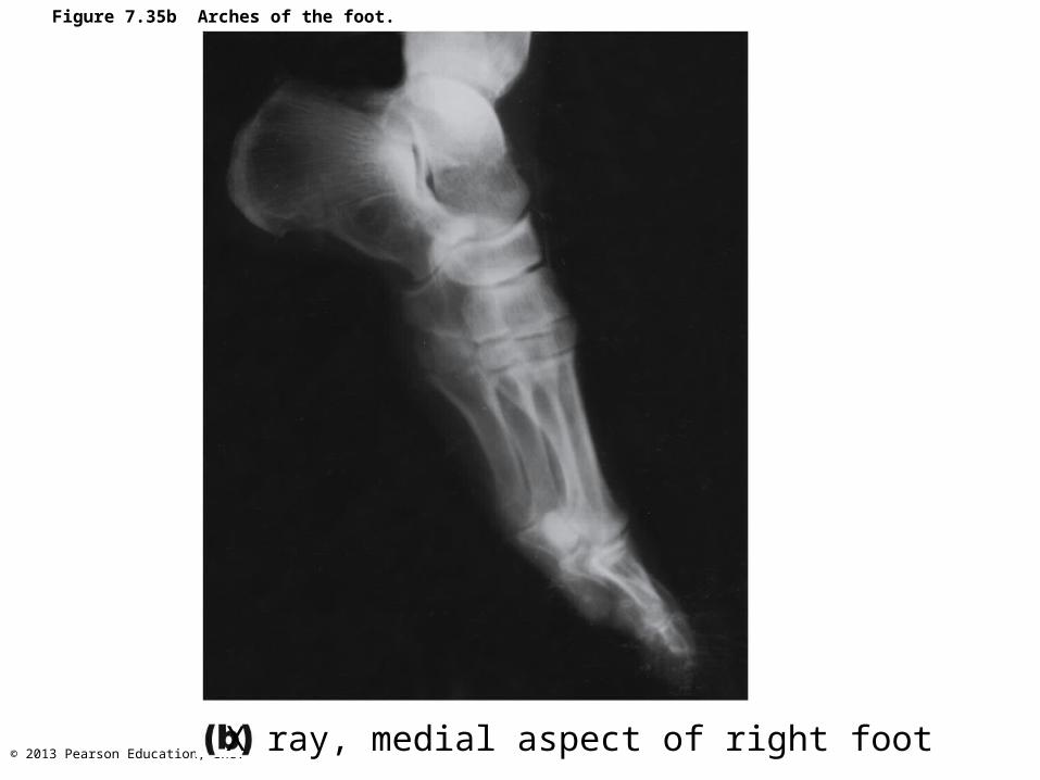
© 2013 Pearson Education, Inc.X ray, medial aspect of right foot
Figure 7.35b Arches of the foot.

© 2013 Pearson Education, Inc.
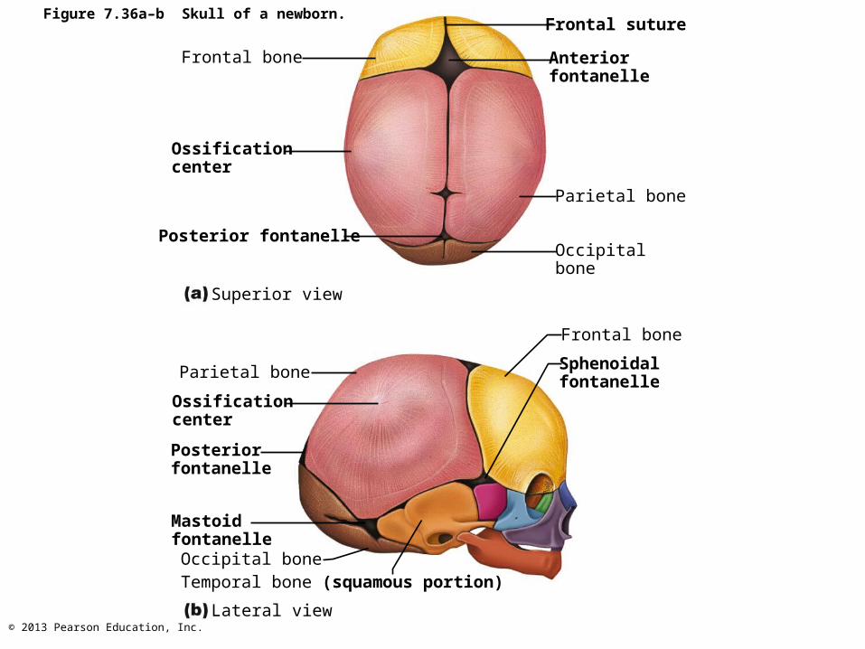
Developmental Aspects: Fetal Skull
• Infant skull has more bones than adult skull– Skull bones such as mandible and frontal
bones are unfused – Skull bones connected by fontanelles
• Unossified remnants of fibrous membranes• Ease birth and allow brain growth• Four fontanelles
– Anterior, posterior, mastoid, and sphenoidal
© 2013 Pearson Education, Inc.
Figure 7.36a–b Skull of a newborn.
Frontal bone
Ossificationcenter
Posterior fontanelle
Superior view
Frontal suture
Anteriorfontanelle
Parietal bone
Occipitalbone
Frontal bone
Sphenoidalfontanelle
Parietal bone
Ossificationcenter
Posteriorfontanelle
Occipital bone
Mastoidfontanelle
Temporal bone (squamous portion)
Lateral view
© 2013 Pearson Education, Inc.
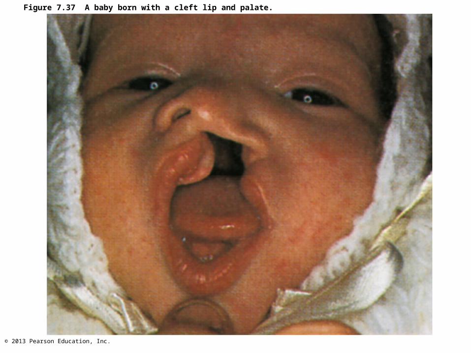
Congenital Abnormalities Of Skull
• Cleft palate– No medial fusion of right and left halves of
palate
© 2013 Pearson Education, Inc.
Figure 7.37 A baby born with a cleft lip and palate.
© 2013 Pearson Education, Inc.
Developmental Aspects: Growth Rates
• At birth, cranium huge relative to face
• At 9 months, cranium is ½ adult size
• Mandible and maxilla are foreshortened but lengthen with age
• Arms and legs grow at faster rate than head and trunk, leading to adult proportions

© 2013 Pearson Education, Inc.
Figure 7.39a Different growth rates of body parts determine body proportions.
Human newborn Human adult

© 2013 Pearson Education, Inc.
Figure 7.39b Different growth rates of body parts determine body proportions.
Newborn 2 yrs 5 yrs 15 yrs Adult
© 2013 Pearson Education, Inc.
Developmental Aspects: Spinal Curvature
• Primary thoracic and sacral curvatures obvious at birth– Give spine a C shape– Convex posteriorly
© 2013 Pearson Education, Inc.
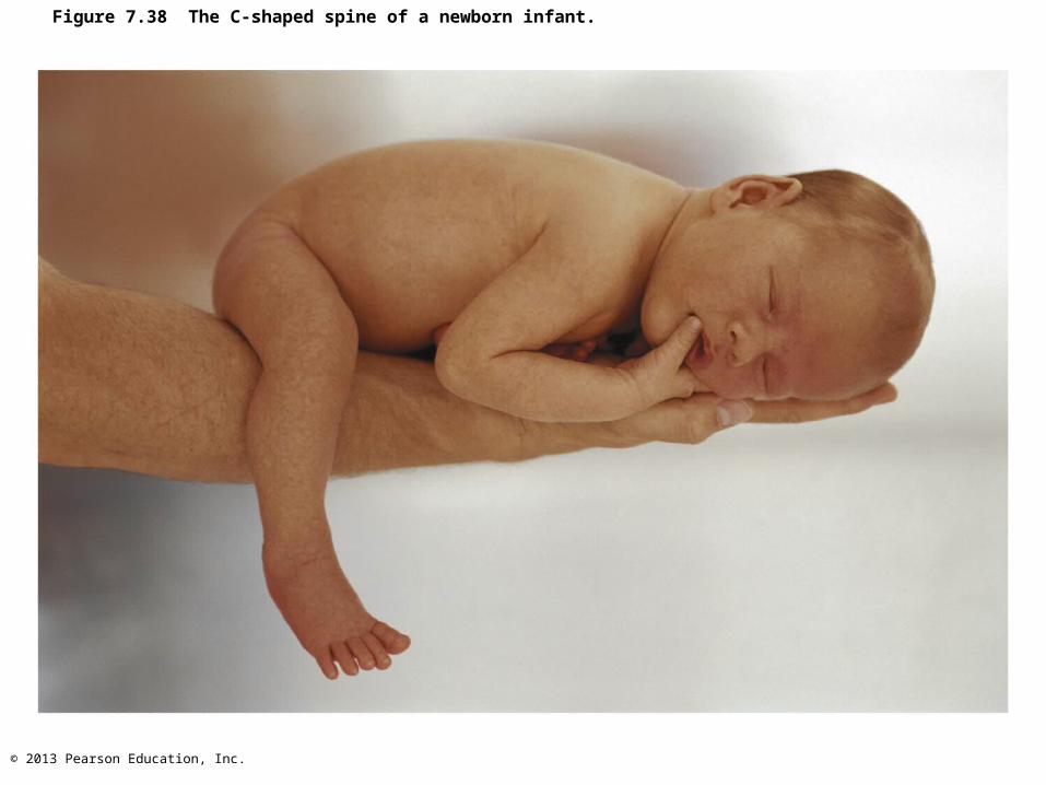
Figure 7.38 The C-shaped spine of a newborn infant.
© 2013 Pearson Education, Inc.
Developmental Aspects: Spinal Curvature
• Secondary curvatures– Cervical and lumbar—convex anteriorly– Appear as child develops (e.g., lifts head,
learns to walk)
© 2013 Pearson Education, Inc.
Developmental Aspects: Old Age
• Intervertebral discs thin, less hydrated, and less elastic– Risk of disc herniation increases
• Several centimeter height loss common by 55 • Costal cartilages ossify
– Rigid thorax causes shallow breathing and less efficient gas exchange
• All bones lose mass, so fracture risk increases
Top Related