Zoology, chemical composition, pharmacology, quality ...
21
Liu et al. Chin Med (2021) 16:46 https://doi.org/10.1186/s13020-021-00457-8 REVIEW Zoology, chemical composition, pharmacology, quality control and future perspective of Musk (Moschus): a review Kai Liu † , Long Xie † , Mao Deng, Xumin Zhang, Jia Luo * and Xiaofang Li * Abstract Musk, the dried secretion from the musk sac gland which is located between the navel and genitals of mature male musk deer, is utilized as oriental medicine in east Asia. It has been utilized to treat conditions such as stroke, coma, neurasthenia, convulsions, and heart diseases in China since ancient times. This paper aims to provide a comprehen- sive overview of musk in zoology, chemical composition, pharmacology, clinical applications, and quality control according to the up-to-date literature. Studies found that musk mainly contains macrocyclic ketones, pyridine, ster- oids, fatty acids, amino acids, peptides, and proteins, whilst the main active ingredient is muscone. Modern pharma- cological studies have proven that musk possesses potent anti-inflammatory effects, neuroprotective effects, anti- cancer effects, antioxidant effects, etc. Moreover, muscone, the main active ingredient, possesses anti-inflammatory, neuroprotective, antioxidant, and other pharmacological effects. In the quality control of musk, muscone is usually the main detection indicator, and the common analytical method is GC, and researchers have established novel and convenient methods such as HPLC-RI, RP-UPLC-ELSD, and Single-Sweep Polarography. In addition, quality evaluation methods based on steroids and the bioactivity of musk have been established. As for the identification of musk, due to various objective factors such as the availability of synthetic Muscone, it is not sufficient to rely on muscone alone as an identification index. To date, some novel technologies have also been introduced into the identification of musk, such as the electronic nose and DNA barcoding technology. In future research, more in vivo experiments and clinical studies are encouraged to fully explain the pharmacological effects and toxicity of musk, and more comprehensive methods are needed to evaluate and control the quality of musk. Keywords: Musk, Muscone, Pharmacology, Quality control © The Author(s) 2021. This article is licensed under a Creative Commons Attribution 4.0 International License, which permits use, sharing, adaptation, distribution and reproduction in any medium or format, as long as you give appropriate credit to the original author(s) and the source, provide a link to the Creative Commons licence, and indicate if changes were made. The images or other third party material in this article are included in the article’s Creative Commons licence, unless indicated otherwise in a credit line to the material. If material is not included in the article’s Creative Commons licence and your intended use is not permitted by statutory regulation or exceeds the permitted use, you will need to obtain permission directly from the copyright holder. To view a copy of this licence, visit http://creativeco mmons.org/licenses/by/4.0/. The Creative Commons Public Domain Dedication waiver (http://creativecommons.org/publicdomain/ zero/1.0/) applies to the data made available in this article, unless otherwise stated in a credit line to the data. Introduction Natural musk is the dried secretion from the musk sac gland which located between the navel and genitals of mature male Moschus berezovskii Flerov (Forest musk deer), Moschus sifanicus Przewalski (Alpine musk deer), or Moschus moschiferus Linnaeus (Siberian musk deer) of the Cervidae family [1–3]. Natural musk is initially recorded in Shen Nong’s Classic of the Materia Medica (Shen Nong Ben Cao Jing). It possesses the efficacy of opening the orifices (resuscitating), invigorating blood and promoting menstruation, relieving swelling and pain. Meanwhile, it has been utilized as a kind of medicine to treat stroke, coma, neurasthenia, convulsions, heart dis- eases, ulcerous sores, and other conditions for 2000 years in China [2, 4, 5]. Because of its potential efficacy, musk is often used in combination with other traditional Chi- nese medicines to treat diseases. For example, Xihuang Wan, containing Bovis calculus, Olibanum, Myrrha, and Moschus, is a traditional prescription for clearing heat Open Access Chinese Medicine *Correspondence: [email protected]; [email protected] † Kai Liu and Long Xie contributed equally to this work School of Pharmacy, Chengdu University of Traditional Chinese Medicine, Chengdu 611137, People’s Republic of China
Transcript of Zoology, chemical composition, pharmacology, quality ...
Liu et al. Chin Med (2021) 16:46 https://doi.org/10.1186/s13020-021-00457-8
REVIEW
Zoology, chemical composition, pharmacology, quality control and future perspective of Musk (Moschus): a reviewKai Liu†, Long Xie†, Mao Deng, Xumin Zhang, Jia Luo* and Xiaofang Li*
Abstract
Musk, the dried secretion from the musk sac gland which is located between the navel and genitals of mature male musk deer, is utilized as oriental medicine in east Asia. It has been utilized to treat conditions such as stroke, coma, neurasthenia, convulsions, and heart diseases in China since ancient times. This paper aims to provide a comprehen-sive overview of musk in zoology, chemical composition, pharmacology, clinical applications, and quality control according to the up-to-date literature. Studies found that musk mainly contains macrocyclic ketones, pyridine, ster-oids, fatty acids, amino acids, peptides, and proteins, whilst the main active ingredient is muscone. Modern pharma-cological studies have proven that musk possesses potent anti-inflammatory effects, neuroprotective effects, anti-cancer effects, antioxidant effects, etc. Moreover, muscone, the main active ingredient, possesses anti-inflammatory, neuroprotective, antioxidant, and other pharmacological effects. In the quality control of musk, muscone is usually the main detection indicator, and the common analytical method is GC, and researchers have established novel and convenient methods such as HPLC-RI, RP-UPLC-ELSD, and Single-Sweep Polarography. In addition, quality evaluation methods based on steroids and the bioactivity of musk have been established. As for the identification of musk, due to various objective factors such as the availability of synthetic Muscone, it is not sufficient to rely on muscone alone as an identification index. To date, some novel technologies have also been introduced into the identification of musk, such as the electronic nose and DNA barcoding technology. In future research, more in vivo experiments and clinical studies are encouraged to fully explain the pharmacological effects and toxicity of musk, and more comprehensive methods are needed to evaluate and control the quality of musk.
Keywords: Musk, Muscone, Pharmacology, Quality control
© The Author(s) 2021. This article is licensed under a Creative Commons Attribution 4.0 International License, which permits use, sharing, adaptation, distribution and reproduction in any medium or format, as long as you give appropriate credit to the original author(s) and the source, provide a link to the Creative Commons licence, and indicate if changes were made. The images or other third party material in this article are included in the article’s Creative Commons licence, unless indicated otherwise in a credit line to the material. If material is not included in the article’s Creative Commons licence and your intended use is not permitted by statutory regulation or exceeds the permitted use, you will need to obtain permission directly from the copyright holder. To view a copy of this licence, visit http:// creat iveco mmons. org/ licen ses/ by/4. 0/. The Creative Commons Public Domain Dedication waiver (http:// creat iveco mmons. org/ publi cdoma in/ zero/1. 0/) applies to the data made available in this article, unless otherwise stated in a credit line to the data.
IntroductionNatural musk is the dried secretion from the musk sac gland which located between the navel and genitals of mature male Moschus berezovskii Flerov (Forest musk deer), Moschus sifanicus Przewalski (Alpine musk deer), or Moschus moschiferus Linnaeus (Siberian musk deer) of the Cervidae family [1–3]. Natural musk is initially
recorded in Shen Nong’s Classic of the Materia Medica (Shen Nong Ben Cao Jing). It possesses the efficacy of opening the orifices (resuscitating), invigorating blood and promoting menstruation, relieving swelling and pain. Meanwhile, it has been utilized as a kind of medicine to treat stroke, coma, neurasthenia, convulsions, heart dis-eases, ulcerous sores, and other conditions for 2000 years in China [2, 4, 5]. Because of its potential efficacy, musk is often used in combination with other traditional Chi-nese medicines to treat diseases. For example, Xihuang Wan, containing Bovis calculus, Olibanum, Myrrha, and Moschus, is a traditional prescription for clearing heat
Open Access
Chinese Medicine
*Correspondence: [email protected]; [email protected]†Kai Liu and Long Xie contributed equally to this workSchool of Pharmacy, Chengdu University of Traditional Chinese Medicine, Chengdu 611137, People’s Republic of China

Page 2 of 21Liu et al. Chin Med (2021) 16:46
and detoxifying, reducing phlegm and resolving masses, promoting blood circulation and eliminating swelling, as well as removing stasis and relieving pain. It is mainly used to treat breast cancer, buboes, scrofula, subcutane-ous nodule, deep multiple abscesses, pulmonary abscess, and small intestinal abscesses [1]. In addition to being used for medicinal purposes, natural musk has been used in the perfume industry for hundreds of years in Europe, due to the low output and wide application of natural musk, it cost five times as much as gold once in Europe, and now is prohibitively so [6, 7].
As the source of the natural musk, geographically, musk deer are mainly distributed across at least 13 countries in Asia (Fig. 1). To date, seven species have been discovered in the aggregate worldwide, while the specified sources of natural musk in Chinese Pharma-copoeia (2020 edition) are Moschus berezovskii Flerov, Moschus sifanicus Przewalski, and Moschus moschiferus Linnaeus [1, 8]. Traditionally, people had to kill musk deer to obtain musk in the past, which eventually led to a steep decline in the population of musk deer in
the past 3–4 decades [9]. One study estimated that the musk deer population in China was no more than 0.1 million by the end of the last century, while that in the 1950s was 2.5 million [10]. According to data from the International Union for Conservation of Nature, six out of the seven species are endangered [8, 10, 11]. Moreover, the population of 7 species of musk deer is still decreasing [6, 12]. Accordingly, they are currently listed in Appendix I in the Convention on International Trade in Endangered Species of Wild Fauna and Flora and Category I of the State Key Protected Wildlife List of China [8, 13]. To ensure the sustainable use of natu-ral musk, the Chinese Government stipulated that only 4 Chinese patent medicine are allowed to use natural musk during preparation, namely Angong Niuhuang Pill, Liushen Pill, Babao Dan, and Pien Tze Huang. Also, the group led by the Institute of Materia Med-ica Chinese Academy of Medical Science developed artificial musk, a musk-like mixture mainly contain-ing synthetic muscone and other substitutes, in 1993 in China in response to the shortage of natural musk
Fig. 1 Countries where musk deer are distributed

Page 3 of 21Liu et al. Chin Med (2021) 16:46
[14]. Moreover, the group won the first prize of the National Science and Technology Progress Award in 2015. However, the specific details are not known since the method of manufacturing artificial musk is a state secret. Modern pharmacological and biological experi-ments had shown that artificial musk has similar activi-ties and indications as natural musk [2, 15]. Meanwhile, farming became a vital way to protect musk deer and the only legal way to obtain the natural musk. The farming of musk deer started in 1958 and preserving the wild populations at the same time in China, and expansion of musk deer farming has been made from then on [3, 16].
In this paper, the zoology, chemical composition, pharmacological properties, toxicity, pharmacokinet-ics, and quality control of musk are reviewed. Relevant information about musk and musk deer was collected from the website about Big Data of Traditional Chi-nese Medicine, the Official website of an international organization. Relevant literature on musk was collected from scientific databases including PubMed, ScienceDi-rect, Web of Science, Springer, Wiley, and CNKI, span-ning the years 1906–2020. The purpose of this review is to summarize the relevant information of musk with emphasis on its pharmacological activities and quality control, so as to provide more up-to-date information and inspiration for future research.
ZoologyThe musk deer is a kind of protected and economical ani-mal in China (Fig. 2). Alpine musk deer body hair is sandy brown, the rear is brown. Its body hair is tan and the hair on the back end of the ear is brown. Of the three animals that are sources of musk, the forest musk is the smallest, they weigh 7 to 9 kg and are 70 to 80 cm long, followed by the Siberian musk deer (9–13 kg and 70–90 cm long) and then the Alpine musk deer (10–15 kg and 80–90 cm long). Male ones of the three species possess well-devel-oped canines that expose outside the lips. The canines of the Alpine musk deer are wider than those of the Forest musk. Their snouts are not the same length, the snout of Forest musk deer is short, but the snout of Alpine musk deer is longer. Forest musk deer is similar in shape and hair color to Siberian musk deer. Its hair color is gray-brown or dark brown and darker than that of Siberian musk deer and Alpine musk deer, meanwhile, the hair color on its hip is much deeper and the stripes under its neck are obvious. The hair on the back end of the ear is brown and that on the base of the ear and within the auri-cle is white or yellowish-white. There is no spot on the back of the mature male Forest musk deer. Adult Alpine deer has 4–6 large brown patches on the back of the neck, with a few fuzzy spots on the rear. The hair on the jaw is white and stripes under the neck are light yellow or off-white. Adults of Siberian musk deer distributed in
Fig. 2 Musk deer and musk. A Moschus berezovskii Flerov, B Moschus sifanicus Przewalski, C. Moschus moschiferus Linnaeus, D. Musk, E. Musk sachets

Page 4 of 21Liu et al. Chin Med (2021) 16:46
northeastern China and the Dabie Mountains in Anhui have cinnamon-colored spots. The hair color of deer dis-tributed in the Qinling Mountains and west of Sichuan is darker and without spots. The stripes around the neck are obvious, and there are spots on that of cubs. The tails of all three species of musk deer are short and hidden in the fur [17, 18].
Musk obtained from wild musk deer is soft, oily, and loose. The surfaces of irregular spherical or granular ones are mostly purple-black, oily, and shiny, with a few lines, and the section is dark brown or yellow–brown. The powdery ones are mostly tan or yellow–brown, and consist of a small amount of fine hair and shed inner membrane. Musk obtained from domestic musk deer is granules, short strips, or irregular clumps. The surface of these clumps is uneven, purple-black or dark brown, oily, slightly shiny, with a small amount of hair and shed inner membrane. Musk possesses an intense and peculiar aroma and tastes slightly spicy, slightly bitter, and salty [1].
Chemical compositionForest musk deer is the most widely distributed and most farmed species in China. In addition, after a literature search, it was found that researchers have studied the chemical composition of forest musk the most, therefore, this section will discuss forest musk. The composition of natural musk is complex and variable [19]. It mainly con-tains macrocyclic ketones, pyridine, steroids, fatty acids, amino acids, peptides, and proteins [2, 19–21]. Moreo-ver, the active ingredients in musk are mainly macrocy-clic ketones, steroids, and some peptides. Some chemical structures of active components in musk are shown in Fig. 3.
Macrocyclic ketonesMuscone (3-methylcyclopentadecan-1-one) (1), one of the macrocyclic ketones, was isolated by Walbaum in 1906 and characterized by Ruzicka et al. in 1926 [22–24]. After decades of research, it is considered as the major medicinal active and odor-contributing ingredient of nat-ural musk [21, 22, 25–27]. Moreover, 4-methylcyclopen-tadecan-1-one (2), normuscone (cyclopentadecanone) (3), cyclotetradecanone (4), 3-methylcyclotridecan-1-one (5), cyclohexadec-8-en-1-one (6), cyclododecanone (7) have been isolated from musk [21].
SteroidsSteroids in musk are variable and they are the second-largest lipid component in musk, and these compounds contain many androstane derivatives, with which the androgenic effects of musk are closely linked [19–21]. Some steroids have been isolated from musk thus far, such
as Cholesterol (8), Cholestan-3-ol (9), Cholest-7-en-3β-ol (10), 3α-hydroxy-5β-androstan-17-one (11), 3-ethyl-3-hydroxy-5α-androstan-17-one (12), 5α-androstane-3α,17β-diol (13), 4α-methyl-5α-cholest-8(14)-en-3β-ol (14), 3,11-dihydroxy-(3α,5β,11α)-androstan-17-one (15), 3-acetate, (3β,17β)-androst-5-ene-3,17-diol (16), 3α-hydroxyandrost-4-en-17-one (17), 3α-ureido-androst-4-en-17-one (18), 4,6-cholestadien-3β-ol (19), 4α-methyl-5α-cholest-7-en-3β-ol (20), 5β-cholestan-3α-ol (21), 5β-androstan-3α,11α,17β-triol (22), 22,23-Dibromostigmasterol acetate (23), Androsterone, trifluoroacetate (24), Cholest-5-ene-3,16,22,26-tetrol (25), Cholesta-3,5-diene (26), lanosterol (27), and dehy-droepiandrosterone sulfate (28) [21].
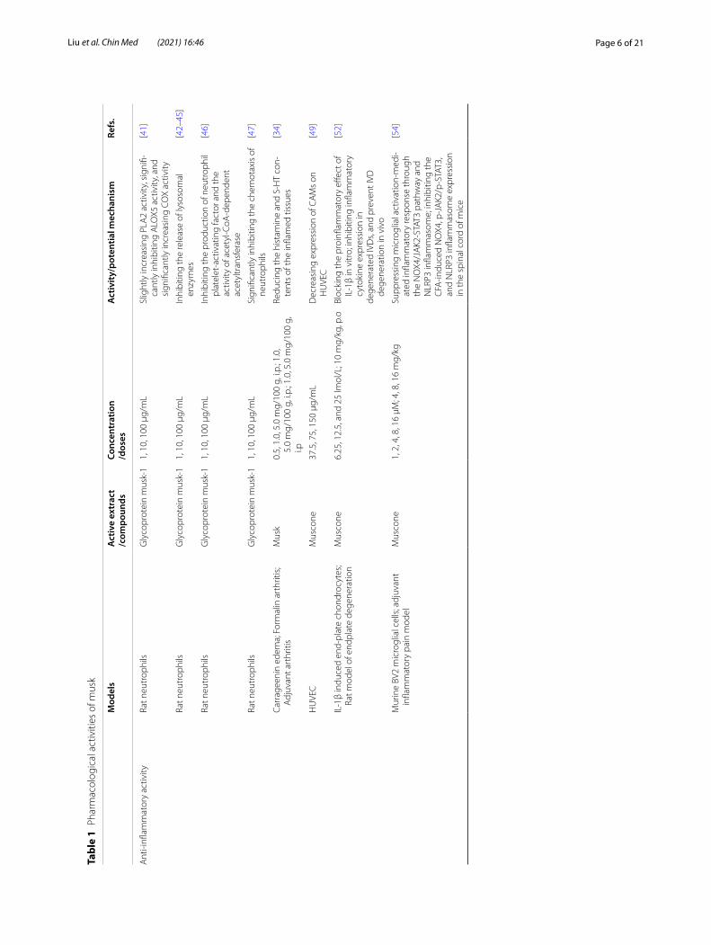
Pharmacological effectsAccording to relevant literature, musk and its main active ingredient, muscone, possess various pharmacological effects such as anti-inflammatory activity, neuroprotec-tive activity, and cardiovascular-protective activity. All the specific details are shown in Table 1 and some rele-vant molecular mechanisms are depicted in Figs. 4, 5, 6.
Anti‑inflammatory effectsInflammation is a kind of biological function triggered by the rupture of mechanical tissue or the reaction caused by physical, chemical, or biological agents in the body. The diseases associated with inflammation include cardi-ovascular disease, arthritis, cancer, diabetes, Alzheimer’s disease, Parkinson’s disease, etc. [30]. Studies have shown that the anti-inflammatory effect of traditional Chinese medicine (TCM) is achieved by inhibiting the expres-sion of master transcription factors, pro-inflammatory cytokines, chemokines, intercellular adhesion molecules, and pro-inflammatory mediators [31]. Modern stud-ies have proven that natural musk is an anti-inflamma-tory agent [32, 33] and some molecular mechanisms are depicted in Fig. 4.
Subcutaneous injection of musk Tween 80 emulsion could reduce croton oil-induced inflammatory response in male albino rats [32]. Taneja et al. [34] investigated the inhibitory effect and mechanism of musk on acute and chronic inflammation models, including the carra-geenan-induced edema and formalin arthritis model. The mechanism study indicated that the anti-inflam-matory effect of musk may be related to the reduction of histamine and 5-hydroxytryptamine (5-HT) content in inflammatory tissues. Another study also showed that musk has antihistamine and anti-5-HT effects [35]. Moreover, the aqueous extract of musk residues that have been extracted with diethyl ether and 95% ethyl alcohol and a polypeptide (musk-1) with a molecular weight less than 10,000 Da in this extract have also attracted great

Page 5 of 21Liu et al. Chin Med (2021) 16:46
interest from researchers [36, 37]. In the early stage, Zhu et al. found that intravenous administration of the aque-ous extract and musk-1 counteracted effectively cro-ton oil-induced ear inflammation in mice, respectively [37]. Further studies demonstrated that this aqueous extract was effective in a variety of inflammatory mod-els. In addition, the anti-inflammatory effect of intrave-nous musk-1 on croton oil-induced ear inflammation in mice was 36 times greater than that of hydrocortisone [38]. Moreover, it indicated that musk could modulate the immune function of the body and the presence of
adrenal glands is necessary for the anti-inflammatory effect of musk [38, 39]. Mechanism studies indicated that the aqueous extract could inhibit platelet aggrega-tion and arachidonic acid metabolism pathway, increase cyclic adenosine monophosphate levels [39, 40]. Subse-quently, Wang et al. conducted a series of experiments to study the anti-inflammatory mechanism of musk-1 using rat neutrophils as subjects. The results showed that musk-1 could inhibit 5-lipoxygenase activity in neutro-phils [41], the release of lysozyme [42–45], and platelet-activating factor production and acetyl-CoA-dependent
Fig. 3 Chemical structures of some cyclic ketones and steroids in musk
Page 6 of 21Liu et al. Chin Med (2021) 16:46
Tabl
e 1
Phar
mac
olog
ical
act
iviti
es o
f mus
k
Mod
els
Act
ive
extr
act
/com
poun
dsCo
ncen
trat
ion
/dos
esA
ctiv
ity/
pote
ntia
l mec
hani
smRe
fs.
Ant
i-infl
amm
ator
y ac
tivity
Rat n
eutr
ophi
lsG
lyco
prot
ein
mus
k-1
1, 1
0, 1
00 μ
g/m
LSl
ight
ly in
crea
sing
PLA
2 ac
tivity
, sig
nifi-
cant
ly in
hibi
ting
ALO
X5 a
ctiv
ity, a
nd
sign
ifica
ntly
incr
easi
ng C
OX
activ
ity
[41]
Rat n
eutr
ophi
lsG
lyco
prot
ein
mus
k-1
1, 1
0, 1
00 μ
g/m
LIn
hibi
ting
the
rele
ase
of ly
soso
mal
en
zym
es[4
2–45
]
Rat n
eutr
ophi
lsG
lyco
prot
ein
mus
k-1
1, 1
0, 1
00 μ
g/m
LIn
hibi
ting
the
prod
uctio
n of
neu
trop
hil
plat
elet
-act
ivat
ing
fact
or a
nd th
e ac
tivity
of a
cety
l-CoA
-dep
ende
nt
acet
yltr
ansf
eras
e
[46]
Rat n
eutr
ophi
lsG
lyco
prot
ein
mus
k-1
1, 1
0, 1
00 μ
g/m
LSi
gnifi
cant
ly in
hibi
ting
the
chem
otax
is o
f ne
utro
phils
[47]
Carr
agee
nin
edem
a; F
orm
alin
art
hriti
s; A
djuv
ant a
rthr
itis
Mus
k0.
5, 1
.0, 5
.0 m
g/10
0 g,
i.p.
; 1.0
, 5.
0 m
g/10
0 g,
i.p.
; 1.0
, 5.0
mg/
100
g,
i.p
Redu
cing
the
hist
amin
e an
d S-
HT
con-
tent
s of
the
infla
med
tiss
ues
[34]
HU
VEC
Mus
cone
37.5
, 75,
150
μg/
mL
Dec
reas
ing
expr
essi
on o
f CA
Ms
on
HU
VEC
[49]
IL-1
β in
duce
d en
d-pl
ate
chon
droc
ytes
; Ra
t mod
el o
f end
plat
e de
gene
ratio
nM
usco
ne6.
25, 1
2.5,
and
25
lmol
/L; 1
0 m
g/kg
, p.o
Bloc
king
the
proi
nflam
mat
ory
effec
t of
IL-1
β in
vitr
o; in
hibi
ting
infla
mm
ator
y cy
toki
ne e
xpre
ssio
n in
dege
nera
ted
IVD
s, an
d pr
even
t IVD
de
gene
ratio
n in
viv
o
[52]
Mur
ine
BV2
mic
rogl
ial c
ells
; adj
uvan
t in
flam
mat
ory
pain
mod
elM
usco
ne1,
2, 4
, 8, 1
6 μM
; 4, 8
, 16
mg/
kgSu
ppre
ssin
g m
icro
glia
l act
ivat
ion-
med
i-at
ed in
flam
mat
ory
resp
onse
thro
ugh
the
NO
X4/J
AK2
-STA
T3 p
athw
ay a
nd
NLR
P3 in
flam
mas
ome;
inhi
bitin
g th
e C
FA-in
duce
d N
OX4
, p-J
AK2
/p-S
TAT3
, an
d N
LRP3
infla
mm
asom
e ex
pres
sion
in
the
spin
al c
ord
of m
ice
[54]

Page 7 of 21Liu et al. Chin Med (2021) 16:46
Tabl
e 1
(con
tinue
d)
Mod
els
Act
ive
extr
act
/com
poun
dsCo
ncen
trat
ion
/dos
esA
ctiv
ity/
pote
ntia
l mec
hani
smRe
fs.
Neu
ropr
otec
tive
effec
tsG
luta
mat
e-in
duce
d PC
12 c
ells
Mus
cone
0.1,
1, 1
0 μM
Att
enua
ting
ROS
gene
ratio
n an
d Ca
2 +
influ
x, v
ia N
R1 a
nd C
aMKI
I-de
pend
ed A
SK-1
/JN
K/p3
8 si
gnal
ing
path
way
s
[61]
MC
AO
rat m
odel
Mus
cone
1 m
g/kg
, i.g
Dow
n-re
gula
ting
the
expr
essi
on o
f EA
AC
1mRN
A in
the
isch
emic
hip
-po
cam
pus
[62]
MC
AO
rat m
odel
Mus
cone
1 m
g/kg
, i.g
Redu
cing
NR1
pro
tein
exp
ress
ion,
th
ereb
y re
duci
ng e
xcita
tory
glu
tam
ate
toxi
city
[63]
MC
AO
rat m
odel
; oxy
gen–
gluc
ose
depr
i-va
tion
cell
mod
elM
usco
ne0.
5, 1
mg/
kg, i
.g.;
0.9,
1.8
μM
Act
ivat
ing
the
PI3K
/Akt
sig
nalin
g pa
thw
ay[6
7]
In v
itro
bloo
d–br
ain
barr
ier m
odel
Mus
cone
8 μM
Inhi
bitin
g P-
gp a
nd M
MP-
9 ex
pres
sion
[59]
The
trau
mat
ic b
rain
inju
ry m
odel
Mus
cone
1.8
mg/
kg, n
asal
adm
inis
trat
ion
redu
cing
the
wat
er c
onte
nt o
f bra
in
tissu
e, a
llevi
atin
g ce
rebr
al e
dem
a,
prom
otin
g se
cret
ion
of B
DN
F an
d N
GF
by o
lfact
ory
ensh
eath
ing
cells
[71]
Trau
mat
ic b
rain
inju
ry ra
t mod
elM
usco
ne1.
8 m
g/kg
, int
rana
sal a
dmin
istr
atio
nRe
duci
ng c
ereb
ral e
dem
a an
d ac
tivat
ing
the
PKA
-CRE
B si
gnal
pat
hway
[72]
Com
plet
e ce
rebr
al is
chem
ia/r
eper
fusi
on
rat m
odel
Mus
cone
0.9,
1.8
, 3.6
mg/
kg, i
.gRe
duci
ng o
xida
tive
stre
ss d
amag
e, d
elay
-in
g ne
uron
al d
eath
effe
cts,
and
inhi
bit-
ing
exci
toto
xici
ty c
ause
d by
EA
A
[64]
Pent
ylen
etet
razo
l ind
uced
rat e
pile
psy
mod
elM
usco
ne10
, 50,
100
mg/
kg, i
.pIn
hibi
ting
the
c-Fo
s, c-
jun
expr
essi
on[7
3, 7
4]
PC12
cel
ls; M
CA
O ra
t mod
elM
usco
ne10
0, 3
00 n
g/m
L; 0
.08,
0.1
6 m
gIn
hibi
ting
apop
tosi
s an
d Fa
s pa
thw
ay[6
6]
LPS-
trea
ted
mic
eM
usco
ne2
mg/
kg, i
.pRe
pres
sing
neu
roin
flam
mat
ion
in th
e pr
efro
ntal
cor
tex
of m
ice
caus
ed b
y its
sup
pres
sion
on
mic
rogl
ia a
ctiv
a-tio
n an
d pr
oduc
tion
of in
flam
mat
ory
cyto
kine
s vi
a ac
ting
on T
LR4
path
way
an
d RA
S ca
scad
e
[25]

Page 8 of 21Liu et al. Chin Med (2021) 16:46
Tabl
e 1
(con
tinue
d)
Mod
els
Act
ive
extr
act
/com
poun
dsCo
ncen
trat
ion
/dos
esA
ctiv
ity/
pote
ntia
l mec
hani
smRe
fs.
Card
iova
scul
ar-p
rote
ctiv
e eff
ects
H2O
2 ind
uced
H9c
2 ca
rdio
myo
cyte
s; H
2O2 i
nduc
ed H
UVE
CM
usk
50 μ
g/m
LSc
aven
ging
RO
S an
d im
prov
ing
the
activ
-ity
of i
ntra
cellu
lar a
ntio
xida
nt e
nzym
es[7
8, 7
9]
H2O
2 ind
uced
HU
VEC
Mus
cone
3, 1
5, 3
0 μg
/mL
Stab
ilizi
ng c
ell m
itoch
ondr
ial m
embr
ane
pote
ntia
l, re
duci
ng c
ell p
erm
eabi
lity,
an
d pr
even
ting
Ca2+
influ
x
[80]
Myo
card
ial i
nfar
ctio
n ra
t mod
elM
usco
ne2
mg/
kg, i
.gRe
duci
ng th
e ex
pres
sion
of t
rans
form
-in
g gr
owth
fact
or-β
1, T
NF-
α, IL
-1β
and
NF-
κB
[82]
Myo
card
ial i
nfar
ctio
n ra
t mod
elM
usco
ne2
mg/
kg, i
.gSt
imul
atin
g an
giog
enes
is v
ia u
preg
ulat
-in
g H
IF-1
α an
d VE
GFA
[83]
Myo
card
ial i
nfar
ctio
n m
ice
mod
elM
usco
ne2
mg/
kg, i
.gIn
hibi
ting
NF-
κB a
nd N
LRP3
infla
mm
a-so
me
activ
atio
n, th
ereb
y im
prov
ing
card
iac
func
tion
[84]
Neo
nata
l rat
car
diac
myo
cyte
sM
usco
ne0.
215,
0.4
3 or
0.8
6 μg
/mL
Alle
viat
ing
the
incr
ease
of l
actic
aci
d de
hydr
ogen
ase
rele
ase,
mal
ondi
al-
dehy
de p
rodu
ctio
n, c
reat
ine
kina
se
activ
ity, c
aspa
se-3
act
ivity
, [Ca
2 +
]i,
apop
tosi
s ra
te a
nd e
xpre
ssio
n of
Bax
pr
otei
n, a
nd re
duct
ion
of s
uper
oxid
e di
smut
ase
activ
ity, M
MP,
and
expr
es-
sion
of B
cl-2
pro
tein
[85]
Ant
i-can
cer e
ffect
sBr
east
can
cer m
ice
mod
elM
usco
ne2
mg/
kg, i
.gRe
duci
ng th
e ex
pres
sion
of V
EGF
[88]
Hep
G2
cells
; xen
ogra
ft li
ver c
ance
r mod
elM
usco
ne0.
663
μM; 0
.1 m
mol
/kg,
0.2
ml/2
0 g
bwIn
crea
sing
cel
lula
r apo
ptos
is th
roug
h en
dopl
asm
ic re
ticul
um s
tres
s re
spon
ses
and
indu
cing
aut
opha
gy th
roug
h A
MP
kina
se/m
TOR
com
plex
1 s
igna
ling
path
way
[89]

Page 9 of 21Liu et al. Chin Med (2021) 16:46
Tabl
e 1
(con
tinue
d)
Mod
els
Act
ive
extr
act
/com
poun
dsCo
ncen
trat
ion
/dos
esA
ctiv
ity/
pote
ntia
l mec
hani
smRe
fs.
Prom
otin
g eff
ect o
n st
em c
ell t
hera
pyH
uman
GM
SCs
Mus
cone
3, 6
, 9 m
g/L
Incr
easi
ng th
e pr
olife
ratio
n an
d m
igra
-tio
n, p
rom
otin
g th
e ad
ipog
enic
diff
er-
entia
tion
and
inhi
bitin
g th
e os
teog
enic
di
ffere
ntia
tion
of G
MSC
s by
inhi
bitin
g th
e W
nt/β
-cat
enin
sig
nalin
g pa
thw
ay
[93]
Skul
l bon
e de
fect
rat m
odel
Mus
k42
, 86,
168
mg/
kgPr
omot
ing
stro
mal
cel
l-der
ived
fact
or 1
an
d m
onoc
yte
chem
otac
tic p
rote
in 1
ex
pres
sion
to p
rom
ote
the
mig
ratio
n of
exo
geno
us b
one
mar
row
mes
en-
chym
al s
tem
cel
ls to
bon
e in
jury
site
s in
rats
[97,
98]
Skul
l bon
e de
fect
rat m
odel
Mus
k42
mg/
kgIn
crea
sing
the
leve
l of S
DF-
1, H
GF,
and
SCF,
and
expr
essi
on o
f MC
P-1
mRN
A,
FGF-
2 m
RNA
, TG
F-β
mRN
A, V
EGF
mRN
A, a
nd d
own-
regu
latin
g th
e ex
pres
sion
of E
GF
mRN
A
[99–
102]
Hum
an G
MSC
s; al
coho
l-ind
uced
ost
e-on
ecro
sis
of th
e fe
mor
al h
ead
Mus
cone
1, 1
0, 2
5 μM
; 1 m
g/kg
Prom
otin
g A
LP a
ctiv
ity a
nd m
RNA
ex
pres
sion
of C
OL1
and
OC
N; r
esto
r-in
g BV
/TV
ratio
and
bon
e de
nsity
of
necr
otic
fem
oral
hea
ds
[103
]
Gen
tam
icin
-indu
ced
AKI
Mus
cone
3.0
mg/
LU
p-re
gula
ting
the
expr
essi
on o
f CXC
R4
and
CXC
R7[1
04]

Page 10 of 21Liu et al. Chin Med (2021) 16:46
acetyltransferase activity [46]. Meanwhile, musk-1 could significantly inhibit the chemotactic response of neutro-phils [47]. These effects may be important mechanisms for their anti-inflammatory effects.
Moreover, muscone was proved to possesses anti-inflammatory effects and utilized to treat inflamma-tory disorders. Excessive inflammation can lead to slow wound healing [48]. He et al. [49] investigated the regula-tory mechanism of muscone on chronic wound inflam-mation. Muscone was found to significantly inhibit the expression of ICAM-1, VCAM-1, and CD44 on the sur-face of human umbilical vein endothelial cells (HUVEC), thereby inhibiting the adhesion of polymorphonuclear leukocytes to HUVEC to suppress excessive inflamma-tion and promote healing of chronic wounds. Interleukin (IL)-1 initiates and controls the inflammatory response [50]. Studies demonstrated that overproduction of IL-1β played an important role in the pathogenesis of human intervertebral disc degeneration [51]. Liang et al. [52] studied the protective effect of muscone on vertebral end-plate degeneration. In vitro, muscone inhibited the IL-1β-induced phosphorylation of extracellular signal-regulated kinases 1/2 and c-Jun N-terminal kinase in a dose-dependent manner. In vivo, muscone inhibited the expression of prostaglandin E2, 6-keto-prostaglandin F1a, IL-1β, and tumor necrosis factor α and recovered
the structural distortion of the degenerative disc. More-over, the therapeutic potential of muscone on cervical spondylotic myelopathy (CSM) was evaluated by Zhou et al. In the chronic cervical cord compression rat model, muscone promoted motor recovery in rats. Molecular studies revealed that muscone could inhibit activation of the NLRP3 inflammasome, NF-κB, and Drp1 in lesions to attenuate inflammatory responses and neuronal dam-age in model rats [53]. In LPS-stimulated BV2 and pri-mary microglial cells, muscone could inhibit the NLRP3 inflammasome and NF-κB activation to suppress mRNA levels of IL-1β, IL-6, and TNF-α and iNOS and Cox-2 protein expression [53]. These results indicated that the potential of muscone to treat CSM is due in part to its anti-neuroinflammatory effects. In addition, intraperi-toneal injection of muscone could reduce inflammatory pain by blocking the NOX4/JAK2-STAT3 pathway and NLRP3 inflammasome, which can cause an inflammatory response [54].
Neuroprotective effectsNatural musk has the action of inducing resuscita-tion and has been utilized as a TCM in treating stroke, coma, neurasthenia, convulsions for thousands of years [4]. Modern studies have demonstrated the neuropro-tective effects of musk. Ayuob et al. found that in a
Fig. 4 The mechanisms of musk against inflammatory diseases
Page 11 of 21Liu et al. Chin Med (2021) 16:46
depression model, inhaling musk could improve behav-ioral changes, elevated serum glucocorticoid levels, memory impairment, neurodegenerative changes, and changes in salivary gland structure induced by chronic unpredictable mild stress [55–58].
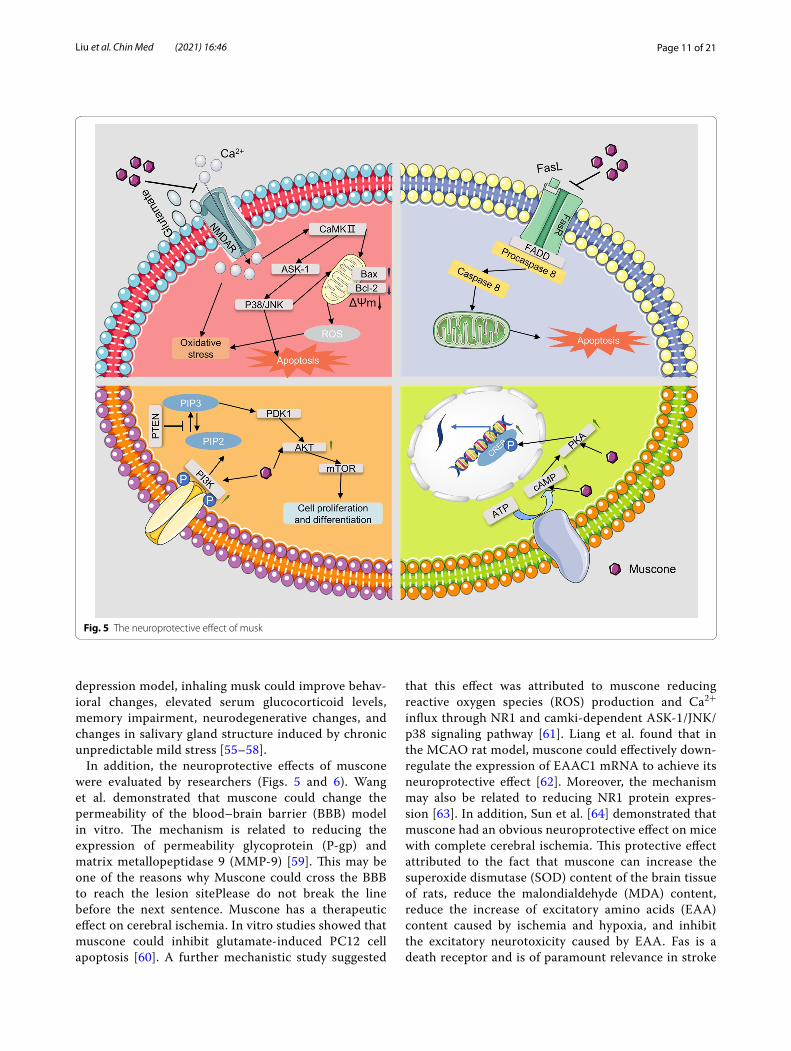
In addition, the neuroprotective effects of muscone were evaluated by researchers (Figs. 5 and 6). Wang et al. demonstrated that muscone could change the permeability of the blood–brain barrier (BBB) model in vitro. The mechanism is related to reducing the expression of permeability glycoprotein (P-gp) and matrix metallopeptidase 9 (MMP-9) [59]. This may be one of the reasons why Muscone could cross the BBB to reach the lesion sitePlease do not break the line before the next sentence. Muscone has a therapeutic effect on cerebral ischemia. In vitro studies showed that muscone could inhibit glutamate-induced PC12 cell apoptosis [60]. A further mechanistic study suggested
that this effect was attributed to muscone reducing reactive oxygen species (ROS) production and Ca2+ influx through NR1 and camki-dependent ASK-1/JNK/p38 signaling pathway [61]. Liang et al. found that in the MCAO rat model, muscone could effectively down-regulate the expression of EAAC1 mRNA to achieve its neuroprotective effect [62]. Moreover, the mechanism may also be related to reducing NR1 protein expres-sion [63]. In addition, Sun et al. [64] demonstrated that muscone had an obvious neuroprotective effect on mice with complete cerebral ischemia. This protective effect attributed to the fact that muscone can increase the superoxide dismutase (SOD) content of the brain tissue of rats, reduce the malondialdehyde (MDA) content, reduce the increase of excitatory amino acids (EAA) content caused by ischemia and hypoxia, and inhibit the excitatory neurotoxicity caused by EAA. Fas is a death receptor and is of paramount relevance in stroke
Fig. 5 The neuroprotective effect of musk

Page 12 of 21Liu et al. Chin Med (2021) 16:46
pathogenesis [65]. It is suggested that neutralizing FasL would be a great choice for stroke treatment. In cer-ebral ischemia rats, muscone exerted a neuroprotec-tive effect through inhibiting apoptosis by suppressing the expression of Fas [66]. Post-stroke recovery is also important for patients and neural stem cells (NSCs) are of importance in this process. Muscone can promote neural stem cell proliferation and differentiation to protect against cerebral ischemia. This effect is attrib-uted to the activation of the PI3K/Akt signaling path-way [67]. Cerebral ischemia is accompanied by edema, and this symptom may lead to death [68]. Muscone can alleviate edema of brain tissue in the ischemic area and significantly reduce the brain water content to play a protective role. In addition, muscone can also change the permeability of the BBB, reduce albumin exposure and leakage, and reduce the degree of edema of brain cells [69]. Jiang et al. [70] found that in the early period after traumatic brain injury, muscone could exert neu-roprotective effects by inhibiting the expression of MMP-9 and reducing cerebral edema.
Moreover, muscone exerts protective effects against traumatic brain injury (TBI). Intranasal administration of muscone can promote the secretion of brain-derived neurotrophic factor and nerve growth factor by olfac-tory ensheathing cells to exert a neuroprotective effect [71]. Another study demonstrated that muscone exerted neuroprotective effects after TBI by activating the PKA-CREB signal pathway [72]. Cheng et al. studied the mech-anism of anti-epilepsy activity of muscone and found
that muscone blocked the expression of c-Fos and c-Jun in the brain during seizures, and this effect had a dose–effect relationship [73, 74]. Recently, He et al. proved that muscone could improve depression-like behavior in rats by repressing lipopolysaccharide (LPS)-induced neuroin-flammation. The underlying mechanism may be its sup-pression of microglia activation and production of IL-1β through acting on TLR4/MyD88 and TLR4/NLRP3 as well as its blockade on the expression of RANTES and MCP-1 (monocyte chemotactic protein 1) via antagoniz-ing renin/Ang II axis [25].
Taken together, the above findings suggest that musk has good neuroprotective effects and has great potential for treating neurological diseases. Some related molec-ular mechanisms are depicted in Figs. 5 and 6, and the details are summarized in Table 1.
Cardiovascular‑protective effectsCardiovascular disease is the deadliest disease worldwide, and its morbidity and mortality rates continue to rise. Studies have shown that some herbs or active ingredients in them have the potential to treat cardiovascular dis-eases, such as curcumin [75], baicalin [76], and berberine [77]. There is evidence that musk is also effective against cardiovascular disease. Quan et al. found that musk can play a protective role against H2O2-induced H9C2 car-diomyocytes injury by eliminating ROS and improving intracellular antioxidant enzyme activity [78]. Moreover, musk can play a protective role in H2O2-induced HUVEC injury by improving intracellular antioxidant enzyme
Fig. 6 The protective effect of musk against microglial cell inflammation

Page 13 of 21Liu et al. Chin Med (2021) 16:46
activity and reducing oxidative stress [79]. Also, research-ers investigated the effect of muscone on cardiovascular disease in vitro and in vivo. Hong et al. demonstrated that muscone can stabilize mitochondrial ΔΨm, reduce cell permeability and reduce Ca2+ influx, thereby inhibit-ing HUVEC cell apoptosis induced by H2O2 [80]. Zhou et al. [81] studied the application of muscone in random skin flap transplantation. Muscone can promote skin flap angiogenesis, activate VEGF expression, reduce apop-tosis, increase SOD levels and decrease MDA levels. Therefore, muscone can improve the survival rate of skin flaps by anti-oxidation, anti-apoptosis, and promoting angiogenesis. Moreover, myocardial infarction (MI) is the leading cause of death and disability in developed coun-tries, and a number of challenges remain in preventing and treating MI. Wang et al. found that muscone could improve cardiac remodeling and dysfunction caused by MI. The mechanistic study revealed that muscone could reduce the expression of transforming growth factor-β1 (TGF-β1), tumor necrosis factor-α (TNF-α), IL-1β, and nuclear factor-κB (NF-κB) to reduce the inflammatory response. Moreover, muscone could reduce myocardial apoptosis by upregulating the Bcl-2/Bax ratio. What’s more, the intervention of muscone significantly induced the phosphorylation of Akt and eNOS, which is related to vascular endothelial function [82]. Further, Du et al. demonstrated that muscone improved cardiac function in mice with MI by enhancing angiogenesis. The underly-ing mechanism of this effect was up-regulating hypoxia-inducible factor 1α (HIF-1α) and vascular endothelial growth factor A (VEGFA) expression levels [83]. Simi-larly, by reducing macrophage-mediated chronic inflam-mation, muscone can improve cardiac function in mice with MI. The mechanism was to inhibit the activation of NF-κB and NLRP3 inflammasome, thereby blocking the production of inflammatory cytokines (IL-1β, TNF-α, and IL-6) [84]. When cardiomyocytes were pretreated with muscone before I/R injury, the increase of LDH release, MDA production, creatine kinase activity, cas-pase-3 activity, [Ca2+]i, apoptosis rate and expression of Bax protein, and reduction of SOD activity, MMP, and expression of Bcl-2 protein can be alleviated. This sug-gested that muscone can protect I/R injury by inhibiting cellular oxidative stress and apoptosis [85].
Anti‑cancer effectsMusk is widely used to treat cancer. It is included in many traditional Chinese medicine formulae for treating can-cer, such as the Xihuang pill [86]. Xu et al. [87] studied the effects of musk and muscone on 22 types of tumor cells. It was found that musk and muscone could widely induce cancer cell growth inhibition and apoptosis. In a nude mouse model of blood stasis syndrome, muscone
can significantly inhibit the growth of breast cancer. The mechanism may be related to the reduction of VEGF expression [88]. Qi et al. [89] found that muscone had a certain anti-cancer effect in hepatocellular carcinoma and this effect attributed to the induction of apopto-sis and autophagy of liver cancer cells. The mechanis-tic study showed that apoptosis was a consequence of endoplasmic reticulum stress through the PERK/ATF4/DDIT3 signaling pathway, and autophagy was closely related to the AMP kinase/mTOR complex 1 signaling pathway. P-gp is a product of the multidrug resistance (MDR) gene, and the high expression of P-gp on the tumor cell membrane is the main mechanism of MDR formation [90]. Wang et al. used human colon carcinoma cell line Caco-2 as a target and proved that muscone can effectively inhibit the function of P-gp [91].
Promoting effect on stem cell therapyNowadays, mesenchymal stem cells (MSCs) are widely used in stem cell therapy [92]. Related reports have dem-onstrated that musk has the effect of promoting mesen-chymal stem cell therapy and the details are summarized in Table 1. In vitro, muscone (3, 6, 9 mg/L) can enhance the proliferation of human gingival mesenchymal stem cells (GMSCs), and 6 mg/L of muscone had the best effect. In vivo, muscone can effectively inhibit osteoblast differentiation and promote GMSC proliferation, migra-tion, and adipogenesis, which is attributed to the inhibi-tion of the Wnt/β-catenin signaling pathway [93]. In a skull defect rat model, muscone (4.2, 8.4, 16.8 μL/100 g) could promote the migration of exogenous stem cells in vivo, and the effect was better at concentrations of 4.2 and 8.4 μL/100 g [94], and the mechanism was related to the promotion of BMSCs proliferation and osteogenic differentiation and the promotion of exogenous BMSCs migration in vivo [95, 96]. Studies have shown that the mechanism by which musk promoted the migration of exogenous bone marrow mesenchymal stem cells to the injury site may be related to its promotion of MCP-1 expression and SDF-1 (stromal cell-derived factor-1) expression in bone defects [97, 98]. Li et al. investigated the mechanism by which musk promotes the healing of bone defects in the skull of rats. The mechanism of musk promoted healing may be related to the increase of serum SDF-1 and hepatocyte growth factor (HGF) lev-els, the up-regulation of mRNA expression of stem cell factor (SCF), MCP-1, fibroblast growth factor 2 (FGF-2), TGF-β, and VEGF, as well as the down-regulation of mRNA expression of epidermal growth factor (EGF) [99–102]. Guo et al. found that muscone had a protec-tive effect on femoral head necrosis caused by alcohol. In vitro, muscone had the potential to promote alkaline phosphatase (ALP) activity and mRNA expression of

Page 14 of 21Liu et al. Chin Med (2021) 16:46
collagen 1 (COL1) and osteocalcin (OCN) in ethanol-treated hBMSCs. In vivo, muscone could restore BV/TV ratio and bone density of necrotic femoral heads [103]. In addition, in an acute kidney injury (AKI) model, muscone enhanced the therapeutic effect of bone marrow mesen-chymal stem cells by promoting cell proliferation, secre-tion, and migration. The mechanism may be related to the expression of C-X-C chemokine receptor (CXCR) 4 and 7 up-regulation [104].
Other effectsIn addition to the pharmacological effects mentioned above, other pharmacological effects of musk and muscone have also been reported, including induc-ing liver drug metabolism enzymes, antibacterial, etc. Muscone can induce certain P-450 isoenzymes, which in turn can alter the metabolism and endogenous sub-strates of drugs. Pretreatment with muscone (75 mg/kg) for 1 day can increase 2.8 times of benzophenantamine demethylase activity in rat microsomes [105]. Tanaka et al. studied the effect of muscone on rat liver microso-mal drug metabolism enzyme system and other enzyme activity parameters in vitro and in vivo and found that muscone could induce liver metabolism enzymes [106]. Muscone mainly induced P450 IIB1 and P450 IIB2 with a slightly weaker effect than phenobarbital [107]. Recently, Phung et al. studied the preventive effect of muscone against cisplatin nephrotoxicity. In LLC-PK1 cells, muscone was proved to prevent cisplatin-induced oxida-tive stress, inflammation, and apoptosis. The mechanistic studies revealed that muscone could inhibit ROS accu-mulation and induce HO-1 expression to exert an antiox-idant effect in cisplatin-treated LC-PK1 cells. Meanwhile, muscone could suppress the phosphorylation of p38, which may mediate production of TNF-α. Moreover, in cisplatin-treated LC-PK1 cells, muscone played an anti-apoptotic role by inhibiting p53, caspase-3, 7, and 8, and restoring the Bcl-2/Bax ratio [108]. In addition, the protective role of muscone in postmenopausal osteopo-rosis was evaluated by Zhai et al. [109] employing bone marrow monocytes, RAW264.7, and female C57BL/6 ovariectomized mice. In vitro, muscone inhibited osteo-clastogenesis in BMMs and RAW264.7 cells. In vivo, the bone loss was prevented by muscone by suppressing osteoclastogenesis. The over-activated RANKL signaling pathways will promote the reproduction of osteoclasts. The molecular study demonstrated that muscone could reduce the levels of RANK and TRAF6, leading to the suppression of downstream NF-kB and MAPK signaling pathways.
Musk extract had inhibitory and bactericidal effects on the growth of pathogenic bacteria such as Staphylo-coccus aureus and Penicillium [110]. Saddiq studied the
inhibitory effects of musk on five opportunistic patho-genic fungi, namely Aspergillus flavus, Aspergillus fumi-gates, Rhizopus stolonifer, Fusarium solani, and Candida albicans. Musk extract (25%) had an inhibitory effect on the above fungi, the inhibition rates were 74.61%, 68.76, 56.92%, 71.57%, and 67.80%, respectively. Subse-quent animal experiments showed that musk extract can reduce lung toxicity caused by A. flavus [111]. AL-Jobori et al. studied the antifungal activity of musk in vitro. Five kinds of fungi were used, including Aspergillus fumigates, Aspergillus niger, Alternaria Spp., Trichomphyton menta-grophytes, and Fusarium Spp. All concentrations (25, 50, 75, or 100%) and amounts (1, 2, 4 mL) inhibited fungal growth and completely eliminated the fungi [112]. Mean-while, musk also inhibited the activity of hydatid cyst [113]. Dong et al. demonstrated that muscone (0.1, 1, 10, 50 mol/L) could reduce high glucose-induced autophagy and apoptosis in RSC 96 cells, and its mechanism was to activate the AKT/mTOR signaling pathway [26].
Clinical applicationMusk possesses a wide range of pharmacological effects. In modern clinical applications, musk and muscone are utilized to treat diseases and they are mostly used in combination with other Chinese herbal medicines. To date, many clinical trials have been listed in the global clinical trial registry (https:// clini caltr ials. gov) and the Chinese Clinical Trial Registry (http:// www. chictr. org. cn).
Internationally, a total of 8 clinical trials related to musk have been registered, among which four are related to musk Shexiang Baoxin Pill (NCT01897805, NCT03072121, NCT04026724, NCT04022031), one is related to Mayinglong musk hemorrhoid oint-ment (NCT01881282), one is related to Gongjin-dan (NCT03219515), one is related to Qishe Pill (NCT01274936), and one is related to Angong Niuhuang Pill (NCT00817609). For example, the therapeutic effect and safety of compound carraghenates cream with May-inglong musk hemorrhoid ointment in the treatment of hemorrhoids, especially regarding the relief of pain. The curative effect of Shexiang Baoxin Pill on coronary artery disease not amenable to revascularization based on west-ern medicine therapy was evaluated. Of these, one trial has been completed (NCT01881282), five trials are of unknown status, and two observational trials related to Shexiang Baoxin Pill have not yet enrolled patients. But unfortunately, none of these clinical trials have published results.
In China, 15 clinical trials of Chinese pat-ent medicines containing musk have been regis-tered since 2012. These tests are mostly related to Shexiang Tongxin Dropping Pill (ChiCTR2000035167,
Page 15 of 21Liu et al. Chin Med (2021) 16:46
ChiCTR2000032429, ChiCTR1900025810, ChiCTR-IPC-17010823, ChiCTR-IPR-16009785, ChiCTR-IPR-16008950, ChiCTR-IPR-15006020,) and Shexiang Baoxin Pill (ChiCTR2000041451, ChiCTR2000034817, ChiCTR1900027946, ChiCTR-TRC-10001237, ChiCTR-TRC-12003513). Moreover, all of these clinical tri-als are related to cardiovascular disease. For example, the Second Affiliated Hospital of the Second Military Medical University studied the therapeutic effect of Shexiang Tongxin Dropping Pill on myocardial perfu-sion among acute myocardial infarct patients (Shexi-ang Tongxin Dropping Pill). Recently, a clinical trial of Shexiang Baoxin Pill in the treatment of coronary micro-vascular dysfunction has been prospectively registered (ChiCTR2000034817). Furthermore, a trail of musk used to treat acute ST-elevation myocardial infarction has also been in preparation (ChiCTR2000037470).
Overall, clinical trials related to musk or Chinese pat-ent containing musk are gradually increasing, especially in China, which is of great significance for more scientific and full utilization of musk.
Toxicity and safetyThe related report indicated that muscone had toxic effects on zebrafish (AB = type) embryo development. Muscone (5, 10, 20, 40, 80, 100 μmol/L) had a lethal effect on zebrafish embryos. When the concentration of muscone reached 80 and 100 μmol/L, the embryo death rate reached 100% at 96 h after fertilization. High-dose muscone had a significant effect on zebrafish embryo development in a time- and dose-dependent manner, which mainly manifested as abnormal development of muscle tissue and heart tissue [114]. Muscone (0.005, 0.01, 0.03, 0.1, 0.2 mM) was toxic to zebrafish embryos by increasing Myh6 and Myh7 mRNA expression and reducing thyroid genes (Trh, Thrβ, and Dio3) expres-sion [115]. Muscone could induce CYP1A2 and CYP3A4 expression in liver cells in vitro and in vivo. In addition, when the dose exceeded 50 mg/kg, muscone had sig-nificant liver toxicity in Kunming mice [116]. Further, pharmacodynamic drug-drug interactions (DDIs) occur when the pharmacological effect of one drug is altered by that of another drug in a combination regimen [117]. Liu et al. [118] demonstrated that muscone would reduce the hypnotic and analgesic effects of ketamine, a widely used general anesthetics, in a dose-independent manner, which may be related to changes in NR1 and delta-opioid receptors. Hence, when a patient is given muscone pre-operatively, it is important to monitor the depth of anes-thesia during the surgery.
PharmacokineticsThe pharmacological activity of a drug in the body is closely related to its absorption, distribution, metabo-lism, and excretion process in the body. In view of the extensive usage of musk in TCM, an in-depth study of the pharmacokinetics of it is necessary. Unfortu-nately, the pharmacokinetics of musk are poorly studied globally. On the other hand, there are some pharma-cokinetics studies on the muscone, the main active component. At the early stage, Zhu et al. established a method employing gas chromatography and applied it to the determination of blood concentration after oral administration of muscone. After oral administration of 80 mg/kg muscone, the parameters indicated that the whole blood concentration–time curve of muscone in rats was best fitted to a two-compartment open model. The T1/2Ka (min), Tmax (min), Cmax (mg/L) and T1/2β (min) were 22, 74.4, 1.44 and 196.1, respectively. These results indicated that muscone was absorbed quickly and eliminated quickly in rats, [119]. After that, Zhu et al. utilized the same method to determine the pharmacoki-netic parameters of intravenously administered muscone in rats, rabbits, and dogs. After intravenous administra-tion of muscone (12, 18 and 24 mg/kg) to rats, the T1/2α, T1/2β, Vss and Vc were 9.4–9.6 min, 118.1–131.2 min, 22.5–23.5 L/kg and 2.3–2.9 L/kg, respectively. The whole blood concentration–time curve was best fitted to a two-compartment open model. Whilst AUC (μg·min−1/mL) were 153.0, 207.7 and 258.2, respectively, which were dose-proportional. After intravenous administration of muscone to rabbits and dogs at a dose of 24 and 18 mg/kg respectively, the whole blood concentration–time curves were both fitted to a three-compartment open model. In rabbits, the T1/2α (min), T1/2β (min), T1/2γ (min), Vss (L/kg) and Vc (L/kg) were 4.82 ± 2.60, 24.87 ± 13.62, 331.92 ± 61.32, 51.65 ± 25.61, 2.13 ± 0.84. In dogs, the T1/2α (min), T1/2β (min), T1/2γ (min), Vss (L/kg) and Vc (L/kg) were 2.78 ± 3.8, 29.98 ± 22.11, 366.39 ± 185.44, 7.25 ± 2.23, 0.38 ± 0.30 [120]. Moreover, Li et al. also demonstrated that muscone can be quickly absorbed in the gastrointestinal tract, and the highest concentration of plasma and brain tissue was reached 1.5 h after intra-gastric administration, indicating that muscone quickly entered the brain tissue through the BBB. The elimina-tion rate constants of muscone in brain tissue and plasma were 0.56 h−1 and 0.45 h−1, respectively, indicating that muscone was eliminated rapidly in the brain and plasma (the concentration in the brain decreases slightly faster than that in the plasma). Therefore, there was no accu-mulation of muscone in the brain [121].

Page 16 of 21Liu et al. Chin Med (2021) 16:46
Quality controlAs we all know, the quality difference of traditional Chi-nese medicine is universal. Taking musk as an example, its quality depends on the physique of the musk deer, harvest time, drying method, etc. In addition, there were reports in the early years that there was counterfeit musk on the market. Therefore, it is indispensable to establish a potentially reliable, sensitive, accurate and repeatable analysis method to ensure the quality of musk. The meth-ods mentioned in this section are listed in Table 2.
Quantitative quality control of muskAccording to Chinese Pharmacopoeia (2020 edition), in addition to morphological and microscopic identifica-tion, as well as loss on drying and ash check, the concen-tration of muscone should exceed 2.0% as determined by GC [1] to control the quality of natural musk. Moreover, other methods have been established to detect muscone, such as GC–MS [122], HPLC-RI [123], RP-UPLC-ELSD
[124], Single-Sweep Polarography [125]. However, the chemical composition of traditional Chinese medicine is complex, and with the advent of synthetic muscone, it is not appropriate to rely solely on muscone as an indicator of biological activity. In view of the fact that steroids are another feature in musk, some methods for the determi-nation of steroid content in musk have been established [126–128]. Moreover, Luo et al. developed a biologi-cal evaluation method to evaluate the clinical efficacy of musk based on the biological potency of its anti-throm-bin activity [129].
Qualitative quality control of muskNatural musk has been a precious Chinese herbal medi-cine since ancient times, and it has been expensive and in short supply for a long time, and hence, this situa-tion stimulated the musk forgery. As synthetic muscone becomes available, new methods of counterfeiting may emerge. Meanwhile, the composition of natural musk is complex. Hence, it is of vital importance to seek
Table 2 Quality control and identification methods for musk
Methods Conditions Indicator/Activity Refs.
Quality control GC Mobile phase, nitrogen; Column, OV-17 column; Detector, FID Muscone [1]
GC/MS Mobile Phase, helium; Column, DB-5 column; Detector, mass detector
Muscone [122]
HPLC-RI Mobile phase, acetonitrile–water (95:5); Column, Zorbax ODS column; Detector, RI detector
Muscone [123]
RP-UPLC-ELSD Mobile phase, methanol–water (78: 22); Column, Waters Acquity BEH C18 column; Detector, ELSD
Muscone [124]
Single-Sweep Polarography Solution, phenylhydrazine hydrochloride-sodium chloride mixed aqueous solution; peak potential, -800 mV(vs.SCE)
Muscone [125]
GC/MS Mobile phase, helium; Column, Rtx-5sil MS column; Detector, mass detector
Steroids [128]
HPLC Mobile Phase, n-hexane-dioxane- ethyl acetate (100:2.5:0.4); Column, Self-loading silicone column (YWG 80-5 μm); Detec-tor, SPD-1 UV–Vis detector
Steroids [126]
GC/MS Mobile phase, helium; Column, HP-1MS column; Detector, mass detector
Steroids [127]
Thrombin titration Titration substrate, 0.5% bovine fibrinogen; Enzyme, 10U/mL bovine thrombin; Titration interval, 1 min; Titrant volume, 2 μL
Anti-thrombin activity [129]
Identification Microscopy Microscope, Nikon E200 Physical characteristics [130]
GC/MS Mobile phase, helium; Column, HP-1MS column; Detector, mass detector
Steroids [128]
FTIR Nicolet 6700 FTIR spectrometer, DTGS mid-infrared detector; Dispersion medium, KBr; Spectral resolution, 4 cm−1; Signal accumulation, 32
Characteristic infrared absorption [131]
ELISA - MP-1 [132]
Electronic nose coupled with chemometrics
An oxide sensor-based electronic nose (A oxide sensor-based electronic nose); musk samples, 0.03 g; Carrier gas, synthetic dry air, 150 mL/min; Injection volume, 1 mL; Injection rate, 1 mL/s, 35 °C
Odors [133]
DNA barcoding DNA extraction, DNeasy tissue blood DNA Extraction Kit, modified CTAB method
Phylogenetic tree [134]

Page 17 of 21Liu et al. Chin Med (2021) 16:46
reasonable and effective ways to identify and compre-hensively evaluate natural musk. Some researchers have established methods for authenticating and evaluating natural musk. Traditionally, microscopic authentica-tion can be used as a fast on-site method [130]. Zhang et al. used GC–MS spectrometry and searched the NIST standard library to quickly determine most of the chemi-cal components in the musk samples, making it easier to screen the fake musk. They found that the types and content of low-content steroids were quite different and had strong characteristics. Therefore, this study focused on the analysis of the steroidal component in the samples collected. Their data showed that the steroids contained in musk were very complex and variable. However, the analysis from multiple samples can capture its charac-teristic components as the basis for identification. The components included in androgen hormones had strong characteristics and regularity. The establishment of fin-gerprints of steroid hormones can simplify data pro-cessing [128]. In addition, Zhou et al. utilized Fourier transform infrared spectroscopy (FTIR) which was fast, sensitive, intuitive, and non-destructive, to identify the authenticity of musk [131]. Ahn et al. established a direct enzyme-linked immunosorbent assay (ELISA) to identify and evaluate different sources of musk for the first time. Firstly, they purified musk protein 1 (MP-1), a unique protein, from musk and made polyclonal antibodies in rabbits. And then a direct ELISA for quantitative analy-sis was developed using anti-MP-1 polyclonal antibodies. Lastly, the ELISA was validated by the determination of the quantity of MP-1. MP-1was detected in four out of nine musk samples, and the concentrations that can be detected ranged from a few nanograms in 1 g of protein. The results demonstrated that this method is useful for evaluating the authenticity of natural musk [132]. The odor is an important property of natural musk and with the development of electronic nose (E-Nose), identifica-tion methods of TCM based on E-Nose are emerging. Ye et al. employed an E-Nose (αFOX-4000) to analyze the aroma of several musk samples, namely 1 artificial musk sample, 5 natural musk samples, and 3 fake musk samples. The data showed that the chemical information between different samples was severely damaged, leading to complex and fuzzy results of musk quality evaluation. Then the original data obtained from the response val-ues of 18 sensors were analyzed by principal component analysis. The adulterates were not only easily discrimi-nated from authentic musk samples based on the above analysis but also showed a clear separation of different quality proportions of adulterated musk [133]. Impor-tantly, DNA barcoding has become a new direction for biological species identification and has attracted the attention of many experts. Zhao et al. designed a pair of
musk mini-DNA barcode identification primers of about 180 bp and successfully identified the fake products [134].
Discussions and future perspectivesThe present review summarizes the zoology, chemical composition, pharmacological effects, toxicity, pharma-cokinetics, and quality control of musk by referring to published reports. Musk is a kind of animal secretion and so far, researchers have identified macrocyclic ketones, pyridine, steroids, fatty acids, amino acids, peptides, and proteins from musk. Pharmacological studies have shown that musk has various pharmacological activities, includ-ing anti-inflammatory effects, neuroprotective effects, cardiovascular protective effects, anti-cancer effects, promoting effects on stem cell therapy, etc. Although the progress in recent decades strongly proves the medicinal value of musk, there are still some notable scientific gaps in the subsequent research.
First, the chemical composition of musk is complex. Many studies now focus only on the biological activ-ity of muscone, ignoring the biological activity of other chemical components. However, studies have shown that muscone is not the only active ingredient in musk. For instance, the androgenic effects of musk are closely related to the androgen derivatives it contained, and dec-ades ago scholars isolated a peptide whose anti-inflam-matory activity was 20 times that of hydrocortisone [36]. As a TCM, the pharmacological effects of musk are the result of all the components working together. There-fore, it is necessary for future research to focus more on the biological activities of other components. Moreover, in pharmacological research, one problem is that many mechanisms of action have not been studied. In addition, there are many traditional uses of musk that have not been proven by modern pharmacological experiments. Furthermore, most of the current pharmacological stud-ies have only conducted animal or in vitro studies, result-ing in a lack of clinical trial data, therefore, researchers should try to convert experimental research into clinical research.
Second, there is insufficient research on the toxicity and safety of the active substances contained in musk, although it has been utilized for treating diseases for thousands of years in China. Toxicity evaluation is indis-pensable before conducting clinical trials and develop-ing new drugs. Therefore, research in this area should attract sufficient attention because there are few rel-evant reports. Moreover, DDIs may occur when two (or more) drugs are administered together. This effect may be synergistic (enhanced potency), antagonistic (reduced potency), or the appearance of a completely new effect that does not occur when taken alone. A study suggested that muscone may affect the anesthetic effect of ketamine

Page 18 of 21Liu et al. Chin Med (2021) 16:46
[118]. Meanwhile, musk is usually used in combination with other traditional Chinese medicines in practical use. Therefore, more DDIs about musk or its active sub-stances with other drugs need to be studied.
Third, the pharmacokinetic behavior of musk needs further study. Pharmacokinetics explains how a drug is absorbed and diffused by the body after administra-tion, the chemical changes that occur in the body, and the way the drug works and is excreted. According to the literature, there is a lack of data on the metabolism and excretion of musk in vivo. Therefore, more studies on the pharmacokinetics of musk in vivo should be conducted.
Fourth, quality evaluation of natural musk is the basis for ensuring the quality and safety of it, so it is of utmost importance to establish more complete quality control methods and standards. It is not only difficult to fully reflect the pharmacological activity and quality of natu-ral musk by prescribing the content of muscone as the only index but also does not meet the overall viewpoint of clinical medicine for TCM. Therefore, it is necessary to improve the existing statutory quality standards. In addition, it is necessary to explore other more holistic quality control methods. There have been studies using DNA-barcoding for the quality evaluation of musk [134, 135]. The results demonstrate that this method is a prom-ising one for the quality control method of natural musk, but more relative studies need to be done to develop this approach more comprehensively. In addition, hyper-spectral imaging is also emerging in the quality control of TCM [136]. Moreover, there are seven species of musk deer, and the Chinese Pharmacopoeia (2020 edition) stipulates that three of them are natural sources of musk. Since the source of musk used in clinical practice is not uniform, and therefore its biological activity may vary, more research should be conducted on the effects of the three musks identified in the regulations.
ConclusionIn the present review, we covered zoology, chemical com-position, pharmacological effects, toxicity, pharmacoki-netics, and quality control of musk as well as the zoology of musk deer. Currently, plenty of pharmacological effects of musk and its main active ingredient, muscone, have been proved by modern pharmacological research, such as anti-inflammatory effects and neuroprotec-tion, but many other pharmacological effects related to traditional applications have yet to be proven. Simulta-neously, other active substances in musk remain to be discovered and studied. Besides, there may be counter-feiting of musk in China due to the imbalance between supply and demand as well as substantial profits, yet the quantitative standards prescribed by the Chinese Phar-macopoeia (2020 edition) may not be able to fully reflect
the comprehensive quality of musk. Therefore, it is of urgency to establish novel, comprehensive, and conveni-ent musk quality evaluation methods.
AbbreviationsGC: Gas chromatography; HPLC: High-performance liquid chromatography; UPLC: Ultra-performance liquid chromatography; TCM: Traditional Chinese medicine; coA: Coenzyme A; 5-HT: 5-Hydroxytryptamine; ICAM-1: Intercel-lular adhesion molecule 1; VCAM-1: Vascular cell adhesion protein 1; HUVEC: Human umbilical vein endothelial cells; IL-1β: Interleukin 1 beta; NLRP3: NOD-, LRR- and pyrin domain-containing protein 3; LDH: Lactic acid dehydroge-nase; ROS: Reactive oxygen species; MCAO: Middle cerebral artery occlusion; P-gp: Permeability glycoprotein; MMP-9: Matrix metallopeptidase 9; SOD: Superoxide dismutase; MDA: Malondialdehyde; EAA: Excitatory amino acids; LPS: Lipopolysaccharide; RANTES: Regulated upon Activation, Normal T Cell Expressed and Presumably Secreted; MCP-1: Monocyte chemotactic protein 1; TGF-β1: Transforming growth factor-β1; TNF-α: Tumor necrosis factor-α; NF-κB: Nuclear factor-κB; HIF-1α: Hypoxia-inducible factor 1 alpha; VEGFA: Vascular endothelial growth factor A; I/R: Ischemia/reperfusion; Bax: Bcl-2-associated X; MDR: Multidrug resistance; MSCs: Mesenchymal stem cells; GMSCs: Gingival mesenchymal stem cells; BMSCs: Bone marrow stromal cells; SDF-1: Stromal cell-derived factor-1; HGF: Hepatocyte growth factor; SCF: Stem cell factor; FGF-2: Fibroblast growth factor 2; EGF: Epidermal growth factor; ALP: Alkaline phosphatase; COL1: Collagen 1; OCN: Osteocalcin; BV/TV: Trabecular bone volume fraction; AKI: Acute kidney injury; CXCR: C-X-C chemokine receptor; ELISA: Enzyme-linked immunosorbent assay; MP-1: Musk protein 1; E-Nose: Electronic nose.
AcknowledgementsNot applicable.
Authors’ contributionsJL and XL organized, conceived, and supervised the study. KL and LX drafted the manuscript. MD and XZ collected and analyzed the data. JL revised the manuscript. All authors read and approved the final manuscript.
FundingThis study was supported by the Sichuan Provincial Administration of Tradi-tional Chinese Medicine (2020JC0038).
Availability of data and materialsNot applicable.
Declarations
Ethics approval and consent to participateNot applicable.
Consent for publicationNot applicable.
Competing interestsThe authors declare that they have no competing interests.
Received: 3 March 2021 Accepted: 10 June 2021
References 1. Pharmacopoeia of the People’s Republic of China, (2020). 2. Tang ZS, Liu YR, Lv Y, Duan JA, Chen SZ, Sun J, et al. Quality markers of
animal medicinal materials: correlative analysis of musk reveals distinct metabolic changes induced by multiple factors. Phytomedicine. 2018;44:258–69.
3. Yang Q, Meng X, Xia L, Feng Z. Conservation status and causes of decline of musk deer (Moschus spp.) in China. Biol Conserv. 2003;109(3):333–42.

Page 19 of 21Liu et al. Chin Med (2021) 16:46
4. Khan IA, Abourashed EA. Leung’s encyclopedia of common natural ingredients: used in food, drugs and cosmetics. Hoboken: Wiley; 2011.
5. Gong Y. Essentials of Chinese Materia Medica and Medical Formulas: new century traditional Chinese Medicine. Amsterdam: Elsevier Sci-ence; 2017.
6. Homes V. On the scent: conserving musk deer: the uses of musk and europe’s role in its trade: Traffic Europe Brussels; 1999.
7. Williams AS. The synthesis of macrocyclic musks. Synthesis. 1999;1999(10):1707–23.
8. Shukla M, Joshi BD, Kumar VP, Thakur M, Mehta AK, Sathyakumar S, et al. Species dilemma of musk deer (Moschus spp) in India: molecular data on cytochrome c oxidase I suggests distinct genetic lineage in Uttarakhand compared to other Moschus species. Anim Biotechnol. 2019;30(3):193–201.
9. Mooki-Ierjee BD, Wilson RA. The chemistry and fragrance of natural musk compounds. In: T. Theimer E, editor. Fragrance chemistry: the science of the sense of smell; 2012. p. 433.
10. Li X, Jiang X. Implication of musk deer (Moschus spp) depletion from hunter reports and dung transect data in northwest Yunnan, China. J Nat Conserv. 2014;22(5):474–8.
11. IUCN2019. The IUCN red list of threatened species. Version 2019-2. 2019. https:// www. iucnr edlist. org/ search? query= musk% 20dee r& searc hType= speci es.
12. Hawkins TH. Musk and the musk deer. Nature. 1950;166(4215):262. 13. Fan Z, Li W, Jin J, Cui K, Yan C, Peng C, et al. The draft genome
sequence of forest musk deer (Moschus berezovskii). Gigascience. 2018;7(4):giy038.
14. Zhu X, Gao Y, Li S. Development of artificial musk. Chin Tradit Pat Med. 1996;18(7):38–41.
15. Shen F, Bai J-Y, Meng Y, Xiao X, Zhang S, Zhu X-Y, et al. Establishment of indirect enzyme-linked immunosorbent assay for artificial musk. J Asian Nat Prod Res. 2014;16(12):1171–4.
16. He L, Li L-h, Wang W-x, Liu G, Liu S-q, Liu W-h, et al. Welfare of farmed musk deer: changes in the biological characteristics of musk deer in farming environments. Appl Anim Behav Sci 2014;156:1–5.
17. Animal husbandry volume editorial board of China agricultural encyclopedia general editorial board. China Agricultural Encyclopedia • Animal Husbandry Volume. Beijing: China Agriculture Press; 1996.
18. Institute of Zoology of Chinese Academy of Sciences, Kunming Institute of Zoology of Chinese Academy of Sciences, Chengdu Institute of Zoology of Chinese Academy of Sciences, Shanghai Institute of Plant Physiology and Ecology of Chinese Academy of Sciences, Institute of Hydrobiology of Chinese Academy of Sciences. China Animal Scientific Database 2020. http:// www. zoolo gy. csdb. cn/ page/ showT reeMap. vpage? uri= cnmam malia. table Taxa& id=.
19. Sokolov VE, Kagan MZ, Vasilieva VS, Prihodko VI, Zinkevich EP. Musk deer (Moschus moschiferus): reinvestigation of main lipid components from preputial gland secretion. J Chem Ecol. 1987;13(1):71–83.
20. Jaechul DO, Kitatsuji E, Yoshii E. Study on the components of musk. I. Ether soluble components. Chem Pharm Bull. 1975;23(3):629–35.
21. Li D, Chen B, Zhang L, Gaur U, Ma T, Jie H, et al. The musk chemical composition and microbiota of Chinese forest musk deer males. Sci Rep. 2016;6(1):18975.
22. Sommer C. The role of musk and musk compounds in the fragrance industry. Series Anthropogenic Compounds. Berlin: Springer; 2004. p. 1–16.
23. Ruzicka L. Zur Kenntnis des Kohlenstoffringes VII. Über die Konstitu-tion des Muscons. Helv Chim Acta. 1926;9(1):715–29.
24. Walbaum H. Das natürliche Moschusaroma. J Prakt Chem. 1906;73(1):488–93.
25. He M-C, Shi Z, Qin M, Sha N-N, Li Y, Liao D-F, et al. Muscone ameliorates LPS-induced depressive-like behaviors and inhibits neuroinflammation in prefrontal cortex of mice. Am J Chin Med. 2020;48:559–77.
26. Dong J, Li H, Bai Y, Wu C. Muscone ameliorates diabetic peripheral neuropathy through activating AKT/mTOR signalling pathway. J Pharm Pharmacol. 2019;71(11):1706–13.
27. Wang J, Xing H, Qin X, Ren Q, Yang J, Li L. Pharmacological effects and mechanisms of muscone. J Ethnopharmacol. 2020;262:113120.
28. Schinz H, Ruzicka L, Geyer U, Prelog V. 198. Muscopyridin, eine Base C16H25N aus natürlichem Moschus. Helv Chim Acta. 1946;29(6):1524–8.
29. Biemann K, Büchi G, Walker B. The structure and synthesis of musco-pyridine. J Am Chem Soc. 1957;79(20):5558–64.
30. Kishore N, Kumar P, Shanker K, Verma AK. Human disorders associ-ated with inflammation and the evolving role of natural products to overcome. Eur J Med Chem. 2019;179:272–309.
31. Pan M, Chiou YS, Tsai M, Ho C. Anti-inflammatory activity of traditional Chinese medicinal herbs. J Tradit Complem Med. 2011;1(1):8–24.
32. Mishra RK, Arora RB, Seth S. Anti-inflammatory activity of musk. J Pharm Pharmacol. 1962;14(1):830–1.
33. Morishita S, Mishima Y, Shoji M. Pharmacological properties of musk. Gen Pharm. 1987;18(3):253–61.
34. Taneja V, Siddiqui HH, Arora RB. Studies on the anti-inflammatory activity of Moschus moschiferus (musk) and its possible mode of action. Indian J Physiol Pharm. 1973;17(3):241–7.
35. Seth SD, Mukhopadhyay AB, Bagchi N, Prbhakar MC, Arora RB. Antihista-minic and spasmolytic effects of musk. JPN J Pharm. 1973;23(5):673–9.
36. Yu D, Liu X, Gao S. Studies on the antiinflammatory principle of musk. Acta Pharm Sin. 1980;15(5):306–7.
37. Zhu X, Xu G, Zhang Z. The pharmacological effects of musk. 1. The anti-inflammatory effect of musk on croton oil-induced ear inflammation in mice. Acta Pharm Sin. 1979;14(11):685–7.
38. Zhu X, Wang W, Xu G, Yang Y, Sun S, Xue L. The pharmacological activi-ties of musk. II. The anti-inflammatory activity of the active components of musk. Acta Pharm Sin. 1988;23(6):406–10.
39. Zhu XY, Xu GF, Cheng YS, Sun SM, Yang YL. The pharmacological activi-ties of musk. III. On the mechanisms of its anti-inflammatory activities. Acta Academiae Medicinae Sinicae. 1989;11(1):52–6.
40. Cheng G. Pharmacological activities of musk. IV. Effects of musk on ara-chidonic acid metabolism in leukocytes from rat inflammatory exudate. Acta Academiae Medicinae Sinicae. 1992;14(5):346–50.
41. Wang W, Bai J, Cheng G, Zhu X. The effect of glucoprotein component of musk on arachidonic acid metabolizing enzymes in rat polymorpho-nuclear leukocytes. Chin J Chin Mater Med. 1997;22(5):301.
42. Wang W, Bai J, Cheng G, Zhu X. Effects of the glucoprotein compo-nent of musk on the functions of rat polymorphonuclear leukocytes activated by fMLP in vitro. Acta Academiae Medicinae Sinicae. 1997;19(3):222–6.
43. Wang W, Bai J, Cheng G, Zhu X. Effects of the glucoprotein component of musk on functions of rat polymorphonuclear leukocytes activated by LTB4 in vitro. Chin J Chin Mater Med. 1998;23(4):238.
44. Wang WJ, Bai JY, Cheng GF, Zhu XY. Effects of musk glucoprotein on the function of rat polymorphonuclear leukocytes activated by IL-8 in vitro. Chin J Chin Mater Med. 2001;26(1):50–3.
45. Wang W, Bai J, Zhou L, Cheng G, Zhu X. Effect of musk glucoprotein on certain functions of rat neutrophil activity by PAF in vitro. Chin Tradit Herbal Drugs. 1998;5:322–4.
46. Wang WJ, Zhou LE, Bai JY, Cheng GF, Zhu XY. Effects of musk glucopro-tein on PAF production and cytosolic Ca2+ level in rat polymorphonu-clear leukocytes in vitro. Chin J Chin Mater Med. 2000;25(12):733–6.
47. Wang W-j, Zhong M, Guo Y, Zhou L-e, Cheng G-f, Zhu X-y. Effects of musk glucoprotein on chemotaxis of polymorphonuclear leukocytes in vivo and in vitro. Chin J Chin Mater Med. 2003;28(1):59–62.
48. Rodrigues M, Kosaric N, Bonham CA, Gurtner GC. Wound healing: a cellular perspective. Physiol Rev. 2019;99(1):665–706.
49. He X, Li P, Qiu Q. Inhibitory effects of muscone on PMNS adhenrence to HUVEC and and the expression of ICAM-1, VCAM-1 and CD44 of HUVEC. Chin J Immunol. 2006;22(2):148.
50. Dinarello CA. Interleukin-1 in the pathogenesis and treatment of inflammatory diseases. Blood. 2011;117(14):3720–32.
51. Le Maitre CL, Freemont AJ, Hoyland JA. The role of interleukin-1 in the pathogenesis of human intervertebral disc degeneration. Arthritis Res Ther. 2005;7(4):R732–45.
52. Liang QQ, Zhang M, Zhou Q, Shi Q, Wang YJ. Muscone protects verte-bral end-plate degeneration by antiinflammatory property. Clin Orthop Relat Res. 2010;468(6):1600–10.
53. Zhou LY, Yao M, Tian ZR, Liu SF, Song YJ, Ye J, et al. Muscone suppresses inflammatory responses and neuronal damage in a rat model of

Page 20 of 21Liu et al. Chin Med (2021) 16:46
cervical spondylotic myelopathy by regulating Drp1-dependent mito-chondrial fission. J Neurochem. 2020;155(2):154–76.
54. Yu S, Zhao G, Han F, Liang W, Jiao Y, Li Z, et al. Muscone relieves inflam-matory pain by inhibiting microglial activation-mediated inflammatory response via abrogation of the NOX4/JAK2-STAT3 pathway and NLRP3 inflammasome. Int Immunopharmacol. 2020;82:106355.
55. Ayuob NN, Ali SS, Suliaman M, El Wahab MGA, Ahmed SM. The antide-pressant effect of musk in an animal model of depression: a histopatho-logical study. Cell Tissue Res. 2016;366(2):271–84.
56. Ayuob NN, Ali SS, Suliaman M, El Wahab MGA, Ahmed SM. Correction to: The antidepressant effect of musk in an animal model of depression: a histopathological study. Cell Tissue Res. 2018;371(2):377–8.
57. Abd-El-Wahab MG, Ali SS, Ayuob NN. The role of musk in relieving the neurodegenerative changes induced after exposure to chronic stress. Am J Alzheimers Dis Other Demen. 2018;33(4):221–31.
58. Ayuob NN, Abdel-Tawab HS, El-Mansy AA, Ali SS. The protective role of musk on salivary glands of mice exposed to chronic unpredictable mild stress. J Oral Sci. 2019;61(1):95–102.
59. Wang G, Wang N, Liao H. Effects of muscone on the expression of P-gp, MMP-9 on blood–brain barrier model in vitro. Cell Mol Neurobiol. 2015;35(8):1105–15.
60. Sun R, Zhang Z, Huang W, Lv L, Yin J. Protective effects and machanism of muskone on pheochromocytoma cell injure induced by glutamate. Chin J Chin Mater Med. 2009;34(13):1701.
61. Yu L, Wang N, Zhang Y, Wang Y, Li J, Wu Q, et al. Neuroprotective effect of muscone on glutamate-induced apoptosis in PC12 cells via antioxi-dant and Ca(2+) antagonism. Neurochem Int. 2014;70:10–21.
62. Hui L, Hu C, Yin G, Ding-fang C. Effect of muscone on neuronal glutamate transporter EAAC1 expression in rats with acute cerebral ischemia. Chin J Integr Med. 2003;9(4):285–8.
63. Hui L, Benyan L. The study of muscone on attenuating excitotox-ity during acute cerebral ischemia. Pharmacol Clin Chin Mater Med. 2005;21(3):12.
64. Sun R, Zhang Z, Huang W, Lv L, Ren H. Protective effects of muskone on rats with complete cerebral ischemia. Tradit Chin Drug Res Clin Pharm. 2009;20(3):197–200.
65. Rosenbaum DM, Gupta G, D’Amore J, Singh M, Weidenheim K, Zhang H, et al. Fas (CD95/APO-1) plays a role in the pathophysiology of focal cerebral ischemia. J Neurosci Res. 2000;61(6):686–92.
66. Wei G, Chen DF, Lai XP, Liu DH, Deng RD, Zhou JH, et al. Muscone exerts neuroprotection in an experimental model of stroke via inhibition of the fas pathway. Nat Prod Commun. 2012;7(8):1069–74.
67. Zhou Z, Dun L, Wei B, Gan Y, Liao Z, Lin X, et al. Musk ketone induces neural stem cell proliferation and differentiation in cerebral ischemia via activation of the PI3K/Akt signaling pathway. Neuroscience. 2020;435:1–9.
68. Katzman R, Clasen R, Klatzo I, Meyer JS, Pappius HM, Waltz AG. Report of joint committee for stroke resources. IV. Brain edema in stroke. Stroke. 1977;8(4):512–40.
69. Lv L, Zhang Z, Huang W, Ren H, Sun R. The protective effects and influ-ence of blood brain barrier transfering function on CMAO model in rats caused by muskone. Pharmacol Clin Chin Mater Med. 2009;2:31–5.
70. Jiang G, Luo C, Peng X, Xiao G, Teng Z. Effects of muscone on the expression of MMP9 protein after traumatic brain injury in rats. J Emerg Tradit Chin Med. 2018;27(1):90–3.
71. Jiang T, Huang LF, Zhou SJ, Cui JJ, Ye Q. Brain protection of muscone in rats with brain injury. Chin J Integr Tradit West Med. 2016;36(6):724–8.
72. Jiang T, Huang L, Zhang X, Liang X. Nasal administration of muscone promotes cAMP-PKA-CREB signaling in rats with traumatic brain injury. Int J Clin Exp Med. 2019;12(5):5902–8.
73. Cheng J, Bai Y, Zhang X, Zhang L, Zhao Q. Influence of muscone on c-Fos expression in brains of rat kindling model of epilepsy chronically induced by pentylenetetrazol. Chin Tradit Pat Med. 2016;38(7):1443–9.
74. Cheng J, Bai Y, Zhang X, Zhang L, Zhao Q. Influence of muscone on c-jun expression in brain of epilepsy chronically induced by pentylene-tetrazol. Chin Arch Tradit Chin Med. 2016;34(12):2962–5.
75. Li H, Sureda A, Devkota HP, Pittala V, Barreca D, Silva AS, et al. Curcumin, the golden spice in treating cardiovascular diseases. Biotechnol Adv. 2020;38:107343.
76. Shen M, Wang L, Yang G, Gao L, Wang B, Guo X, et al. Baicalin protects the cardiomyocytes from ER stress-induced apoptosis: inhibition of
CHOP through induction of endothelial nitric oxide synthase. PLoS ONE. 2014;9(2):e88389.
77. Chen K, Li G, Geng F, Zhang Z, Li J, Yang M, et al. Berberine reduces ischemia/reperfusion-induced myocardial apoptosis via activat-ing AMPK and PI3K-Akt signaling in diabetic rats. Apoptosis. 2014;19(6):946–57.
78. Quan H, Yang X, Jin P, Li L, Jin D, Luo Y. Protective effect of Moschus and the alternative artificial moschus on H9c2 cardiomyocytes impaired by H2O2. J Chin Med Mater. 2018;41(4):961–5.
79. Quan H, Jin P, Li L, Han Y, Tan T, Luo Y. Protective effect of musk and its alternative artificial musk on HUVECs impaired by H2O2. Chin J Hosp Pharm. 2018;38(17):1783–7.
80. Hong Y, Jiang F. Effects of muscone on human vascular endothelial cells apoptosis induced by oxidative stress. Chin J Tradit Chin Med Pharm. 2011;26(9):2178–80.
81. Zhou K, Zhang Y, Lin D, Tao X. Effects of muscone on random skin flap survival in rats. J Reconstr Microsurg. 2016;32(3):200–7.
82. Wang X, Meng H, Chen P, Yang N, Lu X, Wang ZM, et al. Beneficial effects of muscone on cardiac remodeling in a mouse model of myocardial infarction. Int J Mol Med. 2014;34(1):103–11.
83. Du Y, Ge Y, Xu Z, Aa N, Gu X, Meng H, et al. Hypoxia-inducible factor 1 alpha (HIF-1alpha)/vascular endothelial growth factor (VEGF) pathway participates in angiogenesis of myocardial infarction in muscone-treated mice: preliminary study. Med Sci Monit. 2018;24:8870–7.
84. Du Y, Gu X, Meng H, Aa N, Liu S, Peng C, et al. Muscone improves car-diac function in mice after myocardial infarction by alleviating cardiac macrophage-mediated chronic inflammation through inhibition of NF-κB and NLRP3 inflammasome. Am J Transl Res. 2018;10(12):4235–46.
85. Wu Q, Li H, Wu Y, Shen W, Zeng L, Cheng H, et al. Protective effects of muscone on ischemia-reperfusion injury in cardiac myocytes. J Ethnop-harmacol. 2011;138(1):34–9.
86. Su L, Jiang Y, Xu Y, Li X, Gao W, Xu C, et al. Xihuang pill promotes apop-tosis of Treg cells in the tumor microenvironment in 4T1 mouse breast cancer by upregulating MEKK1/SEK1/JNK1/AP-1 pathway. Biomed Pharmacother. 2018;102:1111–9.
87. Xu L, Cao Y. Native musk and synthetic musk ketone strongly induced the growth repression and the apoptosis of cancer cells. BMC Comple-ment Altern Med. 2016;16(1):511.
88. Yong-hui L, Chang J, Lu-jun X, Shao-xian G, Rui W, Ye S, et al. Effects of musk ketone on the growth of blood stasis breast cancer model and expression of VEGF. J Xi’an Jiaotong Univ. 2014;4:547–50.
89. Qi W, Li Z, Yang C, Jiangshan Dai J, Zhang Q, Wang D, et al. Inhibitory mechanism of muscone in liver cancer involves the induction of apop-tosis and autophagy. Oncol Rep. 2020;43(3):839–50.
90. Dong J, Qin Z, Zhang W-D, Cheng G, Yehuda AG, Ashby CR, et al. Medicinal chemistry strategies to discover P-glycoprotein inhibitors: an update. Drug Resist Update. 2020;49:100681.
91. Wang S, Tan N, Ma C, Wang J, Jia P, Liu J, et al. Inhibitory effects of ben-zaldehyde, vanillin, muscone and borneol on P-Glycoprotein in Caco-2 cells and everted gut sac. Pharmacology. 2018;101(5–6):269–77.
92. Guadix JA, Zugaza JL, Gálvez-Martín P. Characteristics, applications and prospects of mesenchymal stem cells in cell therapy. Medicina Clínica (English Edition). 2017;148(9):408–14.
93. Yuan WX, Wang XX, Zheng DH, Ma D, Cui Q, Yang F, et al. Muscone pro-motes the adipogenic differentiation of human gingival mesenchymal stem cells by inhibiting the Wnt/β-Catenin signaling pthway. Drug Des Devel Ther. 2019;13:3291–306.
94. Xie XW, Hou FW, Li N. Effects of musk ketone at different concentrations on in vivo migration of exogenous rat bone marrow mesenchymal stem cells. Chin J Integr Tradit West Med. 2012;32(7):980–5.
95. Hou F-y, Xie X-w, Xi F-q, Xu S-h, Li S-h, Song M. Effects of muscone-con-taining serum on proliferation and differentiation of rat mesenchymal stem cells. J Xi’an Jiaotong Univ. 2013;34(1):110–4.
96. Hou F, Xie X, Shensong L, Shao H, Zhang L. Effect of musk ketone on in vivo migration of exogenous bone marrow mesenchymal stem cells in skull defect rats. Chin J Tissue Eng Res. 2017;21(13):2043–8.
97. Xie X-w, Zhao Y-l, Li N, Xu S-h, Wang Y-s, Jiang H, et al. Effects of musk on MCP-1 expression of skull bone defect model rats. Chin J Inform Tradit Chin Med. 2013;5:49–51.

Page 21 of 21Liu et al. Chin Med (2021) 16:46
98. Zhao Y, Xie X, Li N, Xu S, Jiang W, Li S, et al. Effect of musk on SDF-1 expression in rat skull bone defect model. Chin J Osteoporos. 2013;19(4):386–90.
99. Li Y, Li N, Xie X, Song M, Xu S, Li D. Study of the effect of musk on the relationship between stromal cell-derived factor 1 and hepatocyte growth factor in the rat model of skull bone defect. Chin J Osteoporos. 2016;22(11):1477–80.
100. Li Y, Li N, Xie X, Song M, Xu S, Jiang G. Effect and significance of musk on the expression of SCF and MCP-1 mRNA in the skull bone defect rat model. Chin J Osteoporos. 2017;23(3):286–356.
101. Li N, Li Y, Xie X, Zhao Y, Song M. Effects of musk on mRNA expressions of FGF-2 and EGF in the rat model of skull bone defect. J Xi’an Jiaotong University (Med Sci). 2017;38(3):453–6.
102. Li N, Yingfu L, Xie X, Li J, Xu W, Huang J. The effect of musk on mRNA expression of transforming growth factor β and vascular endothelial growth factor in bone tissues of cranial bone defect model rats. J Tradit Chin Med. 2017;58(14):1229–31.
103. Guo YJ, Luo SH, Tang MJ, Zhou ZB, Yin JH, Gao YS, et al. Muscone exerts protective roles on alcohol-induced osteonecrosis of the femoral head. Biomed Pharmacother. 2018;97:825–32.
104. Liu PF, Feng YT, Dong C, Yang DD, Li B, Chen X, et al. Administration of BMSCs with muscone in rats with gentamicin-induced AKI improves their therapeutic efficacy. PLoS ONE. 2014;9(5):e97123.
105. Peng R, Zhu XY, Yang CS. Induction of rat liver microsomal cytochrome P-450 by muscone (3-methylcyclopentadecanone). Biochem Pharma-col. 1986;35(8):1391–4.
106. Tanaka E, Kurata N, Kohno M, Yoshida T, Kuroiwa Y. Induction of cytochrome P-450 and related drug-oxidizing activities in muscone (3-methylcyclopentadecanone)-treated rats. Biochem Pharmacol. 1987;36(24):4263–7.
107. Tanaka E, Funae Y, Imaoka S, Misawa S. Characterization of liver microso-mal cytochrome P450 from rats treated with muscone (3-methylcyclo-pentadecanone). Biochem Pharmacol. 1991;41(3):472–3.
108. Phung HM, Lee S, Hwang JH, Kang KS. Preventive effect of muscone against cisplatin nephrotoxicity in LLC-PK1 cells. Biomolecules. 2020;10(10):1444.
109. Zhai X, Yan Z, Zhao J, Chen K, Yang Y, Cai M, et al. Muscone ameliorates ovariectomy-induced bone loss and receptor activator of nuclear factor-kappab ligand-induced osteoclastogenesis by suppressing TNF receptor-associated factor 6-mediated signaling pathways. Front Pharmacol. 2020;11:348.
110. Saddiq AA. Potential effect of natural musk and probiotic on some pathogens strain. Int Res J Microbiol. 2011;2(5):146–52.
111. Saddiq AAN. Antiagnostic effect of musk and sidr leaves on some of the opportunistic fungi that cause Lung toxicity. Life Sci J. 2014;11(2s):99–108.
112. Al-Jobori KM, Al-Ameed AI, Witwit NM. In vitro antifungal activity of musk. Bull Environ Pharmacol Life Sci. 2015;4:38–44.
113. Al-Jobori KMM, Faraj AA, Witwit NM. Inhibitory effectiveness of musk on viability of protoscolices of hydatid cysts. Int J Curr Microbiol Appl Sci. 2016;5(4):998–1006.
114. Chen Y, Zhong Y, Dong W, Chunjie L, Wang L, Yunzhu P, et al. Develop-mental toxicity of muscone on zebrafish embryos. Chin J Pharmacol Toxicol. 2014;28(2):267–73.
115. Li M, Yao L, Chen H, Ni X, Xu Y, Dong W, et al. Chiral toxicity of muscone to embryonic zebrafish heart. Aquat Toxicol. 2020;222:105451.
116. Liu S, Cheng Y, Rao M, Tang M, Dong Z. Muscone induces CYP1A2 and CYP3A4 enzyme expression in L02 human liver cells and CYP1A2 and CYP3A11 enzyme expression in Kunming mice. Pharmacology. 2017;99(5–6):205–15.
117. Niu J, Straubinger RM, Mager DE. Pharmacodynamic Drug-Drug Inter-actions. Clin Pharmacol Ther. 2019;105(6):1395–406.
118. Liu C, Huang Z, Li Z, Li J, Li Y. Muscone reduced the hypnotic and anal-gesic effect of ketamine in mice. J Chin Med Assoc. 2020;83(2):148–55.
119. Zhu Y, Cheng G, Zhu X. Study on quantitative in determination of muscone in plasma by gas chromatogrphy. Chin J Clin Pharmacol. 1992;3(8):167–72.
120. Zhu YW, Cheng GF, Zhu XY. Pharmacokinetics of muscone in rats, rab-bits and dogs. Acta Pharm Sin. 1993;28(3):177–80.
121. Li J, Wang N, Lu H, Huang T. Pharmacokinetic study of muscone in rats. Tradit Chin Drug Res Clin Pharm. 2000;11(4):208–10.
122. Chen Q, Li P, Zhang Z, Zhen F, He J. Determination of muscone in musk by GC-MS. Herald Med. 2009;28(5):647–8.
123. Mino Y, Ota N. Rapid analysis of muscone in musk by high-performance liquid chromatography. Chem Pharm Bull. 1986;34(2):906–8.
124. Jin C, Yan C, Luo Y, Li B, He J, Xiao X. Fast and direct quantification of underivatized muscone by ultra performance liquid chromatogra-phy coupled with evaporative light scattering detection. J Sep Sci. 2013;36(11):1762–7.
125. He Y, Tang X, Xiang S, Xu J. Determination of muscone in musk by sigle-sweep polarography. West China J Pharma Sci. 2002;17(1):53–4.
126. Yu B, Guo X, Tu G. Study on composition of musk steroids by HPLC. Chin Tradit Herbal Drugs. 1989;20(11):11–4.
127. Su G, Wu A, Gan X, Yue B, Li J. Quantitative analysis of musk compo-nents by GC/MS. Sichuan J Zool. 2009;28(4):509–16.
128. Zhang H, Tao Y, Hong X, Wang Z. Steriods in musk by gas chromatogra-phy/mass spectrometry. Chin Tradit Pat Med. 2005;27(1):79–83.
129. Luo Y, Tan T, Liang X, Zhao H, Liao Z, Yang M. Quality evaluation of musk based on the biological potency of its anti-thrombin activity. Chin J Chin Mater Med. 2018;43(10):2112–7.
130. Fu Y, Yang H, Ye YQ. Rapid identification of Moschus by microscopy. Chin J Pharma Anal. 2012;32(4):706–8.
131. Zhou J, Jin C, Luo Y, Wu Y, Li J, Luo Y, et al. Identification of musk by fourier transform infrared spectroscopy. Spectrosc Spect Anal. 2010;30(9):2368–71.
132. Ahn KS, Hahn B-S, Lee JP, Chang SY, Lee HK, Jeong CS, et al. Evaluation of musk by enzyme-linked immunosorbent assay. Biol Pharm Bull. 2002;25(4):418–21.
133. Ye T, Jin C, Zhou J, Li X, Wang H, Deng P, et al. Can odors of TCM be cap-tured by electronic nose? The novel quality control method for musk by electronic nose coupled with chemometrics. J Pharm Biomed Anal. 2011;55(5):1239–44.
134. Zhao Y, Zhou J, Yuan Y, Huang L, Jin Y, Jiang C. Mini-DNA barcoding molecular identification of traditional Chinese medicinal Moschus. Mod Chin Med. 2019;21(9):1186–91.
135. Nakanishi H, Yoneyama K, Hayashizaki Y, Hara M, Takada A, Saito K. Establishment of widely applicable DNA extraction methods to identify the origins of crude drugs derived from animals using molecular tech-niques. J Nat Med. 2019;73(1):173–8.
136. Vermaak I, Viljoen A, Lindström SW. Hyperspectral imaging in the qual-ity control of herbal medicines—the case of neurotoxic Japanese star anise. J Pharm Biomed Anal. 2013;75:207–13.
Publisher’s NoteSpringer Nature remains neutral with regard to jurisdictional claims in pub-lished maps and institutional affiliations.