Zebraï¬sh endoderm formation is regulated by combinatorial Nodal
12
DEVELOPMENT 2189 RESEARCH ARTICLE INTRODUCTION In the zebrafish embryo, precursors of the ectoderm derive from the animal pole region, while precursors of the endoderm and mesoderm originate from partially overlapping territories near the equatorial region, or margin, of the embryo (Kimmel et al., 1990; Warga and Nusslein-Volhard, 1999). The endodermal progenitors arise from the first four rows of marginal cells, while mesodermal precursors arise from the entire marginal region (Kikuchi et al., 2004). Fate-mapping experiments have shown that when single cells located near the margin are labelled at the late blastula stage, their progeny frequently populate both germ layers (Warga and Nusslein-Volhard, 1999). Therefore, in the most vegetal rows of cells, mesodermal and endodermal fates are intermingled and both germ layers share common mesendodermal precursors. The molecular mechanisms that allow segregation of these two germ layers are poorly understood. In zebrafish, as in other vertebrates, signalling by secreted TGF factors of the Nodal family is crucial for the formation of the mesoderm and endoderm (Schier, 2003). The nodal-related genes cyclops and squint are expressed in the first two rows of cells and are potent inducers of endoderm and mesoderm when overexpressed (Erter et al., 1998; Feldman et al., 1998; Gritsman et al., 2000; Peyrieras et al., 1998; Rebagliati et al., 1998; Sampath et al., 1998). cyclop;squint double mutants or mutants lacking the maternal and zygotic contribution of oep (MZoep), which encodes a Nodal co- receptor, lack all endoderm and have little or no mesoderm (Agathon et al., 2003; Gritsman et al., 1999; Zhang et al., 1998). Several lines of evidence suggest that in addition to being required for endoderm and mesoderm formation, differential Nodal signalling may also be involved in the separation of these two germ layers. First, many studies have documented that high levels of Nodal signalling promote endoderm formation and expression of endodermal determination genes, while lower levels of Nodal signalling promote mesoderm formation and expression of mesodermal genes such as brachyury (Alexander et al., 1999; Alexander and Stainier, 1999; Clements et al., 1999; Erter et al., 1998; Faucourt et al., 2001; Gritsman et al., 2000; Henry et al., 1996; Jones et al., 1995; Piccolo et al., 1999; Rodaway et al., 1999; Sun et al., 1999; Yasuo and Lemaire, 1999). Second, overexpression of low doses of lefty1, which encodes a potent endogenous antagonist of Nodal, suppresses endoderm formation while higher doses also affect the mesoderm (Thisse et al., 2000; Thisse and Thisse, 1999). Similarly, in zygotic oep mutants that have reduced Nodal signalling, the endoderm is absent while the mesoderm is modestly affected (Schier et al., 1997; Strahle et al., 1997). By contrast, MZoep embryos resemble cyclops;squint double mutants and lack all endoderm and most mesoderm (Agathon et al., 2003; Gritsman et al., 1999; Zhang et al., 1998). Finally, it has been demonstrated that Squint acts as a morphogen capable of inducing different cell fates and that it does not require a relay mechanism (Chen and Schier, 2001; Le Good et al., 2005). Zebrafish endoderm formation is regulated by combinatorial Nodal, FGF and BMP signalling Morgane Poulain 1 , Maximilian Fürthauer 2, *, Bernard Thisse 2 , Christine Thisse 2 and Thierry Lepage 3,† In the zebrafish embryo, the mesoderm and endoderm originate from common precursors and segregate during gastrulation by mechanisms that are largely unknown. Understanding how the signalling pathways that regulate endoderm and mesoderm formation interact is crucial to understanding how the germ layers are established. Here, we have analysed how the FGF and BMP pathways interact with Nodal signalling during the process of endoderm formation. We found that activation of the FGF/ERK pathway disrupts endoderm formation in the embryo and antagonizes the ability of an activated form of Tar/Acvr1b to induce endoderm at the animal pole. By contrast, inhibition of FGF signalling increases the number of endodermal precursors and potentiates the ability of Tar*/Acvr1b to induce endoderm at the animal pole. Using a pharmacological inhibitor of the FGF receptor, we show that reducing FGF signalling partially rescues the deficit of endoderm precursors in bon mutant embryos. Furthermore, we found that overexpression of BMPs compromises endoderm formation, suggesting that formation of endoderm precursors is negatively regulated by BMPs on the ventral side. We show that simultaneous inhibition of the FGF/Ras and BMP pathways results in a dramatic increase in the number of endoderm precursors. Taken together, these data strongly suggest that BMP and FGF-ERK pathways cooperate to restrict the number of endodermal progenitors induced in response to Nodal signalling. Finally, we investigated the molecular basis for the FGF-MAPK-dependent repression of endoderm formation. We found that FGF/ERK signalling causes phosphorylation of Casanova/Sox32, an important regulator of endoderm determination, and provide evidence that this phosphorylation attenuates its ability to induce sox17. These results identify a molecular mechanism whereby FGF attenuates Nodal-induced endodermal transcription factors and highlight a potential mechanism whereby mesoderm and endoderm fates could segregate from each other. KEY WORDS: Endoderm, Zebrafish, FGF, BMP, MAP kinase, Mesoderm, Casanova, Bon, ERK Development 133, 2189-2200 (2006) doi:10.1242/dev.02387 1 National Institute for Medical Research, Division of Developmental Biology, The Ridgeway, Mill Hill, London NW7 1AA, UK. 2 Institut de Génétique et Biologie Moléculaire et Cellulaire, CNRS/INSERM/ULP, BP 163, 67404 Illkirch cedex, CU de Strasbourg, France. 3 UMR 7009 CNRS, Université de Paris VI, Observatoire Océanologique de Villefranche sur Mer, 06230 Villefranche-sur-Mer, France. *Present address: Max Planck Institute of Molecular Cell Biology and Genetics, Pfotenhauerstrasse 108, D-01307 Germany † Author for correspondence (e-mail: [email protected]) Accepted 31 March 2006
Transcript of Zebraï¬sh endoderm formation is regulated by combinatorial Nodal

DEVELO
PMENT
2189RESEARCH ARTICLE
INTRODUCTIONIn the zebrafish embryo, precursors of the ectoderm derive from theanimal pole region, while precursors of the endoderm and mesodermoriginate from partially overlapping territories near the equatorialregion, or margin, of the embryo (Kimmel et al., 1990; Warga andNusslein-Volhard, 1999). The endodermal progenitors arise from thefirst four rows of marginal cells, while mesodermal precursors arisefrom the entire marginal region (Kikuchi et al., 2004). Fate-mappingexperiments have shown that when single cells located near themargin are labelled at the late blastula stage, their progeny frequentlypopulate both germ layers (Warga and Nusslein-Volhard, 1999).Therefore, in the most vegetal rows of cells, mesodermal andendodermal fates are intermingled and both germ layers sharecommon mesendodermal precursors. The molecular mechanismsthat allow segregation of these two germ layers are poorlyunderstood.
In zebrafish, as in other vertebrates, signalling by secreted TGF�factors of the Nodal family is crucial for the formation of themesoderm and endoderm (Schier, 2003). The nodal-related genescyclops and squint are expressed in the first two rows of cells and arepotent inducers of endoderm and mesoderm when overexpressed
(Erter et al., 1998; Feldman et al., 1998; Gritsman et al., 2000;Peyrieras et al., 1998; Rebagliati et al., 1998; Sampath et al., 1998).cyclop;squint double mutants or mutants lacking the maternal andzygotic contribution of oep (MZoep), which encodes a Nodal co-receptor, lack all endoderm and have little or no mesoderm (Agathonet al., 2003; Gritsman et al., 1999; Zhang et al., 1998).
Several lines of evidence suggest that in addition to beingrequired for endoderm and mesoderm formation, differentialNodal signalling may also be involved in the separation of thesetwo germ layers. First, many studies have documented that highlevels of Nodal signalling promote endoderm formation andexpression of endodermal determination genes, while lower levelsof Nodal signalling promote mesoderm formation and expressionof mesodermal genes such as brachyury (Alexander et al., 1999;Alexander and Stainier, 1999; Clements et al., 1999; Erter et al.,1998; Faucourt et al., 2001; Gritsman et al., 2000; Henry et al.,1996; Jones et al., 1995; Piccolo et al., 1999; Rodaway et al.,1999; Sun et al., 1999; Yasuo and Lemaire, 1999). Second,overexpression of low doses of lefty1, which encodes a potentendogenous antagonist of Nodal, suppresses endoderm formationwhile higher doses also affect the mesoderm (Thisse et al., 2000;Thisse and Thisse, 1999). Similarly, in zygotic oep mutantsthat have reduced Nodal signalling, the endoderm is absentwhile the mesoderm is modestly affected (Schier et al., 1997;Strahle et al., 1997). By contrast, MZoep embryos resemblecyclops;squint double mutants and lack all endoderm and mostmesoderm (Agathon et al., 2003; Gritsman et al., 1999; Zhang etal., 1998). Finally, it has been demonstrated that Squint acts as amorphogen capable of inducing different cell fates and that it doesnot require a relay mechanism (Chen and Schier, 2001; Le Goodet al., 2005).
Zebrafish endoderm formation is regulated by combinatorialNodal, FGF and BMP signallingMorgane Poulain1, Maximilian Fürthauer2,*, Bernard Thisse2, Christine Thisse2 and Thierry Lepage3,†
In the zebrafish embryo, the mesoderm and endoderm originate from common precursors and segregate during gastrulation bymechanisms that are largely unknown. Understanding how the signalling pathways that regulate endoderm and mesodermformation interact is crucial to understanding how the germ layers are established. Here, we have analysed how the FGF and BMPpathways interact with Nodal signalling during the process of endoderm formation. We found that activation of the FGF/ERKpathway disrupts endoderm formation in the embryo and antagonizes the ability of an activated form of Tar/Acvr1b to induceendoderm at the animal pole. By contrast, inhibition of FGF signalling increases the number of endodermal precursors andpotentiates the ability of Tar*/Acvr1b to induce endoderm at the animal pole. Using a pharmacological inhibitor of the FGFreceptor, we show that reducing FGF signalling partially rescues the deficit of endoderm precursors in bon mutant embryos.Furthermore, we found that overexpression of BMPs compromises endoderm formation, suggesting that formation of endodermprecursors is negatively regulated by BMPs on the ventral side. We show that simultaneous inhibition of the FGF/Ras and BMPpathways results in a dramatic increase in the number of endoderm precursors. Taken together, these data strongly suggest thatBMP and FGF-ERK pathways cooperate to restrict the number of endodermal progenitors induced in response to Nodal signalling.Finally, we investigated the molecular basis for the FGF-MAPK-dependent repression of endoderm formation. We found thatFGF/ERK signalling causes phosphorylation of Casanova/Sox32, an important regulator of endoderm determination, and provideevidence that this phosphorylation attenuates its ability to induce sox17. These results identify a molecular mechanism whereby FGFattenuates Nodal-induced endodermal transcription factors and highlight a potential mechanism whereby mesoderm andendoderm fates could segregate from each other.
KEY WORDS: Endoderm, Zebrafish, FGF, BMP, MAP kinase, Mesoderm, Casanova, Bon, ERK
Development 133, 2189-2200 (2006) doi:10.1242/dev.02387
1National Institute for Medical Research, Division of Developmental Biology, TheRidgeway, Mill Hill, London NW7 1AA, UK. 2Institut de Génétique et BiologieMoléculaire et Cellulaire, CNRS/INSERM/ULP, BP 163, 67404 Illkirch cedex, CU deStrasbourg, France. 3UMR 7009 CNRS, Université de Paris VI, ObservatoireOcéanologique de Villefranche sur Mer, 06230 Villefranche-sur-Mer, France.
*Present address: Max Planck Institute of Molecular Cell Biology and Genetics,Pfotenhauerstrasse 108, D-01307 Germany†Author for correspondence (e-mail: [email protected])
Accepted 31 March 2006

DEVELO
PMENT
2190
The endodermal determination program initiated by Nodalsignalling requires the maternally expressed Smad2 factor (Dick etal., 2000; Gaio et al., 1999) and eomesodermin (Bjornson et al.,2005) as well as several zygotic transcription factors such as theMix-like homeobox proteins Bon; the product of the bonnie andclyde gene (bon) (Alexander et al., 1999; Trinh et al., 2003) andMezzo (Poulain and Lepage, 2002). Mix-like proteins act in parallelwith the zinc-finger-containing factor Gata5, which is encoded bythe faust gene to induce the sox-related gene casanova/sox32(Kikuchi et al., 2004; Reiter et al., 1999). casanova/sox32 mutantsdo not express the endodermal marker sox17, and lack endodermalprecursors and organs derived from the gut tube (Alexander et al.,1999; Alexander and Stainier, 1999; Aoki et al., 2002a; Dickmeis etal., 2001; Kikuchi et al., 2001; Kikuchi et al., 2004; Reiter et al.,1999; Reiter et al., 2001; Sakaguchi et al., 2001). In the absence ofCasanova activity, cells that normally contribute to the endodermchange their fate and differentiate into mesodermal cells. Therefore,Casanova is a key transcription factor in the endoderm determinationnetwork and is required for the expression of sox17.
In addition to Nodal, the FGF and BMP signalling pathways havebeen shown to play crucial roles in formation and patterning ofmesoderm and endoderm in vertebrates. Basic FGF was firstidentified as a mesoderm-inducing factor that promotes ventralmesoderm formation (Kimelman, 1991). Then, studies in Xenopusand zebrafish demonstrated that FGF is required for posteriormesoderm formation through the maintenance of the expression ofTbox transcription factors such as No Tail and Tbx-16 (Bottcher andNiehrs, 2005; Griffin et al., 1995a; Griffin and Kimelman, 2003;Schulte-Merker and Smith, 1995). Because overexpression of adominant-negative FGF receptor blocks the dorsal mesoderminducing activity of Activin, FGF ligands have been proposed to actas competence factors needed by presumptive mesodermal cells torespond to TGF-� signals (Cornell et al., 1995; Zhao et al., 2003).In zebrafish, FGF signals may relay the action of TGF� ligands overlong distances, allowing activation of the pan-mesodermal markerbrachyury in cells distant from the Nodal source (Reiter et al., 1999).Consistent with these observations, FGF is required downstream ofNodal signalling to induce the co-receptor Oep in cells distant fromthe source of Nodal, a mechanism that contributes to theamplification and propagation of Nodal signals (Mathieu et al.,2004). Thus, the FGF pathway might be involved in the initiation of,as well as in the maintenance of, mesodermal populations. Finally,studies in zebrafish have shown in addition to its effects onanteroposterior patterning of the mesoderm, FGF signalling controlspatterning along the DV axis by repressing the expression of BMPs(Fürthauer et al., 2004). Although a large body of evidence supportsthe role of FGF in mesoderm formation and patterning, theimplication of FGF signalling in formation of the endoderm hasreceived considerably less attention and the results obtained areunclear. In Xenopus, studies with vegetal pole explants by Henry etal. showed that FGF is required for expression of the pancreaticmarker pdx1 but not for expression of the intestinal marker intestinalfatty acid binding protein (IFABP) (Henry et al., 1996). By contrast,Gamer and Wright (Gamer and Wright, 1995) found that bFGF is apotent inhibitor of pdx1 expression in vegetal pole explants. A thirdstudy concluded that overexpression of eFGF inhibits expression ofmixer, while inhibition of FGF signalling in animal caps induces theectopic expression of the key endodermal regulator mixer and of thegene marker endodermin (Cha et al., 2004). These observationssuggest that in Xenopus, FGF signalling may antagonize endodermspecification. However, nothing is known about the role of FGFsignalling in endoderm specification in other vertebrates.
Similarly, the roles of BMPs in patterning of the mesoderm andectoderm are well documented but only a few studies focused on therole of BMPs on endoderm formation. Sasai et al. (Sasai et al., 1996)reported that, in Xenopus, overexpression of noggin or chordininduces endoderm in animal caps. Furthermore, they showed thatendoderm induction by chordin is strongly potentiated by inhibitionof FGF signalling and counteracted by activation of FGF signalling.Taken together these observations suggest that both BMP and FGFsignalling antagonize endoderm formation in Xenopus but themolecular mechanism responsible for this antagonism is not knownand these observations have not been extended to other vertebrates.
Here, we have attempted to unravel the interactions between theNodal, FGF and BMP pathways during formation of the endodermin zebrafish. We show that both the FGF/MAPK and the BMPpathways antagonize endoderm formation in response to Nodalsignals. We have found that activation of the FGF/MAPK pathwayby overexpression of FGF ligands or constitutively active versionsof Ras or ERK caused a severe reduction in the number ofendodermal precursors in the whole embryo and antagonized theability of Tar/Acvr1b to induce endoderm at the animal pole. Bycontrast, the triple inhibition of Fgf8, Fgf17b and Fgf24 caused astrong increase in the number of endodermal precursors whileinhibition of FGF/ERK signalling potentiated the ability ofTar*/Acvr1b to induce endoderm at the animal pole. Furthermore,we found that overexpression of BMPs also inhibits endodermformation and that simultaneous inhibition of the FGF/Ras and BMPpathways causes formation of an excess of endoderm in the embryo.Furthermore, we provide evidence that FGF/ERK signalling resultsin phosphorylation of Casanova and that this phosphorylationattenuates its activity. These results suggest that the FGF and BMPpathways counteract the Nodal signalling pathway and limitformation of endoderm. Importantly, they identify Casanova as a keyfactor at the crossroads between signalling pathways and highlighta potential molecular mechanism that may help explain theseparation of the mesoderm and endoderm.
MATERIALS AND METHODSZebrafish strains, embryo manipulation, inhibitor treatmentsAdult zebrafish were maintained at 28.5°C using standard procedures(Westerfield, 1994). Wild-type embryos were collected by natural spawningfrom the AB strain. Mutant embryos were obtained by inter-crossingheterozygous carrier fish identified by random crossing. We used the bonnieand clydem425 mutant allele (Kikuchi et al., 2000). Embryos were genotypedfollowing the procedure published by Kikuchi et al. (Kikuchi et al., 2000).To inhibit FGFR activity, embryos were treated with SU5402 (Mohammadiet al., 1997), at 15 �M after 1000 cell stage at 28.5°C in the dark. Thisconcentration gave the most consistent results but some variability wasobserved in the effects of the drug, depending of the batch of inhibitor, thecell line and the time of addition of the drug. SU5402 activity was monitoredby its ability to inhibit MAPK activity and sprouty4 expression (see Fig. S1in the supplementary material).
RNA and oligonucleotides microinjectionConstructs for microinjection of Fgf8 (Fürthauer et al., 1997), DN-Fgfr1 andnoggin1 (Furthauer et al., 1999), DN-Ras and CA-Ras (Whitman andMelton, 1992), Erk2* (Emrick et al., 2001), tar*/acvr1b (Peyrieras et al.,1998), and BMP2b, BMP4 and BMP7 have been previously described(Schmid et al., 2000). All capped mRNA were synthesised from templateslinearised with Asp718 using the SP6 mMessage mMachine kit (Ambion).After synthesis, capped RNAs were purified on Sephadex G50 columns andquantitated by spectrophotometry. Injections were performed either in theyolk at the one-cell stage or in the animal blastomere at the 16- to 128-cellstages. The Fgf8, Fgf17b and Fgf24 morpholino antisense oligonucleotideswere injected at respectively 0.2, 0.125 and 0.5 �M. At these doses, theFgf24 morpholino phenocopied the ikarus mutation that disrupts the fgf24
RESEARCH ARTICLE Development 133 (11)

DEVELO
PMENT
gene (Fischer et al., 2003), while the Fgf8 morpholino phenocopied theacerebellar mutation, which disrupts the fgf8 gene. When co-injected, theseoligos caused severe defects in the posterior region of the embryo, which isknown to require FGF signalling. The sequence of the morpholinos used are:MO Fgf8, 5�-GAGTCTCATGTTTATAGCCTCAGTA-3�; MO Fgf24:5�-GACGGCAGAACAGACATCTTGGTCA-3�; MO Fgf17b, 5�-AGT-GTTCAATATCCAGGGCTCTCCT-3�. noggin1 mRNA was injected at 25�g/ml.
Whole-mount in situ hybridisationIn situ hybridisation was performed following a protocol adapted fromThisse et al. (Thisse et al., 2004) with antisense RNA probes and stagedembryos. In some cases, the lineage tracer (FLDX) was detected after in situhybridisation using an anti-fluorescein antibody coupled to alkalinephosphatase and Fast Red as substrate. The probes used in this publicationhave been described previously: sox17 (Alexander and Stainier, 1999),sprouty4 (Fürthauer et al., 2001), nkx2.5 (Chen and Fishman, 1996) andfoxi1 (Fürthauer et al., 2004). All probes were synthesized with the T7 RNApolymerase after linearization by NotI.
To count the number of cells expressing sox17, the embryos werephotographed under different angles so that the whole surface was covered.Four to six different pictures were taken by progressively rotating theembryo and landmarks were used to delimit nonoverlapping areas.Alternatively, flat preparations of the blastoderm were made after removingthe yolk platelets (see Fig. S2 in the supplementary material).
Immunochemistry and western blotCapped mRNA of FLAG-tagged casanova/sox32 (100 ng/�l) were injectedinto embryos at the two-cell stage. When the embryos reached 30% epiboly(5 hpf), the chorion was removed with pronase and the yolk was removedusing an ‘eye hair knife’ in an agarose chamber in MBS 1� (Modified BarthSaline Buffer) supplemented with gentamycin (Peng, 1991; Sagerstrom etal., 1996). Fifteen blastoderms were pooled, allowed to recover for 20minutes after dissection in MBS 1� and then lysed in SDS sample buffer.Proteins were fractioned by sodium dodecyl sulphate-polyacrylamide gelelectrophoresis (SDS-PAGE) and electrophoretically transferred to PVDFmembranes. The replicate was blocked for 1 hour in 1% BSA, BRBT (Tris-HCl 10 mM, NaCl 150 mM, EDTA 1 mM, pH 7.5, Tween 0.01%) andincubated overnight at 4°C with a monoclonal antibody [�-Casanova-Phospho at 1/100 or �-Flag M2 (Kodak) at 1/1000]. After washing in BRBT,the membrane was incubated with the secondary anti body at 1/10000 (anti-Rabbit HRP or anti-mouse HRP conjugated antibodies, Amersham). Boundantibodies were revealed by ECL western blotting detection reagent (Pierce).The �-Casanova-Phospho antibody was made in rabbit by Eurogentec andwas purified by affinity chromatography using HPLC (BioRad system).Immunostaining was performed essentially as described (Shinya et al.,2001).
RNA extraction and reverse transcription-polymerase chainreactionTotal RNA from staged embryos was extracted by the method ofChomczynski (Chomczynski, 1987). For RT-PCR, cDNA synthesis andPCR were performed as described by Sagerström (Sagerström, 1996).Primers pairs for histone H4 were synthesized according to previouspublished reports. Other primer pairs used were derived from the ORF ofeach protein: Zsox-17 forward, 5�-ACGAGGTGGAGTTTGAGCAC; Zsox-17 reverse, 5�-GGCTGCTCTAAAAGCTGCTG (amplified fragment 465bp); Casanova forward, 5�-CAGCATTCTGTCCAGCAGAG; Casanovareverse, 5�-CAAAATCAGCAGCAATCTGG (amplified fragment 480 bp).Cycling parameters were as follows: initial denaturation at 94°C for 1minute, annealing at 55°C for 1 minute, extension at 72°C for 1 minute 30seconds. Thirty cycles were used for ease of comparison. Each experimentwas repeated three times using one or three whole embryos.
Site-directed mutagenesis and construction of expressionplasmidsTo make pCS2 cas-S47A construct, the AGC codon encoding serine inposition 47 of pCS2 cas was mutated GCC (alanine) by splicing PCR usingthe following oligonucleotides: Casanova-S47A Fw, 5�-TCGGGCCCAT-
TAGCCCCGGTGTCTGTC-3�; Casanova-S47A Rev; 5�-GACAGACA-CCGGGGCTAATGGGCCCGA-3�. The exchange was verified bysequencing. pCS2 cas-WT-Flag and pCS2 cas-S47A-Flag were constructedas follows. A ClaI-EcoRI fragment containing Casanova was PCR amplifiedwith the following oligonucleotides (restriction sites sequences underlinedand ATG in bold): Fw-Casanova-ClaI-ATG, 5�-CCCATCGATATGTA-TCTCGACCGGATG-3�; Casanova-EcoRI-Rev, 5�-CTTGAATTCCCTTT-TTGCTGTGGTCCAA-3� and cloned into the pCS2-Flag Vector digestedwith EcoRI and ClaI.
RESULTSFGF/ERK signalling represses endoderm formationTo test the role of FGF signalling on endoderm formation, embryoswere injected at the one- or two-cell stage with synthetic Fgf8mRNA and analysed by in situ hybridization for the expression ofsox17, as a read out of endoderm specification. Injection of fgf8RNA drastically reduced the number of sox17-expressing cells. Inthe most severe cases, only a few residual sox17 progenitors werepresent in the injected embryos (Fig. 1C). To test if Fgf8overexpression interfered with Nodal expression, we examined theexpression of lefty1, which is a target of Nodal signalling (Thisseand Thisse, 1999). Expression of lefty1 was not affected in theembryos overexpressing Fgf8 (data not shown), indicating that theloss of endodermal precursors caused by Fgf8 was not aconsequence of interfering with Nodal expression. Similarly,overexpression of an activated form of Ras (CA-RAS) (Whitmanand Melton, 1992) or of an activated form of human ERK2* (Emricket al., 2001) strongly reduced the number of sox17-expressing cells(Fig. 1B,D). As FGFs are expressed (Draper et al., 2003; Fürthaueret al., 2004) and MAPK activity is detected in the endomesodermal
2191RESEARCH ARTICLERegulation of endoderm formation
Fig. 1. FGF/ERK signalling antagonises endoderm formation.(A-F) Expression of sox17 at 50% epiboly (A,B) or 80% epiboly (C-F).Injection of ERK2* RNA at 50 ng/�l (B), Fgf8 ligands RNA at 10 ng/�l(C) or RNA encoding constitutively activated form of Ras at 5 ng/�l (D)at the one-cell stage inhibits endoderm specification. (E,F) The triplemorpholino knockdown of Fgf8, Fgf17b and Fgf24 causesoverproduction of endoderm precursors. (A,B) Animal pole views;(C,D) dorsal views; (E,F) lateral views.
DEVELO
PMENT
2192
territory during gastrulation (Fig. S1 in the supplementary material),FGFs are good candidates for endogenous factors that negativelyregulate formation of the endodermal precursors. To test thishypothesis we attempted to block FGF signalling usingmorpholinos. Injection of morpholinos directed against either Fgf8or Fgf17b or Fgf24 mRNA alone did not affect significantly thenumber of endoderm precursors. By contrast, the triple knockdownof Fgf8, Fgf17b and Fgf24 resulted in a large increase in the numberof endodermal precursors that formed (Fig. 1E,F; see Table 3).Taken together, these results show that endogenous FGF signalsrestrict endoderm formation during gastrulation.
FGF/ERK signalling antagonizes the ability ofTar*/Acvr1b to induce endoderm at the animalpoleTo document the repressive effect of FGF/MAPK signalling onendoderm formation and circumvent cell-migration dependenteffects, we used the ability of Nodal signals to induce endoderm atthe animal pole as a model to examine the consequences ofactivating or inhibiting the FGF pathway. We used a constitutivelyactivated form of Tar*/Acvr1b, a type I receptor for the Nodalfactors, to activate Nodal signalling (Aoki et al., 2002b; Peyrieras etal., 1998; Renucci et al., 1996). To block FGF signalling, we used adominant-negative form of FGFR1 (DN-FGFR1) (Fürthauer et al.,2004). In agreement with the known role of FGF in mesodermformation, injection of the dominant-negative form of FGFR1potently inhibited endogenous mesodermal expression (data notshown). To block FGF signalling, we also used the pharmacologicalagent SU5402, a specific inhibitor of FGFR function (Mohammadiet al., 1997).
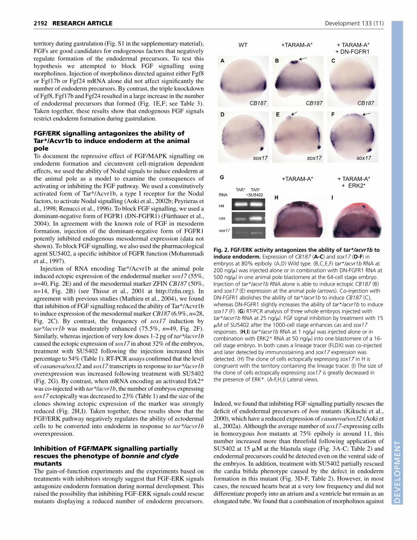
Injection of RNA encoding Tar*/Acvr1b at the animal poleinduced ectopic expression of the endodermal marker sox17 (55%,n=40, Fig. 2E) and of the mesodermal marker ZFIN CB187 (50%,n=14, Fig. 2B) (see Thisse et al., 2001 at http://zfin.org). Inagreement with previous studies (Mathieu et al., 2004), we foundthat inhibition of FGF signalling reduced the ability of Tar*/Acvr1bto induce expression of the mesodermal marker CB187 (6.9%, n=28,Fig. 2C). By contrast, the frequency of sox17 induction bytar*/acvr1b was moderately enhanced (75.5%, n=49, Fig. 2F).Similarly, whereas injection of very low doses 1-2 pg of tar*/acvr1bcaused the ectopic expression of sox17 in about 32% of the embryos,treatment with SU5402 following the injection increased thispercentage to 54% (Table 1). RT-PCR assays confirmed that the levelof casanova/sox32 and sox17 transcripts in response to tar*/acvr1boverexpression was increased following treatment with SU5402(Fig. 2G). By contrast, when mRNA encoding an activated Erk2*was co-injected with tar*/acvr1b, the number of embryos expressingsox17 ectopically was decreased to 23% (Table 1) and the size of theclones showing ectopic expression of the marker was stronglyreduced (Fig. 2H,I). Taken together, these results show that theFGF/ERK pathway negatively regulates the ability of ectodermalcells to be converted into endoderm in response to tar*/acvr1boverexpression.
Inhibition of FGF/MAPK signalling partiallyrescues the phenotype of bonnie and clydemutantsThe gain-of-function experiments and the experiments based ontreatments with inhibitors strongly suggest that FGF-ERK signalsantagonize endoderm formation during normal development. Thisraised the possibility that inhibiting FGF-ERK signals could rescuemutants displaying a reduced number of endoderm precursors.
Indeed, we found that inhibiting FGF signalling partially rescues thedeficit of endodermal precursors of bon mutants (Kikuchi et al.,2000), which have a reduced expression of casanova/sox32 (Aoki etal., 2002a). Although the average number of sox17-expressing cellsin homozygous bon mutants at 75% epiboly is around 11, thisnumber increased more than threefold following application ofSU5402 at 15 �M at the blastula stage (Fig. 3A-C; Table 2) andendodermal precursors could be detected even on the ventral side ofthe embryos. In addition, treatment with SU5402 partially rescuedthe cardia bifida phenotype caused by the defect in endodermformation in this mutant (Fig. 3D-F, Table 2). However, in mostcases, the rescued hearts beat at a very low frequency and did notdifferentiate properly into an atrium and a ventricle but remain as anelongated tube. We found that a combination of morpholinos against
RESEARCH ARTICLE Development 133 (11)
Fig. 2. FGF/ERK activity antagonizes the ability of tar*/acvr1b toinduce endoderm. Expression of CB187 (A-C) and sox17 (D-F) inembryos at 80% epiboly. (A,D) Wild type. (B,C,E,F) tar*/acvr1b RNA at200 ng/�l was injected alone or in combination with DN-FGFR1 RNA at500 ng/�l in one animal pole blastomere at the 64-cell stage embryo.Injection of tar*/acvr1b RNA alone is able to induce ectopic CB187 (B)and sox17 (E) expression at the animal pole (arrows). Co-injection withDN-FGFR1 abolishes the ability of tar*/acvr1b to induce CB187 (C),whereas DN-FGFR1 slightly increases the ability of tar*/acvr1b to inducesox17 (F). (G) RT-PCR analysis of three whole embryos injected withtar*/acvr1b RNA at 25 ng/�l. FGF signal inhibition by treatment with 15�M of SU5402 after the 1000-cell stage enhances cas and sox17responses. (H,I) tar*/acvr1b RNA at 1 ng/�l was injected alone or incombination with ERK2* RNA at 50 ng/�l into one blastomere of a 16-cell stage embryo. In both cases a lineage tracer (FLDX) was co-injectedand later detected by immunostaining and sox17 expression wasdetected. (H) The clone of cells ectopically expressing sox17 in H iscongruent with the territory containing the lineage tracer. (I) The size ofthe clone of cells ectopically expressing sox17 is greatly decreased inthe presence of ERK*. (A-F,H,I) Lateral views.

DEVELO
PMENT
Fgf8 and Fgf24 also rescued the cardia bifida of bon mutants, whilecausing only a modest increase in the number of endodermalprecursors present at 80% epiboly (Table 2). Thus, inhibition of FGFsignalling is able to compensate for the reduced activity of themolecular cascade leading to endoderm formation. We thenattempted to rescue the lack of endoderm of embryos injected withantisense morpholino oligonucleotides directed againstcasanova/sox32 (Dickmeis et al., 2001). Treatment with SU5402,did not rescue endoderm formation in these embryos (not shown).These results suggest that blocking the FGF-MAPK pathway cannotcompensate for the absence of Casanova but can attenuate theendodermal deficiency of mutants with a reduced expression ofcasanova/sox32.
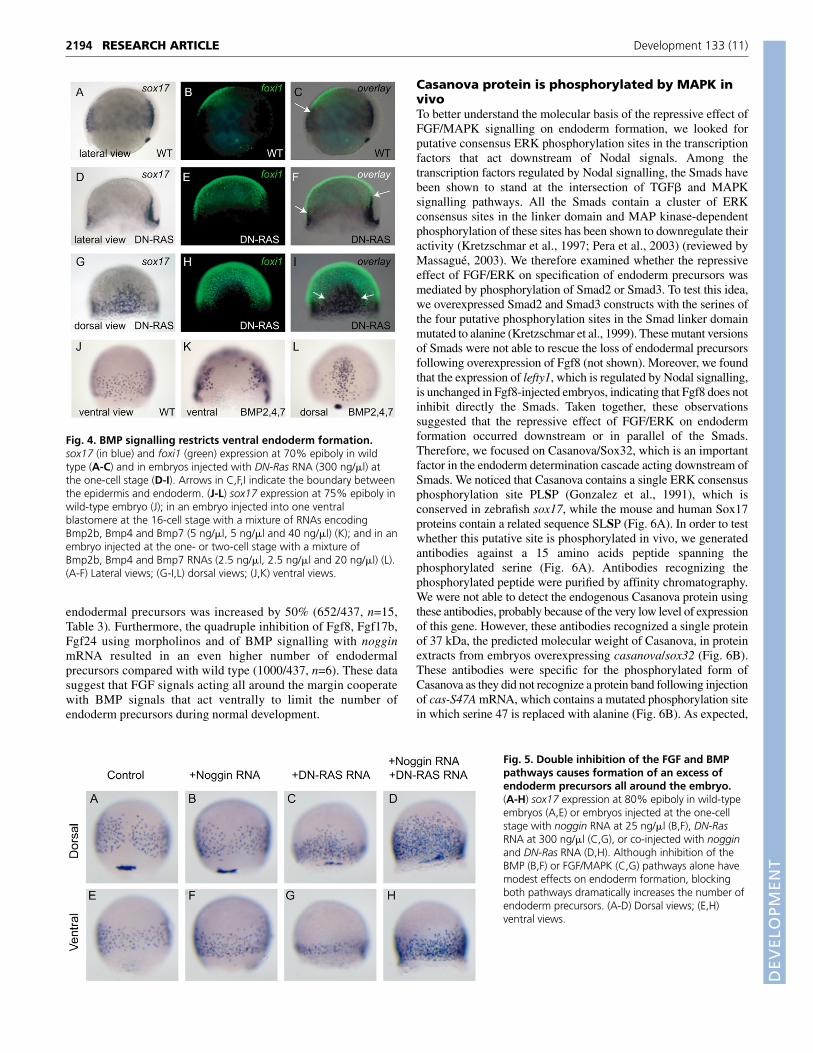
BMP and FGF signalling cooperate to restrictendoderm formationTaken together, our results so far indicate that FGF signalling isinvolved in regulating endoderm formation. However, in the courseof experiments in which we tried to interfere with MAPKsignalling at the level of Ras, we found that overexpression ofRNA encoding a dominant-negative form of Ras (DN-Ras)(Whitman and Melton, 1992) did not, result in an overall excess ofendoderm (354/477, n=88/93) (compare Fig. 5A,E with 5C,G).The number of endodermal cells was slightly increased dorsally(Fig. 5C), but in fact, fewer sox17-expressing cells were present inlateral and ventral regions (Fig. 5G). This observation suggeststhat, in addition to FGF signalling, the extent of endodermformation is also regulated by signals emanating from the ventralside of the embryo. As BMPs are known to be essential for thespecification of ventral cell fates (Kishimoto et al., 1997; Schmidet al., 2000), we investigated whether their activity modulatesendoderm formation. The gene encoding the transcription factorFoxi1 (Nissen et al., 2003) displays a BMP-dependent expressionin the prospective epidermis and can therefore be considered as amolecular readout of ongoing BMP signalling. In the ventralregion of wild-type embryos, the expression domains of foxi1 andsox17 are strikingly complementary (Fig. 4A-C). Moreover, whenFGF signalling is blocked using DN-Ras, DN-FGFR1 or SU5402,the expression of the bmp genes and their target foxi1 expandsdorsally (Fig. 4D-I) (Fürthauer et al., 2004). In these FGF-signalling inhibited embryos, sox17 expression becomes restrictedto the residual dorsal BMP-free zone that does not express foxi1,consistent with a potential role of BMPs in restricting endodermformation. In support of this hypothesis, co-injection of the threeBMP ligands at the ventral margin created a local gap in sox17expression (97.3%, n=38, Fig. 4K). Injection of the same
molecules at the one-cell stage produced a range of effects goingfrom a loss of endoderm on the ventral side (Fig. 4L, n=14/53) toan almost complete loss of endoderm precursors all around theembryo (not shown; n=39/53). We therefore investigated whetherBMP signalling is responsible for the inhibition of endodermformation in the ventrolateral region of FGF-depleted embryos. Inagreement with a previous study, we found that the number ofendodermal progenitors was not increased in zebrafish embryosmutant for bmp2b or bmp7 (Tiso et al., 2002) (data not shown).Similarly, inhibition of BMP signalling with noggin caused only aslight increase in the number of endodermal precursors that formed(Fig. 5B,F, n=84/84 and Table 3). However, we found thatsimultaneously inhibiting MAPK signalling and BMP signallingcaused a massive excess of endodermal precursors to form allaround the embryo (Fig. 5D-H, n=70/88, Table 3). By countingindividual sox17-positive cells in embryos at 75% epiboly co-injected with noggin and DN-Ras we found that the number of
2193RESEARCH ARTICLERegulation of endoderm formation
Table 1. FGF/ERK activity antagonizes the ability of Tar*Acvr1b to induce endoderm at the animal poleTAR* (1 ng/�l) TAR* (1 ng/�l) TAR* (200 ng/�l)
RNA injected at the 16-cell stage TAR* (1 ng/�l) + SU5402 + Erk2* TAR* (200 ng/�l) + DN-FGFR1
Embryos showing ectopic sox17 expression (%) 32 54 23 55 75.5Total number of embryos 152 85 83 40 49
Table 2. Inhibition of the FGF pathway increases the number of sox17-expressing cells and partially rescues heartmorphogenesis in bonnie and clyde mutants
bon–/– + Fgf8 bon–/– bon–/– + SU5402 and Fgf24 MOs
Average number of cells expressing sox17 per embryo 11 (n=12) 37 (n=13) Not determinedat about 80% epiboly
Average number of sox17-expressing cells at about 22 (n=38) Not determined 28 (n=25)90% epiboly
Percentage of embryos with cardia bifida at 30 hpf 19 (n=62) 9 (n=403) 2 (n=86)
Fig. 3. Inhibition of the FGF/MAPK pathway rescues thephenotype of bon mutants. (A-C) Dorsal views of embryos at 80%epiboly showing expression of sox17 in wild-type embryos (A), bonmutant embryos (B) and bon mutant embryos treated with SU5402 at15 �M after 1000-cell stage (C). (D-F) Partial rescue of heartmorphogenesis following SU5402 treatment into bon mutant embryosmonitored by expression of nkx2.5 at 30 hours. The failure of heartprimordia to migrate and fuse in the midline in bon mutants waspartially restored by inhibition of FGF pathway (F). The genotype of therescued embryos was verified by RFLP.
DEVELO
PMENT
2194
endodermal precursors was increased by 50% (652/437, n=15,Table 3). Furthermore, the quadruple inhibition of Fgf8, Fgf17b,Fgf24 using morpholinos and of BMP signalling with nogginmRNA resulted in an even higher number of endodermalprecursors compared with wild type (1000/437, n=6). These datasuggest that FGF signals acting all around the margin cooperatewith BMP signals that act ventrally to limit the number ofendoderm precursors during normal development.
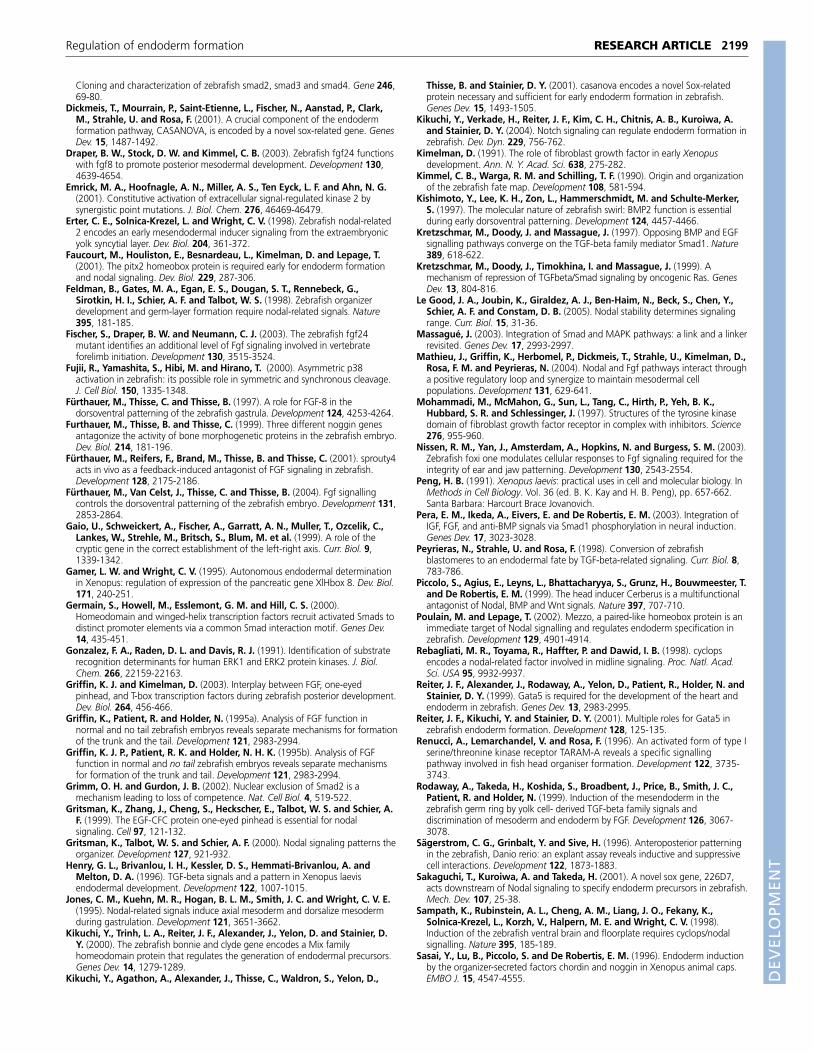
Casanova protein is phosphorylated by MAPK invivoTo better understand the molecular basis of the repressive effect ofFGF/MAPK signalling on endoderm formation, we looked forputative consensus ERK phosphorylation sites in the transcriptionfactors that act downstream of Nodal signals. Among thetranscription factors regulated by Nodal signalling, the Smads havebeen shown to stand at the intersection of TGF� and MAPKsignalling pathways. All the Smads contain a cluster of ERKconsensus sites in the linker domain and MAP kinase-dependentphosphorylation of these sites has been shown to downregulate theiractivity (Kretzschmar et al., 1997; Pera et al., 2003) (reviewed byMassagué, 2003). We therefore examined whether the repressiveeffect of FGF/ERK on specification of endoderm precursors wasmediated by phosphorylation of Smad2 or Smad3. To test this idea,we overexpressed Smad2 and Smad3 constructs with the serines ofthe four putative phosphorylation sites in the Smad linker domainmutated to alanine (Kretzschmar et al., 1999). These mutant versionsof Smads were not able to rescue the loss of endodermal precursorsfollowing overexpression of Fgf8 (not shown). Moreover, we foundthat the expression of lefty1, which is regulated by Nodal signalling,is unchanged in Fgf8-injected embryos, indicating that Fgf8 does notinhibit directly the Smads. Taken together, these observationssuggested that the repressive effect of FGF/ERK on endodermformation occurred downstream or in parallel of the Smads.Therefore, we focused on Casanova/Sox32, which is an importantfactor in the endoderm determination cascade acting downstream ofSmads. We noticed that Casanova contains a single ERK consensusphosphorylation site PLSP (Gonzalez et al., 1991), which isconserved in zebrafish sox17, while the mouse and human Sox17proteins contain a related sequence SLSP (Fig. 6A). In order to testwhether this putative site is phosphorylated in vivo, we generatedantibodies against a 15 amino acids peptide spanning thephosphorylated serine (Fig. 6A). Antibodies recognizing thephosphorylated peptide were purified by affinity chromatography.We were not able to detect the endogenous Casanova protein usingthese antibodies, probably because of the very low level of expressionof this gene. However, these antibodies recognized a single proteinof 37 kDa, the predicted molecular weight of Casanova, in proteinextracts from embryos overexpressing casanova/sox32 (Fig. 6B).These antibodies were specific for the phosphorylated form ofCasanova as they did not recognize a protein band following injectionof cas-S47A mRNA, which contains a mutated phosphorylation sitein which serine 47 is replaced with alanine (Fig. 6B). As expected,
RESEARCH ARTICLE Development 133 (11)
Fig. 4. BMP signalling restricts ventral endoderm formation.sox17 (in blue) and foxi1 (green) expression at 70% epiboly in wildtype (A-C) and in embryos injected with DN-Ras RNA (300 ng/�l) atthe one-cell stage (D-I). Arrows in C,F,I indicate the boundary betweenthe epidermis and endoderm. (J-L) sox17 expression at 75% epiboly inwild-type embryo (J); in an embryo injected into one ventralblastomere at the 16-cell stage with a mixture of RNAs encodingBmp2b, Bmp4 and Bmp7 (5 ng/�l, 5 ng/�l and 40 ng/�l) (K); and in anembryo injected at the one- or two-cell stage with a mixture ofBmp2b, Bmp4 and Bmp7 RNAs (2.5 ng/�l, 2.5 ng/�l and 20 ng/�l) (L).(A-F) Lateral views; (G-I,L) dorsal views; (J,K) ventral views.
Fig. 5. Double inhibition of the FGF and BMPpathways causes formation of an excess ofendoderm precursors all around the embryo.(A-H) sox17 expression at 80% epiboly in wild-typeembryos (A,E) or embryos injected at the one-cellstage with noggin RNA at 25 ng/�l (B,F), DN-RasRNA at 300 ng/�l (C,G), or co-injected with nogginand DN-Ras RNA (D,H). Although inhibition of theBMP (B,F) or FGF/MAPK (C,G) pathways alone havemodest effects on endoderm formation, blockingboth pathways dramatically increases the number ofendoderm precursors. (A-D) Dorsal views; (E,H)ventral views.
DEVELO
PMENT
the level of exogenous phosphorylated Casanova was markedlyincreased following co-injection with ERK2* (Fig. 6C). Similarly,the phosphorylated form of Casanova was no longer detected whenCasanova was co-expressed with zebrafish MKP3, which encodes azebrafish MAPK phosphatase (Fig. 6D). Treatment with SU5402resulted in a reduction of the level of phosphorylated Casanovaprotein in embryos injected with cas mRNA (Fig. 6E). By contrast,the MAPK P38 inhibitor SB205580 did not have any effect on thelevel of phosphorylated Casanova protein (Fig. 6F). These resultssuggest that Casanova protein is a target of ERK in vivo and that theFGF pathway is largely responsible for the phosphorylation ofCasanova protein.
Phosphorylation of Casanova/Sox32 attenuates itsactivityWe next examined whether phosphorylation of Casanova modifiesits activity. First, we compared the ability of low doses of cas-WT orcas-S47A to induce ectopic sox17 expression. When 20 pg of cas-WT RNA was injected, it caused ectopic expression of sox17 in 35%of the embryos (n=381, Fig. 7J, Table 4). When the same dose ofmutated cas-S47A was injected, the percentage of embryosdisplaying sox17 was slightly increased to 47% (n=214).Furthermore, in situ hybridisation signals were consistently strongerin the cas-S47A than in the cas-WT-injected embryos (Fig. 7A,B, seeFig. S3A-C in the supplementary material). These results suggestthat the presence of the ERK target site has a negative effect on theability of Casanova to induce sox17 expression.
We then tested the effect of activating or inhibiting FGF-ERK signalling on the ability of ectopic wild-type or mutatedCasanova to induce sox17 expression at the animal pole. Injectionof Fgf8 (Fig. 7C), a constitutively active Ras (Fig. 7E) or anactivated version of ERK (Fig. 7G; see Fig. S3D,E in thesupplementary material) strongly decreased the ability ofCasanova to induce sox17 (Table 4). Importantly, activating theMAPK pathway with these reagents had only modest effects onthe ability of the mutated form of Casanova to induce ectopicexpression of sox17 at the animal pole (Fig. 7D,F,H). This resultindicates that the ERK consensus phosphorylation site is requiredfor the attenuation of Casanova activity in response to FGFpathway activation. In agreement with this conclusion, inhibitionof the FGF-ERK pathway by treatment with SU5402, or by co-injection with zebrafish MKP3, potentiated the ability ofexogenous Casanova to induce sox17 (Table 4, Fig. 7J). Takentogether, these results suggest that FGF-ERK activity antagonisesendoderm formation by acting at the level of transcription factorsinduced by Nodal signalling through direct phosphorylation ofCasanova.
DISCUSSIONWe have investigated how the Nodal, FGF and BMP pathwaysinteract during the process of endoderm formation in thezebrafish embryo. We found a double repressive effect of the FGFand BMP pathways on induction of endodermal precursorsby Nodal signalling. When either of these pathways is
2195RESEARCH ARTICLERegulation of endoderm formation
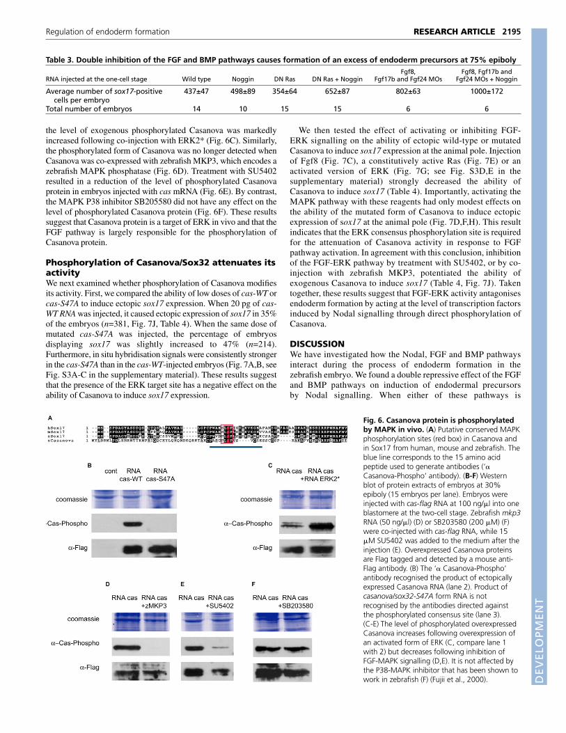
Table 3. Double inhibition of the FGF and BMP pathways causes formation of an excess of endoderm precursors at 75% epibolyFgf8, Fgf8, Fgf17b and
RNA injected at the one-cell stage Wild type Noggin DN Ras DN Ras + Noggin Fgf17b and Fgf24 MOs Fgf24 MOs + Noggin
Average number of sox17-positive 437±47 498±89 354±64 652±87 802±63 1000±172cells per embryo
Total number of embryos 14 10 15 15 6 6
Fig. 6. Casanova protein is phosphorylatedby MAPK in vivo. (A) Putative conserved MAPKphosphorylation sites (red box) in Casanova andin Sox17 from human, mouse and zebrafish. Theblue line corresponds to the 15 amino acidpeptide used to generate antibodies (‘�Casanova-Phospho’ antibody). (B-F) Westernblot of protein extracts of embryos at 30%epiboly (15 embryos per lane). Embryos wereinjected with cas-flag RNA at 100 ng/�l into oneblastomere at the two-cell stage. Zebrafish mkp3RNA (50 ng/�l) (D) or SB203580 (200 �M) (F)were co-injected with cas-flag RNA, while 15�M SU5402 was added to the medium after theinjection (E). Overexpressed Casanova proteinsare Flag tagged and detected by a mouse anti-Flag antibody. (B) The ‘� Casanova-Phospho’antibody recognised the product of ectopicallyexpressed Casanova RNA (lane 2). Product ofcasanova/sox32-S47A form RNA is notrecognised by the antibodies directed againstthe phosphorylated consensus site (lane 3).(C-E) The level of phosphorylated overexpressedCasanova increases following overexpression ofan activated form of ERK (C, compare lane 1with 2) but decreases following inhibition ofFGF-MAPK signalling (D,E). It is not affected bythe P38-MAPK inhibitor that has been shown towork in zebrafish (F) (Fujii et al., 2000).

DEVELO
PMENT
2196
overactivated, the number of endodermal precursors formed isseverely reduced. By contrast, when the FGF pathway is blocked,embryonic cells are more easily induced to form endoderm byactivation of Nodal signalling. Moreover, we showed thatinhibition of FGF signalling partially rescued the deficit ofendoderm of bonnie and clyde mutants, and that the simultaneousinhibition of both the FGF and BMP pathways results in an excessof endodermal precursors. Finally, we identified a molecularmechanism that explains the repressive action of FGF onendoderm formation. We show that FGF signals control thenumber of endodermal precursors formed via phosphorylation ofthe transcription factor Casanova.
FGF signalling and endoderm formationAlthough a large number of studies have implicated FGF signallingin the control of mesoderm formation and patterning, only a fewstudies, mainly in Xenopus, have addressed the role of FGFsignalling in formation of the endodermal germ layer. In Xenopus,numerous observations indicate a repressive role of FGF signallingon endoderm formation. Gamer and Wright (Gamer and Wright,1995) found that bFGF is a potent inhibitor of the expression of thepancreatic marker pdx1. Bouwmeester et al. (Bouwmeester et al.,1996) found that co-injection of XFD, a dominant-negative versionof Fgfr1, potentiates the ability of chordin to induce expression ofendodermal markers in animal caps assays. Finally, a recent studyshowed that overexpression of eFGF inhibits expression of mixerand that inhibition of FGF signalling in animal caps induces theectopic expression of mixer and endodermin (Cha et al., 2004).Taken together, these studies suggest that in Xenopus FGF signallingantagonizes the molecular pathways that control endodermspecification.
To investigate whether FGF and Nodal signalling interactduring endoderm formation in zebrafish, we ectopically expressedFGF signals. Overexpression of FGFs, as well as overexpressionof activated versions of the FGF receptor, activated forms of Rasand the MAP kinase ERK, all strongly interfered with formationof the endoderm. Reciprocally, inhibition of FGF signalling withantisense morpholinos oligonucleotides caused a dramaticincrease in the number of endodermal precursors. By contrast,interfering with the MAPK pathway using DN-Ras only increasedthe number of endoderm precursors present on the dorsal side aswell as the number of endoderm precursors induced by ectopicactivation of the Nodal pathway at the animal pole. Finally, weshowed that treatments with SU5402 or injection of morpholinosagainst Fgf8 and Fgf24 partially rescued the deficit of endodermprecursors and the associated cardia bifida phenotype of thebonnie and clyde mutant, which have a reduced expression ofcasanova/sox32. This finding is consistent with the results ofDavid and Rosa (David and Rosa, 2001), who showed that graftedwild-type endodermal cells rescue the cardia bifida defects ofcasanova/sox32 mutants. Therefore, attenuation of FGFsignalling partially compensates for the lack of a downstreameffector of Nodal probably by increasing the activity of Casanova.This provides a further indication that in the intact embryo,endoderm formation in response to Nodal signalling is negativelyregulated by FGF.
Studies with zebrafish mutants and experiments with inhibitorshad previously shown that FGF and Nodal signalling cooperateduring mesoderm formation (Griffin and Kimelman, 2003; Griffinet al., 1995b; Mathieu et al., 2004). Our results show that duringendoderm formation there is a strong antagonism between the FGFand the Nodal signalling pathways.
RESEARCH ARTICLE Development 133 (11)
Fig. 7. Casanova activity is negatively regulated by the FGF-ERKsignalling pathway. Expression of sox17 (blue) in embryos at 30%epiboly (A,B) or 50% epiboly (C-H). In C-E, FLDX used as lineagemarker was revealed by immunochemistry (red labelling). Embryosinjected at the 16-cell stage with RNA encoding Casanova-WT (A) orCasanova-S47A (B), each at 10 ng/�l (see also Fig. S2 in thesupplementary material). Mutation of the putative phosphorylation siteslightly increases Casanova activity. Co-injection with fgf8 RNA at 10ng/�l (C), CA-Ras RNA at 5 ng/�l (E) or ERK* RNA at 50 ng/�l (G)inhibits the ability of Casanova-WT to induce sox17 (see Fig. S3 in thesupplementary material). However Casanova-S47A is insensitive to Fgf8(D), CA-Ras (F) or ERK* (H) overexpression. (I) RT-PCR analysis fromthree embryos injected with cas RNA at 10 ng/�l alone or together withERK2* RNA at 50 ng/�l. (J) Differential activity of Casanova WT andCasanova S47A, based on the percentage of embryos ectopicallyexpressing sox17 (see Table 4). The activity of exogenous Casanova-WTwas compared with the activity of Casanova-S47A, treated or not withMAPK, FGF inhibitors or an activated form of Erk2.

DEVELO
PMENT
Endoderm formation and BMP signallingPrevious studies had suggested that, in Xenopus, endodermformation is regulated negatively by signals emanating from theventral side. In particular, Sasai et al. reported that overexpressionof noggin or chordin induces endoderm in animal caps and that thiseffect is strongly potentiated by inhibition of FGF signalling (Sasaiet al., 1996). However, Tiso et al. found that, in zebrafish, the numberof endoderm precursors is neither affected by overexpression ofBMP nor reduced in the swirl mutant which has a disrupted bmp2gene (Tiso et al., 2002). By contrast, we found that overexpressionof a cocktail of bmp2, bmp4 and bmp7 potently affects the numberof endoderm precursors and that overexpression of noggin increases,although only slightly, the number of endodermal precursors. Morestrikingly, we found that while inhibition of the BMP signallingpathway with noggin mRNA results in only a modest increase in thenumber of endodermal precursors, when noggin and DN-Ras are co-injected, they cause formation of a massive excess of endodermalprecursors. Finally, the largest number of endodermal precursorswas observed following simultaneous inhibition of BMP signallingwith noggin and of FGF signalling with morpholinos, furtherimplicating BMP signals in restricting endoderm formation. Takentogether, these experiments reveal a previously unknown role forBMP signals in repressing endoderm formation. Therefore, ourresults show that in addition to being positively regulated by signalsfrom the TGF� family, formation of the endoderm is negativelyregulated by a combination of FGF and BMP signals (Fig. 8).
Repression of endoderm formation by FGF or BMPmay not be mediated by phosphorylation orcompetition for Smad2/3Phosphorylation of Smad in their inter-linker region has beenimplicated in modulating the response to Tgf� signals (Massagué,2003; Pera et al., 2003). In Xenopus gastrulae, it has been shown thatMAP kinase-dependent phosphorylation of Smad2 inhibits itstranslocation into the nucleus. As a consequence, animal blastomeresloose their competence to respond to Tgf� and differentiate intoectoderm instead of mesoderm (Grimm and Gurdon, 2002).Consequences of Smad2 phosphorylation are not clear as differentstudies gave opposite results. One report has shown that in humancells, activation of Ras caused the exclusion of Smad2 and Smad3from the nucleus (Kretzschmar et al., 1999). These data contrast withan earlier report in which Erk2-dependant phosphorylation of Smad2correlated with an increase in the nuclear localisation and activity ofSmad2 (de Caestecker et al., 1998). Our finding that a Nodal targetgene such as lefty1 is expressed normally following overexpressionof Fgf8 suggests that, in zebrafish, the antagonism between endodermformation and FGF/ERK signalling does not rely on an inhibitoryphosphorylation of Smads. The inability of a Smad2 mutant than cannot be phosphorylated by ERK to rescue endoderm formation in FGFoverexpressing embryos reinforces this conclusion.
Although the inhibitory effect of FGF signalling on endodermformation can be explained by an inhibitory phosphorylation ofCasanova, the molecular mechanism responsible for the inhibitoryaction of BMP is not presently known. Both the Nodal signalling
pathway and the BMP signalling pathway share a commondownstream component, Smad4. It is therefore tempting to speculatethat the inhibitory action of BMP on endoderm formation may becaused by a competition between Smad1/Smad5, which are
2197RESEARCH ARTICLERegulation of endoderm formation
Table 4. FGF/ERK signalling negatively regulates the activity of Casanova at the animal poleCasanova-WT Casanova-WT Casanova-WT Casanova-S47A
RNA injected at the 16-cell stage Casanova-WT Casanova-S47A + SU5402 + Mkp3 + Erk2* + Erk2*
% Embryos showing ectopic sox17 30 46 49 64 3 54expression
Total number of embryos 381 214 182 205 167 118
Fig. 8. Schematic representation of the repressive effects of theBMP and FGF pathways on endoderm formation. (A) Wild-typecontext: (a-c) schematic representation of signalling activities of Nodal,FGF and BMP signalling. This representation is speculative and based onthe potential range of signals and the expression pattern and range ofantagonists (Schier and Talbot, 2005). (B) Formation of the endoderm isnegatively regulated by a combination of FGF and BMP signals. (C) DN-Ras overexpressing embryos: inhibition of MAPK signalling promotesendoderm formation on the dorsal side but causes a loss of endodermprecursors on the ventral side owing to increased expression of theBMPs. (D) Triple inhibition of FGF signalling with morpholinos orcombined inhibition of Ras and BMP signalling with DN-Ras + noggin:simultaneous removal of the BMP and FGF dependent inhibitionspromotes endoderm formation all around the embryo.
Aa Nodal signalling activity domain
Ab FGF signalling activity domain
Ac BMP signalling activity domain
B Wild-type conditions
C +DN-RAS
D MO FGFs or DN-RAS+Noggin
BMP inhibition by FGF
Endoderm induction by Nodal
Endoderm inhibition by BMP
Endoderm inhibition by FGF
Limit of endodermal domain
Endodermal domain
V D

DEVELO
PMENT
2198
activated by BMP signalling, and Smad2/Smad3, which areactivated by Nodal signalling for binding to Smad4 (Candia et al.,1997). However, we found that overexpression of Smad4 was notable to rescue the loss of sox17 expression in BMP-overexpressingembryos (not shown), therefore, a competition at the level of Smadproteins does not seem to be the cause of the inhibitory action ofBMPs on endoderm formation.
Casanova/Sox32 as a transcription factor at thecrossroad of the FGF and Nodal signallingpathwaysWe have uncovered a potential molecular mechanism whereby FGFsignalling acts to attenuate endoderm formation by identifyingCasanova, a Nodal target gene, as a major target of this repression.First, we found that overexpressed Casanova is phosphorylated andthat the level of phosphorylation is correlated with activation orinhibition of this pathway. Second, we showed that activation ofFGF/ERK attenuates the ability of Casanova to induce sox17 whenectopically expressed at the animal pole, while inhibition ofFGF/ERK makes Casanova a more potent inducer of endoderm.Finally, we showed that although the ability of wild type Casanovato induce endoderm at the animal pole is antagonized byoverexpression of FGF or activation of the ERK pathway, a mutatedphosphorylation-insensitive form of Casanova is no longer subjectto this inhibition. Therefore, these data support the hypothesis thatCasanova is at the crossroads of the Nodal and the MAPK signallingpathways, and that the antagonism between these pathways iscaused, at least in part, by phosphorylation of this transcriptionfactor. Nevertheless, interactions between these pathways couldoccur at other levels. Fgf8 or CA-Ras overexpression decrease theexpression of sox17 and of casanova/sox32. This suggests thatCasanova may not be the only target of this repressive mechanism.In addition to Casanova, MAPK consensus sites are present infactors acting at the level of, or downstream of, Casanova, such asEomesodermin, Gata5 and Sox17. Future experiments shouldaddress whether Eomesodermin, Gata5 and Sox17 are also involvedin the crosstalk between the MAPK and Nodal signalling pathways.Alternatively, FGF may additionally affect the expression ofcasanova/sox32 by compromising the activity of upstreamregulators in the cascade. For example, the homeobox proteins Bonand Mezzo act upstream of Casanova and are both able to bindpurified Smad2 (Germain et al., 2000) (M.P. and T.L., unpublished).Phosphorylation of Smad2/3 may affect their interaction with Mixerand Mezzo.
In zebrafish, the endodermal and mesodermal precursors originatefrom a common endomesodermal territory and both require Nodalsignalling. The molecular mechanisms that allow these two cell fatesto segregate during gastrulation are not well understood. It waspreviously known that factors promoting endoderm formation, suchas Casanova or Mezzo repress mesoderm formation whenoverexpressed (Aoki et al., 2002a; Kikuchi et al., 2001; Poulain andLepage, 2002). Our finding that endogenous FGF signals stronglyantagonize endoderm formation by downregulating Casanova showsthat a reciprocal negative interaction exists between factors thatpromote mesoderm formation and transcription factors required forendoderm formation. This mutual antagonism may therefore help tounderstand how mesodermal- or endodermal-specific generegulatory networks are established in the precursors of these twogerm layers, allowing different cell fates to be segregated.
In conclusion, we have shown that the FGF and BMP signalsantagonize endoderm formation by Nodal factors. Furthermore, wehave shown that Casanova is subject to an inhibitory
phosphorylation in response to FGF signalling and therefore standsat the intersection between the FGF and Nodal signalling pathways.This phosphorylation may represent a general mechanism wherebyFGF attenuates Nodal-induced endodermal transcription factors, andtherefore these results may help to understand how mesoderm andendoderm segregate from each other.
We thank F. Rosa for the tar*/acvr1b, nkx2.5 and casanova/sox32 plasmids,and for the casanova/sox32 morpholino oligonucleotides. We thank N. G.Ahn for sending us ERK2* plasmid, J. Massagué for Smad2 and Smad3constructs, and M. Whitman for CA-Ras and DN-Ras plasmids. We thank D.Meyer for the bonnie and clyde strain. We thank our colleagues at the MarineStation of Villefranche for help and support, and Lydia Besnardeau forexcellent technical help. We thank Clare Hudson and Hitoyoshi Yasuo forfruitful discussions and careful reading of the manuscript. We particularlythank Jean-Phillipe Chambon for helping us with HPLC chromatography andwestern blotting. We thank Laurent Gilleta for taking care of the zebrafish.This work was supported by the CNRS, by the Asssociation pour la Recherchecontre le Cancer, by the Ligue Nationale Contre le Cancer, by the Ministère dela Recherche and by the National Institute of Health. M.F. was supported byCNRS and HFSP.
Supplementary materialSupplementary material for this article is available athttp://dev.biologists.org/cgi/content/full/133/11/2189/DC1
ReferencesAgathon, A., Thisse, C. and Thisse, B. (2003). The molecular nature of the
zebrafish tail organizer. Nature 424, 448-452.Alexander, J. and Stainier, D. Y. (1999). A molecular pathway leading to
endoderm formation in zebrafish. Curr. Biol. 9, 1147-1157.Alexander, J., Rothenberg, M., Henry, G. L. and Stainier, D. Y. (1999).
casanova plays an early and essential role in endoderm formation in zebrafish.Dev. Biol. 215, 343-357.
Aoki, T. O., David, N. B., Minchiotti, G., Saint-Etienne, L., Dickmeis, T.,Persico, G. M., Strahle, U., Mourrain, P. and Rosa, F. M. (2002a). Molecularintegration of casanova in the Nodal signalling pathway controlling endodermformation. Development 129, 275-286.
Aoki, T. O., Mathieu, J., Saint-Etienne, L., Rebagliati, M. R., Peyrieras, N. andRosa, F. M. (2002b). Regulation of nodal signalling and mesendodermformation by TARAM-A, a TGFbeta-related type I receptor. Dev. Biol. 241, 273-288.
Bjornson, C. R., Griffin, K. J., Farr, G. H., 3rd, Terashima, A., Himeda, C.,Kikuchi, Y. and Kimelman, D. (2005). Eomesodermin is a localized maternaldeterminant required for endoderm induction in zebrafish. Dev. Cell 9, 523-533.
Bottcher, R. T. and Niehrs, C. (2005). Fibroblast growth factor signaling duringearly vertebrate development. Endocr. Rev. 26, 63-77.
Bouwmeester, T., Kim, S., Sasai, Y., Lu, B. and De Robertis, E. M. (1996).Cerberus is a head-inducing secreted factor expressed in the anterior endodermof Spemann’s organizer. Nature 382, 595-601.
Candia, A. F., Watabe, T., Hawley, S. H., Onichtchouk, D., Zhang, Y.,Derynck, R., Niehrs, C. and Cho, K. W. (1997). Cellular interpretation ofmultiple TGF-beta signals: intracellular antagonism between activin/BVg1 andBMP-2/4 signaling mediated by Smads. Development 124, 4467-4480.
Cha, S. W., Hwang, Y. S., Chae, J. P., Lee, S. Y., Lee, H. S., Daar, I., Park, M. J.and Kim, J. (2004). Inhibition of FGF signaling causes expansion of theendoderm in Xenopus. Biochem. Biophys. Res. Commun. 315, 100-106.
Chen, J. N. and Fishman, M. C. (1996). Zebrafish tinman homolog demarcatesthe heart field and initiates myocardial differentiation. Development 122, 3809-3816.
Chen, Y. and Schier, A. F. (2001). The zebrafish Nodal signal Squint functions as amorphogen. Nature 411, 607-610.
Chomczynski, P. and Sacchi, N. (1987). Single-step method of RNA isolation byacid guanidinium thiocyanate-phenol-chloroform extraction. Anal. Biochem.162, 156-159.
Clements, D., Friday, R. V. and Woodland, H. R. (1999). Mode of action of VegTin mesoderm and endoderm formation. Development 126, 4903-4911.
Cornell, R. A., Musci, T. J. and Kimelman, D. (1995). FGF is a prospectivecompetence factor for early activin-type signals in Xenopus mesoderminduction. Development 121, 2429-2437.
David, N. B. and Rosa, F. M. (2001). Cell autonomous commitment to anendodermal fate and behaviour by activation of Nodal signalling. Development128, 3937-3947.
de Caestecker, M. P., Parks, W. T., Frank, C. J., Castagnino, P., Bottaro, D. P.,Roberts, A. B. and Lechleider, R. J. (1998). Smad2 transduces common signalsfrom receptor serine-threonine and tyrosine kinases. Genes Dev. 12, 1587-1592.
Dick, A., Mayr, T., Bauer, H., Meier, A. and Hammerschmidt, M. (2000).
RESEARCH ARTICLE Development 133 (11)
DEVELO
PMENT
Cloning and characterization of zebrafish smad2, smad3 and smad4. Gene 246,69-80.
Dickmeis, T., Mourrain, P., Saint-Etienne, L., Fischer, N., Aanstad, P., Clark,M., Strahle, U. and Rosa, F. (2001). A crucial component of the endodermformation pathway, CASANOVA, is encoded by a novel sox-related gene. GenesDev. 15, 1487-1492.
Draper, B. W., Stock, D. W. and Kimmel, C. B. (2003). Zebrafish fgf24 functionswith fgf8 to promote posterior mesodermal development. Development 130,4639-4654.
Emrick, M. A., Hoofnagle, A. N., Miller, A. S., Ten Eyck, L. F. and Ahn, N. G.(2001). Constitutive activation of extracellular signal-regulated kinase 2 bysynergistic point mutations. J. Biol. Chem. 276, 46469-46479.
Erter, C. E., Solnica-Krezel, L. and Wright, C. V. (1998). Zebrafish nodal-related2 encodes an early mesendodermal inducer signaling from the extraembryonicyolk syncytial layer. Dev. Biol. 204, 361-372.
Faucourt, M., Houliston, E., Besnardeau, L., Kimelman, D. and Lepage, T.(2001). The pitx2 homeobox protein is required early for endoderm formationand nodal signaling. Dev. Biol. 229, 287-306.
Feldman, B., Gates, M. A., Egan, E. S., Dougan, S. T., Rennebeck, G.,Sirotkin, H. I., Schier, A. F. and Talbot, W. S. (1998). Zebrafish organizerdevelopment and germ-layer formation require nodal-related signals. Nature395, 181-185.
Fischer, S., Draper, B. W. and Neumann, C. J. (2003). The zebrafish fgf24mutant identifies an additional level of Fgf signaling involved in vertebrateforelimb initiation. Development 130, 3515-3524.
Fujii, R., Yamashita, S., Hibi, M. and Hirano, T. (2000). Asymmetric p38activation in zebrafish: its possible role in symmetric and synchronous cleavage.J. Cell Biol. 150, 1335-1348.
Fürthauer, M., Thisse, C. and Thisse, B. (1997). A role for FGF-8 in thedorsoventral patterning of the zebrafish gastrula. Development 124, 4253-4264.
Furthauer, M., Thisse, B. and Thisse, C. (1999). Three different noggin genesantagonize the activity of bone morphogenetic proteins in the zebrafish embryo.Dev. Biol. 214, 181-196.
Fürthauer, M., Reifers, F., Brand, M., Thisse, B. and Thisse, C. (2001). sprouty4acts in vivo as a feedback-induced antagonist of FGF signaling in zebrafish.Development 128, 2175-2186.
Fürthauer, M., Van Celst, J., Thisse, C. and Thisse, B. (2004). Fgf signallingcontrols the dorsoventral patterning of the zebrafish embryo. Development 131,2853-2864.
Gaio, U., Schweickert, A., Fischer, A., Garratt, A. N., Muller, T., Ozcelik, C.,Lankes, W., Strehle, M., Britsch, S., Blum, M. et al. (1999). A role of thecryptic gene in the correct establishment of the left-right axis. Curr. Biol. 9,1339-1342.
Gamer, L. W. and Wright, C. V. (1995). Autonomous endodermal determinationin Xenopus: regulation of expression of the pancreatic gene XlHbox 8. Dev. Biol.171, 240-251.
Germain, S., Howell, M., Esslemont, G. M. and Hill, C. S. (2000).Homeodomain and winged-helix transcription factors recruit activated Smads todistinct promoter elements via a common Smad interaction motif. Genes Dev.14, 435-451.
Gonzalez, F. A., Raden, D. L. and Davis, R. J. (1991). Identification of substraterecognition determinants for human ERK1 and ERK2 protein kinases. J. Biol.Chem. 266, 22159-22163.
Griffin, K. J. and Kimelman, D. (2003). Interplay between FGF, one-eyedpinhead, and T-box transcription factors during zebrafish posterior development.Dev. Biol. 264, 456-466.
Griffin, K., Patient, R. and Holder, N. (1995a). Analysis of FGF function innormal and no tail zebrafish embryos reveals separate mechanisms for formationof the trunk and the tail. Development 121, 2983-2994.
Griffin, K. J. P., Patient, R. K. and Holder, N. H. K. (1995b). Analysis of FGFfunction in normal and no tail zebrafish embryos reveals separate mechanismsfor formation of the trunk and tail. Development 121, 2983-2994.
Grimm, O. H. and Gurdon, J. B. (2002). Nuclear exclusion of Smad2 is amechanism leading to loss of competence. Nat. Cell Biol. 4, 519-522.
Gritsman, K., Zhang, J., Cheng, S., Heckscher, E., Talbot, W. S. and Schier, A.F. (1999). The EGF-CFC protein one-eyed pinhead is essential for nodalsignaling. Cell 97, 121-132.
Gritsman, K., Talbot, W. S. and Schier, A. F. (2000). Nodal signaling patterns theorganizer. Development 127, 921-932.
Henry, G. L., Brivanlou, I. H., Kessler, D. S., Hemmati-Brivanlou, A. andMelton, D. A. (1996). TGF-beta signals and a pattern in Xenopus laevisendodermal development. Development 122, 1007-1015.
Jones, C. M., Kuehn, M. R., Hogan, B. L. M., Smith, J. C. and Wright, C. V. E.(1995). Nodal-related signals induce axial mesoderm and dorsalize mesodermduring gastrulation. Development 121, 3651-3662.
Kikuchi, Y., Trinh, L. A., Reiter, J. F., Alexander, J., Yelon, D. and Stainier, D.Y. (2000). The zebrafish bonnie and clyde gene encodes a Mix familyhomeodomain protein that regulates the generation of endodermal precursors.Genes Dev. 14, 1279-1289.
Kikuchi, Y., Agathon, A., Alexander, J., Thisse, C., Waldron, S., Yelon, D.,
Thisse, B. and Stainier, D. Y. (2001). casanova encodes a novel Sox-relatedprotein necessary and sufficient for early endoderm formation in zebrafish.Genes Dev. 15, 1493-1505.
Kikuchi, Y., Verkade, H., Reiter, J. F., Kim, C. H., Chitnis, A. B., Kuroiwa, A.and Stainier, D. Y. (2004). Notch signaling can regulate endoderm formation inzebrafish. Dev. Dyn. 229, 756-762.
Kimelman, D. (1991). The role of fibroblast growth factor in early Xenopusdevelopment. Ann. N. Y. Acad. Sci. 638, 275-282.
Kimmel, C. B., Warga, R. M. and Schilling, T. F. (1990). Origin and organizationof the zebrafish fate map. Development 108, 581-594.
Kishimoto, Y., Lee, K. H., Zon, L., Hammerschmidt, M. and Schulte-Merker,S. (1997). The molecular nature of zebrafish swirl: BMP2 function is essentialduring early dorsoventral patterning. Development 124, 4457-4466.
Kretzschmar, M., Doody, J. and Massague, J. (1997). Opposing BMP and EGFsignalling pathways converge on the TGF-beta family mediator Smad1. Nature389, 618-622.
Kretzschmar, M., Doody, J., Timokhina, I. and Massague, J. (1999). Amechanism of repression of TGFbeta/Smad signaling by oncogenic Ras. GenesDev. 13, 804-816.
Le Good, J. A., Joubin, K., Giraldez, A. J., Ben-Haim, N., Beck, S., Chen, Y.,Schier, A. F. and Constam, D. B. (2005). Nodal stability determines signalingrange. Curr. Biol. 15, 31-36.
Massagué, J. (2003). Integration of Smad and MAPK pathways: a link and a linkerrevisited. Genes Dev. 17, 2993-2997.
Mathieu, J., Griffin, K., Herbomel, P., Dickmeis, T., Strahle, U., Kimelman, D.,Rosa, F. M. and Peyrieras, N. (2004). Nodal and Fgf pathways interact througha positive regulatory loop and synergize to maintain mesodermal cellpopulations. Development 131, 629-641.
Mohammadi, M., McMahon, G., Sun, L., Tang, C., Hirth, P., Yeh, B. K.,Hubbard, S. R. and Schlessinger, J. (1997). Structures of the tyrosine kinasedomain of fibroblast growth factor receptor in complex with inhibitors. Science276, 955-960.
Nissen, R. M., Yan, J., Amsterdam, A., Hopkins, N. and Burgess, S. M. (2003).Zebrafish foxi one modulates cellular responses to Fgf signaling required for theintegrity of ear and jaw patterning. Development 130, 2543-2554.
Peng, H. B. (1991). Xenopus laevis: practical uses in cell and molecular biology. InMethods in Cell Biology. Vol. 36 (ed. B. K. Kay and H. B. Peng), pp. 657-662.Santa Barbara: Harcourt Brace Jovanovich.
Pera, E. M., Ikeda, A., Eivers, E. and De Robertis, E. M. (2003). Integration ofIGF, FGF, and anti-BMP signals via Smad1 phosphorylation in neural induction.Genes Dev. 17, 3023-3028.
Peyrieras, N., Strahle, U. and Rosa, F. (1998). Conversion of zebrafishblastomeres to an endodermal fate by TGF-beta-related signaling. Curr. Biol. 8,783-786.
Piccolo, S., Agius, E., Leyns, L., Bhattacharyya, S., Grunz, H., Bouwmeester, T.and De Robertis, E. M. (1999). The head inducer Cerberus is a multifunctionalantagonist of Nodal, BMP and Wnt signals. Nature 397, 707-710.
Poulain, M. and Lepage, T. (2002). Mezzo, a paired-like homeobox protein is animmediate target of Nodal signalling and regulates endoderm specification inzebrafish. Development 129, 4901-4914.
Rebagliati, M. R., Toyama, R., Haffter, P. and Dawid, I. B. (1998). cyclopsencodes a nodal-related factor involved in midline signaling. Proc. Natl. Acad.Sci. USA 95, 9932-9937.
Reiter, J. F., Alexander, J., Rodaway, A., Yelon, D., Patient, R., Holder, N. andStainier, D. Y. (1999). Gata5 is required for the development of the heart andendoderm in zebrafish. Genes Dev. 13, 2983-2995.
Reiter, J. F., Kikuchi, Y. and Stainier, D. Y. (2001). Multiple roles for Gata5 inzebrafish endoderm formation. Development 128, 125-135.
Renucci, A., Lemarchandel, V. and Rosa, F. (1996). An activated form of type Iserine/threonine kinase receptor TARAM-A reveals a specific signallingpathway involved in fish head organiser formation. Development 122, 3735-3743.
Rodaway, A., Takeda, H., Koshida, S., Broadbent, J., Price, B., Smith, J. C.,Patient, R. and Holder, N. (1999). Induction of the mesendoderm in thezebrafish germ ring by yolk cell- derived TGF-beta family signals anddiscrimination of mesoderm and endoderm by FGF. Development 126, 3067-3078.
Sägerstrom, C. G., Grinbalt, Y. and Sive, H. (1996). Anteroposterior patterningin the zebrafish, Danio rerio: an explant assay reveals inductive and suppressivecell interactions. Development 122, 1873-1883.
Sakaguchi, T., Kuroiwa, A. and Takeda, H. (2001). A novel sox gene, 226D7,acts downstream of Nodal signaling to specify endoderm precursors in zebrafish.Mech. Dev. 107, 25-38.
Sampath, K., Rubinstein, A. L., Cheng, A. M., Liang, J. O., Fekany, K.,Solnica-Krezel, L., Korzh, V., Halpern, M. E. and Wright, C. V. (1998).Induction of the zebrafish ventral brain and floorplate requires cyclops/nodalsignalling. Nature 395, 185-189.
Sasai, Y., Lu, B., Piccolo, S. and De Robertis, E. M. (1996). Endoderm inductionby the organizer-secreted factors chordin and noggin in Xenopus animal caps.EMBO J. 15, 4547-4555.
2199RESEARCH ARTICLERegulation of endoderm formation

DEVELO
PMENT
2200
Schier, A. F. (2003). Nodal signaling in vertebrate development. Annu. Rev. CellDev. Biol. 19, 589-621.
Schier, A. F. and Talbot, W. S. (2005). Molecular genetics of axis formation inZebrafish. Annu. Rev. Genet. 39, 561-613.
Schier, A. F., Neuhauss, S. C., Helde, K. A., Talbot, W. S. and Driever, W.(1997). The one-eyed pinhead gene functions in mesoderm and endodermformation in zebrafish and interacts with no tail. Development 124, 327-342.
Schmid, B., Furthauer, M., Connors, S. A., Trout, J., Thisse, B., Thisse, C. andMullins, M. C. (2000). Equivalent genetic roles for bmp7/snailhouse andbmp2b/swirl in dorsoventral pattern formation. Development 127, 957-967.
Schulte-Merker, S. and Smith, J. C. (1995). Mesoderm formation in response toBrachyury requires FGF signalling. Curr. Biol. 5, 62-67.
Shinya, M., Koshida, S., Sawada, A., Kuroiwa, A. and Takeda, H. (2001). Fgfsignalling through MAPK cascade is required for development of the subpallialtelencephalon in zebrafish embryos. Development 128, 4153-4164.
Strahle, U., Jesuthasan, S., Blader, P., Garcia-Villalba, P., Hatta, K. andIngham, P. W. (1997). one-eyed pinhead is required for development of theventral midline of the zebrafish (Danio rerio) neural tube. Genes Funct. 1, 131-148.
Sun, B. I., Bush, S. M., Collins-Racie, L. A., LaVallie, E. R., DiBlasio-Smith, E.A., Wolfman, N. M., McCoy, J. M. and Sive, H. L. (1999). derriere: a TGF-betafamily member required for posterior development in Xenopus. Development126, 1467-1482.
Thisse, B., Wright, C. V. and Thisse, C. (2000). Activin- and Nodal-related factorscontrol antero-posterior patterning of the zebrafish embryo. Nature 403, 425-428.
Thisse, B., Heyer, V., Lux, A., Alunni, V., Degrave, A., Seiliez, I., Kirchner, J.,
Parkhill, J. P. and Thisse, C. (2004). Spatial and temporal expression of thezebrafish genome by large-scale in situ hybridization screening. Methods CellBiol. 77, 505-519.
Thisse, C. and Thisse, B. (1999). Antivin, a novel and divergent member of theTGFbeta superfamily, negatively regulates mesoderm induction. Development126, 229-240.
Tiso, N., Filippi, A., Pauls, S., Bortolussi, M. and Argenton, F. (2002). BMPsignalling regulates anteroposterior endoderm patterning in zebrafish. Mech.Dev. 118, 29-37.
Trinh, L. A., Meyer, D. and Stainier, D. Y. (2003). The Mix family homeodomaingene bonnie and clyde functions with other components of the Nodal signalingpathway to regulate neural patterning in zebrafish. Development 130, 4989-4998.
Warga, R. M. and Nusslein-Volhard, C. (1999). Origin and development of thezebrafish endoderm. Development 126, 827-838.
Westerfield, M. (1994). The Zebrafish Book. Eugene OR: Institute ofNeurosciences, University of Oregon.
Whitman, M. and Melton, D. A. (1992). Involvement of p21ras in Xenopusmesoderm induction. Nature 357, 252-254.
Yasuo, H. and Lemaire, P. (1999). A two-step model for the fate determination ofpresumptive endodermal blastomeres in Xenopus embryos. Curr. Biol. 9, 869-879.
Zhang, J., Talbot, W. S. and Schier, A. F. (1998). Positional cloning identifieszebrafish one-eyed pinhead as a permissive EGF-related ligand required duringgastrulation. Cell 92, 241-251.
Zhao, J., Cao, Y., Zhao, C., Postlethwait, J. and Meng, A. (2003). An SP1-liketranscription factor Spr2 acts downstream of Fgf signaling to mediate mesoderminduction. EMBO J. 22, 6078-6088.
RESEARCH ARTICLE Development 133 (11)