Yujun Guo Kent State University August 13. 2003 PRESENTATION A Binarization Approach for CT-MR...
21
Yujun Guo Kent State University August 13. 2003 PRESENTATION A Binarization Approach for CT- MR Registration Using Normalized Mutual Information
-
date post
21-Dec-2015 -
Category
Documents
-
view
231 -
download
0
Transcript of Yujun Guo Kent State University August 13. 2003 PRESENTATION A Binarization Approach for CT-MR...

Yujun Guo
Kent State UniversityAugust 13. 2003
PRESENTATION
A Binarization Approach for CT-MR Registration
Using Normalized Mutual Information

Why and WhatWhy medical image registration? Medical image registration has been applied to the diagnosis of
breast cancer, cardiac studies, and a variety of neurological disorders including brain tumors.
Why mutual information? Maximization of mutual information of voxel intensities has
been proved to be one of the most popular registration methods for three-dimensional multimodal medical image registration.
What’s the goal of this paper? To develop a registration software. To implement a new approach for multimodality medical image registration. Improve accuracy with no loss of speed. The result could be used in clinical cases.

Modalities Involved in Registration
Computed Tomography (CT) gives anatomical information
Magnetic Resonance (MR) imaging gives anatomic information
Positron Emission Tomography (PET) gives functional information
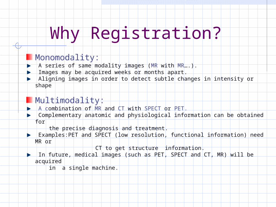
Why Registration?Monomodality: A series of same modality images (MR with MR….). Images may be acquired weeks or months apart. Aligning images in order to detect subtle changes in intensity or shape
Multimodality: A combination of MR and CT with SPECT or PET. Complementary anatomic and physiological information can be obtained for
the precise diagnosis and treatment. Examples:PET and SPECT (low resolution, functional information) need MR or
CT to get structure information. In future, medical images (such as PET, SPECT and CT, MR) will be acquired
in a single machine.

Registration MethodsLandmark-based: Based on identification of corresponding point landmarks or fiducial marks in two images. Accurate, but inconvenient, and cannot applied retrospectively. Labor-intensive and their accuracy depends on the accurate indication of corresponding
landmarks in all modalities.
Surface-based: Corresponding surfaces are delineated and a transformation computed that minimizes
some measure of distance between the two surfaces. Segmentation needed, and surfaces are not easily identified in functional modalities(PET).
Voxel-based: Use the intensities in the two images alone without any requirement to segment or
delineate corresponding structure. It includes sum of squared intensity difference (SSD), correlation coefficient (CC),
variance of intensity ratio (VIR), and mutual information (MI). Mutual information is widely used in multimodality registration.

Mutual Information Criterion
Mutual information is applied to measure the statistic dependence between the image intensities of corresponding voxels in both images, which is assumed to be maximal if the images are geometrically aligned.
a b BA
ABAB bPaP
baPbaPBAI
)()(
),(log),(),(
)|()(
)|()(
),()()(
ABHBH
BAHAH
BAHBHAH

Normalized Mutual Information
Extension of Mutual Information
Maes et. al.:
Studholme et. Al.:
Compensate for the sensitivity of MI to changes in image overlap
)()(
)(2),(
),(),(),(
BHAH
AMIBANMI
BAMIBAHBANMI
),(
)()(),(
BAH
BHAHBANMI

Flow chart based on NMI

Multi-resolution
Why Multi-resolutionMethods for detecting optimality can not guarantee that a global optimal value will be found.Time to evaluate the registration criterion is proportional to the number of voxels.
The result at coarser level is used as the starting point for the finer level.Currently multi-resolution approaches:
Sub-sampling Averaging

A Binarization Approach
Two stages:
1. Region Growing:Segmentation into Background and Foreground.
2. Two levels Registration:Binarized 2-bin images are input to the lower level.
Down-sampled binarized images as the input to the first level.
Result of the first level as the initial estimate for the second level.
The second level performs the registration of full images, using Maximization of Normalized Mutual Information.

A Binarization ApproachRegion Growing Implementation
Finding Starting Points:Easy to select a point as seed for background. Similarity Criteria:Threshold T can be extracted from the histogram
Typical histogram for CT image (left) & MR image (right)
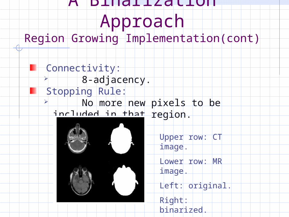
A Binarization ApproachRegion Growing Implementation(cont)
Connectivity: 8-adjacency.
Stopping Rule: No more new pixels to be included in
that region.
Upper row: CT image.
Lower row: MR image.
Left: original.
Right: binarized.

Implementation Specifics
Interpolation:Nearest neighbor interpolation --- First StageTrilinear partial volume interpolation --- Second Stage
Optimization:Downhill simplex method.
Bin SizesTwo bins --- first stage256 bins --- second stage
Platform:using C++, and IDLPC---2.4G Pentium 4, 512MB SDRAM

Implementation Specifics
Data Set (9 patients)
Data set from The Retrospective Registration Evaluation Project database by Vanderbilt University.Images for 9 patients76 image pairs were available to be registered.
41 CT-MR pairs of 7 out of 9 patients 35 PET-MR pairs of 7 out of 9 patients
Implementation Specifics
Data Set (cont.)
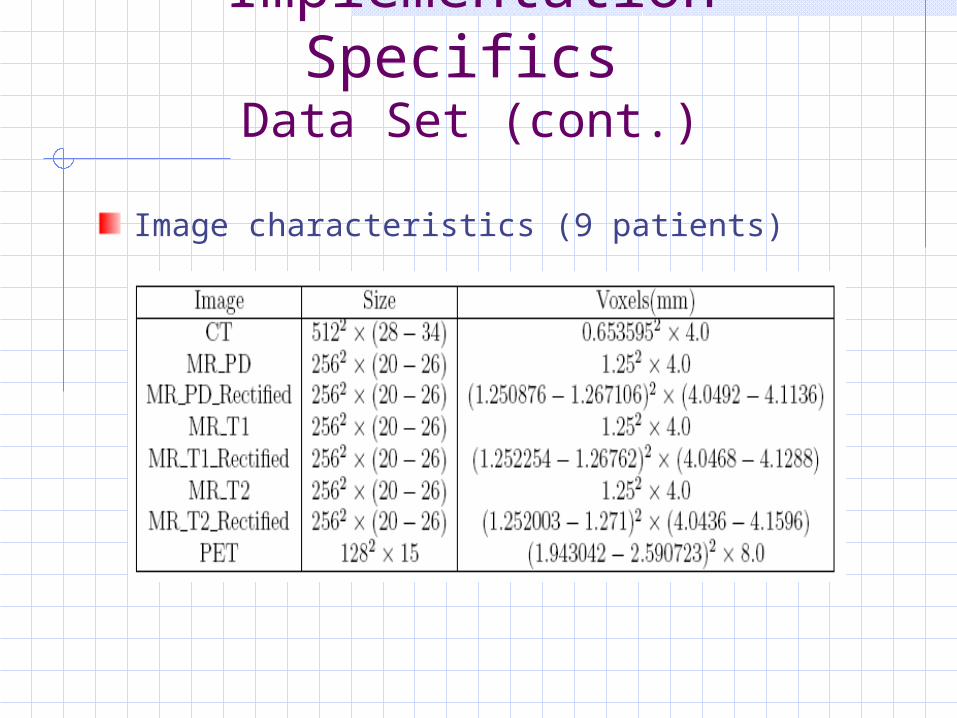
Image characteristics (9 patients)

Registration TimeRegistration time
Time required to perform registration for 41 CT-MR pairs .
Validation
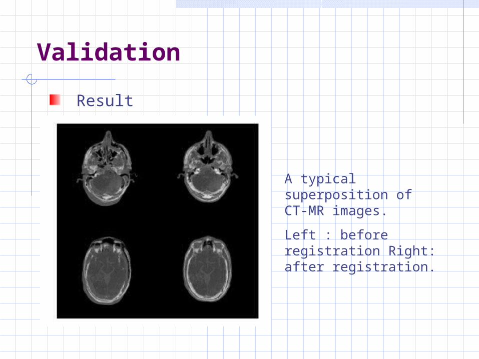
Result
A typical superposition of CT-MR images.
Left : before registration Right: after registration.
Validation
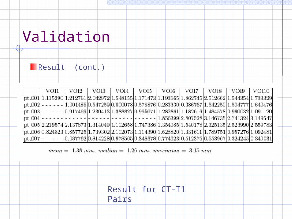
Result (cont.)
Result for CT-T1 Pairs

Validation--AccuracyMedian and maximum error between the prospective gold-standard and several retrospective registration techniques. Ours is labeled as LO2.
Median Error
Maximum Error
Conclusion
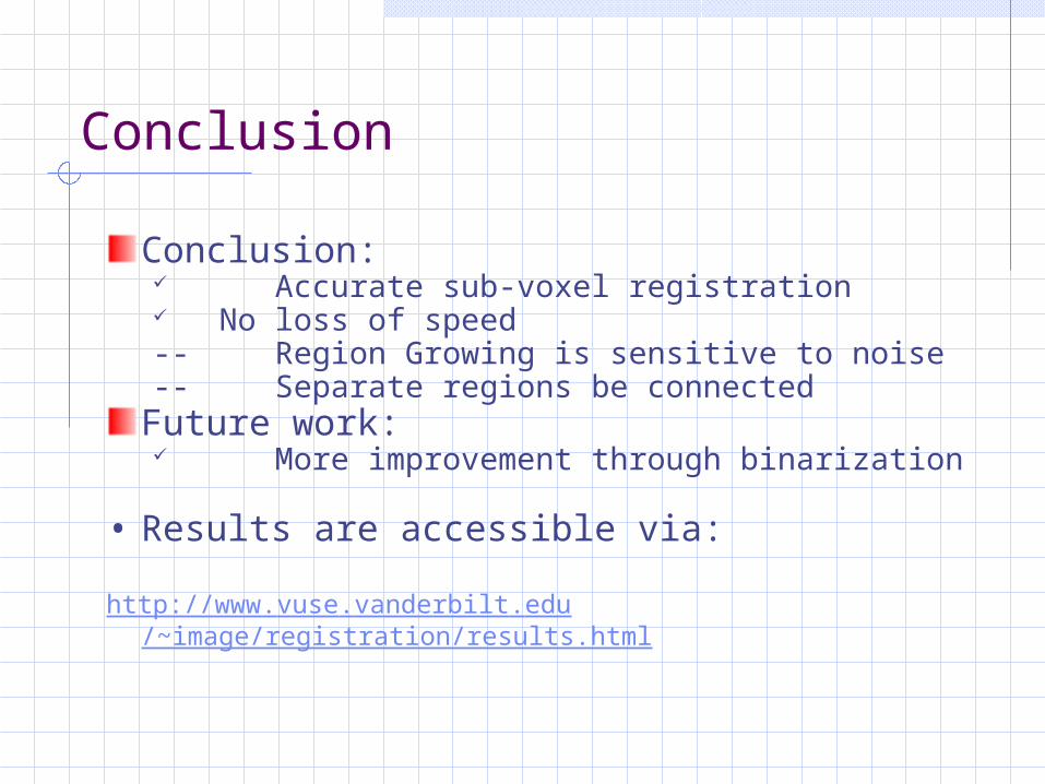
Conclusion: Accurate sub-voxel registration No loss of speed-- Region Growing is sensitive to noise-- Separate regions be connected
Future work: More improvement through binarization
• Results are accessible via:
http://www.vuse.vanderbilt.edu/~image/registration/results.html

Thank you


![arXiv:1705.03260v1 [cs.AI] 9 May 2017 · 2018. 10. 14. · Vegetables2 Normalized Log Size Vehicles1 Normalized Log Size Vehicles2 Normalized Log Size Weapons1 Normalized Log Size](https://static.fdocuments.us/doc/165x107/5ff2638300ded74c7a39596f/arxiv170503260v1-csai-9-may-2017-2018-10-14-vegetables2-normalized-log.jpg)

