Xray beam restrictors 1436711280968
28
XRAY BEAM RESTRICTORS -KRITHIKA
-
Upload
sarin-vidya -
Category
Health & Medicine
-
view
263 -
download
12
Transcript of Xray beam restrictors 1436711280968
XRAY BEAM RESTRICTORS
-KRITHIKA

THREE CATEGORIES
• APERTURE DIAPHRAGMS
• CONES AND CYLINDERS
• COLLIMATORS

APERTURE DIAPHRAGMS
• Simplest type
• Made up of a sheet of lead with a hole at its centre.
ADVANTAGE:
- Simplicity ( lead being soft, the aperture can be easily altered to any desirable size and shape)
DISADVANTAGE:
- large penumbra at the periphery of the xray field

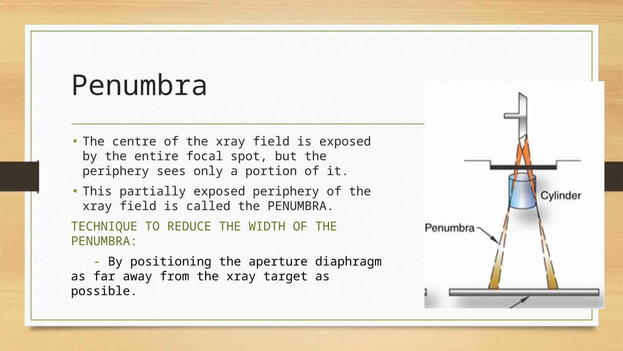
Penumbra
• The centre of the xray field is exposed by the entire focal spot, but the periphery sees only a portion of it.
• This partially exposed periphery of the xray field is called the PENUMBRA.
TECHNIQUE TO REDUCE THE WIDTH OF THE PENUMBRA:
- By positioning the aperture diaphragm as far away from the xray target as possible.

CONES AND CYLINDERS
• CONES:
- flare shaped.
• CYLINDERS:
- beam restriction at the far end of the barrel, so less penumbra.
- may be equipped with extensions to increase their length for better beam restriction.

COLLIMATORS
• Best all round xray beam restrictor device.
• ADVANTAGES:
- Provides an infinite variety of rectangular xray fields.
- light beam shows the centre and the exact configuration of the xray field.

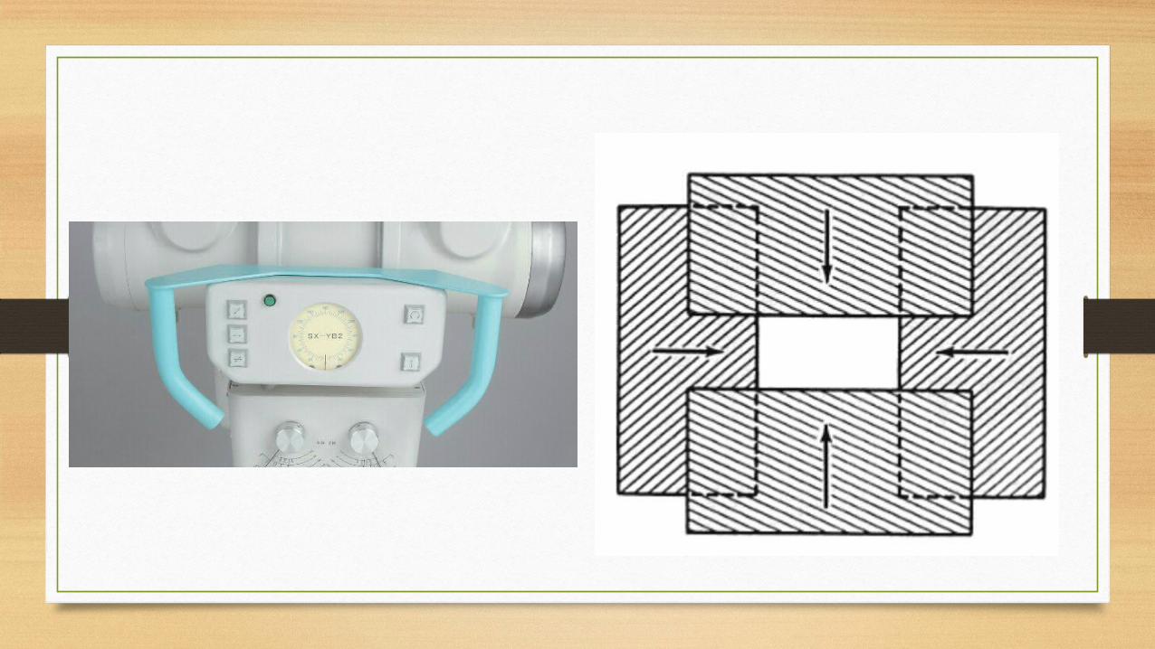
STRUCTURE OF A COLLIMATOR
• Two sets of shutters to control the dimension.
- each shutter contains 4 or more lead plates, which move in independent pairs.
- when the shutters are closed, they meet at the centre.
• Light beam from a light bulb in the collimator.

• The light beam is deflected by a mirror mounted in the path of the xray beam at an angle of 45 degree.
• The target of the xray tube and the light bulb should be the exactly same distance from the centre of the mirror.


• A collimator can also identify the center of the xray field which is accomplished by painting a cross line on a thin sheet of plexiglass mounted on the end of the collimator.
• A backup system is available in case if the light burns out with the help of a calibrated scale which will determine the xray field size for various target-film distances.

AUTOMATIC COLLIMATORS
• POSITIVE BEAM LIGHTING DEVICES
• Shutters are motor driven.
• When a cassette is loaded into the film holder, sensors will identify the alignment of the cassette.
• Then the information is relayed to the collimator motors, which will position the shutters to exactly match the size of the film being used.
• These devices must be accurate to within 2% of the source to image distance (SID).

• A perfectly aligned collimator will leave an unexposed border on all sides of the developed film

TESTING XRAY BEAM AND LIGHT BEAM ALIGNMENT
• Alignment has to be checked periodically.
MATERIALS REQUIRED:
• Four L shaped wires.
• 14 to 17 inch xray film
• Lead letter R

Procedure
• Place the film on the top of the x-ray table
• Open the shutter to a convenient size ( 10x10in)
• Carefully position the L shaped wires at the corner of the light field
• Place the R in the right lower corner.
• Then make an xray exposure. ( 40in.,3.3mAs, 40kVp) to mark the position of the x-ray field on the film.

• Without touching the film or wires, enlarge the field size to 12x12 in. and expose the film for the second time.
The dark centre shows the position of the xray beam and the wires indicate the position of the light beam.

HOW TO ADJUST A MISALIGNED MIRROR?
• Return the processed xray film to its original position. The R in the right lower corner assists in orienting the film properly.
• Position the light beam to the images of the wires, as we dis earlier for the film exposure and adjust the mirror in the collimator until the light beam coincides exactly with the xray field (the dark area on the film)

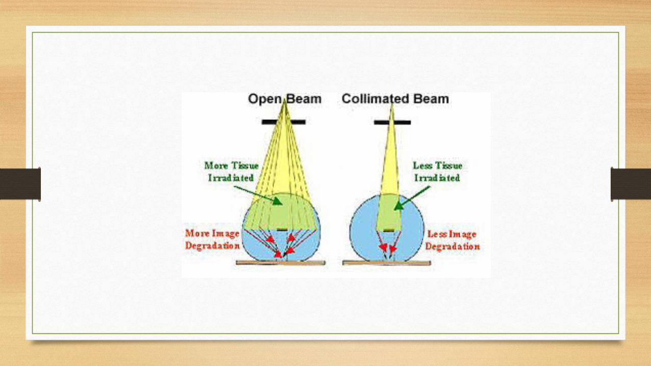
FUNCTION OF XRAY BEAM RESTRICTORS
• PATIENT PROTECTION
• TO DECREASE SCATTER RADIATION

PATIENT PROTECTION
• Mechanism by which collimation protects the patient:
-smaller the xray field, smaller the volume of the patient that is irradiated.
For example, if a 20x20 cm field is collimated to a 10x10cm, the area of the patient that is irradiated decreases from 400 to 100cm2. (since area is a square function, one half of decrease in x-ray beam diameter effects a 4 fold decrease in patient exposure)

IDEAL SHAPE OF THE FIELD
• This depends upon the part of the patient to be examined and not the shape of either the film or collimator.
• Round field are ideal for certain parts like gallbladder and paranasal sinuses.
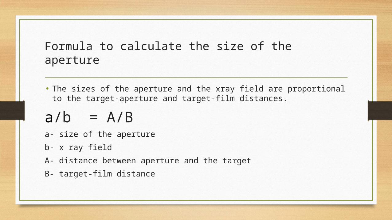
Formula to calculate the size of the aperture
• The sizes of the aperture and the xray field are proportional to the target-aperture and target-film distances.
a/b = A/Ba- size of the aperture
b- x ray field
A- distance between aperture and the target
B- target-film distance

DECREASED SCATTER RADIATION
• Quantity of scatter radiation reaching the film is directly proportional to the field size.
• So collimators by decreasing the field size, the reduce the amount of scatter radiation.
• Xray field of or more than 30x30cm size= maximum scatter radiation
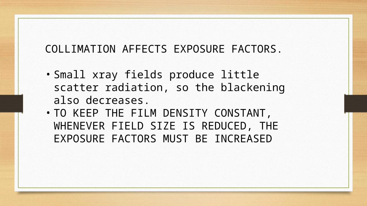
COLLIMATION AFFECTS EXPOSURE FACTORS.
• Small xray fields produce little scatter radiation, so the blackening also decreases.
• TO KEEP THE FILM DENSITY CONSTANT, WHENEVER FIELD SIZE IS REDUCED, THE EXPOSURE FACTORS MUST BE INCREASED

SUMMARY
• THREE TYPES OF XRAY BEAM RESTRICTORS
• BASIC FUNCTION IS TO REGULATE THE SIZE AND SHAPE OF THE XRAY BEAM.
• 2 ADVANRAGES OF CLOSELY COLLIMATED BEAMS:
- SMALLER AREA OF THE PATIENT EXPOSED,\.
- LESS SCATTER RADIATION

Reference
• Christensen’s physics of diagnostic radiology

THANK YOU