X-ray Operating Procedures and Safety Manualresearch.uthscsa.edu/safety/doc/UHS X-ray...
50
X-ray Operating Procedures and Safety Manual November 2015 Revision 10
Transcript of X-ray Operating Procedures and Safety Manualresearch.uthscsa.edu/safety/doc/UHS X-ray...

X-ray Operating
Procedures and Safety Manual
November 2015 Revision 10

ii
TABLE OF CONTENTS
PAGE # 1.0 Introduction 5
1.1 Purpose 5 1.2 Emergency Telephone Numbers 6 1.3 Responsibilities 6
1.3.1 Radiation Safety Officer 6 1.3.2 Individual Faculty, Staff and Students 7
1.3.3 Radiation Safety Committee 1.3.4 Radiation Protocol Committee
8 8
1.3.5 Radiation Safety Division 8 1.4 Corrective Actions 8 1.5 Imminently Dangerous to Life & Health 9 2.0 Approval and Authorization 10
2.1 Registration and Certification 10 2.2 Regulations 10 2.3 Procedure for X-ray Producing Devices Authorization 10 2.4 Procedure for Research Protocols involving X-ray Devices 11 2.5 X-ray Unit Purchase/Authorization 11
2.6 Termination of Authorized User/Operator 11 3.0 Personnel Monitoring and Dosimetry 12
3.1 Exposure Limits of Personnel 12 3.2 Radiation Exposure Assessment & Dosimeter Application 12 3.3 How to Wear a Whole Body Dosimeter 12 3.4 How to Wear an Extremity Monitor 13 3.5 How to Wear a Fetal Monitor 14 3.6 Protective Devices 14 3.7 Do’s and Don’ts of Dosimetry 15 3.8 Declared Pregnant Worker 15
3.8.1 Application 15 3.8.2 Concerns 16
3.9 Personnel Dosimetry Records 16 4.0 Radiation Protection 17
4.1 Introduction 17 4.2 Basic Principles 17
4.2.1 Time 17 4.2.2 Distance 17 4.2.3 Shielding 18
4.3 Radiation Exposure Sources 18 4.4 Biological Effects of Radiation 18 4.5 ALARA 19 4.6 Regulatory Concerns 19
4.6.1Restricted Areas 19 4.6.2 Postings 19 4.6.3 Exposure of the Patient 20 4.6.4 Exposure of the Individual (Staff) 21 4.6.5 Holding of Patient or Film/Image Receptor 21 4.6.6 Exposure of Individuals Other than the Patient 21 4.6.7 Reports to Workers 22
5.0 Medical Physics 23

iii
5.1 Surveys 23 5.2 Physicist Qualifications 23 5.3 Patient Dose Calculations 24
6.0 General Operating Requirements 7.0 Computed Tomography
25 27
7.1 Radiation Safety Precautions 27 7.2 Quality Assurance Policy 27 7.3 CT Protocol Guidelines 29 7.4 Radiation Protocol Committee 29 7.5 Training 30
8.0 Dental Units 31 8.1 Radiation Safety Precautions 31 8.2 Training 31
9.0 Fluoroscopy Units 32 9.1 Radiation Safety Precautions 32 9.2 Training 33
10.0 Fluoroscopy Guided Interventional Procedures 34 10.1 Fluoroscopy Guided Interventional Procedures Definition 34 10.2 Radiation Safety Precautions 34 10.3 FGI Protocol Guidelines 35 10.4 Radiation Protocol Committee 36 10.5 Training 37
11.0 Mammography 38 11.1 Radiation Safety Precautions 38 11.2 Mammography Physician/Operator Requirements 38 11.3 In-House Policies 39 11.4 Training 39
12.0 Radiographic Units 40 12.1 Radiation Safety Precautions 40 12.2 Training 40
13.0 Bone Densitometers 41 13.1 Radiation Safety Precautions 41 13.2 Training 41
14.0 Reportable Events and TDSHS Responses 42 14.1 Sentinel Event 42 14.2 In-House Radiation Protocol Committee 42 14.3 TDSHS Responses
14.3.1 NOV’s 14.3.2 Complaints
42 42 42
15.0 Record Keeping 43 15.1 Record Keeping for Departments 43
15.1.1 Radiation Monitoring & Exposure Records 43 15.1.2 X-ray Producing Devices Repairs or Maintenance 43 15.1.3 Medical Physicist Patient Dose Evaluations 43 15.1.4 Film Retention 43 15.1.5 CT Films 43 15.1.6 Mammography Films 44
15.2 Record Keeping Requirements for Radiation Safety Office 44 15.2.1 Protective Device Evaluations 44 15.2.2 X-ray Producing Devices Inventories 44 15.2.3 X-ray Producing Devices Installation Reports 44 15.2.4 Medical Physics Surveys 44 15.2.5 Radiation Monitoring & Exposure Records 44

iv
15.2.6 Radiation Protocol Committee Minutes & Reviews 44 15.2.7 Radiation Safety Training and Experience 44 15.2.8 TDSHS Regulatory Items 44 15.2.9 TDSHS Inspections/Corrective Actions 44
Appendices A: Personnel Dosimetry Application
A-1 Declared Pregnant Worker Application A-2 Pregnant Worker Handout A-4

5
1.0 INTRODUCTION 1.1 Purpose The objective of the University Health System Radiation Safety Program is to assist in all
levels of management in fulfilling the UHS commitment to furnish a place of employment and learning that is as free as possible from recognized radiation hazards that are likely to cause harm to UHS personnel or our community. It is vital that faculty, staff, and students have enough information available to aid them in the safe conduct of their daily work activities relating to radiation.
To that end, the Texas Department of State Health Services has granted a registration and certifications to University Health System authorizing the use of radiation producing devices. An essential component of that authorization is the X-ray Operating Procedures and Safety Manual. The purpose of the UHS X-ray Operating Procedures and Safety Manual is to assist in both personnel and management in complying with the objectives of the Texas Department of State Health Services, Bureau of Radiation Control regulations and the institutional health and safety policies. The fundamental objective of the medical and dental use of radiation is to obtain optimum diagnostic information or therapeutic effect with minimum exposure of the patient, the personnel concerned, and the general public. The Radiation Safety Division addresses many of the items in this Handbook in periodic Radiation Safety training sessions. This Handbook is not intended to be an exhaustive or fully comprehensive reference, rather a guide for authorized users and other qualified individuals. Further advice concerning hazards associated with specific devices and the development of new or unfamiliar activities should be obtained through consultation with the Radiation Safety Committee, the Radiation Safety Officer, or the Radiation Safety Division. All operators of x-ray producing devices must be familiar with the requirements set forth in this Handbook and must conduct their operations in accordance with them. __signature on file____________ Jennifer Cerecero, MS
Radiation Safety Officer The University of Texas Health Science Center at San Antonio

6
1.2 Emergency Telephone Numbers
Contact
8am-5pm
After Hours
Radiation Safety Office Dental Building Room 1.343T Radiation Safety Officer (Jennifer Cerecero)
(210)567-2955
(210)567-2800
University Hospital Police
(210)358-2465
In case of incidents involving unusual radiation exposure or patient safety involving radiation producing devices, all personnel are required to notify the Radiation Safety Office immediately. After 5:00 pm, University (UTHSCSA) or UHS Police will assist in contacting Radiation Safety personnel.
1.3 Responsibilities
1.3.1 Radiation Safety Officer (RSO) The Radiation Safety Officer is responsible for:
1. Reviewing all proposals for use of x-ray producing devices and recommending action to the Radiation Safety Committee.
2. Inspecting facilities and equipment through radiation safety evaluations and monitoring all facilities in which radiation-producing equipment resides.
3. Ensuring all personnel have been adequately training and comply with the requirements of the Texas Department of State Health Services for operating x-ray producing devices.
4. Prescribing special conditions and requirements as may be necessary for safe and proper use of all x-ray producing devices for UHS research, education, and patient care.
5. Acting as consultant in the design of all new facilities using x-ray producing devices for the purpose of providing protection against radiation exposure.
6. Preparing and disseminating information on radiation safety for faculty, staff, and students as necessary.
7. Authorizing, receiving, storing, and processing incoming x-ray producing devices. 8. Providing personnel monitoring services, including the reviewing and recording of
commercially processed dosimeter reports. 9. Reviewing and performing lead apron/protective device evaluations and removing
any devices that are not in compliance. 10. Reviewing completed medical physics testing and recommending action to the
various departments. 11. Preparing registration and certification amendments and technical renewals as well
as acting as the primary contact for correspondence with state radiation control authorities on a timely basis.
12. Investigating incidents involving radiation exposures including overexposures, incidents, theft, loss of devices, and accidents.
7
13. Notifying the Texas Department of State Health Services of all reportable incidents including overexposures, theft, loss of x-ray producing devices and submitting reports as required.
14. Reacting to any situation that is imminently dangerous to life and health and/or not in compliance with regulatory standards or University policy. Corrective actions shall include the authority to stop or shut down use of radiation producing devices until the situation is deemed safe by the Radiation Safety Officer.
15. Serving on the Radiation Protocol Committee as a consultant for Radiation Safety. 16. Ensuring that radiation doses are maintained as low as reasonably achievable
(ALARA). 17. Maintaining records required by the Texas Department of State Health Services for
inspection purposes.
1.3.2 Individual Faculty, Staff and Students All personnel at UHS are expected to follow these responsibilities:
1. Wearing personnel dosimeters when appropriate based on the radiation risk. 2. Utilizing all appropriate radiation protection measures including:
a. Wearing all appropriate personal protective equipment including leaded gloves, lead aprons, or leaded glasses where appropriate.
b. Using additional protective barriers and other shields when possible. c. Using mechanical devices whenever their aid will reduce exposure. d. Follow the technique chart provided for each unit. e. Complying with requests from the Radiation Safety Office regarding personnel
dosimetry and operating procedures. f. Verifying appropriate training is completed prior to operating x-ray producing
devices. g. Providing signature verification of annual review of operating and safety
procedures. 3. Notifying the Radiation Safety Office of any new radiation producing devices and
repairs to existing equipment. 4. Contacting the Radiation Safety Officer for shielding calculations for rooms proposed
for a different type of x-ray modality or for a new installation of a x-ray producing device.
5. Notifying the Radiation Safety Office of any stolen or lost x-ray producing devices. 6. Complying with proper procedure when terminating employment or the use of x-ray
producing devices.
1.3.3 Radiation Safety Committee The Radiation Safety Committee will be appointed by the President of the University of Texas Health Science Center at San Antonio, with membership including faculty representatives from Basic Sciences, Dental School, Medical School, Nuclear Medicine, Radiation Oncology; a radiologist, a veterinarian; a person from the administration office of UTHSCSA and from Bexar County Hospital District, doing business as University Health System; and a nursing representative. This committee reports to the UHS Quality & Risk Management and the UHS Environment of Care Committee. Hereafter, in the Handbook, the Radiation Safety Committee will be referred to as the RSC. The RSC is responsible for:
1. Approving policies and practices regarding the registration and certification of
radiation producing devices at University Health System and the implementation of the approved policies is delegated to the Radiation Safety Officer.
2. Serving in an advisory and consultative capacity to the Environment of Care department.

8
3. Reviewing the human use protocols involving radiation producing devices for research. The overall feasibility of the protocol will be determined by the Institutional Review Board.
4. Reviewing findings of the Radiation Protocol Committee (RPC). 5. Reviewing periodic audits performed by the RSO. 6. Acting as an avenue of appeal in cases of disputes or exceptions. 7. Maintaining minutes of the meeting delineating the date, members present, members
absent, review actions including committee response, appended conditions, recommended actions, Audits, RPP, ALARA reviews, and RSO reports.
1.3.4 Radiation Protocol Committee The Radiation Protocol Committee (RPC) is an
University Health System Committee responsible for reviewing and approving Fluoroscopy Guided Interventional (FGI) Protocols and Computed Tomography (CT) Protocols. The committee is responsible for the following items: 1. Restricting the use of fluoroscopic systems for interventional purposes to
radiologists, radiation oncologists, or physicians that have completed the radiation safety awareness training required by the Texas Department of State Health Services.
2. Determining a method to be used to monitor radiation output or exposure for CT and FGI.
3. Determining and reviewing a recommended reference level for CT and FGI. 4. Determining and reviewing actions to be taken for cases where the reference level is
exceeded including patient follow-up for CT and FGI. 5. Reviewing all established protocols at intervals not to exceed 14 months. 6. Interacting with the Radiation Safety Committee and Environment of Care Committee
as needed. 7. Keeping meeting minutes including the dates of meetings, names of individuals in
attendance, discussion, and any action completed during the meeting for the required time period of the Texas Department of State Health Services.
8. Keeping protocol revisions or changes for the required time period of the Texas Department of State Health Services.
1.3.5 Radiation Safety Division The Radiation Safety Division, under the direction of the
Radiation Safety Officer is responsible for:
1. Conducting safety evaluations of facilities and equipment through performing radiation surveys and monitoring all facilities in which radiation-producing equipment resides. Surveys include radiation exposure values and record checks.
2. Authorizing orders, receiving, storing, and disposal of radiation producing devices, and maintaining records on all of the preceding transactions.
3. Performing annual lead apron/protective device evaluations. 4. Performing annual inventory checks on radiation producing devices. 5. Coordinating medical physics testing of radiation producing devices. 6. Distribution and monitoring of personnel monitoring devices. 7. Reacting to any situation that is imminently dangerous to life and health and/or not in
compliance with regulatory standards or University policy. Corrective actions shall include the authority to stop or shut down use of radiation producing devices until the situation is deemed safe by the Radiation Safety Officer.

9
1.4 Corrective Action Items of non-compliance or deficiency noted during an evaluation, an inspection, or a walk through will generate corrective actions depending upon the severity of the deficiency noted. The following action will be taken:
1. Serious deficiency: Any uncorrected deficiency deemed to be serious in the
opinion of the Safety Specialist will be evaluated by the RSO. The RSO will establish a corrective action plan, which may include an on-site re-evaluation within a specified time period or additional training. a. Failure by the department or supervisor to correct a serious deficiency within the
time frame specified will result in an Escalated Deficiency Notification follow-up.
2. Other deficiencies: Other deficiencies observed will be followed up by an e-mail (preferred) or written notification to the respective department by the evaluating Safety Specialist. The evaluating Safety Specialist will retain documentation of this notification in the appropriate investigator file.
Extenuating or mitigating circumstances will be considered by the Radiation Safety Committee and the Environment of Care Committee.
1.5 Imminently Dangerous to Life & Health (IDLH) If a Safety Specialist notes any condition where
there is risk of imminent danger to life, health, or facilities, this condition will be brought to the immediate attention of the RSO or appropriate Safety Manager(s) and the Environmental Health & Safety Officer (Director of Environmental Health & Safety). Corrective action may include immediate shut down of all operations as required by the Texas Department of State Health Services.

10
2.0 APPROVAL AND AUTHORIZATION
2.1 Registration and Certifications University Health System has been issued a registration and certifications to possess radiation producing devices by the Texas Department of State Health Services. The registration, R26368, currently covers the use of radiation producing devices for healing arts at the following sites:
1. University Hospital Main, Site 000 2. Robert B. Green Campus, Site 001 3. University Family Health Center Southwest, Site 002 4. University Family Health Center Southeast, Site 003 5. University Center for Community Health, Site 004 6. Bexar County Jail, Site 006 7. Correctional Health Care Services, 600 Mission Road, Site 007 8. Correctional Health Care Services, 3621 Farm Road, Site 008 9. University Health Center North, Site 009 10. University Health Center Northwest, Site 010 11. University Health Center Diagnostic Pavilion, Site 011 12. University Health Surgical Center – Medical Center, Site 012 The certifications currently cover the use of mammography units at the following sites:
1. M00275 – University Hospital Main 2. M00856 – Robert B. Green Campus 3. M00857 - Cancer Therapy and Research Center Stereotactic Unit only 4. M00858 – Cancer Therapy and Research Center
University Health System is required to keep copies of the registration, certifications, and all regulations that apply to the specific sites at each specific site [25 TAC §289.203(b)].
2.2 Regulations All radiation producing machines are regulated by state and federal laws (e.g. the
Texas Administrative Code (TAC) and the Mammography Quality Standards Act administered by the Food and Drug Administration). University Health System will comply with the required regulations. This handbook establishes procedures to comply with the regulations enforced by the Texas Department of State Health Services (TDSHS) Bureau of Radiation Control. [25 TAC §289.227(i)(2)]. The specific TDSHS regulations that must be followed by University Health System are as follows: Notices, Instructions, and Reports to Workers (TAC 289.203), Fees for Registration (TAC 289.204), Hearing and Enforcement Procedures (TAC 289.205), Registration of Radiation Machine Use and Services (TAC 289.226), Use of Radiation Machines in the Health Arts (TAC 289.227), Certification of Mammography Systems and Mammography Machines Used for Interventional Breast Radiography (TAC 289.230), General Provisions and Standards for Protection Against Machine-Produced Radiation (TAC 289.231), and Radiation Control Regulations for Dental Radiation Machines (TAC 289.232).
2.3 Procedure for X-ray Producing Devices Authorization In order to be authorized to operate a x-
ray producing device, the individual must meet the appropriate operator requirements. Each operator shall meet the appropriate credentialing requirements of rules issued pursuant to Medical Radiologic Technologist Act Texas Civil Statutes Article 4512m, See [§289.226(t)] [For information about credentialing, contact the MRT Program at (512) 834-6617.] Mammography physicians and technologists must meet the requirements outlined in Chapter 11 of this handbook. Students are defined as individuals enrolled in a radiologic technology program which meets the requirements of the Texas Department of State Health Services, Medical Radiologic Technologists Board, (25 TAC 143.5).

11
Students will NOT work in a radiographic exposure room or mobile unit without a staff technologist present. Students will NOT establish any techniques. This is the responsibility of the staff technologists. Students may work the controls, setting the factors established and posted by the staff technologists.
Individuals who operate radiation machines shall be instructed and able to demonstrate
competence with the facility's operating and safety procedures. 2.4 Procedure for Research Protocols Involving X-ray Devices The RSC must review all human
use research protocols that involve the use of radiation producing devices for research purposes and not standard of care. The RSC will review the protocol including the radiation worksheet and determine if the radiation exposure, training of individuals, and consent language is appropriate for the research study. All RSC reviews will be sent to the Institutional Review Board for final approval.
2.5 X-ray Unit Purchase/Authorization Contact the Radiation Safety Office when planning to
purchase and install a new unit/radiation producing device. All units must be registered within 30 days of installation except for mammography [TAC 289.226(f)]. Mammography units must follow a specific process for certification that is outlined in Chapter 11. Shielding requirements should be determined prior to the installation of a new unit.
Relocating a unit, major repairs or replacement of tube head requires notification of the Radiation Safety Officer. Disposal, transfer or sale of an x-ray unit is to be reported to the Radiation Safety Officer so the unit may be deleted from the registration list or certification (for mammography). For receipts, transfers, and disposals of interventional breast radiography machines, the records must include the date of receipt, transfer or disposal; the name and signature of the individual making the record; and the manufacturer’s model name and serial number on the control panel. The name of the individual or company receiving working units that are transferred or sold is required when notifying the Texas Department of State Health Services of the deletion. [§289.226(n)].
Notify the Radiation Safety Officer of the installation of a unit. Obtain the pink copy of the FDA form 2579 “Report of Assembly of a Diagnostic X-ray System”. A copy of the report must be sent to the Radiation Safety Officer and a copy is to be kept at the location of the unit. These reports should include the date of installation, manufacturer’s model and serial number of the control panel, name and signature of the person making the record.
2.6 Termination of Authorized User/Operator Employment termination includes separation from the
University Health System or the employee terminates operations involving radiation producing devices. Upon termination the designated department personnel must complete the following steps:
1. Notify the Radiation Safety Office as soon as possible of the termination. 2. Return all personnel dosimeters.

12
3.0 PERSONNEL MONITORING AND DOSIMETRY 3.1 Exposure Limits of Personnel
The maximum permissible radiation dose limits are found in 25TAC §289.202(f) and may be summarized:
Table 3.1
Regulatory Dose Limits Maximum Annual Individual Dose (mrem/year)
Whole body; head and trunk; active blood forming organs; or gonads 5,000
Hands and forearms; feet and ankles; skin of whole body 50,000
Lens of the eye 15,000 Minors 500
Declared Pregnant Worker 500 mrem / 9 months General Public 100
3.2 Radiation Exposure Assessment & Dosimeter Application Personnel are monitored with
commercial dosimeters. Persons working in low exposure areas are furnished with bimonthly or quarterly dosimeters. Monthly dosimeters are assigned to personnel working in higher exposure risk positions (i.e. X-ray technicians, Radiology residents, etc).
Dosimeters will be issued to personnel who enter a high radiation area and who enter a restricted area (Restricted areas will only be authorized by joint approval of the Radiation Safety Committee and the Radiation Safety Officer.)
In accordance with 25TAC §289.202(f), dosimeters will be issued to any person likely to receive greater than 10% of the annual allowable limit. In this case, all University Health System personnel utilizing x-ray units are likely to exceed the 10% of the allowable limit.
An individual’s dosimeter history may be reviewed by the Radiation Safety Officer and if found to be less than 10% of the annual dose for an adult worker, consideration may be given to discontinue the dosimeter.
Please review carefully the Section 2 “Dosimetry Service Assessment & Exposure History Form” and mark yes or no to each of the questions. This section is a risk assessment relating to the radionuclide and quantity used/stored in the laboratory and/or the radiation producing device utilized.
A copy of the dosimeter application is attached in Appendix A. The application can be accessed through the EH&S website, research.uthscsa.edu/safety under Radiation Forms.

13
3.3 How to Wear and Wear a Whole Body Dosimeter
STEP 1 STEP 2 STEP 3
STEP 4
The dosimeter is to be worn outside of the apron (TAC 289.202(r)(1)(A). When only one monitoring device is worn at the neck outside of the protective device, then the reported DDE value multiplied by 0.3 will be the assigned EDE. [§289.231(m)(3)(B)]. How to wear the dosimeters is shown below:
Individuals that may receive doses in excess of 25% of the occupational exposure limit (Angiography, Cardiac Cath, etc.) may be issued two dosimeters (one at the collar outside the lead and one at the waist underneath the lead) in order to calculate a more accurate assigned dose value. When this occurs, the assigned the EDE for external radiation shall be assigned the value of the sum of the DDE reported for the individual monitoring device located at the waist under the protective apron multiplied by 1.5 and the DDE reported for the individual monitoring device located at the neck (collar) outside the protective apron multiplied by 0.04. [§289.231(m)(3)(C)].

14
3.4 How to Wear an Extremity Monitor The extremity monitor, known as ring badge, must be worn with the label facing the radiation source and underneath of the glove.
STEP 1 STEP 2 3.5 How to Wear a Fetal Monitor The fetal monitor should be placed in the umbilical region of the
female as seen below but under the lead apron:
STEP 1 STEP 2
3.6 Protective Devices Protective devices such as leaded aprons, vests, skirts, eye glasses, gloves,
gonadal shields, thyroid shields, or shin shields are to be visually inspected annually for defects such as holes, tears or cracks. A record of the inspection listing the devices, the results and the identity of the individual conducting the inspection is to be maintained. Any device found defective will be removed from service until repaired or discarded. Labels of inspection should be placed on the lead aprons, vests, skirts and gloves. Do not use a lead apron, vest, etc, if a label is not on the device. Remove from service and call Radiation Safety to inspect and label the device. [§289.227(i)(4)(B)].
The thickness of the protective device is to be as follows:
1. 0.5 millimeter thickness of lead equivalent material is required for protective devices that
will be used to shield for direct beam radiation such as the gonadal shield and when using fluoroscopic units in sterile fields (example: fluoroscopy units).
2. 0.25 millimeter thickness of lead equivalent material is required for protective devices
that will be used to protect for primary (once-scattered) scatter radiation (example: diagnostic units).

15
Protective devices are provided in all areas with permanent x-ray units in place and in areas of routine C-arm use. Mobile diagnostic units will have at least one protective apron with the unit.
Protective devices are to be used to reduce exposure to radiation and keep radiation exposure as low as reasonably achievable (ALARA). Protective devices must be provided in the following situations:
1. when it is necessary for an individual other than the patient to remain in the room
or hold a patient, [§289.227(i)(8)]; 2. when it is necessary to protect other patients who cannot be moved out of the
room or further than 6 feet (i.e., CCU, MICU, EC, Recovery) [§289.227(i)(12)]; 3. when the gonads are in or within 5 cm of the x-ray beam, shields must be used
UNLESS the use of the shield interferes with the diagnostic procedure [§289.227(i)(13)];
4. when fluoroscopic procedures are being performed, protective devices such as lead drapes, hinged sliding panels and lead aprons shall be in place.
5. If sterile fields or special procedures prohibit drapes, all persons in the room must wear 0.5mm lead equivalent lead aprons. [§289.227(m)(8)(B)(i)]
Protective shields are to be stored, hung and maintained in accordance with manufacturer's recommendations.
3.7 Do’s and Don’ts of Dosimetry
Do’s 1. Do store dosimeters in a safe area, low radiation area when not being worn and
should not be taken home [TAC 289.202(r)(3)] 2. Do wear the dosimeter assigned to you when being exposed to ionizing radiation
[TAC 289.202 (r)(1)(E)] 3. Do wear dosimeter when working with radiation 4. Do wear the dosimeter where designated (example: whole body badge on chest
area). If a fetal/embryo dosimeter, it is to be worn at the umbilicus (belly-button) under the lead apron.
5. Do turn in or exchange your dosimeter with the supervisor at end of monitoring period [TAC 289.202(r)(2)]
6. Do notify your supervisor immediately if the dosimeter is lost
Don’ts 1. Do not wear another person’s dosimeter 2. Do not ever expose deliberately [TAC 289.202(r)(3)] 3. Do not willfully damage the dosimeter [TAC 289.202(r)(3)]
3.8 Declared Pregnant Worker
3.8.1 Application A radiation worker who is pregnant may voluntarily declare her pregnancy, but is not required to do so. The declaration automatically reduces the regulatory occupational limit to 500 millirem for the entire nine months. An embryo/fetal dosimeter will be issued and is to be worn at the waist level. The form "Pregnancy Declaration" may be obtained from the Radiation Safety Office or on the EH&S website, research.uthscsa.edu/safety under Radiation Forms. The actual application is located in

16
Appendix A of this Handbook. It must be completed and returned to Radiation Safety to initiate the necessary actions.
Should a radiation worker choose not to declare, the regulatory occupational limit for the embryo/fetus remains at the whole body limits shown in Table 3.1.
3.8.2 Concerns No pregnant female or possibly pregnant female or individual under the age of 18 years old shall be considered to hold a patient during a radiation exposure. The pregnant worker handout in Appendix A is provided to all personnel declaring themselves pregnant to the Radiation Safety Office. It answers some of the frequently asked questions and concerns that pertain to a pregnant radiation worker. Please review and any additional questions can be directed towards the Radiation Safety Officer.
3.9 Personnel Dosimetry Records
The Radiation Safety Office is responsible for the occupational dose records and issuing the individual dosimeters to the various departments. Each department is responsible for issuing and exchanging the individual dosimeters. New dosimeters will be issued before/by the 1st of the month and expired dosimeters must be returned by the 10th business day after the wear period ends.
Occupational dose histories are maintained by the Radiation Safety Officer with copies of the dosimeter report issued to each department for viewing upon request. Please ask your dosimetry contact within your department for your history.
If you work for another employer and receive an occupational dose, you should report that dose to the Radiation Safety Office so that it can be included in your annual record of occupational dose.

17
Radioactive Source
0 ft 3 ft 6 ft 9 ft
20 mrem 5 mrem 2 mrem Dose
4.0 RADIATION PROTECTION
4.1 Introduction The Radiation Protection Procedures outlined in this Handbook are designed to protect three types of individuals:
1. Clinical Personnel: Workers in a clinical setting utilizing radiation producing devices in University Health System.
2. General Public/Staff: Persons inside or outside of the University Health System, who might be exposed unknowingly, and without their permission.
3. Patients or Subjects: Patients must be protected against unnecessary exposure to radiation.
4.2 Basic Principles It is the responsibility of any person involved in radiation procedures to maintain his or her own exposure below the regulatory limits. The philosophy "As Low As Reasonably Achievable" (ALARA) is to be used as guidance in reducing occupational exposures. The following principles, which apply when radiation producing devices are being operated, will help personnel reduce their exposure:
4.2.1 Time Since accumulated dose is directly proportional to exposure time, the less time or
duration for the radiation exposure, the less radiation exposure one receives.
4.2.2 Distance The rate of radiation exposure is inversely proportional to the square of the distance from the source. Thus, maintaining more distance from a source of radiation offers increasingly helpful levels of radiation protection.
Employee exposure drops dramatically with increased distance

18
4.2.3 Shielding Utilizing the protective devices outlined in Chapter 3.6 between the individual and the radiation producing device will dramatically reduce the radiation exposure to the personnel.
4.3 Radiation Exposure Sources
1. External Sources: These are radiation producing machines, which are not in direct contact with the body, but which may expose an individual to radiation.
2. Protection from External Sources: This is established by the use of shields and containers made of lead, or other suitable materials; by use of distance as afforded by instruments with long handles, remote handling devices, etc; and by reduction of time spent in the vicinity of radioactive materials, through rapid and careful work.
4.4 Biological Effects of Radiation If an organism is given a significantly large dose of ionizing
radiation within a relatively short period of time, there will be definite effects due to the irradiation. For example, a dose of several hundred rads delivered rapidly to the whole body of a mammal produces the "acute radiation syndrome" with severe illness or possibly death. Exposures of less than that required to produce the acute radiation syndrome may still produce genetic effects and will affect growth and development, the incidence of neoplasm, and the life span.
These effects have been observed at doses greatly in excess of these presently recommended by International, National, and State radiation protection agencies. At the present acceptable levels of radiation exposure, no cellular changes in mammals can be detected. There is no lower level to the amount of radiation that can produce gene mutations.
All these aspects of radiation damage were taken into consideration when the National Council on Radiation Protection and Measurements (NCRP), the unofficial authority on radiation protection, established recommended maximum permissible dose (MPD) values for different segments of the population.
There are two objectives in the creation of maximum permissible dose values. The primary objective in establishing MPD values for a person who works with radiation in his occupation is to keep their exposure below a level at which adverse effects will occur during his lifetime. Another objective is to minimize the incidence of non-stochastic effects for the employee. These dose limits do not include any dose received by an individual as a patient or the dose from natural background radiation.
It must be emphasized that the risk to individuals exposed to the dose limits for the population is considered to be very small; however, risk increases with increasing dose. For this reason, it is desirable to keep radiation exposure as low as achievable with due consideration to medical objectives, feasibility, and efficiency of operation. For the same reason, small deviations in the exposure of an individual above prescribed levels are unimportant except as an indication of adequate protection practices. For more information the Nuclear Regulatory Commission Regulatory Guide 8.29 "Instruction Concerning Risks from Occupational Radiation Exposure" can be accessed online at research.uthscsa.edu/safety.
Nuclear Regulatory Commission Regulatory Guide 8.13 "Instruction Concerning Prenatal Radiation Exposure" can be accessed online at research.uthscsa.edu/safety. Any woman that is of childbearing age, particularly any woman that is planning a family or is pregnant, should read this. The Radiation Safety Officer is always available to provide additional information and to assess the personal work conditions of the declared pregnant worker. Contact the Radiation Safety Office if you have any questions or to schedule an appointment (567-2955).
19
4.5 As Low as Reasonably Achievable (ALARA)The specific objectives of radiation protection can be defined as the prevention of clinically significant radiation-induced deterministic effects and the limitation of stochastic effects (cancer and genetic effects) to what has been deemed a reasonable level. In this context, the ALARA philosophy can be defined as making every reasonable effort to maintain radiation doses to individuals and the general public below regulatory dose limits, while taking into account social, economical, practical and public policy considerations. The regulated dose limits for stochastic effects are not based on a threshold value, but instead on what constitutes an acceptable risk to individuals and the public. It is therefore reasonable to minimize the risk that can be presumed to exist even at levels below the regulatory dose limits.
The current system of radiological protection reflected in the National Council on Radiation Protection and Measurements (NCRP) Report No. 116, “Limitation of Exposure to Ionizing Radiation” is based on three general criteria:
1. Justification – the need to justify any activity which involves radiation exposure on the basis that the expected benefits to society exceed the overall societal cost.
2. Optimization - the need to ensure that the benefits of such justifiable activities or practices is maximized for the minimum associated societal detriment
3. Limitation – the need to employ individual dose limits to ensure that the procedures of justification and optimization do not result in individuals or groups of individuals exceeding levels of acceptable risk
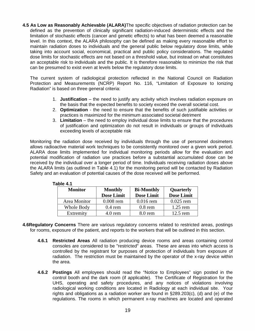
Monitoring the radiation dose received by individuals through the use of personnel dosimeters allows radioactive material work techniques to be consistently monitored over a given work period. ALARA dose limits implemented for individual monitoring periods allow for the evaluation and potential modification of radiation use practices before a substantial accumulated dose can be received by the individual over a longer period of time. Individuals receiving radiation doses above the ALARA limits (as outlined in Table 4.1) for the monitoring period will be contacted by Radiation Safety and an evaluation of potential causes of the dose received will be performed. Table 4.1
Monitor Monthly Dose Limit
Bi-Monthly Dose Limit
Quarterly Dose Limit
Area Monitor 0.008 rem 0.016 rem 0.025 rem Whole Body 0.4 rem 0.8 rem 1.25 rem
Extremity 4.0 rem 8.0 rem 12.5 rem
4.6Regulatory Concerns There are various regulatory concerns related to restricted areas, postings for rooms, exposure of the patient, and reports to the workers that will be outlined in this section.
4.6.1 Restricted Areas All radiation producing device rooms and areas containing control
consoles are considered to be "restricted" areas. These are areas into which access is controlled by the registrant for purposes of protection of individuals from exposure of radiation. The restriction must be maintained by the operator of the x-ray device within the area.
4.6.2 Postings All employees should read the “Notice to Employees” sign posted in the control booth and the dark room (if applicable). The Certificate of Registration for the UHS, operating and safety procedures, and any notices of violations involving radiological working conditions are located in Radiology at each individual site. Your rights and obligations as a radiation worker are found in §289.203(c), (d) and (e) of the regulations. The rooms in which permanent x-ray machines are located and operated

20
are Radiation Areas and are restricted. §289.202(aa). The radiation area is designated by “Caution Radiation Area”.
4.6.3 Exposure of the Patient Individuals (patients) shall not be exposed to the useful beam
except for healing arts purposes and unless such exposure has been authorized by a licensed practitioner of the healing arts. This provision specifically prohibits deliberate exposure for the following purposes:
1. Exposure of an individual for training, demonstration, or other non-healing
arts purposes.
2. Exposure of an individual for the purpose of healing arts screening except as authorized by the Texas Department of State Health Services to the institution for a specific procedure requested.
Techniques employed in radiography and radiation therapy should be those which achieve the desired objectives with minimum dose to the patient. Persons performing the x-ray procedures should follow the guides listed below, to reduce the patient exposure:
1. The useful beam should be limited to the smallest area practical, and
consistent with the objectives of the radiological examination or treatment.
2. The voltage and the source-skin distance (SSD) employed in medical radiological examinations should be as great as is practical and consistent with the diagnostic objectives of the study.
3. Protection of the embryo or fetus during radiological examination or
treatment of women know to be pregnant should be given special consideration.
Note: Ideally, abdominal radiological examination of a woman of childbearing age should be performed during the first ten (10) days following the onset of a menstrual period to minimize the possibility of irradiation of an embryo. In practice, medical needs should be the primary factors in deciding the timing of the examination.
4. Suitable protective devices to shield the gonads of patients who are
potentially procreative must be used when the examination or method of treatment may include the gonads in the useful beam or be within 5 centimeters of the useful beam unless it will interfere with the diagnostic procedure. Gonadal shielding shall be of at 0.5 millimeter lead equivalent material. [§289.227(i)(13)]
5. Fluoroscopy should not be used as a substitute of radiography, but
should be reserved for the study of dynamics or spatial relationships or for guidance in spot-film recording of critical details.
6. X-ray film, intensifying screens, and other image recording devices,
should be as sensitive as is consistent with the requirements of the examination.
7. Film processing materials and techniques should be those recommended

21
by the x-ray film manufacturer. 4.6.4 Exposure of the Individual (Staff)
Reduction of radiation exposure to an individual from external sources of radiation may be achieved by any one or any combination of the following measures:
1. Increasing the distance of the individual from the source.
2. Reducing the duration of exposure.
3. Using protective barriers between the individual and the source.
For dental and medical x-ray equipment, shielding and distance are the factors most readily controlled. Protective shielding includes:
a) That incorporated into the equipment b) Mobile or temporary devices, such as moveable screens c) Lead impregnated aprons and gloves d) Permanent protective barriers and structural shielding, such as walls containing
lead or concrete.
For mammography equipment, only the staff required for the medical procedure or training shall be in the room unless such individual’s assistance is required. Since accumulated dose is directly proportional to exposure time, the less time or duration for the radiation exposure, the less radiation exposure one receives.
4.6.5 Holding of Patient or Film/Image Receptor When a patient or film/image receptor must be supported during a radiation exposure, use a mechanical holding device when the circumstance permits. Patients should be held only after it is determined that available mechanical devices are inadequate. [§289.227(i)(8)] The following situations may require a patient to be held:
1. Trauma Cases 2. Small children and babies 3. Restrained and Combative Patients 4. OR Patients
The human holder will be protected with appropriate protective lead garments and positioned out of the direct beam. To assist in minimizing exposure, it is important for the radiologic technologist to collimate carefully to the area of clinical interest.
In selecting a holder, no pregnant woman or possibly pregnant woman or individual under 18 years old will be considered. No individual shall be used routinely to hold film/image receptor or patients.
4.6.6 Exposure of Individuals Other than the Patient Except for other patients who cannot
be moved out of the room such as ICU, SICU or CCU or a person holding, only the staff and ancillary personnel required for the medical procedure or training shall be in the room during the radiation exposure.
All individuals, other than the patient being examined, shall be positioned such that no part of the body will be struck by the direct beam unless protected by an apron, gloves, or other shielding having 0.5 millimeter lead equivalent material.

22
Staff and ancillary personnel shall be protected from primary scatter by protective aprons or whole body protective barriers or not less than 0.25 millimeters of lead equivalent material.
Other patients who are in line with the primary scatter and who cannot be removed from the room, shall be protected by whole body protective barriers of 0.25 millimeter lead equivalent material or so positioned that the nearest portion of their body is at least 6 feet from both the tube head and the nearest edge of the image receptor.
4.6.7 Reports to Workers 1. Read the “Notice to Employees” sign posted in the control booth and the dark
room (if applicable). 2. The Certificate of Registration for the UHS, operating and safety procedures, and
any notices of violations involving radiological working conditions are located in Radiology at each individual site.
3. Your rights and obligations as a radiation worker are found in §289.203(c), (d) and (e) of the regulations.
4. The rooms in which permanent x-ray machines are located and operated are Radiation Areas and are restricted. §289.202(aa). The radiation area is designated by “Caution Radiation Area”.

23
5.0 MEDICAL PHYSICS
5.1 Surveys Radiation safety surveys or equipment performance evaluations (EPE) in accordance with §289.227, §289.230, and §289.232 will be performed by a licensed Medical Physicist through the Radiation Safety Office. The surveys must be completed per the following table:
Type of Machine Frequency Computed Tomography (CT) Annually not to exceed 14 months from
the date of the prior EPE Fluoroscopy Annually not to exceed 14 months from
the date of the prior EPE Radiographic Podiatric use only
4 years from date of prior EPE
All other Radiographic 2 years from the date of the prior EPE Dental 4 years from the date of prior EPE Mammography Annually not to exceed 13 months from
the date of the prior EPE All equipment performance evaluations must be performed according to the following:
a. Within 30 days after initial installation of new machines (except mammography as this is required prior to patient use),
b. Within 30 days after reinstallation of a machine, or c. Within 30 days after a repair of a machine component that would effect the
radiation output that includes but is not limited to timer, tube, and power supply. d. Within 30 days on major change in equipment operation, for example introduction
of a new software package (CT specifically) Contact the Radiation Safety Officer when such repair is performed on a unit.
Radiation Safety will maintain records of the equipment performance evaluations for all radiation producing devices at University Health System. These records include the measurements and numerical readings, indication of pass or fail for each test, and is reviewed and signed by the licensed medical physicist. If the equipment performance evaluation indicates a fail for a test and it requires a repair, the correction or repair must begin within 30 days of the failure and shall be completed no longer than 90 days from discovery unless authorized by the Texas Department of State Health Services.
Measurements of the radiation output for all systems must be performed utilizing a calibrated dosimetry system. The dosimetry system calibration must be traceable to a national standard, be calibrated within 24 months from the date or prior calibration, and the record of the dosimetry system calibration must include the manufacturer’s name, model, and serial number of each instrument, the date of calibration, and the name of the individual recording the information.
5.2 Physicist Qualifications The person performing evaluation of diagnostic and mammographic system performance in accordance with these regulations shall hold a current Texas license under the Medical Physics Practice Act, Texas Occupations Code, Chapter 602 in the appropriate discipline. The physicist will also be certified by the American Board of Radiology in diagnostic radiology to perform evaluations of mammographic systems for MQSA requirements. For mammography, the medical physicist must meet the initial qualifications or the alternative initial qualifications outlined in §289.230(r)(3)(A-B). For mammography, the physicist must also complete the 15 hours of continuing education over a 36 month period with 8 hours in digital mammography.

24
5.3 Patient Dose Calculations The licensed medical physicist will calculate patient skin doses for all procedures involving CT and Interventional Guided Fluoroscopy that exceed the reference levels outlined in Chapters 7 and 10. These records will be maintained within the individual University Health System departments where the procedure occurred.

25
6.0 GENERAL OPERATING REQUIREMENTS
The following general operating procedures should be followed when operating any x-ray producing device within University Health System:
1. Ordering of X-ray Exams No x-ray exams shall be taken unless ordered by a practitioner. [§289.202(b)and §289.227(b)]. All x-ray exams should be ordered in accordance with 289.231 (b)(1). The only exception to this is in mammography where self-referred patients are allowed.
2. Operator Position During Exposure
The operator must be able to continuously view and communicate with the patient. [§289.227(i)(9)]
3. Operator should apply the concept of As Low As Reasonably Achievable during any
exposure and utilize protective aprons, gloves, leaded glasses, booth, etc.
4. Operator must wear personnel monitoring device during all exposures. The only personnel not utilizing personnel monitoring devices are individuals only operating dental units. Personnel monitoring is outlined in Chapter 3 of this manual.
5. Operators must adhere to the radiation protection guidelines outlined in Chapter 4 including
postings, notices to workers, holding of patients, exposure of the patient, and exposure of the staff at a minimum.
6. Use of a Technique Chart
The use of a technique chart aides in reducing the exposure to the operator and the patient. It must be used for all exposures. The chart is to be posted in the vicinity of the control panel of each x-ray machine or electronically displayed. [§289.227(i)(1)]. The technique chart should be used by all x-ray producing machines operators. Failure to do so could result in higher patient dose and a violation of the Texas regulations. The technique chart must include technical factors, anatomical examination, patient thickness for examination being performed, and source-to-image distance needed to make the clinical radiographs when the radiographic system is in manual mode.
7. Restriction and Alignment of the Beam
The useful x-ray beam shall be restricted to the area of clinical interest. [§289.227(L)(1)(A)(i)]. Use the centering and collimator provided on the x-ray machine.
8. The x-ray tube housing shall not be held by an individual during any an
x-ray exposure.
9. All x-ray operators shall read and understand the written operating and safety procedures on an annual basis. This involves keeping documentation for the TDSHS inspector including the following:
i. Name and signature of individual ii. Date individual read the operating and safety procedures iii. Initials of Radiation Safety Officer
10. Film Processing (if applicable)

26
Unexposed film is stored in light tight bins shielded or away from x-ray exposure, generally in the dark room. Loaded cassettes in the x-ray room will be stored such that they are shielded from scatter and fogging.
Films shall be developed by the time and temperature recommended by the x-ray film manufacturer. [§289.227(p)] The time and temperature of the automatic processor is to be posted on the unit. Do not process film unless the developer temperature is the posted temperature. Run blank films through the process at the beginning of the work day.
Expiration dates on film and chemicals should be checked periodically. New film or chemicals should be rotated so the oldest are used first. Do no use film or chemicals that are past the expiration date.
Chemicals will be replaced by the film processor service vendor according to the manufacturer’s recommended interval not to exceed every three months.
Safe light(s) in the dark room are to have filters and wattage compatible with the film/chemistry and to be three feet from the working surface.
Light leak tests are to be performed every six months. Any light leak detected requires initiation of correction within 72 hours and completed within 15 days.
11. Alternative Processing Systems (if applicable)
The users of daylight processing systems, laser processors, self-processing film units, or other alternative processing systems must follow the manufacturer’s recommendations for image processing. Documentation that the department followed the manufacturer’s recommendations must include the date and initials of the individual completing the document and must be maintained for inspection purposes by the Texas Department of State Health Services.
12. Digital Imaging Acquisition Systems (if applicable) The users of digital imaging acquisition systems must follow quality assurance/quality control protocols for image processing as established by the manufacturer and if no manufacturer’s protocol is available, please contact Radiation Safety for the Standard Operating Procedure for University Hospital. The frequency at which the quality assurance/quality control protocol is performed must be documented including the date and initials of the individual completing the document and kept available for inspection by the Texas Department of State Health Services.

27
7.0 COMPUTED TOMOGRAPHY 7.1 Radiation Safety Precautions
1. The CT control panel shall provide visual indication of the production of x-rays whether the shutter is open or closed.
2. The technique factors shall be accurate to meet the manufacturer’s specifications. If these specifications are not available the factors shall be accurate to within 10% of the indicated setting.
3. The system must require operator initiation of each individual scan or series of scans. 4. The emergency buttons/switches must be clearly labeled as to their functions. 5. There must be two-way aural communication between the patient and the operator at the
control panel via an intercom system. 6. Windows, mirrors, closed-circuit television, or an equivalent shall be provided to permit
continuous observation of the patient during irradiation. If the viewing system fails, no treatments or scans can be performed until this is repaired.
7. Ensure that all personnel in the room are wearing lead aprons, gloves and other appropriate protective devices where necessary. Lead aprons must be utilized for the Ceretom and the BodyTom.
8. Operator must wear personnel monitoring device during all exposures. Personnel monitoring is outlined in Chapter 3 of this manual.
9. Utilize the lead drapes for the CereTom and individuals in the room should be at least 2 meters away when scanning the patients. For the BodyTom, distance and lead aprons must be utilized since lead drapes are not available for the unit.
10. Record of radiation output must be maintained for each exam to include: o Patient identification, o Type and date of examination, o Identification of the CT system used, and o If the CT system is capable of calculating and displaying these values:
CTDIvol, DLP, or Recommendations as identified in AAPM Task Group 111
11. If the radiation output exceeds the reference levels outlined in Section 7.3, the following follow-up actions must be taken:
1. Any patient exams that exceed the reference levels outlined in Section 7.3 must be reported to the Chief Technologist and the Radiology director.
2. Investigation internally and review as to why the reference level was exceeded. 3. Determine if there is any clinical significance to the patient or skin effects. If there
are any skin effects, a dermatology consult should be strongly recommended to the patient and the patient’s primary care team.
4. Report all cases that exceed the reference levels to the Radiation Protocol Committee outlined in Section 7.4.
7.2 Quality Assurance Policy
SYSTEM PERFORMANCE � Control & System Requirements For A CT Single Tube Includes The Following:
o CT conditions of operation (slice thickness, # of slices, gantry angle, etc.) are indicated prior to initiation of the scan [§289.227(n)(1)(C)]

28
o CT conditions of operation visible from any position for scan initiation [§289.227(n)(1)(B)] o Visible indication of x-ray production [§289.227(n)(1)(F)(i)] o Operator required to initiate each scan or series [§289.227(n)(1)(F)(ii)] o Termination of x-ray exposure signal visible to the operator [§289.227(n)(1)(G)(i)] o Termination of scan greater than 0.5 sec by operator possible at any time
[§289.227(n)(1)(G)(iii)] o Must reset manually after termination of 0.5 sec or greater [§289.227(n)(1)(G)(iiii)] o Visual determination (light indicators) of the tomo plane [§289.227(n)(1)(D)(i)] o Visual determination of x-ray production at the gantry [§289.227(n)(1)(H)(i)] o Emergency buttons and/or switches clearly labeled [§289.227(n)(1)(F)(iii)] o Technique chart provided electronically [§289.227(i)(1)] o Leaded glass permits continuous observation of patient [§289.227(n)(2)(B)] o Two way aural communication provided [§289.227(n)(2)(A)]
� Medical Physicist Will Determine The Dose Measurement [§289.227(n)(3)]
o With a properly calibrated dosimetry system [§289.227(n)(3)(B)] o Annually [§289.227(n)(3)(A)(i)] o After major maintenance affecting radiation output to include kV board change, change
in filtration, and at the recommendation of the service engineer, excluding a tube change [§289.227(n)(3)(A)(iI)]
o Maintain Physics report for three years [§289.227(n)(3)(C)]
� Quality Image Maintenance - Many factors effect Image Quality: o Proper alignment of X-ray tube, DAS, detector and table o KV and mA adjustments within specifications o Current Calibration files o Tube Warmup every time the system recommends it o Daily Fastcals o Appropriate pixel size, slice thickness, reconstruction algorithm, and special processing
selections during Scan Rx o Patient remains motionless during scan acquisition
� At Least Three people Must Cooperate To Produce Optimum Images:
o Service representative aligns the system and adjusts kV and mA o Operator follows facility guidelines to maintain daily image quality, prescribe the exams,
and update the calibration files o Patient follows operator (and autovoice) instructions during exam
� A QA Program Helps Locate The Source Of Image Quality Problems:
o Replaces patient with phantom o Provides standard Scan Rx parameters o Provides System Performance tests and comparisons
� QA Program Schedule
o The QA will be performed bi-weekly at main UHS utilizing the Quick IQ protocol for all QA protocols.
o The QA program at the RBG Campus will be performed daily during normal hours of operation utilizing the manufacturer’s calibration recommendations.
o The CT phantom provided by the vendor will be used to run the QA procedure. o Manufacturer procedures will be followed to obtain phantom images o The phantom images are stored digitally [§289.227(n)(5)(A-B)] o For the Ceretom and the Bodytom, the air calibration must be performed everyday
(every time it is moved to a new loation)

29
� Maintenance
o Preventative maintenance by CT unit’s vendor service engineer performed monthly for each CT unit
o Service agreement with the vendor includes onsite contact. 7.3 CT Protocol Guidelines University Health System will follow the recommendations for CT
reference levels for specific CT scan regions as outlined in the AAPM and ACR. The reference levels are outlined in the following table:
CT Scan Region CTDI Reference Level (mGy) Adult Head 80 Adult Torso 50 Pediatric Head <2 years old 2-5 years old
50 60
Pediatric Torso <10 years old (16 cm phantom) <10 years old (32 cm phantom)
25 20
Brain Perfusion 600 Cardiac Retrospectively gated (spiral) Prospectively gated (sequential)
150 50
The radiation output is printed on the patient images and stored in the patient medical records. The technologist should review the CTDI value on the films. If the radiation output exceeds the reference levels outlined above, the following follow-up actions must be taken:
1. Any patient exams that exceed the reference levels outlined must be reported to the Chief Technologist and the Radiology director.
2. Investigation internally and review as to why the reference level was exceeded. 3. Determine if there is any clinical significance to the patient or skin effects. If there are
any skin effects, a dermatology consult should be strongly recommended to the patient and the patient’s primary care team
4. Report all cases that exceed the reference levels to the Radiation Protocol Committee outlined in Section 7.4.
In addition, the Chief Technologist performs a random sampling audit of all cases to verify that values are within the reference level requirements.
7.4 Radiation Protocol Committee The Radiation Protocol Committee responsibilities are outlined in
Section 1.3.4. The following are the members that must be present for the review of any cases that exceed the reference levels for CT protocols:
• Radiologist or Radiation Oncologist – CT Procedures • Licensed Medical Physicist – Diagnostic • UHS Radiation Safety Officer • Representative from Nursing Staff • CT Chief Technologist
The committee will meet at an interval not to exceed 14 months and will discuss any cases that have
occurred that exceeded the CT reference levels. Recommendations will be made for future cases

30
and the committee will review any protocol changes. All records will be kept for TDSHS inspection purposes.
7.5 Training The following two items are required for training of all operators.
1. Basic Medical X-ray Training All operators must complete basic medical x-ray training offered by Radiation Safety. This course will cover the basics of CT units, x-ray physics basics, radiation protection, biological effects, and radiation dosimetry.
2. Operating and Safety Procedures All operators must sign annually stating that they have reviewed and understand the operating and safety procedures outlined in this manual.

31
8.0 DENTAL UNITS 8.1 Radiation Safety Precautions
1. Close the door to the exam room or x-ray room. 2. Place the lead apron on the patient. 3. Position the film/holder. 4. Do not hold the film with your fingers. 5. Step into the operator booth or step 6 feet away. 6. Make sure no one else is in the room. 7. Personnel dosimetry is not required for operators of dental units only. If the individual is
operating more than just a dental unit, the dosimetry is required.
8.2 Training The following two items are required for training of all operators. 1. Basic Dental X-ray Training All operators must complete basic dental x-ray training offered by
Radiation Safety. This course will cover the basics of dental x-ray units, x-ray physics basics, radiation protection, biological effects, and radiation dosimetry.
2. Operating and Safety Procedures All operators must sign annually stating that they have reviewed and understand the operating and safety procedures outlined in this manual.

32
9.0 FLUOROSCOPY UNITS
9.1 Radiation Safety Requirements
1. All fluoroscopy units must have the medical physics equipment performance evaluation results posted on the side of the unit for the fluoroscopist to view. The measurement results cannot exceed 10 R/min or 100 mGy/min.
2. Ensure that all personnel in the room are wearing lead aprons, gloves and other appropriate
protective devices where necessary. More specifically, the thickness of lead must be at least 0.5 mm thickness of lead equivalent material. This is outlined in Chapter 4 under Protective Devices.
3. Check to determine that all protective shields and devices such as protective aprons and
drapes are in place before the procedure begins.
4. Ensure that all personnel are wearing the personnel monitoring device during all exposures. The only personnel not utilizing personnel monitoring devices are individuals only operating dental units. Personnel monitoring is outlined in Chapter 3 of this manual.
5. Reset the 5-minute cumulative timing device before each fluoroscopic procedure
[§289.227(m)(7)(A)].
6. Make sure that only those persons absolutely necessary for the examination are in the room.
7. Close all doors leading to the examination room before the procedure begins.
8. Use of the Fluoroscopic Machines - Stationary and Mobile
Begin with the collimators closed, and open collimators so the x-ray beam is restricted to the area of clinical interest [§289.227(m)(8)(B)(ii)]
9. For mobile (C-Arm) units, a 30-centimeter (cm) source-to-skin distance (SSD) is to be used.
To achieve the 30 cm, the spacer attached to the x-ray tube must be utilized. [§289.227(m)(3)(A)(iv)(III)]. The spacer is a plastic tube-like device, 10 cm in length that clips or slides onto the x-ray tube head of the C-arm.
10. A 20cm source-to-skin distance (20cm SSD) may be used for mobile fluoroscopy when the
patient is too large to fit between the image intensifier and the tube head with the spacer, the surgeon requires additional room for the procedure or the tube head will not fit under the surgical table due to the structure of the surgical tables. The system cannot have a SSD less than 10 cm.
11. When the spacer is off (allowing the 20cm SSD), as precautionary measures, use sterile
drapes on the image intensifier, minimize the field size to clinical field of interest, position patient as close to the image intensifier as possible, and minimize fluoroscopy time.
33
12. Use pulsed mode fluoroscopy when possible to reduce dose to operator. Reduce or minimize the use of mag mode on the units.
13. Utilize the audible signal that is provided to sound after 5 minutes of irradiation during an examination or procedure. This signal will sound until manually reset but assists in minimizing the amount of fluoroscopy beam-on time.
14. Replace the spacer on the C-arm when finished with the procedure each time it is removed.
[§289.227(m)(3)(A)(iv)(III)].
9.2 Training The following two items are required for training of all operators.
1. Basic C-arm Fluoroscopy Training All operators must complete basic c-arm fluoroscopy training offered by Radiation Safety. This course will cover the basics of fluoroscopy, x-ray physics basics, radiation protection, biological effects, and radiation dosimetry.
2. Operating and Safety Procedures All operators must sign annually stating that they have reviewed and understand the operating and safety procedures outlined in this manual.

34
10.0 FLUOROSCOPY GUIDED INTERVENTIONAL PROCEDURES 10.1 Fluoroscopy Guided Interventional Procedures Definition For purposes of this manual, Fluoroscopy-Guided Interventional (FGI) Procedures is defined as interventional diagnostic or therapeutic procedures performed via percutaneous or other access routes, usually with local anesthesia or intravenous sedation, which uses external ionizing radiation in the form of fluoroscopy to localize or characterize a lesion, diagnostic site, or treatment site, to monitor the procedure, and to control and document therapy. Fluoroscopy guided interventional procedures include:
a. TIPS creation (transjugular intrahepatic portosystemic shunt) b. Embolization (any location,any lesion) c. Stroke therapy d. Biliary drainage e. Angioplasty with or without stent placement f. Stent-graft placement g. Chemoembolization h. Angiography and intervention for gastrointestinal hemorrhage i. Carotid stent placement j. RF (radiofrequency) cardiac ablation k. Complex placement of cardiac EP (electrophysiology) devices, and l. PCI (percutaneous coronary intervention) (single or multiple vessel)
10.2 Radiation Safety Precautions
1. All fluoroscopy units must have the medical physics equipment performance evaluation results posted on the side of the unit for the fluoroscopist to view. The measurement results cannot exceed 10 R/min or 100 mGy/min.
2. Ensure that all personnel in the room are wearing lead aprons, gloves and other appropriate
protective devices where necessary. More specifically, the thickness of lead must be at least 0.5 mm thickness of lead equivalent material. This is outlined in Chapter 4 under Protective Devices.
3. Check to determine that all protective shields and devices such as protective aprons and
drapes are in place before the procedure begins.
4. Ensure that all personnel are wearing the personnel monitoring device during all exposures. The only personnel not utilizing personnel monitoring devices are individuals only operating dental units. Personnel monitoring is outlined in Chapter 3 of this manual.
5. Reset the 5-minute cumulative timing device before each fluoroscopic procedure
[§289.227(m)(7)(A)].
6. Make sure that only those persons absolutely necessary for the examination are in the room.
7. Close all doors leading to the examination room before the procedure begins.
8. Use of the Fluoroscopic Machines - Stationary and Mobile

35
Begin with the collimators closed, and open collimators so the x-ray beam is restricted to the area of clinical interest [§289.227(m)(8)(B)(ii)]
9. For mobile (C-Arm) units, a 30-centimeter (cm) source-to-skin distance (SSD) is to be used.
To achieve the 30 cm, the spacer attached to the x-ray tube must be utilized. [§289.227(m)(3)(A)(iv)(III)]. The spacer is a plastic tube-like device, 10 cm in length that clips or slides onto the x-ray tube head of the C-arm.
10. A 20cm source-to-skin distance (20cm SSD) may be used for mobile fluoroscopy when the
patient is too large to fit between the image intensifier and the tube head with the spacer, the surgeon requires additional room for the procedure or the tube head will not fit under the surgical table due to the structure of the surgical tables. The system cannot have a SSD less than 10 cm.
11. When the spacer is off (allowing the 20cm SSD), as precautionary measures, use sterile
drapes on the image intensifier, minimize the field size to clinical field of interest, position patient as close to the image intensifier as possible, and minimize fluoroscopy time.
12. Use pulsed mode fluoroscopy when possible to reduce dose to operator. Reduce or minimize the use of mag mode on the units.
13. Utilize the audible signal that is provided to sound after 5 minutes of irradiation during an examination or procedure. This signal will sound until manually reset but assists in minimizing the amount of fluoroscopy beam-on time.
14. Replace the spacer on the C-arm when finished with the procedure each time it is removed.
[§289.227(m)(3)(A)(iv)(III)]. Where sterile fields or special procedures prohibits the use of normal protective barriers or drapes, all of the following conditions must be met: 1. All persons, except the patient, in the room where fluoroscopy is performed, shall wear
protective aprons which provide a shielding equivalent of 0.5 millimeter of lead. 2. The fluoroscopist and all other personnel in the room, except the patient, shall have
appropriate personnel monitoring devices. 3. The fluoroscopic field shall be reduced to the absolute minimum required for the
procedure being performed (area of clinical interest). 4. Use pulsed mode fluoroscopy when possible to reduce dose to operator.
10.3 FGI Protocol GuidelinesUniversity Health System will follow a set reference level of 5 Gy or higher, or greater than 90 minutes of interventional fluoroscopy on any one case. The following are procedures that are outlined to cover the monitoring of radiation exposure, documenting patient dose in the medical record, and follow-up action items if the reference value is exceeded.
1. It is the responsibility of the physician performing the fluoroscopy- guided procedure to
inform the patient about the potential radiation- related injury associated with the proposed procedure as part of the consent process.
2. The technologist assigned to the procedure shall monitor the cumulative air kerma (CAK) reading (procedure time if CAK is not present). The technologist shall notify the Attending physician performing the procedure when a CAK reading of 3 Gy (60 minutes if CAK not available) has been reached, and every 1 Gy (15 minutes if CAK not available) thereafter, documenting each notification in the procedure log book.

36
3. At the conclusion of each procedure, the technologist shall record the total fluoroscopy time
and/or CAK reading, the fluoroscopy system used, date and time of the examination in the medical record system for the patient. If the CAK or dose area products are not available, the fluoroscopic mode including high-level or pulsed-mode of operation, cumulative fluoroscopic time, and number of films or recorded exposures must be entered into the medical record for the patient.
4. The physician will enter the required information listed above into the patient’s electronic
medical record via the dictated report.
5. In cases where the reference level for FGI procedures or the final CAK reading is 5 Gy or higher, or greater than 90 minutes of fluoro time, the technologist shall contact their supervisor and relay the information to them.
6. Actions or Patient Follow-up:
1. The supervisor will then contact the medical physicist and review the data with him or her
2. The medical physicist will then perform a calculation to estimate the total dose delivered to the patient. If the calculation results in a cumulative dose of greater than 15 Gy to a single field, during a six month time frame, a sentinel event must be reported. Please see Chapter 14.1 for further information about Sentinel Events.
3. A patient who has a final reading of 5 Gy or higher (or greater than 90 minutes of fluoro time) shall be given written follow-up instructions upon discharge relating to a possible radiation injury. The patient shall be instructed to perform self-examinations of the irradiated area and to contact the physician within two to ten weeks if any findings or concerns. Clinical follow-up shall be arranged for positive self-exam. Patients that remain hospitalized will be tracked and assessed by a physician during the two to ten week period for potential skin injury.
4. Any procedures or patient cases that exceed the reference level listed above will be reported to the Radiation Protocol Committee, Section 10.4, for review at the next scheduled meeting.
10.4 Radiation Protocol Committee The Radiation Protocol Committee responsibilities are outlined in Section 1.3.4. The following are the members that must be present for the review of any cases that exceed the reference levels for FGI protocols:
• Licensed Physician involved in Interventional Procedures • Licensed Medical Physicist – Diagnostic • UHS Radiation Safety Officer • Representative from Nursing Staff • Angiography Technologist • Additional Physicians from various specialties involved in FGI procedures that exceed the
reference levels (i.e. Cardiac Cath, Neurosurgery, Vascular, etc.) The committee will meet at an interval not to exceed 14 months and will discuss any cases that have
occurred that exceeded the FGI reference levels. Recommendations will be made for future cases and the committee will review any protocol changes. All records will be kept for TDSHS inspection purposes.

37
10.5 Training The following two items are required for training of all technologists, nursing staff, and Radiologists or Radiation Oncologists present during procedures.
1. Basic C-arm Fluoroscopy Training All operators must complete basic c-arm fluoroscopy training offered by Radiation Safety. This course will cover the basics of fluoroscopy, x-ray physics basics, radiation protection, biological effects, and radiation dosimetry.
2. Operating and Safety Procedures All operators must sign annually stating that they have reviewed and understand the operating and safety procedures outlined in this manual.
The following two items are required for training of all physicians not certified in Radiology or Radiation Oncology but performing FGI procedures:
1. FGI Physician Training: All physicians, other than radiologists and radiation oncologists, performing FGI procedures must complete a minimum of 8 hours of Category I CMEU training in radiation safety awareness training offered by the UHS Radiation Safety office. In addition, there is a requirement to also complete a 1 hour fluoroscopic machine training provided by a radiologist, licensed medical physicist (LMP), or a physician who has already completed the 1 hour training provided by the radiologist or LMP. Physicians have until May 1, 2015 to complete the necessary training. If not completed by this date or employed after this date, they must complete the training prior to use of the fluoroscopy machines for FGI procedures.
2. Operating and Safety Procedures All operators must sign annually stating that they have reviewed and understand the operating and safety procedures outlined in this manual.

38
11.0 MAMMOGRAPHY
11.1 Radiation Safety Precautions Mammography facilities shall meet the quality standards in subsections (r)-(gg) of 25 TAC §289.230 and be accredited by an FDA-approved accreditation body. In order to qualify for certification, new facilities must apply to the agency certifying body in accordance with certain requirements and to an FDA-approved accreditation body and receive acceptance of the accreditation application. All mammography units must be certified by the Texas Department of State Health Services. A facility with an existing certification may begin using a new or replacement machine before receiving an updated certification if the registrant submits to the agency certifying body and to the FDA-approved accreditation body, documentation with a medical physicist’s report. The medical physicist’s report is required prior to using the machine on patients [25 TAC §289.230(n)(5)]. For mammography equipment, only the staff required for the medical procedure or training shall be in the room unless such individual’s assistance is required. For mammography, a chart or manual shall be provided or electronically displayed in the vicinity of the control panel of each mammography machine that specifies the technique factors to be utilized versus patient’s anatomical size. Film Retention - Each registrant that performs mammograms shall maintain mammography films and reports in a permanent medical record for a minimum of five years. If no additional mammograms of the patient are performed at the facility, the films and reports shall be maintained for a minimum of ten years. [§289.230(t)(4)]. The mammographic image identification must include name of patient, date of birth, date of examination, view and laterality, facility name and location (city, state zip code), technologist identification, cassette/screen identification, and mammography machine identification (if more than one machine in facility).
Upon closure or termination of the facility, the registrant shall maintain the mammography films for 5 years. If the facility complies with the following:
a. Within 180 days of closing, the registrant shall directly notify each patient or patient’s representative with instructions on how to retrieve or authorize disposal of the patient’s records; and
b. Within 60 days of closing, the registrant shall publish a notice in one or more newspapers covering the geographical area served by the closing facility. The notice shall include:
i. Contact information on retrieving patient records; and ii. Information that the records will be destroyed if not retrieved by the
patient or the patient’s representative within 5 years; and c. If records have not been retrieved by the patient or the patient’s representative
following the 5-year period after closing, the registrant may destroy the records.
11.2 Mammography Physician/Operator Requirements
A physician who interprets mammograms shall hold a current Texas license issued by the Texas Board of Medical Examiners and shall be certified by the American Board of Radiology or the American Osteopathic Board of Radiology and shall have had 60 hours of documented continuing medical education credits in mammography. Continuing education credits shall be approved by the Accreditation Council for Continuing Medical Education (ACCME) or the Committee on Postdoctoral Training of the American Osteopathic Association (CPTAOA). Documented time spent in residency specifically devoted to mammography will be accepted if the residency

39
program has been approved by the Accreditation Council for Graduate Medical Education (ACGME). In addition, the physician shall read a minimum of 240 mammograms within the six month period prior to qualifying as an interpreting physician. After qualifying, the physician must have interpreted at least 960 mammograms within the last 24 months. For physicians interpreting digital mammograms, it is required that they complete an 8 hours of Continuing Education in Digital Mammography and 15 hours of Continuing Education in Digital Mammography every 3 years.
For qualification, operators of mammography equipment must have an additional 40 hours of formal mammography training as outlined in TAC §289.230(r)(2)(A), and performed the specified 25 mammography exams under direct supervision or hold both a current and valid Texas certification under the Medical Radiologic Technologist Act and an American Registry of Radiologic Technologist (ARRT) Certificate of Advanced Qualification in Mammography. After qualification, operators shall also obtain 15 hours of continuing education (8 hours digital mammography) over a 36 month period as specified in §289.230(r)(2)(C) and 200 mammograms within the last 24 months.
11.3 In-House Policies The Operating Procedures related to the following items listed below (in
accordance with Texas Regulations) are located in Radiology/Mammography in-house policies within the facility:
1. Quality assurance program 2. Image quality and corrective action for images of poor quality 3. Repeat analysis 4. Procedures and techniques for mammography patients with breast implants 5. Procedure to handle complaints 6. Self-referral mammography
The printer QC will be performed and documented in a printer book. The printer and the monitor QC books will be maintained for mammography at the main site, CTRC for inspection purposes as outlined by the medical physicist.
11.4 Training Additional mandatory mammography training may be required by the agency
(TDSHS) based on the recommendations of the ACR and the FDA and should be developed on a case by case basis. The agency could require pre-approval of the additional training, documentation of the additional mandatory training shall be reviewed by the date specified by the agency, and the records of all additional mandatory training shall be maintained by the registrant for inspection by the agency. This applies to both interpreting physicians and MRTs.
Operating and Safety Procedures All operators must sign annually stating that they have reviewed and understand the operating and safety procedures outlined in this manual.

40
12.0 RADIOGRAPHIC UNITS 12.1 Radiation Safety Precautions
1. Ensure that all personnel in the room are wearing lead aprons, gloves and other appropriate protective devices where necessary. More specifically, the thickness of lead must be at least 0.25 mm thickness of lead equivalent material. This is outlined in Chapter 4 under Protective Devices.
2. Check to determine that all patient protective shields and devices such as protective aprons
and drapes are in place if necessary to reduce the dose to the areas not being imaged.
3. Ensure that all personnel are wearing the personnel monitoring device during all exposures. Personnel monitoring is outlined in Chapter 3 of this manual.
4. During the exposure, the operator must be positioned using the concept of ALARA, such as
standing 6 feet away, wear lead apron and never be in the line of the direct beam. [§289.227(i)(10)]
5. Close all doors to the x-ray room.
6. Stand behind the operator's shield when taking the exposure.
7. Ensure that no one else is in the room. Warn the staff in the area of the intent to take the x-
ray.
8. Utilize the technique chart for machine settings.
12.2 Training The following two items are required for training of all operators. 1. Basic Medical X-ray Training All operators must complete basic medical x-ray training offered
by Radiation Safety. This course will cover the basics of medical x-rays, x-ray physics basics, radiation protection, biological effects, and radiation dosimetry.
2. Operating and Safety Procedures All operators must sign annually stating that they have reviewed and understand the operating and safety procedures outlined in this manual.

41
13.0 BONE DENSITOMETERS 13.1 Radiation Safety Precautions
1. Wear dosimeter during working hours if issued.
2. Follow manufacturer’s operating instructions. 3. Close door to the room. 4. Necessary personnel only in the room. 5. Do not hold patient. 6. Position patient. 7. Initiate exposure and step back 6 feet. 8. Inform the Radiation Safety Officer of any problems requiring maintenance with the unit.
13.2 Training The following two items are required for training of all operators.
1. TDSHS Training Requirements Persons that only operate a bone DEXA no longer have to
have a hardship exemption. If they are not certified as a Medical Radiological Technologist (MRT) or Licensed Medical Radiological Technologist (LMRT) or are registered as a Non Certified Technologist (NCT), and they only operate a bone DEXA unit:
a. Documentation of one of the following and a certificate that they have completed 4 hours of radiation safety training at 2 years. 1. Current Certification from the International Society for Clinical Densitometry 2. or completion of the ARRT bone density exam 3. or 20 hours of training: 16 hrs of specified training using bone densitometer
equipment presented by an MRT or a licensed medical physicist. 2. Operating and Safety Procedures All operators must sign annually stating that they have
reviewed and understand the operating and safety procedures outlined in this manual.
42
14.0 REPORTABLE EVENTS AND TDSHS RESPONSES 14.1 Sentinel Event
The Joint Commission defines a Sentinel Event related to fluoroscopy as “Prolonged fluoroscopy with cumulative dose greater than 15 Gy to a single field...”
1. If the peak skin dose estimate is greater than 15 Gy to a single field, the physicist will contact the attending.
2. The Attending physician will document a note in the patient’s electronic medical record giving the estimated dose. The physician will contact the patient if any additional information needs to be given to the patient beyond the discharge paperwork initially provided.
3. The medical physicist will also notify the Radiation Safety Officer. 4. In accordance with UHS Policy 5.08, the Director of Risk Management will be
notified in person or by phone by the area where the exposure occurred. In addition, an electronic Risk Assessment Form (eRAF) will be completed by the Sr. Technologist or PCC and submitted to the Risk Management Department.
14.2 In-House Radiation Protocol Committee
All patient cases that exceed the CT and FGI reference levels outlined in Sections 7.3 and 10.3 will be reported to the Radiation Protocol Committee. The Radiation Protocol Committee will report any necessary findings to the Radiation Safety Committee which reports to the Quality and Risk Management Committee and the Environment of Care Committee.
14.3 TDSHS Responses
14.3.1 NOV’S Notices of Items of Non-Compliance from the Department of State Health Services generated from inspections conducted during site visits requires a response on the part of University Health System. The RSO will respond on behalf of the University Health System outlining corrective action from necessary to prevent reoccurrences.
14.3.2 Complaints University Health System must respond if a complaint has been made to the DSHS regarding a situation involving radiation. The RSO will respond on behave of University Health System.

43
15 RECORD KEEPING 15.1 Record Keeping Requirements for Departments All departments utilizing radiation producing
devices are required to keep copies of records as follows: 15.1.1 Radiation Monitoring and Exposure Records All departmental copies personnel
dosimetry monitoring reports must be retained until the next monitoring report is released. Radiation Safety keeps the permanent monitoring reports as outlined in 15.2.5.
15.1.2 X-ray Producing Devices Repairs or Maintenance Records of repairs or
maintenance for radiation producing machines must be kept for 5 years. 15.1.3 Medical Physicist Patient Dose Evaluations Records of patient dose calculations
must be kept in their medical record based on requirements outlined for medical records retention.
15.1.4 Film Retention Patient films & images must be maintained according to the TSMBE
medical records retention schedule. If the individual was younger than 18 at the time of imaging, then films must be maintained until the patient reaches age 21 or for seven years (whichever is longer). Films acquired for individuals over the age of 18 should be retained for a minimum of 7 years from the date of last treatment by the physician. X-ray films exposed for the purpose of inspection should be maintained for two years past the current calendar year. No films should be destroyed if they are involved in any civil, criminal or administrative proceeding that has not yet been finally resolved. [165 TAC §165.1(b)]. Mammography Films retention is outlined in Chapter 11.
15.1.5 CT Films CT films resulting from quality control tests must be maintained for a total of
10 years. 15.1.6 Mammography Films Retention of clinical images for current, closed or terminated
registrants.
(A) Each registrant that performs mammograms shall maintain mammography films and reports in a permanent medical record for a minimum of five years. If no additional mammograms of the patient are performed at the facility, the films and reports shall be maintained for a minimum of ten years.
(B) Each registrant that performs mammograms shall within 30 days of request by or on behalf of the patient, permanently or temporarily transfer the original mammograms and copies of the patient’s reports to a medical institution, a physician or to the patient directly.
(C) If the medical records are permanently forwarded, the receiving institution or
physician shall maintain and become responsible for the original films until the fifth or tenth anniversary, as specified in subparagraph (A) of the paragraph.
(D) Upon closure or termination, the registrant shall maintain the mammography films for
5 years. If the facility complies with the following:

44
(i) within 180 days of closing, the registrant shall directly notify each patient or patient's representative with instructions on how to retrieve or authorize disposal of the patient's records; and
(ii) within 60 days of closing, the registrant shall publish a notice in one or more newspapers covering the geographical area served by the closing facility. The notice shall include:
(I) contact information on retrieving patient records; and
(II) information that the records will be destroyed if not retrieved by
the patient or the patient's representative within 5 years; and
(iii) if records have not been retrieved by the patient or the patient's representative following the 5-year period after closing, the registrant may destroy the records.
15.2 Record Keeping Requirements for Radiation Safety Office
15.2.1 Protective Device Evaluations The annual protective device evaluations must be maintained for 3 years.
15.2.2 X-ray Producing Devices Inventories Annual inventories must be maintained for 3
years. 15.2.3 X-ray Producing Devices Installation Reports, Disposals, or Transfers All of these
records must be kept until termination of the registration.
15.2.4 Medical Physics Surveys Equipment performance evaluations and corrections must be kept for 10 years. Records of the dosimetry system calibrations used for the medical physics surveys must be kept for 5 years.
15.2.5 Radiation Monitoring and Exposure Records This series provides a record of
radiation testing and monitoring of employees, embryo/fetus dose, visitors, individual members of public, facilities, and the surrounding environment and is used to comply with federal and state reporting and licensing requirements and insurance carrier reporting requirements. These records shall be kept as permanent records.
15.2.6 Radiation Protocol Committee Minutes & Reviews All minutes and protocol revisions
must be kept for 5 years. 15.2.7 Radiation Safety Training and Experience All training and experience records must
be maintained until termination of the registration or 5 years after the personnel leave the facility.
15.2.8 TDSHS Regulatory Items This series includes the Texas Regulations, Current
Certificate of Registration, and Current Operating and Safety Procedures. These records must be kept until termination of the registration.
15.2.9 TDSHS Inspections/Corrective Actions This series includes the notice of violation or
notice of compliance and any documentation of corrective actions from an inspection. These items must be kept until the next TDSHS inspection.

APPENDIX
A

For EHS Use Only: Account: ______________ Series: ________ Frequency: ____ Spare Issued: Yes/No Spare#: _______________________
University Health System – University of Texas Health Science Center at San Antonio Environmental Health & Safety Department
Radiation Safety Division
DOSIMETRY SERVICE ASSESSMENT AND EXPOSURE HISTORY FORM
Section 1: Participant Data As required in the Texas Administrative Code, Chapter 25, §289.202, the following information regarding your radiation exposure history this calendar year is necessary for assessment of dosimetry service. Please complete the following items, then sign and return this form to: Radiation Safety Division, EH&S
Full Name: ________________________________________________________________________________________________ Last First Middle
*Social Security Number: __________________________ Date of Birth: ________________ Gender: Female Male Over the age of 18? Yes/No Office phone number: _______________________________________
List any other name(s) under which you have been monitored: _________________________________________________________
Section 2: Circle the appropriate response: UTHSCSA DOSIMETRY POLICY
(a) I will work with fluoroscopy/radiographic equipment: Yes No If yes, dosimeter required (b) I will work with PET or nuclear medicine isotopes: Yes No If yes, dosimeter required (c) I will work with brachytherapy sources: Yes No If yes, dosimeter required (d) I am a voluntarily declared pregnant worker: Yes No If yes, contact Radiation Safety Office (e) I will work with dental x-ray equipment: Yes No If yes, no dosimeter required
Classification: Faculty Technician/Staff Nurse Resident Other-Specify ________________________
With which Department will you be working? ____________________
Section 3: Previous employment(s) involving radiation exposure this calendar year Have you been occupationally exposed to radiation sources this calendar year at another institution? Yes No Does any concurrent employment to UHS require exposure to radiation sources this calendar year? Yes No Facility Name: ______________________________________ Department:________________________________________ Mailing Address: ____________________________________ ______________________________________________________________ ______________________________________________________________ Dates: _______________ through ______________________
Facility Name: _____________________________________ Department: _______________________________________ Mailing Address: _____________________________________________ _______________________________________________________________ _______________________________________________________________ Dates: _______________ through ______________________
Section 4: Signature I authorize the release of my radiation exposure history to the University of Texas Health Science Center San Antonio and will notify Environmental Health & Safety in the event of changes to the above information. Applicant: _________________________________________ Date: _____________________________________________ Signature Permanent Address: ________________________________ ___________________________________ _________________________________
Return the completed form to: Environmental Health & Safety, Room 1.343T Dental School ([email protected] or Fax (210)567-2965))
watsonj
Typewritten Text
A-1

Return the completed form to: Radiation Safety Division, Environmental Health & Safety, Room 1.343T Fax: (210) 567-2965
PREGNANCY DECLARATION, INSTRUCTION & DOSIMETRY EVALUATION
Section 1 – Voluntary Pregnancy Statement I, ______________________________, voluntarily declare my pregnancy to the University of Texas (Please Print Legibly) Health Science Center at San Antonio Radiation Safety Division, as stated in 25 Texas Administrative Code (TAC) Section 289.202(c)(7) and Section 289.202(rr). The estimated date of conception for this pregnancy is . I understand the radiation dose to my embryo/fetus during my entire pregnancy will not be allowed to exceed 0.5 rem (5 millisievert) (unless that dose has already been exceeded between the time of conception and submitting this letter). I also understand that meeting the lower dose limit may require a change in job or job responsibilities during my pregnancy. DOB: , *SSN
Signature:_________________________________________________ Date: * See Reverse for Details
Section 2 – Instruction to Employee
Oral and written information about prenatal occupational radiation exposure has been provided to the individual declaring pregnancy at this time. This information includes: 1. The risk associated with prenatal radiation exposure and methods available for minimizing risk. 2. A review of previous personnel monitoring results for the individual, if available. 3. The fetal dose limit of 0.5 rem during the gestation period for occupationally exposed declared pregnant
workers. 4. The U.S. NRC Regulatory Guide 8.13 Instructions Concerning Prenatal Radiation Exposure. 5. An opportunity to ask questions and receive answers concerning the information provided. [Fetal dose limits and time period information are taken from TAC §289.202(m)(1) – (4).]
Section 3 – Dosimetry Evaluation
Additional fetal dosimetry devices are available for those individuals likely to receive fetal doses in excess of 10% of the established fetal dose limit, as proscribed in the 25 TAC §289.202(q)(1)(A) and §289.202(m)(1) – (m)(4). The utilization and application of such devices has been discussed with the individual declaring pregnancy at this time. Yes No
1. Likely to exceed 10% of fetal dose limits? □ □ 2. Fetal dosimetry devices offered? □ □
3. Fetal dosimetry devices accepted? □ □
Note: Fetal dosimetry service is issued for the duration of the declared pregnancy.
Section 4 – Acknowledgments
I acknowledge that the above information relating to occupational exposure to radiation has been discussed and an opportunity to ask questions was available. Declarer: ____________________________________ Date: __________________ Radiation Safety Representative: _________________ Date: __________________
watsonj
Typewritten Text
A-2

Return the completed form to: Radiation Safety Division, Environmental Health & Safety, Room 1.343T Fax: (210) 567-2965
Revised 01/09
*Disclosure of your Social Security Number (“SSN”) is required of you in order for The University of Texas Health Science Center at San Antonio to enroll you into the Dosimetry Program and to complete the Radiation Occupational Exposure Dosimeter Application and History, as mandated by 25 Texas Administrative Code (TAC) Section 289.202(c)(3) and Section 289.202(rr). Further disclosure of your SSN is governed by the Public Information Act (Chapter 552 of the Texas Government Code) and other applicable law.
1. With few exceptions, you are entitled on your request to be informed about the information The University of Texas Health Science Center at San Antonio collects about you;
2. Under Sections 552.021 and 552.023 of the Texas Government Code, you are entitled to receive and review the information; and
3. Under Section 559.004 of the Texas Government Code, you are entitled to have The University of Texas Health Science Center at San Antonio correct information about you that is held by The University of Texas Health Science Center at San Antonio and that is incorrect, in accordance with the procedures set forth in The University of Texas System Business Procedures Memorandum 32, Texas Public Information Act.
The information that The University of Texas Health Science Center at San Antonio collects will be retained and maintained as required by Texas records retention laws (Section 441.180 et seq. of the Texas Government Code) and rules. Different types of information are kept for different periods of time.
You may send any SSN questions to Andrea Marks MBA, CPA By mail to: 7703 Floyd Curl Drive, San Antonio, TX 78229-
3900 By fax to: (210)567-7020 In person at: Academic Administrative Building, Room 4.448

The University of Texas Health Science Center
Pregnant Employee Guide to Radiation
Questions? Please call 567-2955 or come by the Environmental Health and Safety Office Radiation Safety Division, DTL 1.343T
Pregnant Employee’s Guide to Radiation This document attempts to explain the risks associated with radiation and pregnancy and compares these risks with other risks to the unborn child. This will assist the pregnant, occupationally exposed employee in assessing the potential risk to the unborn child during the course of employment. Also discussed are methods of minimizing the radiation dose and the risk to the unborn child and maintaining the radiation doses as low as reasonably achievable. There are things in our surroundings and in our lifestyles that can affect an unborn child. It is especially important that individuals who work with sources of radiation understand the risks of radiation to the unborn child. Everyone is exposed daily to various kinds of radiation: heat, light, ultraviolet, microwave, ionizing, and so on. All human activities involve exposure to radiation. People are exposed to different amounts of background ionizing radiation depending on where they live, what they eat and drink, and how they live. Background radiation comes from many sources: radon, soil, rocks, cosmic rays, water, air, consumer products, etc. The average person is exposed to approximately 360 mrem per year from these background sources of ionizing radiation.
References: Cunningham, Gary F. et al. Williams Obstetrics, 19th Edition; Appleton
and Lange: Norwalk, CT, 1993. National Council on Radiation Protection and Measurements.
Exposure of the population in the United States and Canada from natural background radiation. NCRP Report No. 94,1987.
National Council on Radiation Protection and Measurement. Radiation Exposure to the U.S. Population from consumer products and miscellaneous sources. NCRP Report No. 95, 1987.
U.S. Nuclear Regulator Commission. Instruction concerning prenatal radiation exposure. Regulator Guide 8.13, 1987.
Effects on the Embryo/Fetus of Exposure to Radiation and Other Environmental Hazards
To understand the potential effects of different levels of radiation on an embryo/fetus, it is helpful to compare them to the naturally occurring effects and the environmentally produced risks such as smoking and drinking. This will allow someone to contrast these risks with those produced by exposure to ionizing radiation. The natural risks for birth defects are as follows: 3-5% of all births have some type of abnormality detectable at birth and 3-5% of all births have some type of condition or disease that develops later in life (not detectable at birth). The risk of a known pregnancy ending in a miscarriage or stillbirth is 20-30%. The following table compares the radiation risks (childhood cancer, abnormalities) and non-radiation risks (stillbirth or spontaneous abortion due to high-risk occupations such as the lead industry, fetal alcohol syndrome and prenatal death due to alcohol or smoking) with their natural occurrence as birth defects.
watsonj
Typewritten Text
watsonj
Typewritten Text
A-4

Natural Occurrence of Birth Defects v. Excess Defects Due to Types of Risk
Effect # Occurring From Natural Causes
Type of Risk Excess Occurrence Due to Risk
NATURAL RISK
Per 1000
RADIATION RISK
Per 1000
Childhood Cancer
Cancer death 1.4
Radiation dose of 1 rem 0.6 received before birth
Abnormalities Small head size 40 Mental retardation 4
Radiation dose of 1 rem received during specific periods after conception 4-7 weeks 6 8-11 weeks 9 8-15 weeks 4
NATURAL RISK
Per 1000
NONRADIATION RISK
Per 1000 Occupation Stillbirth or 200 spontaneous abortion
Occupational Risk Work in high-risk 90 occupations
Alcohol Consumption Fetal alcohol syndrome 1-2 Prenatal infant death 23 (around time of birth)
Alcohol Risk 2-4 drinks per day 100 >4 drinks per day 200 Chronic alcoholic 350 (>10 drinks per day) Chronic alcoholic 170 (>10 drinks per day)
Smoking Prenatal infant death 23
Smoking Risk <1 pack per day 5 >1 pack per day 10
Nuclear Regulatory Commission and the Texas Bureau of Radiation Control
Position
Regulations and guidance are based on the conservative assumption that any amount of radiation, no matter how small, can have a harmful effect on an adult, child, or unborn child. Because it is known that the unborn child is more sensitive to radiation than adults, particularly during certain stages of development, a special dose limit for protection of the unborn child has been established. Such a limit could result in job discrimination for women of childbearing age, and perhaps an invasion of privacy (if pregnancy tests were required). Therefore, the regulatory agencies have taken the position that special protection of the unborn child should be voluntary and should be based on decisions made by workers and by employers who are well informed about the risks involved. It is important that the employee understand the risk to the unborn child from radiation received as a result of the occupational exposure of the mother. Radiation Dose Limits Because of the sensitivity of the unborn child, the Texas Administrative Code and the Code of Federal Regulations Part 20 has recommended that the dose equivalent to the unborn child from occupational exposure of the expectant mother be limited to 500 mrem for the entire pregnancy. This radiation exposure limit can only be enforced if the pregnancy is declared by the mother. A declared pregnancy is one in which a woman voluntarily informs her employer, in writing, of her pregnancy and gives the estimated date of conception. An employee can declare her pregnancy by filling out a Pregnancy Declaration form available in the Radiation Safety Office.
Advice for Employee and Employer Although the risks to the unborn child are small under normal working conditions, it is a regulatory requirement to limit the radiation dose from occupational exposure to not more than 500 mrem for the total pregnancy and to not more than 50 mrem in any month. Employee and employer should work together to decide the best method for accomplishing this goal. Some methods that might be used include: reducing the time spent in radiation areas, wearing some shielding over the abdominal area, and keeping an extra distance from radiation sources when possible. The Radiation Safety Officer will be able to estimate the probable dose to the unborn child during the normal nine-month pregnancy period based on the exposure history. If the predicted dose approaches the limitthe employee and employer should work out schedules or procedures to confine the dose to less than the 500 mrem required limit. Internal Hazards Workers should be aware that radiation exposure to the fetus could be from internal sources as well as from external sources. In workplaces such as nuclear medicine clinics and research laboratories where unsealed radioactive materials are routinely used, there is a greater risk of radioactive material entering the body. Pertinent standard radiation precautions include the following:
1. Never smoke, eat, drink, or apply cosmetics where radioactive materials are used.
2. Never pipette by mouth. 3. Use disposable gloves while handling radioactive
materials. 4. Wash hands and monitor for radioactive contamination
frequently. 5. Wear lab coats or other protective clothing around loose
radioactive material. 6. Use certified ventilation hoods when handling volatile or
potentially volatile radionuclides.