X-ray Crystallography BMB/Bi/Ch173...
48
1. Purify your protein 2. Crystallize protein 3. Collect diffraction data 4. Get experimental phases 5. Generate an electron density map 6. Build a model 7. Refine the model 8. Publish Petsko and Ringe X-ray Crystallography BMB/Bi/Ch173 02/06/2017 “Catalysis-dependent selenium incorporation and migration in the nitrogenase active site iron-molybdenum cofactor“ Spatzal, Perez, Howard & Rees eLife 2015
Transcript of X-ray Crystallography BMB/Bi/Ch173...
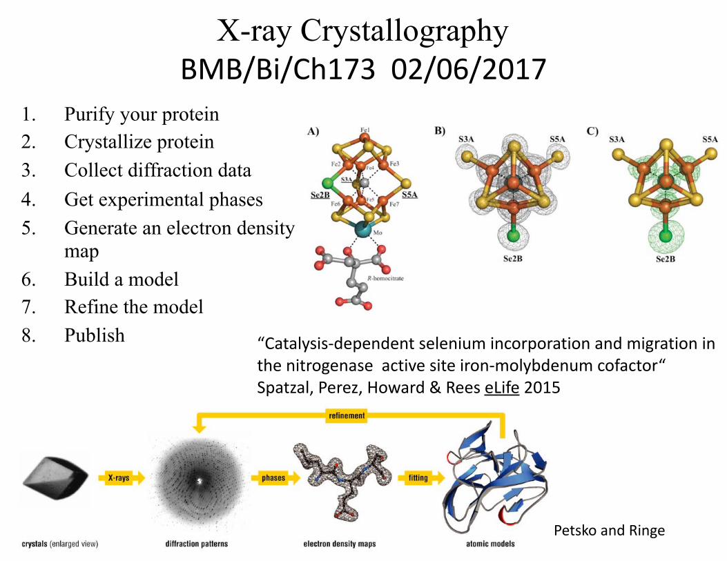
1. Purify your protein 2. Crystallize protein 3. Collect diffraction data4. Get experimental phases5. Generate an electron density
map 6. Build a model 7. Refine the model 8. Publish
PetskoandRinge
X-ray Crystallography BMB/Bi/Ch17302/06/2017
“Catalysis-dependentseleniumincorporationandmigrationinthenitrogenaseactivesiteiron-molybdenumcofactor“Spatzal,Perez,Howard&ReeseLife2015
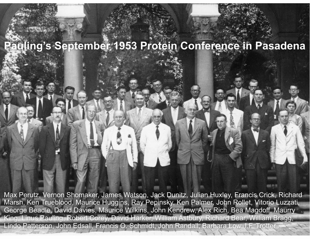
Pauling’s September 1953 Protein Conference in Pasadena
Max Perutz, Vernon Shomaker, James Watson, Jack Dunitz, Julian Huxley, Francis Crick, Richard Marsh, Ken Trueblood, Maurice Huggins, Ray Pepinsky, Ken Palmer, John Rollet, Vitorio Luzzati, George Beadle, David Davies, Maurice Wilkins, John Kendrew, Alex Rich, Bea Magdoff, Maurry King, Linus Pauling, Robert Corey, David Harker, William Astbury, Richard Bear, William Bragg, Lindo Patterson, John Edsall, Francis O. Schmidt, John Randall, Barbara Low, I.F. Trotter

Satellite Tobacco Mosaic Virus (STMV) Crystal
MacPherson


Useful texts and links• Rhodes “Crystallography made crystal clear” • Drenth “Principles of protein X-ray crystallography” • Lattman & Loll “Protein crystallography: a concise guide” • MacPherson “Preparation and analysis of protein crystals” • Ed. Rossmann & Arnold “Crystallography of biological
macromolecules: volume F”
Bernhard Rupp “Biomolecular Crystallography” http://www.ruppweb.org/Xray/101index.html

Nobel prizes related to X-ray Crystallography• 1901 Physics (1st) – Röntgen – X-rays • 1905 Physics – von Lenard – cathode rays • 1914 Physics – von Laue – X-ray diffraction by crystals • 1915 Physics – Bragg & Bragg – first crystal structure • 1946 Chemistry – Sumner – First enzyme crystals • 1962 Chemistry – Perutz & Kendrew – First protein structure • 1962 Medicine – Watson, Crick & Wilkins – DNA structure • 1964 Chemistry – Hodgkin – Protein crystallography • 1976 Chemistry – Lipscomb – Borane structure (Rees mentor) • 1982 Chemistry – Klug – Crystallographic EM • 1985 Chemistry – Hauptman & Karle – Direct methods (Isabella credited) • 1988 Chemistry – Deisenhofer, Huber & Michel – Photosynthetic reaction center • 1997 Chemistry – Agre & Walker – F1 ATPase structure, aquaporins • 2003 Chemistry – MacKinnon – Ion channel structures • 2006 Chemistry – Kornberg – RNA polymerase structure • 2009 Chemistry – Ramakrishnan, Steitz and Yonath – Ribosome structures • 2012 Chemistry – Leftkowitz & Kobilka - GPCRs

The international year of crystallography (2014)
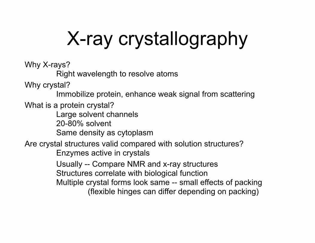
X-ray crystallographyWhy X-rays? Right wavelength to resolve atoms Why crystal? Immobilize protein, enhance weak signal from scattering What is a protein crystal? Large solvent channels 20-80% solvent Same density as cytoplasm Are crystal structures valid compared with solution structures? Enzymes active in crystals Usually -- Compare NMR and x-ray structures Structures correlate with biological function Multiple crystal forms look same -- small effects of packing (flexible hinges can differ depending on packing)

Crystal lattices and symmetry
• A crystal is a regular, 3-dimensional repeating array. The fundamental building block is the unit cell
• The crystal is built up by translations along x, y and z (which are not necessarily orthogonal)
x
y
z
Note that x, y, z form a right-handed coordinate system

Unit cell
• Described by three vectors, a, b, and c, which are related by angles α, β and γ
• Lattice built up by translating the unit cell along each of the lattice vectors

Choice of unit cell
1. Right-handed axis system 2. The basis vectors should
coincide as much as possible with directions of highest symmetry
3. Cell should be smallest one that satisfies previous condition. This may mean the choice of a non-primitive unit cell.
4. a > b > c 5. Angles either all <90° or ≥90°
There are many different cell choices for a lattice;I, II and III all constitute unit cells from which the entire lattice can be generated
Criteria for choosing a unit cell:

Primitive and non-primitive unit cells
• Primitive cells have a single point in each unit cell (1/8 of each corner point)
• Non-primitive cells have more than one point in each unit cell

Contents of a unit cell• A unit cell does not have to
contain a full object but it must contain the sum of the repeating object
• The contents of a unit cell can have no intrinsic symmetry, or can contain objects related by symmetry operations.
2-fold3-fold
4-fold 6-fold
5-fold
7-fold
8-fold
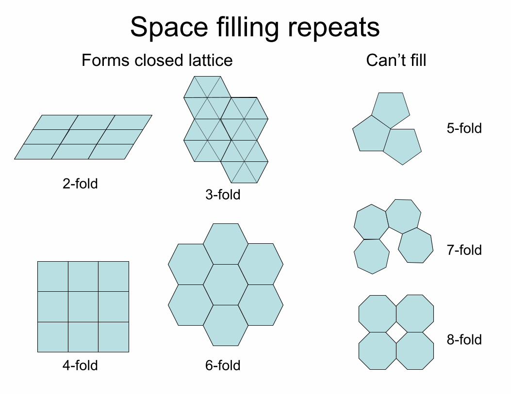
Space filling repeatsForms closed lattice Can’t fill
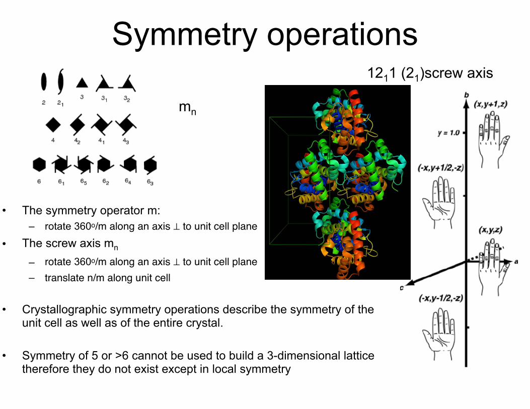
Symmetry operations1211 (21)screw axis
• The symmetry operator m: – rotate 360o/m along an axis ⊥ to unit cell plane
• The screw axis mn – rotate 360o/m along an axis ⊥ to unit cell plane– translate n/m along unit cell
• Crystallographic symmetry operations describe the symmetry of the unit cell as well as of the entire crystal.
• Symmetry of 5 or >6 cannot be used to build a 3-dimensional lattice therefore they do not exist except in local symmetry
mn

Examples of screw axis
Rotate 360/m = 120 Translate n/m n/m unit cells = 1/3
360/m = 120
n/m unit cells = 2/3
360/m = 60
n/m unit cells = 1/6
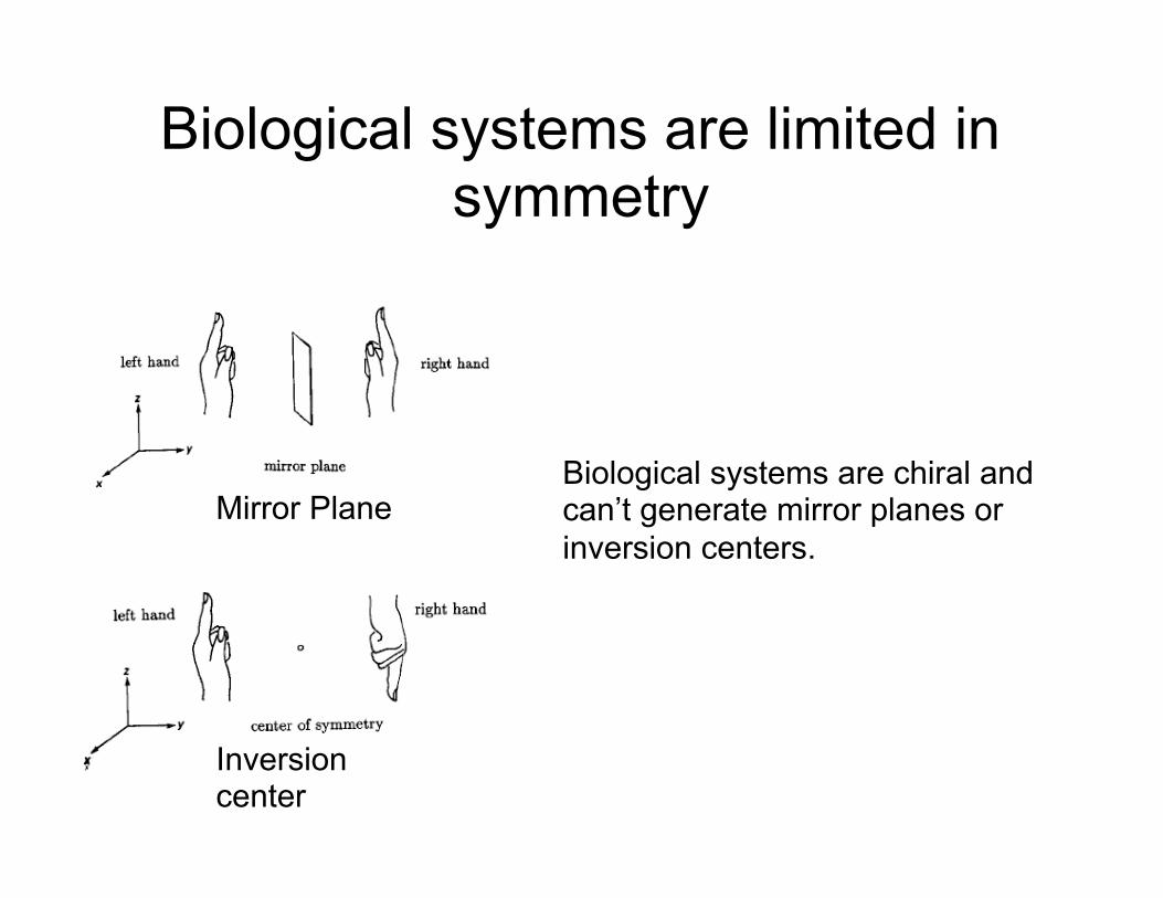
Biological systems are limited in symmetry
Biological systems are chiral and can’t generate mirror planes or inversion centers.
Inversion center
Mirror Plane

Point groups in proteins

Space groups• a complete description of a
crystal lattice – defines a unit cell type – a set of symmetry operations
• 230 possible 3D space groups – only 65 “biological space
groups” due to chirality (no mirror or inversion centers)
– Crystal systems define classes of space groups
• Higher symmetry means less data needed for completeness but more molecules in the unit cell (larger cells…)
Crystal System
Bravais Type
Condition of geometry Minimum symmetry
Triclinic P None 1
Monoclinic P, C α=γ=90 1 2-fold parallel to b
Orthrombic P, I, F α=β=γ=90 Three ⊥ 2-folds
Tetragonal P, I a=b; α=β=γ=90 1 4-fold parallel to c
Trigonal P, R Hex: a=b; α=β=90; γ=120 Rhomb: a=b=c; α=β=γ
1 3-fold axis
Hexagonal P a=b; α=β=90; γ=120 1 6-fold axis
Cubic P, F, I a=b=c; α=β=γ=90 4 3-folds along diagonal
Defined in excruciating detail in The International Tables for Crystallography volume A
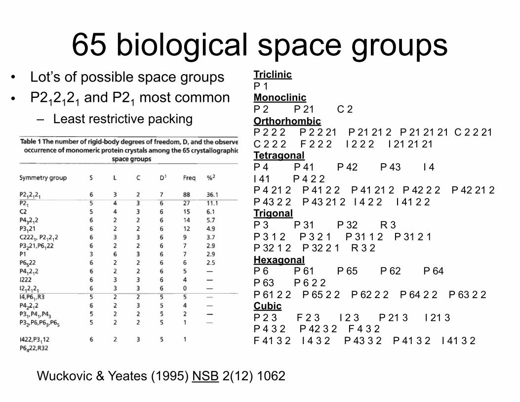
65 biological space groups• Lot’s of possible space groups • P212121 and P21 most common
– Least restrictive packing
Triclinic P 1 Monoclinic P 2 P 21 C 2 Orthorhombic P 2 2 2 P 2 2 21 P 21 21 2 P 21 21 21 C 2 2 21 C 2 2 2 F 2 2 2 I 2 2 2 I 21 21 21 Tetragonal P 4 P 41 P 42 P 43 I 4 I 41 P 4 2 2 P 4 21 2 P 41 2 2 P 41 21 2 P 42 2 2 P 42 21 2 P 43 2 2 P 43 21 2 I 4 2 2 I 41 2 2 Trigonal P 3 P 31 P 32 R 3 P 3 1 2 P 3 2 1 P 31 1 2 P 31 2 1 P 32 1 2 P 32 2 1 R 3 2 Hexagonal P 6 P 61 P 65 P 62 P 64 P 63 P 6 2 2 P 61 2 2 P 65 2 2 P 62 2 2 P 64 2 2 P 63 2 2 Cubic P 2 3 F 2 3 I 2 3 P 21 3 I 21 3 P 4 3 2 P 42 3 2 F 4 3 2 F 41 3 2 I 4 3 2 P 43 3 2 P 41 3 2 I 41 3 2
Wuckovic & Yeates (1995) NSB 2(12) 1062
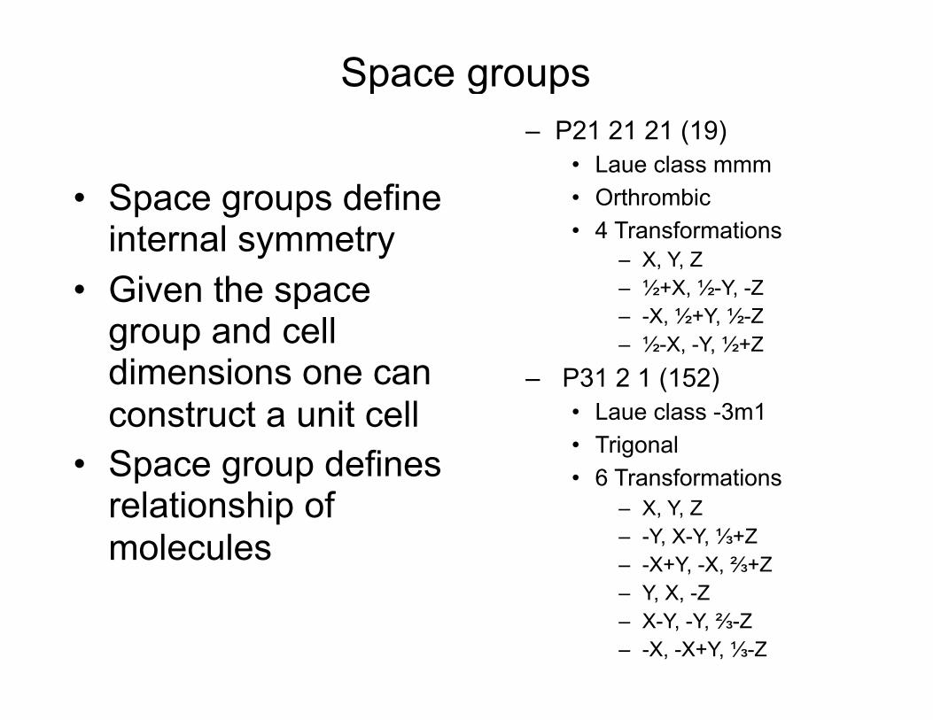
Space groups
• Space groups define internal symmetry
• Given the space group and cell dimensions one can construct a unit cell
• Space group defines relationship of molecules
– P21 21 21 (19) • Laue class mmm • Orthrombic • 4 Transformations
– X, Y, Z – ½+X, ½-Y, -Z – -X, ½+Y, ½-Z – ½-X, -Y, ½+Z
– P31 2 1 (152) • Laue class -3m1 • Trigonal • 6 Transformations
– X, Y, Z – -Y, X-Y, ⅓+Z – -X+Y, -X, ⅔+Z – Y, X, -Z – X-Y, -Y, ⅔-Z – -X, -X+Y, ⅓-Z
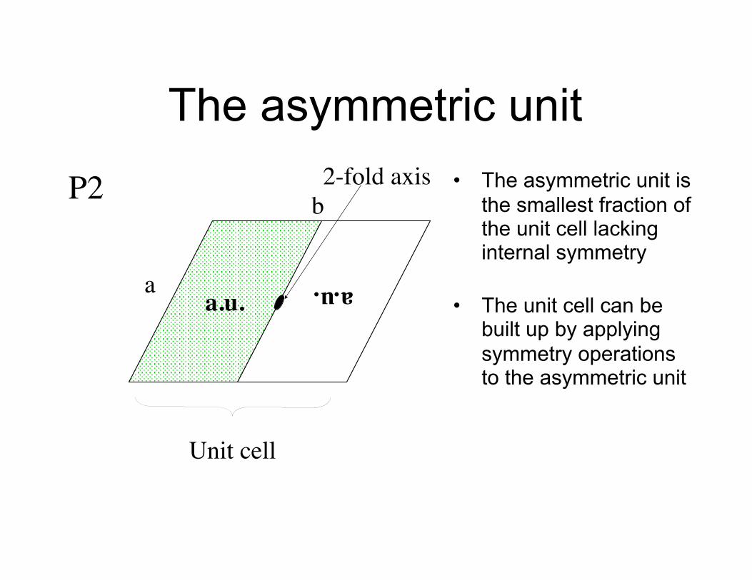
a.u.a.u.
The asymmetric unit• The asymmetric unit is
the smallest fraction of the unit cell lacking internal symmetry
• The unit cell can be built up by applying symmetry operations to the asymmetric unit
P2 2-fold axis
Unit cell
a
b

MacPherson

Joseph Fourier
• Théorie analytique de la chaleur - 1822
• The observation: a periodic function can be described as the sum of simple sine and cosine functions that have wavelengths as integrals of the function
• Also first predicted the Greenhouse Effect

Fourier Series A set of functions from a
Fourier synthesis that can be summed to reproduce a periodic function
Rhodes Crystallography Made Crystal Clear
t
€
f (x) = F cos2π (hx +α)
€
s(t)
€
cos2π (x)
€
− 13 cos2π (3x)
€
15 cos2π (5x)
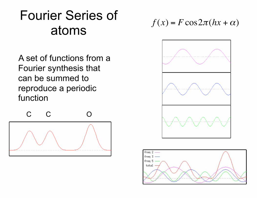
C C O
Fourier Series of atoms
A set of functions from a Fourier synthesis that can be summed to reproduce a periodic function
€
f (x) = F cos2π (hx +α)

How can we describe the diffraction pattern of a protein in a crystal?
Because there is no lens to refocus x-rays, we have to understand reciprocal space.
Diffraction: Scattering followed by interference

Diffraction by a wave
Diffraction: deviation of light from rectilinear propagation, is a characteristic of wave phenomena which occurs when a portion of a wave front is obstructed in some way. When various portions of a wave front propagate past some obstacle, and interfere at a later point past the obstacle, the pattern formed is called a diffraction pattern.
Obstruction (slit) is smaller than the wavelength

Diffraction patternWith 2 slits you now get patterns of interference. -Constructive when peaks or troughs intersect
-Destructructive when peaks and troughs intersect.

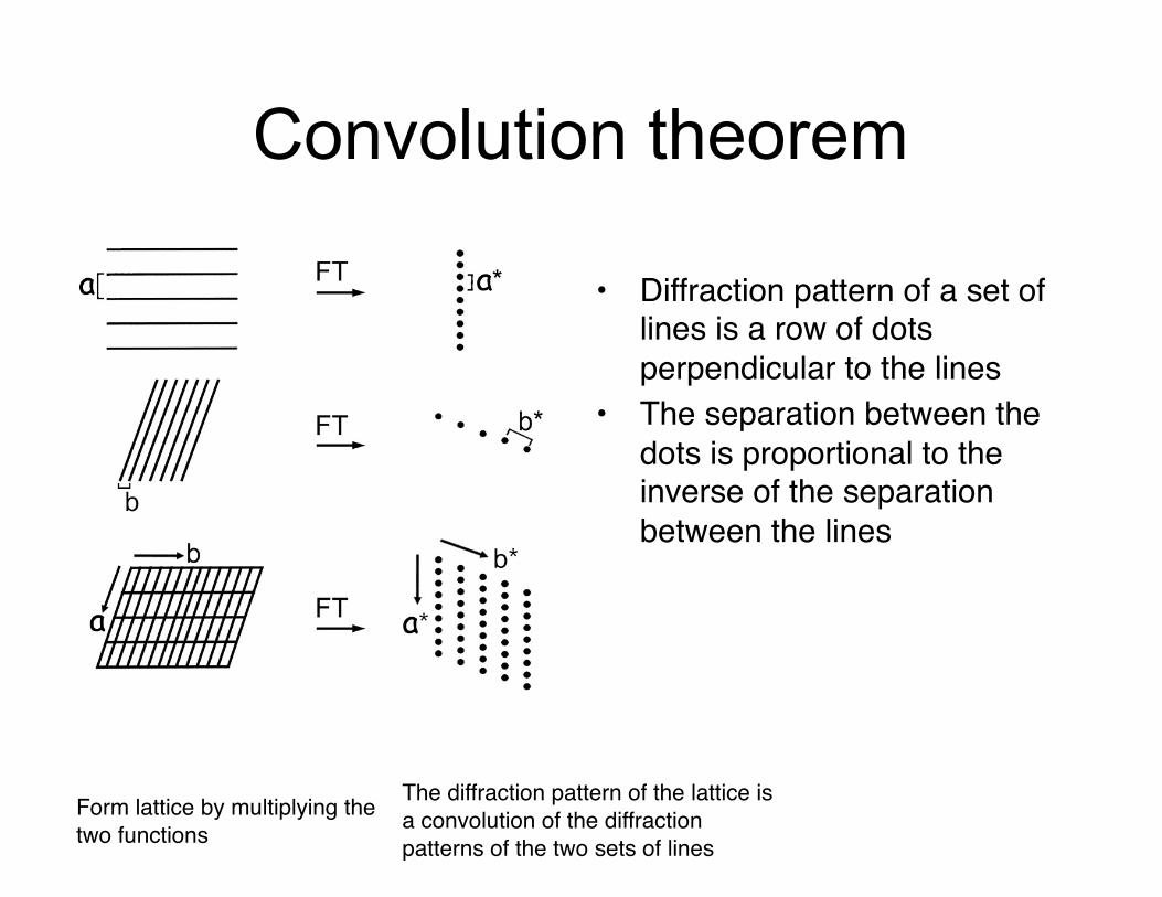
Convolution theorem
• The FT of the convolution of two functions is the product of their FTs
• The diffraction pattern of a lattice is a lattice
• The diffraction pattern of a molecular crystal – product of the transform of
• the molecule (molecular transform) • the diffraction pattern of a lattice (reciprocal lattice)
– Sampling of molecular transform at reciprocal lattice points

The convolution of two functions
Convolution theorem
Form lattice by multiplying the two functions
The diffraction pattern of the lattice is a convolution of the diffraction patterns of the two sets of lines
• Diffraction pattern of a set of lines is a row of dots perpendicular to the lines
• The separation between the dots is proportional to the inverse of the separation between the lines

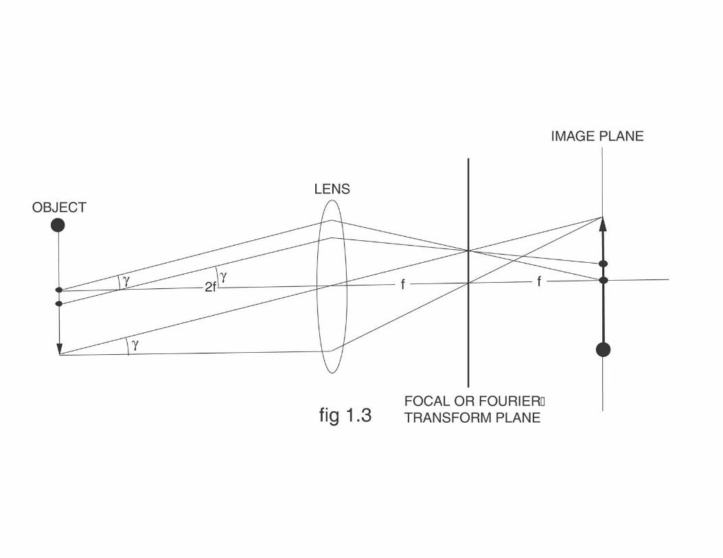
Allofthelenscontributestoeachpoint
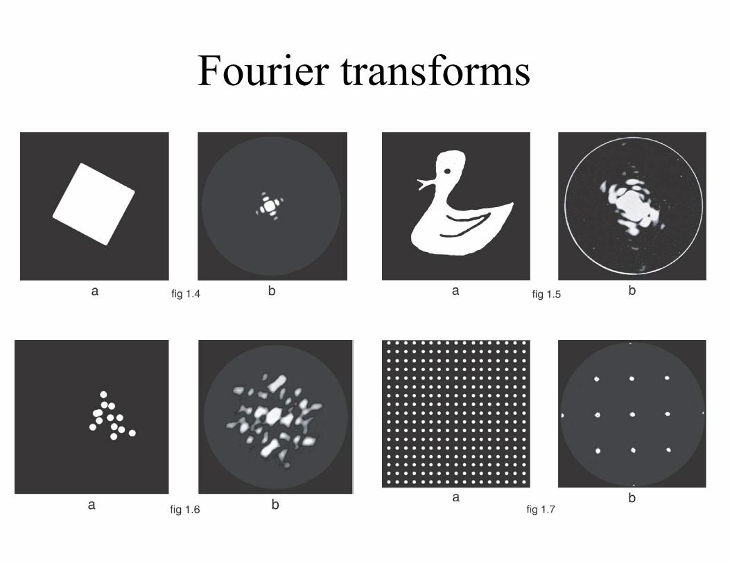
Fourier transforms
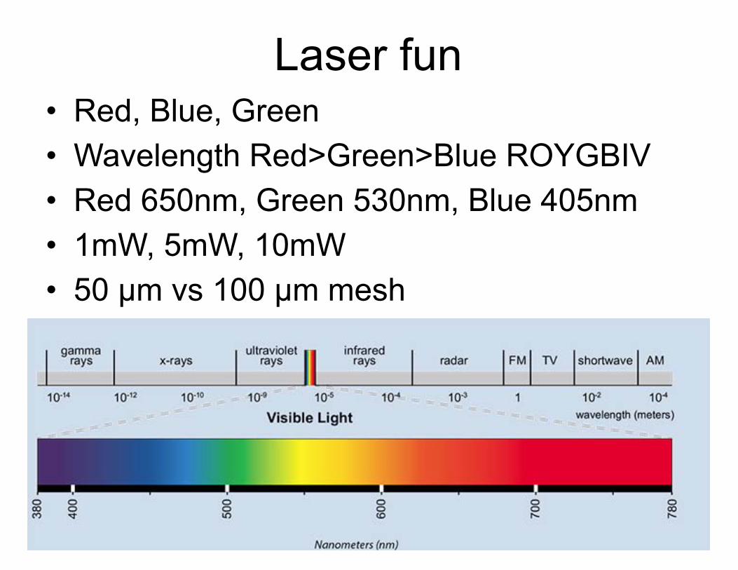
Laser fun• Red, Blue, Green • Wavelength Red>Green>Blue ROYGBIV • Red 650nm, Green 530nm, Blue 405nm • 1mW, 5mW, 10mW • 50 µm vs 100 µm mesh


The transform of the convolution of two functions is the product of the transforms
* =
x =
Fourier transform
of this
= this
F.T. F.T.
a∗b
F.T.(a∗b)
F.T.

Same molecular transform sampled by different lattices
Modified from Lipson & Taylor, 1964
a) Molecular transform b) Lattice c) Convolution of lattice and transform
d - f ) Same molecular transform sampled by different lattices
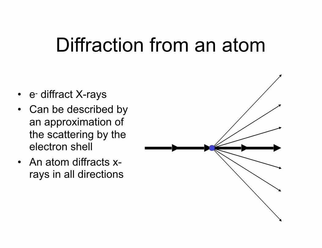
Diffraction from an atom
• e- diffract X-rays • Can be described by
an approximation of the scattering by the electron shell
• An atom diffracts x-rays in all directions

When do we get a diffraction pattern?
Bragg’s Law
When we get constructive interference from two diffracted waves
€
nλ = 2d sinθ

Diffraction planes reflect X-rays
Bragg’s Law
€
θ
€
θ€
λ
€
θ
€
nλ = 2d sinθ
€
d

Bragg’s Law
The difference in travel is equal to a multiple of the wavelength.
All of the atoms close to the Bragg plane contribute to the diffraction
€
θ
€
θ€
λ
€
θ
€
θ
€
θ
€
λ
€
λ
€
nλ = 2d sinθ
d €
2d sinθ
€
d
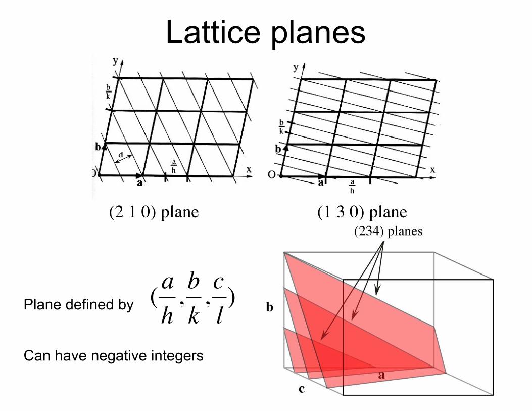
Lattice planes
(2 1 0) plane (1 3 0) plane
Plane defined by
Can have negative integers
€
(ah,bk,cl)

Gettingtoreciprocalspace
d1
1/d1
d2
1/d2
TheanglethatsatisfiesBragg’slawisinverselyproportionaltothelatticespacing
Reciprocalspacecanbedefinedbyavectornormaltotherealplanegivingusthereciprocallattice
θ1 θ2
€
d =nλ2sinθ
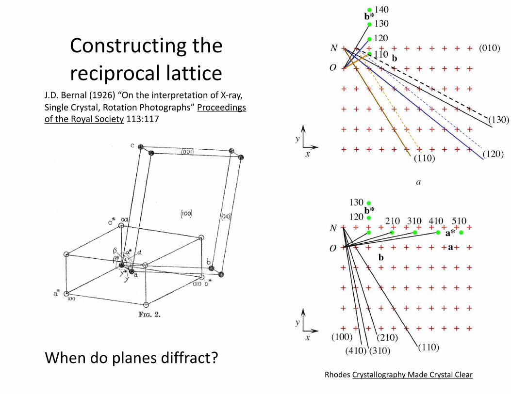
Constructingthereciprocallattice
J.D.Bernal(1926)“OntheinterpretationofX-ray,SingleCrystal,RotationPhotographs”ProceedingsoftheRoyalSociety113:117
RhodesCrystallographyMadeCrystalClear
Whendoplanesdiffract?

EwaldSphere
€
θ
1/d
€
1/λ
€
θ
€
θ
€
1/λ
DiffractedX-ray
Crystalorigin
Reciprocallatticeorigin
AconstructiontoindicatewhichBraggplanesdiffractforagivenorientation
€
12d
=1λsinθ