X.-Juan Sun Maxillary sinus floor elevation using a...
11
Maxillary sinus floor elevation using a tissue-engineered bone complex with OsteoBone t and bMSCs in rabbits X.-Juan Sun Z.-Yuan Zhang S.-Yi Wang S. A. Gittens X.-Quan Jiang L. Lee Chou Authors’ affiliations: X.-Juan Sun, Z.-Yuan Zhang, S.-Yi Wang, Department of Oral and Maxillofacial Surgery, School of Stomatology, Ninth People’s Hospital, School of Medicine, Shanghai Jiao Tong University, Shanghai, China S. A. Gittens, Faculty of Pharmacy and Pharmaceutical Sciences, University of Alberta, Edmonton, Alta, Canada L. Lee Chou, School of Dental Medicine, Boston University, MA, USA Correspondence to: X.-Quan Jiang Oral Bioengineering Lab School of Stomatology Ninth People’s Hospital School of Medicine Shanghai Jiao Tong University Shanghai 200011 China Tel.: þ 86 21 63135412 Fax: þ 86 21 63135412 e-mail: [email protected] Key words: bone marrow stromal cells (bMSCs), inorganic material (OsteoBonet), rabbits, sinus floor elevation, tissue engineering Abstract Objectives: To evaluate the effects of maxillary sinus floor elevation by a tissue-engineered bone complex with OsteoBonet and bone marrow stromal cells (bMSCs) in rabbits. Material and methods: Autologous bMSCs from adult New Zealand rabbits were cultured and combined with OsteoBonet at a concentration of 20 10 6 cells/ml in vitro. Twenty-four animals were used and randomly allocated into groups. For each time point, 16 maxillary sinus floor elevation surgeries were made bilaterally in eight animals and randomly repaired by bMSCs/material (i.e. OsteoBonet), material, autogenous bone and blood clot (n ¼ 4 per group). A polychrome sequential fluorescent labeling was also performed post-operatively. The animals were sacrificed 2, 4 and 8 weeks after the procedure and evaluated histologically as well as histomorphometrically. Results: New bone area significantly decreased from weeks 2 to 8 in the blood clot group, while bone area in the autologous bone reduced from weeks 4 to 8. In both groups, a significant amount of fatty tissue appeared at week 8. Accordingly, augmented height in both groups was also significantly decreased from weeks 2 to 8. The bone area in the material-alone group as well as in the bMSCs/material group, on the other hand, increased over time. Significantly more newly formed bone area and mineralization was observed in the center of the raised space in the bMSCs/material group than in the material-alone group. The augmented height was maintained in these two groups throughout the course of this study. Conclusion: These results suggest that OsteoBonet can successfully be used as a bone graft substitute and that the combination of this material with bMSCs can effectively promote new bone formation in sinus elevation. Osseointegrated implants are considered to be an ideal alternative to replace missing teeth. However, the bone height from the alveolar crest to the sinus floor at the poster- ior maxillary region is usually not adequate enough due to sinus pneumatization as well as the lack of stability caused by maxillary bone loss at edentulous sites required for osseointegrated implantation. Among the various techniques used to regain the height of resorbed maxilla, maxillary sinus floor elevation is regarded as an effective way to restore the upper jaw (Jensen & Shulman 1996). This procedure is based on the eleva- tion of the Schneiderian membrane from the floor of the maxillary sinus and the introduction of either a bone graft or a bone substitute (Graziani et al. 2004). Date: Accepted 28 December 2007 To cite this article: Sun X-J, Zhang Z-Y, Wang S-Y, Gittens SA, Jiang X-Q, Chou LL. Maxillary sinus floor elevation using tissue- engineered bone complex with OsteoBonet and bMSCs in rabbits. Clin. Oral Impl. Res. 19, 2008; 804–813 doi: 10.1111/j.1600-0501.2008.01577.x 804 c 2008 The Authors. Journal compilation c 2008 Blackwell Munksgaard
Transcript of X.-Juan Sun Maxillary sinus floor elevation using a...

Maxillary sinus floor elevation using atissue-engineered bone complex withOsteoBonet and bMSCs in rabbits
X.-Juan SunZ.-Yuan ZhangS.-Yi WangS. A. GittensX.-Quan JiangL. Lee Chou
Authors’ affiliations:X.-Juan Sun, Z.-Yuan Zhang, S.-Yi Wang,Department of Oral and Maxillofacial Surgery,School of Stomatology, Ninth People’s Hospital,School of Medicine, Shanghai Jiao Tong University,Shanghai, ChinaS. A. Gittens, Faculty of Pharmacy andPharmaceutical Sciences, University of Alberta,Edmonton, Alta, CanadaL. Lee Chou, School of Dental Medicine, BostonUniversity, MA, USA
Correspondence to:X.-Quan JiangOral Bioengineering LabSchool of StomatologyNinth People’s HospitalSchool of MedicineShanghai Jiao Tong UniversityShanghai 200011ChinaTel.: þ 86 21 63135412Fax: þ 86 21 63135412e-mail: [email protected]
Key words: bone marrow stromal cells (bMSCs), inorganic material (OsteoBonet), rabbits,
sinus floor elevation, tissue engineering
Abstract
Objectives: To evaluate the effects of maxillary sinus floor elevation by a tissue-engineered
bone complex with OsteoBonet and bone marrow stromal cells (bMSCs) in rabbits.
Material and methods: Autologous bMSCs from adult New Zealand rabbits were
cultured and combined with OsteoBonet at a concentration of 20 � 106 cells/ml
in vitro. Twenty-four animals were used and randomly allocated into groups. For each
time point, 16 maxillary sinus floor elevation surgeries were made bilaterally in
eight animals and randomly repaired by bMSCs/material (i.e. OsteoBonet), material,
autogenous bone and blood clot (n¼4 per group). A polychrome sequential
fluorescent labeling was also performed post-operatively. The animals were sacrificed
2, 4 and 8 weeks after the procedure and evaluated histologically as well as
histomorphometrically.
Results: New bone area significantly decreased from weeks 2 to 8 in the blood clot group,
while bone area in the autologous bone reduced from weeks 4 to 8. In both groups, a
significant amount of fatty tissue appeared at week 8. Accordingly, augmented height in
both groups was also significantly decreased from weeks 2 to 8. The bone area in the
material-alone group as well as in the bMSCs/material group, on the other hand, increased
over time. Significantly more newly formed bone area and mineralization was observed in
the center of the raised space in the bMSCs/material group than in the material-alone
group. The augmented height was maintained in these two groups throughout the course
of this study.
Conclusion: These results suggest that OsteoBonet can successfully be used as a bone graft
substitute and that the combination of this material with bMSCs can effectively promote
new bone formation in sinus elevation.
Osseointegrated implants are considered to
be an ideal alternative to replace missing
teeth. However, the bone height from the
alveolar crest to the sinus floor at the poster-
ior maxillary region is usually not adequate
enough due to sinus pneumatization as well
as the lack of stability caused by maxillary
bone loss at edentulous sites required for
osseointegrated implantation. Among the
various techniques used to regain the height
of resorbed maxilla, maxillary sinus floor
elevation is regarded as an effective way to
restore the upper jaw (Jensen & Shulman
1996). This procedure is based on the eleva-
tion of the Schneiderian membrane from
the floor of the maxillary sinus and the
introduction of either a bone graft or a
bone substitute (Graziani et al. 2004).
Date:Accepted 28 December 2007
To cite this article:Sun X-J, Zhang Z-Y, Wang S-Y, Gittens SA, Jiang X-Q,Chou LL. Maxillary sinus floor elevation using tissue-engineered bone complex with OsteoBonet and bMSCsin rabbits.Clin. Oral Impl. Res. 19, 2008; 804–813doi: 10.1111/j.1600-0501.2008.01577.x
804 c� 2008 The Authors. Journal compilation c� 2008 Blackwell Munksgaard

Since its introduction by Tatum and
Boyne (Boyne & James 1980; Tatum
1986), the use of autogenous bone grafts
in sinus augmentation has been considered
to be the ‘gold standard’ because of their
excellent survival with loaded implants and
the degree of functionality they afford
(Kent & Block 1989; Raghoebar 1993;
Neukam 1994; Nishibor 1994). However,
harvesting autogenous bone is generally
associated with several limitations, includ-
ing morbidity, infection, pain and blood
loss. In fact, reports suggest that the com-
plication rate of autogenous iliac grafts is
8% (Ueda et al. 2001). Autografts taken
from an intraoral donor site (e.g. mandible,
tuberosities) are limited in supply (Schim-
ming & Schmelzeisen 2004; Boyne et al.
2005).
To address the foregoing limitations as-
sociated with autogenous sources of bone,
an array of alternatives, including allograft,
xenograft and synthetic materials, has been
explored. Unfortunately, these materials
are associated with their own drawbacks.
Allografts and xenografts, for example, are
susceptible to immunorejection and carry
the risk of disease transmission (Moore
et al. 2001; Simon et al. 2002; Ueda et al.
2005). Some synthetic materials, on the
other hand, have limited potential for os-
teoconduction; using them alone has
usually failed to achieve the bone volume
expected (Wiltfang et al. 2002; Engelke
et al. 2003). Highlighting the importance
as well as the difficulty associated with
finding a suitable matrix is the fact that
bone regeneration within the sinus is de-
creased due to air pressure and low vascu-
larization within the area (Smiler 1992;
Schimming & Schmelzeisen 2004).
The ideal bone substitutes should be
biocompatible, and not only be osteocon-
ductive, but osteogenic and/or osteoinduc-
tive as well. Tissue engineering, namely
the application of scientific principles to
the design, construction, modification and
growth of living tissue using biomaterials,
cells and factors, alone or in combination,
may provide a means of developing
novel synthetic materials that possess the
aforementioned characteristics (Langer &
Vacanti 1993; Sittinger et al. 1996; Chang
et al. 2004).
Bone marrow stromal cells (bMSCs)
have been regarded as multipotent cells
residing in the bone marrow. Under ade-
quate culture conditions, bMSCs can dif-
ferentiate into various lineages of
mesenchymal tissue, including bone, car-
tilage, fat, tendon, muscle and marrow
stroma (Caplan 1991; Pittenger et al.
1999). In addition, they are relatively easy
to harvest and easily expandable in vitro
(Prockop 1997). These advantages have
made bMSCs ideal seed cells for tissue
engineering. In fact, the combination of
bMSCs with three-dimensional (3D) scaf-
folds has been thought to be the most
promising strategy to facilitate bone regen-
eration (Salgado et al. 2004).
To our knowledge, there are very few
clinical or animal studies that report using
the principles of tissue engineering to aug-
ment maxillary sinus (Schimming &
Schmelzeisen 2004; Ueda et al. 2005).
The scaffolds used in these experiments
are mainly polymeric materials, while the
seed cells are mostly sourced from the
periosteum (Schmelzeisen et al. 2003;
Schimming & Schmelzeisen 2004;
Springer et al. 2006; Zizelmann et al.
2007). In light of the fact that they can be
molded into a 3D scaffold and their degra-
dation rates can be customized, the poly-
mers used in these studies, like the poly
(lactide-co-glycolide), are inherently versa-
tile. Under certain conditions, however,
the hydrolyzation of these polymers can
produce an acidic environment and cause
tissue inflammatory and foreign body reac-
tion (Bostman et al. 1990). Although syn-
thetic bone graft substitutes ought to
exhibit biomechanical properties similar
to the bone that is being used for replace-
ment (Moore et al. 2001), most organic
materials lack the mechanical competence
needed (Rose & Oreffo 2002). Some inor-
ganic materials, however, are mechanically
suitable (Moore et al. 2001).
One example of such a material is Os-
teoBonet. This novel inorganic ceramic
material has an average porosity diameter
of 100–300 mm and an interporosity dia-
meter of 350–500 mm. As its ratio of cal-
cium and phosphorus is similar to that of
normal bone tissue, this material is biode-
gradable and possesses favorable mechan-
ical properties. Moreover, the presence of
calcium, phosphorus and silicon ions in-
side the scaffold has been shown to pro-
mote osteoblastic differentiation of seeded
cells as well as cell proliferation (Sun et al.
1997; Knabe et al. 2005).
The aim of the present study was to
explore the effects of the novel inorganic
materials OsteoBonet and autologous
bMSCs in rabbit’s sinus elevation.
Material and methods
Animals
Twenty-four male New Zealand rabbits,
each weighing from 2 to 2.5 kg, were
used for this study. [All animals were
obtained from the Ninth People’s Hospital
Animal Center (Shanghai, China), and all
procedures involving the use of rabbits
were approved by the Animal Research
Committee of the Ninth People’s Hospi-
tal.] The animals were randomly allocated
into 2-, 4- and 8-week observation groups
with eight rabbits in each group. At each
time point, 16 maxillary sinus floor eleva-
tion surgeries in all eight animals were
made and randomly repaired, with the
following four groups: group A consisted
of a tissue-engineered bMSCs/OsteoBonet
(Yenssen Biotech, Jiangsu, China) complex
(four cases); group B consisted of OsteoB-
onet alone (four cases); as a positive con-
trol, group C consisted of autogenous bone
obtained from iliac bone (four cases); and as
a negative control, group D consisted of
blood clot (four cases).
bMSCs isolation, culture and osteoblasticcharacteristic tests
Three milliliters of bone marrow was aspi-
rated from the fibula of a rabbit and cul-
tured, as described previously in Jiang et al.
(2005), in Dulbecco’s modified Eagle’s med-
ium (Gibco, Grand Island, N.Y., USA) with
10% fetal bovine serum (Hyclone, PERBIO,
Auckland, New Zealand). After 5 days,
non-adherent cells were removed and fresh
medium was added. The remaining adher-
ent cells were mainly mesenchymal stromal
cells. After the first passage, the following
three supplements for inducing osteogenesis
were added: 10�8 mmol/l dexamethasone
(Dex), 50mg/ml C-ascorb and 10 mmol/l b-
glycerophosphate (Sigma, St Louis, MO,
USA). The cells were then incubated con-
tinuously at 371C in 5% CO2. The cells at
passage 2 were used in our study.
After culturing in the induced medium
for 14 days, the cells were measured by
alkaline phosphatase (ALP) staining and
the Von Kossa test, as described in Jiang
(Jiang et al. 2005). Briefly, the cells induced
Sun et al . Maxillary sinus floor elevation using a tissue-engineered bone complex
c� 2008 The Authors. Journal compilation c� 2008 Blackwell Munksgaard 805 | Clin. Oral Impl. Res. 19, 2008 / 804–813

were fixed for 10 min at 41C and incubated
with a mixture of naphthol AS–MX phos-
phate, N,N-dimethylformamide and fast
blue BB salt (ALP kit, Hongqiao, Shanghai,
China). The Von Kossa staining method
consisted of the cells being fixed in 70%
ethanol and stained with 5% silver nitrate,
5% Na2SO3 and then observed.
Preparation of cell material complex usedin vitro and in vivo
bMSCs were collected and washed with
PBS, counted and then combined with the
material (i.e. OsteoBonet) at a final con-
centration of 20 � 106 cells/ml for the si-
nus surgeries. Extra cell material
complexes were further cultured in com-
plete medium at 371C in vitro; cell attach-
ment and spreading were visually assessed
4 h as well 4 days later through scanning
electron microscopy (Philips Quanta-200,
FEI, Eindhoven, Holland).
Autogenous bone was harvested from the
iliac crest, as described by Ueda et al.
(2001). Briefly, a 15-mm incision was
made over the iliac crest after local anesthe-
sia, which exposed the ilium. A 15 � 5 mm
corticocancellous bone block was harvested
from the right iliac crest. The periosteum
and skin flap were replaced and sutured.
The corticocancellous bone block was cut
into small particles before they were grafted
to the maxillary sinus. The blood to be used
as blood clot for the experiments was col-
lected from the ear vein with a sterile
syringe and allowed to clot naturally before
the surgery.
Maxillary sinus floor augmentationprocedure
The rabbits were anesthetized with 0.5
mg/kg sodium pentobarbital intravenously.
0.5 ml of 1% lidocaine with epinephrine
(1 : 100,000) was injected subcutaneously
for local anesthesia. According to the sur-
gical method performed by Asai et al.
(2002) and Xu et al. (2003), a 2.5 cm
vertical midline incision was made and
the skin and periosteum were subsequently
raised to expose the nasal bone and nasoin-
cisal suture line. Using a round bur, two
oval nasal bone windows (8 � 4 mm) were
outlined bilaterally on the nasal bone. The
window was located approximately 20 mm
anterior to the nasofrontal suture line and
10 mm lateral to the midline. Then, fenes-
trae were made by osteotomy during con-
tinuous cooling with sterile saline solution.
Care was taken to avoid the damage to the
antral membrane, which moved back
and forth with the respiratory rhythm. A
Freer elevator (Medical equipment limited
company, Shanghai, China) was used to
gently push the membrane inward (Fig.
1a). The membrane was then raised from
the floor and lateral walls of the antrum to
provide a large compartment. One of four
different grafts, which consisted of either
bMSCs/material, material alone, autoge-
nous bone particles or blood clot, was
slightly filled without compression into
the compartment. The size of the cavity
created at this structure was standardized
by volume among the groups. Sutures were
then placed to close the periosteum and
skin (Fig. 1b and c).
Sequential fluorescent labeling
A polychrome sequential labeling method
(Roldan et al. 2004) was carried out to label
the mineralized tissue and assess the time
course of new bone formation and remodel-
ing. Two and 4 weeks after the operation,
the animals were administered with
25 mg/kg of tetracycline and 30 mg/kg of
alizarin complexon intraperitoneally, re-
spectively. Twenty milligrams per kilo-
gram of calcein green (Sigma) was
administered 3 days before the animals
were sacrificed at week 8.
General and histological observation
The rabbits were sacrificed at 2, 4 or 8
weeks after surgery, exsanguinated and
perfused via the jugular vein with 10%
buffered formaldehyde. The maxillae were
dissected and cut into smaller blocks,
which included the nasal and maxillary
sinus, then fixed in the same solution.
The block was divided in the transverse
plane at the rostrocaudal midpoints of the
osteotomy site. One half was decalcified,
embedded in paraffin, sectioned into 4-mm-
thick sections and stained with hematox-
ylin–eosin. The other half was dehydrated
gradually in ethyl alcohol and was finally
embedded in polymethymetacrylate. The
specimens were cut into 150-mm thick
sections using a microtome (Leica, Ham-
burg, Germany), and were subsequently
ground and polished to a final thickness
of about 40 mm (Donth & Breuner 1982;
Rohrer & Schubert 1992). Undecalcified
sections were observed for fluorescent la-
beling using a microscope under ultraviolet
light. Decalcified sections were subject to
histologic and histomorphometrical obser-
vations.
Histomorphometric analysis
The measurements were performed with
decalcified specimens using a personal
computer-based image analysis system
(Image-Pro Plust, Media Cybernetic, Silver
Fig. 1. Illustration of the surgical procedure: the maxillary sinus was opened and the membrane was
subsequently pushed inwards (a). Filling the grafts into the sinus (b). The area (outlined by a red line) in the
X-ray radiograph reflects the sinus (c).
Sun et al . Maxillary sinus floor elevation using a tissue-engineered bone complex
806 | Clin. Oral Impl. Res. 19, 2008 / 804–813 c� 2008 The Authors. Journal compilation c� 2008 Blackwell Munksgaard

Springs, MD, USA). Four randomly se-
lected sections from the serial sections
collected from each sample were analyzed
manually. The newly formed bone area
(i.e. the percentage of newly formed bone
area in the raised area observed) and the
augmented height (i.e. the maximum
length in the augmented space) were
recorded and compared as described pre-
viously by Ueda et al.(2001) and Ohya
et al.(2005).
Statistical analysis
Statistically significant differences
(Po0.05) between the various groups and
implantation times were measured using
ANOVA and SNK post hoc. All statistical
analysis was carried out using an SAS 8.2
statistical software package (SAS, Cary,
NC, USA). All the data are expressed as
mean� standard deviation.
Results
Cell culture, ALP and Von Kossa staining
Cell clones formed 5–7 days after initial
seeding and reached confluence after ap-
proximately 12–14 days. bMSCs at passage
2 displayed the typical fibroblastic spindle-
shaped phenotype and were used for further
studies (Fig. 2a). Two weeks after culture
in osteogenic medium, areas of ALP-posi-
tive staining and calcium deposits were
observed (Fig. 2b and c), which suggested
that cells induced by Dex led to the bMSC
differentiation into osteoblastic cells: an
occurrence that is well documented in the
literature (Cornet et al. 2002, 2004).
Adhesion and spreading of bMSCs on thematerial
Under the scanning electronic microscope,
the OsteoBonet appeared porous in struc-
ture; the pores were interconnected with
each other (Fig. 3a). Four hours after the
bMSCs were combined with the material,
cells attached to the surface of the scaffold
in vitro. After 4 days, cell spreading on the
implant surfaces was observed (Fig. 3b).
Additionally, these results suggested that
the material was suitable for the proposed
in vivo studies as it facilitated bMSCs
adhesion and spreading onto its surface.
General observations
After surgery, all rabbits recovered well.
Slight post-surgical edema was observed
at the recipient site in each animal. This
disappeared 2–3 days after the procedure.
There was no sign of infection at any time.
Histological findings
At 2 weeks after implantation
In both group A (bMSCs/material) and
group B (material alone), the augmented
space was convex and newly formed trabe-
culae were mainly found close to the parent
bony wall and raised membrane (Fig. 4a
and c). In the same position of the aug-
mented area, more newly formed bone was
observed in group A (Fig. 4b) than in group
B (Fig. 4d). All trabeculae and material
particles were embedded in fibrous connec-
tive tissue.
Group C (autogenous bone) showed tra-
beculae of newly formed bone around the
grafted bone. The lacunae around the os-
teocytes were large. The newly formed
trabeculae were embedded in fibrovascular
tissue with no evidence of inflammation
(Fig. 7a and b).
In group D (blood clot), the augmented
space was concave, and newly formed tra-
beculae embedded in fibrovascular tissue
were found at the center of the cavity. No
remnants of blood clots were visible. More-
over, there was no evidence of inflamma-
tion at the site (Fig. 8a and b).
At 8 weeks after implantation
In both group A and group B, newly formed
bone found adjacent to the bony wall of the
Fig. 2. The second passage bMSCs displayed the typical spindle-shaped fibroblastic phenotype ( � 10) (a).
Alkaline phosphatase-positive stain area (b) and Von Kossa-positive stain area (c) 14 days after having been
induced by dexamethasone ( � 16).
Fig. 3. OsteoBonet: a new inorganic material with a porous structure with an average pore diameter of 200mm
(a). Four days after the bMSCs were combined with OsteoBonet, they could be seen attaching and spreading to
the inner surface of the scaffold (b).
Sun et al . Maxillary sinus floor elevation using a tissue-engineered bone complex
c� 2008 The Authors. Journal compilation c� 2008 Blackwell Munksgaard 807 | Clin. Oral Impl. Res. 19, 2008 / 804–813

cavity was denser than at 2 weeks (Fig. 5a
and c). More mineralized bone in the
bMSCs/material sites were also observed
in the center of the augmented area than in
the material sites alone (Fig. 5b and d).
In the autogenous bone site, the aug-
mented space filled with trabeculae that
were more mature than at 2 weeks. Appar-
ent lamellar bone structure was also ob-
served adjacent to the cortical bone wall of
the cavity.
In the blood clot site, the augmented
space appeared to be smaller than that 2
weeks after implantation. Trabeculae in
the augmented space were more mature
than that at 2 weeks.
At 8 weeks after implantation
In both group A (bMSCs/material) and B
(material alone), the augmented space re-
mained convex. Several formed bones were
observed that showed many interconnec-
tions. This newly formed bone bound to
the material particles tightly in certain
areas (Fig. 6a and c). In the bMSCs/mate-
rial sites, the thick lamellar bone was more
frequently found in the center of the space
(Fig. 6b). In comparison, the general ten-
dency of bone maturation in the bMSCs/
material sites was greater than in the ma-
terial-alone sites (Fig. 6d).
In group C (autogenous bone), cortical
bone formation was observed adjacent to
the raised membrane. The grafted bone and
newly formed trabeculae observed at 2 or 4
weeks were reduced. The cortical bone was
also embedded with fatty tissue (Fig. 7c
and d).
In group D (blood clot), the augmented
space reduced dramatically. The sinus
membrane that was raised almost turned
back, forming a sinus cavity again (Fig. 8c).
Few newly formed bones were found under
the raised sinus membrane. This area was
also embedded with fatty cells (Fig. 8d).
Fluorescence microscopy
The deposition of mineralized bone matrix
was observed in the bMSCs/material and
material-alone sites at different time points
as demonstrated by green tetracycline (yel-
low), complexon (red) and calcein green
(green) (Fig. 9a and b). All the three differ-
ent fluorescent-labeling areas in group A
(bMSCs/material) were larger than those in
group B (material alone). As evidenced by
the fluorescence of tetracycline (yellow) at
week 2 and alizarin complexon (red) at
week 4 in group A (Fig. 9a), the miner-
alization of new bone was present more
frequently in group A than in group B in
the center of the augmented area. These
data suggest that bMSCs contributed to the
enhanced mineralized area over time.
Histomorphometric analysis
Bone area
At 2 weeks, bone area of the autogenous
bone site (34.79� 7.18%) was significantly
higher than that of group A (bMSCs/
Fig. 4. Histological findings at 2 weeks after implantation. Most newly formed trabecular bone was found
along the periphery of the raised sinus and the surface of the OsteoBonet near the parent cortical bone wall in
both group A (a, b) and group B (c, d). NB, nasal bone; M, augmented sinus membrane; P, particles; B, bone.
a–d, hematoxylin–eosin; a, c, � 1.25; b, d, � 10.
Fig. 5. Histological findings at 4 weeks after implantation. There was more newly formed bone than that at 2
weeks found in both group A (a, b) and group B (c, d). NB, nasal bone; M, augmented sinus membrane;
P, particles; B, bone. a–d, hematoxylin–eosin; a, c, � 1.25; b, d, � 10.
Sun et al . Maxillary sinus floor elevation using a tissue-engineered bone complex
808 | Clin. Oral Impl. Res. 19, 2008 / 804–813 c� 2008 The Authors. Journal compilation c� 2008 Blackwell Munksgaard

material) (21.04� 2.67%) (Po0.05) and
group B (material) (19.22� 2.25%)
(Po0.05). No significant difference was ob-
served between other groups. At 4 weeks,
bone area of group A (23.47� 3%), group
B (22.95� 2.18%) and group C (autoge-
nous bone) (28.75� 5.8%) was all signifi-
cantly higher than that of group D (blood
clot) (15.7� 3.33%) (Po0.05). There was
no significant difference between the other
groups. At 8 weeks, the bone area of group
A (35.36� 10.57%) was significantly
higher than that of group C (19.58�2.45%) and group D (13.84� 4.03%)
(Po0.05). No significant differences were
detected between groups C and D. There
was 30% more bone area in group A than
in group B, although this was not statisti-
cally significant due to, perhaps, high in-
tra-group variation.
In the bMSCs/material site, bone area
increased significantly from 4 to 8 and 2 to
8 weeks (Po0.05). Bone area in the mate-
rial site did not show a significant increase
between any observation times. In group
C, bone area decreased over time. There
was a significant decrease in the bone area
from 2 or 4 weeks to 8 weeks (Po0.05). In
group D, from 2 weeks (27.62� 6.44%)
to 4 weeks (15.76� 3.33%) and from 2 to
8 weeks, significant bone area decrease was
also observed (Po0.05) (Fig. 10).
Augmented height
For both control groups, the augmented
height decreased significantly from 2 to 8
weeks (Po0.05). In group C (autogenous
bone group), significant differences were
observed at different times after implanta-
tion (Po0.05). In group D (blood clot), a
significant difference could be observed
between 2 and 8 and 4 and 8 weeks
after surgery (Po0.05). However, for group
A (bMSCs/material) and group B (material
alone), the augmented height was not sig-
nificantly different (Po0.05). Both groups
A (bMSCs/material) and B (material alone)
had a higher augmented height than that in
group D at each observation time point
(Po0.05); they were also significantly dif-
ferent from group C at weeks 4 and 8
(Po0.05). The augmented height in group
C was higher than in group D at 2 and 4
weeks (Po0.05), but this difference was no
longer present at 8 weeks (Fig. 11).
Discussion
The use of rabbits as a model for maxilla
sinus elevation is well-documented: rabbits
have the same ventilation with air ex-
changes through the nasal cavity as hu-
mans and a well-defined ostium opening to
their nasal cavities as well (Kumlien &
Schiratzki 1985; Scharf et al. 1995). As
air pressure causes movement of the max-
illary sinus membrane, the grafted material
in the sinus is subjected continuously to
Fig. 6. Histological findings 8 weeks after implantation. Newly formed bone with many interconnections was
observed in most of the convex augmented space in both group A (a, b) and group B (c, d).NB, nasal bone; M,
augmented sinus membrane. a–d, hematoxylin–eosin; a, c, � 1.25; b, d, � 10.
Fig. 7. Histological findings of group C (autogenous bone). Fibrovascular tissue was observed surrounding
newly formed trabecular bone 2 weeks after implantation at magnifications of � 1.25 (a) and � 10 (b).
Cortical bone, which was surrounded by fatty tissue, was observed adjacent to the raised membrane 8 weeks
after implantation at magnification of � 1.25 (c) and at � 10 (d). NB, nasal bone; M, raised sinus membrane;
B, bone.
Sun et al . Maxillary sinus floor elevation using a tissue-engineered bone complex
c� 2008 The Authors. Journal compilation c� 2008 Blackwell Munksgaard 809 | Clin. Oral Impl. Res. 19, 2008 / 804–813
this air pressure, which will ultimately
affect the augmented bone healing process
and structure (Garey et al. 1991; Asai et al.
2002). Under these conditions, the grafting
material is considered to be an important
determinant of the success or failure of a
bone augmentation procedure (Xu et al.
2004). Results from numerous clinical
cases have already suggested that freeze-
dried demineralized bone cannot withstand
air pressure in the sinus (Jensen & Shul-
man 1996; Block & Kent 1997; Wheeler
1997). Some studies suggest that even
alloplastic materials have problems in
these ischemic areas (Schmelzeisen &
Schimming 2003). Hence, in addition to
being capable of stimulating osteoinduc-
tion, osteoconduction and maturation of
new bone structure, while resisting resorp-
tion, maxillary sinus grafts should also be
able to provide adequate stability (Block &
Kent 1997). Blood clots and autologous
bone have been explored previously as
grafts (Block & Kent 1997; Xu et al.
2004). Consequently, these materials
were used as controls in this study.
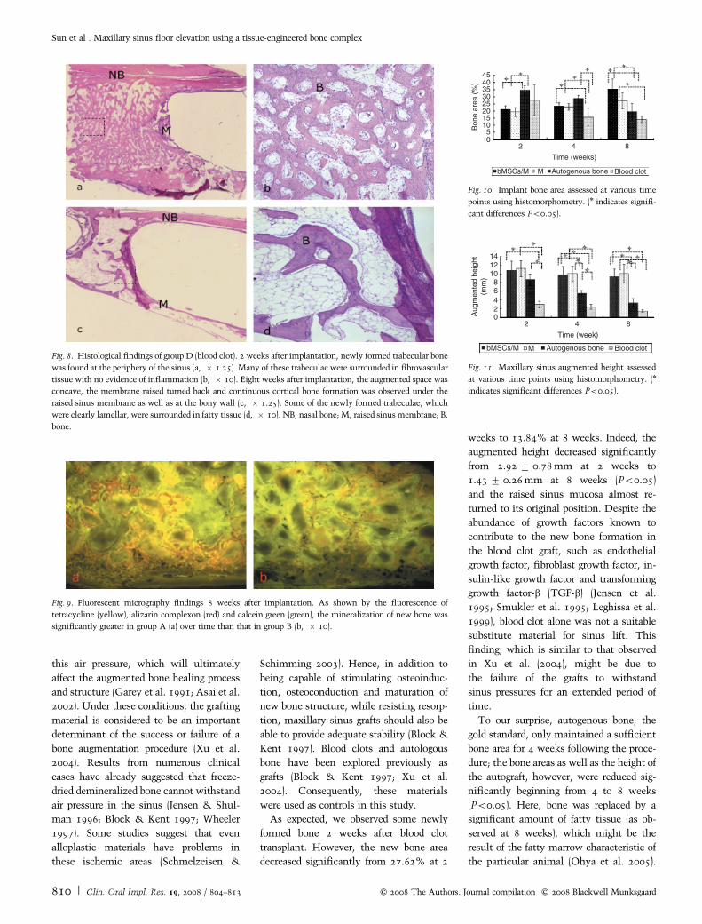
As expected, we observed some newly
formed bone 2 weeks after blood clot
transplant. However, the new bone area
decreased significantly from 27.62% at 2
weeks to 13.84% at 8 weeks. Indeed, the
augmented height decreased significantly
from 2.92� 0.78 mm at 2 weeks to
1.43� 0.26 mm at 8 weeks (Po0.05)
and the raised sinus mucosa almost re-
turned to its original position. Despite the
abundance of growth factors known to
contribute to the new bone formation in
the blood clot graft, such as endothelial
growth factor, fibroblast growth factor, in-
sulin-like growth factor and transforming
growth factor-b (TGF-b) (Jensen et al.
1995; Smukler et al. 1995; Leghissa et al.
1999), blood clot alone was not a suitable
substitute material for sinus lift. This
finding, which is similar to that observed
in Xu et al. (2004), might be due to
the failure of the grafts to withstand
sinus pressures for an extended period of
time.
To our surprise, autogenous bone, the
gold standard, only maintained a sufficient
bone area for 4 weeks following the proce-
dure; the bone areas as well as the height of
the autograft, however, were reduced sig-
nificantly beginning from 4 to 8 weeks
(Po0.05). Here, bone was replaced by a
significant amount of fatty tissue (as ob-
served at 8 weeks), which might be the
result of the fatty marrow characteristic of
the particular animal (Ohya et al. 2005).
Fig. 9. Fluorescent micrography findings 8 weeks after implantation. As shown by the fluorescence of
tetracycline (yellow), alizarin complexon (red) and calcein green (green), the mineralization of new bone was
significantly greater in group A (a) over time than that in group B (b, � 10).
40353025201510
50
45
2 4 8
Time (weeks)
Bon
e ar
ea (
%)
Fig. 10. Implant bone area assessed at various time
points using histomorphometry. (n indicates signifi-
cant differences Po0.05).
02468
101214
2 4 8
Time (week)
Aug
men
ted
heig
ht(m
m)
bMSCs/M M Autogenous bone Blood clot
Fig. 11. Maxillary sinus augmented height assessed
at various time points using histomorphometry. (n
indicates significant differences Po0.05).
Fig. 8. Histological findings of group D (blood clot). 2 weeks after implantation, newly formed trabecular bone
was found at the periphery of the sinus (a, � 1.25). Many of these trabeculae were surrounded in fibrovascular
tissue with no evidence of inflammation (b, � 10). Eight weeks after implantation, the augmented space was
concave, the membrane raised turned back and continuous cortical bone formation was observed under the
raised sinus membrane as well as at the bony wall (c, � 1.25). Some of the newly formed trabeculae, which
were clearly lamellar, were surrounded in fatty tissue (d, � 10). NB, nasal bone; M, raised sinus membrane; B,
bone.
Sun et al . Maxillary sinus floor elevation using a tissue-engineered bone complex
810 | Clin. Oral Impl. Res. 19, 2008 / 804–813 c� 2008 The Authors. Journal compilation c� 2008 Blackwell Munksgaard

Similar results were reported in other stu-
dies (Watanabe et al. 1999; Wada et al.
2001). Another explanation might be that
cortical and cancellous bone in the auto-
genous bone graft could not withstand
sinus pressures for long periods of time,
and as such, start to lose their density and
height during the first several weeks (Jen-
sen & Shulman 1996). Some published
clinical data showed that the absorption
rate using autogenous bone in sinus aug-
mentation was 47% 6–7 months after
surgery (Johansson et al. 2001). Watanabe
et al. also linked the absorption rate of
different donor sources to different ratios
of cortical bone or cancellous bone (Wata-
nabe et al. 1999). For example, chin bone
grafts demonstrated greater volume main-
tenance than did the iliac bone simply
because chin bone contains more cortical
bone. Secondly, bone grafts usually need
functional loading stimulation (via im-
plants, for example) to counter the innate
response of resorbing bone implants (Schle-
gal et al. 2003; Ohya et al. 2005). In this
preliminary study, we did not provide any
functional loading stimulation. Conse-
quently, the grafts might have failed to
receive the necessary stimulation to pre-
vent graft resorption. In fact, the resorption
of autogenous bone graft has not only been
reported in a sinus lifting model but has
also been described in studies that explored
ridge augmentation and bone defect’s re-
storation (Chenug et al. 1994; Roccuzzo
et al. 2007).
In our study, both group A and B showed
the convex augmented space, which sug-
gested that the grafted material can with-
stand sinus air pressure and maintain the
augmented space. The statistical results
showed that both groups maintained their
augmented height throughout the observa-
tion period (P40.05). OsteoBonet alone
achieved increased bone area while the
addition of bMSCs to this material demon-
strated the most promising results.
Throughout the experiment, both these
groups showed increased bone area and
maturation of newly formed bone. Lamel-
lar bone structure in histological sections
was observed 8 weeks after surgery.
In these experiments, no animals
showed signs of infection after surgery.
Histologically, newly formed bone was
found growing into the pores of the mate-
rial from the host bone and had a direct
bond with the graft without obvious in-
flammatory cell infiltration. Furthermore,
cells were found spreading along the mate-
rial surface after being cultured in vitro.
These results suggest that OsteoBonet has
a good biocompatibility and can facilitate
bMSCs adhesion onto its surface. Similar
results have also been observed by Xu et al.
(2004) when using deproteinized bone. Ac-
cording to Fleming et al. (2000), high
calcium phosphate concentrations appear
to improve the integration of implants into
host bone. One possible mechanism is that
the high concentrations of calcium and
phosphate ions may initiate biomineraliza-
tion or may influence osteoblast differen-
tiation in the cells found in adjacent tissues
(Damien et al. 1994). Moreover, silica ion
in the material also affords biocompatibil-
ity. When comparing the effects of different
calcium phosphate particles on the growth
of osteoblasts in vitro, Sun et al. (1997)
found that, under the same experimental
conditions, cell population, the concentra-
tions of TGF-b and ALP cultured on the
silica surface were similar with those on
normal plates. Knabe et al. (2005) found
that novel glass ceramics with silica sup-
ported cellular proliferation as well as the
expression of osteogenic markers as much
as, or better than, tri-calcium-phosphate.
Hard tissue engineering may potentially
provide a better alternative than currently
available bone grafts (Petrovic et al. 2006).
To this end, we used a new inorganic
material (i.e. OsteoBonet) as a scaffold
and bMSCs as seed cells to form a compo-
site of grafted material for maxillary sinus
floor elevation. We found that new bone
was formed mainly in areas close to the
parent bone in both group A and group B 2
weeks after transplantation. From 4 weeks
to 8 weeks, the bone area in group A
(bMSCs/material) increased significantly
from 23.47� 3% to 35.36� 10.57%
(Po0.05). However, the increase in bone
area in group B (material alone) at any time
was not significant. In comparison with
the autogenous group, the bone area in the
bMSCs/material group was much higher at
8 weeks (Po0.05); however, the difference
of bone volume between the material group
and autogenous bone was not significant.
At 8 weeks, the mean bone area in group A
(bMSCs/material) was 30% more than
that in the material-alone group, but differ-
ence of bone area between the bMSCs/
material group and the material-alone
group was not significant. This might be
due to the large variability associated with
the limited number of animals used in this
study.
OsteoBonet has pores with a suitable
diameter that is good for cell adhesion and
growth (Sun et al. 1997). We observed more
new bone formation in the bMSCs/mate-
rial group than in group material after
transplantation. This result concurred
with the results from the fluorescent label-
ing experiments. From the fluorescent
images taken, all three fluorescent colors
were intense and were found extensively in
the bMSCs/material site, which indicated
that the mineralization of newly formed
bone occurred after 2 weeks, while the
material-alone group only demonstrated
intense fluorescence at around week 8.
This suggests that bMSCs have contributed
to the increased bone area as well as en-
hanced mineralization. In the material-
alone group, the results suggested that
new bone formation depended on the os-
teoconductive property of the material, as
the newly formed bone was observed
mainly at the periphery of the parent
bone. When bMSCs were used along with
the OsteoBonet, these cells filtrated into
the pores of the material and more bone
formed in the inner area.
In this study, we simply focused on new
bone formation and the change of augmen-
ted height to reflect the general effects of
sinus elevation. According to the data, the
rate of resorption seemed slow. In some
areas, however, we found osteoclasts on
the surface of the interface between newly
formed bone and the material (data not
shown). This suggests that osteoclastic
resorption might be one of the methods
through which resorption occurred. Ac-
cording to Cornell (1999), calcium phos-
phate-rich surface layers can stimulate
osteoclastic resorption. In future studies,
the degradation of OsteoBonet will be
explored using quantitative methods to
calculate the degradation rate, and using
histochemical staining to assess osteoclasts
activity.
Besides the augmented height and bone
area, the quality of newly formed bone is
also an important factor affecting the sta-
bility of implants. At 2 weeks, the new
bone was composed of irregular trabeculea
with large bone lacuna in both group A
Sun et al . Maxillary sinus floor elevation using a tissue-engineered bone complex
c� 2008 The Authors. Journal compilation c� 2008 Blackwell Munksgaard 811 | Clin. Oral Impl. Res. 19, 2008 / 804–813

(bMSCs/material) and group B (material-
alone). However, 8 weeks after surgery,
new bone was more mature (as evidence
by lamellar structure formation and the
presence of bone marrow therein) in group
A (bMSCs/material) (data not shown).
Schmelzeisen et al. (2003) used a tissue-
engineered bone complex of periosteum-
derived osteoblasts with a polymer to lift
patients’ sinus and found lamellar bone
formation within 4 months. They sug-
gested that lamellar bone could allow for
reliable implantation. Bone marrow sug-
gested the maturation and remodeling of
newly formed bone, but little bone in large
areas of fatty marrow obviously would
decrease the stability of implants. Thus,
how new bone structure, including the
presence of bone marrow, would affect
the real mechanical stability of engineered
bone for implantation must be addressed in
future studies.
In summary, the combination of bMSCs
and OsteoBonet material can be used suc-
cessfully as a bone graft for maxillary sinus
lift in rabbits when compared with the use
of autogenous bone and blood clot. Because
the final goal of elevating maxillary sinus is
for dental implant placement and occlusion
restoration, further experiments using lar-
ger animal models with dental implants, as
well as a longer observation time, will be
needed in order to provide more clinically
relevant data.
Acknowledgements: This work was
supported by National Natural Science
Foundation of China 30400502,
30772431. Science and Technology
Commission of Shanghai Municipality
04dz05601, 05DJ14006, 055407034,
07DZ22007, Shanghai Rising-star
Program 05QMX1426. Shanghai
Education Committee 03BC39,
04YQHB081, Y0203, 07SG19 and National
High Technology and Development
Program of China 2002AA205011.
References
Asai, S., Shimizu, Y. & Ooya, K. (2002) Maxillary
sinus augmentation model in rabbits: effect of
occluded nasal ostium on new bone formation.
Clinic Oral Implants Research 13: 405–409.
Block, M.S. & Kent, J.N. (1997) Sinus augmenta-
tion for dental implants: the use of autogenous
bone. Journal of Oral and Maxillofacial Surgery
55: 1281–1286.
Bostman, O., Hirvensalo, E. & Warknen, J. (1990)
Foreign body reactions to fracture fixation
implants of biodegradable synthetic polymers.
Journal of Bone Joint Surgery British volume 72:
592–596.
Boyne, P.J. & James, R.A. (1980) Grafting of the
maxillary sinus floor with autogenous marrow
and bone. Journal of Oral Surgery 38: 613–616.
Boyne, P.J., Lilly, L.C., Marx, R.El., Moy, P.K.,
Nevins, M., Spagnoli, D.B. & Triplett, R.G.
(2005) De novo bone induction by recombinant
human bone morphogenetic protein-2 (rhBMP-2)
in maxillary sinus floor augmentation. Journal
of Oral and Maxillofacial Surgery 63: 1693–
1707.
Caplan, A.I. (1991) Mensenchymal stem cells. Jour-
nal of Orthopaedic Research 9: 641–650.
Chang, S.C., Chung, H. & Chen, Y.R. (2004)
Cranial repair using BMP-2 gene engineered
bone marrow stromal cells. Journal of Surgical
Research 119: 85–91.
Chenug, L.K., Samman, N., Tong, A.C. & Tide-
man, H. (1994) Mandibular reconstruction with
the Dacro Urethane tray: a radiologic assessment
of bone remolding. Journal of Oral and Maxillo-
facial Surgery 52: 370–380.
Cornell, CW. (1999) Osteoconductive materials and
their role as substitutes for autogenous bone
grafts. Orthopedic Clinics of North America 30:
591–598.
Cornet, F., Anselme, K., Grard, T., Rouahi, M.,
Noel, B., Hardouin, P. & Jeanfils, J. (2002) The
influence of culture conditions on extracellular
matrix proteins synthesized by osteoblasts derived
from rabbit bone marrow. Journal of Biomedical
Materials Research 63: 400–407.
Cornet, F., Broux, O., Anselme, K., Hardouin, P. &
Jeanfils, J. (2004) Effect of dexamethasone on
moesin gene expression in rabbit bone marrow
stromal cells. Molecular and Cellular Biochem-
istry 265: 79–83.
Damien, C.J., Ricci, J.L., Christel, P., Alxander, H.
& Patat, J.L. (1994) Formation of calcium-phos-
phate rich layer on absorbable calcium carbonate
bone graft substitutes. Calcified Tissue Interna-
tional 55: 151–158.
Donth, K. & Breuner, G. (1982) A method for the
study of undecalcified bone s and teeth with
attached soft tissues. The Sage-Schliff (sawing
and grinding) technique. Journal of Oral Pathol-
ogy 11: 318–326.
Engelke, W., Schwarzwaller, W., Behnsen, A. &
Jacobs, H.G. (2003) Subantorscopic laterobasal
sinus floor augmentation (SALSA): an up-to-5-
year clinical study. International Journal of Oral
& Maxillofacial Implants 18: 135–143.
Fleming, J.E. Jr., Cornell, C.N. & Muscler, G.F.
(2000) Bone cells and matrices in orthopedic
tissue engineering. Orthopedic Clinics of North
America 31: 357–374.
Garey, D.J., Whittaker, J.M., James, R.A. & Lozada,
J.L. (1991) The histologic evaluation of the im-
plant interface with heterograft and allograft ma-
terials – an eight month autopsy report. Part 2.
Journal of Oral Implantology 17: 404–408.
Graziani, F., Donos, N., Needleman, I., Gabriele,
M. & Tonetti, M. (2004) Comparison of implant
survival following sinus floor augmentation pro-
cedures with implants placed in pristine posterior
maxillary bone: a systematic review. Clinical
Oral Implants Research 15: 677–682.
Jensen, O.T., Greer, R.O., Johnson, L. & Kasse-
baum, D. (1995) Vertical guided bone-graft aug-
mentation in a new canine mandibular model.
International Journal of Oral & Maxillofacial
Implants 10: 335–344.
Jensen, O.T. & Shulman, L.B. (1996) Report of the
sinus consensus conference of 1996. Internal
Journal of Oral & Maxillofacial Implants 13
(Suppl.): 11–45.
Jiang, X.Q., Chen, J.G., Gittens, S., Chen, C.J.,
Zhang, X.L. & Zhang, Z.Y. (2005) The ectopic
study of tissue-engineered bone with hBMP-4
gene modified bone marrow stromal cells in
rabbits. Chinese Medical Journal (England) 118:
281–288.
Johansson, B., Grepe, A. & Wannfors, K. (2001) A
clinical study of changes in the volume of bone
grafts in the atrophic maxilla. Dento Maxillo
Facial Radiology 30: 157–161.
Kent, J.N. & Block, M.S. (1989) Simultaneous
maxillary sinus floor bone grafting and placement
of hydroxyapatite-coated implants. Journal of
Oral and Maxillofacial Surgery 47: 238–242.
Knabe, C., Berger, G., Reif, D., Gildenhaar, R.,
Howlett, C.R., Zreiqat, H., Stiller, M. & Berger,
G. (2005) The effect of bioactive glass ceramics on
the expression of bone-related genes and proteins
in vitro. Clinical Oral Implants Research 16:
119–127.
Kumlien, J. & Schiratzki, H. (1985) The vascular
arrangement of the sinus mucosa. A study in
rabbits. Acta Oto-Laryngologica 99: 122–132.
Langer, R. & Vacanti, J.P. (1993) Tissue engineering.
Science 260: 920–926.
Leghissa, G.C., Zaffe, D., Assenza, B. & Botticelli,
A.R. (1999) Guided bone regeneration using tita-
nium grids: report of 10 cases. Clinical Oral
Implants Research 10: 62–68.
Moore, W.R., Graves, S.E. & Bain, G.I. (2001)
Synthetic bone graft substitutes. ANZ Journal of
Surgery 71: 354–361.
Neukam, F.W. (1994) Experimental study using free
autogenous bone graft for sinus floor augmenta-
tion. International Journal of Oral & Maxillofa-
cial Implants 6: 125–131.
Nishibori, M. (1994) Short-term healing of autoge-
nous and allogenetic bone grafts after sinus aug-
mentation: a report of 2 cases. Journal of
Periodontology 65: 958–966.
Ohya, M., Yamada, Y., Ozawa, R., Ito, K., Takaha-
shi, M. & Ueda, M. (2005) Sinus floor elevation
applied tissue-engineered bone. Comparative
study between mesenchymal stem cells/platelet-
Sun et al . Maxillary sinus floor elevation using a tissue-engineered bone complex
812 | Clin. Oral Impl. Res. 19, 2008 / 804–813 c� 2008 The Authors. Journal compilation c� 2008 Blackwell Munksgaard

rich plasma (PRP) and autogenous bone with PRP
complexes in rabbits. Clinical Oral Implants
Research 16: 622–629.
Petrovic, L., Schlegel, A.K. & Schultze-Mosgan, S.
(2006) Different substitute biomaterials as poten-
tial scaffolds in tissue engineering. International
Journal of Oral & Maxillofacial Implants 21:
225–231.
Pittenger, M.F., Mackay, A.M. & Beck, S.C. (1999)
Multilineage potential of adult human mesench-
ymal stem cells. Science 284: 143–147.
Prockop, D.J. (1997) Marrow stromal cells stem
cells for non-hematopoietic tissues. Science 276:
71–74.
Raghoebar, G.M. (1993) Augmentation of the max-
illary sinus floor with autogenous bone for the
placement of endosseous implants: a preliminary
report. Journal of Oral and Maxillofacial Surgery
51: 1198–1203.
Roccuzzo, M., Ramieri, G., Bunino, M. & Berrone,
S. (2007) Autogenous bone graft alone or asso-
ciated with titanium mesh for vertical alveolar
ridge augmentation: a controlled clinical trial.
Clinical Oral Implants Research 18: 286–294.
Rohrer, M.D. & Schubert, C.C. (1992) The cutting-
grinding technique for histologic preparation of
undecalcified bone and bone-anchored implants.
Improvements in instrumentation and proce-
dures. Oral Surgery Oral Medicine Oral Pathol-
ogy 74: 73–78.
Roldan, J.C., Jepsen, S., Schmidt, C., Knuppel, H.,
Rueqer, D.C., Acil, Y. & Terheyden, H. (2004)
Sinus floor augmentation with simultaneous pla-
cement of dental implants in the presence of
platelet-rich plasma or recombinant human bone
morphogenetic protein-7. Clinical Oral Implants
Research 15: 716–723.
Rose, F.R. & Oreffo, R.O. (2002) Breakthroughs and
views bone tissue engineering: hope vs. hype.
Biochemical and Biophysical Research Commu-
nications 292: 1–7.
Salgado, A.J., Coutinno, O.P. & Reis, R.L. (2004)
Bone tissue engineering: state of the art and future
trends. Macromolecular Bioscience 4: 743–765.
Scharf, K.E., Lawson, W., Shapiro, J.M. & Gannon,
P.J. (1995) Pressure measurement in the normal
and occlude rabbit maxillary sinus. Laryngoscope
105: 570–574.
Schimming, R. & Schmelzeisen, R. (2004) Tissue-
engineered bone for maxillary sinus augmenta-
tion. Journal of Oral and Maxillofacial Surgery
62: 724–729.
Schlegal, K.A., Fichter, G. & Schulze, M. (2003)
Histologic findings in sinus augmentation with
autogenous bone chips vs. a bovine bone substi-
tute. International of Journal Oral & Maxillofa-
cial Implants 18: 53–58.
Schmelzeisen, R., Schimming, R. & Sittinger, M.
(2003) Making bone: implant insertion into tis-
sue-engineered bone for maxillary sinus floor
augmentation-a preliminary report. Journal of
Cranio-Maxillo-Facial Surgery 3: 34–39.
Simon, C.G. Jr., Khatri, C.A., Wight, S.A. & Wang,
F.W. (2002) Preliminary report on the biocompat-
ibility of a moldable, resorbable, composite bone
graft consisting of calcium phosphate cement and
poly(lactide-co-glycolid) microsphere. Journal of
Orthopedic Research 20: 473–482.
Sittinger, M., Bujia, J., Rotter, N., Reitzel, D. &
Minuth, W.W. (1996) Tissue engineering and
autologous transplant formation: practical ap-
proaches with resorbable biomaterials and new
cell culture techniques. Biomaterials 17: 237–
242.
Smiler, D.G. (1992) Sinus lift grafts and endosseous
implants. Treatment of the atrophic posterior
maxilla. Dental Clinics of North America 36:
151–186; discussion 187–188.
Smukler, H., Barboza, E.P. & Burliss, C. (1995) A
new approach to regeneration of surgically reduced
alveolar ridges in dogs: a clinical and histologic
study. International Journal of Oral & Maxillo-
facial Implants 10: 537–551.
Springer, I.N., Nocini, P.F., Schlegel, K.A., Santis,
D.D., Park, J., Warnke, P.H., Terheyden, H.,
Zimmermann, R., Chiarini, L., Gardner, K., Fer-
rari, F. & Wiltfang, J. (2006) Two techniques for
the preparation of cell-scaffold constructs suitable
for sinus augmentation: steps into clinical appli-
cation. Tissue Engineering 12: 2649–2656.
Sun, J.S., Tsuang, Y.H., Liao, C.J., Liu, H.C., Hang,
Y.S. & Lin, F.H. (1997) The effects of calcium
phosphate particles on the growth of osteoblasts.
Journal of Biomedical Materials Research 37:
324–334.
Tatum, H. (1986) Maxillary sinus and implant
reconstruction. Dental Clinics of North America
30: 207–229.
Ueda, M., Tohnai, I. & Nakai, H. (2001) Tissue
engineering research in oral implant surgery. Ar-
tificial Organs 25: 164–171.
Ueda, M., Yamada, Y., Ozawa, R. & Okazaki, Y.
(2005) Clinical case reports of injectable tissue-
engineering bone for alveolar augmentation with
simultaneous implant placement. International
Journal of Periodontics and Restorative Dentistry
25: 129–137.
Wada, K., Niimi, A., Watanabe, K., Sawai, T. &
Ueda, M. (2001) Maxillary sinus floor augmenta-
tion in rabbits. International Journal of Perio-
dontics and Restorative Dentistry 21: 253–263.
Watanabe, K., Niimi, A. & Ueda, M. (1999) Auto-
genous bone grafts in the rabbit maxillary sinus.
Oral Surgery Oral Medicine Oral Pathology 88:
26–32.
Wheeler, S.L. (1997) Sinus augmentation for dental
implants: the use of alloplasic materials. Journal
of Oral and Maxillofacial Surgery 55: 1287–1293.
Wiltfang, J., Merten, H.A., Schlegel, K.A., Schultze-
Mosqau, S., Kloss, F.R., Ruppricht, S. & Kessler,
P. (2002) Degradation characteristics of alpha and
beta tri-calcium-phosphate (TCP) in minipigs.
Journal of Biomedical Materials Research 63:
115–121.
Xu, H., Shimizu, Y., Asai, S. & Ooya, K. (2003)
Experiment sinus grafting with the use of depro-
teinized bone particles of different sizes. Clinical
Oral Implants Research 14: 548–555.
Xu, H., Shimizu, Y., Asai, S. & Ooya, K. (2004)
Grafting of deproteinized bone particles inhibits
bone resorption after maxillary sinus floor eleva-
tion. Clinical Oral Implants Research 15: 126–
133.
Zizelmann, C., Schoen, R., Metzger, M.C., Schmel-
zeisen, A., Dott, B., Bormann, K.H. & Gellrich,
N.C. (2007) Bone formation after sinus augmen-
tation with engineered bone. Clinical Oral Im-
plants Research 18: 69–73.
Sun et al . Maxillary sinus floor elevation using a tissue-engineered bone complex
c� 2008 The Authors. Journal compilation c� 2008 Blackwell Munksgaard 813 | Clin. Oral Impl. Res. 19, 2008 / 804–813

本文献由“学霸图书馆-文献云下载”收集自网络,仅供学习交流使用。
学霸图书馆(www.xuebalib.com)是一个“整合众多图书馆数据库资源,
提供一站式文献检索和下载服务”的24 小时在线不限IP
图书馆。
图书馆致力于便利、促进学习与科研,提供最强文献下载服务。
图书馆导航:
图书馆首页 文献云下载 图书馆入口 外文数据库大全 疑难文献辅助工具