X Annual Meeting of the Mexican Association of Hepatology obesity and digestive diseases unit....
40
July-August, Vol. 14 No.4, 2015: 571-578 INDEX SECTION BASIC RESEARCH 001 PARTICIPATION OF THE ANTIOXIDANT BARRIER IN CELL TRANSFORMATION PROCESS OF THE LINE LIVER WRL-68 , SÁ H SÁ H , SÁNCHEZ-VALLE V, SÁNCHEZ-VALLE V, 1 R Z , R R R Z , VALVERDE-RAMÍREZ M, VALVERDE-RAMÍREZ M, 2 , , URIBE M, URIBE M, 3 L R E I ROJAS-DEL CASTILLO E 2 1 RESEARCH LABORATORY. MEDICA SUR CLINIC AND FOUNDATION, MEXICO CITY, MEXICO. 2 DEPARTMENT. GENOMIC MEDICINE AND ENVIRONMENTAL TOXICOLOGY, INSTITUTE OF BIOMEDICAL RESEARCH, UNAM, MEXICO CITY, MEXICO. 3 OBESITY AND DIGESTIVE DISEASES UNIT. MEDICASUR CLINIC AND FOUNDATION, MEXICO CITY, MEXICO. 002 IL-17 A AND F ISOFORMS AND THEIR RECEPTORS IN EXPERIMENTAL CHOLESTASIS AND THE IL17A/F HETERODIMER INDUCES A PROFIBROGENIC PROFILE IN HEPATIC STELLATE CELLS IN VITRO T , BU O BUENO-TOPETE M, 1 R S M ZEPEDA-MORALES S, 1 R - DEL-TORO- E A O ARREOLA S, 1 1 1 1 1 I , FAFUTIS-MORRIS M, 2 N L G GARCÍA-BENAVIDES L, 3 A- , VE A VE A- A , VEGA-MAGAÑA N, VEGA-MAGAÑA N, 1 BA , ST BAS , BASTIDAS-RAMÍREZ B, BASTIDAS-RAMÍREZ B, 1 PE Z R SU PER SU EZ PEREIRA-SUÁREZ PEREIRA-SUÁREZ A 2 1 INSTITUTO DE ENFERMEDADES CRÓNICO-DEGENERATIVAS, CUCS,UDEG, 2 LABORATORIO DE INMUNOLOGÍA, CUCS, UDEG, 3 INSTITUTO DE TERAPÉUTICA EXPERIMENTAL Y CLÍNICA, CUCS, UDEG, GUADALAJARA, JALISCO, MEXICO. 003 EVALUATION OF THE HEPATOPROTECTIVE ACTIVITY OF SILYMARIN, SILIBININ AND SILIFOS IN MODELS IN VITRO AND IN VIVO OF LIVER DAMAGE INDUCED BY CCL4 AND ACETAMINOPHEN R , T TORRES-GONZÁLEZ L, 1,2 1,2 1,2 1,2 1,2 D , O WAKSMAN-DE TORRES N, 2 PÉ R PÉREZ-MESEGUER J, R PÉ PÉREZ-MESEGUER J, 2 Ñ - , MUÑOZ-ESPINOSA LE, - , Ñ MUÑOZ-ESPINOSA LE, 1 1 1 1 1 S - SALAZAR- S - SALAZAR- D , AR ARANDA R, 2 CORDERO-PÉREZ P 1,2 1,2 1,2 1,2 1,2 1 UNIDAD DE HÍGADO, SERVICIO DE GASTROENTEROLOGÍA, DEPARTAMENTO DE MEDICINA INTERNA, HOSPITAL UNIVERSITARIO “DR. JOSÉ E. GONZÁLEZ”, MONTERREY, NUEVO LEON, MEXICO. 2 DEPARTAMENTO DE QUÍMICA ANALÍTICA, FACULTAD DE MEDICINA, UANL, MONTERREY, NUEVO LEON, MEXICO. 004 EFFECT OF HEPATOCYTE GROWTH FACTOR IN CELLS INFECTED WITH HCV S L A A BA A O O L A S- BA A- SO O L A L A AS- BAUTISTA-OSORIO E, LOZANO-SEPÚLVEDA SA, SALAS- BAUTISTA-OSORIO E, LOZANO-SEPÚLVEDA SA, SALAS- L B VI O VILLALOBOS TB, R RIVAS-ESTILLA AM LABORATORIO DE INFECTOLOGÍA MOLECULAR, DEPARTAMENTO DE BIOQUÍMICA Y MEDICINA MOLECULAR, FACULTAD DE MEDICINA, UNIVERSIDAD AUTÓNOMA DE NUEVO LEÓN, MONTERREY, NUEVO LEON, MEXICO. 005 SPIRONOLACTONE EFFECT ON SECONDARY DAMAGE BY HEPATIC ISCHEMIA/REPERFUSION IN WISTAR RATS M , J ÉN EZ JIMÉNEZ-PÉREZ JC, 1 Q N PERALES-QUINTANA MM, 1 L CASILLAS- R RAMÍREZ A, 2 Ñ , ZE MUÑOZ-ESPINOSA LE, 1 T , N TORRES-GONZÁLEZ L, 1 PA - A R H Z T VI Z PAT - AVIR H ZAPATA-CHAVIRA HA, ZAPATA-CHAVIRA HA, 1 E Z C R C ER Z CORDERO-PÉREZ P CORDERO-PÉREZ P 1 1 UNIDAD DE HÍGADO, HOSPITAL UNIVERSITARIO “DR. JOSÉ E. GONZÁLEZ”, UNIVERSIDAD AUTÓNOMA DE NUEVO LEÓN, MONTERREY, NUEVO LEON, MEXICO. 2 HOSPITAL REGIONAL DE ALTA ESPECIALIDAD DE CIUDAD VICTORIA “BICENTENARIO 2010”, CIUDAD VICTORIA, TAMAULIPAS, MEXICO 006 GDF11 INDUCES AN ANTITUMORIGENIC EFFECT IN HEPG2 CELLS M, B, G OR P P G OR M, P B, P GERARDO-RAMÍREZ M, PÉREZ-AGUILAR B, PALESTINO- GERARDO-RAMÍREZ M, PÉREZ-AGUILAR B, PALESTINO- N A O L , D N A , I D N N A A , IO L , DOMÍNGUEZ M, NUÑO N, MIRANDA RU, BUCIO L, SOUZA V, DOMÍNGUEZ M, NUÑO N, MIRANDA RU, BUCIO L, SOUZA V, I O G U G GUTIÉRREZ-RUIZ MC, GÓMEZ-QUIROZ LE LABORATORIO DE FISIOLOGÍA CELULAR. DEPARTAMENTO DE CIENCIAS DE LA SALUD. UNIVERSIDAD AUTÓNOMA METROPOLITANA. MEXICO CITY, MEXICO 007 THE PROTECTIVE EFFECT OF THE HGF AGAINST THE TOXICITY INDUCED BY ISONIAZID AND RIFAMPICIN IN A MOUSE MODEL OF PROGRESSIVE TUBERCULOSIS MO BE O R BELLO-MONROY O, 1 ZC EN R N C ENRÍQUEZ-CORTINA C, 1 O Z ROSALES-CRUZ DP, DP, 1 Á U Á EZ U JUÁREZ-HERNÁNDEZ U, JUÁREZ-HERNÁNDEZ U, 2 S- ES S- ES RAMOS-ROBLES B, RAMOS-ROBLES B, 2 MIRANDA MIRANDA , R , RU, RU, 1 O L BUCIO L, L O BUCIO L, 1 , SOUZA V, , SOUZA V, 1 O D MATA-ESPINOSA D, O D MATA-ESPINOSA D, 2 S- BARRIOS- S- BARRIOS- P PAYÁN J, 2 M - AR MARQUINA-CASTILLO B, 2 N , H HERNÁNDEZ-PANDO R, 2 G I R MC GUTIÉRREZ-RUIZ MC, I R M G C GUTIÉRREZ-RUIZ MC, 1 GÓMEZ-QUIROZ LE GÓMEZ-QUIROZ LE 1 1 LABORATORIO DE FISIOLOGÍA CELULAR. DEPARTAMENTO DE CIENCIAS DE LA SALUD. UNIVERSIDAD AUTÓNOMA METROPOLITANA. MEXICO CITY, MEXICO. 2 DEPARTAMENTO DE PATOLOGÍA. INSTITUTO NACIONAL DE CIENCIAS MÉDICAS Y NUTRICIÓN “SALVADOR ZUBIRÁN”. MEXICO CITY, MEXICO. 008 CTGF EXPRESSION DURING LIVER FIBROSIS IN RATS - , AR H T ARÉVALO-SÁNCHEZ TA, 1 N EN MORENO-GONZÁLEZ J, 1 ROMERO- B B BELLO I, BELLO I, 1 N N SÁNCHEZ-JERÓNIMO O, SÁNCHEZ-JERÓNIMO O, 1 E M O E M O RAMÍREZ-MENDOZA A, RAMÍREZ-MENDOZA A, 1 KE EN D KERSHENOBICH D, 2 R YE , S GUTIÉRREZ-REYES G, 1 GUZMÁN C 1 1 LABORATORIO DE HÍGADO, PÁNCREAS Y MOTILIDAD, UNIDAD DE MEDICINA EXPERIMENTAL, FACULTAD DE MEDICINA, UNAM/HOSPITAL GENERAL DE MÉXICO, MEXICO CITY, MEXICO. 2 INSTITUTO NACIONAL DE CIENCIAS MÉDICAS Y NUTRICIÓN SALVADOR ZUBIRÁN, MEXICO CITY, MEXICO. X Annual Meeting of the Mexican Association of Hepatology June 10-13, 2015. Riviera, Nayarit, Mexico.
Transcript of X Annual Meeting of the Mexican Association of Hepatology obesity and digestive diseases unit....

July-August, Vol. 14 No.4, 2015: 571-578
INDEX SECTION
BASIC RESEARCH
001PARTICIPATION OF THE ANTIOXIDANT BARRIER INCELL TRANSFORMATION PROCESS OF THE LINELIVER WRL-68
,SÁ H SÁ H ,SÁNCHEZ-VALLE V,SÁNCHEZ-VALLE V,11111 R Z , R R R Z , VALVERDE-RAMÍREZ M, VALVERDE-RAMÍREZ M,22222 , , URIBE M, URIBE M,33333
L R E I ROJAS-DEL CASTILLO E22222
1RESEARCH LABORATORY. MEDICA SUR CLINIC AND FOUNDATION, MEXICOCITY, MEXICO. 2DEPARTMENT. GENOMIC MEDICINE AND ENVIRONMENTALTOXICOLOGY, INSTITUTE OF BIOMEDICAL RESEARCH, UNAM, MEXICOCITY, MEXICO.3 OBESITY AND DIGESTIVE DISEASES UNIT. MEDICASUR CLINIC ANDFOUNDATION, MEXICO CITY, MEXICO.
002IL-17 A AND F ISOFORMS AND THEIR RECEPTORSIN EXPERIMENTAL CHOLESTASIS AND THE IL17A/FHETERODIMER INDUCES A PROFIBROGENICPROFILE IN HEPATIC STELLATE CELLS IN VITRO
T ,BU O BUENO-TOPETE M,11111 R S M ZEPEDA-MORALES S,11111 R - DEL-TORO- E A O ARREOLA S,1 1 1 1 1 I , FAFUTIS-MORRIS M,22222 N L G GARCÍA-BENAVIDES L,33333
A- ,VE A VE A- A ,VEGA-MAGAÑA N,VEGA-MAGAÑA N,11111 BA , ST BAS , BASTIDAS-RAMÍREZ B, BASTIDAS-RAMÍREZ B,11111 PE Z R SU PER SU EZ PEREIRA-SUÁREZ PEREIRA-SUÁREZAA 22222
1INSTITUTO DE ENFERMEDADES CRÓNICO-DEGENERATIVAS, CUCS,UDEG,2LABORATORIO DE INMUNOLOGÍA, CUCS, UDEG, 3INSTITUTO DETERAPÉUTICA EXPERIMENTAL Y CLÍNICA, CUCS, UDEG, GUADALAJARA,JALISCO, MEXICO.
003EVALUATION OF THE HEPATOPROTECTIVEACTIVITY OF SILYMARIN, SILIBININ AND SILIFOS INMODELS IN VITRO AND IN VIVO OF LIVER DAMAGEINDUCED BY CCL4 AND ACETAMINOPHEN
R ,T TORRES-GONZÁLEZ L,1,2 1,2 1,2 1,2 1,2 D , O WAKSMAN-DE TORRES N,22222
PÉ R PÉREZ-MESEGUER J,R PÉ PÉREZ-MESEGUER J,22222 Ñ - , MUÑOZ-ESPINOSA LE, - , Ñ MUÑOZ-ESPINOSA LE,1 1 1 1 1 S -SALAZAR-S -SALAZAR- D ,AR ARANDA R,22222 CORDERO-PÉREZ P1,21,21,21,21,2
1 UNIDAD DE HÍGADO, SERVICIO DE GASTROENTEROLOGÍA,DEPARTAMENTO DE MEDICINA INTERNA, HOSPITAL UNIVERSITARIO “DR.JOSÉ E. GONZÁLEZ”, MONTERREY, NUEVO LEON, MEXICO. 2DEPARTAMENTO DE QUÍMICA ANALÍTICA, FACULTAD DE MEDICINA, UANL,MONTERREY, NUEVO LEON, MEXICO.
004EFFECT OF HEPATOCYTE GROWTH FACTOR INCELLS INFECTED WITH HCV
S L A ABA A O O L A S-BA A- SO O L A L A AS-BAUTISTA-OSORIO E, LOZANO-SEPÚLVEDA SA, SALAS-BAUTISTA-OSORIO E, LOZANO-SEPÚLVEDA SA, SALAS- L B VI O VILLALOBOS TB, R RIVAS-ESTILLA AM
LABORATORIO DE INFECTOLOGÍA MOLECULAR, DEPARTAMENTO DEBIOQUÍMICA Y MEDICINA MOLECULAR, FACULTAD DE MEDICINA,UNIVERSIDAD AUTÓNOMA DE NUEVO LEÓN, MONTERREY, NUEVO LEON,MEXICO.
005SPIRONOLACTONE EFFECT ON SECONDARYDAMAGE BY HEPATIC ISCHEMIA/REPERFUSION INWISTAR RATS
M ,J ÉN EZ JIMÉNEZ-PÉREZ JC,11111 Q N PERALES-QUINTANA MM,11111 L CASILLAS- R RAMÍREZ A,22222 Ñ , Z E MUÑOZ-ESPINOSA LE,11111 T , N TORRES-GONZÁLEZ L,11111
PA - A R HZ T VI Z PAT - AVIR HZAPATA-CHAVIRA HA,ZAPATA-CHAVIRA HA,11111 E Z C R C ER Z CORDERO-PÉREZ P CORDERO-PÉREZ P11111
1UNIDAD DE HÍGADO, HOSPITAL UNIVERSITARIO “DR. JOSÉ E. GONZÁLEZ”,UNIVERSIDAD AUTÓNOMA DE NUEVO LEÓN, MONTERREY, NUEVO LEON,MEXICO.2HOSPITAL REGIONAL DE ALTA ESPECIALIDAD DE CIUDAD VICTORIA“BICENTENARIO 2010”, CIUDAD VICTORIA, TAMAULIPAS, MEXICO
006GDF11 INDUCES AN ANTITUMORIGENIC EFFECT INHEPG2 CELLS
M, B, G O R P PG O R M, P B, PGERARDO-RAMÍREZ M, PÉREZ-AGUILAR B, PALESTINO-GERARDO-RAMÍREZ M, PÉREZ-AGUILAR B, PALESTINO- N A O L ,D N A , I D N N A A , IO L ,DOMÍNGUEZ M, NUÑO N, MIRANDA RU, BUCIO L, SOUZA V,DOMÍNGUEZ M, NUÑO N, MIRANDA RU, BUCIO L, SOUZA V,
I O G U G GUTIÉRREZ-RUIZ MC, GÓMEZ-QUIROZ LELABORATORIO DE FISIOLOGÍA CELULAR. DEPARTAMENTO DE CIENCIAS DELA SALUD. UNIVERSIDAD AUTÓNOMA METROPOLITANA. MEXICO CITY,MEXICO
007THE PROTECTIVE EFFECT OF THE HGF AGAINST THETOXICITY INDUCED BY ISONIAZID AND RIFAMPICININ A MOUSE MODEL OF PROGRESSIVETUBERCULOSIS
MO BE O R BELLO-MONROY O,11111 Z C EN R N C ENRÍQUEZ-CORTINA C,11111 O Z ROSALES-CRUZDDDP,DP,11111 Á U Á EZ U JUÁREZ-HERNÁNDEZ U, JUÁREZ-HERNÁNDEZ U,22222 S- ES S- ES RAMOS-ROBLES B, RAMOS-ROBLES B,22222 MIRANDA MIRANDA
,RR ,RU,RU,11111 O L BUCIO L, L O BUCIO L,11111 , SOUZA V, , SOUZA V,11111 O D MATA-ESPINOSA D, O D MATA-ESPINOSA D,22222 S- BARRIOS- S- BARRIOS- P PAYÁN J,22222 M - AR MARQUINA-CASTILLO B,22222 N , H HERNÁNDEZ-PANDO R,22222
G I R MCGUTIÉRREZ-RUIZ MC,I R MG CGUTIÉRREZ-RUIZ MC,11111 GÓMEZ-QUIROZ LE GÓMEZ-QUIROZ LE11111
1LABORATORIO DE FISIOLOGÍA CELULAR. DEPARTAMENTO DE CIENCIASDE LA SALUD. UNIVERSIDAD AUTÓNOMA METROPOLITANA. MEXICO CITY,MEXICO. 2DEPARTAMENTO DE PATOLOGÍA. INSTITUTO NACIONAL DECIENCIAS MÉDICAS Y NUTRICIÓN “SALVADOR ZUBIRÁN”. MEXICO CITY,MEXICO.
008CTGF EXPRESSION DURING LIVER FIBROSIS INRATS
- ,AR H TARÉVALO-SÁNCHEZ TA,11111 N EN MORENO-GONZÁLEZ J,11111 ROMERO- B B BELLO I,BELLO I,11111 N N SÁNCHEZ-JERÓNIMO O, SÁNCHEZ-JERÓNIMO O,11111 E M O E M O RAMÍREZ-MENDOZA A, RAMÍREZ-MENDOZA A,11111
KE EN DKERSHENOBICH D,22222 R YE , S GUTIÉRREZ-REYES G,11111 GUZMÁN C11111
1LABORATORIO DE HÍGADO, PÁNCREAS Y MOTILIDAD, UNIDAD DE MEDICINAEXPERIMENTAL, FACULTAD DE MEDICINA, UNAM/HOSPITAL GENERAL DEMÉXICO, MEXICO CITY, MEXICO. 2INSTITUTO NACIONAL DE CIENCIASMÉDICAS Y NUTRICIÓN SALVADOR ZUBIRÁN, MEXICO CITY, MEXICO.
X Annual Meeting of the Mexican Association of Hepatology
June 10-13, 2015. Riviera, Nayarit, Mexico.

Index Section. X Annual Meeting of the Mexican Association of Hepatology. , 2015; 14 (4): 571-578572
009IGFBP-1, -3 AND -6 PROTEIN EXPRESSION IN LIVERFROM RATS WITH DIFFERENT FIBROSIS STAGES
SÁN H Ó IMO SÁNCHEZ-JERÓNIMO O,S H Ó M ÁN I O SÁNCHEZ-JERÓNIMO O,11111 Í Z RAMÍREZ-MENDOZA A, Í Z RAMÍREZ-MENDOZA A,11111 R ROMERO- R ROMERO- B E O BELLO II,11111 O EZ MORENO-GONZÁLEZ,11111 O SÁ N T ARÉVALO-SÁNCHEZ TA,11111
K E DER N KERSHENOBICH D,22222 ES GUTIÉRREZ-REYES G,11111 G M GUZMÁN C11111
1LABORATORIO DE HÍGADO, PÁNCREAS Y MOTILIDAD, UNIDAD DE MEDICINAEXPERIMENTAL, FACULTAD DE MEDICINA, UNAM/HOSPITAL GENERAL DEMÉXICO, MEXICO CITY, MEXICO. 2INSTITUTO NACIONAL DE CIENCIASMÉDICAS Y NUTRICIÓN SALVADOR ZUBIRÁN, MEXICO CITY, MEXICO.
010ASSESSMENT OF THE IGFBP PROTEINS 2 AND 5DURING FIBROGENESIS IN RAT LIVER TISSUE
R ROMERO-BELLO II,11111 EZ J SÁNCHEZ-JERÓNIMO O,11111 ARÉVALO-S H Á TSÁ H TSÁNCHEZ TA,SÁNCHEZ TA,11111 EN Á MORENO-GONZÁLEZ J, E N Á MORENO-GONZÁLEZ J,11111 - RAMÍREZ-MENDOZA - RAMÍREZ-MENDOZAAA,11111 K EN C KERSHENOBICH D,22222 S GUTIÉRREZ-REYES G,11111 GUZMÁN C11111
1LABORATORIO DE HÍGADO, PÁNCREAS Y MOTILIDAD, UNIDAD DE MEDICINAEXPERIMENTAL, FACULTAD DE MEDICINA, UNAM/HOSPITAL GENERAL DEMÉXICO, MEXICO CITY, MEXICO. 2INSTITUTO NACIONAL DE CIENCIASMÉDICAS Y NUTRICIÓN SALVADOR ZUBIRÁN, MEXICO CITY, MEXICO.
011CADMIUN SUBCRHONIC EXPOSURE POTENTIATESLIVER DAMAGE IN HYPERCHOLESTEROLEMICMURINE MODEL
R C R C ROSALES-CRUZ DP,ROSALES-CRUZ DP,11111 L O L O BELLO-MONROY O, BELLO-MONROY O,11111 Z G - G - Z DOMÍNGUEZ-PÉREZ DOMÍNGUEZ-PÉREZMM,11111 Q Í E C ENRÍQUEZ-CORTINA C,11111 U - GÓMEZ-QUIROZ LE,11111 ÉR I GUTIÉRREZ-R MC R MCRUIZ MC,RUIZ MC,11111 O AS D T L O D AS L ROJAS-DEL CASTILLO E, ROJAS-DEL CASTILLO E,22222 R C - C - R Z BUCIO-ORT Z L, BUCIO-ORTIZ L,11111 SOUZA- SOUZA-
A R ARROYO V11111
1DEPARTAMENTO CIENCIAS DE LA SALUD, DCBS. UNIVERSIDAD AUTÓNOMAMETROPOLITANA-IZTAPALAPA, MEXICO CITY, MEXICO. 2DEPARTAMENTODE MEDICINA GENÓMICA Y TOXICOLOGÍA AMBIENTAL. UNIVERSIDADNACIONAL AUTÓNOMA DE MÉXICO. MEXICO CITY, MEXICO.
012ASSESSMENT OF IGFBP7 IN LIVERS AT DIFFERENTSTAGES OF FIBROSIS IN A MURINE MODEL
R EN R EN RAMÍREZ-MENDOZA A,RAMÍREZ-MENDOZA A,11111 C ER C ER SÁNCHEZ-JERÓNIMO O, SÁNCHEZ-JERÓNIMO O,11111 R R ROMERO- ROMERO- B O EL BELLO II,11111 ÉV C AL ARÉVALO-SÁNCHEZ TA,11111 M Z O MORENO-GONZÁLEZ J,11111
K E DER N KER EN DKERSHENOBICH D,KERSHENOBICH D,22222 ES ES GUTIÉRREZ-REYES G, GUTIÉRREZ-REYES G,11111 G M G M GUZMÁN C GUZMÁN C11111
1LABORATORIO DE HÍGADO, PÁNCREAS Y MOTILIDAD, UNIDAD DE MEDICINAEXPERIMENTAL, FACULTAD DE MEDICINA, UNAM/HOSPITAL GENERAL DEMÉXICO, MEXICO CITY, MEXICO. 2INSTITUTO NACIONAL DE CIENCIASMÉDICAS Y NUTRICIÓN “SALVADOR ZUBIRÁN”, MEXICO CITY, MEXICO.
013EFFECT OF A NATURAL ANTIOXIDANT COMPOUNDON HCV EXPRESSION AND REPLICATION
G L GOVEA-SALAS M,11111 T L RIVAS-ESTILLA AM,2 2 2 2 2 L AN -LOZANO-SEPÚLVEDASSSA,SA,22222 S- A T SALAS-VILLALOBOS TB, S - A T SALAS-VILLALOBOS TB,22222 I Z N AGUILAR-GONZÁLEZ CN, I E N Z AGUILAR-GONZÁLEZ CN,11111
R Í EZ H R RODRÍGUEZ-HERRERA R,11111 ES - BELMARES-CERDA RE,11111 L - MORLETT-C V E JC VE JCHÁVEZ JACHÁVEZ JA11111
1FOOD RESEARCH DEPARTMENT, SCHOOL OF CHEMISTRY. AUTONOMOUSUNIVERSITY OF COAHUILA, SALTILLO, COAHUILA, MEXICO. 2DEPARTMENTOF BIOCHEMISTRY AND MOLECULAR MEDICINE, SCHOOL OF MEDICINE,AUTONOMOUS UNIVERSITY OF NUEVO LEON. MONTERREY, NUEVO LEON,MEXICO.
014HEPATIC CHOLESTEROL OVERLOAD PROMOTESAN AGGRESSIVE HEPATOCARCINOMA PHENOTYPEIN A N-DIETHYLNITROSAMINE-INDUCEDCARCINOGENIC MODEL
EN R N ENRÍQUEZ-CORTINA C,E R N N ENRÍQUEZ-CORTINA C,11111 BEL O R BELLO-MONROY O, B O EL R BELLO-MONROY O,11111 H AN SCHWANKE- H A N SCHWANKE- V E AZ Z VAZQUEZ E,11111 O R T TOLEDO R,11111 YO S A SOUZA-ARROYO V,11111 A A - MIRANDA-
L RA LABRA R,11111 U EZ R C É GUTIÉRREZ-RUIZ MA. C,11111 Ó GÓMEZ-QUIROZ LE,11111
C I CALVISI D,C I CALVISI D,22222 BU BUCIO-ORTIZ L BU BUCIO-ORTIZ L11111
1UNIVERSIDAD AUTÓNOMA METROPOLITANA. 2INSTITUTE OF PATHOLOGY,UNIVERSITY MEDICINE OF GREIFSWALD, GREIFSWALD, GERMANY.
015S-ADENOSYLMETHIONINE ENHANCESANTIOXIDANT ENZYME SYSTEMS, GLUTATHIONEBIOSYNTHESIS AND SWITCHES MAT2/MAT1TURNOVER IN HCV EXPRESSING CELLS
L AN - LOZANO-SEPÚLVEDA SA,11111 B AU A SO O BAUTISTA-OSORIO E,11111 C R R - CORDERO-P É PÉ PÉREZ P,PÉREZ P,22222 SP S I A SPI SA MUÑOZ-ESPINOSA L, MUÑOZ-ESPINOSA L,22222 E I S ES I RIVAS-ESTILLA AM RIVAS-ESTILLA AM11111
1LABORATORIO DE INFECTOLOGÍA MOLECULAR, DEPARTAMENTO DEBIOQUÍMICA Y MEDICINA MOLECULAR, FACULTAD DE MEDICINA,UNIVERSIDAD AUTÓNOMA DE NUEVO LEÓN, NUEVO LEON, MEXICO.2UNIDAD DE HÍGADO DEL HOSPITAL “JOSÉ ELEUTERIO GONZÁLEZ”,MONTERREY, NUEVO LEON, MEXICO.
016BACTERIAL TRANSLOCATION ASSOCIATED TOINCREASE OF TH1/TH17 CYTOKINES ANDDECREASE OF OCCLUDIN EXPRESSION INEXPERIMENTAL CHOLESTASIS
V AÑ EG M VEGA-MAGAÑA N,11111 P Z ED AL ZEPEDA-MORALES S,11111 O - DEL TORO-A A R AR A ARREOLA S,ARREOLA S,11111 L C A D C A D L GARCÍA-BENAVIDES L, GARCÍA-BENAVIDES L,33333 Q E Q E RAMOS-MÁRQUEZ M, RAMOS-MÁRQUEZ M,11111
D G R DELGADO-RIZO V,22222 T O R FAFUTIS-MORRIS M,22222 N T - O BUENO-TOPETE M11111
1INSTITUTO DE ENFERMEDADES CRÓNICO-DEGENERATIVAS, CUCS U DE G.GUADALAJARA, JALISCO, MEXICO. 2LABORATORIO DE INMUNOLOGÍA, CUCSU DE G. GUADALAJARA, JALISCO, MEXICO. 3INTEC, CUCS U DE G.GUADALAJARA, JALISCO, MEXICO.

573Index Section. X Annual Meeting of the Mexican Association of Hepatology. , 2015; 14 (4): 571-578
CLINICALRESEARCH – HEPATITIS C VIRUS
001EFFICACY AND SAFETY WITH THE USE OF TRIPLETHERAPY WITH PROTEASE INHIBITORS INPATIENTS WITH HCV GT1 IN LATIN AMERICA.RESULTS OF THE LALREAN COHORT
E,R D RIDRUEJO E,1,21,21,21,21,2 N A R MANERO E,22222 O HOLGUIN J,33333 , GONZALEZ ML,33333
AD ER ADROVER R,D R A E ADROVER R,44444
C Z DCOCOZZELLA D,44444 , AD GADANO A,55555 C , MARCIANO S,55555 , CHEINQUER H,66666
D - U ,DAGHER-ABOU L,- U ,D DAGHER-ABOU L,77777 AR C Z , GARASSINI-CHAVEZ M, AR C Z , GARASSINI-CHAVEZ M,77777 SOZA A, SOZA A,88888
E BA R BARRERA F,88888 S- H MORAES-COELHO HS,99999 E D KERSHENOBICH D,1010101010
H SÁ SÁNCHEZ-ÁVILA JF,1010101010 Z E A L S N MUÑOZ-ESPINOZA LE,1111111111 A R R A PARANA R,1212121212
SC I N MI,SCHINONI MI,I N M ,SC ISCHINONI MI,1212121212 I E , PARISE E, I E , PARISE E,1313131313 A , SILVA M, A , SILVA M,22222 H I ON BEHALF OF LATIN H I ON BEHALF OF LATIN AN ED O AWAM , I A AMERICAN LIVER RESEARCH, EDUCATION AND AWARENESS
O N K ( )N O K ( )NETWORK (LALREAN)NETWORK (LALREAN)1HEPATOLOGÍA, DEPARTAMENTO MEDICINA. CENTRO DE EDUCACIÓNMÉDICA E INVESTIGACIÓN CLÍNICAS NORBERTO QUIRNO “CEDIC”. BUENOSAIRES, ARGENTINA. 2UNIDAD DE HEPATOLOGÍA Y TRASPLANTE HEPÁTICO.HOSPITAL UNIVERSITARIO AUSTRAL, BUENOS AIRES, ARGENTINA (BAA).3HEPATOLOGÍA. CENTRO MÉDICO IMBANACO. CALI, COLOMBIA.4HEPATOLOGÍA. LA PLATA, BAA. 5HEPATOLOGÍA Y TRASPLANTEHEPÁTICO. HOSPITAL ITALIANO. BAA. 6HEPATOLOGÍA YGASTROENTEROLOGÍA. HOSPITAL DE CLINICAS, UNIVERSIDAD FEDERALDE RIO GRANDE DO SUL (UFRGS), BRASIL. 7HEPATOLOGÍA. CENTROMÉDICO DOCENTE LA TRINIDAD. CARACAS, VENEZUELA. 8DEPARTAMENTODE GASTROENTEROLOGÍA, ESCUELA DE MEDICINA. PONTIFICIAUNIVERSIDAD CATÓLICA DE CHILE. CHILE. 9HEPATOLOGÍA, UNIV FEDERALDE RIO DE JANEIRO, BRASIL. 10INNCMSZ, MEXICO CITY, MEXICO. 11UNIDADDE HEPATOLOGÍA. DEPARTAMENTO DE MEDICINA INTERNA. HOSPITALUNIVERSITARIO, UANL, MONTERREY, MEXICO. 12DEPARTAMENTO DEHEPATOGASTROENTEROLOGÍA, HOSPITAL UNIVERSITARIO, UNIV.FEDERAL DE BAHÍA, BRASIL. 13ESCUELA DE MEDICINA, UNIV. FEDERAL DESÃO PAULO, BRASIL.
002OXIDATIVE STRESS EVALUATION IN LIVERDAMAGE INDUCED BY HCV
E ,G N GALICIA-MORENO M,11111 MEDINA AVILA Z,11111 U O A ROSIQUE ORAMASDDD,D,11111 L E E EZ E L PÉREZ HERNÁNDEZ JL, PÉREZ HERNÁNDEZ JL,22222 , S O S O , KERSHENOBICH D, KERSHENOBICH D,1,31,31,31,31,3 R R GUTIÉRREZ GUTIÉRREZ
R GR GREYES GREYES G11111
1UNIDAD EN INVESTIGACIÓN EN MEDICINA EXPERIMENTAL, LABORATORIOHIPAM, FACULTAD DE MEDICINA, UNAM. MEXICO CITY, MEXICO. 2HOSPITALGENERAL DE MÉXICO “EDUARDO LICEAGA”, MEXICO CITY, MEXICO.3INSTITUTO NACIONAL DE CIENCIAS MÉDICAS Y NUTRICIÓN “SALVADORZUBIRÁN”. MEXICO CITY, MEXICO.
003EVALUATION OF PRO-INFLAMMATORY CYTOKINESIN CHRONIC HEPATITIS C PATIENTS
ME AV L ,MEDINA-AVILA Z, ,ME AV L MEDINA-AVILA Z,11111 I A O GALICIA-MORENO M, I O A GALICIA-MORENO M,11111 ROSIQUE-ORAMAS ROSIQUE-ORAMASDDD,D,11111 H U A HIGUERA F, H U A HIGUERA F,22222 R D ROBLES-DÍAZ G, R D ROBLES-DÍAZ G,11111 K E , KERSHENOBICH D, , K E KERSHENOBICH D,11111
I R G GGUTIÉRREZ-REYES G11111
1LABORATORIO HIPAM, DEPARTAMENTO DE MEDICINA EXPERIMENTAL,UNIVERSIDAD NACIONAL AUTÓNOMA DE MÉXICO. MEXICO CITY, MEXICO.2LIVER CLINIC, GENERAL HOSPITAL OF MEXICO, MEXICO CITY, MEXICO.
004USE OF RAPID SCREENING ASSAYS TO DETECT HCVCARRIERS IN A SOCIAL SECURITY FAMILYMEDICINE CLINIC IN MEXICO
ME I ER M R MENA-QUINTERO A, MOLINA-CORNELIO IPI ER R ME M MENA-QUINTERO A, MOLINA-CORNELIO IPGASTROENTEROLOGY DEPARTMENT. HIGH SPECIALTY IN MEDICINE UNIT,MERIDA, YUCATAN. INSTITUTE OF SOCIAL SECURITY IN MEXICO. MEXICO.
005DISTRIBUTION OF SNP RS 738409 OF PNPLA3 GENEAND ITS ASSOCIATION WITH METABOLICSYNDROME IN MEXICAN POPULATION WITHCHRONIC HCV
R AM , AST - , S- O RUIZ-RAMOS D, CASTRO-GÓMEZ JF, SIXTOS-ALONSO MS, , AS - S O R AM T , - RUIZ-RAMOS D, CASTRO-GÓMEZ JF, SIXTOS-ALONSO MS, T C VI C CANTU-LLANOS E, SANCHEZ-AVILA JF
GASTROENTEROLOGY, INSTITUTO NACIONAL DE CIENCIAS MÉDICAS YNUTRICIÓN “SALVADOR ZUBIRÁN”, MEXICO CITY, MEXICO.
006FACTORS ASSOCIATED WITH RESPONSE TOTREATMENT WITH PEGIFN AND RIBAVIRIN INPATIENTS WITH HCV IN THE NORTHEAST OFMEXICO
C I I A R AC I A R AIZAGUIRRE-GARCIA JR, SILVERA-LINARES A, ORTEGA-IZAGUIRRE-GARCIA JR, SILVERA-LINARES A, ORTEGA- EZ G E GONZALEZ K, CORDERO-PÉREZ P, O S MU A MUÑOZ-ESPINOSA LE
UNIDAD DE HÍGADO. HOSPITAL UNIVERSITARIO “DR. JOSÉ E. GONZÁLEZ”,UNIVERSIDAD AUTÓNOMA DE NUEVO LEÓN, MONTERREY, NUEVO LEON,MEXICO.
007EFFICACY AND SAFETY OF PEGYLATEDINTERFERON MONOTHERAPY IN THE TREATMENTOF CHRONIC HCV IN PATIENTS WITH CHRONICKIDNEY DISEASE
E L C B N O R RCASTILLO-BÁRCENA E, SANDOVAL-SALAS R, MORENO- A Á A Á ALCÁNTAR RALCÁNTAR R
GASTROENTEROLOGY DEPARTMENT, SPECIALTY HOSPITAL “DR.BERNARDO SEPÚLVEDA GUTIÉRREZ” NATIONAL MEDICAL CENTER XXICENTURY, IMSS, MEXICO CITY, MEXICO.
008TRIPLE THERAPY WITH PEGINTERFERON ALFA 2a,RIBAVIRIN, AND BOCEPREVIR IN PATIENTS WITHCHRONIC HCV ISSEMYM MEDICAL CENTEREXPERIENCE
R L EZ F R NLÓPEZ-COSSÍO JA, FRANQUEZ-FLORES BJ, GONZÁLEZ-Z H H Z HUEZO MSHUEZO MS
ISSEMYM MEDICAL CENTER (CMI), DEPARTMENT OFGASTROENTEROLOGY, STATE OF MEXICO, MEXICO.
009INFECTION DEMOGRAPHICS OFHCV USING RAPID TESTS
D T C U NCASTAÑEDA-HUERTA N,11111 Z EL O , E VELARDE-RUIZ VELASCO J,11111
PIN ME PINEDO-GÓMEZ J,N PI ME PINEDO-GÓMEZ J,22222 SU R ST SUDER-CASTRO S, R ST SU SUDER-CASTRO S,11111 ASTAÑ - ER CASTAÑEDA-HUERTA A AÑ - E ST R CASTAÑEDA-HUERTA,YY,11111 , C GARCÍA-JIMÉNEZ S,11111 L E F Z L ÁLVAREZ-LÓPEZ F11111
1DEPARTAMENTO DE GASTROENTEROLOGÍA. HOSPITAL CIVIL DEGUADALAJARA FRAY ANTONIO ALCALDE, GUADALAJARA, JALISCO,MEXICO. 2DEPARTAMENTO DE GASTROENTEROLOGÍA. HOSPITAL CIVIL DEGUADALAJARA JUAN I MENCHACA, GUADALAJARA, JALISCO, MEXICO.

Index Section. X Annual Meeting of the Mexican Association of Hepatology. , 2015; 14 (4): 571-578574
CLINICAL RESEARCH-ALCOHOLIC ANDNON ALCOHOLIC FATTY LIVER DISEASE
001PREVALENCE OF LIVER FIBROSIS EVALUATED BYNONINVASIVE METHODS IN METABOLICALLYHEALTHY OBESE PATIENTS
G T Á ER E, OGUTIERREZ-GROBE Y, JUÁREZ-HERNÁNDEZ E, URIBE-RAMOSG E OT Á R E, GUTIERREZ-GROBE Y, JUÁREZ-HERNÁNDEZ E, URIBE-RAMOSM MO O , U M, Z T NH C MH MO O , U M, C EZ T NMH, RAMOS-OSTOS MH, URIBE M, CHAVEZ-TAPIA NCMH, RAMOS-OSTOS MH, URIBE M, CHAVEZ-TAPIA NCOBESITY AND DIGESTIVE DISEASES UNIT, MEDICA SUR CLINIC &FOUNDATION, MEXICO CITY, MEXICO.
002COMPARISON OF NONINVASIVE SCORES (APRI,FIB-4) AND EARLY FIBROSIS STAGES USINGDIFFERENT HISTOPATHOLOGICALCLASSIFICATIONS IN PATIENTS WITHNONALCOHOLIC STEATOHEPATITIS
C - R T CORONADO-TERRAZAS JA,11111 R E Z RAMOS-GÓMEZ MV,11111 B AN AR BARRANCO-F O F O FRAGOSO B,FRAGOSO B,11111 AN G AN G SALAMANCA-GARCÍA M, SALAMANCA-GARCÍA M,11111 BLANCO-VELA CI BLANCO-VELA CI22222
1DEPARTMENT OF GASTROENTEROLOGY, CMN 20 DE NOVIEMBRE, ISSSTE,MEXICO CITY, MEXICO. 2DEPARTMENT OF GASTROENTEROLOGY,HOSPITAL ESPAÑOL, MEXICO CITY, MEXICO.
003BILE ACIDS METABOLISM AND GUT MICROBIOTAIN PATIENTS WITH NON ALCOHOLIC FATTY LIVER
A - L G L S AG L - L S AGUILAR-OLIVOS NE,AGUILAR-OLIVOS NE,11111 R S O O R S LÓPEZ-CONTRERAS B, LÓPEZ-CONTRERAS B,22222 Á Á MORÁN- MORÁN- R RAMOS S,22222 A I E N A R CANIZALES-QUINTEROS S,22222 ÁV P C C EZ T I CHÁVEZ-TAPIA NC,44444
S H Á L SÁNCHEZ-VALLE V,33333 BE I URIBE M,44444 N MÉNDEZ-SÁNCHEZ N11111
1LIVER RESEARCH UNIT, FUNDACIÓN CLÍNICA MÉDICA SUR, MEXICO CITY,MEXICO. 2UNIDAD DE GENÓMICA DE POBLACIONES APLICADA A LA SALUD,INSTITUTO NACIONAL DE MEDICINA GENÓMICA, MEXICO CITY, MEXICO.3 RESEARCH LABORATORY. FUNDACIÓN CLÍNICA MÉDICA SUR,MEXICO CITY, MEXICO.4 OBESITY AND DIGESTIVE DISEASES UNIT. MEDICASUR CLINIC ANDFOUNDATION.
004EVALUATION OF THE ANTIOXIDANT BARRIER ANDOXIDATIVE STRESS MARKERS IN PATIENTS WITHNONALCOHOLIC HEPATIC STEATOSIS
S H Z SÁNCHEZ-VALLE V,S H Z SÁNCHEZ-VALLE V,22222 R R BARBERO-BECERRA V, R R BARBERO-BECERRA V,22222 H A CHÁVEZ-TAPIA N, H E A CHÁVEZ-TAPIA N,11111
U B URIBE M11111
1OBESITY AND DIGESTIVE DISEASES UNIT. MEDICASUR CLINIC ANDFOUNDATION. 2 RESEARCH LABORATORY, MEDICA SUR CLINIC AND FOUNDATION,MEXICO CITY, MEXICO.
005RELATION BETWEEN IGFBP-3 AND BMI ON BLOODDONORS
R R Q ROSIQUE ORAMAS D,11111 ED Z MEDINA AVILA Z,11111 C GUZMÁN C,11111
AR N VAL D FARAGÓN VALVERDE F,A VA R N L D FARAGÓN VALVERDE F,11111 I O GALICIA MORENO M, I O GALICIA MORENO M,11111 R REYES R REYES Z ZERMEÑO G,11111 BEJAR Y,33333 CHAVÉZ MAYOL J,33333 C ER CORDERO
P ÉR PÉREZ P,22222 PI O S MUÑOZ-ESPINOSA LE,22222 T MONTALVO E,33333
K EN C ER BI KER EN BIC KERSHENOBICH D,KERSHENOBICH D,1,41,41,41,41,4 T E R YE G T R EYE G GUTIERREZ-REYES G GUTIERREZ-REYES G11111
1LABORATORIO HÍGADO-PÁNCREAS Y MOTILIDAD. UNIVERSIDAD NACIONALAUTÓNOMA DE MÉXICO. 2HOSPITAL UNIVERSITARIO. UNIVERSIDADAUTÓNOMA DE NUEVO LEÓN, NUEVO LEON, MEXICO. 3HOSPITAL GENERALDE MÉXICO. 4INSTITUTO NACIONAL DE CIENCIAS MÉDICAS Y NUTRICIÓN“SALVADOR ZUBIRÁN”, MEXICO CITY, MEXICO.
006INSULIN RESISTANCE AND METABOLIC SYNDROMEIN PATIENTS WITH ADVANCED FIBROSIS WITHNON ALCOHOLIC STEATOHEPATITIS ACCORDINGTO NAFDL SCORE AND FIB 4
H - L MA Z R DHERNANDEZ-FLORES L, LOZA-MEJIA S, ZAMARRIPA-DORSEYFFFFGASTROENTEROLOGY DEPARTMENT, HOSPITAL JUÁREZ DE MÉXICO,MEXICO CITY, MEXICO
007ALCOHOL CONSUMPTION AND CARDIOVASCULARRISK IN PATIENTS WITH FATTY LIVER DISEASE
B CR A A BRIZUELA-ALCÁNTARA DC,11111 J EZ H U EZ JUÁREZ-HERNÁNDEZ E,11111 R S- RAMOS- O OSTOS MH,11111 MÉ Z ÁN N N MÉNDEZ-SÁNCHEZ N,22222 URIBE M,11111 CHÁVEZ-TAPIA
NNNCNC11111
1OBESITY AND DIGESTIVE DISEASES UNIT, 2LIVER UNIT, MEDICA SURCLINIC & FOUNDATION, MEXICO CITY, MEXICO.
008ACUTE KIDNEY INJURY AS A PREDICTOR OFMORTALITY IN PATIENTS WITH SEVEREALCOHOLIC HEPATITIS
B -V R AR BARRAGÁN-VALAREZO MA, Á M VILA-LANGARICA MA, AVILÉS-G L A R A L Á BA G G ÁL A BAR G L A L GONZÁLEZ A, RAMÍREZ-ESCOBAR S, GONZÁLEZ-ANGULO A,GONZÁLEZ A, RAMÍREZ-ESCOBAR S, GONZÁLEZ-ANGULO A,
Z I ZAMARRIPA-DORSEY FGASTROENTEROLOGY DEPARTMENT, JUAREZ HOSPITAL OF MEXICO,MEXICO CITY, MEXICO.
009CLINICAL UTILITY OF THE RANGE OF RDW ASMARKER OF FIBROSIS IN NAFLD
C O A Ñ ME A A S J O S CARMONA CASTAÑEDA JA, MEJÍA LOZA S, ZAMARRIPAD F D FDORSEY FDORSEY FGASTROENTEROLOGY DEPARTMENT. HOSPITAL JUÁREZ DE MÉXICO,MEXICO CITY, MEXICO.
CLINICAL RESEARCH-LIVER CIRRHOSIS
001CYANOACRYLATE FOR ESOPHAGEAL ANDGASTRIC VARICES IN PATIENTS WITH CIRRHOSIS.SYSTEMATIC REVIEW AND META-ANALYSIS
O R A Y ORNELAS-ARROYO S,11111 F TÉLLEZ-ÁVILA F,22222 SÁ Z N SÁNCHEZ-JIMÉNEZBB,11111 R E ORNELAS-ARROYO V,11111 I L - LÓPEZ-GIL S,11111 R URIBE M,11111
BA I N - IÉR Z TBARRIENTOS-GUTIÉRREZ T,B I N - É Z A I R TBARRIENTOS-GUTIÉRREZ T,33333 N CHAVEZ-TAPIA NC N CHAVEZ-TAPIA NC11111
1OBESITY AND DIGESTIVE DISEASES UNIT, MEDICA SUR CLINIC &FOUNDATION. MEXICO CITY, MEXICO. 2GASTROINTESTINAL ENDOSCOPYDEPARTMENT, INSTITUTO NACIONAL DE CIENCIAS MÉDICAS Y NUTRICIÓNSALVADOR ZUBIRÁN. MEXICO CITY, MEXICO. 3INSTITUTO NACIONAL DESALUD PÚBLICA. CUERNAVACA, MORELOS, MEXICO.
002ANTIBIOTICS FOR SPONTANEOUS BACTERIALPERITONITIS IN CIRRHOTIC PATIENTS;SYSTEMATIC REVIEW AND META-ANALYSIS
S ÁN - SÁNCHEZ-JIMÉNEZ B,11111 C N MÉNDEZ-SÁNCHEZ N,22222 URIBE M,11111
C V P A T I C VEZ T PIA CHAVEZ-TAPIA NCCHAVEZ-TAPIA NC11111
1OBESITY AND DIGESTIVE DISEASES UNIT, 2LIVER UNIT, MEDICA SURCLINIC & FOUNDATION, MEXICO CITY, MEXICO.

575Index Section. X Annual Meeting of the Mexican Association of Hepatology. , 2015; 14 (4): 571-578
003SELECTIVE VASOPRESSIN TYPE 2 RECEPTORANTAGONIST FOR PATIENTS WITH CIRRHOSIS;SYSTEMATIC REVIEW AND META-ANALYSIS
SÁ H E ,SÁNCHEZ-JIMÉNEZ B, ,SÁ H E SÁNCHEZ-JIMÉNEZ B,11111 AG L - S N AGUILAR-OLIVOS N, A - G L S N AGUILAR-OLIVOS N,11111 T BARRIENTOS- T BARRIENTOS- I ,G GUTIÉRREZ T,33333 C Z , MENDEZ-SANCHEZ N,22222 B , I E URIBE M,11111 CHAVEZ-
P A T I TAPIA NC11111
1OBESITY AND DIGESTIVE DISEASES UNIT, 2 LIVER UNIT, MEDICA SURCLINIC AND FOUNDATION, MEXICO CITY, MEXICO. 3INSTITUTO NACIONALDE SALUD PÚBLICA, MEXICO CITY, MEXICO.
004PORTAL VEIN THROMBOSIS IN PATIENTS WITHLIVER CIRRHOSIS: A FINDING OR A MARKER OFWORSE PROGNOSIS?
E N BO U D O Z H A E-BORJAS-ALMAGUER OD, CORTEZ-HERNÁNDEZ C, ALEJANDRE- G J O N EL , BO S-L G J, O N BO ES-LOYA JV, GARCIA-GARCIA J, SILVA-RAMOS HN, BOSQUES-LOYA JV, GARCIA-GARCIA J, SILVA-RAMOS HN, BOSQUES-
L , PA F D G A PADILLA FJ, MALDONADO-GARZA HJSERVICE OF GASTROENTEROLOGY, UNIVERSITY HOSPITAL, MONTERREY,NUEVO LEON, MEXICO.
005MANAGEMENT OF HEPATIC CIRRHOSISSECONDARY TO STEATOHEPATITIS WITH VITAMINE-PENTOXIFYLLINE-METFORMIN PILOT STUDY
,J MÉ JIMÉNEZ-LUEVANO MA,11111 U I , A E UGALDE IA,11111 R RAMÍREZ-FLORES S,22222
A ,R Í EZ V L RODRÍGUEZ-VILLA P,22222 I N A , JIMÉNEZ-PARTIDA MA,11111 E S- CERVANTES-Í ,R R Í EZ ,RODRÍGUEZ G,RODRÍGUEZ G,11111 AV C R O AVO C R BRAVO-CUELLAR A BRAVO-CUELLAR A11111
1HOSPITAL GENERAL VALENTÍN GÓMEZ FARÍAS (ISSSTE), ZAPOPAN,JALISCO, MEXICO. 2CENTRO UNIVERSITARIO DE CIENCIAS DE LA SALUD(CUCS), GUADALAJARA, JALISCO, MEXICO.
006ENDOSCOPIC BEHAVIOR OF PATIENTS WITHSEVERE HYPERTENSIVE GASTROPATHY TREATEDWITH LONG-ACTING OCTREOTIDE IN COMPARISONWITH PROPANOLOL- PILOT STUDY
M U ,J ÉN J MÉN U ,JIMÉNEZ-LUEVANO MA,JIMÉNEZ-LUEVANO MA,11111 C D C D MUÑOZ-SÁNCHEZ DM, MUÑOZ-SÁNCHEZ DM,11111 RAMÍREZ- RAMÍREZ- F R FLORES S,22222 , G - A RODRÍGUEZ-VILLA P,22222 Z AR D JIMÉNEZ-PARTIDA MA,11111
VA ,C N O CERVANTES-RODRÍGUEZ G,11111 - EL AR BRAVO-CUELLAR A11111
1HOSPITAL GENERAL VALENTÍN GÓMEZ FARÍAS (ISSSTE), ZAPOPAN,JALISCO, MEXICO. 2CENTRO UNIVERSITARIO DE CIENCIAS DE LA SALUD(CUCS), GUADALAJARA, JALISCO, MEXICO.
007MAIN CAUSES OF HOSPITAL READMISSIONSAMONG DECOMPENSATED CIRRHOTIC PATIENTSAT HGZ NO. 1 IMSS
C RCONTRERAS-OMAÑA R,11111 - A LUGO-MEDINA M,11111 R LIRA-VERA JE22222
1HOSPITAL GENERAL DE ZONA NO. 1 IMSS, PACHUCA, HIDALGO, MEXICO.2CENTRO DE INVESTIGACIÓN EN ENFERMEDADES HEPÁTICAS YGASTROENTEROLOGÍA, PACHUCA, HIDALGO, MEXICO.
008PORTAL HYPERTENSION WITH DEVELOPMENT OFDUODENAL VARICES AND SUCCESSFULENDOSCOPIC TREATMENT WITH INJECTION OFCYANOACRYLATE
I ER T F R O , E ROJAS-LOUREIRO G, HIGUERA-DE LA TIJERA F, PÉREZ- R Z AR Í MA T N Ñ TORRES E, GÁLVEZ-MARTÍNEZ M, SERVÍN-CAAMAÑO A
HOSPITAL GENERAL DE MÉXICO. MEXICO CITY, MEXICO.
009SARCOPENIA IN PATIENTS WITH LIVER CIRRHOSISAND PORTAL HYPERTENSION OF THE INSTITUTONACIONAL DE CIENCIAS MÉDICAS Y NUTRICIÓNSALVADOR ZUBIRÁN
A MO PE EZ ,C - , Z MÉ CRUZ-SANCÉN NA, MORA-BULNES S, LÓPEZ-MÉNDEZ EE,H F - SÁ ME SÁ H F ME - SÁNCHEZ-ÁVILA JF, GÓMEZ-REYES ESÁNCHEZ-ÁVILA JF, GÓMEZ-REYES E
INSTITUTO NACIONAL DE CIENCIAS MÉDICAS Y NUTRICIÓN SALVADORZUBIRÁN, DEPARTMENT OF GASTROENTEROLOGY, MEXICO CITY, MEXICO.
010AUDITORY P3B, P3A, CRITICAL FLICKERFREQUENCY AND PSYCHOMETRIC TESTS TODETECT MINIMAL ENCEPHALOPATHY
SA T G GSARABIA-ALDANA C, SANTANA-VARGAS D, GARCÍA-FORONDA T G SA GSARABIA-ALDANA C, SANTANA-VARGAS D, GARCÍA-FORONDA R E T N C EZ H C, HIGUERA-DE LA TIJERA M, PÉREZ-HERNÁNDEZ JL
LIVER CLINIC, HOSPITAL GENERAL DE MÉXICO, MEXICO CITY, MEXICO.
011SERUM FERRITIN LEVELS AS A PREDICTOR OFDECOMPENSATION IN PATIENTS WITH LIVERCIRRHOSIS
C I , B GR B ARR C B I , BAR GRAMÍREZ-ESCOBAR SC, ÁVILA-LANGARICA MC, BARRAGÁN-RAMÍREZ-ESCOBAR SC, ÁVILA-LANGARICA MC, BARRAGÁN- O AM IVA E É Á AR DVALAREZO MA, AVILÉS-GONZÁLEZ AG, ZAMARRIPA-DORSEY
FFGASTROENTEROLOGY SERVICE, HOSPITAL JUÁREZ DE MÉXICO, MEXICOCITY, MEXICO.
012BONE MINERAL DENSITY ALTERATION ANDVITAMIN D DEFICIENCY IN PATIENTS WITH LIVERCIRRHOSIS
A R C BE N E Z S EN SBE AN R C E Z S EN SBETANCOURT-SÁNCHEZ F, CERPA-CRUZ S, BARRIENTOS-BETANCOURT-SÁNCHEZ F, CERPA-CRUZ S, BARRIENTOS- VA Ó MO ÁV R VÁVALOS R, ÁLVAREZ-LÓPEZ F, MORA-HUERTA A, VELARDE-
R R RUIZ VELASCO JARUIZ VELASCO JAGASTROENTEROLOGY, ENDOCRINOLOGY AND RHEUMATHOLOGYDEPARTMENTS, “HOSPITAL CIVIL FRAY ANTONIO ALCALDE”,GUADALAJARA, JALISCO, MEXICO.
013HEPATIC VENOUS PRESSURE GRADIENT AS APREDICTOR OF ADVANCED LIVER FIBROSIS
N ÁZ EZ H - HERNÁNDEZ-VELÁZQUEZ B,11111 R C C CORTEZ-HERNÁNDEZ C,11111
BO BORJAS-ALMAGUER O, BO BORJAS-ALMAGUER O,22222 YA , ALEJANDRE-LOYA J, , YA ALEJANDRE-LOYA J,11111 SQ ES- BOSQUES- SQ ES- BOSQUES- PA PADILLA F,11111 L D A , - R MALDONADO-GARZA H,11111 G AR C GARCÍA-GARCÍA J11111
1GASTROENTEROLOGY UNIT,2 INTERNAL MEDICINE DEPARTMENT,UNIVERSITY HOSPITAL JOSÉ ELEUTERIO GONZÁLEZ, MONTERREY, NUEVOLEON, MEXICO.
014FIBROSCAN AS PREDICTOR OFDESCOMPENSATION IN CIRRHOSIS
, E AMA A R R O J -MARTÍNEZ-RAMÍREZ G, ZAMARRIPA-DORSEY F, MEJÍA-LOZA Í E Í S, A R , U S, A Í R Í E, U Í S, GARCÍA-RUÍZ E, LÓPEZ-LURÍA AS, GARCÍA-RUÍZ E, LÓPEZ-LURÍA A
GASTROENTEROLOGY DEPARTMENT. HOSPITAL JUÁREZ DE MÉXICO,MEXICO CITY, MEXICO.

Index Section. X Annual Meeting of the Mexican Association of Hepatology. , 2015; 14 (4): 571-578576
015PREVALENCE OF INFECTIOUS COMPLICATIONSAMONG HOSPITALIZED DECOMPENSATEDCIRRHOTIC PATIENTS VS. COMPENSATEDCIRRHOTIC OUTPATIENTS
C CONTRERAS-OMAÑA R,11111 R VILLALOBOS-ARREOLA ES,22222
G Ó VA GIRÓN-SANDOVAL S,33333 A - LIRA-VERA JE33333
1HOSPITAL GENERAL DE ZONA NO. 1 IMSS, PACHUCA, HIDALGO, MEXICO.2HOSPITAL GENERAL SSA, PACHUCA, HIDALGO, MEXICO. 3CENTRO DEINVESTIGACIÓN EN ENFERMEDADES HEPÁTICAS Y GASTROENTEROLOGÍA,PACHUCA, HIDALGO, MEXICO.
016
AUDITORY P300 EVENT RELATED POTENTIALS TODETECT MINIMAL ENCEPHALOPATHY
PÉR E ÁN J N R A , AS-PÉREZ HERNÁNDEZ JL, SANTANA-VARGAS AD, BARAJAS-P Á N R ASÉR E N J A , -PÉREZ HERNÁNDEZ JL, SANTANA-VARGAS AD, BARAJAS- T E , L FD A T TOLEDO D, GARCÍA FORONDA CG, HIGUERA DE LA TIJERA MF
LIVER CLINIC. HOSPITAL GENERAL DE MÉXICO. MEXICO CITY, MEXICO.
CLINICAL RESEARCH-HEPATOCELLULAR CARCINOMA
001HEPATOCELLULAR CARCINOMA IN NON-CIRRHOTIC LIVER WITH IMAGING ATYPICALPATTERN IN A PATIENT WITH HEMOPHILIA A ANDVIRUS INFECTION OF HEPATITIS C
E ES L A ST L M O A A -ES L ESM O L A A AG -ESTRADA-LEDESMA AL, MONTAÑO-LOZA A, AGUAYO-ESTRADA-LEDESMA AL, MONTAÑO-LOZA A, AGUAYO- V EÑ J L N TI AR Á UVILLASEÑOR JF, JARAMILLO-BUENDÍA C, GONZÁLEZ-HUERTA
SS MDEPARTAMENTO DE GASTROENTEROLOGÍA, DEPARTAMENTO DEPATOLOGÍA, CENTRO MÉDICO NACIONAL DE OCCIDENTE DEL IMSS,GUADALAJARA, JALISCO, MEXICO.
002GENETICS VARIANTS OF ADH1B, ADH1C ANDCYP2E1 IN COLOMBIAN PATIENTS WITHCIRRHOSIS AND/OR HEPATOCELLULARCARCINOMA
G A- I L G IA- L GAVIRIA-CALLE M,GAVIRIA-CALLE M,11111 A L Q MI Q AMIL DUQUE-JARAMILLO A, DUQUE-JARAMILLO A,11111 I O I I O I DI FILIPPO-VILLA DI FILIPPO-VILLADD,11111 T E L Z H MARTÍNEZ-HERNÁNDEZ L,1,21,21,21,21,2 E T R CALLE-TAVERA L,1,21,21,21,21,2 V É VÉLEZ-
R D RIVERA JD,1,21,21,21,21,2 N E AGUDELO JUAN,1,21,21,21,21,2 R R RESTREPO-GUTIERREZJCJCJC,JC,1,21,21,21,21,2 U U HOYOS-DUQUE S, HOYOS-DUQUE S,1,21,21,21,21,2 A R AR CORREA-ARANGO-POSADA G, CORREA-ARANGO-POSADA G,11111
N N NAVAS-NAVAS MC11111
1GRUPO DE GASTROHEPATOLOGÍA, FACULTAD DE MEDICINA,UNIVERSIDAD DE ANTIOQUIA, COLOMBIA. 2HOSPITAL PABLO TOBÓN URIBE,COLOMBIA.
CLINICAL RESEARCH - AUTOIMMUNE ANDCHOLESTATIC LIVER DISEASE
001FREQUENCY OF PROGRESSIVE FAMILIALINTRAHEPATIC CHOLESTASIS DETECTED BYIMMUNOHISTOCHEMISTRY BY EXPRESSION OFBSEP AND MDR3 IN LIVER BIOPSY OF CHILDRENWITH IDIOPATHIC NEONATAL HEPATITIS
G - G L AÍ E G - ÍG EZ L A-GONZÁLEZ-RODRÍGUEZ R, FLORES-CALDERÓN J, SIORDIA-GONZÁLEZ-RODRÍGUEZ R, FLORES-CALDERÓN J, SIORDIA- R , AM Í Á L A Ó A N REYES G, RAMÓN-GARCÍA G, MORÁN-VILLOTA S
HOSPITAL DE PEDIATRÍA, CENTRO MÉDICO NACIONAL SIGLO XXI “DR.SILVESTRE FRENK FREUND”, MEXICO CITY, MEXICO.
002CORRELATION BETWEEN THE DEGREE OF FATIGUEAND BIOCHEMICAL ALTERATIONS IN PATIENTSWITH PRIMARY BILIARY CIRRHOSIS
C RCONTRERAS-OMAÑA R,C R CONTRERAS-OMAÑA R,11111 - A LUGO-MEDINA M, - A LUGO-MEDINA M,11111 R LIRA-VERA JE R LIRA-VERA JE22222
1HOSPITAL GENERAL DE ZONA NO. 1 IMSS, PACHUCA, HIDALGO, MEXICO.2CENTRO DE INVESTIGACIÓN EN ENFERMEDADES HEPÁTICAS YGASTROENTEROLOGÍA, PACHUCA, HIDALGO, MEXICO.
003PREVALENCE OF AUTOIMMUNE LIVER DISEASES:DATA FROM A THIRD-LEVEL HOSPITAL IN MEXICOCITY DURING A SIX-YEAR PERIOD
V I C EA AL VALDIVIA-CORREA B,33333 O F CHABLÉ-MONTERO F,22222 - JUAREZ-H E H E HERNÁNDEZ E,HERNÁNDEZ E,33333 N CHAVEZ-TAPIA N, N CHAVEZ-TAPIA N,33333 URIBE M, URIBE M,33333 MÉNDEZ- MÉNDEZ-
S H Á SÁNCHEZ N11111
1LIVER RESEARCH UNIT, 2DEPARTMENT OF PATHOLOGY, MEDICA SURCLINIC & FOUNDATION, MEXICO CITY, MEXICO.3 OBESITY AND DIGESTIVE DISEASES UNIT. MEDICASUR CLINIC ANDFOUNDATION, MEXICO CITY, MEXICO.
004AUTOIMMUNE HEPATITIS-PRIMARY BILIARYCIRRHOSIS OVERLAP SYNDROME INTERNATIONALCLASSIFICATIONS AND LONG TERM FOLLOW-UPIN NORTHEAST MEXICAN PATIENTS
MU Z MUÑOZ L,11111 EZ LÓPEZ Y,11111 L GUEL T,11111 AR ALARCÓN G,22222 V ÁVALOS V,11111
C D C D CORDERO P1CORDERO P11LIVER UNIT, 2PATHOLOGICAL ANATOMY AND CYTOPATHOLOGY;UNIVERSITY HOSPITAL “DR. JOSÉ ELEUTERIO GONZÁLEZ”. MONTERREY,NUEVO LEON, MEXICO.
005HERBALIFE AS A PREDISPOSING AUTOIMMUNEHEPATITIS, A CASE REPORT
E Z V R E A N AR L FEN Z VAR R E L A FENRÍQUEZ COVARRUBIAS P, HIGUERA DE LA TIJERA F,ENRÍQUEZ COVARRUBIAS P, HIGUERA DE LA TIJERA F, P ÁN É ER , P E PÉREZ HERNÁNDEZ J, PÉREZ TORREZ E
HOSPITAL GENERAL DE MÉXICO, MEXICO CITY, MEXICO.
006CHOLANGIOPATHY ASSOCIATED WITH IGG4, ACASE REPORT
L ILÁZARO-PACHECO I,11111 A S ARISTI-URISTA G,22222 - CASTILLO- G T I O GUITARRERO S,22222 D A H U T HIGUERA DE LA TIJERA M,11111 P PÉREZ-
H H HERNÁNDEZ JHERNÁNDEZ J11111
1GENERAL HOSPITAL OF MEXICO, 2DEPARTMENT OF PATHOLOGY,GENERAL HOSPITAL OF MEXICO, UNAM. MEXICO CITY, MEXICO.
CLINICAL RESEARCH - LIVER TRANSPLANTION
001RENAL FUNCTION PRESERVATION WITH SHORTTERM CONVERSION TO SIROLIMUS INORTHOTOPIC LIVER TRANSPLANT
M O U MU O MUÑOZ-ESPINOSA L,MUÑOZ-ESPINOSA L,11111 O O ÉR D O D O ÉR CORDERO-PÉREZ P, CORDERO-PÉREZ P,11111 VE VE SILVERA-LINARES SILVERA-LINARESAA,11111 H A ZAPATA-CHAVIRA H,22222 C L R B ESCOBEDO-VILLARREAL M,22222 PEREZ-R I Z R I EZ RODRIGUEZ E,RODRIGUEZ E,22222 I S H S H I SANCHEZ-MARTINEZ M SANCHEZ-MARTINEZ M33333
1UNIDAD DE HÍGADO. 2SERVICIO DE TRASPLANTES. 3SERVICIO DENEFROLOGÍA. HOSPITAL UNIVERSITARIO “DR. JOSÉ E. GONZÁLEZ»UNIVERSIDAD AUTÓNOMA DE NUEVO LEÓN, MONTERREY, NUEVO LEON,MEXICO.

577Index Section. X Annual Meeting of the Mexican Association of Hepatology. , 2015; 14 (4): 571-578
002LAL-D IN LIVER TRANSPLANT PATIENTS FOR LIVERCRYPTOGENIC CIRRHOSIS AND NON-ALCOHOLICSTEATOHEPATITIS
J N AR JOANICO-AGUILAR R, LÓPEZ-MÉNDEZ YI, CHÁVEZ-N A J R JOANICO-AGUILAR R, LÓPEZ-MÉNDEZ YI, CHÁVEZ- , G EZ LVE R S XT NVELÁZQUEZ JH, SEGURA-GONZÁLEZ RA, SIXTOS-ALONSO
R ,MS , MS, LEAL-VILLALPANDO PR, CONTRERAS-SALDÍVAR AG,L B N VI Á A O GVIL BÁ- A O N GVILATOBÁ-CHAPA M, CASTRO-NARRO GEVILATOBÁ-CHAPA M, CASTRO-NARRO GE
DEPARTAMENTO DE GASTROENTEROLOGÍA, TRASPLANTE HEPÁTICO.INSTITUTO NACIONAL DE CIENCIAS MÉDICAS Y NUTRICIÓN SALVADORZUBIRÁN, MEXICO CITY, MEXICO.
003REMOTE ISCHEMIC PRECONDITIONING IN LIVERTRANSPLANTATION
Z PAT - AV R ,ZAPATA-CHAVIRA HA,PA - A ,Z T V R ZAPATA-CHAVIRA HA,11111 C R - CORDERO-PÉREZ P, C - R CORDERO-PÉREZ P,22222 Ñ - MUÑOZ- Ñ - MUÑOZ- ,ES ESPINOSA LE,22222 J M I ÉN E C JIMÉNEZ-PÉREZ JC,22222 Q N A PERALES-QUINTANA
MMM,11111 Z N A A ZAPATA-SALAZAR NA,22222 R G A Z ORTIZ-GARZA O,22222 HERNÁNDEZ- G G GUEDEA M,GUEDEA M,11111 G C G C GUEVARA-CHARLES A, GUEVARA-CHARLES A,11111 O I E E O I ESCOBEDO-VILLARREAL ESCOBEDO-VILLARREAL
MMM,11111 - G PÉREZ-RODRÍGUEZ E11111
1SERVICIO DE TRASPLANTES, 2UNIDAD DE HÍGADO, HOSPITALUNIVERSITARIO “DR. JOSÉ E. GONZÁLEZ”, UNIVERSIDAD AUTÓNOMA DENUEVO LEÓN, MONTERREY, NUEVO LEON, MEXICO.
004PHYSICAL ACTIVITY AND INTAKE OF MACRO ANDMICRO NUTRIENTS IN POST OLT PATIENTS
G SE E , - DSE G E , - DSEGURA-GONZÁLEZ AR, GÓMEZ-REYES E, LÓPEZ-MÉNDEZSEGURA-GONZÁLEZ AR, GÓMEZ-REYES E, LÓPEZ-MÉNDEZ , H A YI H Q J N SYI, CHÁVEZ-VELÁZQUEZ JH, JOANICO-AGUILAR R, SIXTOS-
O I R AG A MA T SA AL MA O SA I R AG ALONSO MA, CONTRERAS-SALDIVAR AG, LEAL-VILLALPANDOALONSO MA, CONTRERAS-SALDIVAR AG, LEAL-VILLALPANDO R H R RP, VILATOBÁ-CHAPA M, CASTRO-NARRO GE
DEPARTAMENTO DE GASTROENTEROLOGÍA, INSTITUTO NACIONAL DECIENCIAS MÉDICAS Y NUTRICIÓN SALVADOR ZUBIRÁN, MEXICO CITY,MEXICO
005EFFICACY AND SAFETY OF TREATMENT WITH PEG-IFN + RBV + PROTEASE INHIBITOR IN LIVERTRANSPLANT PATIENTS WITH HCV
N A J R J N AR JOANICO-AGUILAR R, LÓPEZ-MÉNDEZ YI, CHÁVEZ-JOANICO-AGUILAR R, LÓPEZ-MÉNDEZ YI, CHÁVEZ- O R ,VE SO N VELÁZQUEZ JH, SIXTOS-ALONSO MS, SEGURA-GONZÁLEZ R,
Í AR , TG T SA G SA Í AR , TGÁLVEZ-CALVO E, CONTRERAS-SALDÍVAR AG, VILATOBÁ-GÁLVEZ-CALVO E, CONTRERAS-SALDÍVAR AG, VILATOBÁ- PA E ,C H D A, L CHAPA M, SÁNCHEZ-CEDILLO A, LEAL-VILLALPANDO PR,
O R G R GAMBOA A, CASTRO-NARRO GEDEPARTAMENTO DE GASTROENTEROLOGÍA, TRASPLANTE HEPÁTICO.INSTITUTO NACIONAL DE CIENCIAS MÉDICAS Y NUTRICIÓN SALVADORZUBIRÁN. MEXICO CITY, MEXICO.
006ETIOLOGY OF CIRRHOSIS AND ITS RELATION TOTHE INCIDENCE OF METABOLIC SYNDROME, OLT
E V P E -C Z EL EZ MÉ Z MCHÁVEZ-VELÁZQUEZ JH, LÓPEZ-MÉNDEZ YI, GÓMEZ-REYES N AN XE, J A SI TE, N J AN A SIXTE, SEGURA-GONZÁLEZ AR, JOANICO-AGUILAR R, SIXTOS-E, SEGURA-GONZÁLEZ AR, JOANICO-AGUILAR R, SIXTOS-
, ER O C A AL A A EAALONSO MS, HUERTA ÁVILA EE, VILATOBÁ-CHAPA M, LEAL- L L N VI AS A OVILLALPANDO P, CONTRERAS-SALDÍVAR AG, CASTRO-NARRO
GGG EG EINSTITUTO NACIONAL DE CIENCIAS MÉDICAS Y NUTRICIÓN SALVADORZUBIRÁN, MEXICO CITY, MEXICO.
007BODY COMPOSITION AND QUALITY OF LIFE WITHNIGHT BCAA SUPPLEMENTATION IN PATIENTSVALUED FOR OLT
C V Z VE , - , Z MÉ ZCHÁVEZ-VELÁZQUEZ JH, GÓMEZ-REYES E, LÓPEZ-MÉNDEZVE , , E M EC Z VE - Z É ZCHÁVEZ-VELÁZQUEZ JH, GÓMEZ-REYES E, LÓPEZ-MÉNDEZ , Á N A XYI O L AR J R S TYI, SEGURA-GONZÁLEZ AR, JOANICO-AGUILAR R, SIXTOS-
, ER O C A AL A A EAALONSO MS, HUERTA ÁVILA EE, VILATOBÁ-CHAPA M, LEAL-L L N VI AS A OVIL L N AS A OVILLALPANDO P, CONTRERAS-SALDÍVAR AG, CASTRO-NARROVILLALPANDO P, CONTRERAS-SALDÍVAR AG, CASTRO-NARRO
GG EINSTITUTO NACIONAL DE CIENCIAS MÉDICAS Y NUTRICIÓN SALVADORZUBIRÁN, MEXICO CITY, MEXICO.
008DEFICIENCY AND INSUFFICIENCY OF MAGNESIUMAND VITAMIN D AND THEIR CORRELATION WITHTHE METABOLIC PROFILE AFTER LIVERTRANSPLANTATION
VI C - C R CRUZ-SANCÉN NA, SÁNCHEZ-ÁVILA JF, CASTRO-NARRO GE,L VI BÁ G E VIL BÁ G E VILATOBÁ-CHAPA M, GÓMEZ-REYES EVILATOBÁ-CHAPA M, GÓMEZ-REYES E
DEPARTMENT OF GASTROENTEROLOGY, INSTITUTO NACIONAL DECIENCIAS MÉDICAS Y NUTRICIÓN SALVADOR ZUBIRÁN, MEXICO CITY,MEXICO.
CLINICAL RESEARCH - DRUG INDUCEDLIVER INJURY
001RISK FACTORS FOR DEVELOP ACUTE LIVERFAILURE AND DEATH IN PATIENTS WITHIDIOSYNCRATIC DRUG INDUCED LIVER INJURY
A J H U I E H U A- IJE HIGUERA-DE LA TIJERA F,HIGUERA-DE LA TIJERA F,11111 Z MA R N Z MAR N GÁLVEZ-MARTÍNEZ M, GÁLVEZ-MARTÍNEZ M,11111 AS- AS- SALAS- SALAS- D G GORDILLO F,11111 , E PÉREZ-TORRES E,11111 , A A CAMACHO-AGUILERA J,22222
AL S ALEXANDERSON-ROSAS EG,22222 A D CRUZ-ESTRADA A,2 2 2 2 2 PPÉREZ-N D LH H N D LHERNÁNDEZ JL,HERNÁNDEZ JL,11111 N AAM VÍ AÑ VÍN AAMAÑ SERVÍN-CAAMAÑO A SERVÍN-CAAMAÑO A22222
1LIVER CLINIC, 2INTERNAL MEDICINE DEPARTMENT. HOSPITAL GENERAL DEMÉXICO, DR. EDUARDO LICEAGA, MEXICO CITY, MEXICO.
002IMPACT OF DRUG-INDUCED LIVER DAMAGE INJUAREZ HOSPITAL OF MEXICO
L EZ Z G AV L C AVILÉS-GONZÁLEZ A, MEJIA-LOZA S, AVILA-LANGARICA M,A Á R O B A R BA Á AL R R O BARRAGÁN-VALAREZO M, RAMIREZ-ESCOBAR S,BARRAGÁN-VALAREZO M, RAMIREZ-ESCOBAR S,
I Z ZAMARRIPA-DORSEY FDEPARTMENT OF GASTROENTEROLOGY. JUAREZ HOSPITAL OF MEXICO,MEXICO CITY, MEXICO.
CLINICAL RESEARCH - PEDIATRICS
001ACUTE LIVER FAILURE. EXPERIENCE IN A THIRDLEVEL PEDIATRIC HOSPITAL
Á B A S R E O L , R RREYES CERECEDO A, GONZÁLEZ ORTIZ B, ARIAS FLORES R, N F R R N FLORES CALDERÓN JFLORES CALDERÓN J
DEPARTMENT OF GASTROENTEROLOGY. UMAE PEDIATRIC HOSPITAL,CMN SXXI IMSS, MEXICO CITY, MEXICO
Index Section. X Annual Meeting of the Mexican Association of Hepatology. , 2015; 14 (4): 571-578578
002IMPACT ON THE TIMELY DETECTION OF BILIARYTRACT ATRESIA THROUGH IMPLEMENTATION OFSTOOL COLOR CARD
F R E J S O FLORES CALDERÓN J, REYES CERECEDO A, VILLASISF R S E J O FLORES CALDERÓN J, REYES CERECEDO A, VILLASIS K E MKEEVER MA
DEPARTMENT OF PEDIATRICS HOSPITAL, GASTROENTEROLOGY , UMAESIGLO XXI, IMSS. MEXICO CITY, MEXICO
003BILE DUCT PAUCITY EXPERIENCE IN A THIRD LEVELPEDIATRIC HOSPITAL
R A F A R J RM Í L N , Y EROMERO-GARCÍA B, FLORES-CALDERÓN J, MONRROY-TENIZAZZZZDEPARTAMENTO DE GASTROENTEROLOGÍA PEDIÁTRICA/ HOSPITAL DEPEDIATRÍA, CENTRO MÉDICO NACIONAL SXXI. IMSS, MEXICO CITY, MEXICO.
004CONGENITAL HEPATIC FIBROSIS, A CASE REPORT
L C LÁZARO-PACHECO I,11111 A - I R I ARISTI-URISTA G,22222 H C R CHOREÑO-GARCIAOO,22222 I E T R HIGUERA-DE LA TIJERA M,11111 - PÉREZ-HERNÁNDEZ J11111
1HOSPITAL GENERAL OF MÉXICO, 2DEPARTMENT OF PATHOLOGY,HOSPITAL GENERAL DE MÉXICO, UNAM SCHOOL OF MEDICINE, MEXICOCITY, MEXICO.
005SMALL INTESTINAL BACTERIAL OVERGROWTHFREQUENCY IN PEDIATRIC PATIENTS WITHCHRONIC LIVER DISEASE
R A B SP S R EM Í , I A S-ROMERO-GARCÍA B, ESPINOSA-SAAVEDRA D, FLORES-C D Ó C D Ó CALDERÓN J,CALDERÓN J, A - Z G I AG - Z AGRAZ-ORTIZ DAGRAZ-ORTIZ DDEPARTMENT OF PEDIATRIC GASTROENTEROLOGY, HOSPITAL DEPEDIATRÍA, CMN SXXI, IMSS, MEXICO CITY, MEXICO.
CLINICAL RESEARCH - MISCELLANEOUS
001INTRAHEPATIC CHOLANGIOCARCINOMAMUCINOUS MULTICENTER AS SECOND PRIMARY
C O YES CÓRDOBA REYES J,C O Y ES CÓRDOBA REYES J,11111 AR R A ARISTI URISTA G, R AR A ARISTI URISTA G,22222 U VELAZQUEZ U VELAZQUEZ A E L Z ALVAREZ A,22222 N P H PÉREZ HERNÁNDEZ J11111
1GASTROENTEROLOGÍA, HOSPITAL GENERAL DE MÉXICO. O.D. 2SERVICIODE PATOLOGÍA HGM, FACULTAD DE MEDICINA, UNAM. MEXICO CITY,MEXICO.
002DETECTION OF LIVER FIBROSIS BY NONINVASIVEMETHODS IN PATIENTS WITH PSORIASIS
J E - N D JUÁREZ-HERNÁNDEZ E,J - D E N JUÁREZ-HERNÁNDEZ E,11111 O E CORRALES-ROSAS B, O E CORRALES-ROSAS B,22222 VE VERA- VE VERA- I DR IZAGUIRRE D,11111 R L LEÓN-DORANTES G,33333 U URIBE M,11111 Á C V - CHÁVEZ-
T P A I T PIA TAPIA NCTAPIA NC11111
1DEPARTMENT OF GASTROENTEROLOGY, MEDICA SUR CLINIC &FOUNDATION, MEXICO CITY, MEXICO. 2DERMATOLOGY UNIT, CENTROMÉDICO NACIONAL SIGLO XXI, MEXICO CITY, MEXICO. 3MEXICANASSOCIATION FOR PSORIASIS. MEXICO CITY, MEXICO.
003BANTI SYNDROME INCIDENCEIN A TERTIARY CENTRE
A L BAVI A A E R -AVIL A A EZ BAR -AVILA-LANGARICA MA, AVILÉS-GONZÁLEZ A, BARRAGAN-AVILA-LANGARICA MA, AVILÉS-GONZÁLEZ A, BARRAGAN- V AM ME AL EZ M Í J A VALAREZO MA, RAMÍREZ-ESCOBAR S, MEJÍA-LOZA SA,
G - SE F Y G - SEY FGONZÁLEZ-ANGULO A, ZAMARRIPA-DORSEY FGONZÁLEZ-ANGULO A, ZAMARRIPA-DORSEY FJUAREZ HOSPITAL OF MEXICO, MEXICO CITY, MEXICO
004GIANT LIVER CYST TREATED WITH PERCUTANEOUSDRAINAGE AND SCLEROTHERAPY.CASE REPORT
P R ÉR , PÉR , R PÉREZ-IXCOY PL, PÉREZ-HERNÁNDEZ JL, PÉREZ TORRES EPÉREZ-IXCOY PL, PÉREZ-HERNÁNDEZ JL, PÉREZ TORRES EGENERAL HOSPITAL OF MEXICO, MEXICO CITY, MEXICO.
005FLUOROSCOPY GUIDED PERCUTANEOUS LIVERBIOPSY. HOSPITAL JUAREZ DE MEXICOEXPERIENCE
A L A N ANGELES-LABRA AR,1 1 1 1 1 J EN S U EZ B I JUÁREZ-BARRIENTOS TE,11111 Z A R ZAMARIPA- D R DORSEY F,11111 Z MEJÍA-LOZA S,11111 O A R U S RODRÍGUEZ-BLAS A22222
1SERVICIO DE GASTROENTEROLOGÍA, MEXICO CITY, MEXICO. 2SERVICIODE RADIOLOGÍA E IMAGEN, HOSPITAL JUÁREZ DE MÉXICO. MEXICO CITY,MEXICO.

July-August, Vol. 14 No.4, 2015: 579-610
ABSTRACT SECTION
X Annual Meeting of the Mexican Association of HepatologyJune 10-13, 2015. Riviera, Nayarit, Mexico.
BASIC RESEARCH
001PARTICIPATION OF THE ANTIOXIDANT BARRIER IN
CELL TRANSFORMATION PROCESS OF THE LINELIVER WRL-68
ÁN - SÁNCHEZ-VALLE V,11111 L D A R M VALVERDE-RAMÍREZ M,22222 URIBE M,33333 E ROJAS-DELL EI IL ECASTILLO ECASTILLO E22222
1RESEARCH LABORATORY. MEDICA SUR CLINIC AND FOUNDATION, MEXICO CITY,MEXICO. 2DEPARTMENT. GENOMIC MEDICINE AND ENVIRONMENTAL TOXICOLOGY,
INSTITUTE OF BIOMEDICAL RESEARCH, UNAM, MEXICO CITY, MEXICO.3 OBESITY AND DIGESTIVE DISEASES UNIT. MEDICASUR CLINIC AND FOUNDATION,
MEXICO CITY, MEXICO.
Introduction. Liver cancer is one of the leading causes ofdeath worldwide, representing a global health problem. For hisvaried etiology (exposure to biological agents, biochemical andxenobiotics), although not fully understood mechanisms andmolecular pathways involved in the process of liver carcinogen-esis. However, it has been suggested to oxidative stress (EOX)as a possible starter of this process. Clinical reports relate thepresence of damage to bio-molecules (DNA, lipids, proteins)and EOX in different liver diseases such as cancer; this as a re-sult of the increased concentration of reactive oxygen species(ROS) and the reduction of antioxidants (AOX). Objective. Toassess the participation of the antioxidant barrier in the proc-ess of cellular transformation in a model of human hepatic ori-gin untransformed (WRL-68); though inhibition of theantioxidant glutathione (GSH), catalase (CAT) and chronic ex-posure to environmental carcinogens mixture (As-Pb-Cd). Ma-terial and methods. WRL-68 cells were cultured and evaluatedfour study groups (untreated control, mixture of metals, anti-oxidant inhibitors mixture and metals mixture + antioxidantinhibitors). The treatments were renewed every 72 h, crops andcell passaging made every 5 days during 25 days of exposure.With the harvested cells was evaluate the intracellular concen-tration of ROS (oxidation of Rhodamine-123); lipoperoxidation(T-BARS); genotoxicity (comet assay); AOX activity and con-centration (spectrophotometry); cellular transformation (mor-phology and anchorage-free culture); protein expression(immunohistochemistry). Results. A significant increase in thegeneration of reactive oxygen species, cytotoxicity, genotoxicity,lipoperoxidation, gene and morphological changes associatedwith cell transformation was observed; treated with exclusiveblend of metals more antioxidant inhibitors group. Conclu-sion. Our results demonstrate the direct involvement of anti-oxidant barrier inhibiting the transformation of the WRL-68line; by preventing ROS formation and establishment of EOX.The authors declare no conflict of interest.
002IL-17 A AND F ISOFORMS AND THEIR RECEPTORS
IN EXPERIMENTAL CHOLESTASIS AND THE IL17A/FHETERODIMER INDUCES A PROFIBROGENIC
PROFILE IN HEPATIC STELLATE CELLS IN VITRO
N - O T BUENO-TOPETE M,N T - O BUENO-TOPETE M,11111 A- R S ZEPEDA-MORALES S, A- R S ZEPEDA-MORALES S,11111 D T O DEL-TORO-ARREOLA S, D O T DEL-TORO-ARREOLA S,11111
F T R FAFUTIS-MORRIS M,22222 C A D VI GARCÍA-BENAVIDES L,33333 A- AÑ V VEGA-MAGAÑA N,11111
D BASTIDAS-RAMÍREZ B,11111 E R I ÁR PEREIRA-SUÁREZ A22222
1INSTITUTO DE ENFERMEDADES CRÓNICO-DEGENERATIVAS, CUCS,UDEG,2LABORATORIO DE INMUNOLOGÍA, CUCS, UDEG, 3INSTITUTO DE TERAPÉUTICA
EXPERIMENTAL Y CLÍNICA, CUCS, UDEG, GUADALAJARA, JALISCO, MEXICO.
Background. IL-17 plays a central role in the pathogenesisof fibrosis associated with various etiologies. Significant de-crease in liver fibrosis in IL-17RA knockout mice has beendemonstrated. However, the expression of IL-17 isoforms andtheir receptors in cholestatic liver fibrosis has not been ex-plored yet. Aditionally, there is not enough information aboutIL-17A/F heterodimer in vitro signaling on hepatic stellatecells (HSC). Objetives. To analyze the expression of IL-17A,IL-17F and their receptors IL-17RA and IL-17RC in the liverof rats with cholestasis; aditionally we investigated the partici-pation of IL-17A/F on HSC signaling. Material and meth-ods. Male Wistar rats were sacrificed at 8 and 30d after bileduct ligation (BDL). Hepatic IL-17A, IL17-F, IL-17RA and IL-17RC expression was determined by qRT-PCR. Protein levelsof IL-17 and RORgammaT were analyzed by Western Blot.Activated HSC were stimulated with IL-17A/F, then the tran-scriptional factors Stat-3p, NF-κB and Smad-2p and profibro-genic genes collagen I, III and TGF-beta were evaluated byqRT-PCR. Results. Hepatic gene expression of IL-17A, IL-17-F and IL-17RC dramatically increased at 8 and 30d post BDL.IL- 17RA significantly increased at 30d post BDL. The overallIL-17RC level was positively correlated with both IL-17A andIL-17F. At the protein level, IL-17 and RORgammaT signifi-cantly increased 8 and 30d post BDL. In vitro, Stat-3p, NF-κB, Smad-2p and collagen I, III and TGF-beta significantlyincreased in HSC stimulated with IL-17A/F. Conclusions. IL-17 (A and F) isoforms and their receptors are critical media-tors of liver damage in experimental cholestatic fibrosis. Th17cells might represent an important source of IL-17. Het-erodimeric IL-17A/F potentially induces profibrogenic genes inHSC cultures.The authors declares that there is no conflict of interest.
003EVALUATION OF THE HEPATOPROTECTIVE
ACTIVITY OF SILYMARIN, SILIBININ AND SILIFOS INMODELS IN VITRO AND IN VIVO OF LIVER DAMAGE
INDUCED BY CCL4 AND ACETAMINOPHEN
R T N TORRES-GONZÁLEZ L,1,2 1,2 1,2 1,2 1,2 K O ,SM R WAKSMAN-DE TORRES N,22222
- PÉREZ-MESEGUER J,22222 P O I SA MUÑOZ-ESPINOSA LE,1 1 1 1 1 R SALAZAR-ARANDA R,22222
D D CORDERO-PÉREZ PCORDERO-PÉREZ P1,21,21,21,21,2
1 UNIDAD DE HÍGADO, SERVICIO DE GASTROENTEROLOGÍA, DEPARTAMENTO DEMEDICINA INTERNA, HOSPITAL UNIVERSITARIO “DR. JOSÉ E. GONZÁLEZ”,
MONTERREY, NUEVO LEON, MEXICO. 2 DEPARTAMENTO DE QUÍMICA ANALÍTICA,FACULTAD DE MEDICINA, UANL, MONTERREY, NUEVO LEON, MEXICO.
Abstract section. X Annual Meeting of the Mexican Association of Hepatology. , 2015; 14 (4): 579-610580
Background. The extracts of medicinal plants, are assessedincreasingly models hepatoprotection. Select the appropriatemodel and the experimental conditions for assessing the bio-logical activity of natural products is a guideline to follow.Aim. To evaluate the hepatoprotective activity of Silymarin,Silibinin and Silifos in vitro and in vivo in induced liver dam-age by carbon tetrachloride (CCl4) or acetaminophen (APAP).Material and methods. Hepatotoxicity of CCl4 and APAP atdifferent concentrations and times in HepG2 was evaluated.Viability by MTT, AST, ALT, LDH and mediators of oxidativestress (total antioxidants, TBARS, SOD and GSH), to selectthe best model of hepatotoxicity was determined. Cytotoxic ac-tivity of Silymarin, Silibinin and Silifos at 10, 100 and 150 μg/mL for 12 h through the afore mentioned parameters wereevaluated. The hepatoprotective activity of these agents wasassessed in the damage induced by the best hepatotoxic agentin HepG2 and Wistar rats, which were pre-treated orally every12 h for 3 days before intoxication (intraperitoneal injection)and 24 h after sacrificed. At least 3 replicates were performed.Results. Regarding hepatotoxicity, the CCl4 was better thanAPAP. Silymarin, Silibinin and Silifos showed no cytotoxicityat the doses tested and the best of these compounds hepato-protective activity in HepG2 cells and Wistar rats was shownby silibinin followed by silymarin. Conclusions. 1. The besthepatotoxic agent for bioassay-guided fractionation bioassaysduring CCl4 conditions was evaluated. 2. The hepatoprotectiveagents were not toxic, based on the evaluated parameters. 3.Pre-treatment of HepG2 cells with 150 μg/mL and Silibininpretreatment Silibinin Wistar rats at 70 mg/kg reduced thedamage induced by CCl4, indicative of its hepatoprotective ac-tivity.This work has been fully sponsored by CONACYT Convoca-toria Científica Básica 2012-180977.
004EFFECT OF HEPATOCYTE GROWTH FACTOR IN
CELLS INFECTED WITH HCV
I T O S VI BOBAUTISTA-OSORIO E, LOZANO-SEPÚLVEDA SA, SALAS-VILLALOBOST O VI BI S OBAUTISTA-OSORIO E, LOZANO-SEPÚLVEDA SA, SALAS-VILLALOBOST ,TB, T L RIVAS-ESTILLA AM
LABORATORIO DE INFECTOLOGÍA MOLECULAR, DEPARTAMENTO DE BIOQUÍMICA YMEDICINA MOLECULAR, FACULTAD DE MEDICINA, UNIVERSIDAD AUTÓNOMA DE
NUEVO LEÓN, MONTERREY, NUEVO LEON, MEXICO.
Background. The hepatitis C virus (HCV) affects the liverby increasing the oxidative stress in hepatocytes. It has dem-onstrated the role of hepatocyte growth factor (HGF) in he-patic regeneration, decrease of oxidative stress and the viralload in infected patients, however there are not informationabout the molecular mechanisms implicated in the effect ofHGF in modulating of HCV replication. Aim. Evaluate themodulation of genetic expression of antioxidants proteins inthe presence of HGF in cells infected with HCV. Materialand methods. Transient transfection assays to overexpressthe viral structural and non-structural proteins of HCV wereperformed in presence and abscence of HGF. The Huh 7 cellswere transfected with 250 ng of each plasmid: pFK1-VHC,pNS5A-HCV and pE2-HCV. Then transfected cells were treat-ed with 50 ng/μl of HGF 24 h. Later, the total RNA was ex-tracted to quantify the mRNA by real-time PCR ofsuperoxidase dismutase 1 and 2 (SOD1 and SOD2), methio-nine adenosyltransferase 1 and 2 (MAT1 and MAT2), catalase(CAT), thioredoxin (TRX), and 18S ribosomal RNA. The lev-els of HCV-RNA were quantified using TaqMan probes of theIRES region of each plasmid. Simultaneously the levels of re-
active oxygen species (ROS) in transfected cells were assessedby the DHCF-DA assay. Results. The genetic expression ofSOD1, SOD2, MAT2A, TRX and CAT were decreased in pa-rental Huh7 cells with HGF compared to untreated control at24 h. Cells with pFK1 showed higher mRNA levels of thoseproteins compared to pE2 and pNS5A and decreased withHGF treatment at 24 h. MAT1A was not detected because isoften silenced in hepatocellular carcinoma. The levels of ROSwere reduced 14, 10 and 40% in pFK1, pNS5A and pE2 withHGF respect to untreated control. Conclusions. The HGFsignificantly reduces the levels of SOD1, SOD2, MAT1A,MAT2A, TRX and CAT in cells expressing the HCV proteins.The HGF alters the mRNA of antioxidant proteins and theROS levels, demonstrating the antiviral activity mediated bymodulation of the oxidative stress.No conflicts of interest between the authors. This work wassubsidized by CONACYT/CB-2010-01-I0017 awared to PhD.Rivas-Estilla AM.
005SPIRONOLACTONE EFFECT ON SECONDARY
DAMAGE BY HEPATIC ISCHEMIA/REPERFUSION INWISTAR RATS
C- JIMÉNEZ-PÉREZ JC,11111 T PERALES-QUINTANA MM,11111 R CASILLAS-RAMÍREZ,A,22222 - Ñ L MUÑOZ-ESPINOSA LE,11111 ES O L - Z TORRES-GONZÁLEZ L,11111 A A P - A R ZAPATA-CHAVIRA
AHA,11111 R - P CORDERO-PÉREZ P11111
1UNIDAD DE HÍGADO, HOSPITAL UNIVERSITARIO “DR. JOSÉ E. GONZÁLEZ”,UNIVERSIDAD AUTÓNOMA DE NUEVO LEÓN, MONTERREY, NUEVO LEON, MEXICO.
2HOSPITAL REGIONAL DE ALTA ESPECIALIDAD DE CIUDAD VICTORIA “BICENTENARIO2010”, CIUDAD VICTORIA, TAMAULIPAS, MEXICO
Background. Ischemia-reperfusion (IR) involves the formationof reactive oxygen species and an excessive inflammatory re-sponse. Recent studies have shown that spironolactone (SPI) re-duces the damage induced by IR in brain, heart and kidney, buthas not been reported its effect on the liver. Objective. To eval-uate the effect of SPI during injury induced by IR in rat liver.Material and methods. The study was performed with 15 maleWistar rats (200-250g) and divided into 3 groups (n = 5). Afteranesthesia with Fentobarbital (60 mg/kg): in SHAM group wasoperated without ischemia, the group with IR was underwent to20 min of hepatic ischemia (occlusion hepatoduodenal ligament,which contains the hepatic artery, portal vein and bile duct) fol-lowed by 60 min of reperfusion, and ESP group received 2.6 mg/kg orally 20 h before IR and the same process of IR group wasperformed. For this Project were measured the degree of hepaticlesion morphology and serum concentrations of ALT, AST,LDH, TNFα, IL-6, IL-1a, total antioxidant, lipid peroxidation(TBARS) and catalase activity. The data were analyzed in a sta-tistical software program SPSS 15.0 using ANOVA test withTukey contrasting. Results. After the IR liver tissue damage wasevident, characterized by widespread acute inflammatory infil-trate, and disorganization of hepatic hemorrhage trabeculae,and presence of apoptotic bodies. Likewise, serum levels of liverenzymes, cytokines IL-6, TNF-α, levels of MDA and catalase wereincreased in IR group compared with SHAM group, but onlyshowed significantly increase in AST, ALT, MDA and catalase (P< 0.05). Histologically the group level with pretreatment SPIpresent cellular architecture preserved, isolated pockets of in-flammation and apoptotic bodies isolated. The evaluated media-tors are shown in the table 1. Conclusions. SPI prevented theliver damage induced by IR, characterized by decrease of histo-logical changes, liver transaminase levels and increase antioxi-dant enzyme catalase.

581Abstract section. X Annual Meeting of the Mexican Association of Hepatology. , 2015; 14 (4): 579-610
006GDF11 INDUCES AN ANTITUMORIGENIC EFFECT IN
HEPG2 CELLS
- U ST O D N AR E MÍ U GERARDO-RAMÍREZ M, PÉREZ-AGUILAR B, PALESTINO-DOMÍNGUEZ M,Ñ , R M , , U U É CÑ , , U , U É EZ R MCNUÑO N, MIRANDA RU, BUCIO L, SOUZA V, GUTIÉRREZ-RUIZ MC,NUÑO N, MIRANDA RU, BUCIO L, SOUZA V, GUTIÉRREZ-RUIZ MC,
G L GÓMEZ-QUIROZ LELABORATORIO DE FISIOLOGÍA CELULAR. DEPARTAMENTO DE CIENCIAS DE LA
SALUD. UNIVERSIDAD AUTÓNOMA METROPOLITANA. MEXICO CITY, MEXICO
Background. Growth differentiation factor 11 (GDF11) is amember of the TGFβ family, which has been characterized asa potent player in development and differentiation. It has beenreported that GDF11 content diminish with longevity, the res-toration of levels of this protein was associated to muscle tis-sue repair, at the same time it has been observed that GDF11can antagonize the effect of canonical growth factors such asEGF and FGF, inducing cell cycle arrest and even apoptosis.Taking in consideration this evidence, the aim of the presentwork was to address the effect of GDF11 in a well-character-ized cancer cell line HepG2, as a first approach of GDF11 inliver cancer. Material and methods. The human hepatob-lastoma cell line HepG2 was cultured in standard conditions,cells were treated or not with 100 ng/mL GDF11. Wound heal-ing and Spheroid forming assays were performed. Proteincontent was measured by Western blotting. Results. Datashow that GDF11 induced a decrease in the number and sizeof the spheroid, in comparison with not treated cells at sevendays of treatment, in addition wound healing assays revealeda better repair process in not treated cells, when comparingwith GDF11 treated plates at three days of treatment. Thesedata were correlated with a decrease in Akt activation, whichlead a signaling pathways associated to proliferation and sur-vival. In conclusion our results suggest that GDF11 couldhave an antitumorigenic effect in the hepatoblastoma cell line,supporting that GDF11 could be considered as a possible ther-apeutic target.Grant from Conacyt and UAM.
007THE PROTECTIVE EFFECT OF THE HGF AGAINST THETOXICITY INDUCED BY ISONIAZID AND RIFAMPICIN
IN A MOUSE MODEL OF PROGRESSIVETUBERCULOSIS
M OBELLO-MONROY O,11111 N I ENRÍQUEZ-CORTINA C,11111 O , R ROSALES-CRUZ DP,11111
U E Á Z JUÁREZ-HERNÁNDEZ U,22222 AM R RAMOS-ROBLES B,22222 , D MIRANDA RU,11111 L BUCIO L,11111
O A VSOUZA V,O A VSOUZA V,11111 O D MATA-ESPINOSA D, O D MATA-ESPINOSA D,22222 R S- J, BARRIOS-PAYÁN J, R S J - , BARRIOS-PAYÁN J,22222 Q IN MARQUINA- Q N I MARQUINA- L O CASTILLO B,22222 Á Z A E N N R HERNÁNDEZ-PANDO R,22222 ÉR I GUTIÉRREZ-RUIZ MC,11111 GÓMEZ-
LQUIROZ LE11111
1LABORATORIO DE FISIOLOGÍA CELULAR. DEPARTAMENTO DE CIENCIAS DE LASALUD. UNIVERSIDAD AUTÓNOMA METROPOLITANA. MEXICO CITY, MEXICO.
2DEPARTAMENTO DE PATOLOGÍA. INSTITUTO NACIONAL DE CIENCIAS MÉDICAS YNUTRICIÓN “SALVADOR ZUBIRÁN”. MEXICO CITY, MEXICO.
Background. Tuberculosis is a disease that is responsible oftwo million of deceases every year worldwide, this fact is main-ly associated to an increase of the number of infections withmultidrug resistant strains (MDR), and also to the miscarryon the conventional treatments due to liver failure. It hasbeen proposed the elevation in the doses of drugs, such as ri-fampicin (RIF) and isoniazid (INH) could be useful for theelimination of the bacteria, but this also could be related to anincrease in liver damage. The aim of the present study was toaddress the effect of the hepatocyte growth factor (HGF) inthe liver and lung in the treatment with high doses of RIF andINH in mice infected with a MDR strain of Mycobacterium tu-berculosis. Material and methods. In this preclinical assaywe used Balb/c, which were infected with a MDR strain of My-cobacterium tuberculosis. Once mice developed the disease wetreated them with RIF and INH (150 and 75 mg/kg, i.g. respec-tively), and cotreated or not with HGF (10 ug/kg). After that,mice we sacrificed at 30 and 60 days post-treatment. We deter-mined colony-forming units, H&E staining, reactive oxygenspecies (ROS) determination by DHE staining in liver andlungs. Results. Data show that high doses of both drugs in-crease the production of ROS, steatosis in the liver. The treat-ment with HGF significantly diminished both conditions.Interestingly, HGF also induced a decrease in the colony-forming units in the lung by increasing ROS, contributing tothe lung repair. In conclusion, HGF could be considered as agood adjuvant in the treatment of tuberculosis due to the pro-tective effect in the liver and lungs.Conacyt 131707.
008CTGF EXPRESSION DURING LIVER FIBROSIS IN
RATS
A S H R O Á ARÉVALO-SÁNCHEZ TA,11111 R E Z MORENO-GONZÁLEZ J,11111 R ROMERO-BELLO I,11111
C ER , SÁNCHEZ-JERÓNIMO O,11111 EZ M O RAMÍREZ-MENDOZA A,11111 KE EN D KERSHENOBICH D,22222
S GUTIÉRREZ-REYES G,11111 Z GUZMÁN C11111
1LABORATORIO DE HÍGADO, PÁNCREAS Y MOTILIDAD, UNIDAD DE MEDICINAEXPERIMENTAL, FACULTAD DE MEDICINA, UNAM/HOSPITAL GENERAL DE MÉXICO,
MEXICO CITY, MEXICO. 2INSTITUTO NACIONAL DE CIENCIAS MÉDICAS Y NUTRICIÓNSALVADOR ZUBIRÁN, MEXICO CITY, MEXICO.
Background and aims. Connective Tissue Growth Factor(CCN2/CTGF) is protein involved in wound healing. In-creased serum levels of CCN2/CTGF have been related to fi-brosis in lung, skin and kidney. In vitro, hepatic stellate cellsexpress this protein under TGF-β1 induction. CCN2/CTGFhas been suggested as a fibrosis biomarker in patients infectedwith hepatitis B virus. However, no evidence of the dynamicsof its hepatic expression during liver fibrosis is available. Theaim of this study was to assess CCN2/CTGF expression dur-ing liver fibrosis in a murine model. Material and methods.Three month male Wistar rats weighing 250 ± 20 g were ad-
Table 1 (005). Evaluated mediators on SHAM, IR and SPI groups.
Group ALT AST LDH IL-1β IL-6 TNF-α Total MDA Catalase(U/L) (U/L) (U/L) (ng/mL) (ng/mL) (ng/mL) antioxidants (μm) activity
(mM) (nmol/min/mL)
Sham 54 ± 6 149 ± 63 5,245 ± 5,345 1.07 ± 0.49 0.14 ± 0.31 0.89 ± 0.58 3.10 ± 0.14 10.62 ± 0.71 8.95 ± 15.03
I/R 673 ± 409* 1,407 ± 787* 24,747 ± 13,878* 1.27 ± 0.31 0.68 ± 1.53 1.08 ± 0.76 3.18 ± 0.05 13.22 ± 0.34 177.22 ± 84.46
SPI 2.6 260 ± 128** 559 ± 176** 14,195 ± 12,793 1.24 ± 0.74 2.14 ± 0.59 2.16 ± 0.63 2.98 ± 0.34 17.16± 3.84* 661.55 ± 61.16*,**mg/kg +I/R
*p < 0.05 vs. SHAM. **p < 0.05 vs. IR.

Abstract section. X Annual Meeting of the Mexican Association of Hepatology. , 2015; 14 (4): 579-610582
ministered a different number of CCl4 doses intraperitoneally(250 μl; 33% v/v in olive oil) in order to induce different fibro-sis stages: F1 (8 doses), F2 (12 doses), F3 (20 doses) y F4 (40doses) according to METAVIR score. A control group was in-cluded (F0) as well as a group that received 20 doses followedby a moth of recovery (F3-R). Livers were collected and fibro-sis was established by histology (H&E, Sirius red). CCN2/CTGF expression was assessed by RT-PCR using specificprimers and normalized with 18S. Results were analyzed byOne-way ANOVA followed by Tukey test or Student’s t-testwhen appropriate. Mean ± SD. P < 0.05 was considered sig-nificant. Results. Liver expression of CCN2/CTGF was signif-icantly increased in all fibrosis groups compared to control,however no difference was found among the different stages(F0 = 0.085 ± 0.140; F1 = 0.449 ± 0.095; F2 = 0.598 ± 0.086;F3 = 0.616 ± 0.130; F4 = 0.663 ± 0.149 OD; n = 6). During fi-brosis reversion, CCN2/CTGF expression was lower comparedto the F3 group that had received the same number of CCl4doses (F3 = 0.616 ± 0.130; F3-R = 0.010 ± 0.001 OD; n = 6).Conclusions. CCN2/CTGF is overexpressed in liver fibrosisinduced by CCl4 independently of the degree of damagepresent in the tissue. This gen is down regulated during fibro-sis reversion.
009IGFBP-1, -3 AND -6 PROTEIN EXPRESSION IN LIVERFROM RATS WITH DIFFERENT FIBROSIS STAGES
N , SÁNCHEZ-JERÓNIMO O,11111 - A E O , RAMÍREZ-MENDOZA A,11111 O I B O I ROMERO-BELLO II,11111
MO N ÁMORENO-GONZÁLEZ,11111 R SÁ O N ARÉVALO-SÁNCHEZ TA,11111 O SH H KERSHENOBICH D,22222
R E S R ES GUTIÉRREZ-REYES G,GUTIÉRREZ-REYES G,11111 N Z Z N GUZMÁN C GUZMÁN C11111
1LABORATORIO DE HÍGADO, PÁNCREAS Y MOTILIDAD, UNIDAD DE MEDICINAEXPERIMENTAL, FACULTAD DE MEDICINA, UNAM/HOSPITAL GENERAL DE MÉXICO,
MEXICO CITY, MEXICO. 2INSTITUTO NACIONAL DE CIENCIAS MÉDICAS Y NUTRICIÓNSALVADOR ZUBIRÁN, MEXICO CITY, MEXICO.
Background and aims. Insulin like grown factor bindingproteins (IGFBPs) have been implied in processes like cellularproliferation, apoptosis and extracellular matrix production.Recently, the role of some IGFBPs in fibrogenesis in lung andskin has been established, however few data exist on liver re-gardless of being their main source. These proteins are mainlyproduced by the liver but there is poor evidence about IGFBPsin liver fibrosis. We aimed to evaluate the amount of IGFBP-1,-3 and -6 in the liver of rats with diverse fibrosis stage. Mate-rial and methods. In order to induce diverse degrees of liverfibrosis, male Wistar rats weighing 250 ± 20 g were includedin groups to receive different intraperitoneal doses of CCl4(250 μl; 33% V/V in olive oil). Fibrosis stage was established byhistological analysis of liver tissue (Sirius red staining) andaccording to METAVIR score. A control group (F0) withoutliver fibrosis and four groups with fibrosis (F1, F2, F3 andF4) were included. Liver samples from every group were ob-tained, total proteins were isolated from 100 mg of tissue andIGFBP-1, -3 and -6 were measure by MILIPLEX kit. Data waspresented as Mean ± SEM and analyzed by One-way ANOVAand Tukey pos hoc test. P < 0.05 was considered significant.Results. The amount of IGFBP-1 in liver tissue was signifi-cantly lower in F4 group compared to F0 and F1 groups (F0 =14.7 ± 3.0; F1 = 15.5 ± 5.0; F2 = 13.2 ± 4.7; F3 = 13.23 ±4.5; F4 = 7.2 ± 2.3 ng/100 mg of tissue). IGFBP-3 decreasedsignificantly in group F4 compared to F1 and F2 groups (F0= 4.32 ± 0.48; F1 = 4.8 ± 1.51; F2 = 4.4 ± 1.60; F3 =3.96±1.12; F4 = 2.98 ± 0.96 ng/100mg). Furthermore, theamount of IGFBP-6 decreased in F2, F3 and F4 groups com-pared to F0 and F1 groups (F0 = 2.19 ± 0.32; F1 = 2.30 ±0.86; F2 = 1.28 ± 0.50; F3 = 1.32 ± 0.36; F4 = 0.9 ± 0.22 ng/
100 mg). Conclusions. IGFBP-1 and -3 are diminished in F4group, while IGFBP-6 decreased from earlier stages. Changesin hepatic synthesis of these proteins trough chronic liverdamage could be associated to the progression of liver fibrosisinduced by CCl4.Acknowledgement: This study was funded by “Estímulo An-tonio Ariza Cañadilla para la Investigación en Hepatología”,Fundación Mexicana para la Salud Hepática and ConsejoNacional de Ciencia y Tecnología CB-2013-01-221137 (Mexico).
010ASSESSMENT OF THE IGFBP PROTEINS 2 AND 5DURING FIBROGENESIS IN RAT LIVER TISSUE
R ME L ROMERO-BELLO II,R L ME ROMERO-BELLO II,11111 SÁN - I O SÁNCHEZ-JERÓNIMO O, SÁ I N - O SÁNCHEZ-JERÓNIMO O,11111 ÉVA C T , ARÉVALO-SÁNCHEZ TA, É , VA C T ARÉVALO-SÁNCHEZ TA,11111
Z G MORENO-GONZÁLEZ J,11111 A R A RAMÍREZ-MENDOZA A,11111 B C D I KERSHENOBICH D,22222
- ,GUTIÉRREZ-REYES G,11111 N Z GUZMÁN C11111
1LABORATORIO DE HÍGADO, PÁNCREAS Y MOTILIDAD, UNIDAD DE MEDICINAEXPERIMENTAL, FACULTAD DE MEDICINA, UNAM/HOSPITAL GENERAL DE MÉXICO,
MEXICO CITY, MEXICO. 2INSTITUTO NACIONAL DE CIENCIAS MÉDICAS Y NUTRICIÓNSALVADOR ZUBIRÁN, MEXICO CITY, MEXICO.
Background and aim. Insulin-like growth factor bindingproteins (IGFBP) are primarily produced by the liver. IGFBP-5 has been involved in fibrogenic processes in different organsincluding lung, skin and intestine. IGFBP-5 enhances activat-ed hepatic stellate cell survival in culture by inhibiting theirapoptosis. Despite the structural similarity among this proteinfamily, no evidence has involved IGFBP-2 in fibrogenesis.These proteins might have a role in hepatic fibrogenesis. Forthis reason, we aimed to assess IGFBP-2 and -5 in liver ratswith different degrees of fibrosis induced by CCl4. Materialand methods. Three months old male Wistar rats weighing250 ± 20 g were organized in groups of 10 animals, and ad-ministrated with 8, 12, 20 and 40 intraperitoneal doses ofCCl4 (250 μl; 33% V/V in olive oil) to induce different degreesof liver fibrosis. A control group (C) without fibrosis was in-cluded. Liver samples were obtained from each group; tissueproteins were extracted by freezing-thawing cycles and quanti-fied by suspension array technology. Histological evaluationof liver was performed by Masson’s trichrome stain and grad-ed according to METAVIR score. Data was presented as mean± SEM and analyzed by one-way ANOVA followed by Tukeytest. p < 0.05 was considered significant. Results. Liver fibro-sis increased according to the number of CCl4 doses adminis-tered. IGFBP-5 was decreased in the group of 40 doses(cirrhosis) compared to the groups C, 8D, 12D and 20D (C =89.23 ± 14.45; 8D = 146.39 ± 32.96; 12D = 119.36 ± 29.93;20D = 89.41 ± 17.86; 40D = 45.15 ± 6.00 ng protein/100 mgtissue). While IGFBP-2 was significantly different in thegroup 40D compared to 8D (C = 0.26 ± 0.02; 8D = 0.42 ±0.10; 12D = 0.23 ± 0.04; 20D = 0.22 ± 0.04; 40D = 0.16 ±0.04 ng protein/100 mg tissue). Conclusions. These resultsshow that IGFBP-2 and IGFBP-5 present similar patterns insynthesis during liver fibrosis induced by CCl4, both proteinsare significantly decreased in cirrhosis. Further studies areneeded to establish their role in hepatic fibrogenesis.Acknowledgement: This study was funded by “Estímulo An-tonio Ariza Cañadilla para la Investigación en Hepatología”,Fundación Mexicana para la Salud Hepática and Consejo Na-cional de Ciencia y Tecnología CB-2013-01-221137 (Mexico).

583Abstract section. X Annual Meeting of the Mexican Association of Hepatology. , 2015; 14 (4): 579-610
011CADMIUN SUBCRHONIC EXPOSURE POTENTIATES
LIVER DAMAGE IN HYPERCHOLESTEROLEMICMURINE MODEL
ROSALES-CRUZ DP, ROSALES-CRUZ DP,11111 BELLO-MONROY O, BELLO-MONROY O,11111 G - DOMÍNGUEZ-PÉREZ M, G - DOMÍNGUEZ-PÉREZ M,11111
R U T A ENRÍQUEZ-CORTINA C,11111 G E GÓMEZ-QUIROZ LE,11111 T U GUTIÉRREZ-RUIZ MC,11111
J ST AS L ROJAS-DEL CASTILLO E,22222 BUCIO-ORTIZ L,11111 R U SOUZA-ARROYO V11111
1DEPARTAMENTO CIENCIAS DE LA SALUD, DCBS. UNIVERSIDAD AUTÓNOMAMETROPOLITANA-IZTAPALAPA, MEXICO CITY, MEXICO. 2DEPARTAMENTO DEMEDICINA GENÓMICA Y TOXICOLOGÍA AMBIENTAL. UNIVERSIDAD NACIONAL
AUTÓNOMA DE MÉXICO. MEXICO CITY, MEXICO.
Introduction. Non-alcoholic liver disease (NAFLD) is ahighly common disease that can progress to steatohepatitis,fibrosis and cirrhosis. Oxidative stress plays an importantrole in hepatic damage progression. Cadmium (Cd) is a pro-oxidative metal that we could be exposed to through smoke,some food products and contaminated water, mainly. Aim. Toevaluate the effect in the liver of Cd subchronic exposure in ahypercholesterolemic murine model. Material and meth-ods. C57bl/6 male mice were feed with a hypercholesterolemicdiet (HC; 2% cholesterol and 0.5% sodium cholate) and wereexpose to CdCl2 15 ppm through drinking water for 30 days.Mice were sacrificed and blood serum was isolated for AST de-termination. Hepatic tissue was analyzed by using optic andelectronic transmission microscopy. Antioxidant enzymes likesuperoxide dismutase 1 (SOD-1), gamma glutamylcysteinesynthetase (γ-GCS), glutathione peroxidases 1 and 2 (GPx’s1/2) and glutathione S-transferase (GST), as well as au-tophagy-related proteins like AMP kinase (AMPK), dynamin-related protein 1 (Drp-1), optic atrophy protein 1 (OPA1) andmitofusins 1 and 2 (Mfn 1/2) were evaluated by Western blot.Reactive oxygen species (ROS) were determined by immun-ofluorescence. Results. The HC diet along with Cd exposureincreases AST activity levels (4.1 vs. 12.6 fold), indicating liverdamage; furthermore more inflammatory infiltration, mito-chondrial alteration and autophagosomes formation is ob-served as confirmed by electronic microscopy and relatedprotein expression. Finally there is an antioxidant enzymes in-crease despite the ROS elevated induction. Conclusion. Ourdata suggest that HC diet-induced liver damage is exacerbatedby Cd exposure, which could lead to NAFLD progression.
012ASSESSMENT OF IGFBP7 IN LIVERS AT DIFFERENT
STAGES OF FIBROSIS IN A MURINE MODEL
E O Z ME RAMÍREZ-MENDOZA A,11111 N , SÁNCHEZ-JERÓNIMO O,11111 B O I O EL ROMERO-BELLO II,11111
ÉV C Z T ,ARÉVALO-SÁNCHEZ TA,É EZ ,V C TARÉVALO-SÁNCHEZ TA,11111 R Z J MORENO-GONZÁLEZ J, EZ R J MORENO-GONZÁLEZ J,11111 KE EN KERSHENOBICH D, K E E N KERSHENOBICH D,22222
S GUTIÉRREZ-REYES G,11111 Z GUZMÁN C11111
1LABORATORIO DE HÍGADO, PÁNCREAS Y MOTILIDAD, UNIDAD DE MEDICINAEXPERIMENTAL, FACULTAD DE MEDICINA, UNAM/HOSPITAL GENERAL DE MÉXICO,
MEXICO CITY, MEXICO. 2INSTITUTO NACIONAL DE CIENCIAS MÉDICAS Y NUTRICIÓN“SALVADOR ZUBIRÁN”, MEXICO CITY, MEXICO.
Background. Insulin-like growth factor binding protein 7(IGFBP7) is implicated in diverse physiological processes in-cluding cellular senescence, apoptosis and proliferation. Thisprotein has been related to p53 activity, this latter being aprotein involved in cell cycle regulation and apoptosis. In theliver, IGFBP7 expression is specific to activated hepatic stel-late cells (HSCs). High levels of this protein are able to initiatecellular senescence in HSC as well as other cells. Recent evi-dence suggests a role for both IGFBP7 and p53 during the fi-brotic process in the liver. Objective. To assess IGFBP7protein levels and p53 expression in hepatic tissue of rats with
different degrees of fibrosis. Material and methods. MaleWistar rats weighing 250±20g were given intraperitoneal in-jections of CCl4 (8, 12, 20 y 40 doses; 250 μl, 33% V/V in oliveoil) to induce different degrees of fibrosis. A control groupwithout fibrosis was included. The degree of fibrosis was es-tablished by histological staining according to METAVIRscore. Total protein was obtained from 100 mg of liver foreach sample and IFGBP7 was quantified by suspension array.Specific oligonucleotide sequences were used to evaluate p53expression by RT-PCR. 18S was used as loading control. Datawas presented as Mean ± SEM and was analyzed by one-wayANOVA followed by Tukey post hoc test. P < 0.05 was consid-ered significant. Results. IGFBP7 protein levels significantlydecreased in F4 compared to F0, F1 and F2 (F0 = 0.21 ± 0.07;F1 = 0.23 ± 0.09; F2 = 0.17±0.08; F3 = 0.10 ± 0.06; F4 = 0.07± 0.04 OD). No significant changes were observed in p53 ex-pression at different stages of fibrosis. Conclusions. Theseresults show a significant decrease in IGFBP7 protein levelsin the cirrhotic liver induced by CCl4, which could favor acti-vated HSC survival consequently perpetuating fibrogenesiswithout a role for p53.Acknowledgement: This study was funded by “Estímulo An-tonio Ariza Cañadilla para la Investigación en Hepatología”,Fundación Mexicana para la Salud Hepática and Consejo Na-cional de Ciencia y Tecnología CB-2013-01-221137 (Mexico).
013EFFECT OF A NATURAL ANTIOXIDANT COMPOUND
ON HCV EXPRESSION AND REPLICATION
AL G GOVEA-SALAS M,11111 I , E RIVAS-ESTILLA AM,2 2 2 2 2 L N ,Z - LOZANO-SEPÚLVEDA SA,22222
SAL O SALAS-VILLALOBOS TB,SA O L SALAS-VILLALOBOS TB,22222 I G Z AGUILAR-GONZÁLEZ CN, I G EZ AGUILAR-GONZÁLEZ CN,1 1 1 1 1 ÍG ZRODRÍGUEZ-GÍ EZRODRÍGUEZ- ER E HERRERA R,11111 E L ES R BELMARES-CERDA RE,11111 ET J R MORLETT-CHÁVEZ JA11111
1FOOD RESEARCH DEPARTMENT, SCHOOL OF CHEMISTRY. AUTONOMOUSUNIVERSITY OF COAHUILA, SALTILLO, COAHUILA, MEXICO. 2DEPARTMENT OF
BIOCHEMISTRY AND MOLECULAR MEDICINE, SCHOOL OF MEDICINE, AUTONOMOUSUNIVERSITY OF NUEVO LEON. MONTERREY, NUEVO LEON, MEXICO.
Background. Gallic acid (GA), a natural phenol obtainedfrom plants and fruits, which has antioxidant propertiescould be used as an adjuvant therapy in treatment of viral in-fections, heart diseases and cancer. Hepatitis C virus (HCV) isa public health problem. Current HCV treatments are expen-sive, have side effects and are unavailable for many patients.Aim. We investigated the effects generated by different dosesof GA in HCV-replication, using the subgenomic replicon cellsystem (Huh7-HCV-replicon) that expresses HCV-nonstruc-tural proteins. Material and methods. Cells were exposed to100, 200, 300 μM GA at different times (0-72 h), then we eval-uated GA cytotoxicity in Huh7 replicon cells by MTT assay. Inaddition, total RNA and proteins were extracted from treatedand untreated cells (control). Expression levels of HCV-non-structural proteins NS3 and NS5A proteins were evaluated byWestern blot analysis and real time RT-PCR for each time.Furthermore, the effect of GA on HCV replication was alsoevaluated by RT-PCR. Experiments were performed in tripli-cate and analyzed using a Tukey test (P < 0.05). Results. Weobserved that GA treatment did not produce toxicity in Huh7replicon cells. The expression levels of NS3 and NS5A pro-teins were down-regulated by 200 μM GA, compared with thecontrol without GA (40 and 50%, respectively). Furthermore,GA modulates virus replication (HCV-RNA) negatively, de-creasing it 40% at 24-48 h at the concentrations of 100, 200and 300 μM GA. Conclusions. These results suggest that GAtreatment reduces in vitro HCV protein expression and HCV-RNA replication, causing a transcriptional and translationaleffect by modulating expression of proteins involved in viral

Abstract section. X Annual Meeting of the Mexican Association of Hepatology. , 2015; 14 (4): 579-610584
cycle, as NS3 and NS5A nonstructural proteins, without af-fecting cell viability. For this reason GA could be consider apotential natural adjuvant in the treatment of chronic HCVinfection.No conflicts of interest between authors. Work supported byCONACYT-BASICA-CB2010-01-155082 to AMRE and FON-CYT-COECYT-COAH-2002-C08-C37 to JAMC.
014HEPATIC CHOLESTEROL OVERLOAD PROMOTES
AN AGGRESSIVE HEPATOCARCINOMA PHENOTYPEIN A N-DIETHYLNITROSAMINE-INDUCED
CARCINOGENIC MODEL
EN R N ENRÍQUEZ-CORTINA C,11111 , BELLO-MONROY O,11111 H A , N SCHWANKE-VAZQUEZ E,11111
, ,TOLEDO R,TOLEDO R,11111 U YO U YO SOUZA-ARROYO V, SOUZA-ARROYO V,11111 M R BR I MIR BR MIRANDA-LABRA R, MIRANDA-LABRA R,11111 U É U É EZ GUTIÉRREZ- GUTIÉRREZ- RUIZ MA. C,11111 E, GÓMEZ-QUIROZ LE,11111 C I CALVISI D,22222 U BUCIO-ORTIZ L11111
1UNIVERSIDAD AUTÓNOMA METROPOLITANA. 2INSTITUTE OF PATHOLOGY,UNIVERSITY MEDICINE OF GREIFSWALD, GREIFSWALD, GERMANY.
Introduction. Liver cancer is one of the major causes ofmortality worldwide. There are several risk factors for thedevelopment of this pathology, such as, hepatitis virus B,C, aflotoxin infection and/or insulin resistance and otherdiet-induced metabolic disorders. Nowadays, there is a vastamount of information that suggests that a high cholester-ol intake promotes the pathogenesis and development of he-patic cancer but the mechanism remains to be elucidated.The aim of this work was to identify the role of a hypercho-lesterolemic diet (HC) in the liver microenvironment home-ostasis as a precondition for hepatic cancer development. Asingle dose of N-diethylnitrosamine (10 μg/g, ip) was ad-ministrated or not in C57BL/6 mice 14 days old followed ornot by HC (2% cholesterol, 0.5% sodium cholate, 16 daysold) feeding for different times (0, 2, 5, 7, 14 days and 8months) mice were weight and sacrificed; serum was collect-ed (-80 °C); the liver tissue was weighted and stored forwestern blot, immunofluorescence, reactive oxygen species(ROS) detection in situ, by HPLC. Data showed that theHC diet promotes cholesterol accumulation in both, serumand the liver tissue (7 days). Moreover, an increase in lipid,protein and DNA oxidation was shown (7 days after DENadministration) and it was potentiated with the HC feeding;these results correspond with the ROS production. Fur-thermore, the antioxidant enzymes expression increasedsignificant on the DEN/HC groups. Finally the content ofDNA damage repair proteins were diminish significant inboth experimental groups suggesting that the diet alonecould impair the DNA repair by negatively control theproteins related to DNA damage repair and promotingmutation accumulation that leads to cancer. Importantly,mice treated with DEN/HC for 8 months showed moreand bigger tumors that also were more vascularizedsuggesting a more aggressive phenotype when cholesterolis administrated.CONACYT 153902 GRANT.
015S-ADENOSYLMETHIONINE ENHANCES
ANTIOXIDANT ENZYME SYSTEMS, GLUTATHIONEBIOSYNTHESIS AND SWITCHES MAT2/MAT1
TURNOVER IN HCV EXPRESSING CELLS
SE V LOZANO-SEPÚLVEDA SA,11111 R BAUTISTA-OSORIO E,11111 O O PÉ CORDERO-PÉREZ,P,22222 MU O MUÑOZ-ESPINOSA L,22222 V A AS L RIVAS-ESTILLA AM11111
1LABORATORIO DE INFECTOLOGÍA MOLECULAR, DEPARTAMENTO DE BIOQUÍMICA YMEDICINA MOLECULAR, FACULTAD DE MEDICINA, UNIVERSIDAD AUTÓNOMA DE
NUEVO LEÓN, NUEVO LEON, MEXICO. 2UNIDAD DE HÍGADO DEL HOSPITAL “JOSÉELEUTERIO GONZÁLEZ”, MONTERREY, NUEVO LEON, MEXICO.
Background. SAM decreases Hepatitis C Virus (HCV) ex-pression by unknown mechanisms. SAM is the main precur-sor of glutathione synthesis. Methionine adenosyltransferase1A (MAT1A) enzyme is responsible of its biosynthesis in nor-mal liver, but is replaced by MAT2 in liver regeneration andHCC. Our aim was to elucidate the mechanism(s) by whichSAM decreases HCV expression, using a hepatoma cell line ex-pressing HCV non-structural proteins. Material and meth-ods. Huh7 HCV-replicon and parental cells were treated with1 mM SAM. Total glutathione level was evaluated at differenttimes by Ellman’s recycling method (0-24 h). ROS level wasdetermined by dichlorofluorescein method (0-48 h), PDTCtreatment was used as an antioxidant agent and hydrogenperoxide as a positive oxidative agent. Total RNA and proteinwere extracted (24-72 h), and then cDNA was synthesized andreal time-PCR was performed to quantify HCV-RNA, SOD1,SOD2, catalase, thioredoxin 1, MAT1A and MAT2A expres-sion; GAPDH and RPS18 were measured as endogenousgene. Cellular and viral protein expression were evaluated bywestern blot using antibodies vs. HCV-NS5A, SOD1, SOD2, cat-alase, thioredoxin-1, MAT1A, MAT2A and actin. Results. SAMtreatment decreased HCV-RNA levels 50-70% compared to un-treated control (24-72 h). Total glutathione levels increased upon6 and 12 h post-treatment in replicon cells, but it was earlier inparental cells (2 h post-treatment). Transcriptional antioxidantprotein expression (SOD1, SOD2 and thioredoxin1) was in-creased at different times but this effect was not observed for cat-alase. Interestingly, there was no significant change in ROSlevels in both cell types upon SAM treatment, contrary to whatwas observed with PDTC, where an average of 30% reductionwas detected. Finally, MATIA expression was increased (2.5 fold-times at 48h) and MATII was decreased (from 24 h) upon SAMexposition at both transcriptional and translational level in bothcell lines. Conclusions. A possible mechanism(s) by which SAMdecreases HCV expression could involve modulating antioxidantenzymes systems, biosynthesis of glutathione and switchingMAT2/MAT1 turnover in hepatitis C virus expressing cells.Financial support was provided by CONACYT-SALUD-2011-C01-162077 and BASICA-CB2010-01-155082.

585Abstract section. X Annual Meeting of the Mexican Association of Hepatology. , 2015; 14 (4): 579-610
016BACTERIAL TRANSLOCATION ASSOCIATED TO
INCREASE OF TH1/TH17 CYTOKINES ANDDECREASE OF OCCLUDIN EXPRESSION IN
EXPERIMENTAL CHOLESTASIS
G A VEGA-MAGAÑA N,11111 MO A S, ZEPEDA-MORALES S,11111 EL R S DEL TORO-ARREOLA S,11111
A Í - GARCÍA-BENAVIDES L,33333 S- A RAMOS-MÁRQUEZ M,11111 Z DELGADO-RIZO V,22222
FAFUTIS-MORRIS M,22222 - O M T BUENO-TOPETE M11111
1INSTITUTO DE ENFERMEDADES CRÓNICO-DEGENERATIVAS, CUCS U DE G.GUADALAJARA, JALISCO, MEXICO. 2LABORATORIO DE INMUNOLOGÍA, CUCS U DE G.GUADALAJARA, JALISCO, MEXICO. 3INTEC, CUCS U DE G. GUADALAJARA, JALISCO,
MEXICO.
Background. Bacterial translocation (BT) in patients withhepatic cirrhosis is an important trigger of bacterial peritoni-tis and sepsis, with a mortality rate of 30-50%. BT has beendemonstrated in experimental models of hepatic fibrosis. Inthe model of bile duct ligation (BDL), the BT can be presentwithin the first 24 h following the liver damage. However, ithas not been well described the dynamic of cytokines type TH1/TH17, which is currently considered as an emerging field ofstudy. Similarly, there is little information about molecules in-volved in the integrity of intestinal epithelium as occludin inthis experimental model. Objective. To assess the expressionof cytokines and transcription factors related to TH1/TH17 re-sponse, occludin and bacterial load in the bowel of rats withcholestatic liver injury by BDL. Material and methods.Male Wistar rats were sacrificed at 8 and 30 days after BDL.T-bet, IFN-γ, RORγ and IL-17 were measured by Westernblotting in total protein extracts of intestine. Bacterial loadwas assessed through detection of E. coli by qPCR. Occludinexpression was analyzed by immunohistochemistry. Results.Expression of T-bet, IFN-γ, ROR-γ and IL-17 were increasedduring early fibrosis (8 days) in the small and large intestine,compared with control group. In advanced fibrosis (30 days),the TH1/TH17 response was very similar to early fibrosis insmall and large intestine, with the exception of T-bet, whichwas down-regulated in small bowel. Immunostaining of occlu-din decreased drastically at 8 and 30 days of BDL. Bacterialoverload of E. coli was detected at 8 and 30 days after BDL.Conclusions. Bacterial overgrown associated to bacterialtranslocation is intimately linked to overexpression of IFN-γand IL-17. These cytokines might contribute synergistically toexacerbate the inflammatory process, as well as to weaken im-portant transmembrane proteins of tight junctions, such asoccludin, which in turn would facilitate the intestinal permea-bility.The authors declare that there is no conflict of interest. Thiswork has no subsidy.
CLINICALRESEARCH – HEPATITIS C VIRUS
001EFFICACY AND SAFETY WITH THE USE OF TRIPLE
THERAPY WITH PROTEASE INHIBITORS INPATIENTS WITH HCV GT1 IN LATIN AMERICA.
RESULTS OF THE LALREAN COHORT
I R RIDRUEJO E,1,21,21,21,21,2 MANERO E,22222 J, HOLGUIN J,33333 GONZALEZ ML,33333 , ADROVER R,44444
Z L O O L COCOZZELLA D,COCOZZELLA D,44444 GADANO A, GADANO A,55555 R O S, R O S, MARCIANO S, MARCIANO S,55555 R H , H R , CHEINQUER H, CHEINQUER H,66666
AG E ,DAGHER-ABOU L,77777 R GARASSINI-CHAVEZ M,77777 , A SOZA A,88888 E BA R BARRERA F,88888
EL ,R MORAES-COELHO HS,99999 D KERSHENOBICH D,1010101010 F S H SÁNCHEZ-ÁVILA JF,1010101010
MUÑOZ-ESPINOZA LE,1111111111 R PARANA R,1212121212 C O SCHINONI MI,1212121212 I PARISE E,1313131313
S L I SILVA M,22222 EH F C ES H L I V EA ON BEHALF OF LATIN AMERICAN LIVER RESEARCH,N W O E E N K A AN W E N O K A EAEDUCATION AND AWARENESS NETWORK (LALREAN)EDUCATION AND AWARENESS NETWORK (LALREAN)
1HEPATOLOGÍA, DEPARTAMENTO MEDICINA. CENTRO DE EDUCACIÓN MÉDICA EINVESTIGACIÓN CLÍNICAS NORBERTO QUIRNO “CEDIC”. BUENOS AIRES, ARGENTINA.
2UNIDAD DE HEPATOLOGÍA Y TRASPLANTE HEPÁTICO. HOSPITAL UNIVERSITARIOAUSTRAL, BUENOS AIRES, ARGENTINA (BAA). 3HEPATOLOGÍA. CENTRO MÉDICOIMBANACO. CALI, COLOMBIA. 4HEPATOLOGÍA. LA PLATA, BAA. 5HEPATOLOGÍA Y
TRASPLANTE HEPÁTICO. HOSPITAL ITALIANO. BAA. 6HEPATOLOGÍA YGASTROENTEROLOGÍA. HOSPITAL DE CLINICAS, UNIVERSIDAD FEDERAL DE RIO
GRANDE DO SUL (UFRGS), BRASIL. 7HEPATOLOGÍA. CENTRO MÉDICO DOCENTE LATRINIDAD. CARACAS, VENEZUELA. 8DEPARTAMENTO DE GASTROENTEROLOGÍA,
ESCUELA DE MEDICINA. PONTIFICIA UNIVERSIDAD CATÓLICA DE CHILE. CHILE.9HEPATOLOGÍA, UNIV FEDERAL DE RIO DE JANEIRO, BRASIL. 10INNCMSZ, MEXICO
CITY, MEXICO. 11UNIDAD DE HEPATOLOGÍA. DEPARTAMENTO DE MEDICINA INTERNA.HOSPITAL UNIVERSITARIO, UANL, MONTERREY, MEXICO. 12DEPARTAMENTO DEHEPATOGASTROENTEROLOGÍA, HOSPITAL UNIVERSITARIO, UNIV. FEDERAL DE
BAHÍA, BRASIL. 13ESCUELA DE MEDICINA, UNIV. FEDERAL DE SÃO PAULO, BRASIL.
Introduction. In North America and Europe the efficacy andsafety of treatment with Peginterferon-alfa+ribavirin (PR) +boceprevir (BOC) or telaprevir (TVR) in patients with HCVgenotype 1 in clinical practice is comparable to randomizedclinical trials (RCT). In our region there is no data to confirmthis. Objective. Evaluate efficacy and safety of triple therapyin clinical practice in Latin America. Material and methods.Clinical records from patients treated with triple therapy inArgentina, Brazil, Chile, Colombia, Mexico and Venezuelawere recorded in HepatiC™ data base for analysis. Patients < 18a,HIV/HBV- coinfected or treated in RCT were excluded.Results. 213p were included, 141 (66%) male, age x 52.6y, 110(52%) BMI > 25, x HCV-RNA 3,898,064 IU/mL, Gt1 24(11.2%), Gt1a 61 (28.6%), Gt1b 127 (59.6%), F3-F4 in 129(61%). 119p (56%) naïve and treatment experienced: 53(56%)relapsers, 17(18%) partial responders and 24 (26%) null re-sponders. 73p with IL28B: 20(27%) were CC. 100p (47%) re-ceived PR + TVR (68% cirrhotics and 58% naive) and113(53%) received PR + BOC (52% cirrhotics and 54% naïve).Genotype distribution in PR + TVR vs. PR + BOC was GT1avs. 1b vs. 1(n, %): 26 (26) vs. 60 (60) vs. 13 (13); 35 (30.9) vs.67 (59.2) vs. 11 (9.7) and mean HCV-RNA was 4,601,832 IU/mL vs. 3,140,034 IU/mL, respectively. In group PR + TVR,10p are in treatment. 90p completed with 11 early suspensions[6 breakthrough (BT) y 5 serious adverse events, SAEs]. 80%with end of treatment response (ETR), and those who com-pleted follow-up: 42% SVR, 23% relapses and 35% null re-sponders. In PR + BOC, 19p are in treatment. 94p completedwith 14 early suspensions (10 BT y 4 SAEs). 78% achievedETR, and those who completed follow-up: 67% SVR and 9%relapses. In AEs, group PR + TVR had 8-36% anemia (Hb <12 g/dL), 15-36% neutropenia (< 1,500/mm3) and 13-40%thrombocytopenia (< 150,000/mm3). In group PR + BOC had8-36% anemia, 4-24% neutropenia, and 4-19% thrombocyto-penia. Conclusion. Triple therapy was safe and effective in
--

Abstract section. X Annual Meeting of the Mexican Association of Hepatology. , 2015; 14 (4): 579-610586
this cohort with a high percentage of cirrhotic patients treat-ed in Latin America, similar to that reported in RCTs.
002OXIDATIVE STRESS EVALUATION IN LIVER
DAMAGE INDUCED BY HCV
G C GALICIA-MORENO M,11111 ME D Z MEDINA AVILA Z,11111 O ROSIQUE ORAMAS D,11111 P É PÉREZ JH LHERNÁNDEZ JL,22222 B I , KERSHENOBICH D,1,31,31,31,31,3 ÉR E I YE GUTIÉRREZ REYES G11111
1UNIDAD EN INVESTIGACIÓN EN MEDICINA EXPERIMENTAL, LABORATORIO HIPAM,FACULTAD DE MEDICINA, UNAM. MEXICO CITY, MEXICO. 2HOSPITAL GENERAL DEMÉXICO “EDUARDO LICEAGA”, MEXICO CITY, MEXICO. 3INSTITUTO NACIONAL DECIENCIAS MÉDICAS Y NUTRICIÓN “SALVADOR ZUBIRÁN”. MEXICO CITY, MEXICO.
Background. In patients chronically infected, the virus repli-cation is continuous and can develop a variety of liver diseas-es such as fibrosis, cirrhosis and hepatocellular carcinoma ina period of 5 to 30 years. Researchers reported that in chronichepatitis, immunity initiates the production of ROS and nitricoxide. Furthermore, it is known that HCV produces moreROS than other viruses. Aim. Evaluate the oxidative stressthrough the quantification of carbonyl groups in patient whithepatitis C. Material and methods. Patients with positiveHVC viral panel of Hospital General de Mexico were recruited(n = 19). Also a group of 130 subjects without clinical or bio-chemical stigmata of liver disease with negative HVC viralpanel constituted the control group. Four Whole blood sam-ples were obtained of each patient in the course of a year.Content of carbonyl groups were quantified in the serum ofpatients and controls by a spectrophotometric technique. Re-sults. Values represented as the mean ± SE. Conclusions.Our results demonstrate that oxidation of proteins in patientswith hepatitis C is increased. Carbonylated proteins contentdid not increase proportionally in each of the visits in a year,but levels of this marker of oxidative damage were higherthan the control group. With these results we can demon-strate that oxidative stress has an important role in the liverdamage induced by VHC.This work was supported in part by Programa PAPIITIA203113.
003EVALUATION OF PRO-INFLAMMATORY CYTOKINES
IN CHRONIC HEPATITIS C PATIENTS MEDINA-AVILA Z,MEDINA-AVILA Z,11111 A- R I IA- R GALICIA-MORENO M, GALICIA-MORENO M,11111 O O O O ROSIQUE-ORAMAS D, ROSIQUE-ORAMAS D,11111
, HIGUERA F,22222 BL A ROBLES-DÍAZ G,11111 K E D KERSHENOBICH D,11111 - GUTIÉRREZ- EY REYES G11111
1LABORATORIO HIPAM, DEPARTAMENTO DE MEDICINA EXPERIMENTAL, UNIVERSIDADNACIONAL AUTÓNOMA DE MÉXICO. MEXICO CITY, MEXICO. 2LIVER CLINIC, GENERAL
HOSPITAL OF MEXICO, MEXICO CITY, MEXICO.
Background. Chronic hepatitis C virus (HCV) infection rep-resent an important global health problem, affecting 130-150million people in the world. Cytokines play an important rolein viral clearance, infection control, inflammation, regenera-
tion and fibrosis. However, these molecules are also implicatedin both viral persistence and the liver damage seen duringchronic HCV infection. Aim. Evaluate levels of cytokines inpatients with chronic HCV. Material and methods. Studytwo subject groups of Liver Clinic and Blood Bank of HospitalGeneral de Mexico. Group 1 positive HCV patients and group2: subjects without clinical or biochemical stigmata of liverdamage with negative HVC viral panel. Serum cytokine levelswere determined by Luminex technology. For statistical anal-ysis was performed U-Mann Whitney and were considered sig-nificant differences when P < 0.05. Results. Included 80subjects: 60 controls and 29 patients with HCV infection. Themean age was 39 ± 9.4 and 55 ± 11 years, respectively (P <0.001). Conclusions. Our results showed that levels of cy-tokines in serum in chronic hepatitis C patients are augment-ed; this response demonstrated that inflammation is animportant mechanism in this pathology. We propose to studythe levels of this molecules considering different stages of livercirrhosis induced by HCV.This work was supported in part by Programa PAPIITIA203113.
004USE OF RAPID SCREENING ASSAYS TO DETECT HCV
CARRIERS IN A SOCIAL SECURITY FAMILYMEDICINE CLINIC IN MEXICO
C EL PI E L R IMENA-QUINTERO A, MOLINA-CORNELIO IPGASTROENTEROLOGY DEPARTMENT. HIGH SPECIALTY IN MEDICINE UNIT, MERIDA,
YUCATAN. INSTITUTE OF SOCIAL SECURITY IN MEXICO. MEXICO.
Background and aims. Hepatitis C virus (HCV) representsa global epidemic. In Mexico HCV prevalence is 1.4%. 50 to70% of HCV carriers do not know they are sick, and that theyhave a high probability of evolving to chronic liver disease
Table 1 (002).
Parameters Control Visit 1 Visit 2 Visit 3 Visit 4
Gender F/M (%) 39/91 (30/70) 12/7 (63/37) 12/7 (63/37) 12/7 (63/37) 12/7 (63/37)Age (years) 38.1 ± 0.8 49.2 ± 2.2a 49.2 ± 2.2a 49.2 ± 2.2a 49.2 ± 2.2a
AST (UI/L) 29.9 ± 0.9 77.3 ± 8.8a 82.1 ± 12.0a 73.4 ± 10.1a 80.2 ± 9.0a
ALT (UI/L) 27.8 ± 1.6 82.4 ± 12.4a 86.9 ± 14.1a 75.8 ± 10.1a 78.4 ± 8.2a
Carbonylated prot. 0.05 ± 0.007 0.05 ± 0.01 0.08 ± 0.01a 0.07 ± 0.01a 0.07 ± 0.01a
(nmol Carb prot/mg prot)
a P < 0.05 vs. control.
Table 1 (003). Clinical and biochemical parameters in patients withHCV.
Control HCV P
Gender n (%), F/M 31(52)/29(48) 7 (24)/22(76) 0.014Age (years) 39 ± 9.4 55 ± 11 < 0.001Platelets (103/mm3) 268 ± 57 107.7 ± 51.5 < 0.001Albumin (g/dL) 4.2 ± 0.6 3.8 ± 0.5 0.009ALT (U/L) 26.9 ± 22.2 74.1 ± 54.4 < 0.001AST (U/L) 28.9 ± 12.2 93.4 ± 53.8 < 0.001IL-6 (pg/mL) 0.3 ± 0.6 1.5 ± 3.7 0.048IL-8 (pg/mL) 2.1 ± 2.8 6.5 ± 9.9 0.002GM-CSF (pg/mL) 0.5 ± 1.1 0.5 ± 2.4 0.001TNF-α (pg/mL) 0.4 ± 0.6 0.6 ± 2.0 NS
Data are expressed as mean ± SD. g/dL: grams per deciliter. U/L: unitsper liter. pg/mL: picograms per milliliter. NS: not significant.

587Abstract section. X Annual Meeting of the Mexican Association of Hepatology. , 2015; 14 (4): 579-610
and cirrhosis. Rapid antibody tests for HCV represent an im-portant support for the detection of infected patients due totheir high sensibility and specificity. The aim of this study wasto evaluate de factibility of using rapid screening assays as away to detect HCV carriers in waiting rooms of family medi-cine clinics. Material and methods. Descriptive, observa-tional, prospective study carried in people between 18 and 75years old, affiliated to social security system in Mexico, in thefamily medicine clinic #57, and having risk factors for HCV.The following risk factors were considered: blood transfusionbefore 1995, surgery before 1995, use of intravenous drugs,potentially contaminated needle exposure, first degree relativewith hepatitis C or cirrhosis, high risk sexual activity andhealth worker. Candidates were surveyed with a question-naire for risk factors. If they had risk factors, blood was ob-tained by digit puncture in order to perform a quick assay forhepatitis C OraQuick (OraSure Technologies, Inc). If HCVwas detected, diagnosis was confirmed with third generationELISA and patient was referred to the gastroenterology de-partment for treatment. Results. 1,030 tests were performed(737 female and 293 males). Most of the patients aged between40 and 60 years old. The most frequent risk factor identifiedwas surgery before 1995 (56%), followed by use of potentiallycontaminated needle (41%), high risk sexual practice (31%),family history (20%), blood transfusion (14.8%), health work-er (12%) and use of intravenous drugs (0.4%). Six patientshaving positive quick assays were identified, one of which wasa false positive. Conclusions. Quick assays for HCV are use-ful and easy to screen population in a social security clinic.No conflict of interest exist for any of the authors. OraQuickassays were provided by ROCHE.
005DISTRIBUTION OF SNP RS 738409 OF PNPLA3 GENE
AND ITS ASSOCIATION WITH METABOLICSYNDROME IN MEXICAN POPULATION WITH
CHRONIC HCV
C S T N R D ME I RUIZ-RAMOS D, CASTRO-GÓMEZ JF, SIXTOS-ALONSO MS,L AN H A JFCANTU-LLANOS E, SANCHEZ-AVILA JFA A JL N H FCANTU-LLANOS E, SANCHEZ-AVILA JF
GASTROENTEROLOGY, INSTITUTO NACIONAL DE CIENCIAS MÉDICAS Y NUTRICIÓN“SALVADOR ZUBIRÁN”, MEXICO CITY, MEXICO.
Introduction. Non-alcoholic hepatic steatosis in patientswith chronic hepatitis C virus (HCV) accelerates the progres-sion of hepatic fibrosis, reduce the viral response rate (VRR)and increase the incidence of hepatocellular carcinoma. Poly-morphism on the patatin-like phospholipase-3 (PNPLA3)gene increases risk of steatosis and fibrosis progression inHCV infected patients. The PNPLA3 single nucleotide poly-morphism (SNP) rs 738409 play as a genetic predictor for st-eatosis and fibrosis. This gene is located on the long arm ofchromosome 22 at band 13.31 (22q13.31). It encodes a mem-brane protein with enzymatic activity and participates in theenergy balance of adipocytes. Objective. To describe the SNPrs 738409 in PNPLA3 gene distribution in Mexican patientswith chronic HCV, its response to treatment and associationto metabolic syndrome. Material and methods. The blood
of 85 subjects treated (ribavirin/pegylated interferon alfa-2α)for chronic HCV was analyzed. Clinical and biochemicaldata for metabolic syndrome were retrospectively collected.The polymorphism SNP Rs738409 in PNPLA3 gene wasdetermined according to the following sequenceGGAGATAAGGCCACTGTAGAAGGG[C/G]ATGGGAAGCAGGAACATCCAAGGCCT in genomic DNA obtained fromperipheral blood mononuclear cells. Real-time PCR and PCR-dissociation curves (PCR-TR Light-Cycler v2) were used.Results. The prevalence of metabolic syndrome was 18% (n = 16).The most common cardiovascular risk factors in our popula-tion were hypoalphalipoproteinemia, hypercholesterolemia,overweight and obesity [n = 55, 42, 31, 19 (64.7%, 49.4%,36.4%, 22.35% respectively)]. Carbohydrate metabolism alter-ations with prevalence of 37.6% (n = 32). The prevalence ofgenotype (GG) associated with the development of hepatic st-eatosis was 4.4% (n = 4). The GG and GC genotypes had bet-ter response kinetics at treatment week 12. The CC genotypecarriers maintained a higher viral load. In overall, presence ofthis polymorphism did not modify the VRR. Conclusions.Hypoalphalipoproteinemia and weight problems were themost common risk factors. The data obtained suggest thatSNP Rs738409 GG in PNPLA3 gene is rare and doesn’t seemto alter the VRR. The SNP rs738409 must be assessed in openpopulation to determine the true prevalence and impact in ourpopulation. The high prevalence of metabolic disorders couldnegatively impact the VRR in our population.The author declares that there is no conflict of interest.
006FACTORS ASSOCIATED WITH RESPONSE TOTREATMENT WITH PEGIFN AND RIBAVIRIN INPATIENTS WITH HCV IN THE NORTHEAST OF
MEXICO
G R V ES Z Z I ER N KIZAGUIRRE-GARCIA JR, SILVERA-LINARES A, ORTEGA-GONZALEZ K, R - CORDERO-PÉREZ P, Z S N PI S MUÑOZ-ESPINOSA LE
UNIDAD DE HÍGADO. HOSPITAL UNIVERSITARIO “DR. JOSÉ E. GONZÁLEZ”,UNIVERSIDAD AUTÓNOMA DE NUEVO LEÓN, MONTERREY, NUEVO LEON, MEXICO.
Introduction. HCV prevalence in the north of Mexico is 2%,treatment with Peginterferon and Ribavirin (PR) was thestandard until 2011. Direct acting antivirals have improvedsustained virological response (SVR) mainly in genotype 1(GT1), but the availability in Mexico is very limited. Objec-tive. Analyze factors associated with SVR or treatment fail-ure (TF) with PR therapy in patients with chronic hepatitisC. Materials and methods. 71 patients treated with PR;GT1 n = 53, GTno-1 n = 18. Registered data: RNA-VHC,AST, ALT, BIL, ALB, ALP, GGT, GLU, Cr, INR, Hb, PLT,WBC, NEU; with follow-up at weeks 4, 12, 24, 48 y 72 oftreatment; and significant differences between SVR and TFgroups. Results. Age (53 ± 12), female gender [40(56%)] andBMI (26.9 ± 4.8) were comparable between groups (p > 0.05).Diabetes mellitus type 2 [6(8.4%)], cardiovascular disease[4(5.6%)], alcohol consumption [11(15.5%)] and use of IVdrugs [8 (11.3%)] were not significant between groups (p >0.05), nor in previous treatments: SVR 6% vs. TF 11%
Table 1 (006). Treatment response GT1 vs. GTno-1.
SVR (%) Relapse (%) Breakthrough (%) Partial response (%) Null response (%) Early discontinuation (%)
GT 1 (n = 53) 21 (40) 10 (19) 3 (6) 3 (6) 14 (26) 2 (4)GTno-1 (n = 18) 13 (72) 2 (11) 1 (5) 0 2 (11) 0

Abstract section. X Annual Meeting of the Mexican Association of Hepatology. , 2015; 14 (4): 579-610588
(p = 0.67). GT1 was associated with TF (p = 0.02). Cirrhosiswas associated with TF [SVR 3 (8%) vs. TF 10 (27%), p =0.04]. In SVR group vs. TF group, lower baseline levels werefound of AST (57 ± 29 vs. 96 ± 58, p = 0.001), ALP (86 ± 33vs. 115 ± 69, p = 0.02), GGT (65 ± 89 vs. 110 ± 81, p = 0.05)and INR (1.06 ± 0.13 vs. 1.14 ± 0.16, p = 0.02), and higherbaseline levels of ALB (4.2 ± 0.4 vs. 3.8 ± 0.5, p = 0.004),WBC (6.19 ± 1.9 vs. 5 ± 1.7, p = 0.009) and NEU (3.4 ± 1.3vs. 2.5 ± 1.2, p = 0.008). Negative viral load by week of treat-ment: week 4 (SVR 26% vs. TF 3%), week 12 (SVR 56% vs. TF16%), week 24 (SVR 56% vs. TF 24%) and week 48 (SVR 71%vs. TF 16%). Conclusions. There was a higher SVR in GTno-1 and a higher number of patients with null response inGT1 vs. GT no-1. Patients with GT1, cirrhosis, altered levelsof AST, ALB, ALP, and GGT were associated with TF. Pa-tients with higher WBC and NEW showed SVR. Comorbiditiesand previous treatments did not influence treatment response.
007EFFICACY AND SAFETY OF PEGYLATED
INTERFERON MONOTHERAPY IN THE TREATMENTOF CHRONIC HCV IN PATIENTS WITH CHRONIC
KIDNEY DISEASE
- O A O C E SA S EN - E SA O AS O EN C CASTILLO-BÁRCENA E, SANDOVAL-SALAS R, MORENO-ALCÁNTAR RCASTILLO-BÁRCENA E, SANDOVAL-SALAS R, MORENO-ALCÁNTAR RGASTROENTEROLOGY DEPARTMENT, SPECIALTY HOSPITAL “DR. BERNARDO
SEPÚLVEDA GUTIÉRREZ” NATIONAL MEDICAL CENTER XXI CENTURY, IMSS, MEXICOCITY, MEXICO.
Background. Between 130-210 million individuals worldwidehave chronic hepatitis C virus (HCVC); in Mexico the preva-lence is estimated between 0.7 and 1.4 % for the general popu-lation and 8.4 % in patients with chronic kidney disease(CKD) on hemodialysis (HD) which means nearly 10 times therisk of the general population. The SVR rate in this group ofpatients globally does not exceed 40% with treatment discon-tinuation in 26% of adverse effects. Objective. To evaluatethe sustained viral response (SVR) in patients with HCVC andCKD in HD HCVC of the Specialty Hospital National MedicalCenter XXI Century; secondary objectives were to assess de-mographic characteristics of patients and their clinical and bi-ochemistry evolution and safety during antiviral therapy.Material and methods. A retrospective study including allpatients with HCVC, CKD and replacement therapy of renalfunction with HD between January 1996 to November 2014in a tertiary level of care was performed; who received antivi-ral treatment with pegylated interferon alfa monotherapy 2A(180 mcg/subcutaneous/week) for 24 or 48 weeks dependingon genotype. Results. Twenty-eight patients with chronichepatitis C virus without previous antiviral treatment, all un-der 60 years, 64% men were included and none had evidence ofclinical, biochemical or transient elastography with liver cir-
rhosis ; 75% were carriers of genotype 1 (n = 21) , 17.8% gen-otype 2 (n = 5) and genotype 3 7.14% (n = 2) ; overall SVRrate was 57% (n = 16), only 1 patient (3.5%) discontinuedtreatment for a severe infectious condition, one patient hadthrombocytopenia associated with use of interferon treatmentsetting without meriting suspension. Conclusions. In pa-tients with HCVC in hemodialysis replacement therapy,pegylated interferon monotherapy at doses of 180 MCGRachieves an efficiency equivalent to that reported for dualtherapy in patients without chronic kidney disease, with agood safety profile and tolerance.
008TRIPLE THERAPY WITH PEGINTERFERON ALFA 2a,RIBAVIRIN, AND BOCEPREVIR IN PATIENTS WITH
CHRONIC HCV ISSEMYM MEDICAL CENTEREXPERIENCE
L - , ÁL P F O O L P - O , O ÁL LÓPEZ-COSSÍO JA, FRANQUEZ-FLORES BJ, GONZÁLEZ-HUEZO MSLÓPEZ-COSSÍO JA, FRANQUEZ-FLORES BJ, GONZÁLEZ-HUEZO MSISSEMYM MEDICAL CENTER (CMI), DEPARTMENT OF GASTROENTEROLOGY, STATE
OF MEXICO, MEXICO.
Introduction. In Mexico, the prevalent genotype in HCV in-fected individuals is type 1. Unfortunately, treatments basedon interferon haven’t had a satisfactory response in individu-als infected with this genotype. The development and market-ing of direct-acting antivirals has allowed a dramatic increase insustained viral response (SVR) in these patients, thus pre-venting progression to complications such as cirrhosis andhepatocellular carcinoma. Objective. To describe 6 cases ofHCV genotype 1 infection treated with triple therapy with bo-ceprevir. Material and methods. Patients with HCV geno-type 1 infection who finished treatment with triple therapywith boceprevir where included. Demographic, history or ab-sence of pretreatment viral behavior were documented duringtreatment and results of determinations of IL28B when avail-able. Results. We included 6 patients whose mean age was 51years. 5/6 cases had high viral load (VL) (> 600,000 IU/mL)at the start of treatment. Average VL: 1,153,351 IU/mL. Inthe 8th week of treatment the VL was undetectable in 50%and in 100% at the end of treatment. Adverse effects duringtreatment included: cytopenias, fatigue, dizziness, weight loss,decreased appetite; drug dose adjustment was required in allpatients. One case had a history of orthotopic liver transplan-tation requiring immunosuppressive therapy that requireddose adjustment. Conclusions. Treatment with triple therapyachieved a SVR in 5/6 cases. Adverse effects occurred in 6/6cases but were tolerable without the necessity to stop treat-ment. Drug interactions must be taken into account prior tothe initiation of therapy, particularly in patients with livertransplantation.
Table 1 (008).
Age Sex Genotype IL-28b Baseline VL VL12 End of treatment SVR Status Fibrosis(U/mL) (U/mL)
60 F 1b NA 2,460,000 43,000 Undetected Present Virgin NA40 M 1a NA 14,384 Undetected Undetected Present Virgin NA34 F 1a ND 602,000 Undetected Undetected Present Nonresponder NA58 F 1a CT 2,784,860 12 Undetected Absent Nonresponder F450 F 1a TT 599,665 Undetected Undetected Present Virgin F344 M b CT 1,001,000 Undetected Undetected Present Relapse F1

589Abstract section. X Annual Meeting of the Mexican Association of Hepatology. , 2015; 14 (4): 579-610
009INFECTION DEMOGRAPHICS OF
HCV USING RAPID TESTS
ED U T NCASTAÑEDA-HUERTA N,ED T NU CASTAÑEDA-HUERTA N,11111 Z VEL O J, VELARDE-RU Z VELASCO J, V J Z EL O , VELARDE-RUIZ VELASCO J,11111
E D Ó PINEDO-GÓMEZ J,22222 O - SUDER-CASTRO S,11111 A AÑ - ER CASTAÑEDA-HUERTA Y,11111
C EZ GARCÍA-JIMÉNEZ S,11111 AR Ó ÁLVAREZ-LÓPEZ F11111
1DEPARTAMENTO DE GASTROENTEROLOGÍA. HOSPITAL CIVIL DE GUADALAJARAFRAY ANTONIO ALCALDE, GUADALAJARA, JALISCO, MEXICO. 2DEPARTAMENTO DE
GASTROENTEROLOGÍA. HOSPITAL CIVIL DE GUADALAJARA JUAN I MENCHACA,GUADALAJARA, JALISCO, MEXICO.
Background. Currently, 170 million people worldwide arechronically infected with hepatitis C virus (HCV). In Mexicothe reported prevalence is 1.4%. Groups at high risk ofexposure to the virus may benefit from early detection,however remains controversial whether screening should bedone in the rest of the population without risk factors. Aims.To determine the demographics of HCV infection throughscrutiny through rapid tests (RT) in an open population,including the risk factors associated with the infection.Material and methods. A cross-sectional observationalstudy was conducted using RT anti-HCV to test voluntaryopen population. The possible results were positive, negativeor undetermined. The following variables: age, gender,occupation, risk factors for HCV infection and comorbiditieswere also study. The results are expressed as percentages, av-erages and medias. Results. We included 521 individuals, ofwhom 293 (56%) were women. Of the total study population42% were health staff and the rest were patients attending theGastroenterology consult for the first time, relatives ofpatients and people that request to be proof. The age rangewas 16 to 91 years, median 29 years, 80 were born between1945 and 1964. Twenty one positive results were obtained,equivalent to 4% of the total population, 11 (52%) were bornbetween 1945 and 1964. Two (9.5%) patients with positiveresults had no risk factors for HCV acquisition. In the restintravenous drug use (14%) and blood-transfusion (76%)were found. Conclusions. In this study, we found a three
times higher frequency of HCV compare to the reported inour country. It is also interesting that 9.5% of those infectedhad no risk factors identified. We consider important toextend this kind of studies to determine which patientsshould be screening in our country.
CLINICAL RESEARCH-ALCOHOLIC ANDNON ALCOHOLIC FATTY LIVER DISEASE
001PREVALENCE OF LIVER FIBROSIS EVALUATED BY
NONINVASIVE METHODS IN METABOLICALLYHEALTHY OBESE PATIENTS
R Z I ÁN R GUTIERREZ-GROBE Y, JUÁREZ-HERNÁNDEZ E, URIBE-RAMOS MH,S R M, H RAMOS-OSTOS MH, URIBE M, CHAVEZ-TAPIA NC H S R M, RAMOS-OSTOS MH, URIBE M, CHAVEZ-TAPIA NC
OBESITY AND DIGESTIVE DISEASES UNIT, MEDICA SUR CLINIC & FOUNDATION,MEXICO CITY, MEXICO.
Background and aim. Obesity is a major cause of nonalco-holic liver disease (NAFLD). A phenotype of obese patientswithout metabolic syndrome comorbidities has been described,this patients are called metabolically healthy obese (MHO). Theprevalence of liver fibrosis in MHO patients is unknown. Theaim of this study was to determine the prevalence of liver fibro-sis in MHO patients. Material and methods. A cross-section-al study nested in a clinical trial (NCT01874249) whichevaluated 1,024 patients with NAFLD. The presence of liver fi-brosis was assessed by NAFLD Score, APRI and Fibroscan®(150 patients). The MHO patient classification was performedusing Wildman (W), Wildman Modified (WM), and metabolicsyndrome consensus (MSC) criteria. Data were analyzed byFisher exact test. Results. 428 obese patients were included,84.6% were female, mean body mass index was 33.4 ± 3.2 kg/m2. The prevalence of MHO patients for each criteria was W:5.4%, WM 16.4% and MSC: 25.7%. Metabolically unhealthyobese patients classified by W criteria had a higher prevalenceof liver fibrosis compared with MHO (42 vs. 21.4%), while MSCcriteria, MHO patients had a higher prevalence of advanced fi-brosis compared with metabolically unhealthy patients (12.2 vs.21.8%) (p < 0.05) (Figure 1). Conclusions. According to classi-fication criteria, MHO patients have less prevalence of hepaticfibrosis than metabolically unhealthy patients.
002COMPARISON OF NONINVASIVE SCORES (APRI,
FIB-4) AND EARLY FIBROSIS STAGES USINGDIFFERENT HISTOPATHOLOGICAL
CLASSIFICATIONS IN PATIENTS WITHNONALCOHOLIC STEATOHEPATITIS
D AZ - ER CORONADO-TERRAZAS JA,11111 RAMOS-GÓMEZ MV,11111 O BARRANCO- FRAGOSO B,11111 Í A SALAMANCA-GARCÍA M,11111 C VE BLANCO-VELA CI22222
1DEPARTMENT OF GASTROENTEROLOGY, CMN 20 DE NOVIEMBRE, ISSSTE, MEXICOCITY, MEXICO. 2DEPARTMENT OF GASTROENTEROLOGY, HOSPITAL ESPAÑOL,
MEXICO CITY, MEXICO.
Background. Noninvasive models have been developed andapproved for the prediction of hepatic fibrosis in patients withnonalcoholic liver disease (NAFLD). The aim of this study isto determine which of these models show a greater value forthe prediction of early fibrosis stages using different histopa-hological classifications. Material and methods. The studyincluded 39 hepatic biopsies with confirmed diagnosis of non-alcoholic steatohepatitis (NASH). Fibrosis stage was deter-
Figure 1 (001). MHO: metabolic healthy obese. MUHO: me-tabolic unhealthy obese.
MHO MUHO MHO MUHOWildman modified criteria Consensus Societies
Metabolic Syndrome Criteria
APRINAFLD Score
Fibroscan any fibrosisFibroscan advanced fibrosis
1.4%
1.4%
7.1%
2.5% 4.7%
23.5
%
24.4
%
1.8%
1.8% 2.8% 5.
6%43
.7%
p = 0.05 p = 0.05
21.4
%
12.2
%
21.8
%
42%

Abstract section. X Annual Meeting of the Mexican Association of Hepatology. , 2015; 14 (4): 579-610590
mined by an expert pathologist using the nonalcoholic liverdisease activity score (NAS) and the Brunt classification. Ear-ly fibrosis was defined as fibrosis stage ≤ 2 in both classifica-tions. The evaluated noninvasive models were AST/plateletindex and FIB-4 index. Results. NAS reported 36 (92.3%) cas-es of early fibrosis (Ia, Ib, Ic, II) while the Brunt classificationreported 35 (89.7%) cases in early stages I and II. APRI valuesshowed a mean of 0.823 with a maximum of 2.61 and a mini-mum of 0.21. FIB-4 levels reported a mean of 1.79 with amaximum of 5.32 and a minimum of 0.52. Chi-square (χ2) forthe association of APRI with the stated fibrosis by NAS was0.387 (p = 0.009) and by Brunt was 0.415 (p = 0.45). The χ2
for the association of FIB-4 and fibrosis stated by NAS was0.396 (p = 0.74) and FIB-4 with fibrosis by Brunt was 0.610(p = 0.223). Conclusion. The noninvasive model APRI has asignificant association with early fibrosis stages using NASand Brunt classification. On the other hand, FIB-4 shows apoor performance when associating with the reported fibrosisby both histopathological classifications.
003BILE ACIDS METABOLISM AND GUT MICROBIOTAIN PATIENTS WITH NON ALCOHOLIC FATTY LIVER
AGUILAR-OLIVOS NE, AGUILAR-OLIVOS NE,11111 T , LÓPEZ-CONTRERAS B, T , LÓPEZ-CONTRERAS B,22222 R R MORÁN-RAMOS S, R R MORÁN-RAMOS S,22222
AN I A E CANIZALES-QUINTEROS S,22222 Á - AP A VE I , CHÁVEZ-TAPIA NC,44444 E Z V SÁNCHEZ-VALLE V,33333
I URIBE M,44444 D MÉNDEZ-SÁNCHEZ N11111
1LIVER RESEARCH UNIT, FUNDACIÓN CLÍNICA MÉDICA SUR, MEXICO CITY, MEXICO.2UNIDAD DE GENÓMICA DE POBLACIONES APLICADA A LA SALUD, INSTITUTO
NACIONAL DE MEDICINA GENÓMICA, MEXICO CITY, MEXICO.3 RESEARCH LABORATORY. FUNDACIÓN CLÍNICA MÉDICA SUR,
MEXICO CITY, MEXICO.4 OBESITY AND DIGESTIVE DISEASES UNIT. MEDICASUR CLINIC AND FOUNDATION,
MEXICO CITY, MEXICO.
Background. Non-alcoholic fatty liver disease (NAFLD) isthe most common cause of chronic liver disease. It has beensuggested that bile acids (BA) modulate several metabolicpathways that regulate glucose, lipid, and energy homeostasisand in the other hand we know that microbiota can induce al-terations in BA metabolism. Objective. The aim of this studywas to investigate if there are differences in bile acids andmicrobiota composition between NAFLD patients and healthycontrols. Material and methods. We studied 40 NAFLDpatients diagnosed by ultrasound and 40 healthy controls, of
both gender (24 women and 56 men) and age between 20 to 75years old. Clinical, anthropometric and biochemical data werecollected. Total and fractionated BA were determined by massspectrometry and proteins expression quantified by ELISA as-say and microbiota was determined by pyrosequencing. Wealso evaluated cytokeratin-18, 7α-hydroxylase and 27α-hy-droxylase. Results. Media ± SD value of biochemical and an-thropometric data in control and NAFLD group were asfollows: BMI (25.47 ± 2.97 vs. 27.76 ± 4.88), glucose (91.17 ±6.21 vs. 101.35 ± 21.27), ALT (23.8 ± 9.3 vs. 33 ± 18.17), AST(25.5 ± 5.2 vs. 29.12 ± 10.78), triglycerides (161.89 ± 100.4vs. 210.42 ± 144.55), HOMA-IR (1.16 ± 0.56 vs. 1.84 ± 1.21);all this were significant (p ≤ 0.05). Cholesterol (212.8 ± 43.0vs. 204.77 ± 41.49) and LDL (131.6 ± 36.6 vs. 119.42 ± 38.69)were not different. Bile acids, protein expression and microbiotaquantification in NAFLD patients and healthy controls areshowed in table 1. Conclusion. We did not find significant differ-ences in bile acids composition and microbiota between NAFLDpatients and healthy controls. Cytokeratin-18 and enzymes in-volved in bile acid synthesis were different between groups.
004EVALUATION OF THE ANTIOXIDANT BARRIER ANDOXIDATIVE STRESS MARKERS IN PATIENTS WITH
NONALCOHOLIC HEPATIC STEATOSIS
Á E C SÁNCHEZ-VALLE V,22222 A BARBERO-BECERRA V,22222 Á A , -T CHÁVEZ-TAPIA N,11111 U B URIBE M11111
1OBESITY AND DIGESTIVE DISEASES UNIT. MEDICASUR CLINIC AND FOUNDATION. 2 RESEARCH LABORATORY, MEDICA SUR CLINIC AND FOUNDATION, MEXICO CITY,
MEXICO.
Background. Nonalcoholic fatty liver disease (NAFLD) is aglobal health problem that affects 35% of the Western popula-tion. Begins with the accumulation of fatty acids or steatosis(E), progressing to inflammation, fibrosis and hepatocellularcarcinoma. Reactive oxygen species (ROS), oxidative stress(OXS) and changes in antioxidant barrier have been proposedas potential mechanisms involved in NAFLD, suggesting theirstudy in early and progression stages. Aim. Determine the in-volvement of oxidative stress and antioxidant barrier changesin patients with hepatic steatosis. Material and methods.Anthropometric and biochemical data, as well as serum sam-ples, from 20 healthy volunteers as a control group (Ctl) and20 patients with E (diagnosed by ultrasound) were collectedfrom the CIDyT-Medica Sur Clinic and Foundation, Mexico,
Table 1 (003). Quantification of bile acids, protein expression and microbiota.
Healthy control n = 40 NAFLD n = 40 p valueMedia ± SD Media ± SD
Bile acids (μmol/L)Cholic acid 1.7 ± 0.9 1.7 ± 0.5 0.80Chenodeoxycholic acid 1.9 ± 1.2 2.1 ± 0.9 0.32Deoxycholic acid 1.5 ± 0.2 1.5 ± 0.1 0.87Total bile acids 4.9 ± 1.4 5.3 ± 1.3 0.27
Protein expressionCytokeratin 18 (CK-18) (U/L) 373 ± 0.01 393 ± 0.01 0.00017α-hydroxylase (pg/mL) 442 ± 0.2 369 ± 0.2 0.000127α -hydroxylase (ng/mL) 0.15 ± 0.04 0.27 ± 0.08 0.0001
Microbiota (relative abundace)Bifidobacterium 0.09 ± 0.03 0.06 ± 0.01 0.385Akkermansia 0.13 ± 0.07 0.10 ± 0.03 0.624Bacteroidetes 11.1 ± 0.65 11.5 ± 0.61 0.675
T-student test
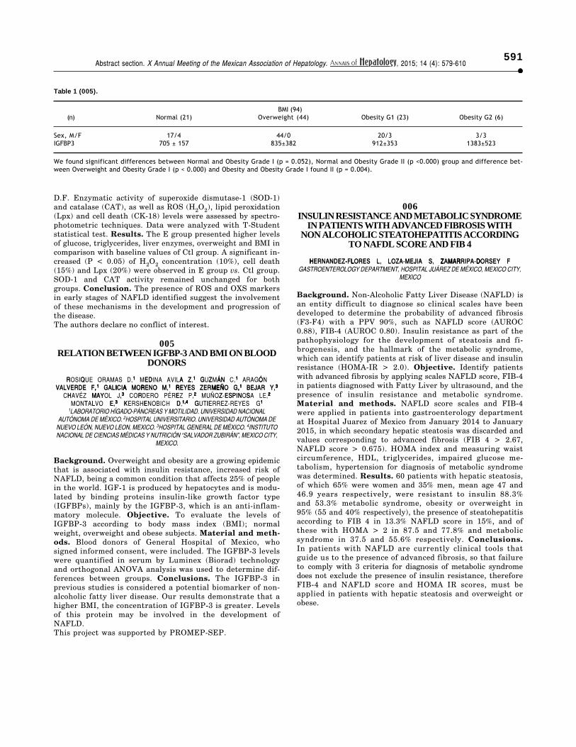
591Abstract section. X Annual Meeting of the Mexican Association of Hepatology. , 2015; 14 (4): 579-610
D.F. Enzymatic activity of superoxide dismutase-1 (SOD-1)and catalase (CAT), as well as ROS (H2O2), lipid peroxidation(Lpx) and cell death (CK-18) levels were assessed by spectro-photometric techniques. Data were analyzed with T-Studentstatistical test. Results. The E group presented higher levelsof glucose, triglycerides, liver enzymes, overweight and BMI incomparison with baseline values of Ctl group. A significant in-creased (P < 0.05) of H2O2 concentration (10%), cell death(15%) and Lpx (20%) were observed in E group vs. Ctl group.SOD-1 and CAT activity remained unchanged for bothgroups. Conclusion. The presence of ROS and OXS markersin early stages of NAFLD identified suggest the involvementof these mechanisms in the development and progression ofthe disease.The authors declare no conflict of interest.
005RELATION BETWEEN IGFBP-3 AND BMI ON BLOOD
DONORS
R Q ROSIQUE ORAMAS D,11111 ED A Z MEDINA AVILA Z,11111 G Á Z N GUZMÁN C,11111 Ó ARAGÓN , ,VALVERDE F,VALVERDE F,11111 I R A I A R GALICIA MORENO M, GALICIA MORENO M,11111 R M EÑ , R MEÑ , REYES ZERMEÑO G, REYES ZERMEÑO G,11111 BEJAR Y, BEJAR Y,33333
M ,H AY CHAVÉZ MAYOL J,33333 R E CORDERO PÉREZ P,22222 Z ES N PI S MUÑOZ-ESPINOSA LE,22222
L ET ,MONTALVO E,33333 D K E KERSHENOBICH D,1,41,41,41,41,4 G I GUTIERREZ-REYES G11111
1LABORATORIO HÍGADO-PÁNCREAS Y MOTILIDAD. UNIVERSIDAD NACIONALAUTÓNOMA DE MÉXICO. 2HOSPITAL UNIVERSITARIO. UNIVERSIDAD AUTÓNOMA DE
NUEVO LEÓN, NUEVO LEON, MEXICO. 3HOSPITAL GENERAL DE MÉXICO. 4INSTITUTONACIONAL DE CIENCIAS MÉDICAS Y NUTRICIÓN “SALVADOR ZUBIRÁN”, MEXICO CITY,
MEXICO.
Background. Overweight and obesity are a growing epidemicthat is associated with insulin resistance, increased risk ofNAFLD, being a common condition that affects 25% of peoplein the world. IGF-1 is produced by hepatocytes and is modu-lated by binding proteins insulin-like growth factor type(IGFBPs), mainly by the IGFBP-3, which is an anti-inflam-matory molecule. Objective. To evaluate the levels ofIGFBP-3 according to body mass index (BMI); normalweight, overweight and obese subjects. Material and meth-ods. Blood donors of General Hospital of Mexico, whosigned informed consent, were included. The IGFBP-3 levelswere quantified in serum by Luminex (Biorad) technologyand orthogonal ANOVA analysis was used to determine dif-ferences between groups. Conclusions. The IGFBP-3 inprevious studies is considered a potential biomarker of non-alcoholic fatty liver disease. Our results demonstrate that ahigher BMI, the concentration of IGFBP-3 is greater. Levelsof this protein may be involved in the development ofNAFLD.This project was supported by PROMEP-SEP.
006INSULIN RESISTANCE AND METABOLIC SYNDROME
IN PATIENTS WITH ADVANCED FIBROSIS WITHNON ALCOHOLIC STEATOHEPATITIS ACCORDING
TO NAFDL SCORE AND FIB 4
- L MA D H Z R H - L Z MAR D HERNANDEZ-FLORES L, LOZA-MEJIA S, ZAMARRIPA-DORSEY FHERNANDEZ-FLORES L, LOZA-MEJIA S, ZAMARRIPA-DORSEY FGASTROENTEROLOGY DEPARTMENT, HOSPITAL JUÁREZ DE MÉXICO, MEXICO CITY,
MEXICO
Background. Non-Alcoholic Fatty Liver Disease (NAFLD) isan entity difficult to diagnose so clinical scales have beendeveloped to determine the probability of advanced fibrosis(F3-F4) with a PPV 90%, such as NAFLD score (AUROC0.88), FIB-4 (AUROC 0.80). Insulin resistance as part of thepathophysiology for the development of steatosis and fi-brogenesis, and the hallmark of the metabolic syndrome,which can identify patients at risk of liver disease and insulinresistance (HOMA-IR > 2.0). Objective. Identify patientswith advanced fibrosis by applying scales NAFLD score, FIB-4in patients diagnosed with Fatty Liver by ultrasound, and thepresence of insulin resistance and metabolic syndrome.Material and methods. NAFLD score scales and FIB-4were applied in patients into gastroenterology departmentat Hospital Juarez of Mexico from January 2014 to January2015, in which secondary hepatic steatosis was discarded andvalues corresponding to advanced fibrosis (FIB 4 > 2.67,NAFLD score > 0.675). HOMA index and measuring waistcircumference, HDL, triglycerides, impaired glucose me-tabolism, hypertension for diagnosis of metabolic syndromewas determined. Results. 60 patients with hepatic steatosis,of which 65% were women and 35% men, mean age 47 and46.9 years respectively, were resistant to insulin 88.3%and 53.3% metabolic syndrome, obesity or overweight in95% (55 and 40% respectively), the presence of steatohepatitisaccording to FIB 4 in 13.3% NAFLD score in 15%, and ofthese with HOMA > 2 in 87.5 and 77.8% and metabolicsyndrome in 37.5 and 55.6% respectively. Conclusions.In patients with NAFLD are currently clinical tools thatguide us to the presence of advanced fibrosis, so that failureto comply with 3 criteria for diagnosis of metabolic syndromedoes not exclude the presence of insulin resistance, thereforeFIB-4 and NAFLD score and HOMA IR scores, must beapplied in patients with hepatic steatosis and overweight orobese.
Table 1 (005).
BMI (94)(n) Normal (21) Overweight (44) Obesity G1 (23) Obesity G2 (6)
Sex, M/F 17/4 44/0 20/3 3/3IGFBP3 705 ± 157 835±382 912±353 1383±523
We found significant differences between Normal and Obesity Grade I (p = 0.052), Normal and Obesity Grade II (p <0.000) group and difference bet-ween Overweight and Obesity Grade I (p < 0.000) and Obesity and Obesity Grade I found II (p = 0.004).
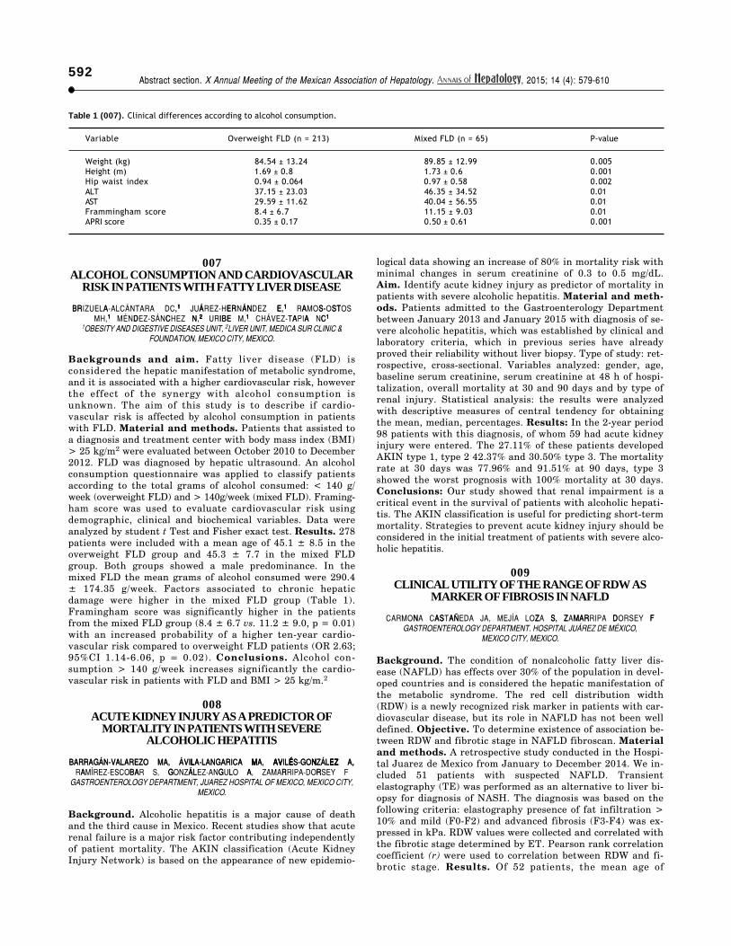
Abstract section. X Annual Meeting of the Mexican Association of Hepatology. , 2015; 14 (4): 579-610592
007ALCOHOL CONSUMPTION AND CARDIOVASCULAR
RISK IN PATIENTS WITH FATTY LIVER DISEASE
BR A ,BRIZUELA-ALCÁNTARA DC,11111 ÁN E Á ER , JUÁREZ-HERNÁNDEZ E,11111 S- ST A RAMOS-OSTOSMH,11111 D C N MÉNDEZ-SÁNCHEZ N,22222 BE I URIBE M,11111 A A C CHÁVEZ-TAPIA NC11111
1OBESITY AND DIGESTIVE DISEASES UNIT, 2LIVER UNIT, MEDICA SUR CLINIC &FOUNDATION, MEXICO CITY, MEXICO.
Backgrounds and aim. Fatty liver disease (FLD) isconsidered the hepatic manifestation of metabolic syndrome,and it is associated with a higher cardiovascular risk, howeverthe effect of the synergy with alcohol consumption isunknown. The aim of this study is to describe if cardio-vascular risk is affected by alcohol consumption in patientswith FLD. Material and methods. Patients that assisted toa diagnosis and treatment center with body mass index (BMI)> 25 kg/m2 were evaluated between October 2010 to December2012. FLD was diagnosed by hepatic ultrasound. An alcoholconsumption questionnaire was applied to classify patientsaccording to the total grams of alcohol consumed: < 140 g/week (overweight FLD) and > 140g/week (mixed FLD). Framing-ham score was used to evaluate cardiovascular risk usingdemographic, clinical and biochemical variables. Data wereanalyzed by student t Test and Fisher exact test. Results. 278patients were included with a mean age of 45.1 ± 8.5 in theoverweight FLD group and 45.3 ± 7.7 in the mixed FLDgroup. Both groups showed a male predominance. In themixed FLD the mean grams of alcohol consumed were 290.4± 174.35 g/week. Factors associated to chronic hepaticdamage were higher in the mixed FLD group (Table 1).Framingham score was significantly higher in the patientsfrom the mixed FLD group (8.4 ± 6.7 vs. 11.2 ± 9.0, p = 0.01)with an increased probability of a higher ten-year cardio-vascular risk compared to overweight FLD patients (OR 2.63;95%CI 1.14-6.06, p = 0.02). Conclusions. Alcohol con-sumption > 140 g/week increases significantly the cardio-vascular risk in patients with FLD and BMI > 25 kg/m.2
008ACUTE KIDNEY INJURY AS A PREDICTOR OF
MORTALITY IN PATIENTS WITH SEVEREALCOHOLIC HEPATITIS
BARRAGÁN-VALAREZO MA, BARRAGÁN-VALAREZO MA, ÁÁ M V É N Z L A E ,L M AV É N EZ ,VILA-LANGARICA MA, AVILÉS-GONZÁLEZ AVILA-LANGARICA MA, AVILÉS-GONZÁLEZ A, A A G L L A B Á G R R RAMÍREZ-ESCOBAR S, GONZÁLEZ-ANGULO A, ZAMARRIPA-DORSEY F
GASTROENTEROLOGY DEPARTMENT, JUAREZ HOSPITAL OF MEXICO, MEXICO CITY,MEXICO.
Background. Alcoholic hepatitis is a major cause of deathand the third cause in Mexico. Recent studies show that acuterenal failure is a major risk factor contributing independentlyof patient mortality. The AKIN classification (Acute KidneyInjury Network) is based on the appearance of new epidemio-
logical data showing an increase of 80% in mortality risk withminimal changes in serum creatinine of 0.3 to 0.5 mg/dL.Aim. Identify acute kidney injury as predictor of mortality inpatients with severe alcoholic hepatitis. Material and meth-ods. Patients admitted to the Gastroenterology Departmentbetween January 2013 and January 2015 with diagnosis of se-vere alcoholic hepatitis, which was established by clinical andlaboratory criteria, which in previous series have alreadyproved their reliability without liver biopsy. Type of study: ret-rospective, cross-sectional. Variables analyzed: gender, age,baseline serum creatinine, serum creatinine at 48 h of hospi-talization, overall mortality at 30 and 90 days and by type ofrenal injury. Statistical analysis: the results were analyzedwith descriptive measures of central tendency for obtainingthe mean, median, percentages. Results: In the 2-year period98 patients with this diagnosis, of whom 59 had acute kidneyinjury were entered. The 27.11% of these patients developedAKIN type 1, type 2 42.37% and 30.50% type 3. The mortalityrate at 30 days was 77.96% and 91.51% at 90 days, type 3showed the worst prognosis with 100% mortality at 30 days.Conclusions: Our study showed that renal impairment is acritical event in the survival of patients with alcoholic hepati-tis. The AKIN classification is useful for predicting short-termmortality. Strategies to prevent acute kidney injury should beconsidered in the initial treatment of patients with severe alco-holic hepatitis.
009CLINICAL UTILITY OF THE RANGE OF RDW AS
MARKER OF FIBROSIS IN NAFLD
N AÑ Z S M F AS , Z AR D CARMONA CASTAÑEDA JA, MEJÍA LOZA S, ZAMARRIPA DORSEY FGASTROENTEROLOGY DEPARTMENT. HOSPITAL JUÁREZ DE MÉXICO,
MEXICO CITY, MEXICO.
Background. The condition of nonalcoholic fatty liver dis-ease (NAFLD) has effects over 30% of the population in devel-oped countries and is considered the hepatic manifestation ofthe metabolic syndrome. The red cell distribution width(RDW) is a newly recognized risk marker in patients with car-diovascular disease, but its role in NAFLD has not been welldefined. Objective. To determine existence of association be-tween RDW and fibrotic stage in NAFLD fibroscan. Materialand methods. A retrospective study conducted in the Hospi-tal Juarez de Mexico from January to December 2014. We in-cluded 51 patients with suspected NAFLD. Transientelastography (TE) was performed as an alternative to liver bi-opsy for diagnosis of NASH. The diagnosis was based on thefollowing criteria: elastography presence of fat infiltration >10% and mild (F0-F2) and advanced fibrosis (F3-F4) was ex-pressed in kPa. RDW values were collected and correlated withthe fibrotic stage determined by ET. Pearson rank correlationcoefficient (r) were used to correlation between RDW and fi-brotic stage. Results. Of 52 patients, the mean age of
Table 1 (007). Clinical differences according to alcohol consumption.
Variable Overweight FLD (n = 213) Mixed FLD (n = 65) P-value
Weight (kg) 84.54 ± 13.24 89.85 ± 12.99 0.005Height (m) 1.69 ± 0.8 1.73 ± 0.6 0.001Hip waist index 0.94 ± 0.064 0.97 ± 0.58 0.002ALT 37.15 ± 23.03 46.35 ± 34.52 0.01AST 29.59 ± 11.62 40.04 ± 56.55 0.01Frammingham score 8.4 ± 6.7 11.15 ± 9.03 0.01APRI score 0.35 ± 0.17 0.50 ± 0.61 0.001

593Abstract section. X Annual Meeting of the Mexican Association of Hepatology. , 2015; 14 (4): 579-610
patients was 44 years, with a range of 11-82 years. Therewere 17 men and 34 women, where 43.2% were advanced stageof fibrosis and 56.8% for mild fibrosis stage. RDW values re-ported (11.8-20.8) with an average of 13.99. In Pearson corre-lation analysis there was significant correlations betweenRDW and fibrotic stage. This reached statistically significancesuch that Pearson’s correlation coefficients were r = 0.305and p = 0.029. Conclusions. The relationship between RDWand the advanced fibrotic stage was determined by correlationwith r = 0.305 and p = 0.029, thus having this ratio is directlyproportional to increased levels of RDW with fibrotic stagewith statistical significance for the sample size. It means thatin care centers in our country could be a useful tool in pre-venting application on NAFLD.The authors declares that there is no conflict of interest.
CLINICAL RESEARCH-LIVER CIRRHOSIS
001CYANOACRYLATE FOR ESOPHAGEAL AND
GASTRIC VARICES IN PATIENTS WITH CIRRHOSIS.SYSTEMATIC REVIEW AND META-ANALYSIS
EL ORNELAS-ARROYO S,11111 L A TÉLLEZ-ÁVILA F,22222 E Z J M SÁNCHEZ-JIMÉNEZ B,11111
YO O AS ORNELAS-ARROYO V,11111 L LÓPEZ-GIL S,11111 U , URIBE M,11111 BARRIENTOS- T GUTIÉRREZ T,33333 C CHAVEZ-TAPIA NC11111
1OBESITY AND DIGESTIVE DISEASES UNIT, MEDICA SUR CLINIC & FOUNDATION.MEXICO CITY, MEXICO. 2GASTROINTESTINAL ENDOSCOPY DEPARTMENT, INSTITUTO
NACIONAL DE CIENCIAS MÉDICAS Y NUTRICIÓN SALVADOR ZUBIRÁN. MEXICO CITY,MEXICO. 3INSTITUTO NACIONAL DE SALUD PÚBLICA. CUERNAVACA, MORELOS,
MEXICO.
Background and aims. Cyanoacrylate has been studied forthe management of the spectrum of gastro esophageal varices.However, its efficacy and rate of adverse events are importantconcerns. The aim of this study was to compare the benefitsand harms of cyanoacrylate in the treatment of gastro es-ophageal varices. Material and methods. The research wasconducted at The Cochrane Central Register of ControlledTrials (CENTRAL) and on MEDLINE up to 2014. Rand-omized clinical trials comparing the use of cyanoacrylate ver-sus other methods for acute bleeding, primary and secondaryprophylaxis in cirrhotic patients were included. Data were an-alyzed using odds ratios (ORs) and 95% confidence intervals.
Significant heterogeneity was considered at a P value ≤ 0.10(χ2) or I2 > 25%. Results. Twelve randomized clinical trialswere included. For acute bleeding, cyanoacrylate achieved bet-ter rates of hemostasis (RR 1.09; IC95% 1.01-1.19), with lessmortality (OR, 0.63; 95% CI, 0.43-0.91), bleeding-related mor-tality (OR, 0.41; 95% CI, 0.22-0.74), and rebleeding (OR 0.52;IC95% 0.38-0.71) compared with other endoscopic methodsfor any type of varices. For gastric varices, cyanoacrylateyielded less rebleeding (RR 0.53; IC 95% 0.34-0.78) than bandligation. For esophageal varices, cyanoacrylate produced lessmortality (OR, 0.53; 95% CI, 0.29-0.97) and rebleeding (OR,0.46; 95% CI, 0.24-0.88) than sclerotherapy. For secondaryprophylaxis, cyanoacrylate showed more adverse events thanother methods. For primary prophylaxis, cyanoacrylateshowed less acute variceal bleeding episodes (10 vs. 38%) andless bleeding- related mortality (0 vs. 10%). Conclusion. Foracute variceal bleeding, cyanoacrylate reduced rebleeding epi-sodes, and exhibited a high hemostasis rate. Additional trialsare necessary to support these data.
002ANTIBIOTICS FOR SPONTANEOUS BACTERIAL
PERITONITIS IN CIRRHOTIC PATIENTS;SYSTEMATIC REVIEW AND META-ANALYSIS
- SÁNCHEZ-JIMÉNEZ B,- SÁNCHEZ-JIMÉNEZ B,11111 E - , MÉNDEZ-SÁNCHEZ N, - , E MÉNDEZ-SÁNCHEZ N,22222 U I M, URIBE M, I U M, URIBE M,11111 CHAVEZ- CHAVEZ- N TAPIA NC11111
1OBESITY AND DIGESTIVE DISEASES UNIT, 2LIVER UNIT, MEDICA SUR CLINIC &FOUNDATION, MEXICO CITY, MEXICO.
Background and aim. Spontaneous bacterial peritonitis(SBP), a complication in cirrhotic patients occurs in the absenceof any intra-abdominal source of infection. Antibiotic therapyshould be initiated to avoid severe complications and death.Third generation cephalosporins are the current standardtreatment. The aim of this study is to compare the differencein mortality, no resolution of SBP, and side effects on differ-ent groups of antibiotics. Material and methods. Electronicsearch was performed in The Cochrane Hepato-Biliary GroupControlled Trials Register, the Cochrane Central Register ofControlled Trials (CENTRAL) in The Cochrane Library,MEDLINE, EMBASE, and Science Citation Index EXPAND-ED until December 2014. References of all identified studieswere hand searched. Randomized studies comparing differenttypes of antibiotics for SBP in cirrhotic patients were includ-
Cephalosporin Any other ATB Odds ratio Odds ratioStudy of subgroup Events Total Events Total Weight M-H, fixed, 95% Cl M-H, fixed, 95% Cl
Angeli 2006 12 55 8 61 17.2% 1.85 (0.69, 4.93)Chen 2005 4 19 5 18 11.8% 0.69 (0.15, 3.14)Felisart 1985 10 37 14 36 30.0% 0.58 (0.22, 1.56)Navasa 1996 11 59 12 64 27.2% 0.99 (0.40, 2.46)Ricart 2000 5 24 3 24 6.9% 1.84 (0.39, 8.77)Tuncer 2003 5 19 3 16 7.0% 1.55 (0.31, 7.81)
Total (95% CI) 213 219 100.0% 1.08 (0.68, 1.71)Total events 47 45
Heterogeneity: χ2 = 3.66, df = 5 (P = 0.60); I2 = 0%Test for overall effect: Z = 0.32 (P = 0.75) 0.01 0.1 1 10 100
Favours (Cephalosporin) Favours (any other ATB)
Figure 1 (002). Mortality: third generation Cephalosporin vs. any other ATB (quinolone, aminoglycoside, penicillin or carba-penem).

Abstract section. X Annual Meeting of the Mexican Association of Hepatology. , 2015; 14 (4): 579-610594
ed. Data were extracted by two different authors, and risk ofbias was analyzed by three authors. Results. Fifteen studieswere included, meta-analysis was performed on three groupsof antibiotics comparing cephalosporin vs. A) quinolone, B)aminoglycoside, and C) any other antibiotic. No significantdifference was observed on mortality comparing cepha-losporines vs. any group using 274 patients in group A, 110patients in group B, and 432 in group C. Difference on resolu-tion of SBP was only statistical significant vs. aminoglycoside(Peto OR 0.33, 95% CI 0.11 to 0.94 p = 0.04) and show no dif-ference on group A and C. No significant difference was ob-served on side effects in any group. Conclusions. Availableinformation cannot support any specific antibiotic regimenfor treatment of SBP in patients with cirrhosis.
003SELECTIVE VASOPRESSIN TYPE 2 RECEPTOR
ANTAGONIST FOR PATIENTS WITH CIRRHOSIS;SYSTEMATIC REVIEW AND META-ANALYSIS
B,SÁNCHEZ-JIMÉNEZ B, B ,SÁNCHEZ-JIMÉNEZ B,11111 AR AGUILAR-OLIVOS N, AR AGUILAR-OLIVOS N,11111 T BARRIENTOS- T BARRIENTOS- T GUTIÉRREZ T,33333 N N MENDEZ-SANCHEZ N,22222 BE I URIBE M,11111 Z T C CHAVEZ-TAPIA NC11111
1OBESITY AND DIGESTIVE DISEASES UNIT, 2 LIVER UNIT, MEDICA SUR CLINIC ANDFOUNDATION MEXICO CITY, MEXICO. 3INSTITUTO NACIONAL DE SALUD PÚBLICA,
MEXICO CITY, MEXICO.
Background and aim. The presence of ascites indicates de-compensation in cirrhotic patients, increasing mortality. In-hibitors of the selective vasopressin type-2 receptors (ARV2)represent an option for ascites refractory to standard treat-ment. The aim of this study is to assess the benefits andharms of ARV2 in this scenario. Material and methods.Electronic search was performed on the Cochrane CentralRegister of Controlled Trials, The Cochrane Library,MEDLINE, EMBASE, Science Citation Index EXPANDEDand Cochrane Hepato-Biliary Group Controlled Trials Regis-ter until April 2014. Trials comparing ARV2 against placeboto manage ascites in cirrhotic patients were included. Two au-thors independently extracted data, and assess bias risk. Re-sults. Eleven randomized clinical trials including 1,940patients were included. The primary outcomes were all-causeand disease-specific mortality, liver transplantation and ad-verse events. Meta-analyses showed no significant differencein effect between an ARV2 and placebo on all-cause mortality
22 vs. 16% (OR 1.27, 95% CI 0.99 to 1.62, I2 = 12%); disease-specific mortality 17 vs. 12% (OR 1.27, 95% CI 0.94 to 1.70,I2 = 0%), liver transplantation 5.2 vs. 5.5% (OR 0.93, 95%CI 0.56 to 1.52, I2 = 0%); total adverse events 83 vs. 80%(OR 1.19, 95% CI 0.92 to 1.53, I2 = 36%). However, ARV2significantly decreased body weight (MD -1.53 kg, 95% CI -1.99to -1.07, I2 = 0%) and significantly increase serum sodium(MD 2.52 mmol/L, 95% CI 1.94 to 3.11, I2 = 64%). Conclu-sions. This meta-analyses showed no benefit of ARV2 in mor-tality or liver transplantation, and even though ARV2improve ascites and serum sodium, this may not be clinicalimportant.
004PORTAL VEIN THROMBOSIS IN PATIENTS WITHLIVER CIRRHOSIS: A FINDING OR A MARKER OF
WORSE PROGNOSIS?
A T Z A -S- ER ÁN , L DAS- ER T ÁN Z , AL D -BORJAS-ALMAGUER OD, CORTEZ-HERNÁNDEZ C, ALEJANDRE-LOYABORJAS-ALMAGUER OD, CORTEZ-HERNÁNDEZ C, ALEJANDRE-LOYA A H BO S- C VA L JV, GARCIA-GARCIA J, SILVA-RAMOS HN, BOSQUES-PADILLA FJ,
L D A - R HMALDONADO-GARZA HJSERVICE OF GASTROENTEROLOGY, UNIVERSITY HOSPITAL, MONTERREY, NUEVO
LEON, MEXICO.
Introduction. The diagnosis of portal vein thrombosis(PVT) has been associated with increased gastrointestinalbleeding and bowel infarction. It is unclear whether the portalvein thrombosis unrelated to malignancy is associated withreduced survival, or is an epiphenomenon of advanced liverdisease. Objective. The objective of this study was to assessclinical outcome in cirrhotic patients with PVT not associatedwith malignancy and its prevalence. Material and methods.Retrospective Search diagnosed with “liver fibrosis and cir-rhosis” or “other cirrhosis” between June 2011-December2014 in one center (Hospital Universitario, UANL). Results.169 patients, 55 women and 114 men, mean age 54.43 ±12.75. 13 had PVT (7.6%). It was divided into 2 groups, pa-tients with PVT and Non-PVT. The PVT group was younger(46.7 vs. 55.1 years p = 0.025). The predominant etiology wasalcohol. Child A patients were more frequent in PVT andChild C in Non-PVT. Mean MELD score was lower in PVT(11.54 ± 5.06 vs. 19.72 ± 8.26 p = < 0.001). The prothrombintime, partial thromboplastin time, INR and indirect bilirubinwere also lower in PVT. Platelets was higher in patients PVT
Experimental Control Risk ratio Risk ratioStudy of subgroup Events Total Events Total Weight M-H, fixed, 95% Cl M-H, fixed, 95% Cl
Cárdenas 2012 5 63 4 57 2.6% 1.13 (0.32, 4.01)Gerbes 2003 0 18 0 20 Not estimableGines 2008 suppl. 34 92 22 47 17.9% 0.79 (0.53, 1.18)Ginés 2008 0 28 0 28 Not estimableGinés 2010 0 38 1 35 1.0% 0.31 (0.01, 7.31)Nevens 2009 suppl. 0 10 0 5 Not estimableOkita 2014 0 25 0 26 Not estimableSakaida 2014 0 82 0 80 Not estimableThuluvath 2006 0 6 0 6 Not estimableWong 2010 4 40 3 36 1.9% 1.20 (0.29, 5.00)Wong 2012 203 720 104 478 76.7% 1.30 (1.05, 1.59)
Total (95% CI) 1,122 818 100.0% 1.19 (0.99, 1.43)Total events 246 134Heterogeneity: χ2 = 5.30, df = 4 (P = 0.26); I2 = 25%Test for overall effect: Z = 1.88 (P = 0.06) 0.01 0.1 1 10 100
Favours (AV2R) Favours (placebo)
Figure 1 (003).

595Abstract section. X Annual Meeting of the Mexican Association of Hepatology. , 2015; 14 (4): 579-610
(182.3 ± 80.4 vs. 121.4 ± 75.3 p = 0.003). None of thepatients received anticoagulant treatment. There was no dif-ference between upper gastrointestinal bleeding (UGB) andspontaneous bacterial peritonitis (SBP) in the groups. En-cephalopathy (grade 3-4) requiring hospital treatment (46.7vs. 30.7 % p = 0.007) and large volume ascites (57 vs. 38.4%p = 0.012) was more common in Non-PVT. Survival was betterfor PVT (16.5 ± 27.9 vs. 4.13 ± 12.2 months p = 0.005). Theonly predictor of mortality after multivariate analysis was theMeld score (HR 1,155, CI-95%, 1,098-1,215, p = < 0.001). Con-clusion. This is the first study reporting the prevalence of PVTin Mexico. We found that PVT itself does not lead to a worseprognosis. The most reliable predictor for clinical outcomeremains the MELD score. The presence of PVT could be just anepiphenomenon and not a marker of advanced liver disease.
005MANAGEMENT OF HEPATIC CIRRHOSIS
SECONDARY TO STEATOHEPATITIS WITH VITAMINE-PENTOXIFYLLINE-METFORMIN PILOT STUDY
I N JIMÉNEZ-LUEVANO MA,11111 G UGALDE IA,11111 A R RAMÍREZ-FLORES S,22222
D RODRÍGUEZ-VILLA P,D RODRÍGUEZ-VILLA P,22222 - JIMÉNEZ-PARTIDA MA, - JIMÉNEZ-PARTIDA MA,11111 C E CERVANTES- C E CERVANTES- G ,Í E RODRÍGUEZ G,11111 - E A L R BRAVO-CUELLAR A11111
1HOSPITAL GENERAL VALENTÍN GÓMEZ FARÍAS (ISSSTE), ZAPOPAN, JALISCO,MEXICO. 2CENTRO UNIVERSITARIO DE CIENCIAS DE LA SALUD (CUCS), GUADALAJARA,
JALISCO, MEXICO.
Background. Steatohepatitis is a public health problem inMexico and the world, and it is an etiological factor increasinglyfrequent of liver cirrhosis. It is estimated that by 2025 could bethe leading cause of cirrhosis in Western countries and theworld. Objective. Assess whether the use of pentoxifylline-metformin-vitamin E is an alternative to treat these patientsversus change of lifestyle and Sitagliptin. Material andmethods. This pilot clinical trial was realized in the gastroen-terology service of Valentín Gómez Farías Hospital in Zapopan,Jalisco over a period of six months. Twenty-nine patients par-ticipated and they were divided into two groups. The experi-mental group with a total of 14 patients received 800 mgpentoxifylline orally, every 24 h + 850 mg metformin orally,every 24 h + 400 U of vitamin E orally, every 24 h, meanwhile,the control group with 15 patients received lifestyle changesbased on 150 min per week of anaerobic exercise + diet of 1,500kilocalories + Sitagliptin 500 mg orally, every 24 h. The dis-gnotic was made based on serological, biochemical, molecular,endoscopic, ecosonography, and body mass index, glucose, cho-lesterol, and PCR. Biopsy was not realized because the obtainedclinical data showed the diagnosis. Results. All patients were inChild A classification; the average age of the experimentalgroup was 62 ± 11, and the control group was 64 ± 10. No sig-nificant difference was found in the progression of Child (p >0.005). The experimental group presented INR. 1.0, cholesterol:159 mg/dL, AST: 26 mg/dL, and ALT: 24 mg/dL, while the con-trol group showed INR: 1.10, cholesterol: 188 mg/dL, AST: 34mg/dL, and ALT: 26 mg/dL. There was no significant differencein adverse effects between both groups. Conclusions. The pilottest showed that the use of pentoxifylline-metformin-vitamin Eis equally effective as the use of changing lifestyle and diet for 6months. Therefore, studies of larger and long term are suggest-ed to know its actual effectiveness.The authors declares that there is no conflict of interest.
006ENDOSCOPIC BEHAVIOR OF PATIENTS WITH
SEVERE HYPERTENSIVE GASTROPATHY TREATEDWITH LONG-ACTING OCTREOTIDE IN COMPARISON
WITH PROPANOLOL- PILOT STUDY
- E E V JIMÉNEZ-LUEVANO MA,11111 Z SÁ N MUÑOZ-SÁNCHEZ DM,11111 MÍ - L S RAMÍREZ-FLORES S,22222
D RODRÍGUEZ-VILLA P,22222 - JIMÉNEZ-PARTIDA MA,11111 - E T CERVANTES- R D GRODRÍGUEZ G,11111 VO C BRAVO-CUELLAR A11111
1HOSPITAL GENERAL VALENTÍN GÓMEZ FARÍAS (ISSSTE), ZAPOPAN, JALISCO,MEXICO. 2CENTRO UNIVERSITARIO DE CIENCIAS DE LA SALUD (CUCS), GUADALAJARA,
JALISCO, MEXICO.
Background. Hypertensive gastropathy is a complication incirrhotic patients resulting from the increase of pressure inporta hepatic system, is a common cause of chronic and se-vere bleeding. It is estimated that 70-80% of cirrhotic patientsdevelop hypertensive gastropathy. The treatment used inthese patients is propranolol. However, there have been fewstudies with other treatments. Objective. Evaluate endoscop-ic response in patients with severe hypertensive gastropathytreated with long-acting Octreotide against Propanolol. Ma-terial and methods. For this pilot clinical trial were evaluat-ed 22 patients in six months, 11 for the control group and 11for the study group. Which were diagnosed with liver cirrhosisby endoscopic, histological, ecosonography, biochemical, sero-logical and molecular studies; and prior to informed consent,the study group was administered 20 mg of long-acting Octre-otide intramuscular single dose, while control group with 40mg propranolol every 12 h, adjusting for dose response wereconducted begin endoscopy and at 2 months of treatment. Pa-tients were staged by Child Pugh classification and valued byparametric tests such as T student and Mann-Whitney. Re-sults. It was observed that in the group treated with Octre-otide was a change of severe gastropathy to moderateGastropathy in 100% of patients, with a significant decreasein the size of esophageal varices. While in the propranolol-treated group there was a decrease in 37% of patients withmoderate to severe gastropathy. Conclusions. Given the im-provement with long-acting octreotide , which was well toler-ated by patients and observed the changes of severeGastropathy to moderate Gastropathy in all patients with adecreased in the size of esophageal varices two months afterthe treatment, consider the Octreotide as an alternative treat-ment for severe hypertensive gastropathy in patients withcontraindications to beta-blockers. Therefore we suggest mul-ticenter randomized studies to know its real effectiveness.The authors declares that there is no conflict of interest.
007MAIN CAUSES OF HOSPITAL READMISSIONS
AMONG DECOMPENSATED CIRRHOTIC PATIENTSAT HGZ NO. 1 IMSS
A M R S- A R AS- MA CONTRERAS-OMAÑA R,CONTRERAS-OMAÑA R,11111 - A - A LUGO-MEDINA M, LUGO-MEDINA M,11111 R R R R LIRA-VERA JE LIRA-VERA JE22222
1HOSPITAL GENERAL DE ZONA NO. 1 IMSS, PACHUCA, HIDALGO, MEXICO. 2CENTRODE INVESTIGACIÓN EN ENFERMEDADES HEPÁTICAS Y GASTROENTEROLOGÍA,
PACHUCA, HIDALGO, MEXICO.
Background. Cirrhotic patients are a vulnerable group toseveral comorbidities that decompensate their medical condi-tion. There is no substantial evidence in our country to estab-lish the main causes of hospital readmission amongdecompensated cirrhotic patients. Objective. To establish themain causes of hospital readmission among decompensatedcirrhotic patients at our Center and compare them versus themain causes stated in literature. Material and methods.

Abstract section. X Annual Meeting of the Mexican Association of Hepatology. , 2015; 14 (4): 579-610596
Observational, cross-sectional, descriptive and retrospectivestudy. Realized at Internal Medicine and Gastroenterologyservices of HGZMF No. 1 IMSS Pachuca, Hidalgo, Mexicofrom January 2012 to August 2014. We applied a data collec-tion card to gather patient’s information and to evaluate theircauses of hospital readmission within the first month and sixmonths after discharge. We used SPSS for descriptive statis-tics. Results. We studied 77 decompensated cirrhotic patientsreadmitted at our Center. Of all of patients, 20 of them wereChild-Pugh A (25.97%), 33 were Child-Pugh B (42.85%) and24 were Child-Pugh C (31.16%). The main causes of hospitalreadmission within the first month after discharge were he-patic encephalopathy (56.25%) and variceal bleeding (43.75%).Hepatic encephalopathy still led hospital readmissions aftersix months of discharge (42.85%), followed by variceal bleed-ing (22.07%), infections (11.68%), ascites (11.68%) and jaun-dice (11.68%). Conclusions. Hepatic encephalopathy is themain cause of hospital readmission among decompensatedcirrhotic patients at our Center, both within the first monthand six months after discharge. Other causes are as followed:variceal bleeding, infections, ascites and jaundice. We suggestto be performed other studies in Centers of our country tovalidate our findings.
008PORTAL HYPERTENSION WITH DEVELOPMENT OF
DUODENAL VARICES AND SUCCESSFULENDOSCOPIC TREATMENT WITH INJECTION OF
CYANOACRYLATE
S G F O - R A A E,ROJAS-LOUREIRO G, HIGUERA-DE LA TIJERA F, PÉREZ-TORRES E, N Í GÁLVEZ-MARTÍNEZ M, SERVÍN-CAAMAÑO A
HOSPITAL GENERAL DE MÉXICO. MEXICO CITY, MEXICO.
Introduction. Duodenal varices have a mortality rate ashigh as 40% in the first episode of bleeding. The studies thatexist until today include small series of cases, without clinicaltrials that allow to sustain which is the best therapy. Cy-anoacrylate is a monomeric tissue adhesive that immediatelyforms a solid polymer in contact with blood. In previous seriesof cases, 100% of the patients with duodenal variceal bleedingthat were treated with cyanoacrylate injection had a good re-sponse. Aim. To describe the experience about the endoscopi-cal management of patients with duodenal varices in theMexico’s General Hospital. Material and methods. Series ofcases that describes the characteristics of hospitalized patientsin the last 3 years because of acute duodenal variceal bleeding,who were successful treated with cyanoacrylate injection. Re-sults. A total of 4 patients, 3 men and 1 woman; of them, 2with alcoholic liver cirrhosis, one case of portal hypertensiondue to portal thrombosis secondary to proteins C and S defi-cit, and one case of splenic vein thrombosis due to pancreati-tis. The cirrhotic patients with history of endoscopic bandligation of esophageal varices with erradication. All of themtreated with cyaoacrylate injection into duodenal varices at adose of 0.5 mL mixed with polyglycanol 0.8 mL without com-
plications. Only 1 patient had bleeeding recurrence and need asecond cyanoacrylate injection 2 months and 6 months later.Conclusions. The endoscopic treatment with cyanoacrylateinjection is technically feasible and effective.
009SARCOPENIA IN PATIENTS WITH LIVER CIRRHOSIS
AND PORTAL HYPERTENSION OF THE INSTITUTONACIONAL DE CIENCIAS MÉDICAS Y NUTRICIÓN
SALVADOR ZUBIRÁN
O Ó U A A CRUZ-SANCÉN NA, MORA-BULNES S, LÓPEZ-MÉNDEZ EE, SÁNCHEZ- Á L EY ÁVILA JF, GÓMEZ-REYES E
INSTITUTO NACIONAL DE CIENCIAS MÉDICAS Y NUTRICIÓN SALVADOR ZUBIRÁN,DEPARTMENT OF GASTROENTEROLOGY, MEXICO CITY, MEXICO.
Background. Loss of skeletal muscle mass (SMM) is an ad-verse event that affects prognosis, and Quality of Life of Pa-tients with liver cirrhosis (LC). Sarcopenia is the syndromecharacterized by gradual and widespread loss of SMM,strength and functional capacity. Series of patients publishedabout this syndrome in LC are still limited in Mexican popula-tion. Objetive. To asses the prevalence of sarcopenia in pa-tients with LC and portal hypertension at theINCMNSZ. Material and methods. Cross-sectional study in105 patients with LC was conducted from February 2012 toNovember 2013. SMM was estimated by bioelectrical imped-ance (BIA), bicipital and tricipital skinfold, BMI, and measure-ment of muscle strength (MS), it was classified as diminishedwhen MS < 30 kg/f in men and < 20 kg/f in women. The SMMwas calculated with Rangel, et al. equation for Mexican popu-lation (2014), the grade of depletion was classified as severe,moderate and normal. Sarcopenia diagnosis was made consid-ering both MS and SMM according to The European WorkingGroup on Sarcopenia on Characters in the Elderly. Results.60% were women. Main etiologies were HCV, idiopathic LC, al-cohol and PBC. Mean age was 57.03 ± 12.82 years old andmajor comorbidities were: DM2, hypertension, hypothy-roidism and obesity. Average BMI was 27.83 ± 5.26 kg/m2.Mean percentage of fat mass for men and women was 30.44%± 6.1 vs. 37.6 ± 6.9% and 69.55% ± 6.1 vs. 62.3 ± 6.9% forfat free mass, respectively. The average MS was 23.71 ± 7.26kg/f for men and 12.56 ± 5.46 kg/f for women. 40.4% of menand 50% of women presented diminished MS. The depletion inSMM was severe in 62.9% of patients, moderate in 25.7% andnormal in 11.4%. Considering both, the SMM and the MS, theprevalence of sarcopenia was 74.6% (50.7% was severe) and25.4% pre-sarcopenia. Conclusions. Sarcopenia in LC ishighly prevalent (74.6% in this population) and is often un-derestimated. It is important to make an early assessment andtreatment, due this entity is related with a decrease of thequality of life, comorbidities and complications.The authors declare that there is not conflict of interest.
Table 1 (008).
Patient Age Sex Etiology of Previous esophageal Re-bleeding Re-bleeding at(years) portal hypertension or gastric varices before 3 months 6 months
1 46 Female Portal vein thrombosis No No No2 74 Male Liver cirrhosis Yes, esophageal No No3 53 Male Liver cirrhosis Yes, esophageal No No4 59 Male Splenic vein thrombosis/pancreatitis Yes, gastric Yes No

597Abstract section. X Annual Meeting of the Mexican Association of Hepatology. , 2015; 14 (4): 579-610
010AUDITORY P3B, P3A, CRITICAL FLICKER
FREQUENCY AND PSYCHOMETRIC TESTS TODETECT MINIMAL ENCEPHALOPATHY
AR D A , T N G , ÍA- O O SARABIA-ALDANA C, SANTANA-VARGAS D, GARCÍA-FORONDA C,AR A N , A- O D , T G Í O SARABIA-ALDANA C, SANTANA-VARGAS D, GARCÍA-FORONDA C, D J R L I ER D JHIGUERA-DE LA TIJERA M, PÉREZ-HERNÁNDEZ JL
LIVER CLINIC, HOSPITAL GENERAL DE MÉXICO, MEXICO CITY, MEXICO.
Introduction. Minimal hepatic encephalopathy (MHE) im-pairs quality of life, the ability to carry out daily activities andproduces an increased risk of traffic accidents. Diagnostictools for MHE in the clinical routine must be easy to use, withhigh sensitivity, valid, objective, and reliable. Psychometricand electrophysiological tests are currently used to detectMHE. Critical flicker frequency (CFF) has been used for thediagnosis of MHE but neurophysiological tests provide moreobjective results. Objective. Compare the psychometric he-patic encephalopathy score (PHES) test, the critical flickerfrequency test auditory P3b and P3a event-related potentialsto detect minimal encephalopathy. Material and methods.Fifty consecutive patients with liver cirrhosis were recruited atthe Liver Clinic of General Hospital of Mexico City. Patientswith alcoholic cirrhosis were discarded. All patients completedPsychometric battery PHES, auditory P3b and P3a and, CFF.Stimuli to elicit ERPs were standard 1000Hz, target 2000Hzpure tones and white noise as distractor. Stimuli lasted 100ms and were presented in semirandom sequences of 400 stim-ulus 2:8 (target: standard) for P3b and 600 stimulus 1:1:8(target:distractor:standard) for P3a. Dicothomic values wereassigned and submitted to contingent table analysis. Signifi-cance was set at alpha below 0.05. Results. Eighteen patientsdid not met inclusion criteria. Comparisons were performed inthe rest of the patients (mean age = 56.84, sd = 9.38, 26 fe-male) Child Pugh A: 21, B: 10, C: 2. Etiology: Cryptogenic:10, HVC: 9 NASH: 8 Autoimmune: 3 HVB: 2, CBP 1. McNe-mar tests showed no significance to PHES vs. CFF (p = 0.26),PHES vs. P3b (P = 0.06), and PHES vs. P3a (p = 0.35). Nodifferences in distributions were found between CFF and P3bor P3a, neither for P3b and P3a. Conclusions. Differences indistributions between PHES, CFF and auditory P3b and P3asuggest inconsistencies in detecting MHE. P3a ERPs showedbetter accuracy in detecting MHE. No differences betweenCFF and ERP where found, we propose a more reliable set oftest to detect MHE.
011SERUM FERRITIN LEVELS AS A PREDICTOR OFDECOMPENSATION IN PATIENTS WITH LIVER
CIRRHOSIS R BA G RO , Á L R -RAMÍREZ-ESCOBAR SC, ÁVILA-LANGARICA MC, BARRAGÁN-VALAREZO
É ÁL A I - F S O MA MA, AVILÉS-GONZÁLEZ AG, ZAMARRIPA-DORSEY FGASTROENTEROLOGY SERVICE, HOSPITAL JUÁREZ DE MÉXICO, MEXICO CITY,
MEXICO.
Background and aim. Iron induces liver damage and fibro-sis, which has been considered as a marker of necro-inflammation.This study was conducted to determinate relative levels of fer-ritin and decompensation in patients with liver cirrhosis. Ma-terial and methods. Cross-sectional descriptive retrospectivestudy on 63 patients diagnosed with liver cirrhosis from Jan-uary 2014 to January 2015 obtained ferritin levels and moni-toring of decompensation. Variables of age, gender, serumferritin, scales Child-Pugh and MELD-Na were analyzed. Av-erage percentages and analysis were applied. Results. 63 pa-tients, 30 males (47.6%), female (52.3%) were analyzed. Were
classified by Child-Pugh A 34.9% B 49.2% and 15.8% C. ScaleMeldNa on average according to Child-Pugh A 9.5 B 13.8Child, Child C 22.5. According alcoholic etiology in 39.6% ofpatients, nonalcoholic steatohepatitis 7.4%, 14.2% hepatitis Cvirus, alcohol overlap with 4.7% hepatitis C virus, autoim-mune 22.2% and 1.5% cryptogenic. Ferritin average was 201.The complications observed were variceal upper gastrointesti-nal bleeding in 32 (50.7%) patients, hepatic encephalopathy,20 (31.7%) and spontaneous bacterial peritonitis 11 (7.9%).Conclusions. According to the results, the serum ferritin lev-el correlates with MeldNa and Child Pugh, as the higher levelof the ferritin is the probability of decompensated patients, soit is not excluded as a marker of prediction to develop them.
012BONE MINERAL DENSITY ALTERATION AND
VITAMIN D DEFICIENCY IN PATIENTS WITH LIVERCIRRHOSIS
O C O A R E S BETANCOURT-SÁNCHEZ F, CERPA-CRUZ S, BARRIENTOS-ÁVALOS R, Z F MO V D A V Ó EL U SC ÁLVAREZ-LÓPEZ F, MORA-HUERTA A, VELARDE-RUIZ VELASCO JA
GASTROENTEROLOGY, ENDOCRINOLOGY AND RHEUMATHOLOGY DEPARTMENTS,“HOSPITAL CIVIL FRAY ANTONIO ALCALDE”, GUADALAJARA, JALISCO, MEXICO.
Background. Bone disease is a major complication of livercirrhosis (hepatic osteodystrophy). The occurrence of oste-oporosis goes as high as 50% it can appear with spontaneousor low impact fractures, producing morbility which affects lifequality and patient survival. Objective. Determine the occur-rence of osteoporosis, vitamin D deficiency and relationshipwith the liver cirrhosis severity. Material and methods.Mineral bone density evaluation and the vitamin D serum lev-els, has a direct relationship with the liver cirrhosis severityand bone fractures. Liver cirrhosis patients were analyzed atany stage of the desease, etiology and gender. Results. Fifty-four patients were included in which, 61 were men, the aver-age age was 53. Osteoporosis was found mildly increased inwomen in a 42% were 39.9% in men. Osteopenia was in-creased in a higher frequency of 36% against 23% in women.The etiology of cirrhosis was due to alcohol in a 46% followedup by 27.8% with hepatitis C. 85.2% of patients were B and CChild-Pugh stages. 72% had a decompensated liver disease.Hepatitis C patients had a bigger rate (53%) of osteoporosis,alcoholic cirrhotic patients 48%. Osteoporosis was more com-mon in patients above 51 years old. Vitamin D deficiency wasfound in 94% of patients. 11.1% had a lumbar fracture. Pear-sons correlation coefficient was calculated with Child-Pugh,MELD and vitamin D with osteoporosis, without finding sig-nificant evidence that proves these factors are independent: p =-0.2, p = -0.15, p = -023 respectively. A weak relationship wasfound between decompensated LC and osteoporosis, p = 0.56.Vitamin D insufficiency showed a significant correlation withdecompensated LC, B and C Child-Pugh stages and MELDabove 15, p = 0.047, p = 0.009, p = 0.49, respectively. Con-clusions. Osteoporosis is a common LC complication and vi-tamin D deficiency was found in most patients, this correlatesdirectly with the severity of the liver disease. Screening ofBMD and serum levels of vitamin D is recommended as a rou-tine test in cirrhotic patients.

Abstract section. X Annual Meeting of the Mexican Association of Hepatology. , 2015; 14 (4): 579-610598
013HEPATIC VENOUS PRESSURE GRADIENT AS APREDICTOR OF ADVANCED LIVER FIBROSIS
N D ÁZ Z HERNÁNDEZ-VELÁZQUEZ B,D N ÁZ EZ HERNÁNDEZ-VELÁZQUEZ B,11111 C R D CORTEZ-HERNÁNDEZ C, C R D CORTEZ-HERNÁNDEZ C,11111 S- BORJAS- S- BORJAS- AL ALMAGUER O,22222 A E- ALEJANDRE-LOYA J,11111 P A BOSQUES-PADILLA F,11111
D A ,- R MALDONADO-GARZA H,11111 A A R J GARCÍA-GARCÍA J11111
1GASTROENTEROLOGY UNIT,2 INTERNAL MEDICINE DEPARTMENT, UNIVERSITYHOSPITAL JOSÉ ELEUTERIO GONZÁLEZ, MONTERREY, NUEVO LEON, MEXICO.
Introduction. Hepatic venous pressure gradient (HVPG)correlates with liver fibrosis of viral etiology and with compli-cations of advanced disease. Liver biopsy is considered thegold standard for diagnosis of liver fibrosis; nevertheless, in-terobserver variability, sample viability and associated compli-cations limit its use. A non-invasive study such as Fibrotesthas been used as predictor of liver fibrosis with inconstant re-sults. Objectives. To evaluate HVPG as a predictor of liverfibrosis. Material and methods. Retrospective study (June2011-December 2014). Patients with diagnosis of chronic liverdisease submitted to hepatic hemodynamic studies with biopsy.Results. Sixty-four patients were evaluated, 11 were elimi-nated because inadequate liver tissue sample. A total of 53subjects, 29 male (54.7%) 24 female (45.3%) with an averageage of 54.11 ± 12.3. Child A 24 (45.3%), B 23 (43.4%), C 6(11.6%). MELD 8 (4-19). Etiology: alcoholic 19 (35.8%), hepatitisC virus 7 (13.2%), autoimmune 11 (20.8%), drug associated 2(3.8%), primary biliary cirrhosis 1 (1.9%), non-alcoholic fattyliver disease 3 (5.7%), cryptogenic 10 (18.9%). With regard toHVPG, 6 (11.4%) patients had normal HVPG (≤ 5 mmHg), 3(5.6%) patients had 5.1-9.9 mmHg, 3 (5.6%) patients had clini-cally significant portal hypertension and 41 (77.4%) had se-vere portal hypertension. The Receiver OperatingCharacteristic (ROC) curve of HVPG for prediction of severefibrosis (3-4) had an area under the curve (AUC) of 0.91 witha HVPG value of 11.4 mmHg, with a sensibility of 95% andspecificity of 75%. ROC curve of HVPG for severe liver fibro-sis for Fibrotest was of 0.727, with a HVPG value of 11.9mmHg, with a sensibility of 90% and specificity of 40%. Con-clusions. HVPG correlates with degree of liver fibrosis. AHVPG greater than 11.4 mmHg is a predictor of advanced liv-er fibrosis. This cutoff can be used to predict liver fibrosisgrade in patients with inadequate liver biopsy.The author declares that there is no conflict of interest.
014FIBROSCAN AS PREDICTOR OF
DESCOMPENSATION IN CIRRHOSIS T E R P Y J A- Z A F Í MARTÍNEZ-RAMÍREZ G, ZAMARRIPA-DORSEY F, MEJÍA-LOZA S,
AR Ó Í AGARCÍA-RUÍZ E, LÓPEZ-LURÍA AGASTROENTEROLOGY DEPARTMENT. HOSPITAL JUÁREZ DE MÉXICO, MEXICO CITY,
MEXICO.
Background. Portal hypertension common complication ofcirrhosis, contributes to the development of ascites, esopha-geal varices (EV) and hepatic encephalopathy (HE). Therenoninvasive methods for predicting stage of portal hyperten-sion to help identify complications and optimize diagnosis andmanagement of cirrhosis. Elastography may reflect progres-sive increase in portal pressure. Publications have established
cutoff 21kpa as a predictor of significative portal hyperten-sion, predicting descompensation. Objetives. Assess the firstevent of the usefulness of fibroscan events as predictor ofdecompensated cirrhosis. Material and methods. Included54 patients with liver cirrhosis, making elastography. Contrack six months. Results included 44 patients. Statisticalanalysis with SPSS version 19 package, descriptive analysis ofquantitative variables. To detect the presence correlationbetween the value of kilopascals in Fibroscan and presence ofbleeding events, VE, ascites and encephalopathy employmentPearson correlation test. 43.2% female, 56.8% male principalalcohol etiology (38.6%). 59% larger and 41%. The smallvarices hemorrhage 36.3%; ascites 52.2% grade 2 and 25%grado1; EH 9%. The Pearson correlation test was not statisti-cally significant for any of the variables measured, thoughshowing a tendency to greater number of kilopascals, mani-festations of more severe portal hypertension. Conclusions.The Fibroscan could be a useful noninvasive predictor decom-pensation, easy to perform test. In our study as it is reported inthe literature, we note that values above 21 are associated withportal hypertension and decompensation. However a largersample and long term is needed to determine its usefulness.
015PREVALENCE OF INFECTIOUS COMPLICATIONS
AMONG HOSPITALIZED DECOMPENSATEDCIRRHOTIC PATIENTS VS. COMPENSATED
CIRRHOTIC OUTPATIENTS R CONTRERAS-OMAÑA R,11111 O AR A VILLALOBOS-ARREOLA ES,22222
AN GIRÓN-SANDOVAL S,AN GIRÓN-SANDOVAL S,33333 IR JE LIRA-VERA JE R J I E LIRA-VERA JE33333
1HOSPITAL GENERAL DE ZONA NO. 1 IMSS, PACHUCA, HIDALGO, MEXICO. 2HOSPITALGENERAL SSA, PACHUCA, HIDALGO, MEXICO. 3CENTRO DE INVESTIGACIÓN ENENFERMEDADES HEPÁTICAS Y GASTROENTEROLOGÍA, PACHUCA, HIDALGO,
MEXICO.
Background. Cirrhosis is a cause of immunodeficiency. Hos-pitalized decompensated cirrhotic patients are at high risk ofdeveloping infectious complications due to their immune sta-tus, bacterial translocation and hemodynamic alterations.Objective. To establish the prevalence of infectious complica-tions among hospitalized decompensated cirrhotic patients vs.compensated cirrhotic outpatients. Material and methods.Prospective cohort study. Realized at Internal Medicine andGastroenterology services of Hospital General SSA Pachuca,Hidalgo, Mexico from December 2012 to January 2014. Com-pensated cirrhotic patients managed as outpatients were in-cluded in one cohort; hospitalized decompensated cirrhoticpatients were included in another cohort. We applied a datacollection card to gather patient’s information, and it wasprocessed using relative risk. Results. We studied 64 patientsin each cohort, for a total of 128. 44 of the hospitalized pa-tients (68.75%) developed infectious complication vs. 22 out-patients (34.37%). The relative risk of developing infections inhospitalized decompensated cirrhotic patients was two timeshigher than a compensated cirrhotic outpatient (RR 2.0) IC95% (1.37-2.91) p 0.00001. Urinary tract infection was themost common infectious complication, with prevalence of35.93% in hospitalized patients vs. 18.75% in outpatients. Oth-er causes of infectious complications, in hospitalized and out-patient respectively, were: spontaneous bacterial peritonitis
Table 1 (013). Area under the curve for grade 3-4 liver fibrosis in biopsy with and mmHg of hepatic venous pressure gradient.
mmHg AUC IC-95% P Sensibility Specificity PPV NPV LR + LR -
11.4 0.910 0.807-1.000 <0.001 95% 75% 92% 81% 3.8 0.06

599Abstract section. X Annual Meeting of the Mexican Association of Hepatology. , 2015; 14 (4): 579-610
(20.31 vs. 6.25%), soft-tissues infections (6.25 vs. 3.12%),pneumonia (3.12 vs. 3.12%) and bacteremia (3.12 vs.0.0%).Conclusions. The relative risk of developing infectionsin hospitalized decompensated cirrhotic patients is two timeshigher than a compensated cirrhotic outpatient (RR 2.0); uri-nary tract infection is the most common cause. We suggestother studies to be performed in order to validate our results.
016
AUDITORY P300 EVENT RELATED POTENTIALS TODETECT MINIMAL ENCEPHALOPATHY
Z N , A- D AJ L O DPÉREZ HERNÁNDEZ JL, SANTANA-VARGAS AD, BARAJAS-TOLEDO D,E N , A- D O DZ AJ L PÉREZ HERNÁNDEZ JL, SANTANA-VARGAS AD, BARAJAS-TOLEDO D, F D E T MF GARCÍA FORONDA CG, HIGUERA DE LA TIJERA MF
LIVER CLINIC. HOSPITAL GENERAL DE MÉXICO. MEXICO CITY, MEXICO.
Introduction. Minimal hepatic encephalopathy (MHE) is re-lated to cognitive impairment in several domains affectingabilities to perform daily living activities and decreasing quali-ty of life. Psychometric and perceptual tests are currentlyused for detecting MHE. Psychometric test over-diagnoseMHE while Critical flicker frequency (CFF) is a better test butavailability is difficult. CFF had proved superior sensitivityand specificity than psychometric test in MHE diagnosis, butnew and more available tools are required. Recently electro-physiological test such as P300 event related potentials (ERP)have been proposed as an alternative to assess cognitive im-pairment in MHE, but literature is poor to this respect andmore supporting evidence is required. Objective. To comparethe psychometric hepatic encephalopathy score (PHES) test,the critical flicker frequency test auditory P300 event-relatedpotentials to detect minimal encephalopathy. Material andmethods. Twenty-five consecutive patients with nonalcoholicliver cirrhosis and 25 controls were recruited at the LiverClinic of General Hospital of Mexico City. All participantscompleted Psychometric battery PHES, auditory P300, andCFF in separated sessions. Stimuli to elicit ERPs were stand-ard 1000 Hz, target 2000 Hz pure tones presented in semi-ran-dom sequences of 400 stimulus 2:8 (target:standard).Dicothomic values were assigned to detection of MHE andsubmited to contingent table analysis while P300 were ana-lyzed with t Student test. Significance alpha level was set be-low or equal to 0.05. Results. McNemar tests showed nosignificance to PHES (p = 0.28), CFF test were sensitive (p =0.050), and P300 distinguished between controls and MHEpatients (p = 0.50). Conclusions. PHES failed to detectMHE. Distributions between CFF and auditory P300 suggestsimilar power for detecting MHE. P300 is more available thanCFF in current clinical practice and results provide evidenceof suitability for assess MHE diagnosis.Interest conflict: authors declare absence of interest conflictfor the present research.
CLINICAL RESEARCH-HEPATOCELLULAR CARCINOMA
001HEPATOCELLULAR CARCINOMA IN NON-
CIRRHOTIC LIVER WITH IMAGING ATYPICALPATTERN IN A PATIENT WITH HEMOPHILIA A AND
VIRUS INFECTION OF HEPATITIS C
R L S ES MA , N O A, ES R L SMA , N O A, ESTRADA-LEDESMA AL, MONTAÑO-LOZA A AGUAYO-VILLASEÑOR JF,ESTRADA-LEDESMA AL, MONTAÑO-LOZA A, AGUAYO-VILLASEÑOR JF, J L MA T JARAMILLO-BUENDÍA C, GONZÁLEZ-HUERTA SM
DEPARTAMENTO DE GASTROENTEROLOGÍA, DEPARTAMENTO DE PATOLOGÍA,CENTRO MÉDICO NACIONAL DE OCCIDENTE DEL IMSS, GUADALAJARA, JALISCO,
MEXICO.
Background and aim. Primary liver cancer accounts for 4%of all new cancers diagnosed worldwide. Hepatocellular carci-noma accounts for 90% of all hepatic neoplasm and only 10 to30% of tumors occur in non-cirrhotic livers. Case report. A51 years old man, with type A mild hemophilia 45.4%, multipletransfusions of factor VIII, social alcoholism, suspended sixmonths ago, infection with hepatitis C virus genotype 1 anewly diagnosed, a viral load of 2,260.000 IU/mL, negative se-rologies for hepatitis B. Referred right upper quadrant ab-dominal pain, decreased appetite, jaundice and weight loss 10kg 4 months duration. Physical examination: jaundice,hepatomegaly of 5 cm, right upper quadrant pain, no stigmataof chronic liver disease. Laboratory alpha-fetoprotein 712 ng/mL, hemoglobin 10.7 g/dL, platelets 336,000, creatinine 4.78mg/dL, prothrombin time (PT) 16/11.7 sec, total bilirubin 8.8mg/dL, directly 7.8 mg/dL, albumin 2.6 g/dL, ALT 162 U/L,AST 523 U/L, liver ultrasound with hyperechoic tumor of 9per 10 cm in segment VI, peripheral vasculature, during thehepatic arterial phase, angiography demonstrates a tumor of9 by 10 cm in the right hepatic lobe, hypodense heterogeneouswithout enhancement to intravenous contrast, with enhance-ment of the pseudocapsule, but without washing late phase orportal vein thrombosis with pulmonary metastases. It wasscheduled for liver biopsy by laparotomy; however, the patienthad worsening of renal failure and died. The autopsy revealedmultifocal conventional moderately differentiated hepatocel-lular carcinoma, mass subtype with multiple satellite nodulessmaller in the rest of the hepatic parenchyma without fibrosisor cirrhosis and pulmonary metastasis. Conclusions. Searchfor hepatitis B and C is recommended in patients with hemo-philia A. Confirmation is required histopathological hepaticcarcinoma in atypical tumors by imaging study in non-cir-rhotic livers as in this case which were in stage C of the clas-sification of Barcelona, being a candidate for treatment withSorafenib according to clinical stage and have no contraindi-cation to their hematologic disease.The authors declare no conflict of interest.
Table 1 (014).
Results Kpa Kpa
Esophageal varices Large 40.8 Small 38.6Gastrointestinal bleeding 1 event 38.2 2 event 44.7Ascites Grade 1 25.8 Grade 281.3Encephalopathy 1 event 61.8 3 event 75

Abstract section. X Annual Meeting of the Mexican Association of Hepatology. , 2015; 14 (4): 579-610600
002GENETICS VARIANTS OF ADH1B, ADH1C AND
CYP2E1 IN COLOMBIAN PATIENTS WITHCIRRHOSIS AND/OR HEPATOCELLULAR
CARCINOMA E AL GAVIRIA-CALLE M,11111 D E- DUQUE-JARAMILLO A,11111 F P V L I PO I DI FILIPPO-VILLA D,11111
M N AR ER MARTÍNEZ-HERNÁNDEZ L,1,21,21,21,21,2 L CALLE-TAVERA L,1,21,21,21,21,2 ÉL I VÉLEZ-RIVERAJDJDJD,JD,1,21,21,21,21,2 D , D , AGUDELO JUAN, AGUDELO JUAN,1,21,21,21,21,2 R E E E R EZ RESTREPO-GUTIERREZ JC, RESTREPO-GUTIERREZ JC,1,21,21,21,21,2 HOYOS- HOYOS-
U DUQUE S,1,21,21,21,21,2 O C EA CORREA-ARANGO-POSADA G,11111 AV AS NAVAS-NAVAS MC11111
1GRUPO DE GASTROHEPATOLOGÍA, FACULTAD DE MEDICINA, UNIVERSIDAD DEANTIOQUIA, COLOMBIA. 2HOSPITAL PABLO TOBÓN URIBE, COLOMBIA.
Background. Studies in some populations provide evidencefor the genetic variants in genes related to alcohol metabo-lism, such as alcohol dehydrogenase (ADH1) and cytochromeP450 (CYP2E1) genes related to enzymatic activity. Somestudies suggest a protective effect of these genetic variants,but in other cases has been associated with an increased riskof liver disease development. Aim. To describe genetic varia-tion of ADH1 (ADH1B*1/*2, ADH1B*2/*2, ADH1C*1/*1,ADH1C*2/*2) and CYP2E1 (CYP2E1 C1/C1, CYP2E1 C2/C2)genes in Colombian patients diagnosed with cirrhosis and/orhepatocellular carcinoma (CHC) assisted in a hospital ofMedellín, Colombia. Material and methods. Patients withcirrhosis and/or CHC who voluntarily agreed were included inthe study and were recruited in a Hepatology Unit in Medellincity, Colombia. Blood sample or hepatic tissue was collectedand preserved at -70 °C. Liver tissue DNA extraction was per-formed using the chloroform-trizol method and peripheralblood sample was extracted with a commercial kit method.PCR-RFLP was performed to determine the gene polymor-phism of interest. A 107pb region of exon 3 of ADH1B genewas amplified, a 146pb of exon 8 of ADH1C was amplified anda fragment of 413pb of promoter region of CYP2E1 was am-plified. The restriction was performed with MaeIII, SspI andRsaI enzyme, respectively. The polymorphism observed in thepatient’s samples was compared with genotype reported data1,000 genomes (http://www.1000genomes.org/index.html) forgeneral population of Medellin, Colombia. Results. We haveidentified the genotype of ADH1B*1*1, ADH1B*2*2 and het-erozygote in 65.5% (19/29), 3.45% (1/29) and 31.03% (9/29)samples, respectively. In ADH1C, the ADH1C*1 genotype andheterozygous (ADH1C*1/*2) was detected in 73.68% (14/19)and 26.32% (5/19) samples, respectively. And the CYP2E1/C1genotype in 88.46% (46/52) samples and heterozygous geno-type in 11.54% (6/52) samples (CYP2E1/C1/C2). Conclu-sions. According to preliminary results, there are notdifferences between the study population and the referencepopulation (1,000 genomes) for ADH1B*1, CYP2E1/C1 andADH1C*1. It should be noted that genotype ADH1C*1 en-codes an enzyme with increased metabolic activity, whichcould represent risk for developing liver disease; however,studies are controversial according to population.This work was supported by Colciencias and the University ofAntioquia, Medellin Colombia.
CLINICAL RESEARCH - AUTOIMMUNE ANDCHOLESTATIC LIVER DISEASE
001FREQUENCY OF PROGRESSIVE FAMILIAL
INTRAHEPATIC CHOLESTASIS DETECTED BYIMMUNOHISTOCHEMISTRY BY EXPRESSION OF
BSEP AND MDR3 IN LIVER BIOPSY OF CHILDRENWITH IDIOPATHIC NEONATAL HEPATITIS
N I D ES ,O , F R A ER GONZÁLEZ-RODRÍGUEZ R, FLORES-CALDERÓN J, SIORDIA-REYES G,- R G M I RAMÓN-GARCÍA G, MORÁN-VILLOTA S- R I G M RAMÓN-GARCÍA G, MORÁN-VILLOTA S
HOSPITAL DE PEDIATRÍA, CENTRO MÉDICO NACIONAL SIGLO XXI “DR. SILVESTREFRENK FREUND”, MEXICO CITY, MEXICO.
Background. Progressive familial intrahepatic cholestasis(PFIC) are genetic alterations that result in mutations in ami-nophospholipid transporter proteins (PFIC1), bile salts(PFIC2) and class III multidrug resistant glycoprotein (MDR3)which mediates translocation of the phosphatidylcholinethrough the hepatocyte canalicular membrane. In our envi-ronment PFIC frequency is not known, its identification inchildren diagnosed with idiopathic neonatal hepatitis couldorientate towards the prevention and treatment of complica-tions of biliary cirrhosis, improvement in nutritional statusand early inclusion in liver transplant programs. Objective.Determine the frequency of PFIC detected by immunohisto-chemistry expression of BSEP and MDR3 in liver biopsy ofchildren diagnosed with idiopathic neonatal hepatitis. Mate-rial and methods. Observational, descriptive, transversalstudy. Clinical features and liver biopsies from patients diag-nosed with idiopathic neonatal hepatitis were reviewed. Immu-nohistochemistry was performed to detect the absence ofBSEP and MDR3 to confirm or rule PFIC. Statistical analy-sis. Results are expressed as mean ± standard deviation,ranges and percentages. Results. 21/35 cases are included; 8(38%) were females and 13 patients (62%) were males. 4 forBSEP, 1 for MDR3 and 2 patients with absence of staining forboth BSEP and MDR3: 7 patients with no response to IHCwere detected. They had history of consanguinity, 4/7, and 1family history of cholestasis. When comparing the clinical, bi-ochemical and histopathological findings among cases withnegative vs. positive IHC, no significant differences werefound. Conclusion. In this study 33% of cases with idiopath-ic neonatal hepatitis, absence of immunohistochemical stain-ing for BSEP and MDR3 was found; to corroborate definitivediagnosis by genetic testing; apparently by immunohisto-chemistry, frequency OF PFIC in our environment is higherthan that reported in literature.
002CORRELATION BETWEEN THE DEGREE OF FATIGUE
AND BIOCHEMICAL ALTERATIONS IN PATIENTSWITH PRIMARY BILIARY CIRRHOSIS
N AS M CONTRERAS-OMAÑA R,11111 A - LUGO-MEDINA M,11111 ER LIRA-VERA JE22222
1HOSPITAL GENERAL DE ZONA NO. 1 IMSS, PACHUCA, HIDALGO, MEXICO. 2CENTRODE INVESTIGACIÓN EN ENFERMEDADES HEPÁTICAS Y GASTROENTEROLOGÍA,
PACHUCA, HIDALGO, MEXICO.
Background. Primary biliary cirrhosis is an autoimmunedisease that develops chronic cholestasis, which most com-mon symptom is fatigue. There is no evidence in our countryto correlate the degree of fatigue against the biochemical al-terations in liver tests. Objective. To correlate the degree of

601Abstract section. X Annual Meeting of the Mexican Association of Hepatology. , 2015; 14 (4): 579-610
fatigue against the biochemical alterations in liver tests of pa-tients with primary biliary cirrhosis. Material and methods.Observational, cross-sectional, descriptive and retrospectivestudy. Realized at Gastroenterology outpatient clinic of HGZ-MF No. 1 IMSS Pachuca, Hidalgo, Mexico from January 2008to December 2013. We applied the Borg Rating of PerceivedExertion Scale and liver tests were performed among patients.We used Excel for descriptive statistical measures and Pearsonproduct-moment correlation coefficient to measure the de-pendence between the degree of fatigue and biochemical alter-ations. Results. We studied 19 patients with Primary biliarycirrhosis. Ten patients (52.63%) had some degree of fatigue,while 9 (47.36%) were asymptomatic. Of all of patients withfatigue, 8 of them (80%) had mild fatigue and 2 patients (20%)had moderate fatigue. As regard of the values of alkalinephosphatase, 12 patients (63.15%) presented elevated values,of which 7 (58.33%) had mild fatigue, 2 (16.66%) had moder-ate fatigue, 3 (25%) were asymptomatic, and there were nopatients with severe fatigue. A determination of 0.601 betweenthe degree of fatigue and alteration of alkaline phosphataselevel was obtained, which indicates a positive correlation.Conclusions. There appear to be a positive correlationbetween the degree of fatigue and alkaline phosphatase valuesin patients with primary biliary cirrhosis. We suggest to beperformed other studies in Centers of our country to validateour findings.
003PREVALENCE OF AUTOIMMUNE LIVER DISEASES:
DATA FROM A THIRD-LEVEL HOSPITAL IN MEXICOCITY DURING A SIX-YEAR PERIOD
L R VALDIVIA-CORREA B,33333 C L , O CHABLÉ-MONTERO F,22222 Á E N E JUAREZ-HERNÁNDEZ E,33333
NCHAVEZ-TAPIA N,33333 M, URIBE M,33333 E H N M Z SÁ MÉNDEZ-SÁNCHEZ N11111
1LIVER RESEARCH UNIT, 2DEPARTMENT OF PATHOLOGY, MEDICA SUR CLINIC &FOUNDATION MEXICO CITY, MEXICO.
3 OBESITY AND DIGESTIVE DISEASES UNIT. MEDICASUR CLINIC AND FOUNDATION.MEXICO CITY, MEXICO.
Background and aim. The prevalence of autoimmune liverdiseases and histopathological findings in Mexican populationare variable. In studies from Northern Europe, the prevalenceis calculated to be 11 to 25 per 100,000 populations, howeverepidemiological studies in Mexico are sparse. Objective. Theaim of this study is to determine the prevalence of autoim-mune liver disease in a group of Mexican patients. Materialand methods. We retrospectively examined seven hundredeighty five liver biopsy specimens between the years 2008 and2013; of these, 67 patients showed compatible characteristicswith autoimmune liver disease. Medical records were analyzedin order to recollect data of demographic characteristics andassociated risk factors. Results. Sixty-seven patients had thefollowing diagnosis: autoimmune hepatitis (AIH), primary bil-iary cirrhosis (PBC), primary sclerosing cholangitis (PSC)and overlap syndrome (OS); 56 were women (83.6%) and 11men (16.4%). The average age in the female group was 49.5years (19-73 years); in the male group, 52 years (28-72 years).The results showed that the prevalence of autoimmune liverdiseases in liver biopsies during this period was 8.5% (Figure1). The distribution of place of birth was as follow: 32 patientswere born in Mexico City (47.76%), 16 patients in Estado deMexico (23.88%) the rest were distributed along differentstates. The most frequent associated comorbidity were otherautoimmune diseases (20.89%), mainly systemic lupus ery-thematosus, ankylosing spondylitis and Sjögren syndrome.Smoking was present in 28 patients (41.79%) and alcohol con-sumption in 32 (47.7%). The main cause of cirrhosis in the fe-
male group with the established diagnosis of autoimmune liv-er disease was AIH (100%); and in the male group PBC (42%).Conclusions. This study shows a probable increasing ten-dency in the prevalence of autoimmune liver disease detectedby biopsies; however it is important to collect more epidemio-logical information in order to create large national prospec-tive cohorts that could generate future hyphoteses and adresskey questions.The authors declare that there are no conflicts of interest.
004AUTOIMMUNE HEPATITIS-PRIMARY BILIARY
CIRRHOSIS OVERLAP SYNDROME INTERNATIONALCLASSIFICATIONS AND LONG TERM FOLLOW-UP
IN NORTHEAST MEXICAN PATIENTS
O MUÑOZ L,11111 PE L , LÓPEZ Y,11111 GUEL T,11111 A R ALARCÓN G,22222 L S ÁVALOS V,11111 O CORDERO P11LIVER UNIT, 2PATHOLOGICAL ANATOMY AND CYTOPATHOLOGY; UNIVERSITY
HOSPITAL “DR. JOSÉ ELEUTERIO GONZÁLEZ”. MONTERREY, NUEVO LEON, MEXICO.
Background. Primary biliary cirrhosis (PBC)/autoimmunehepatitis(AIH) is the most commonly seen overlap syndrome(OS). Although there are no diagnostic criteria established forOS; Paris criteria (PC), the revised score of the internationalautoimmune hepatitis group (RIAIHG) and simplified criteria(SC) have been used for diagnosis. The aim of this study wasto apply RIAIHG, SC and PC and to evaluate long term fol-low-up in northeast Mexican patients. Material and meth-ods. Thirty-six patients were seen at the Liver Unit diagnosedas OS according to clinical, biochemical and histological pa-rameters, with a follow-up of 60 ± 74 (1-404) months (m).Thirty-five PBC and 73 AIH patients were included as con-trols, with a follow-up of 60 ± 60(3-278 m) and 65 ± 63 (2-324m), respectively. RIAIHG, SC and PC were applied. Ursodeox-ycholic acid (UDCA) and UDCA + prednisone (PRED) and/orazathioprine (AZA) were used as a treatment in 15 and 21 pa-tients, respectively. Statistics: SPSS 22.0. Results. Probableand definite diagnosis according to RIAIHG and SC was seenin 72% and 47% of patients, respectively. Meanwhile, 33% wereconfirmed as OS using PC. Compared Sensitivity (S), Specifi-city (E) and positive predictive value (PPV) and negative(NPV) between classifications, RIAIHG had the highest S(72%) even though, E and PPV was high in all them (100%for RIAHG and SC, 97% for PC). NPV was 100% in RIAIHGand SC. Complications on admission and follow-up were: cir-rhosis 19 (53%) and 23 (64%); portal hypertension 11 (31%)and 17 (47%); gastrointestinal bleeding 4 (11%) and 8 (22%);spontaneous bacterial peritonitis 1 (3%) and 6 (17%); enceph-alopathy 2 (6%) and 4 (11%), respectively. Seven (19%) pa-tients died or received a liver transplant. Survival by KaplanMeier analysis was 58% for OS, 76% for CBP and 78% for
Prev
alen
ce (
%)
3.00
2.50
2.00
1.50
1.00
0.50
0.00
Figure 1 (003). Point prevalence distribution.
Point prevalence
2008 2009 2010 2011 2012 2013
0.51% 0.63% 1.39% 1.40% 2.51% 2.04%

Abstract section. X Annual Meeting of the Mexican Association of Hepatology. , 2015; 14 (4): 579-610602
AIH. During follow-up 15 patients treated with UDCA showedan improvement in ALT (p = 0.047), whereas patients treatedwith UDCA + PRED and/or AZA showed an improvent inGGT (p = 0.017). Conclusions. The three international clas-sifications can be complementary for OS diagnosis. RIAIHGdiagnosed a larger number of cases. PC diagnosed 33%. Themajority of patients showed cirrhosis on admission (53%).Overall long term survival was lower in patients with OS,compared with PBC and AIH, regardless which treatment wasused.
005HERBALIFE AS A PREDISPOSING AUTOIMMUNE
HEPATITIS, A CASE REPORT
BI A E ZR U R P, H U D A I R ENRÍQUEZ COVARRUBIAS P, HIGUERA DE LA TIJERA F, PÉREZH Z T R HERNÁNDEZ J, PÉREZ TORREZ E H EZ T R HERNÁNDEZ J, PÉREZ TORREZ E
HOSPITAL GENERAL DE MÉXICO, MEXICO CITY, MEXICO.
Background and aim. Autoimmune hepatitis has a preva-lence of 10-17 per 100,000 in Europe, affecting women 3-4times more often than in men. Most have no identifiable pre-cipitating cause, has been associated with viral infections,drug use and even the subsequent use of herbal drugs. Drugsand herbal products can cause autoimmune hepatitis. UsingHerbalife as a medicinal product and weight reduction hasbeen controversial in the past 10 years and has caused liverfailure in more than 70 cases. Case report. Female 34 yearsold with no medical history, which has intermittent jaundiceboxes Itching refers Herbalife diet intake, being impaired liverfunction, study protocol begins with mitochondrial antibodies(-), neutrophil cytoplasmic antibodies (-), anti-LKM (-) ASMA:negative, antinuclear antibody (+), MRI scans are performedfinding probable data diffuse hepatocellular inflammatory dis-ease liver disease and / or nonspecific cholangitis. Liver biopsyis done by finding incipient liver cirrhosis with moderate ac-tivity, presence of granuloma and eosinophils suggests drugdamage, posterior two-year suspension supplements the pa-tient reported onset of fatigue, weakness, headache, jaundice,pruritus generalized predominantly nocturnal, right upperquadrant pain resulting immunological studies are repeatednegative, negative viral panel to HBV, HCV, CMV immu-noglobulin G but with 4999, ALT and AST increased 5 nor-mal value, autoimmune hepatitis score is calculated at 11, ismanagement starts with prednisone 30 mg dose reduction andazathioprine 50 mg, with partial response to treatment withreduction ALT and AST to normal. Conclusion. The diagno-sis of hepatitis caused by drugs and herbal consumption israre and poorly documented described that some drugs cancause hepatocellular damage that mimics autoimmune hepati-tis and this possibility has been suggested for herbal products.
006CHOLANGIOPATHY ASSOCIATED WITH IGG4, A
CASE REPORT
P EC O AC LÁZARO-PACHECO I,11111 I U G ARISTI-URISTA G,22222 ST L E CASTILLO-GUITARRERO S,22222
G A E I R G A I ER HIGUERA DE LA TIJERA M,HIGUERA DE LA TIJERA M,11111 E J EZ J PÉREZ-HERNÁNDEZ J PÉREZ-HERNÁNDEZ J11111
1GENERAL HOSPITAL OF MEXICO, 2DEPARTMENT OF PATHOLOGY, GENERALHOSPITAL OF MEXICO, UNAM. MEXICO CITY, MEXICO.
Introduction. IgG4-related cholangiopathy, a different typeof cholangitis of unknown origin, by increased serum levels ofIgG4, infiltration of IgG4-positive plasma cells, fibrosis andobliterative phlebitis. Storiform fibrosis with thickening of thebile duct wall is characterized. It is associated with autoim-
mune pancreatitis. Is recognized as a manifestation of biliaryIgG4 related diseases. It is diagnosed by a combination of im-aging, serology, histopathology and responsiveness to ster-oids. Its cholangiographic features are difficult to differentiatefrom primary sclerosing cholangitis, pancreatic cancer orcholangiocarcinoma. Objective. Diagnosis of IgG4-associat-ed cholangitis in patients with chronic pancreatitis of un-known origin. Case report. Male 34 years old, history ofnephropathy of unknown origin at birth steroid treated withappropriate response. Starts with jaundice, dark urine, acoliaassociated with acute HAV hepatitis. Persists hiporexia, jaun-dice, dark urine, acholia, lost weight 10 kg, generalized pruri-tus, intermittent abdominal pain in epigastric region,cholestatic pattern, function normal hepatic synthesis, MRI ofthe abdomen suggests probably autoimmune sclerosingcholangitis, cystic image in pancreas, pancreatic duct is notobserved in their proximal course, dilated, tortuous, irregular-ly contoured distal. Then ERCP and Endoscopic Ultrasoundis performed with needle aspiration cytology inconclusive soexploratory laparotomy with biopsy of the head and body ofpancreas plus liver biopsy was necessary, then reported Im-munohistochemistry positive for IgG4 in plasma cells. Dis-cussion. We must suspect of IgG4-related in patientswith one or more characteristic patterns in tissues or inthose with pancreatitis of unknown origin, sclerosing cholan-gitis, 30% had normal concentrations of IgG4 disease. Con-clusions. IgG4-related cholangiopathy is difficult todifferentiate from primary sclerosing cholangitis, requires ahigh index of suspicion. While IgG4-associated cholangitis re-sponds favorably to steroids no primary sclerosing cholangitis.
CLINICAL RESEARCH - LIVER TRANSPLANTION
001RENAL FUNCTION PRESERVATION WITH SHORT
TERM CONVERSION TO SIROLIMUS INORTHOTOPIC LIVER TRANSPLANT
O M MUÑOZ-ESPINOSA L,11111 P, CORDERO-PÉREZ P,11111 L SILVERA-LINARES A,11111
H ZAPATA-CHAVIRA H,22222 VI O ESCOBEDO-VILLARREAL M,22222 PEREZ-RODRIGUEZEE,EE,22222 C AR N SANCHEZ-MARTINEZ M AR C N SANCHEZ-MARTINEZ M33333
1UNIDAD DE HÍGADO. 2SERVICIO DE TRASPLANTES. 3SERVICIO DE NEFROLOGÍA.HOSPITAL UNIVERSITARIO “DR. JOSÉ E. GONZÁLEZ» UNIVERSIDAD AUTÓNOMA DE
NUEVO LEÓN, MONTERREY, NUEVO LEON, MEXICO.
Introduction. Conversion to Sirolimus (SRL) has been usedin patients with calcineurin inhibitor (CNI) nephrotoxicity inorthotopic liver transplant (OLT). Objective: Compare renaland metabolic outcomes in long-term OLT in patients underCNI vs CNI to SRL conversion (CNI/SRL). Material andmethods. Retrospective study with 64p post-OLT. CNI group(n = 31) vs. CNI/SRL (n = 33). Conversion to SRL: early <12 months (M) or late > 12M. MRDR-6 equation was used toevaluate glomerular filtration rate (GFR). Metabolic out-comes: glucose (GLU), cholesterol (CHOL) and triglycerides(TG). Data was registered at 6M, 12M and cumulative follow-up (Acum), post-OLT in CNI group and post-conversion inCNI/SRL group. Results. Pre-OLT characteristics CNI vs.CNI/SRL: x age 45y vs. 55y (p = 0.003); x follow-up 58 ± 48vs. 68 ± 40 M; diabetes mellitus type 2: 32 vs. 36% (p = 0.80);GLU: 99 ± 33 vs. 123 ± 72 (p = 0.02); GFR: 109 ± 53 vs. 76± 53 (p = 0.02). Etiology CNI vs. CNI/SRL: HCV (23 vs.34%), ALD (0 vs. 27%), NASH (19 vs. 12%), AIH (32 vs. 18%)and others (26 vs. 9%). Conversion to SRL was due to renal
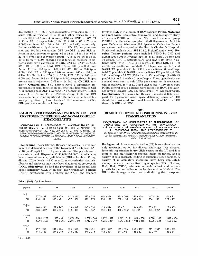
603Abstract section. X Annual Meeting of the Mexican Association of Hepatology. , 2015; 14 (4): 579-610
dysfunction (n = 27), neuropsychiatric symptoms (n = 3),acute cellular rejection (n = 1) and other causes (n = 2).GFR-MDRD (mL/min) at follow-up CNI vs. CNI/SRL 6M: 74± 41 vs. 65 ± 19 (p = 0.02); 12M: 79 ± 46 vs. 65 ± 21 (p =0.009) and Acum: 65 ± 34 vs. 64 ± 27 (p = 0.44), respectively.Patients with renal dysfunction (n = 27): 17p early conver-sion and 10p late conversion. GFR pre-OLT vs. pre-SRL vs.Acum in early conversion group: 62 ± 24 vs. 38 ± 15 vs. 63 ±22 (p < 0.05); late conversion group: 72 ± 32 vs. 46 ± 15 vs.49 ± 26 (p = 0.09), showing renal function recovery in pa-tients with early conversion to SRL. CNI vs. CNI/SRL GLU6M: 119 vs. 129 (p = 0.35); 12M: 124 vs. 130 (p = 0.50) andAcum: 118 vs. 108 (p = 0.05); COL 6M: 168 vs. 220 (p =0.03); 12M: 197 vs. 198 (p = 0.20) and Acum: 205 vs. 198 (p =0.19); TG 6M: 143 vs. 258 (p = 0.05); 12M: 133 vs. 206 (p =0.05) and Acum: 163 vs. 213 (p = 0.34), respectively. Biopsyproven acute rejections: CNI n = 5(16%) vs. CNI/SRL n =2(6%). Conclusions. SRL demonstrated a significant im-provement in renal function in patients that discontinued CNI< 12 months post-OLT, reverting CNI nephrotoxicity. Higherlevels of CHOL and TG in CNI/SRL group at 6M and 12Mwere seen but with no significant difference at cumulative fol-low-up. Significantly lower levels of GLU were seen in CNI/SRL group at cumulative follow-up.
002LAL-D IN LIVER TRANSPLANT PATIENTS FOR LIVERCRYPTOGENIC CIRRHOSIS AND NON-ALCOHOLIC
STEATOHEPATITIS
O C U , L ÁZ Z JJOANICO-AGUILAR R, LÓPEZ-MÉNDEZ YI, CHÁVEZ-VELÁZQUEZ JH,O C , ÁZ EZ U L JJOANICO-AGUILAR R, LÓPEZ-MÉNDEZ YI, CHÁVEZ-VELÁZQUEZ JH, A O L A S, N R- Z , O AL SEGURA-GONZÁLEZ RA, SIXTOS-ALONSO MS, LEAL-VILLALPANDO PR,
ER C , AG A CONTRERAS-SALDÍVAR AG, VILATOBÁ-CHAPA M, CASTRO-NARRO GEDEPARTAMENTO DE GASTROENTEROLOGÍA, TRASPLANTE HEPÁTICO. INSTITUTONACIONAL DE CIENCIAS MÉDICAS Y NUTRICIÓN SALVADOR ZUBIRÁN, MEXICO CITY,
MEXICO.
Background. Ester Storage Disease Cholesterol is producedby null or deficient activity of the Lysosomal Acid Lipase (LAL< 40 pmol/h/spt) for LIPA gene mutation. The prevalence inCaucasians and Hispanics (1:90,000/170,000). Adults mayhave transaminasemia, dyslipidemia (HDL-c levels < 45 mg/dL and LDL-c levels > 130 mg/dL), microvesicular steatosis,fibrosis and cirrhosis may have been diagnosed as cryptogenic(LCC). Objectives. To find the prevalence of lysosomal acidlipase deficiency (LAL-D) in post liver transplant patients(PTHO) cryptogenic liver cirrhosis and NASH and compare
levels of LAL with a group of HCV patients PTHO. Materialand methods. Retrolective, transversal and descriptive studyof patients PTHO by CHC and NASH with a control groupPTHO HCV. Detection samples LAL-D (Lysososmal Acid Li-pase Deficiency Whatman 903 Specimen Collection Paper)were taken and analyzed at the Seattle Children’s Hospital.Statistical analysis with SPSS 22.0, P significant < 0.05. Re-sults. Twenty patients were included PTHO by CHC andNASH (2005-2014). Average age (55 ± 11 years), 10 men and10 women. CHC 10 patients (50%) and NASH 10 (50%). 7 pa-tients (35%) with HDL-c < 45 mg/dL, 2 (10%) LDL-c > 130mg/dL (no results were related). Acid lipase average LCC andNASH (120 pmol/h/spt). In LCC lipase mediates 98 pmol/h/spt(40-120 pmol/h/spt) NASH lipase mediates 128 pmol/h/spt (70-142 pmol/h/spt) 3 LCC (15%) had < 45 pmol/h/spt (2 with 43pmol/h/spt and 1 with 40 pmol/h/spt). These genetically se-quenced were sent to rule LIPA gene mutation, if treatmentwill be positive. 65% of LCC and NASH had < 150 pmol/h/sptPTHO control group patients were tested for HCV. The aver-age level of greater LAL 186 pmol/h/spt, (75-428 pmol/h/spt).Conclusions. The search for Disease Cholesterol Esters de-posit for Lysosomal Acid Lipase deficiency cause of LCCshould be considered. We found lower levels of LAL in LCCthan in NASH and HCV.
003REMOTE ISCHEMIC PRECONDITIONING IN LIVER
TRANSPLANTATION
H ZAPATA-CHAVIRA HA, H ZAPATA-CHAVIRA HA,11111 E Z CORDERO-PÉREZ P, E EZ CORDERO-PÉREZ P,22222 Ñ Z ES N E, MUÑOZ-ESPINOSA LE, Ñ E E Z S N , MUÑOZ-ESPINOSA LE,22222
M ,J ÉN EZ JIMÉNEZ-PÉREZ JC,22222 Q N PERALES-QUINTANA MM,11111 Z A A ZAPATA-SALAZAR,NA,22222 I Z ORTIZ-GARZA O,22222 Á E E N D HERNÁNDEZ-GUEDEA M,11111 G C GUEVARA-CHARLES
A,A,11111 O I O I ESCOBEDO-VILLARREAL MM, ESCOBEDO-VILLARREAL MM,11111 Z R Z D Z R D Z PÉREZ-RODRÍGUEZ E PÉREZ-RODRÍGUEZ E11111
1SERVICIO DE TRASPLANTES, 2UNIDAD DE HÍGADO, HOSPITAL UNIVERSITARIO “DR.JOSÉ E. GONZÁLEZ”, UNIVERSIDAD AUTÓNOMA DE NUEVO LEÓN, MONTERREY,
NUEVO LEON, MEXICO.
Background. Liver transplantation (LT) is considered as theonly treatment option for diverse end-stage liver disease.Ischemia reperfusion injury (IRI) occurs in the LT and is acomplex and multifactorial process, many mediators, and avariety of cells interact, leading to extensive tissue damage. Avariety of inflammatory mediators have been implicated,among them are the reactive oxygen species (ROS), TNF-α,IL-6, IL-1, TGF-β, a-interferon, endothelin-1 and variousgrowth factors and adhesion molecules such as ICAM-1. TheIRI is the damage to the liver graft during the transplant
Table 1 (003). Cytokines levels.
pg/mL PT PR 12 H 24 H 48 H 72 H 7º D 15º D 30º D
IL-6C 327 ± 219 465 ± 179 421 ± 210 425 ± 230 442 ± 236 331 ± 203 356 ± 129 417 ± 184 306 ± 71PIR 210 ± 91 350 ± 401 457 ± 301 396 ± 279 259 ± 127 280 ± 153 337 ± 96 254 ± 106 227 ± 139
TNF-αC 140 ± 116 159 ± 247 199 ± 342 245 ± 331 123 ± 174 38 ± 5 104 ± 129 83 ± 92 125 ± 155PIR 378 ± 400* 459 ± 335 319 ± 373 244 ± 167 351 ± 386 400 ± 135* 31 ± 16 243 ± 296* 342 ± 440*
ICAM-1C 1,689 ± 229 1,508 ± 401 1,676 ±266 1,702 ± 364 1,872 ± 397 1,613 ± 315 1,811 ± 350 1,588 ± 100 1,839 ± 496PIR 1,793 ± 657 1,711 ± 396 2,201 ± 371 1,712 ± 319 1,225 ± 324 1,643 ± 624 1,931 ± 166 1,975 ± 224 1,668 ± 523
VEGFC 297 ± 250 241 ± 276 335 ± 560 287 ± 451 409 ± 358* 169 ± 156 258 ± 197 219 ± 154* 206 ± 232PIR 148 ± 133 244 ± 210 313 ± 157 349 ± 214 163 ± 134 211 ± 92 145 ± 82 63 ± 19 126 ± 81
* p = < 0.05.

Abstract section. X Annual Meeting of the Mexican Association of Hepatology. , 2015; 14 (4): 579-610604
process, to remedy the IRI have adopted different strategies,such as remote ischemic preconditioning (RIP). Objective.Determine whether the RIP modulates the mechanisms in-volved in the IRI in liver transplant recipients by cytokines(TNFα, IL-6), intracellular adhesion molecules (ICAM-1) andfactor of vascular epithelial growth factor (VEGF). Materialand methods. Eight patients, 4 controls (C) and 4 under PIRwere evaluated in phases, pre-transplant (PT), 90 min. Post-reperfusion (PR), 12, 24, 48.72 h (H) and 7, 15, 30 days (D).Results. The results are shown in the table 1. Significant dif-ference (p < 0.05) was found between the groups for the fol-lowing variables: TNFα in the PT phase (p = 0.040), in the 72 Hphase (p = 0.028), in phase 15 D (p = 0.016) in the phase 30 D(p = 0.016); 48H VEGF in phase (p = 0.011), in phase 15 D (p= 0.014). Conclusions. TNF was observed with a significantelevation in PIR group vs. C group in phases 72 H, 15 D and30 D. In C Group VEGF was found significantly higher thanthe PIR group in phases 15 D and 48H. IL6 and ICAM-1 showno changes in any of the phases between groups. We need toincrease the number of patients to infer the involvement ofthese mediators of inflammatory response in the LT.This proyect was fully sponsored by CONACYT-2012-01-182653.
004PHYSICAL ACTIVITY AND INTAKE OF MACRO AND
MICRO NUTRIENTS IN POST OLT PATIENTS
A O Ó Y ,Z A L IA- O Z A Ó L YI,SEGURA-GONZÁLEZ AR, GÓMEZ-REYES E, LÓPEZ-MÉNDEZ YI,SEGURA-GONZÁLEZ AR, GÓMEZ-REYES E, LÓPEZ-MÉNDEZ YI, EZ V A X N C EL U O G L SI T CHÁVEZ-VELÁZQUEZ JH, JOANICO-AGUILAR R, SIXTOS-ALONSO MA,
O R S IVAR AG L - A D P TCONTRERAS-SALDIVAR AG, LEAL-VILLALPANDO RP, VILATOBÁ-CHAPAR VA AG TO S I R L - A D P CONTRERAS-SALDIVAR AG, LEAL-VILLALPANDO RP, VILATOBÁ-CHAPA C A R EM, CASTRO-NARRO GE
DEPARTAMENTO DE GASTROENTEROLOGÍA, INSTITUTO NACIONAL DE CIENCIASMÉDICAS Y NUTRICIÓN SALVADOR ZUBIRÁN, MEXICO CITY, MEXICO
Background. After orthotopic liver transplant (OLT), thesedentary lifestyle and return to dietary habits advantagehigh calorie intake and development of such as metabolic syn-drome. Aim. Determinate the level of physical activity and in-take of macro and micro nutrients in post OLT. Materialand methods. In these study were evaluated 65 patients postOLT, whit the International Physical Activity Questionnaire(IPAQ) to evaluated the physical activity (low < 600 MET-minutes/week, moderate > 600 MET-minutes/week and high> 1,500 MET-minutes/week), and the Cuestionario de Fre-cuencia de Consumo of the Instituto Nacional de Salud Públi-ca, was analyzed whit the SNUT program. The data wereanalyzed with SPSS statistics program version 21. Results.Of the 65 patients evaluated 36 (55%) male and 29 (45%) fe-male with a middle age of 53 years, 1.65 height, weight 68.6Kg, BMI 25 kg/m2 and middle calories intake of 2,200kcal; 15%proteins, 51% carbohydrates and 34% fat. The 62% of maleand 53% of female have intake more than 2,000 kcal, beinghigher fat intake (> 30%) in the female (78%). The 55% haveoverweight or obese. Insufficient intake of calcium was foundin 71%, iron in 77%, vitamin D in 91%, and adequate or highintake of magnesium in 86%, zinc in 78%, with intake < 3 grof sodium in the 95% of patients. The 34% have a low physicalactivity, 52% moderate and 14% high, predominantly low andmoderate physical activity in men. The 45% have a intakemore than 2,000 kcal with a low or moderate physical activity19 and 29% receptively. Conclusions. There is a high intakeof calories, fat, moderate in sodium and deficient in micro nu-trients, with a low or moderate physical activity, contributingto weigh gain post OLT, for these reasons it is important toperform a nutritional intervention and monitoring.The authors declare that there is no conflict of interest.
005EFFICACY AND SAFETY OF TREATMENT WITH PEG-
IFN + RBV + PROTEASE INHIBITOR IN LIVERTRANSPLANT PATIENTS WITH HCV
JO C R , D ÁZ Z JJOANICO-AGUILAR R, LÓPEZ-MÉNDEZ YI, CHÁVEZ-VELÁZQUEZ JH,J R D Á E O C , Z Z JJOANICO-AGUILAR R, LÓPEZ-MÉNDEZ YI, CHÁVEZ-VELÁZQUEZ JH, S- N V V Á EZ C O SIXTOS-ALONSO MS, SEGURA-GONZÁLEZ R, GÁLVEZ-CALVO E,
O S Í R ÁN LT AL H - ICONTRERAS-SALDÍVAR AG, VILATOBÁ-CHAPA M, SÁNCHEZ-CEDILLO L - AL D G L R A, LEAL-VILLALPANDO PR, GAMBOA A, CASTRO-NARRO GE
DEPARTAMENTO DE GASTROENTEROLOGÍA, TRASPLANTE HEPÁTICO. INSTITUTONACIONAL DE CIENCIAS MÉDICAS Y NUTRICIÓN SALVADOR ZUBIRÁN. MEXICO CITY,
MEXICO.
Background. Cirrhosis hepatitis C virus (HCV) is the mostcommon indication for Orthotopic Liver Transplantation(OLT). HCV immediately and universally recurs after OLT inpatients with detectable RNA when THO; of these, 30% devel-op cirrhosis within 5 years. The treatment post THO INFpegis indicated in severe recurrence (SR). The INFpeg/Ribavirin iseffective in 30%, and triple therapy (TT) adding protease in-hibitor (PI) Boceprevir (BCP), Simeprevir (SMP) or Telapre-vir (TLP) with sustained viral response (SVR) to 70%.Objectives. To describe the efficacy and safety of treatmentwith peg-IFN+RBV+PI (BCP or SMP) for 48 weeks in SR inliver transplant patients with HCV. Material and methods.Retrolective and Descriptive study THO and HCV patientstreated with peg-INF+RBV+PI in the INCMNSZ (January2005-April 2015) were included. Were analyzed: gender, age,and SR transplant patients who were treated with peg-+RBV+PI safety and efficacy. The SR is defined as cholestat-ic hepatitis, fibrosing cholestatic hepatitis, acute hepatitis withsignificant necrosis or presence of moderate fibrosis by liverbiopsy 1 year or more after OLT. Results. Of 40 patientsTHO HCV, 6 (15%) received TT with PI, for SR (F2-F4). Five(83%) males, 1 (17%) female; mean age 55 years. Genotypes1b (83%). IL-28b polymorphism CT 4 (67%), TT 2 (33%). Av-erage time for SR was 6 months. Tacrolimus immunosup-pression 4 (67%), sirolimus immunosuppression 2 (33%). Twonaive patients received peg-IFN/RBV/BCP with 100% SVR (12weeks). Three non-responders to previous treatment also had100% SVR. One patient is receiving pwg-IFN+RBV+SMP,has genotype 1a (Q80K Neg) with undetectable viral load atweek 4. None had serious adverse events. The adverse eventwas presented more frequently anemia responded with de-creased Ribavirin. Renal failure occurred in 2 patients whoimproved with calcineurin setting. Conclusions. SVR in pa-tients with post THO peg-IFN+RBV+BOC was 100% andfast response with peg-IFN+RBV+SMP continuous treat-ment. Association between polymorphism IL-28b CT, and se-vere recurrence was found.
006ETIOLOGY OF CIRRHOSIS AND ITS RELATION TOTHE INCIDENCE OF METABOLIC SYNDROME, OLT
ÁV - L , S E V , - YI E,CHÁVEZ-VELÁZQUEZ JH, LÓPEZ-MÉNDEZ YI, GÓMEZ-REYES E, S E I O EG Z A A L S SEGURA-GONZÁLEZ AR, JOANICO-AGUILAR R, SIXTOS-ALONSO MS,
ER I E PHUERTA ÁVILA EE, VILATOBÁ-CHAPA M, LEAL-VILLALPANDO P,O T SAL Í R C AR GCONTRERAS-SALDÍVAR AG, CASTRO-NARRO GEO S Í R C AR T AL GCONTRERAS-SALDÍVAR AG, CASTRO-NARRO GE
INSTITUTO NACIONAL DE CIENCIAS MÉDICAS Y NUTRICIÓN SALVADOR ZUBIRÁN,MEXICO CITY, MEXICO.
Background. Metabolic alterations are frequently found inpatients with Orthotopic Liver Transplantation (OLT). TheMetabolic Syndrome prevalence (MS) reported goes from 43 to58%. MS is related to insulin resistance (RI), predisposes toDM and cardiovascular disease with decreased survival in this

605Abstract section. X Annual Meeting of the Mexican Association of Hepatology. , 2015; 14 (4): 579-610
population. HCV is related to RI that regardless of blood glu-cose levels is associated to twice hypertension (HAS), cardio-vascular disease three times and 8 times more DM.Objective. To describe the incidence of MS in patients OLTand etiologies most frequently associated. Materials andmethods. Retrolective, cross-sectional and descriptive studywhich included post OLT patients. All clinical charts were re-viewed to obtain the demographic and clinical characteristics,and the following variables were analyzed: gender, age at thetime of transplantation, body weight (kg), size (cm), bodymass index (BMI), lipid profile, fasting glucose, and bloodpressure; as well as the pharmacological treatment for diabe-tes mellitus (DM), arterial hypertension (AHT) or dyslipi-demia (DLP), pre and post-transplantation. For MSdiagnosis, the NCEP-ATPIII were used, frequencies and cor-relations of etiologies leading to OLT analyzed. The statisticalanalysis with a SPSS v20.0 (< 0.05) p-value was consideredstatistically significant. Results. Ninety-two patients trans-planted between year 2005-2014, 53% men with a median ageof 47 years (16-67), the main causes were HCV (38%), NASH(11%) , cryptogenic (9%), CHAN (8%) and others. Of the total,30% developed SM to post transplant year. According to theetiology, HCV (39%) developed SM, NASH (25%), cryptogenic(11%), CHAN (7%), CBP (7%) and (3.6%) HAI, overlayingsyndrome and other respectively. In the binary logistic regres-sion analysis adjusted for gender, pre transplant BMI andage, etiology with increased risk of developing SM year wasNASH (OR: 2.16, 95% CI 0.12-37.9) followed by Cryptogenic,CBP, HCV and CHAN and were identified as significant cov-ariates to higher risk of MS; gender (Being male OR: 3.5, CI:1.03-12.2) and pre transplant BMI (OR 1.22, CI 1.03-1.45).Conclusions. MS is a common complication in OLT, etiologymost frequently encountered SM was NASH. Man and BMIpre transplant also influenced.The authors declare that there is no conflict of interest.
007BODY COMPOSITION AND QUALITY OF LIFE WITH
NIGHT BCAA SUPPLEMENTATION IN PATIENTSVALUED FOR OLT
- G E D CHÁVEZ-VELÁZQUEZ JH, GÓMEZ-REYES E, LÓPEZ-MÉNDEZ YI,U G O IC - U SI T SEGURA-GONZÁLEZ AR, JOANICO-AGUILAR R, SIXTOS-ALONSO MS,U G O C S T I - U I SEGURA-GONZÁLEZ AR, JOANICO-AGUILAR R, SIXTOS-ALONSO MS,
L L D A H V HUERTA ÁVILA EE, VILATOBÁ-CHAPA M, LEAL-VILLALPANDO P, T SA R GL CONTRERAS-SALDÍVAR AG, CASTRO-NARRO GE
INSTITUTO NACIONAL DE CIENCIAS MÉDICAS Y NUTRICIÓN SALVADOR ZUBIRÁN,MEXICO CITY, MEXICO.
Background. Protein-energy malnutrition (PEM) is associ-ated with complications pre and post OLT (infections, hospitalstay and mortality). It is suggested calorie-protein supplemen-tation in patients with PEM overnight with carbohydratesand branched chain amino acids (BCAA) to improve nutri-tional status and reduce muscle loss. Objective. Evaluate theeffects of BCAA supplementation night for one month andimpact on body composition, quality of life and dynamometryhand. Material and methods. Twenty-one patients evaluat-ed for (OLT) received a nutritional supplement with BCAAand calories (Enterex Hepatic®, 110 g). Inclusion criteriawere: phase angle < 5.4° mesuared by biolectrical impedance,grade B or C in the subjective global nutritional assessmentor handgrip strength < 30 in men and < 20 in women (meas-ured by no dominant dynamometry). Body composition wereanalyzed using body plethysmography (fat and free mass)and by anthropometric parameters [weight, height, bodymass index (BMI), mean arm circumference (MAC), muscle
arm area (MAA) and tricipital skinfold]. Quality of life usingthe questionnaire SF36v.2 at the beginning and after 30 daysof supplementation night. Adherence to treatment was meas-ured by a 24-h recall and sheet attachment to supplement.Statistical analysis was made using SPSS v.20. Changes wereconsidered as significant p < 0.05. Results. 10 women and 8men were enrolled in a non-controlled clinical trial, the meanage was (42 ± 11 years). Daily supplementation with BCAAand calories significantly tricipital skinfold (14.6 to 15.4mm p< 0.05) and the handgrip strength (18-20 kg/F p < 0.05). Thequestionnaire SF-36 quality of life total (1,590-1,849 p <0.05), physical role (85-169 p < 0.05) and improved vitality all(158-178 p < 0.05). The main adverse effects reported were sa-tiety and nausea. Conclusions. Supplementation with BCAAand calories improves muscle strength and quality of life inpatients referred for evaluation of OLT. As may be an optionin the treatment of these patients.The authors declare that there is no conflict of interest.
008DEFICIENCY AND INSUFFICIENCY OF MAGNESIUMAND VITAMIN D AND THEIR CORRELATION WITH
THE METABOLIC PROFILE AFTER LIVERTRANSPLANTATION
R , H O E, S CRUZ-SANCÉN NA, SÁNCHEZ-ÁVILA JF, CASTRO-NARRO GE, AT M EZ R VILATOBÁ-CHAPA M, GÓMEZ-REYES E
DEPARTMENT OF GASTROENTEROLOGY, INSTITUTO NACIONAL DE CIENCIASMÉDICAS Y NUTRICIÓN SALVADOR ZUBIRÁN, MEXICO CITY, MEXICO.
Introduction. In patients who received liver transplant theprevalence of metabolic alterations and development of meta-bolic syndrome is high. Recent studies have been focused onlevels of 25 hidroxy vitamin D (25-OH-D) and serum magnesi-um in this event. There are not studies on this relation in pa-tients after liver transplantation. Objetives. Asses theprevalence of deficiency and insufficiency of vitamin D andmagnesium and its correlation with the lipid profile of patientsafter liver transplantation. Material and methods. A Nest-ed-cross sectional study was performed in patients who re-ceived liver transplantation in the INCMNSZ. Prevalence ofdeficiency and insufficiency of vitamin D was assessed usingthe serum levels of 25-OH-D according to the USA EndocrineSociety. Magnesium status was defined as deficiency when se-rum levels were < 1.85 mg/dL, insufficiency 1.85-1.92 andnormal 1.93-2.5 mg/dL. Metabolic profile was assessed usingfasting glucose and lipid profile (triglycerides, total, LDL andHDL cholesterol). The variables were described as mean andstandard deviation, correlation was determined using Spear-man’s test. Results. Data of 71 patients were analyzed, where37 were females and 34 males. The mean age was 51.72 ±11.45 years old; the main causes of liver transplant were HCV(47.7%), autoimmune hepatitis (15.49%) and non alcoholicfatty liver disease (12.68%); the most frequent comorbilitieswere DM2 (27%) and hypertension (19.35%). 81.7% used tac-rolimus as an immunosuppressive. The deficiency of vitaminD was 59.2%, insufficiency 28.2%, normal 11.3% and elevated1.4%. In the analysis by gender the deficiency of vitamin Dwas higher in males than females (63.3 vs. 54.1%). The mag-nesium status was deficiency 52.94%, insufficiency 17.65%,and normal 29.4%. The statistically significant correlationswere: 25-OH-D levels were directly correlated with serum mag-nesium, and inversely correlated with total cholesterol andtriglycerides; the serum magnesium levels did not correlatedwith metabolic profile. Conclusions. The prevalence of defi-ciency and insufficiency of vitamin D and magnesium in this

Abstract section. X Annual Meeting of the Mexican Association of Hepatology. , 2015; 14 (4): 579-610606
population is high. Low levels of 25-OH-D are associated withincreased total cholesterol and triglycerides as well as lowerlevels of serum magnesium.
CLINICAL RESEARCH - DRUG INDUCEDLIVER INJURY
001RISK FACTORS FOR DEVELOP ACUTE LIVER
FAILURE AND DEATH IN PATIENTS WITHIDIOSYNCRATIC DRUG INDUCED LIVER INJURY
G D L I ER FHIGUERA-DE LA TIJERA F,D I ER G L FHIGUERA-DE LA TIJERA F,11111 VE M GÁLVEZ-MARTÍNEZ M, V E M GÁLVEZ-MARTÍNEZ M,11111 G O SALAS-GORDILLO G O SALAS-GORDILLOF,11111 E E S , PÉREZ-TORRES E,11111 MA I C CAMACHO-AGUILERA J,22222 L O D - ALEXANDERSON-
S AS ROSAS EG,22222 A C - R CRUZ-ESTRADA A,2 2 2 2 2 Z H ,PÉREZ-HERNÁNDEZ JL,11111 N VÍ SERVÍN- O ACAAMAÑO A22222
1LIVER CLINIC, 2INTERNAL MEDICINE DEPARTMENT. HOSPITAL GENERAL DE MÉXICO,DR. EDUARDO LICEAGA, MEXICO CITY, MEXICO.
Background. Idiosyncratic drug induced liver injury (DILI)can lead to liver failure or death. DILI is a diagnosis of exclu-sion. On the basis of R-value it can be classified into hepatocel-lular, cholestasis, or mixed types. The hallmark of treatment iswithdrawal of the causal agent. Our aim was to describe themain characteristics of patients diagnosed with DILI, and toidentify risk factors for develop acute liver failure (ALF) anddeath. Material and methods. Retrospective study. We col-lected data from medical records of 69 patients diagnosed withDILI, treated between January 2006 and June 2014 at “Hospi-tal General de México”. Results. The following drugs wereidentified as causal agents of DILI: Herbal 28 cases (40.6%);quinolones 9 (13%); ceftriaxone, amoxicillin/clavulanate, keto-conazole 6 (8.7%) each one; antituberculosis drugs, car-bamazepine 3 (4.3%) each one; diclofenac, oral contraceptives 2(2.9%) each one; valproic acid, dapsone, tamoxifen 1 (1.4%)each one. The mean age was 38.8 ± 11.3 years; 52 (75.4%) werefemale; according to R-value 34 (49.3%) had hepatocellular in-jury, 24 (34.8%) mixed, 11 (15.9%) cholestasis; 27 (39.1%) wereobese; ALF occurred in 32 (46.4%) cases, and 10 (14.5%) died.Risk factors for develop ALF were obesity (66.7 vs. 33.3%, P =0.01; OR 4.0, 95% CI = 1.4-11.1); hepatocellular injury (61.8 vs.31.4%, P = 0.01; OR 3.5, 95%CI = 1.3-9.5); and herbal (64.3 vs.34.1%, P = 0.01; OR 3.4, 95%CI = 1.3-9.5). Risk factors fordeath were obesity (37 vs. 0%, P < 0.0001; OR 51.0, 95% CI =2.8-918.7); hepatocellular injury (29.4 vs. 0%; P = 0.003; OR30.4, 95%CI = 1.7-543.9); herbal (32.1 vs. 2.5%, P = 0.003; OR13.2, 95%CI = 2.2-79.9); female gender (19.2 vs. 0%, P = 0.003;OR 30.4, 95%CI = 1.7-543.9). Conclusions. Obesity, herbal,hepatocellular injury are risk factors for ALF, besides them, fe-male gender is a risk factor for death.All authors declare no conflict of interest.
002IMPACT OF DRUG-INDUCED LIVER DAMAGE IN
JUAREZ HOSPITAL OF MEXICO
EZ Z G A L L C AVILÉS-GONZÁLEZ A, MEJIA-LOZA S, AVILA-LANGARICA M,R Á L B Z BA A O C , BAR Á AL O C B , Z BARRAGÁN-VALAREZO M, RAMIREZ-ESCOBAR S, ZAMARRIPA-DORSEY FBARRAGÁN-VALAREZO M, RAMIREZ-ESCOBAR S, ZAMARRIPA-DORSEY F
DEPARTMENT OF GASTROENTEROLOGY. JUAREZ HOSPITAL OF MEXICO, MEXICOCITY, MEXICO.
Introduction. Drug-induced liver injury is one of the mostdifficult diagnoses face gastroenterologists. Different types ofpresentations and various causative agents make the diagno-sis and management particularly difficult. In the United
States an annual incidence of 14 cases per 100,000 inhabit-ants documented. Objective. To determine the incidence ofliver damage from drugs in the Juárez Hospital of Mexico.Material and methods. Patients admitted with abnormalliver function tests and liver damage suspected drug of Janu-ary 1 to December 31, 2014. Type of study: descriptive cross-sectional and retrospective. Variables analyzed: age, gender,type of induced damage, and MELD score, type of agent in-volved, ultrasonographic findings. The results were analyzedwith relative frequency measures center to obtain percentag-es, median and average. Results. Eight cases were found. Theaverage age was 34.5 years (range 29-43 years), with predomi-nance of males (6 cases), the factors involved are 2 drugs(amoxicillin/clavulanate and antiretroviral) and 6 cases perherbology (blue stick 3 cases, Herbalife 1 case, 4life 1 case,Beto Ramon 1 case), 7 patients developed jaundice like symp-toms for which they were consulted, as to the type of damage4 found with hepatocellular pattern, 1 mixed and 3 cholestatic,average MELD was 23.23 (range 7-48), by hepatic ultrasound4 patients with hepatomegaly, none of the patients met crite-ria required of Kings College Hospital, categories of suspicionas CIOMS scale all patients were possible, everyone is under-went liver biopsy which was consistent with liver damage fromdrugs. Conclusions. Liver damage from drugs is difficult todiagnose, interesting that most patients had intake of herbal,posing a new diagnostic challenge due to increased intake ofthese products and the lack of control over the sales, distribu-tion and consumption, so it should take into account when weare faced with a patient with abnormal liver function tests.
CLINICAL RESEARCH - PEDIATRICS
001ACUTE LIVER FAILURE. EXPERIENCE IN A THIRD
LEVEL PEDIATRIC HOSPITAL
EY O Z ES EC O EYES EC O Z O REYES CERECEDO A, GONZÁLEZ ORTIZ B, ARIAS FLORES R,REYES CERECEDO A, GONZÁLEZ ORTIZ B, ARIAS FLORES R, L N JFLORES CALDERÓN J
DEPARTMENT OF GASTROENTEROLOGY. UMAE PEDIATRIC HOSPITAL, CMN SXXIIMSS, MEXICO CITY, MEXICO
Background. Acute liver failures (ALF) is a severe conditionin a previous healthy child that develop hepatic dysfunctionwith coagulopathy with or without encephalopathy, with ahigh mortality rate. There are multiple causes related to theage of the child. Objective. Describe the causes and outcomeof children with ALF in a third level Pediatric Hospital. Mate-rial and methods. All children with ALF from January 2010to March 2015 who meet the criteria for the diagnosis werestudied: hepatic dysfunction with coagulopathy without cor-rection with Vit K (PT > 15 sec or INR 1.5) with encephalop-athy or (PT > 20 sec or INR > 2) without encephalopathywithin 8 weeks of the initial symptoms, without previous liverdisease. Results. Nineteen children were included; the meanage was 36 ± 35 months (between 10 y 132 months), meanweight was 14 ± 11 kg and mean height 85 ± 33 cm. Malegender 11 and female 9. Six cases (32%) were of autoimmuneorigin: autoimmune hepatitis in 4 (21%) cases, hemolytic ane-mia associated to autoimmune hepatitis in 1 (5.2%), and pri-mary sclerosing cholangitis in 1 (5.2%); metabolic disease 2(10.5%); viral 4 (21%): Epstein Barr 2 (10.5%), cytomegalovi-rus 1 (5.2%) and viral hepatitis A in 1 (5.2%); hepatocellularcarcinoma in 1 (5.2%) and undetermined in 6 (32%) cases.Survived 7 (39%) patients and 11 (61%) died within 38 ± 34days after the onset of symptoms. Eight cases were listed for

607Abstract section. X Annual Meeting of the Mexican Association of Hepatology. , 2015; 14 (4): 579-610
liver transplantation (LT). Survival was 100% in the autoim-mune hepatitis cases and 50% in metabolic diseases. One un-determined case and one with CMV Hepatitis survived. Oneno determined cause case is alive one year after living relatedLT. Conclusions. In this study autoimmune hepatitis diseas-es was the most frequent cause of ALF. Survival was relatedto the cause and the possibility of treatment. All autoimmunehepatitis cases survive in contrast with those with metabolic,viral and undetermined diseases. Organ supply is the limitingfactor and a significant number of patient die while waitingLT.The authors declare that there is no conflict of interest.
002IMPACT ON THE TIMELY DETECTION OF BILIARYTRACT ATRESIA THROUGH IMPLEMENTATION OF
STOOL COLOR CARD
N EY A, V I K O E O I EE FLORES CALDERÓN J, REYES CERECEDO A, VILLASIS KEEVER MADEPARTMENT OF PEDIATRICS HOSPITAL, GASTROENTEROLOGY , UMAE SIGLO XXI,
IMSS. MEXICO CITY, MEXICO
Introduction. Biliary atresia (AVB) is a process obstructivebile ducts leading to fibrosis, obliteration of the biliary tractand cirrhosis. Has 60% of restoration of bile flow to makeportoenterostomy < 90 days old. In our setting > 50% ofcases are referred after 3 months so that from January 2013the Program for Early Detection of AVB was implementedby colorimetric card into the National Vaccination (CNV).Aim. To know the process of using the card as well as thereference time to our hospital for children with AVB fromthe first level for diagnosis and treatment. Material andmethods. We included all children with AVB treated be-tween 2010 and 2015. Data for shipping time, age at diagno-sis and surgery were the clinical record. For analysis weredivided into Group 1: period 2010-2012, and Group 2: periodfrom 2013 to 2015. He questioned the family about the re-porting process color card. Results. Forty-five children, 29(64.4%) were females; were 27 (60%) in Group 1 and 18(40%) Group 2. Was performed portoenteroanastomosis 14(51.9%) and 13 (72.2%) patients, respectively (p = 0.1). Theage in days to surgery: 85 ± 38 vs. 121 ± 41 (p = 0.2); terti-ary sent age: 93 ± 44 vs. 116 ± 65 (p = 0.1) and age at diag-nosis: 130 ± 85 vs. 137 ± 60 (p = 0.7), respectively. InGroup 2 color card no information was given in 50%, 40%CNV gave no color card. Reasons for delay in shipment tothird level: conducting studies for hepatitis, prolonged treat-ment for CMV and administrative problems. Conclusions.Our results show that there has been no change in the diag-nosis and treatment for AVB. It is necessary to strengthenthe information and maintain close surveillance program atall levels.The authors declare that no conflict of interest.
003BILE DUCT PAUCITY EXPERIENCE IN A THIRD LEVEL
PEDIATRIC HOSPITAL
L O A R I ROMERO-GARCÍA B, FLORES-CALDERÓN J, MONRROY-TENIZA ZDEPARTAMENTO DE GASTROENTEROLOGÍA PEDIÁTRICA/ HOSPITAL DE PEDIATRÍA,
CENTRO MÉDICO NACIONAL SXXI. IMSS, MEXICO CITY, MEXICO.
Background. Bile duct paucity (BDP) is a cause of neonatalcholestasis. There are two types: syndromatic (Alagille syn-drome) and non-syndromatic associated to genetic, metabolic,infectious and immunological causes. Aim. To describe the
presentation and outcome of children with BDP. Materialand methods. A retrospective and descriptive review of clini-cal, biochemical data and outcome in children with histologicdiagnosis of BPD in the last 10 years was done. Results.Twelve cases were found, 7 syndromic type and 5 the non-syn-dromic. Mean age at diagnosis was 3.5 months, 7 females.Jaundice presentation was present at birth in 8/12. Gestation-al age and birth weight was lower in the non-syndromic thansyndromatic (mean 2.4 vs. 3.5 kg). Syndromic children had fa-cial dysmorphia (5), heart disease (7), embryotoxon (4) andvertebral malformations (3). AP and GGT values were higherin the syndromatic type. During follow up (6 months to 12 yr)the syndromic developed portal hypertension (2), intractablepruritus (3), xanthomatosis (2); one liver transplant was per-formed and 3 remain on the waiting list; one patient admittedwith Kasai developed recurrent cholangitis. Non-syndromicassociated causes were CMV infection in 2, multifactorial in 2and not determined in 1; all cases had improvement outcome.Conclusion. Our results show that biochemical and clinicaldata of children with BDP differentiate between the two typesof BDP. Syndromatic children underwent complications andlisted for liver transplant in contrast with non-syndromatictype who had good outcome. BDP can resemble other causesof neonatal cholestasis and liver biopsy is important to avoidunnecessary surgery.
004CONGENITAL HEPATIC FIBROSIS, A CASE REPORT
C LÁZARO-PACHECO I,11111 T , I ARISTI-URISTA G,22222 O CHOREÑO-GARCIA O,22222
G I E L J HIGUERA-DE LA TIJERA M,11111 - D N J PÉREZ-HERNÁNDEZ J11111
1HOSPITAL GENERAL OF MÉXICO, 2DEPARTMENT OF PATHOLOGY, HOSPITALGENERAL DE MÉXICO, UNAM SCHOOL OF MEDICINE, MEXICO CITY, MEXICO.
Introduction. Congenital hepatic fibrosis is an autosomalrecessive disorder characterized histologically by fibrosis peri-portal and perilobular, with thick bands containing distortedstructures that resemble bile ducts, structures that can expandand form microcysts not communicate with the biliary tract,this derivative lack of ductal plate remodeling resulting per-sistent embryonic bile ducts. It is associated with Caroli disease,choledochal cyst, renal dysplasia and autosomal recessivepolycystic kidney disease and risk of cholangiocarcinoma. Usu-ally diagnosed between 3-10 years of age, affects both sexesequally. With normal laboratory or fluctuating elevated se-rum transaminase levels with elevated alkaline phosphatase.USG: areas of increased echogenicity, splenomegaly andhepatomegaly. Liver biopsy is a very important diagnostictool. Treatment is through control of the manifestations ofportal hypertension, if suspected cholangitis antibiotics; livertransplantation. Case report. Female 2 years 8 months,mother of 18, regular prenatal care, adequate intake of folicacid, iron and calcium, risk of abortion at 3 months of gesta-tion, born by cesarean premature as consecuence of ruptureof membranes with 30.4 sdg APGAR 8/9, weight 1,700 gr size
Table 1 (003). Initial and follow up biochemical data in 12 cases withBDP.
Variables Initial Follow-up
Direct bilirubin (mg/dL) 6.58 0.2-16.5 2.2 0.1-7.6Total bilirubin (mg/dL) 10.0 0.3-14.5 4.4 0.2 to 26
AST (UI) 183.5 63-528 114 27-303ALT (IU) 209 74- 698 142.3 22- 224
Alkaline phosphatase (UI) 613 150-982 277.6 70-840GGT (IU) 467.4 101-1262 264 12.9-890

Abstract section. X Annual Meeting of the Mexican Association of Hepatology. , 2015; 14 (4): 579-610608
50 cm, remained hospitalized for 20 days with headbox with-out requiring mechanical ventilation. At month old she hadbeen hospitalized 10 times for respiratory infections. Duringthese hospitalizations, she was diagnosed polycystic kidneydisease, splenomegaly and hepatomegaly, which was per-formed AMO is reported without abnormalities. Liver biopsywas performed her which reports changes characteristic ofductal plate malformation with extensive fibrosis. Conclu-sions. Diagnosis of Congenital Hepatic Fibrosis should be sus-pected in children with persistent hepatomegaly to begin thestudy and proper handling. The general pediatrician shouldknow this disease to derive timely specialist and not delay itsmanagement.
005SMALL INTESTINAL BACTERIAL OVERGROWTH
FREQUENCY IN PEDIATRIC PATIENTS WITHCHRONIC LIVER DISEASE
ER D ,AR O S C ROMERO-GARCÍA B, ESPINOSA-SAAVEDRA D, FLORES-CALDERÓN J, R AGRAZ-ORTIZ D
DEPARTMENT OF PEDIATRIC GASTROENTEROLOGY, HOSPITAL DE PEDIATRÍA, CMNSXXI, IMSS, MEXICO CITY, MEXICO.
Background. The child with chronic liver disease (CLD) mayexperience complications with some similarities to adult, suchas metabolic abnormalities, cholestasis and infections. Smallintestinal bacterial overgrowth (SIBO) occurs when bacterialcounts are abnormally high in the small intestine (> 105
CFU/mL). From the noninvasive methods, lactulose breathtest is often used for the diagnosis. It is known that the SBI iscommon in adult patients with CLD, but the frequency is un-known in pediatric patients. Objective. To determine the fre-quency of intestinal bacterial overgrowth syndrome inpediatric patients with CLD. Transversal, prospective study.Material and methods. Patients with CLD were includedduring 2 years. Data was collected from the clinical evalua-tion, liver function tests, Child-Pugh score. SIBO was consid-ered positive with a lactulose breath test value ≥ 20 parts permillion (ppm). Statistical analysis: 28 patients were studied,with ages ranging from 3m-15 years (mean age 6 years); 12male (43%) and 16 female (57%), 16 had ascites (57%). A posi-tive lactulose breath test was obtained in 19 cases (68%); ac-cording to Child Pugh score test was positive: 9/16 with ChildA (56%), 6/8 (75%) B and 4/4 (100%) Child C. Patients with se-rum albumin low (< 3.2 g/dL) and ascites showed a high fre-quency of SIBO in 16/16 patients (100%). Conclusion. Thisstudy showed that children with CLD has a high frequency ofSIBO, which increases directly proportional to the severity ofliver disease. Hypoalbuminemia and ascites are parameters toexplore as predictors of SIBO in prospective study with morepatients.
CLINICAL RESEARCH - MISCELLANEOUS
001INTRAHEPATIC CHOLANGIOCARCINOMA
MUCINOUS MULTICENTER AS SECOND PRIMARY
D E BA YE CÓRDOBA REYES J,11111 U G ARISTI URISTA G,22222 A E R Z Z VELAZQUEZ ALVAREZ A,22222
N H JPÉREZ HERNÁNDEZ J11111
1GASTROENTEROLOGÍA, HOSPITAL GENERAL DE MÉXICO. O.D. 2SERVICIO DEPATOLOGÍA HGM, FACULTAD DE MEDICINA, UNAM. MEXICO CITY, MEXICO.
Introduction. Cholangiocarcinoma is a rare malignant tu-mor that originates in the epithelium of the intra or extra he-
patic bile conducts. 3% of malignant tumors. Histologicallymost of cholangiocarcinoma are principally ductal adenomasbut there are other types in which the incidence is very rare asthe papillary, the mucinous, the mucoepidermoid, the adenos-quamous, the squamous or cystadenocarcinoma, with lessthan 10% of cases. Case report. Female 57 years old withthe following importance antecedents genetic load for hyper-tension and pancreatic cancer. In 2010 it was diagnosed leftbreast cancer IIB T2N1M0 with infiltrating ductal carcinomabiopsy lymph node invasion HER2 +. Treated with chemo-therapy (paclitaxel and trastuzumab) and radiotherapy. Mas-tectomy and plastic reconstruction in 2013. Laparotomy 15years ago. Glaucoma in both eyes treated with Timolol. Shestarts her condition four prior months with abdominal pain,type oppressive in right upper quadrant, it was intermittentno mitigating or aggravating, in addition to bloating, nauseaand sometimes vomiting, a week before her admission jaun-dice in sclerae is added, she was assessed by particularly phy-sician and sent to this medical unit. On admission she isgeneralized jaundice, neurologically complete, hydrated, with-out cardiopulmonary alterations, abdomen with hepatomega-ly 2-2-3 cm below the costal margin. Leu 6700 Hb 16.3 Hto 46Plat 173,000 TGO 321 TGP 209 FA 1,926 DHL 1150 pt 6.6Alb 4.1 Glu 233 U 20 Cr 0.7 BT 6.04 BD 3.6 BI 2.4. Chest radi-ograph without suggestive image of pathology. TAC Abdomi-nal hepatic metastases, simply left renal cyst. Liver biopsy:mucinous cholangiocarcinoma. Conclusion. The mucinousvariant of intraductal papillary bile duct tumors is a rare var-iant of presentation of injuries cholangiocarcinoma type.They are characterized by increased mucin production (likepancreatic). They are slow growing and low infiltration rate.Knowing it is important because it has a better prognosisthan other forms for cholangiocarcinoma.
002DETECTION OF LIVER FIBROSIS BY NONINVASIVE
METHODS IN PATIENTS WITH PSORIASIS
- D J E N JUÁREZ-HERNÁNDEZ E,11111 C L R O CORRALES-ROSAS B,22222 U E VERA-IZAGUIRRE D,11111
R L LEÓN-DORANTES G,33333 B M, URIBE M,11111 Á - A A VE PI CHÁVEZ-TAPIA NC11111
1DEPARTMENT OF GASTROENTEROLOGY, MEDICA SUR CLINIC & FOUNDATION,MEXICO CITY, MEXICO. 2DERMATOLOGY UNIT, CENTRO MÉDICO NACIONAL SIGLO
XXI, MEXICO CITY, MEXICO. 3MEXICAN ASSOCIATION FOR PSORIASIS. MEXICO CITY,MEXICO.
Background and aim. Liver fibrosis is considered one of themajor adverse effects secondary to therapeutic agents used inthe treatment of psoriasis. Currently, the prevalence of liverfibrosis in this patient is unknown. The aim of this study wasto describe the prevalence of liver fibrosis in patients diag-nosed with psoriasis. Material and methods. We evaluated127 patients with psoriasis under treatment with one or moremedications. Demographic and biochemical data were recol-lected; liver fibrosis was determined by four noninvasivemethods: NAFLD Score, APRI, Fib4 and FibroScan®. Re-sults. The sample included 46 women (36.2%). The prevalenceof liver fibrosis by each noninvasive method was: NAFLDScore 4.6% (n = 6); APRI 4.6% (n = 6); Fib4 3.9% (n = 6); Fi-broScan® > 9 kPa 15% (n = 9) and FibroScan® > 12 kPa8.7% (n = 11). In logistic regression analysis treatment withAdalimumab was associated with presence of advanced liverfibrosis (> 12 kPa) (OR 12.4; 95%CI 1.8-84.0, p < 0.05). Con-clusions. The prevalence of liver fibrosis in patients with pso-riasis is high. In clinical practice screening of liver fibrosis isimportant to establish adequate treatment schemes.

609Abstract section. X Annual Meeting of the Mexican Association of Hepatology. , 2015; 14 (4): 579-610
003BANTI SYNDROME INCIDENCE
IN A TERTIARY CENTRE
A AR AVIL Z R - OAVILA-LANGARICA MA, AVILÉS-GONZÁLEZ A, BARRAGAN-VALAREZOA A L Z R -A R VI OAVILA-LANGARICA MA, AVILÉS-GONZÁLEZ A, BARRAGAN-VALAREZO C S U A, R BA , N MA, RAMÍREZ-ESCOBAR S, MEJÍA-LOZA SA, GONZÁLEZ-ANGULO A,
Z R D R ZAMARRIPA-DORSEY FJUAREZ HOSPITAL OF MEXICO, MEXICO CITY, MEXICO
Introduction. Banti syndrome or idiopathic portal fibrosis isa disorder of unknown etiology characterized by portal hyper-tension, splenomegaly with or without hypersplenism and pre-served liver function. In the West it is more frequent infemales and in the fifth decade of life. Its pathogenesis is stillunder investigation, have been implicated prothromboticstates and infections. Objective. To determine the frequencyand epidemiological characteristics of Banti syndrome in Jua-rez Hospital of Mexico. Material and methods. A retrospec-tive cross-sectional study was conducted, statistics areexpressed in percentages and averages. I included were pa-tients who underwent liver Doppler ultrasound in the periodMarch 1, 2012 to February 28, 2015 with reports of portal hy-pertension without cirrhosis data Doppler ultrasound or bylaboratory tests. Results. Four cases were found. The aver-age age was 37 years (range 28-45), female gender predomi-nance (ratio 3: 1), average body mass index 26.67 (range23.43-27.6) one case diagnosed with type 2 diabetes mellitus.Banti syndrome occurred in 2.35% of patients with portal hy-pertension by Doppler ultrasound liver. The etiology of one ofthe cases was Evans syndrome, the remaining three cases, noetiology remains. 75% cases presented ascites. In lab tests,mean hemoglobin was 9.02g/dL (range 7.6-11g/dL), thrombo-cytopenia with an average of 135,000 platelets (range from59,000 to 200,000), leukocytes average 5792 (range 3,510-7,860), average neutrophil 3990 (range 2,870-5,240), lym-phocytes average 1,222.5 (range 320-2,120), total bilirubin 1.1mg/dL (range 0.8-1.6), albumin 4 g (range 3.5-4.3). Averagespleen volume Doppler ultrasound liver was 650.75 cc (range376-1,044). Only 25% (1 case) presented variceal bleeding sec-ondary to large esophageal varices Baveno. Conclusion. Thepredominant sex and etiology of one case coincide with thosereported in the literature. It is a disease underdiagnosed inour country, although not the most common cause of portalhypertension, it is important to identify for proper handling.The authors have no conflicts of interest.
004GIANT LIVER CYST TREATED WITH PERCUTANEOUS
DRAINAGE AND SCLEROTHERAPY.CASE REPORT
O ÉR EZ E EPÉREZ-IXCOY PL, PÉREZ-HERNÁNDEZ JL, PÉREZ TORRES EGENERAL HOSPITAL OF MEXICO, MEXICO CITY, MEXICO.
Background. Hepatic cysts are congenital malformations oc-cur in 4 to 5% of the adult population, being more frequent inwomen with a 5: 1 in most cases are asymptomatic and re-quire no treatment, reserving for the producing symptomscaused by the growth of the cyst with compression neighbor-ing organs usually when it reaches a size of 5-10 cm. Comple-mentary diagnostic imaging such as ultrasound and CT scanare required and can be treated expectantly, percutaneous orsurgical drainage. Case report. Male 70 years old with nohistory major, begins with progressive increase in abdominalgirth, right upper quadrant pain, and weight loss unquanti-fied. The physical examination with increased in the waist cir-
cumference from epigastric predominance was observed in themesogastrio a tumor of approximately 10 x 10 cm, involvingthe right upper quadrant and epigastric, mesogastrio. Studyprotocol was started evidencing: altered liver function tests,abdominal USG: enlarged liver, by intraparenchymal cyst, ab-dominal CT: homogeneous liver, hypodense in liquid range,ovoid morphology, smooth edges, thin, without internal septaof 20.2 x 20.1 x 14.8 cm, is sent to interventional radiology forpercutaneous drainage quantified at 4,800 cc, later sclerother-apy performed with Polidocanol in 4 sessions, and 40 days af-ter ultrasonographic screening hepatic cysts are observedseptate so we proceeded to retreat drainage tube with normalliver tests controls. Conclusions. Aspiration alone has notbeen effective in preventing recurrence, surgical interventionhas so far been the only effective treatment available howeverthis involves considerable morbidity. Has been recently re-ported that extraction by puncture, aspiration + alcohol injec-tion is a simple procedure, inexpensive, low morbidity andmortality as well as low recurrence, and could be the treat-ment of choice for symptomatic congenital cysts.
005FLUOROSCOPY GUIDED PERCUTANEOUS LIVER
BIOPSY. HOSPITAL JUAREZ DE MEXICOEXPERIENCE
A N L A ANGELES-LABRA AR,1 1 1 1 1 Á R R JUÁREZ-BARRIENTOS TE,11111 P Y A ZAMARIPA-DORSEYF,F,F,F,11111 S, MEJÍA-LOZA S, S , MEJÍA-LOZA S,11111 O R A RODRÍGUEZ-BLAS A O R A RODRÍGUEZ-BLAS A22222
1SERVICIO DE GASTROENTEROLOGÍA, MEXICO CITY, MEXICO. 2SERVICIO DERADIOLOGÍA E IMAGEN, HOSPITAL JUÁREZ DE MÉXICO. MEXICO CITY, MEXICO.
Background. Histological evaluation of the liver tissue re-mains essential part of the diagnosis, assessing prognosis andin tailoring treatment of multiple liver diseases. The techniqueoriginally described in 1958 by Menghini percussion based he-patic parenchyma, has now fallen into disuse due to its highrate of complications and has been replaced by image guidedmainly tomography and ultrasound techniques. Fluoroscopygive us an image similar to an X-ray in real time to easilyidentify the hepatic parenchyma and facilitates accurate biop-sy with needle visualization. In our center this technique isused for taking liver biopsy, in cases where parenchymal dis-ease is suspected, and CT-guided biopsy for the case of singlehepatic lesion or neoplasia. Material and methods. Liver bi-opsies performed between January and December 2014 werereviewed, the technique used, the requesting service, indica-tions for liver biopsy, complications and the influence of he-patic histology on subsequent patient management wasassessed. Results. A total of 62 percutaneous biopsies, ofwhich 46 (74%) were guided by fluoroscopy, and 16 (26%)were guided by CT. There were no complications reported in38 patients (61%), the main complication was pain at the
A B
Figure 1 (004).

Abstract section. X Annual Meeting of the Mexican Association of Hepatology. , 2015; 14 (4): 579-610610
puncture site which occurred in 24 patients, only 1 patienthad 1 wall hematoma with secondary anemia, one week afterthe procedure, which required hospitalization. There were nodifferences in complications between the two groups. Themain indication for liver biopsy was metastatic liver, in 18patients, followed by chronic liver disease under study in 14 patients,fatty liver, hepatic neoplasia, acute liver failure, acute hepati-
tis and hepatocellular carcinoma suspected. The biopsy hasuseful confirmation in 31 cases, change in treatment in 14not influenced in 10 and was confusing in 3 cases. Conclu-sions. According to this experience percutaneous liver biopsyguided by real-time fluoroscopy appears to be effective andminimally invasive, yet to justify their safety follow up with agreater number of cases is necessary.