Wound Care in Horses
18
Wound Care in Horses Stephanie S. Caston, DVM KEYWORDS • Horses • Equine practice • Wounds • Tendons and ligaments • Healing • Treatment Wounds are common in equine patients. Due to the horse’s nature and the environment in which it lives, wounds frequently involve a significant amount of tissue trauma. Legs caught in fences, panels, wire, or gates are a frequent occurrence, as are lacerations from steel siding, trailer accidents, kicks, and riding accidents. As a result, equine ambulatory practitioners typically see a relatively large number of cases presenting for wound care. Enormous variation exists in treatments, medications, bandages, and bandaging techniques applied to wounds in horses. 1–5 In addition, products marketed to owners for wound care are ubiquitous, numerous, and sometimes dangerous. In some instances, owners may try to treat the wound at home before consulting a veterinarian or they may acquire a horse that has an old wound. This leaves the equine veterinarian treating not only acute wounds but chronic and complicated wounds as well. To further complicate the issue, other factors, including economic constraints, and what is considered an acceptable end result, will often play major roles in the decision-making process. To develop an intelligent, workable plan for dealing with wounds, an equine practitioner must first have a good working knowledge of anatomy. There is no magic ointment, salve, or injection that makes wound healing easy. The practitioner must be well versed in the wound-healing process and recognize what he or she can and cannot do to assist that process. This will also allow the practitioner to recognize when the wound-healing process is not going according to plan and to ascertain what has happened to complicate the process. This article will review evaluation of a wound, the normal process of wound healing, and general management of full- thickness skin wounds, as well as an approach to managing them. WOUND ASSESSMENT The initial assessment of a wound should include careful attention to its anatomical location. Answering a few important questions can help determine what structures the wound affects, and prioritize treatment objectives. First: are structures involved that need more in depth attention? Second: is there involvement of a structure that if The author has nothing to disclose. Department of Veterinary Clinical Sciences, College of Veterinary Medicine, Iowa State University, 1600 South 16th Street, Ames, IA 50010, USA E-mail address: [email protected] Vet Clin Equine 28 (2012) 83–100 doi:10.1016/j.cveq.2012.01.001 vetequine.theclinics.com 0749-0739/12/$ – see front matter © 2012 Elsevier Inc. All rights reserved.
-
Upload
stephanie-s -
Category
Documents
-
view
214 -
download
2
Transcript of Wound Care in Horses

psbTcep
powcwhwt
Wound Care in Horses
Stephanie S. Caston, DVM
KEYWORDS
• Horses • Equine practice • Wounds • Tendons and ligaments• Healing • Treatment
Wounds are common in equine patients. Due to the horse’s nature and theenvironment in which it lives, wounds frequently involve a significant amount of tissuetrauma. Legs caught in fences, panels, wire, or gates are a frequent occurrence, asare lacerations from steel siding, trailer accidents, kicks, and riding accidents. As aresult, equine ambulatory practitioners typically see a relatively large number of casespresenting for wound care. Enormous variation exists in treatments, medications,bandages, and bandaging techniques applied to wounds in horses.1–5 In addition,
roducts marketed to owners for wound care are ubiquitous, numerous, andometimes dangerous. In some instances, owners may try to treat the wound at homeefore consulting a veterinarian or they may acquire a horse that has an old wound.his leaves the equine veterinarian treating not only acute wounds but chronic andomplicated wounds as well. To further complicate the issue, other factors, includingconomic constraints, and what is considered an acceptable end result, will oftenlay major roles in the decision-making process.To develop an intelligent, workable plan for dealing with wounds, an equine
ractitioner must first have a good working knowledge of anatomy. There is no magicintment, salve, or injection that makes wound healing easy. The practitioner must beell versed in the wound-healing process and recognize what he or she can andannot do to assist that process. This will also allow the practitioner to recognizehen the wound-healing process is not going according to plan and to ascertain whatas happened to complicate the process. This article will review evaluation of aound, the normal process of wound healing, and general management of full-
hickness skin wounds, as well as an approach to managing them.
WOUND ASSESSMENT
The initial assessment of a wound should include careful attention to its anatomicallocation. Answering a few important questions can help determine what structures thewound affects, and prioritize treatment objectives. First: are structures involved thatneed more in depth attention? Second: is there involvement of a structure that if
The author has nothing to disclose.Department of Veterinary Clinical Sciences, College of Veterinary Medicine, Iowa StateUniversity, 1600 South 16th Street, Ames, IA 50010, USAE-mail address: [email protected]
Vet Clin Equine 28 (2012) 83–100doi:10.1016/j.cveq.2012.01.001 vetequine.theclinics.com
0749-0739/12/$ – see front matter © 2012 Elsevier Inc. All rights reserved.
ap
tisuieWtmwr
84 Caston
damaged or contaminated could be life threatening or career limiting? Damage tostructures such as thoracic or abdominal cavities, large vessels, synovial structures,fractures, flexor tendon, or suspensory ligament damage complicate wound manage-ment. Visual examination, careful digital exploration with a gloved hand, palpation andmanipulation, ultrasound, and radiographs should all be used as needed to helpdetermine what structures are involved and what is the extent of damage to them.
CATASTROPHIC WOUNDS
Catastrophic wounds should be evaluated in light of the best interests of both thehorse and the client; treatment of such wounds can be difficult and expensive. If awound communicates with the thoracic or abdominal cavity, it should be evaluatedfor occurrences such as fecal contamination of the abdomen, which might makewound care superfluous. If treatment of a catastrophic wound is desired, or possible,first aid should be applied, if necessary, after evaluation of the structures involved andpossibly after consultation with a referral facility. Therapy for shock may be needed;the body cavity should be sealed if possible. Catastrophic wounds are very difficult tomanage in the field, and, ideally, should be referred for further care.6–8 Specific firstid management would best be decided. Complications like pneumothorax anderitonitis are more easily dealt with in a hospital setting.7–9
Hemorrhage is generally a cause for alarm for clients of the ambulatory practitioner.If a wound has damaged a large vessel(s) and severe bleeding is occurring, the vesselshould be located and ligated or clamped. Ligating a severed vessel will not causefurther damage and will stop bleeding; the practitioner should not hesitate to do so.If the source of bleeding cannot be located and bleeding does not cease, it may benecessary to apply a pressure bandage to help control the bleeding and consideranesthesia in the field or at a referral center as appropriate. If they are completelysevered, vessels can retract a significant distance from the primary wound, makingthem difficult to locate. The volume of blood lost and circulatory status should beconsidered prior to sedation or anesthesia, and doses adjusted, accordingly. Bleed-ing from smaller vessels is less problematic; most bleeding from smaller vessels canbe stopped with pressure from a good bandage. Regardless of the size of the vessel,either ligation or pressure or a combination thereof will almost always stop thebleeding; reassuring the owner that everything is going to be fine presents a separatechallenge for the ambulatory practitioner. Under any circumstances, it is rare for awound to cause bleeding that is fatal.
WOUNDS TO SYNOVIAL STRUCTURES
Wounds communicating with synovial structures can frequently have unfavorableoutcomes.10–14 If the wound is anywhere near a synovial structure, communication ofhe wound with the synovial structure should be ruled out. Some wounds that do notnitially appear to involve synovial structures may actually involve them; the position of thekin and underlying tissues at the time of injury may mislead the ambulatory practitioner,nless further evaluation of the wound is undertaken. For example, a limb that sustains an
njury while it is flexed is often then evaluated once the horse is standing with the limb inxtension. In such cases, the skin wound may not be directly over the synovial structure.ounds that may miss synovial structures initially may invade them subsequently; blunt
rauma wounds can erode into synovial structures post wounding. Checking for com-unication of wounds with synovial structures requires familiarity with their anatomy andhere to approach them for synoviocentesis and distention of the joint. An excellent field
eference is available for joint injection techniques.15

85Wound Care in Horses
Tips to evaluate whether a synovial structure has been penetrated include thefollowing:
1. Prepare the joint or tendon sheath is routinely as for injection or synoviocentesis ata site remote from the wound, prior to inserting the needle.
2. Save a synovial fluid sample for cytology and culture if fluid is obtained.3. Distend the synovial structure with a sterile solution, such as saline or lactated
Ringer’s solution.4. In most subacute/chronic wounds that have penetrated a synovial structure, the
synovium has sealed and it is easy to collect synovial fluid; it is not uncommon inacute cases to not be able to aspirate a sample because the synovial fluid isleaking into the wound. Fluid will normally exit the wound if it communicates withthe synovial structure (Fig. 1).
5. An idea of the capacity of each synovial structure is useful. Some structuresrequire a larger volume to distend. The veterinarian should avoid administering somuch fluid in smaller joints so that they are potentially damaged.
6. Often, leaving the needle in place and disconnecting the syringe after the joint feelspressurized is useful in determining if the joint is intact. The injected solution andsynovial fluid will normally come back out of the needle if it the joint capsule isintact.
7. A dilute solution (approximately 1:5) of procaine penicillin G and saline can help aidin visualization of the fluid exiting the wound (see Fig. 1).
8. Radiographs are also often helpful in assessing the synovial structures and thesurrounding area for fractures, metallic foreign bodies, and air in tissues. Aradiographic contrast study may be indicated in some cases to determine if awound communicates with underlying structures.
Older wounds that communicate with a synovial structure frequently present withsevere lameness. However, absence of severe lameness does not ensure theabsence of synovial sepsis as an open synovial structure may not allow pressure to
Fig. 1. A needle has been placed in the dorsal pouch of the coffin joint in the left forelimb.The horse is in right lateral recumbency. A solution of saline and procaine penicillin G is beinginjected, and the opaque, white fluid can be seen readily exiting the wound on the lateralpastern area.
build within; intrasynovial pressure is a major source of pain. The possibility of

vo
dd
86 Caston
fractures associated with a wound should be suspected if the horse is also acutelyseverely lame or if there is crepitus or instability on palpation. While most long bonefractures of the limbs are quite obvious due to the degree of swelling, inability to bearweight, and palpation findings, smaller, incomplete, or nondisplaced fractures maynot be so obvious. Wounds on the head or face may have depression fractures thatare not immediately apparent, especially if there is concurrent swelling. Again,radiographs of any area are rarely contraindicated when evaluating a wound and arefrequently quite helpful in determining if fractures are present.
WOUNDS TO TENDONS AND LIGAMENTS
If a wound is on the palmar/plantar surface of a limb and the flexor tendons orsuspensory ligament are lacerated or damaged, the wound should be cleaned and aheavy bandage applied. A splint is likely warranted if a tendon or ligament issevered.16 For optimum outcome, referral to a clinic that can proceed with repairand/or external coaptation is appropriate for these cases as these structures are vitalto athletic function and injuries to them can be career ending or even life threaten-ing,16–18 Selected cases can be managed in the field, although they may require asignificant time and money commitment from the horse owner.
Injuries to flexor tendons or the suspensory ligament are in stark contrast to injuriesto the extensor tendons that reside on the dorsal aspect of the limb. These tendonsare often damaged during a traumatic equine limb wound that involves the dorsalcannon bone area. The extensor tendons can be damaged or even completelysevered, and prognosis for return to full function may still be very good in mostcases.16,19 In the early period following disruption of the extensor tendons, a splint orery heavily padded bandage may be necessary to prevent the horse from knucklingver, which may slow healing due to mechanical disruption of the wound.20,21
WOUND-HEALING PROCESS
Once the wound has been evaluated and career-ending or life-threatening injurieshave been ruled out, treated, or first aid and referral pursued, routine wound carebegins. Horses suffering traumatic full-thickness skin wounds heal via a process thatis typically divided into 3 phases. These phases—inflammation/debridement, prolif-eration, and maturation/remodeling—overlap temporally. Any process that prolongsor prevents a phase from proceeding to completion can prevent the next phase frombeing completed as well. The entire process is a complex occurrence that includesparticipation of cells, extracellular matrix, and many mediators.22 This process is
escribed in detail; there are also practical implications for wound managementuring each stage.
Inflammation
This phase starts at the time of wounding. The magnitude of the inflammatory reactionis directly correlated to the severity of the trauma and amount of tissue damagesustained with the injury.22 White blood cells—primarily neutrophils—initially migrateto the site of injury to help clean up bacteria and debris, but macrophages will takeover as the major inflammatory cell, killing bacteria, and helping in the debridementprocess, as well.22 They also recruit other cells and thus initiate the angiogenesis,fibroplasia, and epithelialization of the proliferation phase.
Clinical signs seen by the ambulatory clinician during this period include the classicsigns of swelling, heat, redness, and pain (Fig. 2). In addition, wounds become larger
before getting smaller as tissues swell, retract, are debrided, and even slough
dec
87Wound Care in Horses
tissue.23–25 In some cases where the injury that caused the wound involved blunttrauma, surrounding tissue that initially appears viable may not be, and skin flaps maynot survive.24 Inflammation that continues for a protracted period either due toongoing trauma, sepsis, or wound mismanagement will not only prolong the inflam-mation/debridement phase but can also contribute to exuberant granulation tissue.22
In addition, factors that prolong debridement such as sequestration of tissues orpresence of reactive foreign bodies within the wound can prevent the completion ofinflammation and debridement.25,26
Proliferation
Ideally, primary closure could be utilized in all wounds to allow for first-intentionhealing. This is often not possible, so then wounds must heal by second intentionusing proliferation, maturation, and remodeling. During the proliferative phase ofwound healing, granulation tissue starts to fill the wound gap, becoming visible in a
Fig. 2. Signs of inflammation, including swelling, can be appreciated in this wound on theorsal cannon bone region of the right hind limb. This image was taken during the initialxamination of the wound and it is approximately 36 hours since the wound occurred. Theannon bone is exposed in the center of the wound.
wound at about 3–5 days.22,26 Fibroblasts, endothelial cells, and epithelial cells enter

i
wl
88 Caston
the wound during this phase, intiating fibroplasia and angiogenesis, along withcontinued debridement by macrophages. The multitude of new blood vessels alongwith the fibroblastic stroma gives healthy granulation tissue the classic pink-red“cobblestone” appearance22 (Fig. 3). Not only does granulation tissue provide asurface for epithelial cell migration, but it provides both a physical and a physiologicbarrier to infection.22 If the granulation tissue has defects or is unhealthy, epithelial-zation can be impaired.22 Epithelialization can be seen starting to occur around thewound edges at about 4–6 days post injury; its existence at the edges of a wound cangive ambulatory practitioners hints as to when the wound occurred in chronic woundsthat are being seen for the first time (see Fig. 3). During the proliferative period time,wound strength is still relatively poor,26 and even as new epithelium covers the
ound, it is easily traumatized, especially as it becomes thinner near the center ofarger wounds.22,26
Maturation and Remodeling
The last stage of the normal healing process includes contraction and scar tissueformation. During the period starting about the second week after the wound occurs,
Fig. 3. Wound on the dorsal cannon bone region of a left hindlimb. The wound is approxi-mately 6 days old and granulation tissue can be seen starting to fill the wound as well as a rimof new epithelium around the edge of the granulation tissue. The cannon bone is exposed inthe center of the wound.
contraction usually starts to occur.23,24 Generally, in horses contraction is advantageous,
tc
89Wound Care in Horses
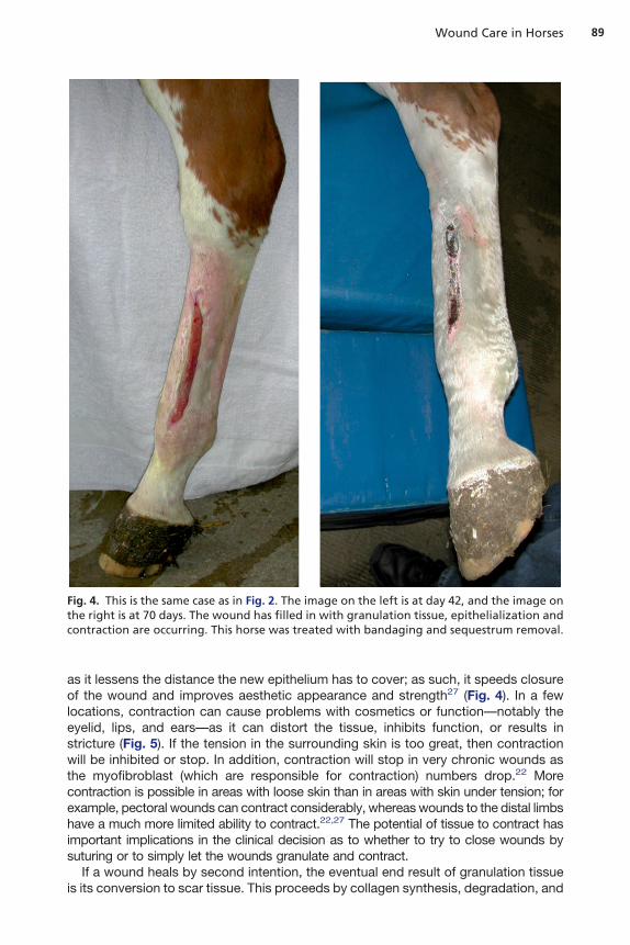
as it lessens the distance the new epithelium has to cover; as such, it speeds closureof the wound and improves aesthetic appearance and strength27 (Fig. 4). In a fewlocations, contraction can cause problems with cosmetics or function—notably theeyelid, lips, and ears—as it can distort the tissue, inhibits function, or results instricture (Fig. 5). If the tension in the surrounding skin is too great, then contractionwill be inhibited or stop. In addition, contraction will stop in very chronic wounds asthe myofibroblast (which are responsible for contraction) numbers drop.22 Morecontraction is possible in areas with loose skin than in areas with skin under tension; forexample, pectoral wounds can contract considerably, whereas wounds to the distal limbshave a much more limited ability to contract.22,27 The potential of tissue to contract hasimportant implications in the clinical decision as to whether to try to close wounds bysuturing or to simply let the wounds granulate and contract.
If a wound heals by second intention, the eventual end result of granulation tissue
Fig. 4. This is the same case as in Fig. 2. The image on the left is at day 42, and the image onhe right is at 70 days. The wound has filled in with granulation tissue, epithelialization andontraction are occurring. This horse was treated with bandaging and sequestrum removal.
is its conversion to scar tissue. This proceeds by collagen synthesis, degradation, and

90 Caston
eventually cross-linking and rearrangement. Remodeling of the scar tissue cancontinue for up to 2 years.22 Over time, the tissue becomes more organized and lessvascular, and the normal ratio of type I collagen to type III collagen (1:4) is restored.It is important for ambulatory practitioners to realize that even when scar tissuereaches full maturity, the wound will be 15%–20% weaker than the original tissue.22
TREATMENT OF THE WOUND
Following initial triage of the wound, it should be cleaned. Sedation will frequently behelpful. Treatment with nonsteroidal anti-inflammatory agents to reduce pain andswelling is also appropriate at this time. Clipping the hair around the wound is of usein getting and keeping the wound clean even if the wound will not be closed withsutures. Applying a water-soluble lubricant in the wound or packing the wound withsaline moistened gauze can help keep hair out of the wound while clipping. If the areais covered in dried blood or mud/dirt, preliminary cleansing can begin with a hose. Thehose should not have a sprayer nozzle attached; the sprayer can force water intotissue planes. The goal is merely to cleanse the surface. After clipping the hair andremoval of the bulk of organic debris, the wound can be further cleansed with mildsoap, dilute iodine, or dilute chlorhexidine soaps and rinsed with saline or anotherisotonic fluid. Large volume lavage utilizing isotonic fluids can also dilute contamina-tion and help with the removal of foreign material.
A regional or local anesthetic block can facilitate debridement. Judicious sharpdebridement can be used to remove a very thin layer of tissue if foreign material isembedded in the tissues. Skin flaps, especially on the head or below mid-forearm/mid- gaskin, should not be removed during sharp debridement. The viability of skinflaps will become readily apparent over the first few days post injury; any portion ofa flap that remains viable can and will be used to aid in the healing process. Thepaucity of movable skin in some areas makes it such a desirable commodity that skinflaps should be given every possible opportunity to remain. Skin flaps on eyelidsshould never be removed at initial treatment. It is surprising in some cases, especiallyon the head and eyes, how much of a skin flap can remain viable. It is very easy to trimskin flaps, but there should not be a rush to do so. If there is any doubt about skin
Fig. 5. Healed wound on the left upper eyelid. Loss of tissue and subsequent second-intention healing including contraction of the wound have caused eversion of the uppereyelid and loss of mobility. This horse had difficulty blinking. Recognition of this probabilityshould have prompted a reconstructive procedure to be performed to prevent this end result.
viability, err on the side of caution. Once you remove the skin, you are committed. As

hoaaoF
91Wound Care in Horses
compared to the upper body, the distal limb has greater skin tension, epithelializationis slower than on the body, and excessive granulation tissue is more likely tooccur.22,28
Once the wound is cleaned and debrided, an effort should be made to close thewound, or to bring the edges as close together as possible. Suture patterns such asnear-far-far-near and vertical mattress patterns—with or without stents—can be usedto pull the skin edges together in cases where there is skin missing or there is enoughswelling to create significant tension. Many suture patterns have been described forclosing specific wounds, and those can be employed as the wound configurationdictates.29,30 Even if the clinician suspects that a skin flap will later slough or thesutures dehisce due to tension or motion, closure of the wound should be attempted,even if only partially. Even a partial closure can help hold tissues in place, coverunderlying structures, and reduce dead space (Fig. 6).
If the wound is highly contaminated or a large amount of dead space is present, adrain can be placed and then removed a few days later. A simple Penrose drainexiting the most ventral aspect of the wound or area of dead space is oftensufficient.30 Areas that cannot be closed or that were closed and later dehisce, willeal by second-intention wound healing. It is good practice to prepare owners for thatccurrence and to counsel them on what to expect so that they do not perceive it asfailure on your part when dehiscence occurs. High motion areas, deep wounds, andreas that are difficult to bandage, such as those that may occur in the pectoral arear upper hindquarters, may dehisce. Owners should be aware of this possibility.
Fig. 6. Degloving injury to a left hindlimb before and after debridement/suturing. Thecannon bone and extensor tendon are exposed prior to closure in the image on the left. Thesestructures are covered and the skin flap is held in place in the image on the right.
ortunately, these areas also heal very well as open wounds.28

dwshcbaatd
wwsbswdssa
92 Caston
After debridement and closure, a good padded bandage should be placed andmaintained. A good bandage is extremely important for reducing the swelling in anacute wound, which can be severe (see Bandaging section) (see Fig. 2). In cases ofdistal limb wounds that already have significant swelling and might otherwise be hardto close because of tension, bandaging for 24 hours prior to wound repair can also bebeneficial. The wound can be assessed, explored, and lavaged as outlined earlier,then reevaluated in 24 hours to see if the edges can be apposed under less tension.
WHAT THE PRACTITIONER AND THE OWNER SHOULD EXPECT1. Make realistic assessments. When assessing and treating wounds, practitionersshould prepare owners for likely outcomes at the time of the initial exam. The woundwill likely look its best at the end of your initial treatment, but owners should be madeaware that complications can occur. On the other hand, realistic expectations forwounds healing by second intention sometimes come as a surprise to owners.Informing owners of the duration of healing is something that is often overlooked;owners should be fully aware that most wounds healing by this process (unless small)will take months to heal. Care and bandaging of a wound can become tedious forowners over time, and they may discontinue treatments in frustration.
2. Monitor wound healing. The presence of wound fluid and the odor of a wounduring bandage changes can disturb some owners and lead them to assume theound is infected or healing poorly, even in cases where healing is progressingatisfactorily. On the other hand, some owners ignore or fail to recognize whenealing is impaired or delayed. Description of what is acceptable, as well as whathanges should initiate a call to the veterinarian, should be given to the owner toetter their ability to monitor healing and assist in care of the wound. For example, theppearance of a healthy granulating wound versus one that is incomplete because ofsequestrum or foreign body, one that has developed exuberant granulation, or one
hat exhibits impaired/delayed healing is an example of a complication that might beiscussed with clients in practice.
3. Be prepared for complications. If bone is exposed, as often happens in lower limbounds, a sequestrum should be an expected outcome. Good debridement of theound and the surface of the bone (with a curette or bone rasp) and closure of thekin, if possible, may help reduce the incidence or degree of sequestrum formation,ut owners should be aware of a possible additional procedure to remove aequestrum in the future. Sequestrum formation is usually evident radiographicallyithin 3 weeks31 and should be suspected if the granulation tissue has a cleft orefect when it has otherwise filled the wound (Fig. 7). Most sequestra can also beeen using ultrasound as well. Some horses exhibit lameness during the time theequestrum is maturing at around 10–14 days; practitioners and owners need to beware of this possibility. These horses can be up to grade 4/5 lame32 when this
occurs. The lameness is usually transient and can be alleviated with NSAID therapy.Sequestrum removal can often be performed in a standing, sedated horse. If thehorse’s nature or the location or extent of the wound makes standing removal difficult,short-term anesthesia can facilitate removal. Radiography and/or ultrasound can behelpful in determining if removal is complete.
4. Consider skin grafting larger wounds. Wounds that result in the loss of a large areaof skin, especially on the limbs, either at the time of initial injury or after sloughing/debridement should be considered candidates for skin grafting. Owners should be
advised that some wounds cannot heal, or heal poorly, with contraction and
etce
hosrs
93Wound Care in Horses
epithelialization alone. Grafting can remedy this problem, and decrease healing timefor many wounds. There are several techniques for free skin grafting frequently usedin horses; the type chosen may depend on equipment available, cost, and expectedcosmetic outcome. Pinch or punch skin grafts are relatively inexpensive, graft harvestand implantation can be performed in a standing horse, can be accepted even in lessthan ideal granulation beds,33–35 and can be safely performed in the field. Success ofach graft is independent of the others, so failure of one or more grafts does not affecthe rest35 (Fig. 8). Other grafting techniques, such as sheet grafting or tunnel graftingan cover larger areas and have a more cosmetic outcome but are often relativelyxpensive and require more specialized instruments than pinch or punch grafts33–35
and must be performed under general anesthesia, most commonly at referralfacilities.33
BANDAGING
Even though it can be difficult, and expensive over time, the importance of a goodbandage to equine wound care cannot be overstated. While there is some evidencethat bandages can promote some degree of exuberant granulation tissue, otherevidence suggests that bandaged wounds may heal faster than those that are notbandaged,36 and a good bandage has many other beneficial effects on wound
ealing.28 The qualifier good is significant; a poor bandage can, at best, be of littler no benefit to the patient and, at worst, be detrimental. A good bandage can helptop bleeding in an acute wound, prevent further contamination and trauma,estrict movement, provide a moist wound-healing environment, and reduce
Fig. 7. This is the same case as in Figs. 2 and 4. The wound is approximately 3 weeks old, anda cleft can be seen in the granulation tissue in the image on the left. The sequestrum is shownin the image on the right at the time of removal.
welling.4,21,28
94 Caston
Unfortunately, bandaging is often discontinued early in the process of wound care,is not performed, or is done improperly. This is likely because application andmaintenance of a good bandage can be time consuming and require a good deal ofwork on the part of the veterinarian and owner. Use of an inadequate amount ofpadding is a very common pitfall. A number of cotton roll products exist, and any canbe used to provide this padding. A compressive layer or 2 over the cotton, such aswide brown gauze followed by a self-adherent elastic bandage, will help secure thebandage and provide compression. Sealing the top and bottom of the bandage withan elastic adhesive tape helps keep the bandage in place and helps keep shavingsand other debris out of the bandage.
Bandage changes can occur every few days in most cases. Early on, bandagechanges may need to be more frequent if bleeding is still occurring or if treatmentssuch as debridement, suturing, or regional limb perfusions are necessary. After thewound is stable, owners can change the bandage every 3–5 days or sooner if it slipsdown or gets torn, worn through, or very wet. In some instances, owners do not feelcomfortable changing the bandage, or the horse requires sedation for bandagechanges. In such cases, a heavily padded, very snug bandage fully covered withelastic adhesive tape may last 7–10 days. This seems a long period, but a goodbandage should last that long and there should be no worry about leaving a bandageon for that duration if it stays intact—when casts are used to cover a wound, they are
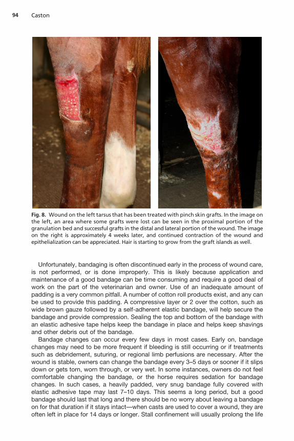
Fig. 8. Wound on the left tarsus that has been treated with pinch skin grafts. In the image onthe left, an area where some grafts were lost can be seen in the proximal portion of thegranulation bed and successful grafts in the distal and lateral portion of the wound. The imageon the right is approximately 4 weeks later, and continued contraction of the wound andepithelialization can be appreciated. Hair is starting to grow from the graft islands as well.
often left in place for 14 days or longer. Stall confinement will usually prolong the life

mpsdo
95Wound Care in Horses
of a bandage, but unless the injury requires, it is not essential. While farm setup andmanagement may preclude stall rest in some cases, limiting the activity level of thehorse with a limb wound decreases the amount of motion and may be useful in theearly phases of wound healing.
When bandaging is discontinued, it may be useful to warn owners that the areabeing bandaged can become edematous with the cessation of bandaging. Increasedexercise, or “weaning” the horse from bandaging by applying a bandage for 12–24hours, then removing the bandage for 12–24 hours, then bandage for 8–12 hours,then remove, and so on, can help counteract this problem.
TREATMENTS, PRODUCTS, AND CONTINUED MANAGEMENT OF THE WOUND
Unfortunately, there is no “silver bullet” that will speed wound healing significantly.Some treatments, products, and procedures have shown modest benefit, but thedegree to which each helps is contingent on the wound properties, location on thebody, and stage of healing, and the time and cost of such interventions may not justifythe marginal clinical relevance. For the practitioner to search for one product that willheal every wound or heal wounds extremely fast or without complications is futile.Certainly, partial-thickness wounds and wounds that are able to be closed by delayedprimary closure will heal quite quickly and often with excellent cosmetic results, butthere is no evidence that their healing is helped by various over-the-counter woundproducts, which only add to the expense of care. Full-thickness wounds that heal bysecond intention must all go through the same healing process, and while somedifferences in healing time and quality can be obtained with proper care, no oneproduct or treatment is right for every case, and few have been shown to make a verydramatic difference in healing time.3,5
What does seem clear is that wound healing can be impaired or delayed with somepractices and with application of some products.20,21,28 Although desiccation ofepithelial cells can result in slowed epithelialization if a moist wound-healing environ-ment is not always maintained, a common misperception among owners is thatwounds should “get air to them.” Other practices can be more damaging; the authorhas seen substances such as formalin, pickling lime, silver nitrate, lye, and calciumhydroxide put on wounds. Most are used in an attempt to reduce or prevent excessivegranulation tissue but, in fact, such substances delay wound healing and can promotescarring.28 There are an astounding number of wound treatment products on the
arket. The ambulatory clinician should carefully scrutinize the ingredients in suchroducts before using any wound treatment; many topical treatments contain agentsuch as phenols and alcohols or other chemicals that are known irritants, caustics, orrying agents. In addition, some products can damage tissue surrounding the woundr other normal tissue that comes into contact with it.28
In some instances, a product applied can be irritating enough to cause self-mutilation and damage to intact skin (Fig. 9). If a wound has granulation tissue that isdiscolored or otherwise unhealthy or that is below the level of the skin, then theapplication of a caustic treatment should be considered as a cause. Caustic agentsare not selective; they are caustic to all of the tissue with which they come in contact.While the intent of these agents is to decrease granulation tissue, they can delaywound healing by destroying epithelial cells. To remedy the urge by some owners toapply products to wounds and avoid potentially caustic or harmful substances, it maybe useful to give owners something to apply that will do no harm. In most cases, this
is simply generic triple antibiotic ointment.2,37
2
3
wspt
96 Caston
COMPLICATED AND CHRONIC WOUNDS
On occasion, the ambulatory equine practitioner is presented with a chronic,nonhealing wound. These can be difficult cases but should be approached with thewound-healing process in mind. There are many reasons why a wound may not behealing, and a systematic approach can help narrow down the possibilities:
1. Is the granulation bed appropriate and intact? If the granulation tissue bed is notintact, if a cleft is present or if the wound itself is a draining tract, then somethingis causing continued drainage. Possible causes include a sequestrum, foreignbody, motion/trauma, or infection of an underlying structure.
. If the granulation tissue is intact across the entire bed of the wound—is itappropriate? Bulging of the granulation tissue above the wound surface, or proudflesh, is one example of an inappropriate course of healing. Granulation tissue withan abnormal color or consistency may also be signs that the bed is not healthy.
. Is something else growing? Sarcoids or squamous cell carcinomas might besuspected if the normal skin edge ends abruptly and a mass begins, rather than ahealing epithelial edge being present38 (Figs. 10 and 11).
Proud Flesh
Exuberant granulation tissue, or proud flesh, is one cause for a wound to havedelayed or impaired healing. Prevention of this occurrence with good woundmanagement is best, but sometimes it is an inevitable outcome. Small to moderateamounts of excessive granulation tissue can be treated with application of cortico-steroid cream and a good bandage, but if there is a large amount of proud flesh, itshould be sharply excised.28 The excess granulation tissue should be trimmed flush
ith the healing epithelial edge, taking care not to remove or damage healthyurrounding skin or new epithelial tissue. Most horses are amenable to this beingerformed while standing under sedation. A tourniquet above the area that is to be
Fig. 9. Thoroughbred weanling that originally sustained a wound to the pectoral area. Acommercial wound powder containing caustic hydrated lime was applied to the pectoralwound. This caused self-mutilation, during which contact with the lime caused a woundunder the lower lip. That wound—seen in image on the left—eventually contracted andcaused deformity of the lip that had to be corrected surgically. (Images courtesy of Dr ScottMcClure.)
rimmed can help reduce the amount of bleeding, but because of the composition of

97Wound Care in Horses
granulation tissue, bleeding will still occur. The owner should be prepared for bleedingif they are present for the procedure, as it can appear to be a dramatic amount,especially since horse blood clots slowly. The bleeding will eventually cease with aheavily padded bandage placed over the wound after trimming. In some cases,
Fig. 10. Sarcoid on the dorsal carpus of a 5-year-old quarterhorse stallion.
Fig. 11. Two cases with chronic nonhealing wounds. The case on the left has chronic excessivegranulation tissue present, while the proliferative tissue in the case on the right is a sarcoid.Both cases underwent treatment for removal of the excess tissue and the underlying wound,
but the enlargement of the leg caused by chronic swelling remained in both cases.
npsa
98 Caston
repeated trimming of the granulation tissue is necessary.28 However, if the woundeeds repeated trimming of granulation tissue and does not appear to be makingrogress, causes for delayed healing should be investigated. In those wounds that areimply too large to heal well by second intention alone, skin grafting should be offereds a treatment option.
Summer Sores
Habronemiasis can occur in open wounds and will sometimes have an appearancesimilar to proud flesh or sarcoids. It can be difficult to tell the difference between these3 problems and a few other conditions, such as squamous cell carcinoma. Summersores often contain calcified granules and are can be quite pruritic. Histopathology isrecommended for any proliferative nonhealing wound as treatments and expectedoutcomes may depend on the diagnosis. Debulking the portion that protrudes beyondthe skin edge and bandaging is almost always an excellent start to getting any ofthese conditions under control, and a biopsy can be submitted from the portion thatis removed.
Excessive Motion
At times, even if the wound is healing appropriately, a large amount of motion candisrupt the granulation bed and delay healing or result in undue scar tissue.20,21,28
Areas where this may occur are typically near joints such as the dorsal fetlock or overa bony eminence, such as the point of the hock. Measures to reduce motion includingstall confinement, a heavily padded bandage with a splint, or a cast can help limitmotion and thus disruption of the healing wound.
Wounds that are chronic and have been left without treatment for a long period oftime may have associated limb swelling that could be permanent due to a largeamount of scar tissue and disrupted vascular/lymphatic function. Even if the woundis amenable to treatment, if the duration of swelling has been prolonged the ownersshould be advised of the possibility of a permanent enlargement (see Fig. 11).Nevertheless, there are many chronic cases in which treating the wound andbandaging the limb may result in surprising improvement in both the appearance ofthe wound, as well as in limb swelling.
SUMMARY
Care of equine wounds in the field can be a challenging endeavor. Many times, woundcare is complicated by chronicity or by prior inappropriate care in addition to the greatdegree of tissue trauma that occurred when the horse was wounded. Recognizinginvolvement of synovial structures, loss of skin, and damage to bone are critical in theinitial examination of wounds and will guide future care. Education of clients is alsoimportant in that preparing them for possible outcomes during healing may helpimprove compliance and proper treatment of wound. Owners and trainers oftenperform much of the daily care and monitoring of equine wounds and thus can greatlyassist or impede the progress. Bandaging is important to management of equinewounds—especially on the limbs—and is sometimes overlooked because of itslabor-intensive nature and the desire for a spray, ointment, or salve that will heal thewound. The practitioner that improves and utilizes his or her understanding of thewound-healing process in concert with his or her knowledge of local anatomy will be
the one who is best equipped to care for wounds in ambulatory practice.
1
99Wound Care in Horses
REFERENCES
1. Stashak TS, Farstvedt EF. Update on wound dressings: indications and best use. In:Stashak TS, Theoret C, editors. Equine wound management. 2nd edition. Ames (IA):Wiley-Blackwell; 2008. p. 109–36.
2. Farstvedt EF, Stashak TS. Topical wound treatments and wound care products. In:Stashak TS, Theoret C, editors. Equine wound management. 2nd edition. Ames (IA):Wiley-Blackwell; 2008. p. 137–59.
3. Theoret C. Wound repair: problems in the horse and innovative solutions. In: StashakTS, Theoret C, editors. Equine wound management. 2nd edition. Ames (AI): Wiley-Blackwell; 2008. p. 47–68.
4. Gomez J. Bandaging and casting techniques for wound management. In: StashakTS, Theoret C, editors. Equine wound management. 2nd edition. Ames (IA): Wiley-Blackwell; 2008. p. 623–58.
5. Dart AJ, Dowling BA, Smith CL. Topical treatments in equine wound management.Vet Clin North Am Equine Pract 2005;21:77–89.
6. Holcombe SJ. Shock: pathophysiology, diagnosis and treatment. In: Auer JA, StickJA, editors. Equine surgery. 3rd edition. St Louis: Saunders Elsevier; 2006. p. 1–9.
7. Holcombe SJ. Physiologic response to trauma: evaluating the trauma patient. In: AuerJA, Stick JA, editors. Equine surgery. 3rd edition. St Louis: Saunders Elsevier; 2006.p. 88–96.
8. Hassel DM. Thoracic trauma in horses. Vet Clin North Am Equine Pract 2007;23:67– 80.
9. Hendrickson DA. Management of deep and chronic wounds. In: Auer JA, Stick JA,editors. Equine surgery. 3rd edition. St Louis: Saunders Elsevier; 2006. p. 299–305.
0. Schneider RK. Synovial and osseous infections. In: Auer JA, Stick JA, editors. Equinesurgery. 3rd edition. St Louis: Saunders Elsevier; 2006. p. 1121–30.
11. Wereszka MM, White NA 2nd, Furr MO. Factors associated with outcome followingtreatment of horses with septic tenosynovitis: 51 cases (1986–2003). J Am Vet MedAssoc 2007;230:1195–200.
12. Schneider RK, Bramlage LR, Moore RM, et al. A retrospective study of 192 horsesaffected with septic arthritis/tenosynovitis. Equine Vet J 1992;24:436–42.
13. Honnas SM, Schumacher J, Cohen ND, Watkins JP, Taylor TS. Septic tenosynovitisin horses: 25 cases (1983–1989). J Am Vet Med Assoc. 1991;199(11):1616–22.
14. Gibson KT, McIlwraith CW, Turner AS, et al. Open joint injuries in horses: 58 cases(1980–1986). J Am Vet Med Assoc 1989;194:398–404.
15. Moyer W, Schumacher J, Schumacher J. Equine joint injection and regional anesthe-sia. 5th edition. Chadds Ford (PA): Academic Veterinary Solutions; 2011. p. 1–144.
16. Davis CS, Smith RKW. Diagnosis and management of tendon and ligament disorders.In: Auer JA, Stick JA, editors. Equine surgery. 3rd edition. St Louis: Saunders Elsevier;2006. p. 1086–111.
17. Taylor DS, Pascoe JR, Meagher DM, et al. Digital flexor tendon lacerations in horses:50 cases (1975–1990). J Am Vet Med Assoc 1995;206:342–6.
18. Foland JW, Trotter GW, Stashak TS, et al. Traumatic injuries involving tendons of thedistal limbs in horses: a retrospective study of 55 cases. Equine Vet J 1991;23:422–5.
19. Mespoulhès-Rievère C, Martens A, Bogaert L, et al. Factors affecting outcome ofextensor tendon lacerations in the distal limb of horses. A retrospective study of 156cases (1994–2003). Vet Comp Orthop Traumatol 2008;21:358–64.
20. Stashak TS. Selected factors that negatively impact healingIn: Stashak TS, TheoretCeditors. Equine wound management. 2nd edition. Ames (IA): Wiley-Blackwell; 2008.
p. 71–84.
100 Caston
21. Hendrickson D, Virgin J. Factors that affect equine wound repair. Vet Clin North AmEquine Pract 2005;21:33–44.
22. Theoret C. Physiology of wound healing. In: Stashak TS, Theoret C, editors. Equinewound management. 2nd edition. Ames (IA): Wiley-Blackwell; 2008. p. 5–28.
23. Shumacher J, Brumbaugh GW, Honnas CM, et al. Kinetics of healing grafted andnongrafted wounds on the distal portion of the forelimbs of horses. Am J Vet Res1992;53:1568–71.
24. Wilmink JA, Stolk PW, Van Weeren PR, et al. Differences in second-intention woundhealing between horses and ponies: macroscopic aspects. Equine Vet J 1999;31:53–60.
25. Schumacher J, Stashak TS. Management of wounds of the distal extremities. In:Stashak TS. Theoret C, editors. Equine wound management. 2nd edition. Ames (IA):Wiley-Blackwell; 2008. p. 375–425.
26. Theoret, CL. The pathophysiology of wound repair. Vet Clin North Am Equine Pract2005;21:1–13.
27. Wilmink JM, Van Weeren PR. Second-intention repair in the horse and pony andmanagement of exuberant granulation tissue. Vet Clin North Am Equine Pract2005;21:12–32.
28. Wilmink JM, Van Weeren PR. Treatment of exuberant granulation tissue. Clin TechEquine Pract 2004;3:141–7.
29. Stashak TS. Selection of suture materials, suture patterns, and drains for woundclosure. In: Stashak TS, Theoret C, editors. Equine wound management. 2nd edition.Ames (IA): Wiley-Blackwell; 2008. p. 193–224.
30. Stashak TS. New and innovative approaches to wound closure. In: Stashak TS,Theoret C, editors. Equine wound management. 2nd edition. Ames (IA): Wiley-Blackwell; 2008. p. 225–37.
31. Moens Y, Verschooten F, De Moor A, et al. Bone sequestration as a consequence oflimb wounds in the horse. Vet Radiol 1980;21:40–4.
32. American Association of Equine Practitioners (AAEP). Lameness scale: definition andclassification of lameness. In: Guide for veterinary service and judging of equestrianevents. 4th edition. Lexington (KY): AAEP; 1991. p. 19.
33. Bristol DG. Skin grafts and skin flaps in the horse. Vet Clin North Am Equine Pract2005;21:125–44.
34. Schumacher J. Skin grafting. In: Auer JA, Stick JA, editors. Equine surgery. 3rdedition. St Louis: Saunders Elsevier; 2006. p. 269–87.
35. Schumacher J, Hanselka DV. Skin grafting of the horse. Vet Clin North Am EquinePract 1989;5:591–614.
36. Woollen N, DeBowes RM, Liepold HW, et al. A comparison of four types of therapy forthe treatment of full-thickness skin wounds of the horse. In: Proceedings of the AnnualConvention of the American Association of Equine Practitioners 1987;33:569–76.
37. Stashak TS. Management practices that influence wound infection and healing. In:Stashak TS, Theoret C, editors. Equine wound management. 2nd edition. Ames (IA):Wiley-Blackwell; 2008. p. 85–106.
38. Knottenbelt DCSarcoid transformation at wound sites. In: Stashak TS, Theoret C,editors. Equine wound management. 2nd edition. Ames (IA): Wiley-Blackwell; 2008.p. 585–608.