Wound Ca re AD VISOR · Official journal of Wound Ca re AD VISOR Practical issues in wound, skin,...
44
Official journal of Wound Care ADVISOR Practical issues in wound, skin, and ostomy management ® What would you do with this ostomy? Providing evidence-based care for individuals with cellulitis Exercise for patients with lower extremity disease Creating high-performance interprofessional teams May/June 2015 • Volume 4 • Number 3 www.WoundCareAdvisor.com A Publication
-
Upload
phungtuyen -
Category
Documents
-
view
223 -
download
0
Transcript of Wound Ca re AD VISOR · Official journal of Wound Ca re AD VISOR Practical issues in wound, skin,...

Official journal of
WoundCareADVISOR
Practical issues in wound, skin, and ostomy management®
What wouldyou do with
this ostomy?
Providing evidence-basedcare for individuals with
cellulitis
Exercise for patients withlower extremity disease
Creating high-performanceinterprofessional teamsMay/June 2015 • Volume 4 • Number 3
www.WoundCareAdvisor.com
A Publication

Advancing wound care one solution at a time
*The skin protection claim does not apply to the Foaming Body Cleanser and the Antifungal Body Powder.
Unless otherwise noted, the product claims, information and recommendations in this document were obtained from the manufacturer and have not been independently verifi ed by McKesson Medical-Surgical. The properties of a product may change or be inaccurate following the posting or printing of the product information in this document, either in the print or online version. Caution should be exercised when using or purchasing any products from McKesson’s online or print document by closely examining the product packaging and the labeling prior to use. Be advised that information contained herein is intended to serve as a useful reference for informational purposes only and is not complete clinical information. This information is intended for use only by competent healthcare professional exercising judgment in providing care. This promotion is not open to the general public and may be terminated by McKesson Medical-Surgical at any time. Due to regulatory restrictions related to gifts and gratuities, this promotion cannot be off ered to healthcare providers licensed in Vermont or to federal government employees.
© 2015 McKesson Medical-Surgical Inc. All trademarks and registered trademarks are the property of their respective owners.
Your residents deserve the best care available. Now, with McKesson Medical-Surgical’s full suite of advanced wound care solutions, it’s easier to give them just that. Whether you’re seeking clinical or business support, proven skin care solutions or quality advanced wound care products, we can help you provide the best care, and save money doing it.
Visit mckessonwoundcare.com to request a complimentary sample of our new Silicone Foam Adhesive Dressing.
Clinical and business support:
• Access to our Clinical Resource Team• Customized webinars• Assistance as you create product formularies• In-servicing• Access to the F314 Wound Care QuickPath• McKesson SpendManagerSM
Advanced skin and wound care:
• Eight THERA™ Advanced Skin Care products that help cleanse, moisturize, protect* and treat the skin
• Our comprehensive wound care products include Wound Cleansers and Guides, Wound Dressings, Cover and Absorptive Dressings and Multi-Layer Compression Bandages
Your residents deserve the best care available. Now, with McKesson Medical-Surgical’s full suite of
NEW!

2 www.WoundCareAdvisor.com May/June 2015 • Volume 4, Number 3 • Wound Care Advisor
staffGroup Publisher
Gregory P. OsbornePublisher
Tyra LondonEditor-in-Chief
Donna Sardina, RN, MHA, WCC, CWCMS, DWC, OMSEditorial Director
Cynthia Saver, RN, MSEditor
Kathy E. GoldbergCopy Editor
Karen C. ComerfordArt Director
David BeverageProduction Manager
Rachel BargeronAccount Managers
Susan Schmidt Renee Artuso
John Travaline
PuBlished ByHealthCom Media
259 Veterans Lane, Doylestown, PA 18901Telephone: 215/489-7000Facsimile: 215/230-6931Chief Executive OfficerGregory P. Osborne
Executive Vice President, SalesBill Mulderry
Digital Program ManagerMichael FerrariFinance Director
MaryAnn FosbennerBusiness Manager
Tina Boyer
Wound Care Advisor (ISSN 2168-4421) is published byHealthCom Media, 259 Veterans Lane, Doylestown, PA18901. Printed in the USA. Copyright © 2015 by Health-Com Media. All rights reserved. No part of this publica-tion may be reproduced, stored, or transmitted in anyform or by any means, electronic or mechanical, includ-ing photocopy, recording, or any information storageand retrieval system, without permission in writing fromthe copyright holder. Send communication to Health-Com Media, 259 Veterans Lane, Doyles town, PA 18901.
The opinions expressed in the editorial and advertis-ing material in this issue are those of the authorsand advertisers and do not necessarily reflect theopinions or recommendations of the National Al-liance of Wound Care and Ostomy®; the EditorialAdvisory Board members; or the Publisher, Editors,and the staff of Wound Care Advisor.
Editorial Mission: Wound Care Advisor providesmultidisciplinary wound care professionals withpractical, evidence-based information on theclinical management of wounds. As the officialjournal of the National Alliance of Wound Careand Ostomy®, we are dedicated to deliveringsuccinct insights and information that our read-ers can immediately apply in practice and useto advance their professional growth.
Wound Care Advisor is written by skin and woundcare experts and presented in a reader-friendly elec-tronic format. Clinical content is peer reviewed.
The publication attempts to select authors who areknowledgeable in their fields; however, it does notwarrant the expertise of any author, nor is it responsi-ble for any statements made by any author. Certainstatements about the use, dosage, efficacy, and charac-teristic of some drugs mentioned here reflect the opin-ions or investigational experience of the author. Anyprocedures, medications, or other courses of diagnosisor treatment discussed or suggested by authors shouldnot be used by clinicians without evaluations of theirpatients’ conditions and possible contraindications ordanger in use, review of any applicable manufacturer’sprescribing information, and comparison with the rec-ommendations of other authorities.
editor-in-chief
Donna Sardina, RN, MHA, WCC, CWCMS, DWC, OMSCo-Founder, Wound Care Education Institute
Lake Geneva, WI
editorial advisory BoardNenette L. Brown, RN, PHN, MSN/FNP, WCC
Wound Care Program CoordinatorSheriff’s Medical Services Division
San Diego, CA
Debra Clair, PhD, APN, RN, WOCN, WCC, DWC Wound Care Provider
Alliance Community HospitalAlliance, OH
Kulbir Dhillon, NP, WCCWound Care Specialist
Skilled Wound CareSacramento, CA
Fred BergVice President, Marketing/Business Development
National Alliance of Wound Care and OstomySt. Joseph, MI
Cindy Broadus, RN, BSHA, LNHA, CLNC, CLNI, CHCRM, WCC, DWC, OMSExecutive Director
National Alliance of Wound Care and OstomySt. Joseph, MI
Catherine Jackson, RN, MSN, WCCClinical Nurse Manager
Inpatient and Outpatient Wound CareMacNeal Hospital
Berwyn, IL
Jeffrey Jensen, DPM, FACFASDean and Professor of Podiatric Medicine & Surgery
Barry University School of Podiatric MedicineMiami Shores, FL
Rosalyn S. Jordan, RN, BSN, MSc, CWOCN, WCCDirector of Clinical Education
RecoverCare, LLCLouisville, KY
Jeff Kingery, RNVice President of Professional Development
RestorixHealthTarrytown NY
Jeri Lundgren, RN, BSN, PHN, CWS, CWCNVice President of Clinical Consulting
Joerns Charlotte, NC
Courtney Lyder, ND, GNP, FAAN Dean and Professor
UCLA School of NursingLos Angeles
Nancy Morgan, RN, BSN, MBA, WOC, WCC, DWC, OMSCo-Founder, Wound Care Education Institute
Plainfield, IL
Steve Norton, CDT, CLT-LANACo-founder, Lymphedema & Wound Care Education, LLC
President, Lymphedema Products, LLCMatawan, NJ
Bill Richlen, PT, WCC, CWS, DWCOwner
Infinitus, LLCChippewa Falls, WI
Lu Ann Reed, RN, MSN, CRRN, RNC, LNHA, WCCAdjunct Clinical Instructor
University of CincinnatiCincinnati, OH
Stanley A. Rynkiewicz III, RN, MSN, WCC, DWC, CCSAdministrator
Deer Meadows Home Health and Support Services, LLCBHP Services
Philadelphia, PA
Cheryl Robillard, PT, WCC, CLTClinical SpecialistAegis TherapiesMilwaukee WI
Donald A. Wollheim, MD, WCC, DWC, FAPWCAOwner and Clinician, IMPLEXUS Wound Care Service, LLC
Watertown, WIInstructor, Wound Care Education Institute
Plainfield, IL

For more details call 1-888-318-8536 or visit us at
HOTELLAS VEGAS
e details call For mor
or visit us at 1-888-318-8536e details call
or visit us at

4 www.WoundCareAdvisor.com May/June 2015 • Volume 4, Number 3 • Wound Care Advisor
FEATURES
12 Helping patients overcome ostomy challenges By Beth Hoffmire Heideman, MSN, RNPhysiologic, psychological, and psychosocial issues demand careful planning, monitoring, and creativity.
24 Providing evidence-based care for patients with lower-extremity cellulitis By Darlene Hanson, PhD, RN; Diane Langemo, PhD, RN, FAAN; Patricia Thompson, MS, RN; Julie Anderson, PhD, RN; and Keith Swanson, MD Find out how to identify and intervene for this potentially dangerous bacterial skin infection.
DEPARTMENTS6 From the Editor
Ankle-brachial index: A dirty word?
9 Clinical Notes
18 Apple BitesMoldable ostomy barrier rings and strips
20 Best PracticesHelping patients with lower-extremity disease benefit from exercise
Get the ‘SKINNI’ on reducing pressure ulcers
30 Business ConsultA collaborative approach to wound care and lymphedema therapy: Part 1
Creating high-performance interprofessional teams
36 Clinician Resources
37 NAWCO News
May/June 2015 • Volume 4 • Number 3www.WoundCareAdvisor.comCONTENTS
page 24
page 12
page 21

Angelini Pharma Inc.8322 Helgerman Court - Gaithersburg - MD 20877 -USA1 (800) 726-2308www.Angelini-us.com
First Line Treatment for Healing Chronic Wounds
100% pure native equine type 1 collagen
Highest collagen content on the market
Easy to use for hard to heal wounds
BIOPADCaring for Wounds
TM
Contact us for more [email protected]
Size: 2” x 2” - 3 per box Order code: 132622
HCPCS code: A6021
D
Contact us for more [email protected]
Medicare Reimbursed
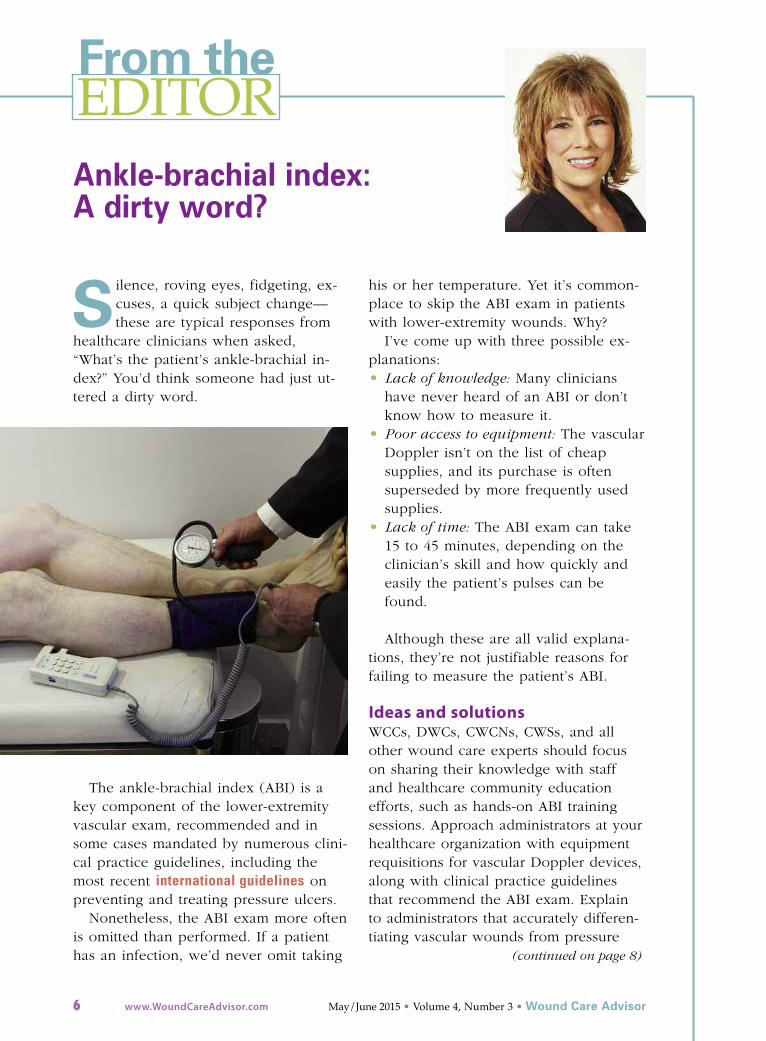
From theEDITOR
S ilence, roving eyes, fidgeting, ex-cuses, a quick subject change—these are typical responses from
healthcare clinicians when asked,“What’s the patient’s ankle-brachial in-dex?” You’d think someone had just ut-tered a dirty word.
The ankle-brachial index (ABI) is akey component of the lower-extremityvascular exam, recommended and insome cases mandated by numerous clini-cal practice guidelines, including themost recent international guidelines onpreventing and treating pressure ulcers.
Nonetheless, the ABI exam more oftenis omitted than performed. If a patienthas an infection, we’d never omit taking
his or her temperature. Yet it’s common-place to skip the ABI exam in patientswith lower-extremity wounds. Why?
I’ve come up with three possible ex-planations: • Lack of knowledge: Many clinicians
have never heard of an ABI or don’tknow how to measure it.
• Poor access to equipment: The vascularDoppler isn’t on the list of cheap supplies, and its purchase is often superseded by more frequently usedsupplies.
• Lack of time: The ABI exam can take15 to 45 minutes, depending on theclinician’s skill and how quickly andeasily the patient’s pulses can befound.
Although these are all valid explana-tions, they’re not justifiable reasons forfailing to measure the patient’s ABI.
Ideas and solutionsWCCs, DWCs, CWCNs, CWSs, and allother wound care experts should focuson sharing their knowledge with staffand healthcare community education efforts, such as hands-on ABI trainingsessions. Approach administrators at yourhealthcare organization with equipmentrequisitions for vascular Doppler devices,along with clinical practice guidelinesthat recommend the ABI exam. Explainto administrators that accurately differen-tiating vascular wounds from pressure
Ankle-brachial index: A dirty word?
6 www.WoundCareAdvisor.com May/June 2015 • Volume 4, Number 3 • Wound Care Advisor
(continued on page 8)

Joerns.comRecoverCare.com
©2015 Joerns
Easy-to-use mattress coverlet adds microclimate management to any therapy support surface.
Best in class moisture vapor transfer*
provides effective moisture management.
Optimizes skin temperature at mattress
interface to improve patient comfort.*
Use ClimateCare on any surface, including the Dolphin™ Fluid Immersion Simulation® system.
Call your Joerns RecoverCare Representative to schedule a trial.Joerns VA & Rental 800.966.6662Joerns Capital 800.826.0270RecoverCare 888.750.78282
Keep your patient cool, dry and comfortablewith ClimateCare™ Advanced MicroClimate Management
*Moisture vapor transfer greater than 130 (g/m2)/hr. Joerns RecoverCare data on file.

wounds may decrease the organization’spressure ulcer prevalence rate.
Other suggestions:• Contract with an outside agency to
perform ABI exams for your facility oragency.
• Designate an official ABI staffer to as-sist with admissions.
• Consider using the Lanarkshire Oxime-try Index as a substitute for ABIs.
For additional information, visit theseother online links:• Online video training• ABI policy and procedure• Measurement and interpretation of
the ankle-brachial index: A scientific
statement from the American Heart As-sociation
The ABI is an extremely beneficialtool that can aid early detection of pe-ripheral arterial disease, in turn helpingto prevent complications and amputa-tions and potentially saving lives. ABIneeds to come off the dirty-word list.
Donna Sardina, RN, MHA, WCC, CWCMS,DWC, OMS
Editor-in-ChiefWound Care Advisor
Cofounder, Wound Care Education InstitutePlainfield, Illinois
8 www.WoundCareAdvisor.com May/June 2015 • Volume 4, Number 3 • Wound Care Advisor
(continued from page 6)

Factors affecting medicationadherence in patients withdiabetes identified
Factors associated with better adherence toantidiabetic medications taken by patientswith diabetes include older age, male sex,higher education, higher income, use ofmail-order vs. retail pharmacies, primarycare vs. nonendocrinology specialist pre-scribers, higher daily total pill burden, andlower out-of-pocket costs.
“Determinants of adherence to diabetesmedications: Findings from a large pharmacyclaims database,” published in DiabetesCare, also found that patients who arenew to diabetes therapy are less likely tobe adherent. The study included morethan 200,000 patients who were treatedfor diabetes with noninsulin medications.
LMW heparin may improvehealing of chronic venous ulcers
International Wound Journal has pub-
lished “Low molecular weight heparin im-proves healing of chronic venous ulcers espe-cially in the elderly,” a study that included284 patients.
The healing rate for those receivinglow-molecular-weight (LMW) heparin wasaround 80% at 12 months, compared toaround 60% for those who didn’t receiveheparin. Older patients received the mostbenefit and also had the lowest recurrencerate.
Comparison of dressings forpediatric donor skin-graft sites
Compared to foam and hydrofiber, calci-um alginate is the optimum dressing forpediatric donor skin-graft sites, accordingto a study in the Journal of Burn Care &Research.
“Management of pediatric skin-graft donorsites: A randomized controlled trial of threewound care products” included 57 children,and the median size of the donor site was63.50 cm2. The median days for healingfor those in the calcium alginate groupwas 7.5, compared to 8 days for hy-drofiber and 9.5 days for foam.
Wound Care Advisor • May/June 2015 • Volume 4, Number 3 www.WoundCareAdvisor.com 9
ClinicalNOTES

Insulin pump clinical safetyappraised
“Insulin pump risks and benefits: A clinicalappraisal of pump safety standards, adverseevent reporting, and research needs,” is aJoint Statement of the European Associa-tion for the Study of Diabetes and theAmerican Diabetes Association DiabetesTechnology Working Group published inDiabetes Care. The article contains severalrecommendations for reducing adverse ef-fects caused by user error, including: • Select appropriate candidates for pump
therapy.• Provide those beginning pump therapy
with appropriate and ongoing educa-tion and support.
• Ensure that healthcare professionalssupporting pump users are themselveswell trained and supported.
The article also notes that the clinicalstudies required before marketing an in-sulin pump are “small and over-reliant onbench testing” and that once a pump ison the market, “insufficient data are madepublicly available on its long-term use in areal-world setting.”
Resistance training may improvelymphedema
According to a poster presented at a Flori-da State University symposium, “Resistance
training improves muscular strength and lym-phedema in breast cancer survivors,” 33 fe-male participants experienced moderate-to high-intensity resistance therapy over 12weeks. The researchers found that partici-pants tolerated therapy well and that lym-phedema was significantly decreased.
Ability to stop insulin varies withbariatric surgery type
“Insulin cessation and diabetes remission af-ter bariatric surgery in adults with insulin-treated type 2 diabetes,” published in Dia-betes Care, found that patients who hadRoux-en-Y gastric bypass surgery weremore likely than those who had laparo-scopic adjustable gastric banding to beable to stop insulin after surgery.
Pilot studies find acupuncturereduces lymphedema
“Acupuncture research at Memorial Sloan
10 www.WoundCareAdvisor.com May/June 2015 • Volume 4, Number 3 • Wound Care Advisor

Kettering Cancer Center” reports acupunc-ture significantly reduces arm circumfer-ence in patients with lymphedema.
The article, published in the Journal ofAcupuncture and Meridian Studies, dis-cusses two pilot studies that indicateacupuncture is safe for patients who havehad breast cancer surgery.
Clinical practice guidelines forostomy surgery released
Diseases of the Colon & Rectum has pub-lished “Clinical practice guidelines for osto-my surgery.” The American Society ofColon and Rectal Surgeons developed theguidelines, which discuss ostomy creation,closure, and complications.
The guidelines state that the “optimalcare for patients undergoing ostomy sur-gery includes preoperative, perioperative,and postoperative care by an ostomynurse specialist.” ■
We asked. You answered. We Delivered.New Heelift® Glide Ultra and AFO Ultra are the result of independent third-pary research with clinicians across North America and Europe. You wanted a cool inner lining that can be wiped clean while providing extra grip. We delivered Heelift Glide Ultra and AFO Ultra, with new Ultra-Grip interior; a breathable fabric lining that holds the leg in place and can be wiped clean. See for yourself how the new Heelifts are everything you’ve been asking for…and more.
To learn more or to request a free sample, visit heelift.com/x

12 www.WoundCareAdvisor.com May/June 2015 • Volume 4, Number 3 • Wound Care Advisor
No one wants an ostomy, butsometimes it’s required tosave a patient’s life. As osto-my specialists, our role is to
assess and intervene for patients with astoma or an ostomy to enhance theirquality of life. We play an active role inhelping patients perform self-care fortheir ostomy and adjust to it psychologi-cally, starting even before surgery.
Preoperative considerations Preparation for the ostomy is the mostcritical aspect of a healthy adjustment.When the ostomy is planned, the patientand family members are more likely to
process the life changes it will entail.They can learn about anticipated postsur-gical changes in the patient’s diet, cloth-ing, and sexuality, and family memberscan become more sensitive to the changein their loved one.
Assessment On initial assessment, evaluate your pa-tient’s body configuration, stoma place-ment, skin integrity, physical limitations,psychological needs, and home caregiv-ing system. Then develop a plan of careto mitigate problems that could impedethe patient’s ability to maintain and man-age the ostomy system.
The human body comes in many con-figurations and sizes. Because each per-son’s body is unique, clinicians may needto get creative to adapt the ostomy sys-tem to a patient’s body. Options foradapting it to your patient’s physicalcharacteristics include using: • a one-piece vs. a two-piece system• a flexible flange, clear drape flange, or
moldable flange.
Factors affecting decisions about an os-tomy include its location, skin integrity,and physical ability. (See Decision guidefor ostomy products.)
LocationThe stoma may be located near an inci-sion, under a peniculum, or in an ab-
Helping patients overcome ostomy challengesPhysiologic, psychological, and psychosocial issues demand careful planning, monitoring, and creativity.By Beth Hoffmire Heideman, MSN, RN

Wound Care Advisor • May/June 2015 • Volume 4, Number 3 www.WoundCareAdvisor.com 13
Decision guide for ostomy products
The chart below suggests appropriate products to use based on your patient’s physical condition orostomy characteristics. It applies to patients with ileostomies, urostomies, or colostomies. Refer pa-tients with special challenges to a certified wound clinician.
OSTOMY SYSTEM BASICS
Description What to use
Retracted stoma (below abdominal plane) • Convex flange• Convex ring• Strip paste
Protruded stoma (above abdominal plane) • Flat flange • Flat ring
Acidic effluence (ileostomy or urostomy) • Extended-wear flange• Extended-wear skin protector• Convex adaptor ring
Basic (neutral) effluence (colostomy) • Standard-wear flange• Standard skin protector• Stoma paste, adaptor rings• Adhesive strips
PERIWOUND SKIN
Eroded or denuded • Stoma powder• Use crusting method: Apply powder, dust off,
apply skin prep; repeat three times.
Fungal rash • Antifungal powder and skin protectant• Skin protectant product• Use crusting method: Apply powder, dust off,
apply skin prep; repeat three times.
Infection or ulcer • Calcium alginate silver powder• Hydrofera blue• Silver hydrofiber• Calcium alginate silver sheet
SPECIAL SITUATIONS
Stoma located in abdominal fold or • One-piece systemabnormal position • Extended-wear products
• Convex adaptor rings• Silicone tape• Pectin ring
Stoma located on a flat surface • One- or two-piece system(regardless of body position) • Standard ostomy system
Difficult adherence • Consider using ostomy belt, medical adhesive spray, or latex bonding cement.
Stoma near incision line • Offset flange opening to right or left.
Hernia • Ostomy hernia belt (requires physician order and prescription specifying ostomy hernia belt)
High-output stoma • Adaptor valves connected to night drainage bag (for urostomy or ileostomy); acquire through patient’s ostomy supply vendor.
Additional recommendations If your patient’s ostomy problems aren’t resolving:• Assess for patterns.• Determine what occurred and identify related issues.• Evaluate for changes in the patient’s psychosocial status.• Carefully observe the stoma as the patient passes effluent.

14 www.WoundCareAdvisor.com May/June 2015 • Volume 4, Number 3 • Wound Care Advisor
dominal fold. Ostomies in these areascan be hard to manage because ofwound dressings, staples, adhesive strips,and body shape.
If the ostomy system is located next toan incision, you may want to adapt it byusing stoma paste strips, moving theflange opening to the right or left, or us-ing a pectin-ring stoma system without aflange. When the stoma is placed undera peniculum, pressure from the weightslows effluence (drainage) flow. To de-crease pressure on the stoma and pro-mote flow, an abdominal support bindercan be used. (See Case study 1: Stoma lo-cation challenge.)
If the stoma is located in an abdomi-nal fold, you can use a one-piece flexibleostomy system to increase adherence.When needed, add stoma paste stripsand either medical adhesive spray or abonding cement.
Skin integrityAlways consider skin integrity whenchoosing an ostomy system. Take into ac-count the patient’s fragility from such fac-tors as age, medications, an irregular ab-dominal plane from previous surgeries orscarring, moisture or oily skin that limitsflange adherence, and comorbidities(such as psoriasis, fungal infections, andulcers). Options for maintaining skin andostomy-system integrity include use ofcrusting, silicone flanges, stoma pastestrips, or topical medication covered withhydrocolloid or extended-wear products.
Physical abilitiesBe aware that a patient with limited mus-cle function may have limited gross andfine motor skills, which makes self-care achallenge. Expect patients with such con-ditions as multiple sclerosis and musculardystrophy to have limited strength. Thosewith amyotrophic lateral sclerosis, Parkin-son’s disease, or stroke are likely to havelimited muscle control. In each case, re-
When the patient declines to participate in the planof care, solving a stoma location problem can bedifficult, as this case study illustrates.
HistoryMary, a 25-year-old moribund, obese, bedbound pa-tient, had a colostomy to assist with healing a stageIV pressure ulcer adjacent to her anus. The stomawas placed under the pannus, and Mary’s familywas responsible for ostomy care.
AssessmentOn assessment, Mary weighed 365 lb. Her diet washigh in fat and salt. The pannus hung below herknees, restricting effluence flow and stretching thestoma. Four assistants and mobility devices wererequired to move her.
Plan• Reduce weight of the pannus to lessen pressure
on the stoma.• Promote weight loss.• Instruct the family in ostomy management.
ActionsTo address Mary’s problems, the healthcare team:• taught the family how to stop the pannus from
applying pressure on the stoma by using a sup-port binder
• educated the family on how to assess the effec-tiveness of pressure-reduction techniques formaintaining stoma function
• promoted weight loss by referring Mary to a reg-istered dietitian
• referred Mary to a social worker for emotionalcounseling related to weight loss
• remeasured the stoma with each wafer change.
OutcomeAlthough the family responded well to teaching,Mary declined to follow through with the plan ofcare (which included weight reduction) and contin-ued to gain weight. As a result, the stoma continuedto stretch and flatten until it became level with theabdominal plane. As it kept enlarging, she chose touse an incontinence pad to collect effluence.
The stoma, 2.5 cm inheight when first placed,became nearly level withthe abdominal plane.
Case study 1: Stoma location challenge

Wound Care Advisor • May/June 2015 • Volume 4, Number 3 www.WoundCareAdvisor.com 15
habilitation support and physical or occu-pational therapy can help the patientlearn how to adapt to the stoma.
Psychological adjustment Hidden issues can make it hard for pa-tients to adjust to the ostomy system. Thepatient who undergoes an unplanned os-tomy has to relearn life skills while griev-ing the change in self-image and dealingwith a sense of having an imperfectbody, loss of control, or feeling like aninfant. To this patient, the ostomy systemmay become the enemy, so to speak.The patient may refuse to learn aboutself-care and ignore ostomy complica-tions. To help patients regain a sense ofcontrol, clinicians must address body im-age with them and provide education.
The following interventions can helpthe patient focus on the positive: • Suggest that the patient keep a diary of
daily activities.• Listen actively as the patient expresses
thoughts and feelings.• Confront false ideations, such as “I’m a
baby now,” “No one will ever touchme again,” or “I smell” with such posi-tive statements as “I’m still an adult,”“My wife loves me,” or “I can use de-odorizers to make sure the ostomydoesn’t smell.”
• Recommend ostomy support groups orspiritual or psychological counseling.
Mental illnessMental illness also can cause ostomy man-agement problems. Mentally ill patientsmay respond differently to an ostomy thanother patients, leading to lack of properostomy self-care. If mental illness goes un-recognized and unaddressed, the stoma orperistomal skin may become damaged.
As a wound care clinician, be sure tocarefully review reports of unresolving os-tomy malfunction issues, note their fre-quency, and observe malfunction patterns.When these malfunctions occur consistent-
As this case study shows, psychosocial and physio-logic problems may converge to cause stoma retrac-tion and other ostomy challenges.
HistoryJim, a 70-year-old man with a history of high anxietyand schizophrenia, had been managing his ostomyindependently in his own home. Five weeks aftermoving to a senior apartment complex, his ostomysystem began to constantly release (pull away) fromhis abdomen and the peristomal skin became denud-ed. Jim’s family decided to sever contact with himbecause of his multiple calls to them for help withthe ostomy. At that point, the patient’s physicianwrote a referral for home care.
AssessmentJim’s stoma height had been 2.5 cm. On assessment,the home care nurse found a rosy red stoma andfound that hyperperistalsis caused it to retractdeeply into the abdomen when the patient experi-enced anxiety or stress, most notably at night. Psy-chological assessment revealed Jim was lonely andfelt rejected by his family.
Plan• Monitor the stoma for physical changes.• Assess the flange-release pattern.• Observe the patient’s behavior.• Provide emotional support to the patient. • Consult a social worker to identify another care-
giving option.
ActionsTo address Jim’s problems, the nurse:• used a convex flange to manage leakage of efflu-
ence under the flange, which occurred with stomaretraction
• used the crusting method (stoma powder and skinprotectant) to promote healing and protect peris-tomal skin
• obtained an order for dicyclomine to reduce hy-perperistalsis, which had caused the stoma to re-tract into the abdomen
• advised the patient to listen to music on the radioat low volume at night to decrease his sense ofloneliness and anxiety.
OutcomeThe social worker was able to connect Jim with anadult day-care program and activities taking placenear where he lived. Over a 2-month period, heachieved an intact ostomy system and continuedwith community outreach supports.
Note that the stoma has retracted into the abdomen.
Case study 2: Effect of psychological distress on ostomy care
16 www.WoundCareAdvisor.com May/June 2015 • Volume 4, Number 3 • Wound Care Advisor
ly, assess the patient for mental illnessand provide a referral to appropriate sup-port services. (See Case study 2: Effect ofpsychological distress on ostomy care.)
Depressed patients may avoid thestoma or ostomy system. They may fail toapply the system or, conversely, leave iton for extended periods to avoid thinkingabout the body-image change it repre-sents. On the other hand, highly anxiouspatients may be hypervigilant and removethe ostomy system frequently to check onthe stoma. In patients with either depres-sion or high anxiety, the stoma and peris-tomal skin may break down.
Bipolar patients may have difficultylearning about self-care because of theirhigh or low affect. They should receivecare from a mental health specialist,along with appropriate medications, tosupport their ability to learn and adjust tothe ostomy.
Unmedicated schizophrenic patientsmay have trouble processing the pres-ence of a stoma. They may perceive thestoma or ostomy system as alien and at-tack it, injuring themselves or damagingthe stoma or peristomal skin. This re-sponse demands careful mental healthobservation and medication monitoring toprevent further bodily harm.
Home caregivers’ behaviorThe patient’s home caregivers also maybe a hidden cause of ostomy systemproblems. They may be unable to acceptthe change in their loved one, and theirnegative reactions may result in the pa-tient’s failure to perform self-care. Thislack of self-care reflects the patient’s dis-tress. Observe carefully for disharmonyamong caregivers and address any issues.Through active listening or referral to asupport group or counseling, you canhelp ease negative behaviors.
Financial constraintsIf because of complications, your patient
needs additional ostomy supplies beyondwhat the insurance company allows: • Ask the physician to write a letter of
medical necessity to the insurancecompany and vendor that explains thereason for product overage.
• Contact the ostomy supply vendor torequest free samples.
• Contact ostomy support group members,who may be able to provide samples.
Overcoming adversityA patient with a malfunctioning ostomysystem or a maladaptive response to it canpose a challenge for the ostomy manage-ment specialist or the wound, ostomy, andcontinence nurse. But with careful plan-ning, monitoring, and creativity, such chal-lenges can be overcome so the patient canhave the highest possible quality of life. ■
Selected referencesBaranoski S, Ayello EA. Wound Care Essentials:Practice Principles. 3rd ed. Philadelphia, PA: Lippin-cott Williams & Wilkins; 2011.
Johnson RJ. Finding Health: A Search for Wellnessand Longevity. CreateSpace Independent PublishingPlatform; 2011.
Shulman L. Brooks/Cole Empowerment Series: TheSkills of Helping Individuals, Families, Groups, andCommunities. 7th ed. Brooks/Cole Cengage Learn-ing; 2009.
Taylor SG, Renpenning K. Self-Care Science, Nurs-ing Theory and Evidence-Based Practice. SpringerPublishing; 2011.
Townsend MC. Psychiatric Mental Health Nursing:Concepts of Care in Evidence-Based Practice. 8th ed.Philadelphia, PA: F.A. Davis; 2105.
Wound, Ostomy and Continence Nurses Society(WOCN). Colostomy and Ileostomy Products andTips: Best Practice for Clinicians. Mt. Laurel, NJ:WOCN; 2013.
Wound, Ostomy and Continence Nurses Society(WOCN). Peristomal Skin Complications: Best Prac-tice for Clinicians. Mt Laurel, NJ: WOCN; 2007.
Wound, Ostomy and Continence Nurses Society(WOCN). Stoma Complications: Best Practice for Cli-nicians. Mt. Laurel, NJ: WOCN; 2014.
Beth Hoffmire Heideman is a wound care nurseat McAuley Seton Home Care in Cheektowaga,New York.

Wound Care Advisor invites you to consider submitting articles forpublication in the new voice for wound,skin, and ostomy management specialists.
as the official journal of wcc®s, dwc®s, omss,
and lleSMs, the journal is dedicated to delivering
succinct insights and pertinent, up-to-date
information that multidisciplinary wound team
members can immediately apply in their practice
and use to advance their professional growth.
we are currently seeking submissions for these
departments:
• Best Practices, which includes case studies,
clinical tips from wound care specialists, and
other resources for clinical practice
• Business Consult, which is designed to help
wound care specialists manage their careers
and stay current in relevant healthcare issues
that affect skin and wound care.
if you’re considering
writing for us, please
click here to review
our author guidelines.
the guidelines will
help you identify an
appropriate topic and learn how
to prepare and submit your
manuscript. following these
guidelines will increase the
chance that we’ll accept your
manuscript for publication.
if you haven’t written before, please consider
doing so now. our editorial team will be happy
to work with you to develop your article so that
your colleagues can benefit from your
experience.
for more information, click here to send an
email to the managing editor.
A guide to diabeticfoot ulcersBy Donna Sardina, RN, MHA, WCC, CWCMS, DWCThis chart explains the differences among
ischemic, neuropathic, and neuroischemic
diabetic foot ulcers, making it easier for you
to select the best treatment for your patient. �
20www.WoundCareAdvisor.com
July/August 2012 • Issue 1, Number 2 • Wound Care Advisor
BestPRACTICES
Ischemic ulcersNeuropathic ulcers
Neuroischemic ulcers
Anatomiclocation• Between toes or tips of toes • Plantar metatarsal heads• Margins of foot, especially on
• Over phalangeal heads• Plantar heel
medial surface of first
• Borders or dorsal aspect of • Over plantar bony prominences metatarsophalangeal joint
feet
and deformities
• Over lateral aspect of fifth
• Areas subjected to weightmetatarsophalangeal joint
bearing on plantar surface• Tips of toes; beneath toenails
• Areas subjected to stress (eg,dorsal portion of hammer toes)
Woundcharacteristics• Deep, pale wound bed
• Red base, with healthy• Pale pink or yellow wound bed
• Even wound marginsgranular appearance
• Even wound margins
• Gangrene or necrosis• Even wound margins
• Rounded or oblong shape over
• Redness at borders of ulcer • Callus formation at bordersbony prominence
• Blanched or purpuricof ulcer
• Callus; may or may not be present
periwound tissue
• Painless, unless complicated • Painless, owing to neuropathy
• Severe painby infection
• Minimal exudate
• Cellulitis• Rounded or oblong shape
• Minimal exudateover bony prominence• Variable exudate
Associatedfindings• Thin, shiny, dry skin• Dry skin
• Thin, shiny, dry skin
• Absent or diminished pulses • Bounding pulses• Absent or diminished pulses
• TBPI < 0.7 mm Hg• TBPI ≥ 0.7 mm Hg
• TBPI < 0.7 mm Hg
• TcPO2 < 30 mmHg• TcPO2 > 30 mm Hg
• TcPO2 < 30 mm Hg
• Skin cool to touch, pale, or • Warm foot• Skin cool to touch, pale, or mottled
mottled
• Evidence of peripheral• Evidence of peripheral neuropathy
• No findings of peripheralneuropathy
• Hair loss on ankle and foot
neuropathy
• Atrophy of small muscles of feet • Thick dystrophic toenails
• Hair loss on ankle and foot • Distended dorsal foot veins • Pallor on elevation; dependent
• Thick dystrophic toenails• Cyanosis
rubor
• Pallor on elevation;dependent rubor• CyanosisSource: Wound Care Education Institute. TBPI = toe brachial pressure index; TcPO2 = transcutaneous oxygen pressure.
Differentiating diabetic foot ulcers
View: Diabetic foot exam
“But I left voicemessages anda note…”By Nancy J. Brent, MS, RN, JD
O ften nurses get named in a lawsuitwhen they are involved in clearlynegligent conduct that causes aninjury to or the death of a patient. Exam-ples include administering the wrong med-ication to the wrong patient or not posi-tioning a patient correctly in the operativesuite prior to surgery. Sometimes, howev-er, the negligent behavior of a nurse is notas clear to the nurse involved in the careof the patient.That was apparently the circumstance inthe reported case, Olsten Health Services,Inc v. Cody.1 In September 2000, Mr. Codywas the victim of a crime that resulted inparaplegia. He was admitted to a rehabili-tation center and discharged on November15, 2000. His physician ordered daily homehealth care services in order to monitor his“almost healed” Stage 2 decubitus pressuresore.2 The home health care agency as-signed a registered nurse (RN) to Mr. Codyand, after Mr. Cody’s health care insurancewould not approve daily visits, a reducedvisit plan was approved by Mr. Cody’sphysician.
A progressive problemOn November 16, 2000, the nurse visitedMr. Cody for the first time. During that visit,she did an admission assessment and notedthat the pressure sore, located at the areaof the tailbone, measured 5 cm by 0.4 cmwide and 0.2 cm deep. She believed thepressure ulcer could be completely healedwithin 3 weeks. The nurse called Mr.Cody’s physician and left him a voice mes-sage concerning her visit and her findings.On November 19, a second visit tookplace and the nurse observed and docu-mented that Mr. Cody’s pressure sore was“100%” pink and no odor was detected.On November 20, she attempted anoth-er visit but did not see Mr. Cody becausethe front gate surrounding his home waslocked. The nurse buzzed the gate door-bell several times to no avail. She left anote on the front gate for the Cody familyand left a voice message for Mr. Cody’sphysician.The next visit took place on November21. The pressure ulcer was now only “90%pink” and had a “fetid” odor; this condi-tion did not improve over the next 24hours. The nurse documented this fact inher nurses’ notes. Again, she left a voicemail message for the physician concerningthese findings.
BusinessCONSULT
32 www.WoundCareAdvisor.com July/August 2012 • Issue 1, Number 2 • Wound Care Advisor
Consider writing an article
Anasept® Antimicrobial Skin & Wound Gel
Pathogenic Bacteria:Acinetobacter baumanniiCarbapenem Resistant E. coli (CRE) Clostridium difficileEscherichia coliMethicillin Resistant Staphylococcus aureus (MRSA)Proteus mirabilisPseudomonas aeruginosaSerratia marcescensStaphylococcus aureusVancomycin Resistant Enterococcus faecalis (VRE)Pathogenic Fungi: Aspergillus nigerCandida albicansKI
LLS T
HE FO
LLOW
ING:

Moldable ostomybarrier rings andstrips By Nancy Morgan, RN, BSN, MBA, WOC, WCC,DWC, OMS
Each issue, Apple Bites brings you a toolyou can apply in your daily practice.Here’s a brief overview on moldable,bendable, and stretchable adhesive ringsand strips used to improve the sealaround a stoma.
BenefitsAdhesive rings and strips can be an alter-native to stoma paste for filling or caulkinguneven skin contours next to and arounda stoma, fistula, or wound. They create awaterproof seal that protects the underly-ing skin from irritation and are used with(not in place of) the ostomy pouch andskin barrier. Moldable rings and strips may • be used with one- or two-piece appliances• prolong ostomy appliance wear time by
creating a better seal• eliminate the need for ostomy paste• be easier to use than ostomy paste for
patients with limited dexterity• benefit patients with chronic leakage,
fitting problems, or highly sensitive skin• be used around fistulas and wounds to
fill crevices and folds.
These rings and strips are also resistant toerosion, and their softness and flexibility allow
them to be placed against the stoma withoutcausing pressure or cutting off blood supply.
UseMoldable rings and strips are adhesive, butnot reusable. You can• stretch and mold rings for use on oval
or irregularly shaped stomas, and cut,bend, or stack them together to improvethe fit of the barrier. They are typicallymoldable rings and strips are typicallymade from pectin-based, hydrocolloid-type ingredients, although compositionvaries by brand.
• break strips into smaller pieces, and rollor mold them. You can cut and rejointhem as needed.
• warm the rings or strips in your handsto increase malleability.
• place moldable rings and strips eitheraround the stoma before applying thewafer or to the wafer directly.
Brand-name examplesExamples of these products include: • Coloplast™ Brava™ Moldable Ostomy
Rings & Brava™ Strip Paste• ConvaTec™ Stomahesive® Strips• Hollister™ Adapt® Barrier Strips & Rings
Access more examples. ■
Nancy Morgan, cofounder of the Wound CareEducation Institute, combines her expertise as aCertified Wound Care Nurse with an extensivebackground in wound care education and pro-gram development as a nurse entrepreneur.
Information in Apple Bites is courtesy of the WoundCare Education Institute (WCEI), copyright 2015.
18 www.WoundCareAdvisor.com May/June 2015 • Volume 4, Number 3 • Wound Care Advisor
AppleBITESBITES
Dose from WCEI

Wear Your Certifi cationWear Your Certifi cationWear Your Certifi cationWith Pride.Check out the new
NAWCO® Online Clothing Store!Choose from a great collection of high quality clothing for work or home. Select from comfortable shirts, blouses, jackets and embroidered scrubs or lab coats. Embroidery is now always free. Order now and receive a free gift with each order. All proceeds go to a candidate scholarship fund.
Click SHOP on our website to visit our store. Always Open 24 hours a day, 7 days a week.
www.nawccb.org
Present Your Wound Care CredentialsWith Distinction.
The NAWCO online Print Shop offers custom business materials that you can order online. Each piece is professionally designed to visually promote you and all your active NAWCO credentials.
Business CardsNote CardsPost Cards
To browse the print shop and order 24 hours a day, 7 days a week.
CLICK HERE

Helping patientswith lower-extremity diseasebenefit fromexercise By Jeri Lundgren, BSN, RN, PHN, CWS, CWCN
Research has shown that exercise canhelp ease symptoms in patients with
arterial insufficiency, venous insufficiency,neuropathic disease, or a combination ofthese conditions. Here’s what you need toknow to ensure your patients reap themost benefits from exercise.
Start on the right foot It’s imperative that the correct diseasestate is diagnosed before an exercise pro-gram is started and that the patient’s pri-
mary care provider clears the patient. Teach the patient to wear well-fitting
shoes and socks while exercising, and pro-vide instructions. Guidelines from theWound, Ostomy and Continence NursesSociety include exercise recommendationsfor each disease state, as noted in the nextsections.
Arterial insufficiency Supervised exercise for 30 to 45 minutesthree times per week for a minimum of 12weeks improves signs and symptoms ofclaudication and significantly improvesmaximal walking time, overall walkingability, and pain-free walking distances.
Other types of exercises to consider include:• strength training• polestriding (a form of walking similar
to cross-country skiing)• upper- or lower-limb exercises.
Venous insufficiency In addition to compression therapy andleg elevation, exercise and physical activi-ty help patients with venous insufficiencyreduce edema. Types of exercises to con-sider include the following:• Elevate legs above the heart several
times a day when possible.• Perform ankle flexion 5 to 10
times every few minutes for 1 to 2 min-utes every 30 minutes throughout theday to avoid venous congestion and todecrease venous reflux.
• Walk briskly at frequent intervals duringthe day to help the calf muscle pumpthe blood up and out of the legs.
• Perform resistance calf muscle exercises(plantar flexion), tip-toe exercises, andwalk on an incline treadmill. You can ac-cess a video describing calf muscle exer-
20 www.WoundCareAdvisor.com May/June 2015 • Volume 4, Number 3 • Wound Care Advisor
BESTPRACTICES
cises that might be helpful for patients(have them omit the jumping section).
• Sit and rock in a rocking chair, us-ing the feet to push down, to plantarflex the ankles.
Neuropathic disease Exercise must be conducted with cautionbecause of the patient’s insensate extremi-ties. It’s best to avoid weight-bearing exercises.
Types of non-weight-bearing exercisesto consider are swimming, water aerobics,bicycling, rowing, and chair and upper-body exercises.
Be aware that resting tachycardia andlack of heart-rate variability during deepbreathing or exercise are signs of auto-nomic neuropathy and are associated witha high risk of coronary heart disease.
Promoting benefits The benefits of exercise for patients suf-fering from lower-extremity disease are of-ten overlooked. Encouraging appropriateexercise for these patients may improvethe disease state and reduce the risk of ul-cer development. ■
Selected referencesWound, Ostomy and Continence Nurses Society.Guide line for Management of Wounds in Patients withLower-Extremity Arterial Disease. Mt. Laurel: NJ:Wound, Ostomy and Continence Nurses Society; 2014.
Wound, Ostomy and Continence Nurses Society.Guideline for Management of Wounds in Patientswith Lower-Extremity Neuropathic Disease. MountLaurel, NJ: Wound, Ostomy and Continence NursesSociety; 2012.
Wound, Ostomy and Continence Nurses Society.Guideline for Management of Wounds in Patients withLower-Extremity Venous Disease. Mount Laurel, NJ:Wound, Ostomy and Continence Nurses Society; 2011.
Jeri Lundgren is president of Senior ProvidersResource, LLC, in Cape Coral, Florida.
Get the ‘SKINNI’on reducingpressure ulcers By Cindy Barefield, BSN, RN-BC, CWOCN
Like many hospitals, Houston MethodistSan Jacinto Hospital uses national
benchmarks such as the National Databaseof Nursing Quality Indicators (NDNQI®) tomeasure quality outcomes. Based onbenchmark reports that showed an in-creased trend of pressure ulcers in critical-ly ill patients in our hospital, the clinicalnurses in our Critical Care Shared Gover-nance Unit-Based Council (CCSGUBC)
Wound Care Advisor • May/June 2015 • Volume 4, Number 3 www.WoundCareAdvisor.com 21

identified an improvement opportunity.As a certified wound, ostomy, and con-
tinence nurse (CWOCN), I serve as a re-source for the critical care units, so Iworked with the council on the initiative.We used the Prosci ADKAR® change mod-el to guide the project. This model incor-porates five steps to ensure a smoothchange process: Awareness, Desire,Knowledge, Ability, and Reinforcement.
Step1: Awareness The first step for the CCSGUBC was toraise awareness of the need for change.During a meeting, we reviewed occur-rences of hospital-acquired pressure ulcersso members would know the problem.
Step2: Desire Awareness prompted council members toembrace the need for change to improvepatient outcomes. Their desire for changefueled a discussion of opportunities forimprovement.
Step3: Knowledge Knowledge was the next step in thechange process. Clinical nurses identifiedthe need for additional resource nurses foreach shift to help with pressure ulcer stag-ing and skin care. This led to the develop-ment of “skin care champions,” who actas resource nurses for the clinical area.Clinical nurses interested in the projectvolunteered for the new role. Currently,there are seven skin care champions.
The skin care champions participated inan interprofessional education programled by the CWOCN, a physical therapy/clinical wound specialist, and a clinical di-etitian. Topics included wounds, pressureulcers, nutrition and wound healing, in-continence-associated dermatitis, and an
overview on documentation of pressureulcers. A review of current literature onbest practices with skin care bundles alsowas included.
To provide additional educational sup-port, all critical care nurses were given ac-cess to free NDNQI Pressure Ulcer Train-ing modules via the hospital intranet.
The skin care champions embraced thechallenge of creating a skin care bundle.As nurses with critical care experience,they were familiar with bundles forcatheter-associated urinary tract infectionand ventilator-associated pneumonia. Theyhad implemented these best practices to
improve patient outcomes and were eagerto do the same for pressure ulcer preven-tion. They were confident that the successof the skin care bundle depended on syn-ergy of all components as a whole ratherthan on a single component.
During an interactive session, the skincare champions developed the compo-nents of the skin care bundle based on aliterature review for topics of importanceto their patient population. They chosethe following topics: Support surface,Keep repositioning, Incontinence manage-ment, Needs/risks, and Improve docu-
22 www.WoundCareAdvisor.com May/June 2015 • Volume 4, Number 3 • Wound Care Advisor

mentation, which form the acronym SKIN-NI. “What’s the SKINNI?” has become acommon question at our organization. Theenergy and enthusiasm for this nurse-ledinitiative have been widespread.
One challenge the skin care championsfaced was adding documentation for thenew skin care bundle to the electronicmedical record (EMR). The clinical dieti-tian on the project team and a technologi-cally savvy skin care champion collaborat-ed to create a process that clinical nursescould use when documenting.
Step4: Ability Ability was the next step in the changeprocess. At this stage, the skin care bundlewas integrated into nursing practice. Theteam has developed many innovative waysto keep the focus on the new process: • The skin care champions and the nurse
leader of the project wear a large but-ton that reads “What’s the SKINNI?” toraise awareness about the skin carebundle throughout the organization.The stick figure used with the messagehas become a symbol for the project.
• Small cardboard signs taped at eachcomputer have the same “What’s theSKINNI?” message to remind nurses todocument the skin care bundle.
• The leader of the CCSGUBC and I sendfrequent e-mails with reminders and re-inforcement messages.
Step5: Reinforcement As with any change, reinforcement andsustainability of this new practice arenecessary to achieve quality outcomes.We’re using several reinforcement strate-gies, including: • The skin care champions and I provide
peer-to-peer feedback informally and
face-to-face using criteria specific to theskin care bundle.
• A Life Saver® candy with a card thatsays “You are a Life Saver® for your pa-tient today” is given to clinical nurseswho correctly document the skin carebundle in the EMR. This provides rein-forcement for the change in practice.Life Saver® cards are distributed asneeded at the discretion of the skincare champions.
• The skin care champions conductmonthly pressure ulcer surveys to eval-uate outcomes and share the resultswith the nursing team.
Success story Skin care champions and members of theCCSGUBC presented the project for thehospital-system Shared Governance Con-ference. It was a great opportunity toshare best practices with nurse colleagues.Over the past year, we have also beenpleased to validate a significant decreasein the rate of pressure ulcers in criticallyill patients. ■
Selected referencesCooper KL. Evidence-based prevention of pressureulcers in the intensive care unit. Crit Care Nurse.2013;33(6):57-67.
Gray-Siracusa K, Schrier L. Use of an interventionbundle to eliminate pressure ulcers in critical care. J Nurs Care Qual. 2011;26(3):216-25.
Hiatt, Jeffrey. ADKAR®:A model for change in busi-ness, government and our community. Loveland,Col.: Prosci Learning Center Publications; 2006.
Institute for Healthcare Improvement. How to guide:Prevent pressure ulcers. Cambridge, MA: Institutefor Healthcare Improvement; 2011. www.ihi.org/re-sources/Pages/Tools/HowtoGuidePreventPres-sureUlcers.aspx
Cindy Barefield, RN is a certified wound, osto-my, and continence nurse at Houston MethodistSan Jacinto Hospital in Texas.
Wound Care Advisor • May/June 2015 • Volume 4, Number 3 www.WoundCareAdvisor.com 23

24 www.WoundCareAdvisor.com May/June 2015 • Volume 4, Number 3 • Wound Care Advisor
Cellulitis is an acute, painful, andpotentially serious spreadingbacterial skin infection that af-fects mainly the subcutaneous
and dermal layers. Usually of an acute on-set, it’s marked by redness, warmth,swelling, and tenderness. Borders of theaffected skin are characteristically irregu-lar. Although cellulitis may occur in manybody areas, this article discusses the mostcommon location—the lower limb.
In cellulitis, bacteria enter through an
opening in the skin caused by a bite, anulcer, a body piercing, or other disconti-nuity. The most common bacteria areStreptococcus pyogenes and Staphylococcusaureus, which are indigenous to the skin.
The body reacts to these microbes as for-eign, leading to presenting signs andsymptoms. On assessment, clinicians maynotice a recent insect bite, surgical inci-sion, or trauma to the leg.
Cellulitis and cutaneous abscesses com-bined cause nearly 600,000 hospital ad-missions annually in the United States—anincrease of 65% since 1999. Cellulitis andother soft-tissue infections account for upto 10% of hospital admissions. Incidenceof cellulitis ranges from 0.2 in 1,000 per-son-years to 24.6 in 1,000 person-years indifferent populations.
In 2006, about 14.5 million cases of cel-lulitis occurred, incurring costs of approxi-mately $3.7 billion overall. Costs may risewhen the condition is misdiagnosed orwhen antibiotics are used inappropriately,as this may prolong treatment or predis-pose patients to complications.
What the literature shows In 2012, Lipsky and colleagues completeda prospective multicenter study of pa-tients with soft-tissue infections to explorethe epidemiology, clinical presentation,treatment, and clinical outcomes. Of the1,033 subjects, 26.9% had cellulitis andthe same percentage had diabetic foot in-fections. In contrast, surgical-site infec-tions affected 16.7% and deep soft-tissue
Providing evidence-based care for patients with lower-extremity cellulitisFind out how to identify and intervene for this potentially dangerous bacterial skin infection.By Darlene Hanson, PhD, RN; Diane Langemo, PhD, RN, FAAN; Patricia Thompson, MS, RN; Julie Anderson, PhD, RN; and Keith Swanson, MD

Wound Care Advisor • May/June 2015 • Volume 4, Number 3 www.WoundCareAdvisor.com 25
abscesses affected 13.6%. The lower legwas the most common cellulitis site(49.6%). Pain was rated as moderate tosevere in 73% (n = 203) of patients. Over-all, patients with cellulitis had more se-vere erythema and local warmth thanthose with other soft-tissue infections.However, abscess, induration, tenderness,and pain were more common and moresevere in patients with deep soft-tissueabscess. Leg warmth was absent in only10 of the 278 cellulitis patients.
Comorbidities most often accompany-ing cellulitis included diabetes, peripheralvascular disease, chronic lung disease,and renal insufficiency. Treatment includ-ed initial I.V. vancomycin in 60% of pa-tients, followed by penicillins, beta-lacta-mase inhibitors, and cephalosporins. Forpatients hospitalized with cellulitis, themean stay was 7.1 days (range, 5.8 to 8.1days).
A 2010 study by Kilburn and colleaguesfound 25 randomized controlled trials re-lated to cellulitis. The review noted thatmacrolides reportedly were more effectivethan penicillins in treating cellulitis andoral antibiotics were more effective thanI.V. antibiotics. But due to lack of re-search-supported findings, reviewerscouldn’t give specific recommendationsfor cellulitis treatment; further study isneeded to determine the best treatment.
A retrospective epidemiologic and out-comes study by Zervos and colleagues (in2012) assessed the origin of complicatedand soft-tissue infections and the appro-priateness of initial antibiotic therapy inhospital patients. In the sample of 1,096patients, the most common soft-tissue in-fections were cellulitis and abscess, usual-ly community acquired. S. aureus was themost common culture-positive skin infec-tion; 74% of these infections were methi-cillin-resistant. More work needs to bedone to examine the impact of skin infec-tions and use of appropriate initial thera-py for such infections.
Risk factors Cellulitis is common in patients with circu-latory problems of the legs, particularlythose with venous disease. Anyone whosustains leg trauma, an insect bite, or asurgical wound is at risk. People who areoverweight or have leg ulcers or lym-phedema are at higher risk. Lymphedemaespecially increases cellulitis risk becausethe lymphatic pathways transport immunecells to fight infection; if these pathwaysare blocked, cellulitis can readily occur.
Cellulitis isn’t contagious because it’s aninfection of the dermis and subcutaneoustissues, which act as a protective layerover the infected tissues. Rarely, it canlead to a deeper, more serious skin infec-tion, such as necrotizing fasciitis.
Diagnosis, staging, andclassification Clinical Resource Efficiency Support Team(CREST) guidelines aid diagnosis. Cellulitisranges from class I to class IV, with IV be-ing the most severe.• Class I: Patients lack systemic signs or
symptoms.• Class II: Patients have comorbid condi-
tions that affect recovery. • Class III: Patients have accompanying
limb-threatening conditions or confu-sion, tachycardia, or other unstable con-ditions.
• Class IV: Patients have severe, life-threatening infections or septicemia.(See Classifying cellulitis.)
Cellulitis is common
in patients with
circulatory problems
of the legs.

26 www.WoundCareAdvisor.com May/June 2015 • Volume 4, Number 3 • Wound Care Advisor
Differentiating cellulitis fromsimilar conditionsCellulitis is diagnosed definitively basedon classic symptoms, which include a uni-lateral hot, erythematous, nonblanchingredness that persists with limb elevation.Skin may be dry and flaking. Commonly,subcutaneous tissue is tender; in severecellulitis, crepitations may occur.
Differentiating cellulitis from other con-ditions may prove challenging. One studywith a sample of 635 patients who’d beendiagnosed with cellulitis found only 425(67%) actually had the condition. Disordersthat can mimic cellulitis include eczema,tinea pedis, and other chronic conditionssuch as erysipelas. Lipodermatosclerosis al-so may be mistaken for cellulitis.
Unlike cellulitis, venous eczema cancause a range of manifestations, such asbilateral symptoms, itching, hemosiderindeposits, and edema. Suspect venouseczema, not cellulitis, in a patient withreddened leg skin, chronic venous diseaseor an ulcer, and a history of appropriateantibiotics with no improvement.
Dependent rubor from peripheral vas-cular disease also may resemble cellulitis.But in this condition, further assessmentreveals short-distance claudication or“rest” pain, lack of hair growth on thelower limb, and redness that completelydisappears on elevation.
Assessing for indurationIf you suspect cellulitis, assess for indura-tion—a hardened mass or formation withdefined edges, with slight swelling andfirmness at the edges or border betweennormal skin and skin affected by celluli-tis. The Bates-Jensen Wound AssessmentTool recommends assessing induration bygently attempting to pinch the affectedarea; with induration, you won’t be ableto pinch the tissue. Use a measuring toolto document how far induration extends.Wound care clinicians typically outlinethe indurated area from visit to visit to
This chart describes characteristics of the fourclasses of cellulitis.
CLASSIFICATION CHARACTERISTICS
Class I • No signs or symptoms of systemic toxicity
• No uncontrolled comorbidities
Class II • Systemic illnessor
• Systemic wellness with comorbid conditions—for instance, peripheral vascular disease, chronic venous insufficiency, or morbid obesity, which may impede resolution of infection
Class III • Significant systemic signs and symptoms, such as acute confusion, tachycardia,tachypnea, or hypotension
• Unstable comorbidities that may interfere with response to therapy
• Limb-threatening infection caused by vascular compromise
Class IV • Sepsis syndrome.• Severe life-threatening
infection, such as necrotiz- ing fasciitis
Based on Clinical Research Efficiency Support Team (CREST). Guide -lines on the Management of Cellulitis in Adults. June 2005.www.acutemed.co.uk/docs/Cellulitis%20guidelines,%20CREST,%2005.pdf
Classifying cellulitis
Treatment of cellulitis depends on its classification.
• Class I: oral antibiotics in an outpatient setting• Class II: oral or I.V. antibiotics in an outpatient
setting• Class III: hospitalization for I.V. antibiotic therapy• Class IV: urgent hospitalization for intensive mul-
tiple therapy and specialist consult
Cellulitis treatment

Wound Care Advisor • May/June 2015 • Volume 4, Number 3 www.WoundCareAdvisor.com 27
determine if induration has increased ordecreased.
Treatment Guidelines for cellulitis treatment hinge onseverity. A triple approach using I.V. an-tibiotics, I.V. fluids, and pain managementis recommended. Light compression issuggested if the ankle-brachial index (ABI)is adequate, but use caution during anacute cellulitis episode. Consider limb ele-vation and analgesics for comfort.
Treatment should be prompt to help pre-vent complications. Using the HAMMMERacronym can help you remember the essen-tial elements of treatment. (See Cellulitistreatment and HAMMMER interventions.)
AntibioticsClinicians typically prescribe a 14-daycourse of antibiotics (unless contraindicat-ed) if they’re unsure whether inflamma-tion stems from infection. Advise patientsto contact their primary care practitioner ifthey don’t notice a response to therapywithin 3 days. Antibiotics are effective inabout 90% of cases. If the affected area isquite small and cellulitis isn’t severe, itmay clear without antibiotics; if exudate ismore than minimal, the patient usuallyneeds antibiotics.
Empirical treatment with semisyntheticpenicillin, first-or second-generationcephalosporins, macrolides, or clin-damycin is advised, primarily because ofthe increasing incidence of methicillin-re-sistant S. aureus (MRSA) or erythromycin-resistant S. pyogenes. When cellulitis sur-rounds an abscess formation with MRSA,about half of the infections resist clin-damycin. Of the S. pyogenes cases resist-ant to macrolides, about 99.5% are sus-ceptible to clindamycin and 100% topenicillin. If the condition doesn’t im-prove, symptoms are extensive, or the pa-tient has a high temperature, hospitaliza-tion and I.V. antibiotics may bewarranted.
I.V. fluids and hydrationAs with any systemic infection, I.V. fluidsare indicated, as the infection can signifi-cantly increase insensible water loss, inturn causing dehydration and possiblymultisystemic failure.
CompressionIn the past, studies recommended againstusing compression, assuming it couldspread bacteremia. Current best practiceincludes light compression therapy usedcautiously. (Acute infections that lead toswelling can cause higher tissue pressuresthan normal and compression could fur-
To help you remember interventions for patientswith cellulitis, think HAMMMER.
H for Hydrate: Urge patients to drink plenty of flu-ids—about 68 oz per day if possible.
A for Analgesia: Provide pain relief on a regular ba-sis.
M for Monitor pyrexia: Is the patient’s temperaturestill rising?
M for Mark off the area: Is the redness spreading?M for Measure limb circumference: Is leg size in-
creasing?E for Elevate the limb: Reduce swelling, if possible.R for Record assessment findings: Ensure accurate
documentation.
Based on Beasley A. Management of patients with cellulitis of thelower limb. Nurs Stand. 2011;(26)11:50-5.
HAMMMER interventions
Recurrent cellulitis can
damage the lymphatic
drainage system of the
affected limb, causing
lymphangitis, chronic
lymphedema, or both.

28 www.WoundCareAdvisor.com May/June 2015 • Volume 4, Number 3 • Wound Care Advisor
ther compromise the limb.) Teach the pa-tient how to apply and care for the com-pression hose. Before considering com-pression in any form, perform a vascularassessment, including ABI measurement.(See Cellulitis: A case study.)
Pain management and skin comfortAssess the patient’s pain level and pro-vide pain management as needed. Nons-teroidal anti-inflammatory drugs hastenhealing when combined with antibiot-ics.Moisturizing the limb can reduce skindryness and flaking and ease discomfort.
Limb elevationElevating the affected leg above heart levelis a key intervention for cellulitis. Raise theankle higher than the knee, the knee high-er than the hip, and the entire leg higherthan heart level. Continue elevation for thefirst 24 to 48 hours while I.V. antibioticsare infusing.
Monitoring for complicationsMeasure the patient’s temperature on anongoing basis. Expect to obtain blood cul-tures as a standard of care. For complexpatients with peripheral arterial disease, as-
Henry Castillo*, a 68-year-oldmigrant farm worker, comes toyour clinic for diabetes manage-ment. On examination, you finda weeping open leg wound withlower-leg redness and swelling.You note early signs and symp-toms of chronic obstructive pul-monary disease, includingshortness of breath on exertionand bilateral inspiratorywheezes.
Mr. Castillo’s history includestype 2 diabetes with peripheralneuropathy and hypertension.He reports he smokes one packof cigarettes daily and drinkstwo or three beers a day.
Initial laboratory tests show aglycosylated hemoglobin(HbA1c) level of 9.7, white bloodcell count of 13,000, hemoglobinlevel of 11.7 g/dL, low-densitylipoprotein level of 187 mg/dL,high-density lipoprotein level of50 mg/dL, and total cholesterollevel of 252 mg/dL.
Further assessment is war-ranted. You observe indurationand dry, flaky skin on his lowerleg, but no obvious signs of pe-ripheral arterial disease. Youstage his cellulitis as class II anddocument absence of peripheral
arterial disease. Oral antibioticsand increased fluids are or-dered. Although Mr. Castillo istreated at home, he will requirehospitalization if his inflamma-tion spreads while on oral an-tibiotics, if he has a suspectedsystemic infection, or if heshows objective signs andsymptoms of infection, includ-ing an elevated temperature ora red streak spreading up to-ward the trunk.
Mr. Castillo is prescribed oralantibiotics with analgesics andmoisturizing lotions to increasehis comfort. He is referred to thewound care center for ankle-brachial index measurement,which reveals adequate circula-tion. The clinician marks the af-fected leg area to help deter-mine if induration is increasingor decreasing, cleans the woundwith normal saline solution, andcarefully applies an antimicro-bial absorbent dressing.
The clinician correctly appliescompression wraps, and teach-es Mr. Castillo how to protectthe compression wraps andwhat to do if they seem tootight. She instructs family mem-bers to make sure he keeps his
leg elevated properly to relievethe accompanying edema. Shealso advises him when to returnto the clinic and teaches himhow to do ankle exercises to in-crease blood flow. She instructsfamily members how to supportthe limb carefully when movingand turning him.
To ensure comprehensivecare, the clinician refers Mr.Castillo to a nutritionist for di-etary management of his lowhemoglobin value and high cho-lesterol and HbA1c levels. Thetreatment plan includes physicaltherapy, wound care, compres-sion therapy, foot exercises, androutine monitoring after his clin-ic visit.
When Mr. Castillo returns tothe clinic, the clinician notes im-provement. Induration and red-ness have decreased, no signsor symptoms of fever are pres-ent, and his wound has healed.She fits him for compressionstockings to decrease the risk ofcellulitis recurrence. For this pa-tient, comprehensive, holisticmanagement resulted in a posi-tive outcome.
*Fictitious name
Cellulitis: A case study

Wound Care Advisor • May/June 2015 • Volume 4, Number 3 www.WoundCareAdvisor.com 29
sess for complications, such as gangreneand poorly healing wounds.
If cellulitis doesn’t respond to ordinarytreatment, suspect complications, such assepticemia. This condition arises when bac-teria spread to the lymph system and blood-stream. Rarely, the infection may spread todeeper fascial tissues (resulting in necrotizingfasciitis) or to the bone (causing osteomyeli -tis). Signs and symptoms of systemic infec-tion include chills, sweating, fatigue, generalmalaise, muscle ache, and a sensation ofheat. These require prompt attention.
Recurrent cellulitis can damage the lym-phatic drainage system of the affectedlimb, causing lymphangitis, chronic lym-phedema, or both. Also, abscesses mayform if the infection becomes highly local-ized in a small area.
Innovations in therapyIn England, a nurse-led “Red Legs” servicehas been established to help meet theneeds of patients with conditions that canbe misconstrued as cellulitis. A team ofhealthcare professionals established inte-grated care pathways for cellulitis diagnosisand treatment. Results were promising andincluded a significant cost savings. Anothergroup of British researchers reported on theeffectiveness of training caregivers aboutcellulitis using simulation methods. In a2011 simulation study by Unsworth and col-leagues, nurses who participated in patientsimulation scenarios had a 45% increase inconfidence levels regarding diagnosing andmanaging cellulitis and recognizing patientdeterioration. Further research is needed sohealthcare professionals can provide cost-effective, evidence-based treatment for themany individuals affected by cellulitis. ■
Selected referencesBeasley A. Management of patients with cellulitis ofthe lower limb. Nurs Stand. 2011;26(11):50-5.
Clinical Resource Efficiency Support Team (CREST).Guidelines on the Management of Cellulitis inAdults. June 2005. www.acutemed.co.uk/docs/Cellulitis guidelines, CREST, 05.pdf
Dalal A, Eskin-Shwartz M, Mimouni D, et al. Interven-tions for the prevention of recurrent erysipelas and cel-lulitis. Cochrane Database Syst Rev. 2012:4:CD009758.
Elwell R. Developing a nurse-led integrated ‘redlegs’ service. Br J Community Nurs. 2014;19(1):12-9.
Harris C, Bates-Jensen B, Parslow N, et al. Bates-Jensen wound assessment tool: pictorial guide vali-dation project. J Wound Ostomy Continence Nurs.2010;37(3):253-9.
Jenkins TC, Knepper BC, Sabel AL, et al. Decreasedantibiotic utilization after implementation of aguideline for inpatient cellulitis and cutaneous ab-scess. Arch Intern Med. 2011;171(12):1072-9.
Kilburn SA, Featherstone P, Higgins B, Brindle R.Interventions for cellulitis and erysipelas. CochraneDatabase Syst Rev. 2010 June 16;(6):CD004299.
Levell NJ, Wingfield CG, Garioch JJ. Severe lowerlimb cellulitis is best diagnosed by dermatologistsand managed with shared care between primary andsecondary care. Br J Dermat. 2011;164(6):1326-8.
Lipsky BA, Moran GJ, Napolitano LM, et al. Aprospective, multicenter, observational study of com-plicated skin and soft tissue infections in hospital-ized patients: clinical characteristics, medical treat-ment, and outcomes. BMC Infect Dis. 2012;12:227.
Stevens DL, Bisno AL, Chambers HF, et al. Practiceguidelines for the diagnosis and management ofskin and soft tissue infections: 2014 update by theInfectious Disease Society of America. Clin InfectDis. 2014;59(2):147-59.
Unsworth J, Tuffnell C, Platt A. Safer care at home:use of simulation training to improve standards. BrJ Community Nurs. 2011;16(7):334-9.
Wingfield C. Diagnosing and managing lower limbcellulitis. Nurs Times. 2012;108(27):18-21.
Wound, Ostomy and Continence Nurses Society(WOCN). Guideline for management of wounds inpatients with lower-extremity venous disease. Mt.Laurel, NJ: Author; 2011.
Zervos MJ, Freeman K, Vo L, et al. Epidemiologyand outcomes of complicated skin and soft tissueinfections in hospitalized patients. J Clin Microbiol.2012;50(2):238-45.
Darlene Hanson is a clinical associate professorat the University of North Dakota (UND) Collegeof Nursing and Professional Disciplines inGrand Forks. Diane Langemo is a professoremeritus, Patricia Thompson is a clinical associ-ate professor, and Julie Anderson is professorand acting director of library sciences at UND.Keith Swanson is a physician specializing in in-ternal medicine and vascular medicine at theAltru Health System in Grand Forks.

A collaborativeapproach towound care andlymphedematherapy: Part 1 By Erin Fazzari, MPT, CLT, CWS, DWC
Have you seen legs like those shownin the images below in your practice?
These images show lymphedema and ve-nous stasis ulcers, illustrating the impor-tance of collaboration between cliniciansin two disciplines: lymphedema andwound care.
My experienceOver the last 12 years as a physical thera-pist specializing in lymphedema therapyand wound care, I’ve had the opportunityto treat many patients with wounds in mul-tiple settings. I’ve also had the opportunityto collaborate with medical professionals in
multidisciplinary treatment centers wherelymphedema therapists and wound careclinicians act as a team. Through this expe-rience—and through review of the litera-ture—I’ve learned that such a team has im-proved patient outcomes.
To help the team reap maximal bene-fits, I’d like to share information related tolymphedema, its management, and howcollaboration in multidisciplinary treatmentcenters can enhance outcomes.
In Part 1 of this two-part series, I dis-cuss pathophysiology related to woundsand lymphedema and begin the discussionof collaboration.
The basicsTo understand the role of lymphedematherapy as it relates to wound care, it’sfirst necessary to take a step back and de-fine both chronic wound and lymphede-ma. A chronic wound is a wound thatdoesn’t heal in an orderly set of stagesand in the predictable amount of time thatmost wounds do. Delayed healing may re-sult from a variety of underlying factors,
30 www.WoundCareAdvisor.com May/June 2015 • Volume 4, Number 3 • Wound Care Advisor
BusinessCONSULT
Lymphedema Chronis stasis wounds

such as poor systemic immune function,malnourishment, chemotherapeutic agents,high bioburden, repetitive mechanicaltrauma, and cytotoxic agents.
Lymphedema is a condition of localizedfluid retention and tissue swelling charac-terized by high-protein edema caused bya compromised lymphatic system. All ex-terior regions of the body (for example,face, neck, torso, extremities, and genitals)can be affected. Common causes of lym-phedema and accompanying diseases thatcan contribute to lymphedema includeheredity, filariasis, trauma, surgeries,lymph node dissections, radiation therapy,malignancy, obesity, diabetes, chronicheart failure, dependent mobility and, ofcourse, venous disease—the principal cul-prit of our wound for discussion, the ve-nous stasis ulcer.
The venous stasis ulcer is one majordebilitating result of advanced venousdisease. Venous ulceration is the mostcommon cause of lower-extremity ulcer,accounting for half of these ulcers andaffecting 1% to 2% of the U.S. popula-tion, with 3% to 5% of patients older thanage 65.
Venous stasis ulcers and lymphedemaSo how are the venous ulcer and lym-phedema related? The venous and lym-phatic systems are closely intertwined.When explaining the systems to patients,I often refer to the lymphatics as the sew-er system of the venous system.
Most wound care clinicians are familiarwith the pathophysiology that results invenous disease and the cascade of eventsthat leads to a venous ulcer. Many clini-cians, however, aren’t as familiar with therole of the lymphatics in this process.
Under optimal circumstances, the ve-nous system is responsible for the re-moval of 90% of interstitial fluid at thecapillary level. The remaining 10% is theresponsibility of the lymphatic system.However, the lymphatics have a built-insafety net to manage excess interstitialfluid that occurs when the veins functioninefficiently or ineffectively. Venous refluxmay be present, but edema in the tissueisn’t yet visible when the lymphatics areable to manage the load. Edema in thelower extremities, as well as other areasof the body, is visible only when both theveins and lymphatics are no longer capa-ble of managing the load.
The lymphatics are also responsible forthe removal of large macromoleculesfrom the interstitial space, including pro-teins that are unable to diffuse back intothe venous system at the capillary level. Avenous stasis ulcer occurs when the in-crease in protein concentrations in the tis-sue results in chronic inflammation andinfiltration of white blood cells and fi-broblasts. This leads to fibrosis of theedematous tissue, dilation and insufficien-cy of lymphatic tissue, and damage to en-dothelial cells, further reducing lymphatic
Wound Care Advisor • May/June 2015 • Volume 4, Number 3 www.WoundCareAdvisor.com 31
The World Health Organization (WHO) recog-nized the benefits of addressing wound careand lymphedema together in October 2009 withthe formation of the World Alliance of Woundand Lymphedema Care (WAWLC). The WAWLC’smission statement, “Working in partnershipwith communities worldwide to advance sus-tainable prevention and care of wounds andlymphedema in settings of limited resources,” isincluded in the 2010 WHO white paper entitled“Wound and Lymphedema Management.” Thispaper was written to establish and share bestpractices for patients with lymphedema whohave chronic wounds.
World action on lymphedema and wound care

flow and enhancing the destructiveprocess.
The body’s physiologic responses illus-trate the close anatomic and physiologicconnection between the two systems.Consequently, it should be a priority forus, as clinicians, to address both the lym-phatic and venous systems when edemais detectable in the tissue.
Worldwide impactThe anatomy and physiology of these sys-tems have a huge impact on our patientpopulation. Each year in North America, 5to 7 million chronic wounds occur. Low-er-extremity venous stasis ulcer is themost common of these, with an incidenceof 2.5 million. In the United States alone,chronic leg wounds account for 2 millionlost workdays per year.
When it comes to lymphedema, 1 in 30people worldwide are estimated to be af-flicted with this debilitating disease, not in-cluding those suffering from venous disease.
World organizations have begun to rec-ognize the importance of addressing lym-phedema and wound care collaboratively.(See World action on lymphedema andwound care.) As noted earlier, anatomyand physiology don’t separate the venousand lymphatic systems, so wound careand lymphedema clinicians need to workcollaboratively to help patients.
Common goalsA good place to start collaborating is tounderstand that the disciplines of lym-phedema therapy and wound care havemany common goals, including:• reducing and stabilizing edema• achieving ulcer healing• preventing recurrence• preventing infection
• maximizing tissue healing.
Multidisciplinary teams are the wave ofthe future of health care. Consider how ateam approach with lymphedema andwound care professionals would enhanceyour practice, and watch for Part 2 of thisseries, which will further address com-mon goals, review the gold standard ofmanagement (complex decongestive ther-apy), and illustrate how collaboration inmultidisciplinary treatment centers can en-hance patient outcomes. ■
Selected referencesDitzler K. Collaborating lymphedema and wound care[lecture]. Penn Medicine at Radnor, Radnor, PA; 2014.
Foldi M, Foldi E. Foldi’s Textbook of Lymphology:For Physicians and Lymphedema Therapists. 3rd ed.Munich, Germany: Urban & Fischer, 2012.
Law K. Addressing the Whole, Not Just the Hole: ACollaborative Approach for Patient Success [Lec-ture]. Penn Medicine at Radnor, Radnor, PA; 2014.
MacDonald J, Asiedu K. WAWLC: World alliancefor wound and lymphedema care. Wounds. 2010;22(3):55-9.
MacDonald J, Geyer, MJ, eds. Wound and lym-phoedema management. World Health Organiza-tion: 2010. http://whqlibdoc.who.int/publica-tions/2010/9789241599139_eng.pdf
Macdonald JM, Sims N, Mayrovitz HN. Lymphedema,lipedema, and the open wound: the role of compres-sion therapy. Surg Clin North Am. 2003;83(3):639-58.
Norton S. CDT theoretical review course for the cer-tified lymphedema therapist. Norton School of Lym-phatic Therapy; 2005.
Pawel D, Franek A, Kolank M. A comparative clini-cal study on five types of compression therapy inpatients with venous leg ulcers. Int J Med Sci. 2013Dec;11(1):34-43.
Sen CK, Gordillo GM, Roy S, et al. Human skinwounds: a major and snowballing threat to publichealth and the economy. Wound Repair Regen.2009 Nov-Dec;17:763-7.
Erin Fazzari is a physical therapist at GoodShepherd Penn Partners: Penn Therapy and Fit-ness, in Philadelphia, Pennsylvania.
32 www.WoundCareAdvisor.com May/June 2015 • Volume 4, Number 3 • Wound Care Advisor

Creating high-performanceinterprofessionalteams With healthcare reform,team-based care hasbecome more crucial thanever. By Terry Eggenberger, PhD, RN, CNE, CNL; Rose O.Sherman, EdD, RN, NEA-BC, FAAN; and KathrynKeller, PhD, RN
Kate Summer, a wound care clini-cian in a urban hospital, is leadingan initiative to reduce pressure ul-
cers. She knows from experience thatmore effective communication and collab-orative planning by the interdisciplinaryteam managing these patients is crucialfor reducing pressure ulcers. But doingthis has been challenging for Kate. Recently, a local university asked her to
present a talk on strategies to reduce pres-sure ulcers to students in interprofessionaleducation classes. She is intrigued that stu-dents from multiple disciplines now attendclasses together—something that never hap-pened during her nursing education.
The language of healthcare is evolvingrapidly. Consider the term interdiscipli-nary. Many clinicians associate this wordwith the interdisciplinary team meetingsrequired for regulatory compliance or dis-charge planning, as in the scenario above.Just getting everyone to attend these meet-ings can be daunting logistically. Typically,the meetings occur weekly and have a
designated start and end time, or are con-vened for a particular project or outcome.
But the concept of healthcare profes-sionals from different disciplines comingtogether and working in teams is changing.Reflecting this change, the term interdisci-plinary has given way to interprofessionalin many settings. Interprofessional team-work refers to the cooperation, coordina-tion, and collaboration expected amongmembers of different professions in deliver-ing patient-centered care collectively.
Creating an interprofessional teamGetting team members on the samepage—or even getting them together inthe same place, as Kate hopes to do—canbe difficult. Helping team members getpast their day-to-day duties, conflicts, andcommunication problems to attain thegoal of working together effectively is asignificant leadership challenge. Nowhereare the stakes higher than in health care,where good patient outcomes hinge on
Wound Care Advisor • May/June 2015 • Volume 4, Number 3 www.WoundCareAdvisor.com 33

team synergy and interdependence. Most medical errors involve communi-
cation breakdowns among team members.Ineffective interprofessional teamworkjeopardizes patient safety, and some ex-perts believe it correlates strongly withhigher mortality. An interprofessional-teamapproach could benefit many situations.Opportunities for team effectiveness existin many key areas, including medicationreconciliation, discharge planning, lengthof stay, care transitions, end-of-life issues,error disclosure, and reducing 30-dayreadmissions.
In any setting, high-performance workteams rarely occur naturally. They must becreated and managed. To instill effectiveteamwork into health care, leaders needto recognize and emphasize its impor-tance. They play a key role in helping ateam develop the ability to collaborate ef-fectively, build relationships and trust, in-novate, and achieve results at a consistent-ly high level.
Promoting more effective team-workIf you’ve worked on a highly effective andsmooth-running team, you know it’s an ex-perience you won’t soon forget. Effectiveteams share the following characteristics:• clear goals that everyone on the team
works towards• clarity about each team member’s role
and contributions • clear and open communication• effective decision making• engagement of all members in the work
of the team• appreciation of diversity in terms of
generation, culture, and thinking• effective conflict management • trust among members
• cooperative relation-ships
• participative lead-ership.
Achieving this level ofteamwork can be chal-lenging. Communicationbreakdowns and conflictare inevitable, especiallyif team members keepchanging.
When managed effec-tively, teams provide anopportunity for growth. Butthis doesn’t always happen.The most common obstacles to effectiveteamwork are blaming others, turf protec-tion, mistrust, and inability to confront is-sues directly. Without complete trust,members are more likely to withhold theirideas, observations, and questions.
People also are more likely to leave ateam that has trust issues. Trust beginswith communication. As a leader, empha-size to team members that relationshipslive within the context of the conversa-tions they have—or don’t have—with oneanother. Without open and frank commu-nication, things can and do go wrong onteams.
Also, never assume healthcare profes-sionals completely understand the uniqueknowledge, skills, and abilities that mem-bers of other disciplines bring to theteam. Seeing through the lens of anotherprofessional as a way of building trustcan be enlightening and enhance one’sunderstanding of roles and responsibili-ties. Each discipline has a unique culture,language, and model through which it ap-proaches patient situations. Spending timewith or shadowing members of other dis-
34 www.WoundCareAdvisor.com May/June 2015 • Volume 4, Number 3 • Wound Care Advisor

ciplines helps professionals understandwhat it’s like to experience patient carefrom another perspective.
As a leader, you can assess whetheryour team is working effectively. (See As-sessing a team’s effectiveness.)
The future of interprofessionalpracticeWith healthcare reform, interprofessionalpractice has become even more crucial.Most new care-delivery initiatives beingtested today involve team-based care. Us-ing the skills of each discipline is impor-tant in reducing healthcare costs and im-proving patient outcomes through sharedresponsibility.
On a multidisciplinary team, each pro-fessional functions independently of theothers, and one person usually makestreatment decisions. In contrast, interpro-fessional teams reach decisions collective-ly. In many healthcare settings, collectivedecision-making will require a major shiftin thinking. In response to Crossing theQuality Chasm: A New Health System forthe 21st Century, a 2001 report from theInstitute of Medicine, healthcare educationprograms increased the emphasis on stu-dents from different healthcare professionscoming together to learn about collabora-tive teamwork from and with each other.
In 2011, the Interprofessional EducationCollaborative published Core Competenciesfor Interprofessional Collaborative Practice.This report states that healthcare profes-sionals need certain core competencies toprovide high-quality, integrated care. To-gether, these competencies offer a struc-ture for best practices. The competenciesfall into four domains:• values and ethics for interprofessional
practice
• roles and responsibilities• interprofessional communication• teams and teamwork.
In today’s academic programs, studentsof medicine, pharmacy, social work, andother related disciplines come togetherside by side to learn how to communi-cate, work in teams, and discuss pertinentissues, such as ethics and policy. This maybe an unusual case where academic set-tings are ahead of their practice partners(especially in acute-care settings) in imple-menting new professional competencies.Healthcare students frequently commentthat they don’t see such behaviors in prac-tice. When educating students about evi-dence-based tools for communication and
Wound Care Advisor • May/June 2015 • Volume 4, Number 3 www.WoundCareAdvisor.com 35
Ask yourself the following questions to gaugewhether team members are working togethereffectively:
• Do team members know each other’s names?
• Do they talk about “my patient” or “our pa-tient”?
• How do they address each other?
• Have clear team goals been established?Have roles been assigned?
• Do team members respect each other’s view-points and expertise?
• Do they understand the scope of practice andkey responsibilities of each discipline on theteam?
• Do they round on patients together?
• Do they feel accountable to attend teammeetings or care-coordination conferences?
• Can patients identify the members of theircare team?
• How does the team manage conflict or dis-agreement about care decisions?
Assessing a team’s effectiveness
(continued on page 41)

Learn about resources useful to yourpractice.
Nutrition and pressure ulcers
Advances in Skin & Wound Care has pub-lished “The role of nutrition for pressure ulcermanagement: National Pressure Ulcer AdvisoryPanel, European Pressure Ulcer Advisory Panel,and Pan Pacific Pressure Injury Alliance WhitePaper.” The white paper includes evi-dence-based nutrition strategies for pre-venting and managing pressure ulcers.
Updated guidance on workplace violence
Download “Guidelines for preventing workplaceviolence for healthcare and social service work-ers,” recently updated by the OccupationalSafety and Health Administration. In 2013,the Bureau of Labor Statistics reportedmore than 23,000 significant injuries causedby assaults at work, with more than 70% of these assaults in healthcare and socialservice settings. Healthcare and social serv-ice workers are almost four times as likelyto be injured as a result of violence than
the average private-sector worker.
How to engage patients and families
Access “Partnering to improve quality and safety:A framework for working with patient and familyadvisors,” from Hospitals in Pursuit of Ex-cellence, which provides a guide forworking with patients and families whoserve as advisors on quality and patientsafety initiatives.
Updated summaries of pressureulcer guidelines
The National Guidelines Clearinghousehas updated its succinct summaries of thefollowing guidelines from the NationalPressure Ulcer Advisory Panel:• Interventions for prevention and treatment of
pressure ulcers• Prevention of pressure ulcers• Special populations• Treatment of pressure ulcers.
You can view these summaries onlineand download them as PDFs.
Sexuality and ostomy
The United Ostomy Association of Ameri-ca has resources related to intimacy, sexuali-ty, and an ostomy that are available in bothEnglish and Spanish. ■
36 www.WoundCareAdvisor.com May/June 2015 • Volume 4, Number 3 • Wound Care Advisor
ClinicianRESOURCES

Note from Executive Director
By Cindy Broadus, RN, BSHA, LNHA,CLNC, CLNI, CHCRM, WCC, DWC, OMS
Nominations for WCCOutstanding Achievement Awards now open
Nominations for the 9th annual WCC Out-standing Achievement Awards are openuntil July 24. These prestigious awardsare given annually to three outstandingWCCs from across the country.
Each award category recognizes oneWCC for his or her contributions in thefield of wound management. Winners willreceive airfare, hotel, and registration forthe 2015 Wild on Wounds (WOW) confer-ence, where they will be recognized. Thisyear’s conference will be held September2-5 at the Rio Hotel and Convention Cen-ter in Las Vegas.
You may nominate more than one in -dividual for any given award category,and each nominee must meet all the cri-teria for the category. One form must beused for each nominee. Learn more aboutthe awards program and nominationprocess.
NAWCO Introductions
In the previous issue of Wound Care Ad-visor, I introduced you to two of the Na-tional Alliance of Wound Care and Osto-my (NAWCO) board members, PresidentDebbie Dvorachek and Vice PresidentKatie Pieper. In this issue, I’d like you tomeet two more board members.
Andrew Joiner, CWCMS Andrew has been a board member for 11years and has been involved in the woundcare marketplace for nearly 30 years—20years with Hill-Rom, 8 years with KCI, and1 year with the Wound Care Education Institute.
“As a CWCMS, I would really like to seethis credential earned by more nonclinicalindustry sales professionals,” Andrew says.“The education really expands the under-standing of how products contribute towound healing vs. ‘when to use’ or ‘whoreimburses’ the product.”
Andrew believes that by understandingthe phases of wound healing, the CWCMScan consult with clinicians based on thequality of materials, clinical research, andmechanisms of action of products. As amember of the board, Andrew works withwound care manufacturers to promote theWild on Wounds (WOW) conference, wherehe visits with exhibitors to explain thebenefits of the program. He vows to con-tinue to promote the opportunities to mar-keting specialists, stating, “The CWCMS isa unique and rewarding program.”
Cheryl Robillard, PT, WCC, CLT, DWC Cheryl is employed as a clinical specialist forAegis Therapies, where she provides educa-tion and training for multidisciplinary staffmembers, with particular emphasis on woundcare in long-term care. As a physical therapist,Cheryl’s expertise includes sharp debride-ment, edema management, and the use of
Wound Care Advisor • May/June 2015 • Volume 4, Number 3 www.WoundCareAdvisor.com 37
NAWCONEWS

38 www.WoundCareAdvisor.com May/June 2015 • Volume 4, Number 3 • Wound Care Advisor
electrophysical modalities to facilitate healing. “I have had the honor of both presenting
and assisting with hands-on labs at Wild OnWounds numerous times,” says Cheryl, whohas been a member since the board’s incep-tion. She notes, “Our role is to support themembership of NAWCO. We ensure theWCC exam and credential are meaningfuland useful, provide ongoing mentoring, andfacilitate career opportunities for the WCCs.”
Cheryl adds, “I am proud that we are themost inclusive of the credentials available.Our goal is to make the WCC credential thepremiere credential available to healthcareprofessionals.”
Below is the complete list of boardmembers:
Debbie Dvorachek—presidentKathryn Pieper—vice presidentCarol Krueger—board memberOttamissiah Moore—board memberCheryl Robillard—board memberRosalyn Jordan—board memberAndrew Joiner—board memberClive Horrocks—board memberMichael Richardson—board member
Watch for upcoming issues of WoundCare Advisor to meet other members of theboard.
Josephine AmarosaJennifer AndersonAmy AndressLaura ApostolicoAnjelyna AraizaTheresa ArturoEllen BakerMarie BakerKaren BarnettKelly BarretteMaria BawarJulia BeckerDavid BennettCindy BennettLaura BergerPatricia BlevinsSheri BooneFarrah BottiggiAlison BowenPatricia Bozzi
Krysta BrattLaura BrownBarbara BrownTiffany BrowningGrace BrunnerLynn BrunsonMary BryanMillicent BurnsMichael BushCarolann CampbellArleen Maria CantaSusan CapplemanCrystal CastilloMichael ChadwickKelly ChapmanClaudia CharryAndrea ChavousMaurice ChevalierValerie ChristensenLisa Chubb
Aloyma CintadoAlisa ClawsonStacy CollingwoodMarjorie Condon, DOGail CoopeySandra CoxJenniffer CriffieldJill CrohanHillary CroyLauren DanziMeranda DardenHazel De CastroTeresa DeGraveRamona DeguzmanCheryl Deliz-ColbyTammy DelValleJacqueline
DemicheleLynn DesmetJudith DespotovichDenise DinichKelsey DockendorfAllison DonahueAlex DreikosenDamien DuignanElizabeth Dunn
Melissa DuvalRachella EarlyEileen Ebert-AguilarCristin EcclesAmanda EllisTammy EngleTammy EppersonShirley ErbMichelle EsparzaJacquelline EstevesDavid Faison JrShannon FieldsDecora FranklinJoe Franz Llenos, MDRachel FrawleyJorge Garayua-
DeJesus, MDMichelle GasperiCynthia GeorgeDenise GerhabTiffany GerkeyJennifer GiacomettiEmily GibsonHeather GlennNicole GomezMelodie Grainger
New certificants
Below are WCC, DWC, and OMS certifi-cants who were certified from Februaryto March 2015.

Wound Care Advisor • May/June 2015 • Volume 4, Number 3 www.WoundCareAdvisor.com 39
Robert GravesCrystal HaagAshley HallDenise HallRandi HandyHelen HarrisonS M ABU Zaheed
Hassan, MDTawny HassettKristy HaveyMaureen HawkinsCynthia HaynieLorraine HeimRetta HenryAngie HickeyKatherine HillyerTiffany HolbeinJennifer HoranLori HowardRita HrcskoDawn HumeErin HummelTreyce HuntCharlotte HutchisonVivian IrizarryJessica Irvine, DOAmanda JacksonClaudine JacksonAshley JacobsNoelle JaureguiRebecca JohnsonWendy JohnsonConnie JohnsonShealyn KamrathTracy KandolinPaul KarstenKathy KearneyNancy KehoeDebra KellerJohn KellerDanielle KetelKimberly KincaidLoria Kirkham
Jennifer KnightCarrie KocerAdrienne KoopmanBrenda Krohn-
GleasonSusan KruppaMaureen KubicekKym LagafuainaTara LeitingShannon LesliePatricia LitzingerMaricar LlamzonChristine LoceyDiane LockettRolando LoresElizabeth LunaStephanie
MachowiakJulie MaloneOwen MarananDiana MartilJulia MartinElsa MartinezCarrie MaterniakVivien Mayo BautistaTammy McCoyJeanette MccreeAngela McCullenHaley McCulloughCynthia MeachamSilvia MehmelDana MelenaCharles MillerVirginia MilliganDana MilnerAlba MirandaBrooke MitchellAdrienne MitchellAlbert MogulChyle MorganTracy MorrisonTameca MossJo Muller
Annamarie MurdockElizabeth NelsonRosemary NewboldJessica NiebruggeJulane NisporicWinifred NjongeJessica ObydyennovAbieyuwa OgbetaTerri OhlingerFrancisco OlioJustin OlsenTanya OswaldShaun OwensCarmen OxfordShanda PaguntalanLyubov PapernoSadie ParcherMansi PatelChristine PerebzakChristopher PerezMaria PfancuffDeborah PhelpsGina PiccioneLeslie PickeringJeffry Pilney, MDLinda PittsJennifer PonfilPhilomena PooleTracy PressAmy PrinceEvelina ProdanovAnnmarie PulitiNiven QuinanolaSusan RaineyJoshua RamburgerErin RaylDeidre ReganAnnette RengierBobbie-Jo RewekantJillian ReynoldsMelissa RhoadesAmy RiggsTorri Ring
Mary RiniElizabeth RiordanMelanie RiserGregg RobertsElizabeth RosabalKathy RoyerJeffrey RussellDanielle SalasChristy SanfordSophia SantiagoMarilene SantosSara ScalfPamela ScarlettaSharon SchantzBarbara SchellhaassLaura SchliesserMichael SeabockShannon SeekerJessica SeichPatricia SenarianShay SeymourAnita ShamounHolly ShearsJohn SilvisCelisha SimpsonBrandi SingelaisDarlene SmithMegan SquierLisa StevensonM Leigh Stone RyanKent Svee, MDStacy SwitzerAngelica TablasGlendell TanMatthew TartEllen TaylorSharon TaylorLisa Taylor ReptaLisa TeppertNicole ThompsonEdward TolpMary TraficanteIsabelle Troadec

40 www.WoundCareAdvisor.com May/June 2015 • Volume 4, Number 3 • Wound Care Advisor
Rebecca TrowbridgeLana TsirakidisJenifer UphoffJuana ValentineKristina VanoverAngela VaughnMelissa VeenendaalDanielle VeliFe VillacentinoTheresa VirtaMaria VistaKhoi VuZachary WalthersBrenda WattsTherese WautersSandra WetzelZoe WhiteZoe WhiteJacob WhiteMary WietlispachThelma WilliamsAmanda WilliamsonKirby WilsonLee WitterNikki WoodCynthia WoodfordWendy YatesEmily YatesTeresa YerianCarla ZahradkaBridget Zuehlke
Jillian AbataSharon Adams
Zarah AguilaBarbara AlbrightDenise AlcodrayAllys Ansah-ArkorfulHendry AnsorieMaribel ArguinzoniLuz ArtigaPriscilla AsonyeDaphne Rose BalaneVanessa BalboaShelia BallSharon BartonDesiree BassettScott BatieTracy BenterKathleen BonnellVirginia BrockMichelle BrunsKatheryn BucciKristin BucekBernadette BufeteDenise ByrdChristine CainTisha CarlsonDonna CavanaughKaiyi ChangFrancine ChavezAngela ClarkWilliam ConveryBrian CornejoGloria CortesSharon CoxMary CummingsDebra CurrierMary DaileyChalmersJennifer DavisKim DeDamosLarisa DimitrashchukWendy DobovichMelissa DohertyGail DombeckCherylann Dornan
Elizabeth DowAldene DoyleSabrina DraftonKathryn EatonDianne EnrightLindy FabryHui-Min FengMarjorie FerrellScott FisherTerri FortnerBarbara FriesenAbra FriessPatricia FryAnna GableJohn GaresMichele GarmanEric GarrettErica GarrettDaisy GementeraCathy GeraciGretchen GrahamLindsay GrimSherry HamptonPhaedra HarrisKari HauserElizabeth HendrixKatrina HintonAngela HoeWendy HoffHolland HolsmanMargaret HughesDiane IveyMichael JamesMindy JenningsNicole JohansonCarol JohnsonShannon Jones
KincaidNancy KelleherKatherine KeoughKathlene KeplerCindy KlopovicPaula Kock
Monica KoderSherry KohlesJulie KolonickJean KotkiewiczPatorosia KuhnAlicia LattimoreJodi LawrenceChuan-Chuan LinWilliam Liston, DOValarie Loman, DOMarieta LuceroMu LwanEva MadruenoMaria Andrea
MalgapuDiane Manus-
BianculliKristina McGuaneAmanda McKnightShelika McLean-
GeorgeJoAnn MedinaSarah MedranoRaisa MeyerovichCheryl MezaKaren MillerMary MinagliaAnthony MitchellBernadette MontalvoCody MorganIrene MorrisDenise MorrisonHope MoyersTonya MulliganChristine MyersGail NataleChrystelle NguyenMarilyn NosworthyGbemisola
OdekanmiLilibet OngSheryl OsbornRoger Owens
Recertified certificants Below are WCC,DWC, and OMS certificants whowere recertifiedfrom February toMarch 2015.

Sherolyn PattersonKatherine PeabodyJudith PeacockTammy PeeblesEvelyn PerezAbbey PippinShaeleen PiquetKaren PortilloMargaret ProsserNancy RagerKiran RambhatlaLinda RasarPatricia ReetzEdda RodriguezJanice Rodriguez
Perez, MDElena RomBeth RoperDigna Rosales
Michael Roth, DPMMichael RothsteinBarbara RovreitMariola SanchezTina SchroederMichael Sears, DPMNatalie Shur-LitvakRenata SienkiewiczDanielle SingsonGina SlamaMichelle StaggKathy SteeleDawn StoneEsther TacusalmeJanet Tait-RoperLydia TallyMarilyn TeamerConnie TerryHolly Thompson
Connie ThompsonCarolyn ThornBelinda ToatleyKaren TolliSteven TolsonGail Troxell MartinEngle TunstellMichelle UrbanAmy Van EllenMichelle VaughnLeta WagonerShannon WalkerLinda WareLaurie WeedaJeanne Wheeler-
LovelandJunecia WhiteAngela WhiteIngrid Wood
Shirley WynneJill YouensKimberly YoungMalgorzata Zebryk
■
Wound Care Advisor • May/June 2015 • Volume 4, Number 3 www.WoundCareAdvisor.com 41
conflict resolution, teachers must supportand model these skills in clinical practiceso students can increase their proficiencyand effectiveness. Academic and clinicalpractice partners must be aware of evolv-ing priorities and trends.
Building and maintaining effectiveteams and interprofessional practice is ajourney that never ends. As new membersjoin the team, leaders should encouragethem to participate in building a collabo-rative team culture and should urge teammembers to nurture these novices. ■
Selected referencesInstitute of Medicine. Crossing the Quality Chasm: ANew Health System for the 21st Century. Washing-ton, DC: National Academy Press; 2001. www.iom.edu/~/media/Files/Report%20Files/2001/Crossing-the-Quality-Chasm/Quality%20Chasm%202001%20%20report%20brief.pdf. Accessed September 16, 2014.
Interprofessional Education Collaborative ExpertPanel. Core Competencies for Interprofessional Col-laborative Practice. Washington, DC: Interprofes-sional Education Collaborative; 2011. www.aacn.nche.edu/education-resources/ipecreport.pdf. Ac-cessed September 16, 2014.
Keller KB, Eggenberger TL, Belkowitz J, et al. Im-plementing successful interprofessional communica-tion opportunities in academia: A qualitative analy-sis. Int J Med Ed. 2013;4:253-9. www.ijme.net/archive/4/interprofessional-communication-in-health-care.pdf. Accessed September 16, 2014.
Lencioni P. The Five Dysfunctions of a Team: ALeadership Fable. San Francisco: Jossey-Bass; 2002.
Sherman RO, McClean G. Developing a high-per-formance OR team. OR Nurse. 2009;3(1):10-12.
The authors work at the Christine E. Lynn Col-lege of Nursing at Florida Atlantic University inBoca Raton, Florida. Terry Eggenberger is anassistant professor, Rose O. Sherman is a pro-fessor and director of the Nursing LeadershipInstitute, and Kathryn Keller is a professor.(Names in scenarios are fictitious.)
(continued from page 35)