WOUND BASICS ASSESSMENT MANAGEMENT - … · WOUND BASICS ASSESSMENT & MANAGEMENT ... Camay, Dial,...
87
Baltimore WOUND BASICS ASSESSMENT & MANAGEMENT June 2016 Webinar Series prepared for State of Maryland Developmental Disabilities Nursing Team
Transcript of WOUND BASICS ASSESSMENT MANAGEMENT - … · WOUND BASICS ASSESSMENT & MANAGEMENT ... Camay, Dial,...
Baltimore
WOUND BASICSASSESSMENT & MANAGEMENT
June 2016 Webinar Series
prepared for
State of Maryland
Developmental Disabilities Nursing Team
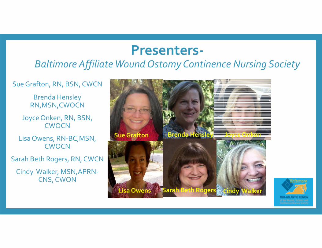
Presenters‐Baltimore Affiliate Wound Ostomy Continence Nursing Society
Sue Grafton, RN, BSN, CWCN
Brenda Hensley RN,MSN,CWOCN
Joyce Onken, RN, BSN, CWOCN
Lisa Owens, RN‐BC,MSN, CWOCN
Sarah Beth Rogers, RN, CWCN
Cindy Walker, MSN,APRN‐CNS, CWON
• Group picture …
Sue Grafton Brenda Hensley Joyce Onken
Lisa Owens Sarah Beth Rogers Cindy Walker
Objectives Webinar Series 1‐Assessment1. Recognize principles of healthy skin care management
2. Identify 4 or more interventions which reduce the risk of pressure injury based on evidence based skin risk assessments
3. Discuss 4 or more components of a comprehensive skin/wound assessment.
4. Differentiate 3 or more interventions and associated wound characteristics that support wound healing.
5. Distinguish 3 or more characteristics of various wound etiologies including moisture associated skin injury, pressure injury, and venous, arterial, and neuropathic ulcers
Objectives Webinar Series 2‐Management
6. Support wound dressing /treatment selections based on wound product categories associated with 3 or more patient centered assessment findings.
7. Appreciate principles of safe negative pressure wound therapy
8. Choose appropriate support surface application based on 2 or more unique patient centered needs
9. Identify community resources applicable to the chronic wound care management across care settings.
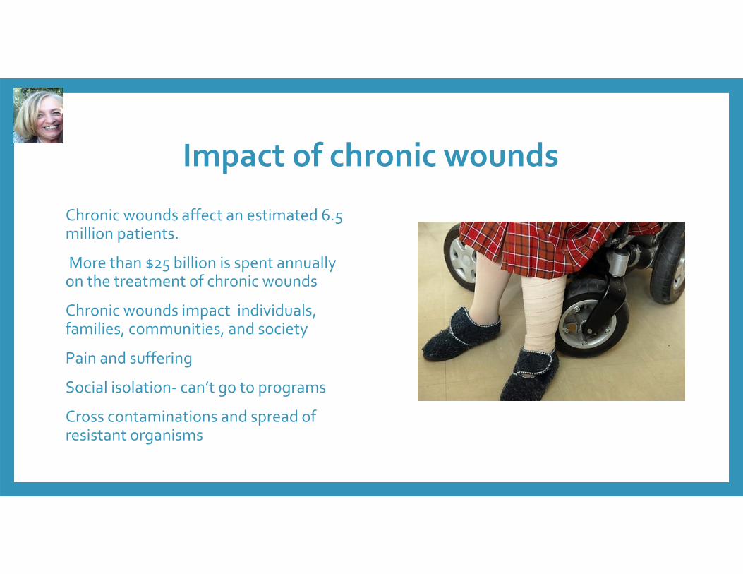
Impact of chronic wounds
Chronic wounds affect an estimated 6.5 million patients.
More than $25 billion is spent annually on the treatment of chronic wounds
Chronic wounds impact individuals, families, communities, and society
Pain and suffering
Social isolation‐ can’t go to programs
Cross contaminations and spread of resistant organisms
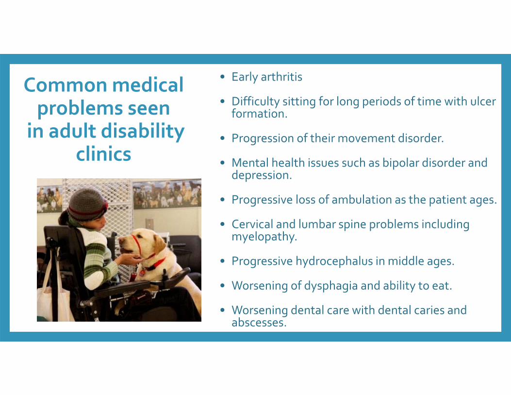
Common medical problems seen
in adult disability clinics
• Early arthritis
• Difficulty sitting for long periods of time with ulcer formation.
• Progression of their movement disorder.
• Mental health issues such as bipolar disorder and depression.
• Progressive loss of ambulation as the patient ages.
• Cervical and lumbar spine problems including myelopathy.
• Progressive hydrocephalus in middle ages.
• Worsening of dysphagia and ability to eat.
• Worsening dental care with dental caries and abscesses.
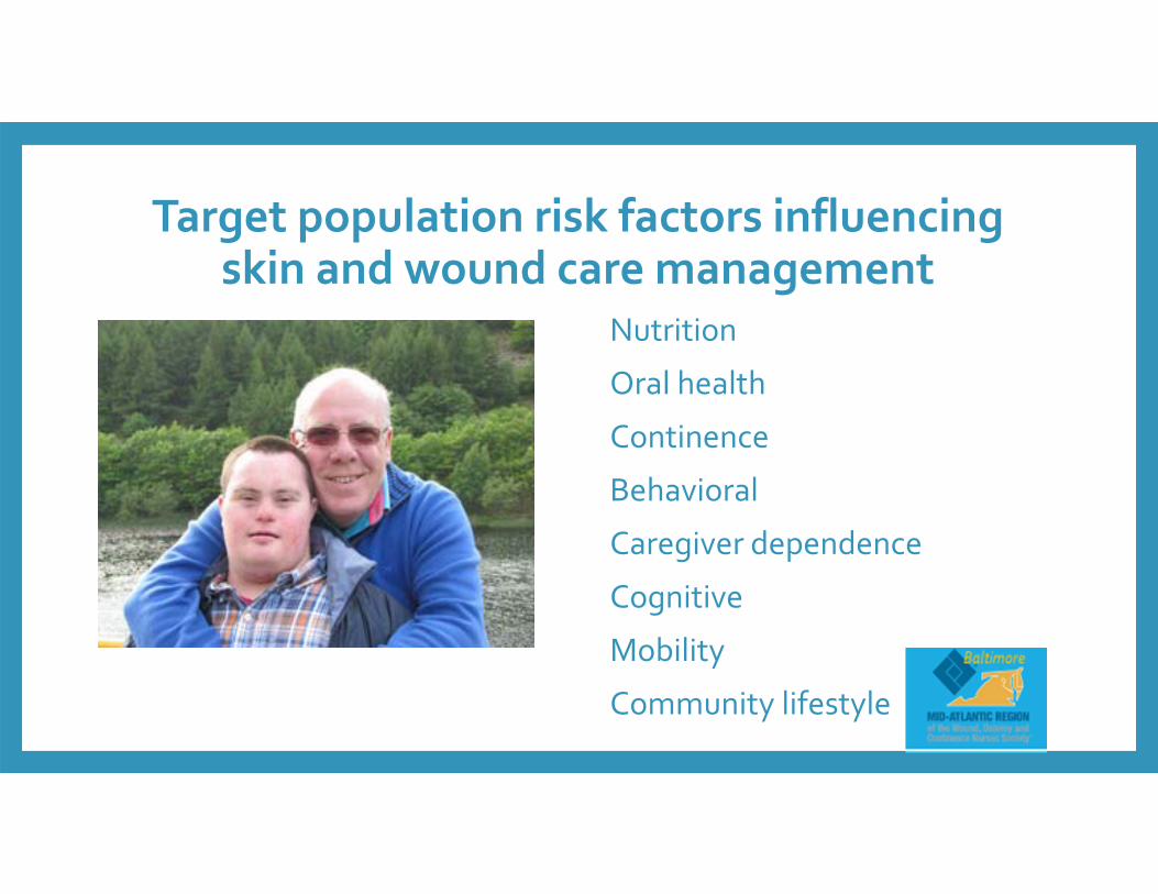
Target population risk factors influencing skin and wound care management
Nutrition
Oral health
Continence
Behavioral
Caregiver dependence
Cognitive
Mobility
Community lifestyle

The Skin
• Weighs 8 lbs/covers 20 sq ft
• Protects body from environment as first line of defense
• Largest organ in our body
• Receives 1/3 of our blood flow
• pH (5.5)
June 28, 2016 9
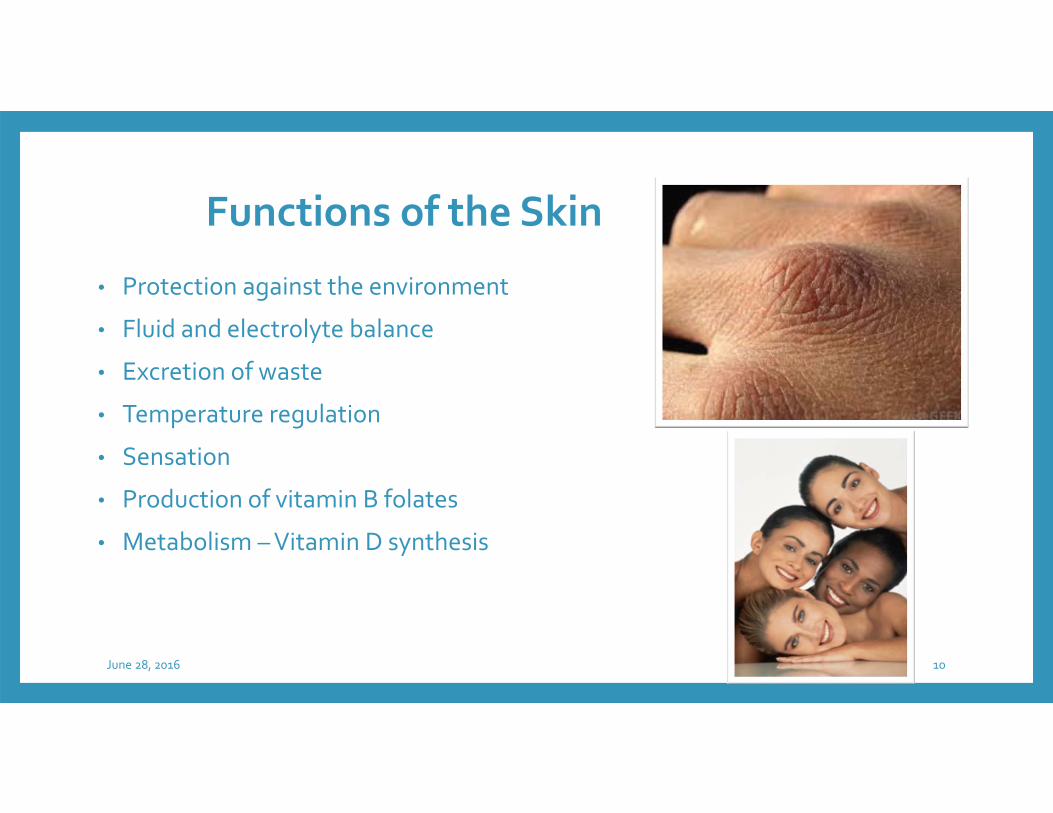
Functions of the Skin
• Protection against the environment
• Fluid and electrolyte balance
• Excretion of waste
• Temperature regulation
• Sensation
• Production of vitamin B folates
• Metabolism –Vitamin D synthesis
June 28, 2016 10
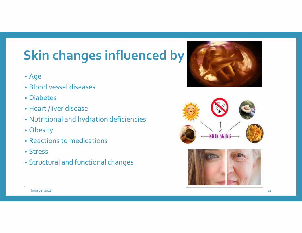
Skin changes influenced by• Age• Blood vessel diseases• Diabetes• Heart /liver disease• Nutritional and hydration deficiencies• Obesity• Reactions to medications• Stress• Structural and functional changes
•
June 28, 2016 11
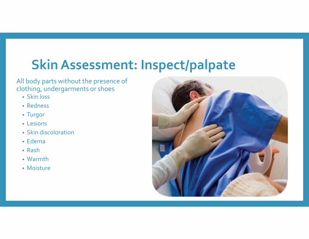
Skin Assessment: Inspect/palpateAll body parts without the presence of clothing, undergarments or shoes
• Skin loss• Redness• Turgor• Lesions• Skin discoloration• Edema • Rash • Warmth• Moisture
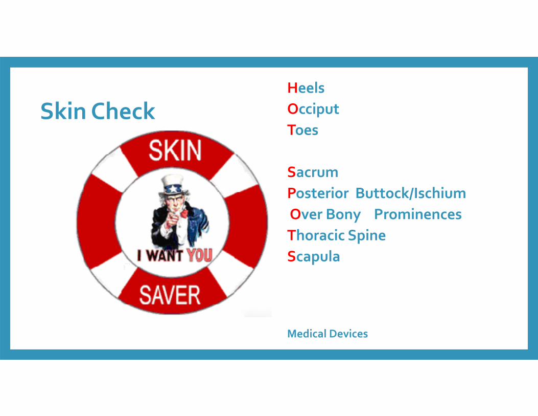
Skin CheckHeels Occiput Toes
SacrumPosterior Buttock/IschiumOver Bony ProminencesThoracic SpineScapula
Medical Devices
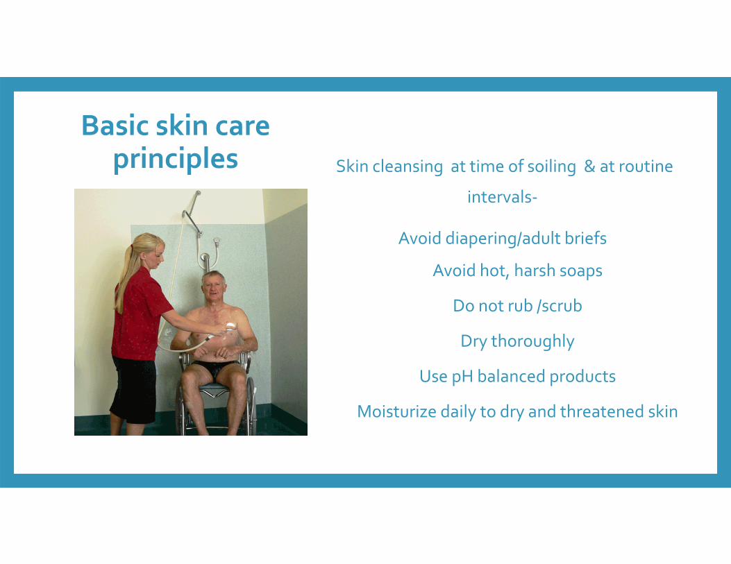
Basic skin care principles Skin cleansing at time of soiling & at routine
intervals‐
Avoid diapering/adult briefs
Avoid hot, harsh soaps
Do not rub /scrub
Dry thoroughly
Use pH balanced products
Moisturize daily to dry and threatened skin
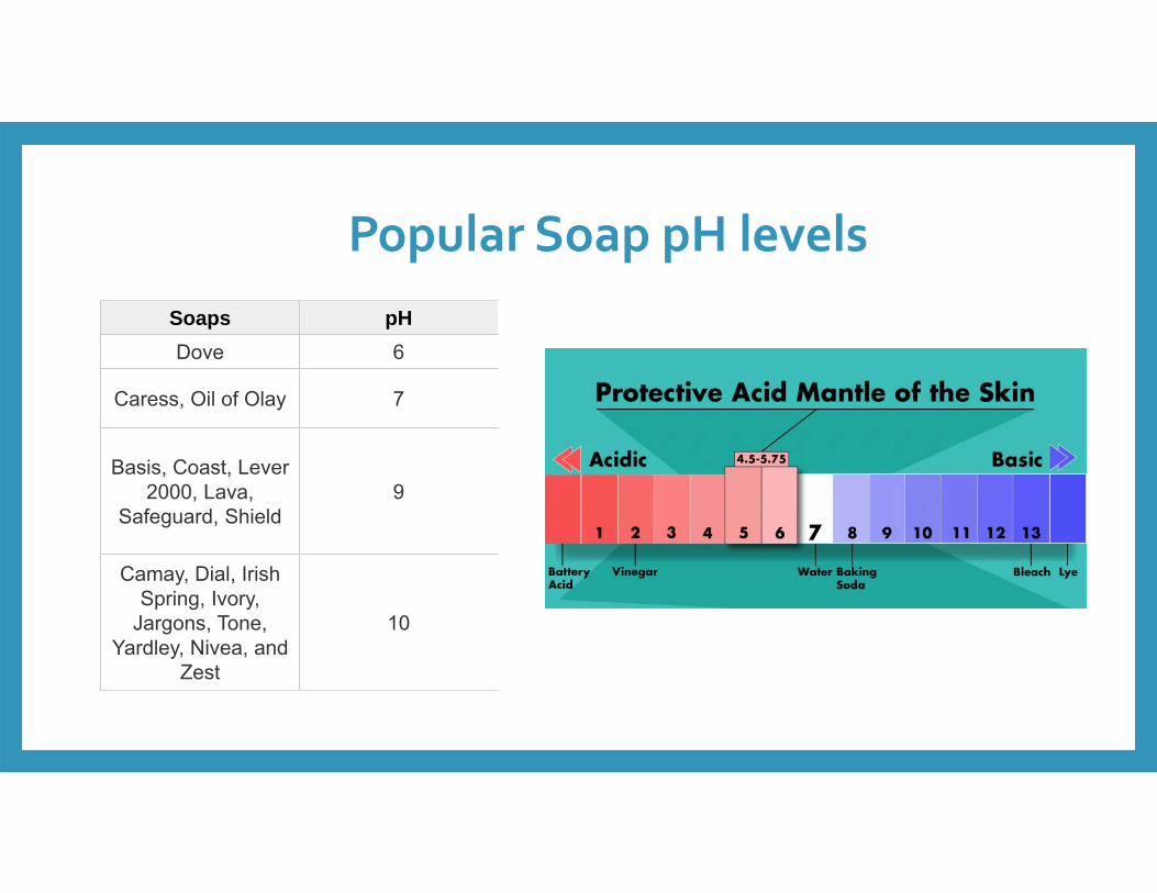
Popular Soap pH levelsSoaps pHDove 6
Caress, Oil of Olay 7
Basis, Coast, Lever 2000, Lava,
Safeguard, Shield9
Camay, Dial, Irish Spring, Ivory,
Jargons, Tone, Yardley, Nivea, and
Zest
10
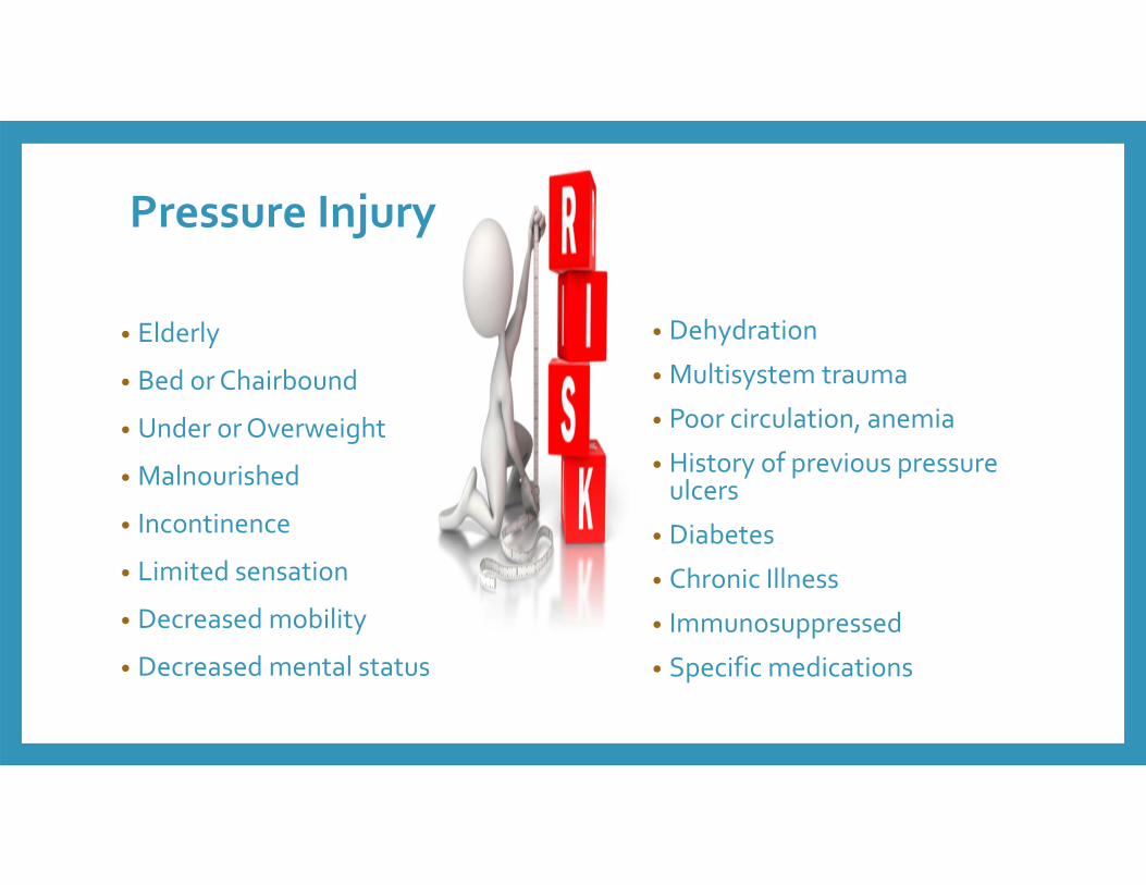
• Elderly
• Bed or Chairbound
• Under or Overweight
• Malnourished
• Incontinence
• Limited sensation
• Decreased mobility
• Decreased mental status
• Dehydration
• Multisystem trauma
• Poor circulation, anemia
• History of previous pressure ulcers
• Diabetes
• Chronic Illness
• Immunosuppressed
• Specific medications
Pressure Injury
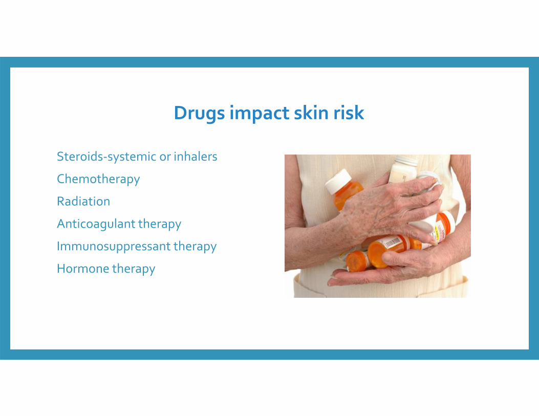
Drugs impact skin risk
Steroids‐systemic or inhalers
Chemotherapy
Radiation
Anticoagulant therapy
Immunosuppressant therapy
Hormone therapy
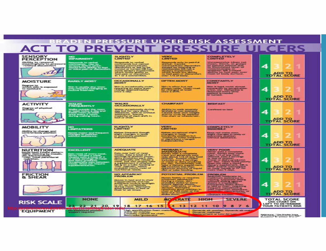
https://jessbrantnerwvudietetics.wordpress.com/tag/braden‐scale/
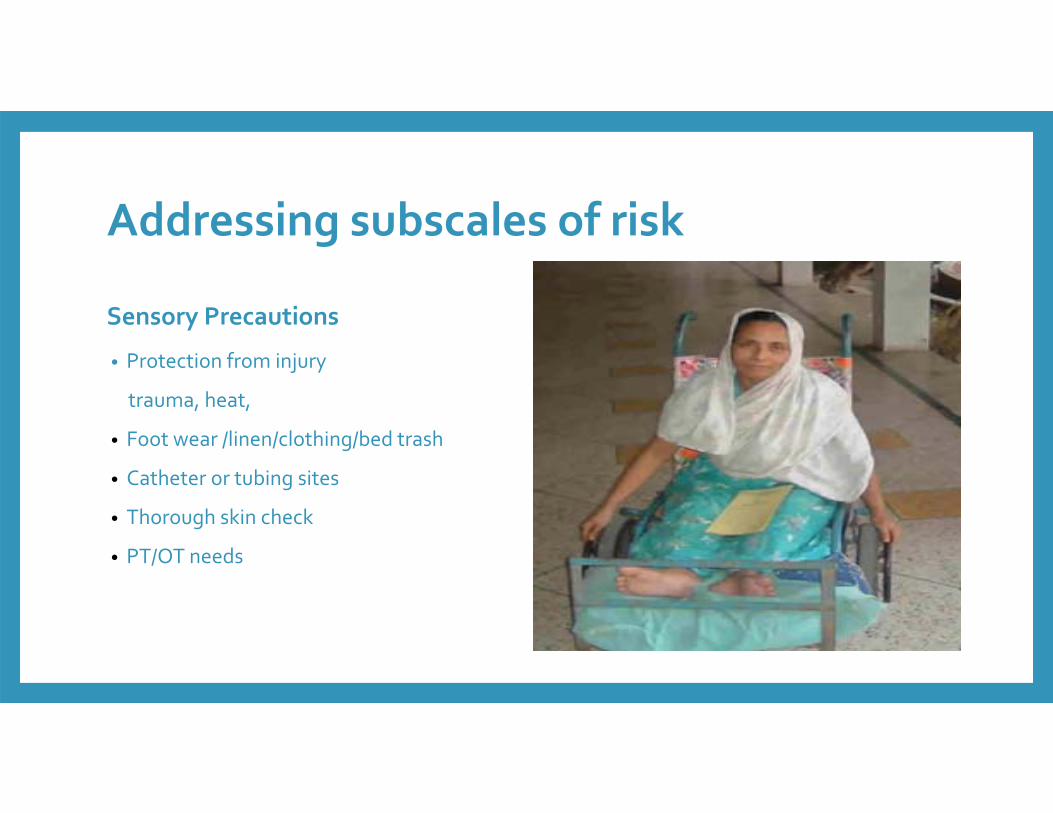
Addressing subscales of risk
Sensory Precautions
• Protection from injury
trauma, heat,
• Foot wear /linen/clothing/bed trash
• Catheter or tubing sites
• Thorough skin check
• PT/OT needs
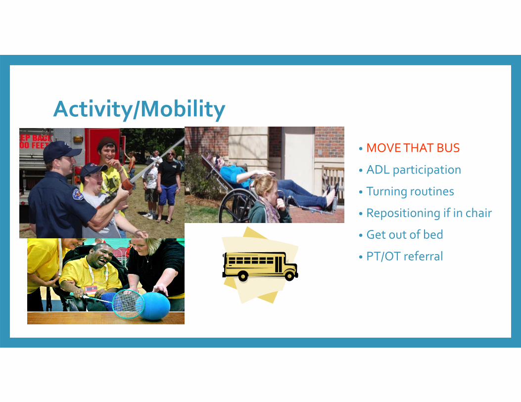
Activity/Mobility
• MOVE THAT BUS
• ADL participation
• Turning routines
• Repositioning if in chair
• Get out of bed
• PT/OT referral
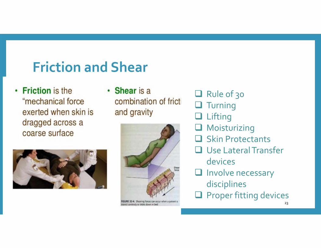
Friction and Shear
6/28/2016 23
Rule of 30 Turning Lifting Moisturizing Skin Protectants Use Lateral Transfer
devices Involve necessary
disciplines Proper fitting devices
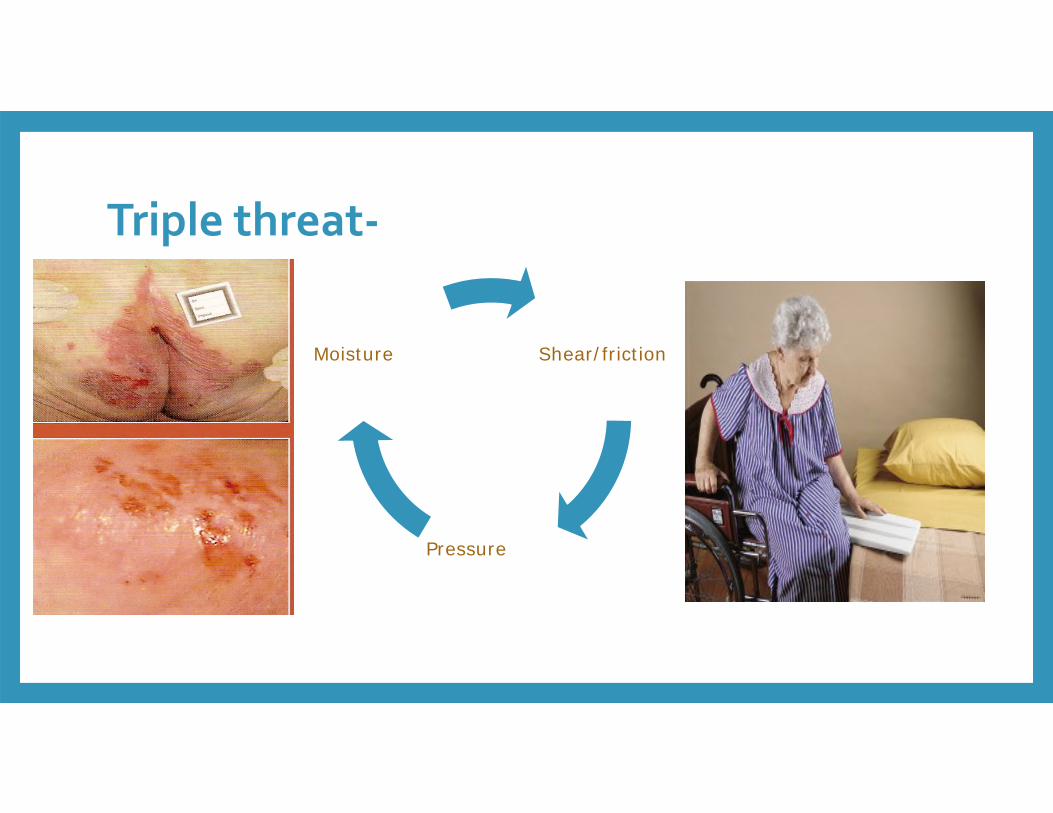
Triple threat‐
Shear/friction
Pressure
Moisture
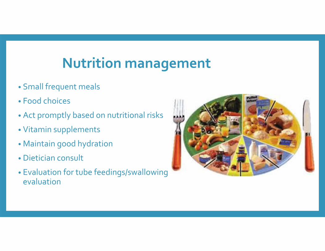
Nutrition management• Small frequent meals
• Food choices
• Act promptly based on nutritional risks
• Vitamin supplements
• Maintain good hydration
• Dietician consult
• Evaluation for tube feedings/swallowing evaluation

Basic prevention principles
Avoid massage over bony prominences
Encourage maximum mobility
Position changes
Float heels
Protect bony prominences
Lift don’t drag
Reposition in bed and chair
Skin care bundle
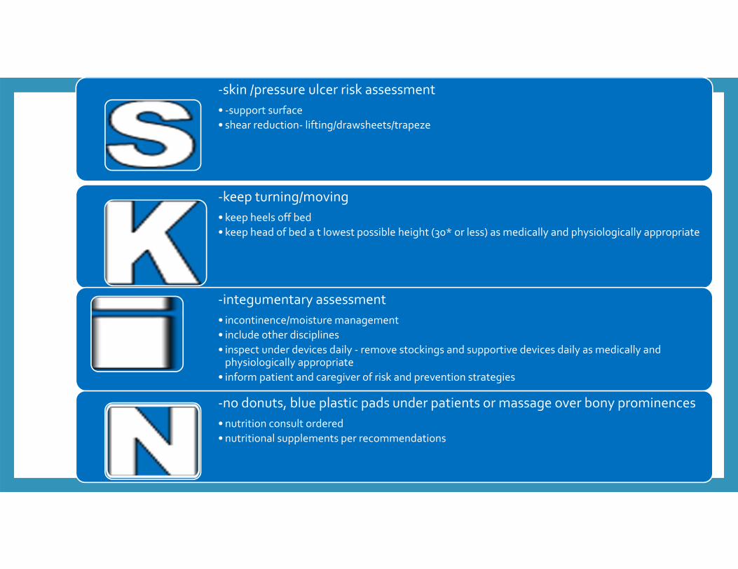
‐skin /pressure ulcer risk assessment• ‐support surface• shear reduction‐ lifting/drawsheets/trapeze
‐keep turning/moving• keep heels off bed• keep head of bed a t lowest possible height (30* or less) as medically and physiologically appropriate
‐integumentary assessment• incontinence/moisture management• include other disciplines • inspect under devices daily ‐ remove stockings and supportive devices daily as medically and physiologically appropriate
• inform patient and caregiver of risk and prevention strategies
‐no donuts, blue plastic pads under patients or massage over bony prominences• nutrition consult ordered• nutritional supplements per recommendations
Pressure Injury
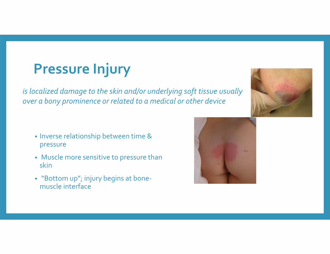
• Inverse relationship between time & pressure
• Muscle more sensitive to pressure than skin
• “Bottom up”; injury begins at bone‐muscle interface
is localized damage to the skin and/or underlying soft tissue usually over a bony prominence or related to a medical or other device
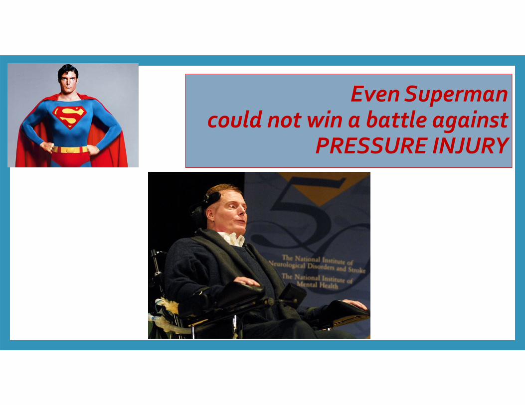
Even Superman could not win a battle against
PRESSURE INJURY
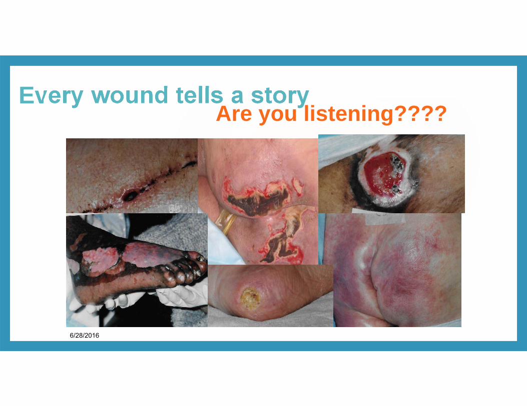
Every wound tells a story
6/28/2016
Are you listening????
Baltimore
QUESTIONS???Recognize principles of healthy skin care management
Identify 4 or more interventions which reduce the risk of pressure injury based on evidence based skin risk assessments
Discuss 4 or more components of a comprehensive skin/wound assessment.
Sarah Beth Rogers, RN, CWCN
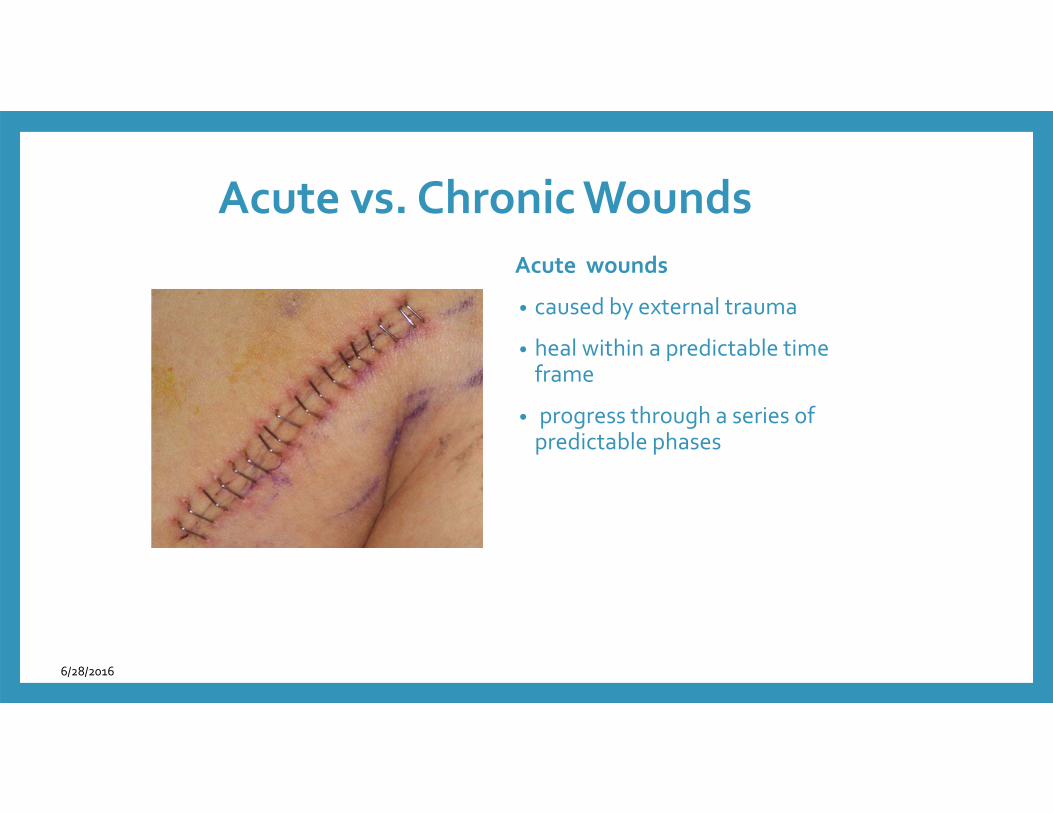
Acute vs. Chronic WoundsAcute wounds
• caused by external trauma
• heal within a predictable time frame
• progress through a series of predictable phases
6/28/2016
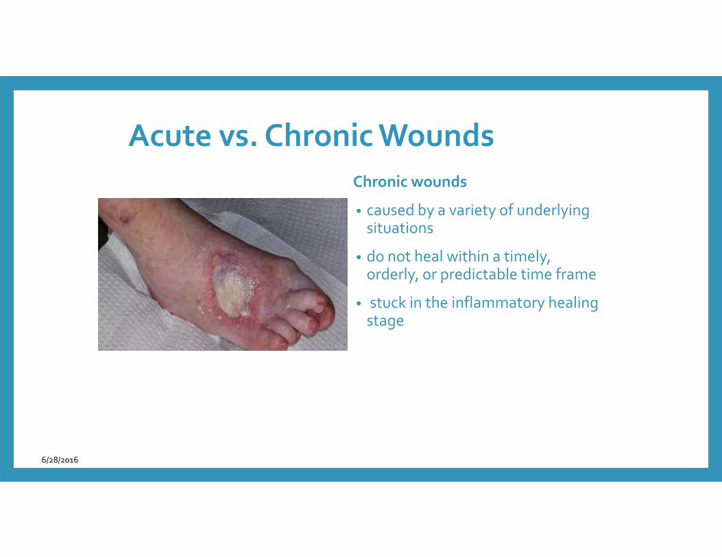
Acute vs. Chronic WoundsChronic wounds
• caused by a variety of underlying situations
• do not heal within a timely, orderly, or predictable time frame
• stuck in the inflammatory healing stage
6/28/2016
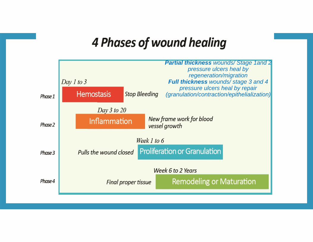
Partial thickness wounds/ Stage 1and 2 pressure ulcers heal by regeneration/migration
Full thickness wounds/ stage 3 and 4 pressure ulcers heal by repair
(granulation/contraction/epithelialization)
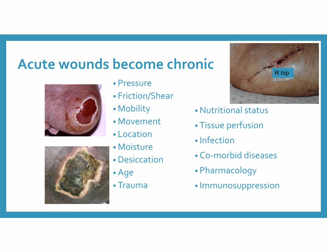
Acute wounds become chronic• Pressure• Friction/Shear•Mobility•Movement• Location •Moisture•Desiccation• Age• Trauma
R hip
• Nutritional status
• Tissue perfusion
• Infection
• Co‐morbid diseases
• Pharmacology
• Immunosuppression
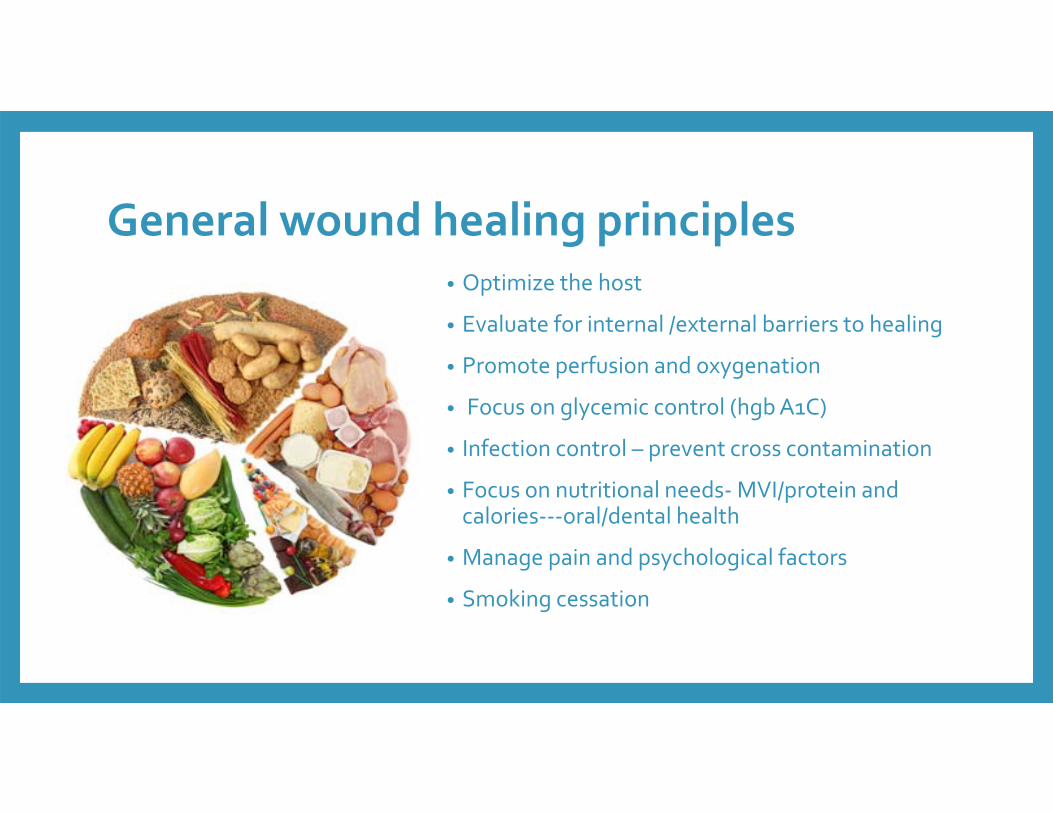
General wound healing principles• Optimize the host
• Evaluate for internal /external barriers to healing
• Promote perfusion and oxygenation
• Focus on glycemic control (hgbA1C)
• Infection control – prevent cross contamination
• Focus on nutritional needs‐MVI/protein and calories‐‐‐oral/dental health
• Manage pain and psychological factors
• Smoking cessation
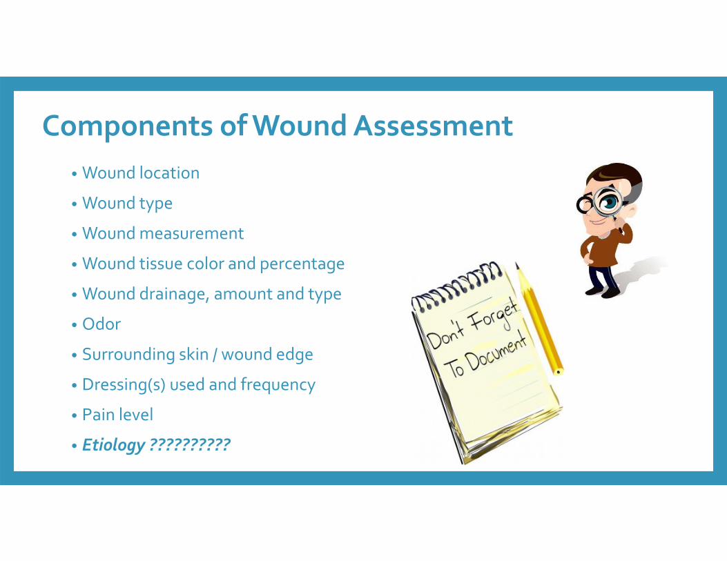
Components of Wound Assessment• Wound location
• Wound type
• Wound measurement
• Wound tissue color and percentage
• Wound drainage, amount and type
• Odor
• Surrounding skin / wound edge
• Dressing(s) used and frequency
• Pain level
• Etiology ??????????
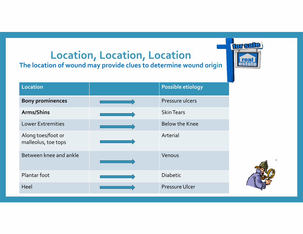
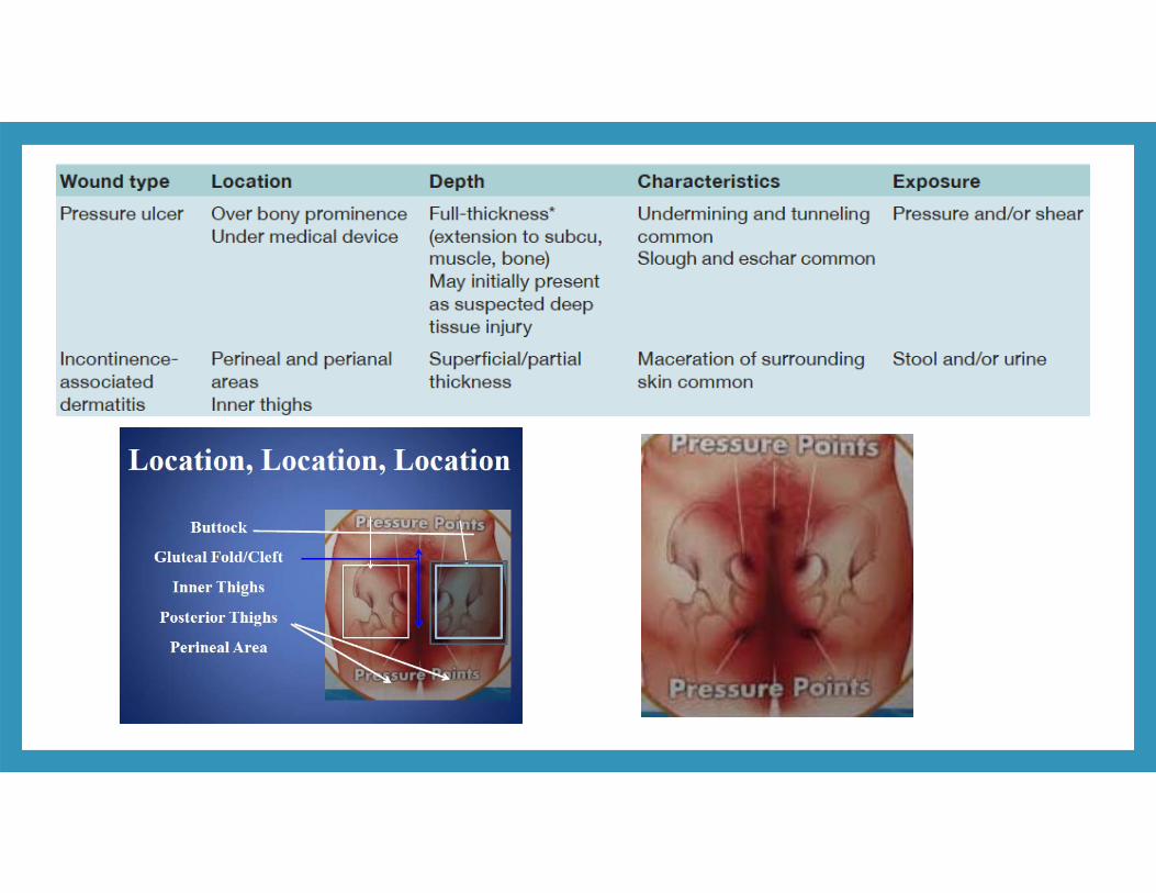
Location, Location, Location The location of wound may provide clues to determine wound origin
Location Possible etiology
Bony prominences Pressure ulcers
Arms/Shins Skin Tears
Lower Extremities Below the Knee
Along toes/foot or malleolus, toe tops
Arterial
Between knee and ankle Venous
Plantar foot Diabetic
Heel Pressure Ulcer
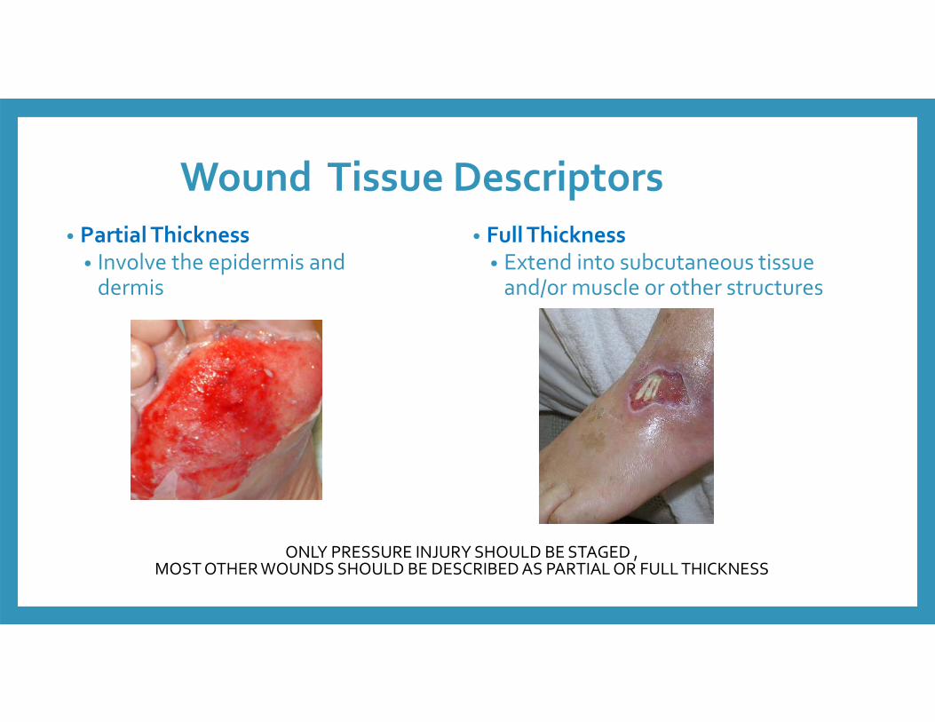
• Partial Thickness• Involve the epidermis and dermis
• Full Thickness• Extend into subcutaneous tissue and/or muscle or other structures
Wound Tissue Descriptors
ONLY PRESSURE INJURY SHOULD BE STAGED , MOST OTHER WOUNDS SHOULD BE DESCRIBED AS PARTIAL OR FULL THICKNESS
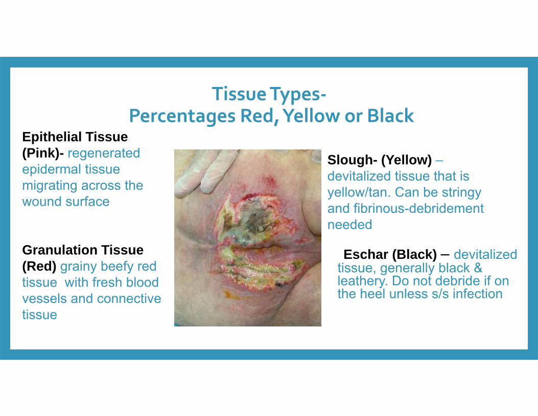
Tissue Types‐Percentages Red, Yellow or Black
Eschar (Black) – devitalized tissue, generally black & leathery. Do not debride if on the heel unless s/s infection
Epithelial Tissue (Pink)- regenerated epidermal tissue migrating across the wound surface
Granulation Tissue (Red) grainy beefy red tissue with fresh blood vessels and connective tissue
Slough- (Yellow) –devitalized tissue that is yellow/tan. Can be stringy and fibrinous-debridement needed
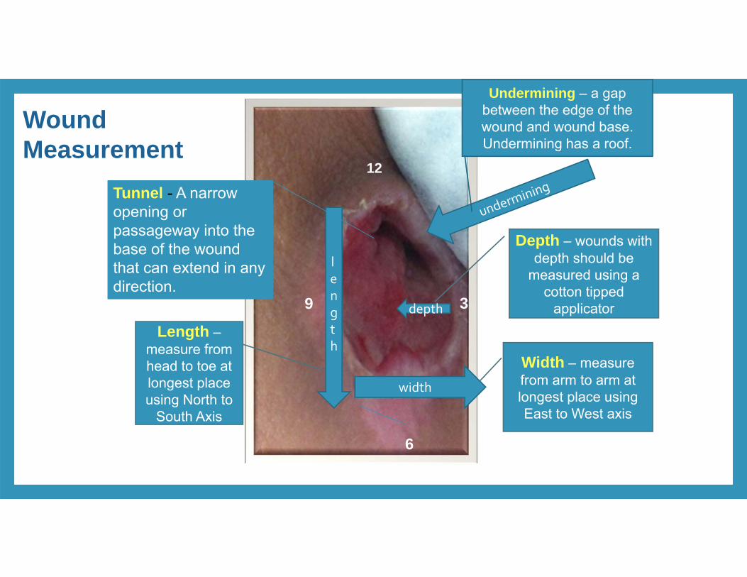
width
length
depth
Length –measure from head to toe at longest place using North to
South Axis
Width – measure from arm to arm at longest place using East to West axis
Depth – wounds with depth should be
measured using a cotton tipped
applicator
Undermining – a gap between the edge of the wound and wound base. Undermining has a roof.
Tunnel - A narrow opening or passageway into the base of the wound that can extend in any direction.
12
6
9 3
Wound Measurement
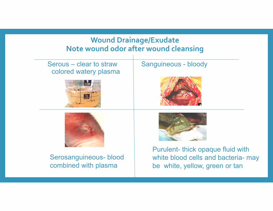
Wound Drainage/ExudateNote wound odor after wound cleansing
Serous – clear to straw colored watery plasma
Sanguineous - bloody
Serosanguineous- blood combined with plasma
Purulent- thick opaque fluid with white blood cells and bacteria- may be white, yellow, green or tan
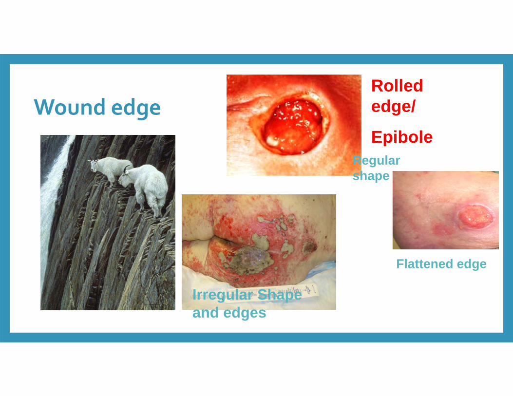
Wound edge
Flattened edge
Irregular shape
Rolled edge/
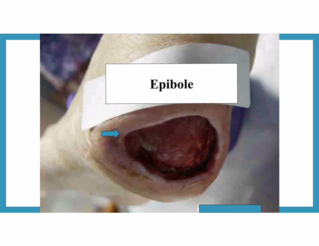
EpiboleRegular shape
Irregular Shape and edges
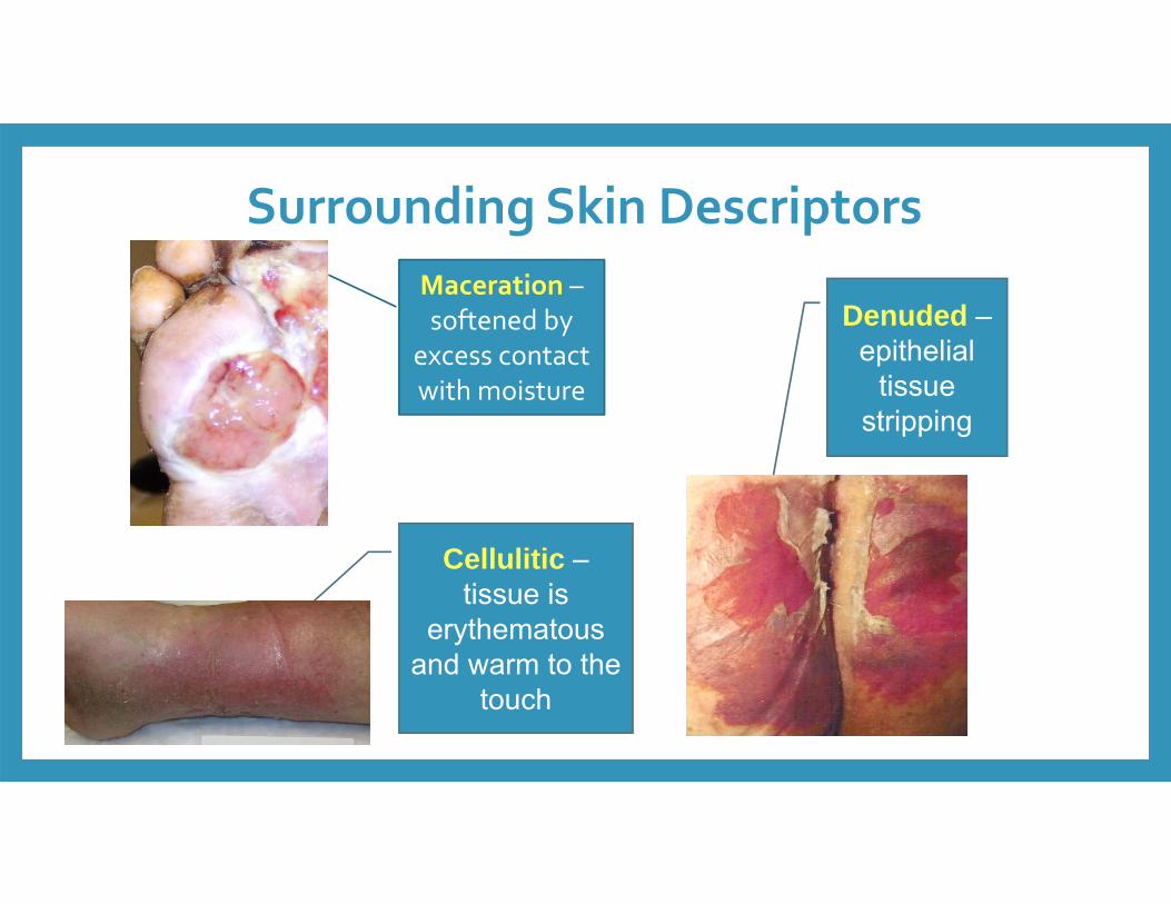
Surrounding Skin DescriptorsMaceration –softened by
excess contact with moisture
Cellulitic –tissue is
erythematous and warm to the
touch
Denuded –epithelial
tissue stripping
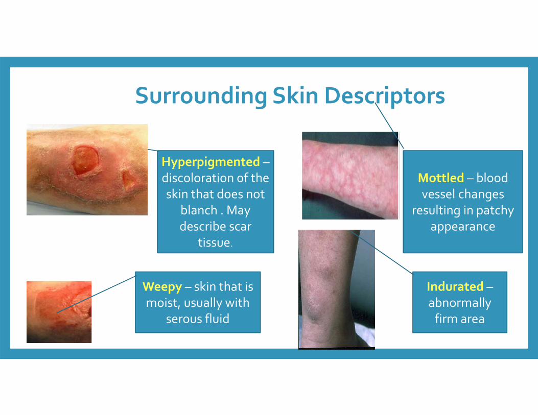
Surrounding Skin Descriptors
Hyperpigmented –discoloration of the skin that does not
blanch . May describe scar
tissue.
Weepy – skin that is moist, usually with
serous fluid
Indurated –abnormally firm area
Mottled – blood vessel changes
resulting in patchy appearance
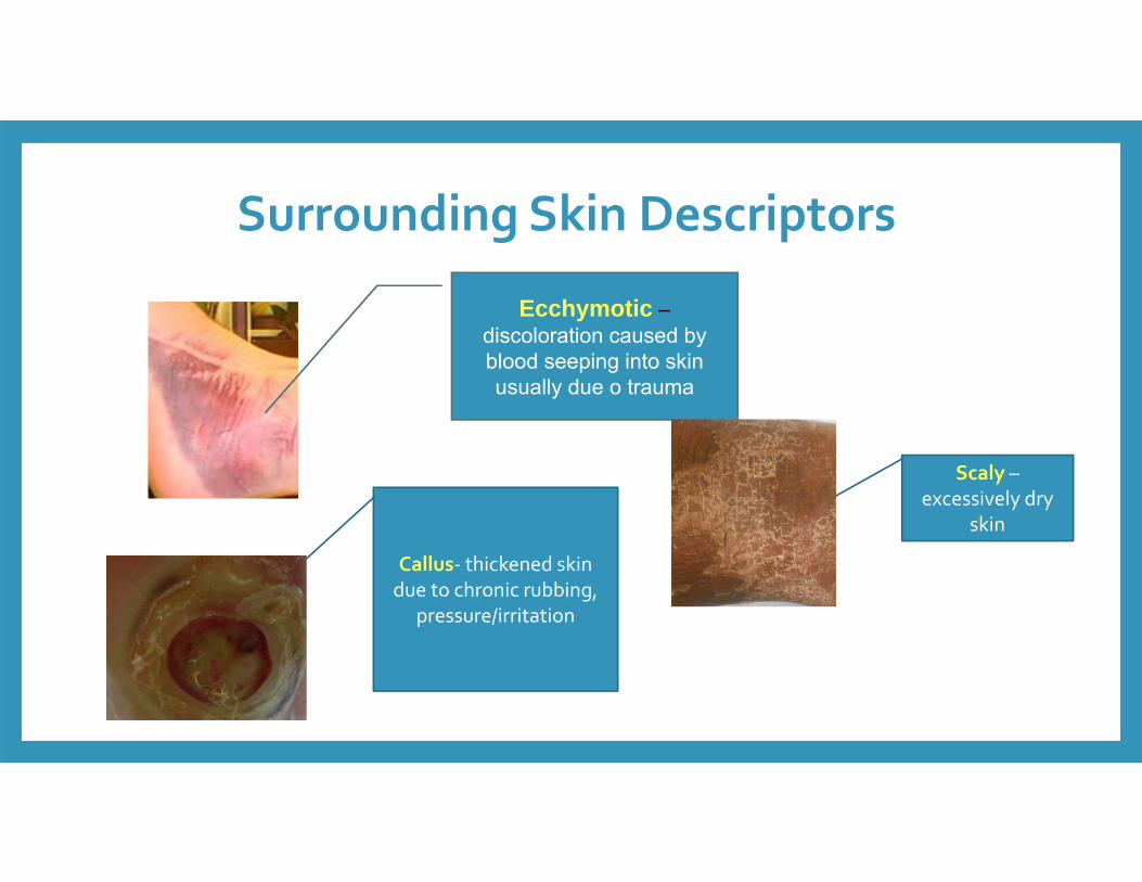
Surrounding Skin Descriptors
Ecchymotic –discoloration caused by blood seeping into skin usually due o trauma
Callus‐ thickened skin due to chronic rubbing,
pressure/irritation
Scaly –excessively dry
skin
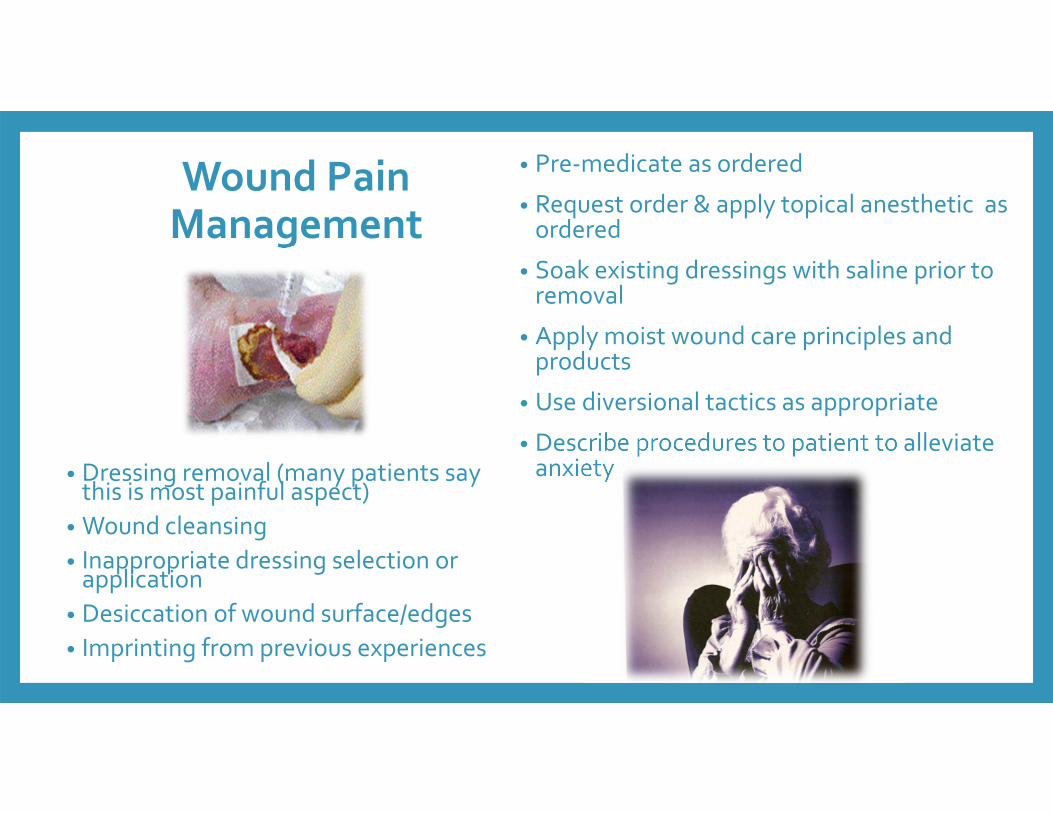
• Dressing removal (many patients say this is most painful aspect)
• Wound cleansing • Inappropriate dressing selection or application
• Desiccation of wound surface/edges• Imprinting from previous experiences
• Pre‐medicate as ordered
• Request order & apply topical anesthetic as ordered
• Soak existing dressings with saline prior to removal
• Apply moist wound care principles and products
• Use diversional tactics as appropriate
• Describe procedures to patient to alleviate anxiety
Wound Pain Management
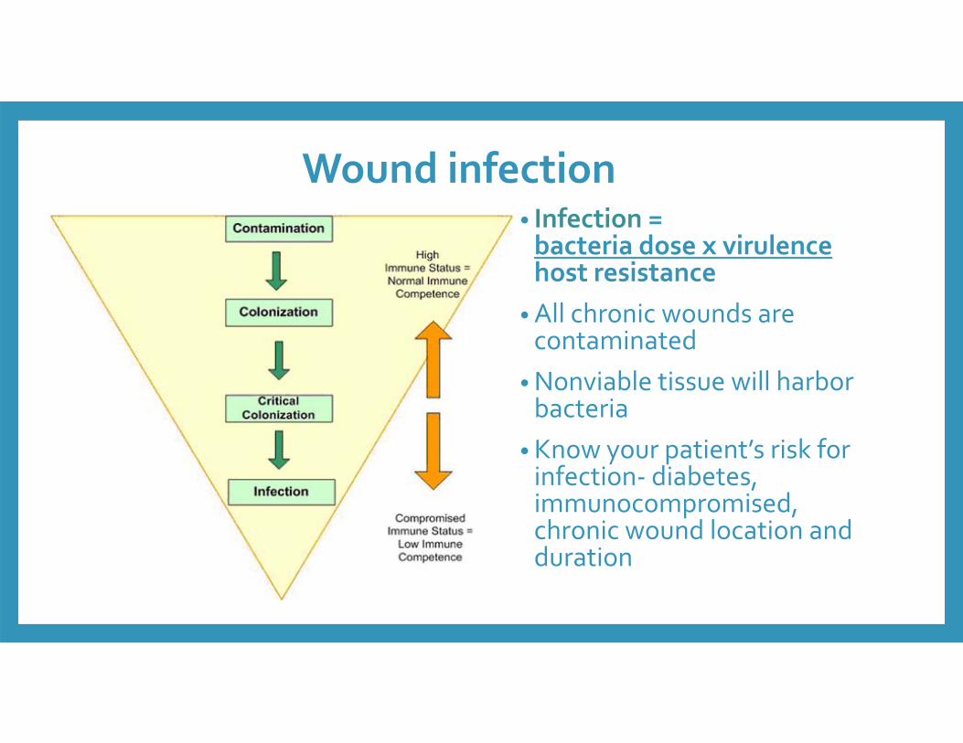
Wound infection• Infection =bacteria dose x virulencehost resistance
• All chronic wounds are contaminated
•Nonviable tissue will harbor bacteria
• Know your patient’s risk for infection‐ diabetes, immunocompromised, chronic wound location and duration
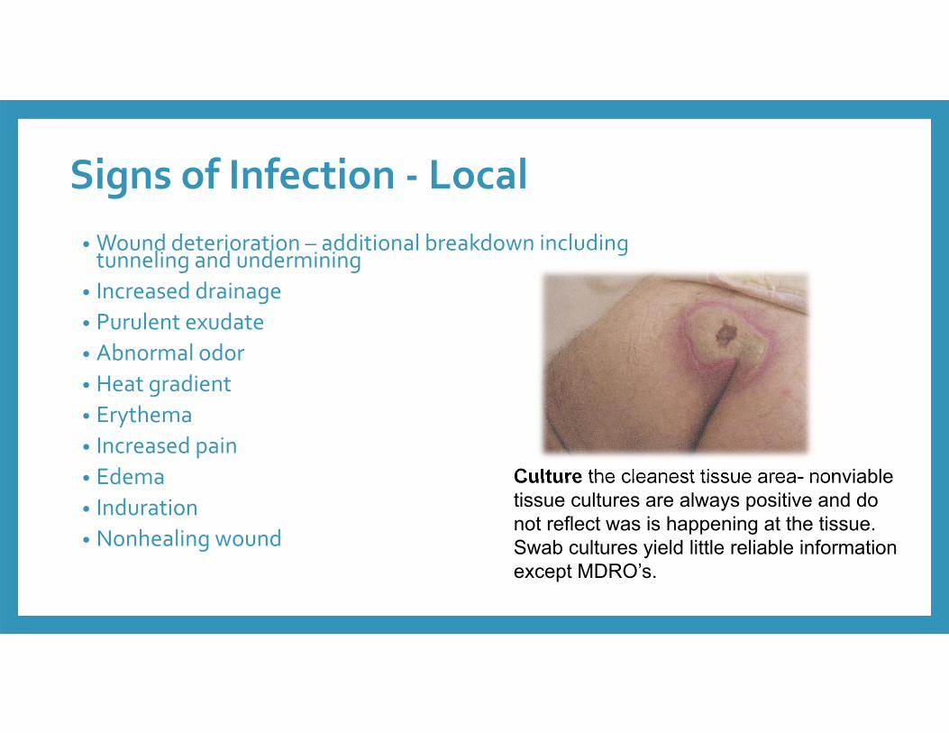
Signs of Infection ‐ Local• Wound deterioration – additional breakdown including tunneling and undermining
• Increased drainage • Purulent exudate• Abnormal odor• Heat gradient• Erythema• Increased pain• Edema• Induration• Nonhealing wound
Culture the cleanest tissue area- nonviable tissue cultures are always positive and do not reflect was is happening at the tissue.Swab cultures yield little reliable information except MDRO’s.
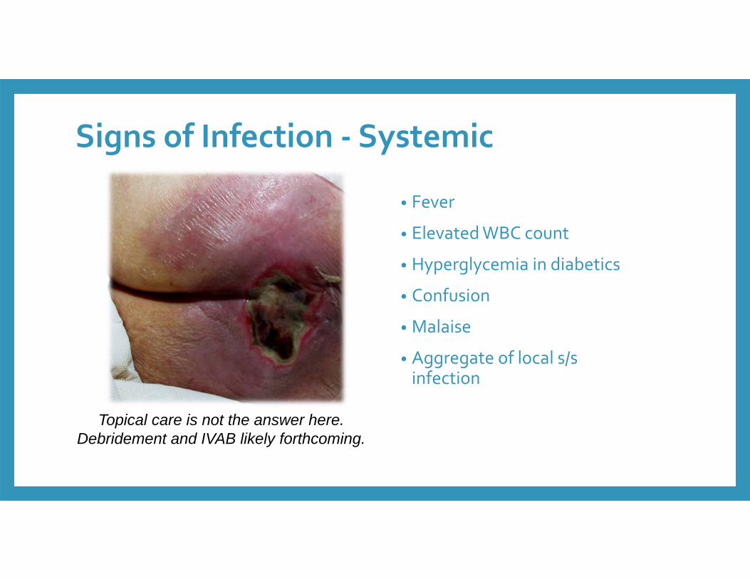
Signs of Infection ‐ Systemic
• Fever
• Elevated WBC count
• Hyperglycemia in diabetics
• Confusion
• Malaise
• Aggregate of local s/s infection
Topical care is not the answer here. Debridement and IVAB likely forthcoming.
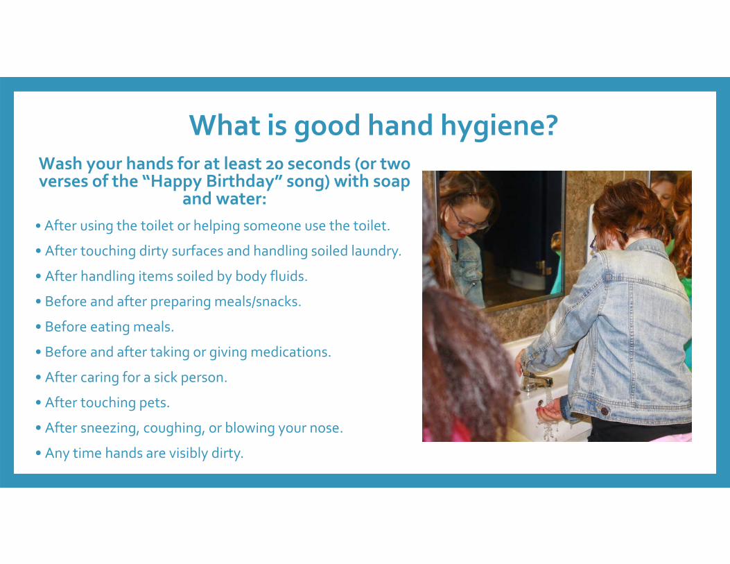
What is good hand hygiene? Wash your hands for at least 20 seconds (or two verses of the “Happy Birthday” song) with soap
and water: • After using the toilet or helping someone use the toilet.
• After touching dirty surfaces and handling soiled laundry.
• After handling items soiled by body fluids.
• Before and after preparing meals/snacks.
• Before eating meals.
• Before and after taking or giving medications.
• After caring for a sick person.
• After touching pets.
• After sneezing, coughing, or blowing your nose.
• Any time hands are visibly dirty.
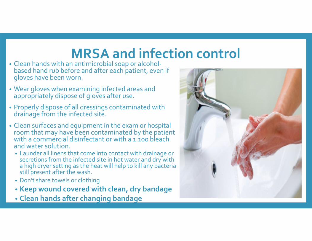
MRSA and infection control• Clean hands with an antimicrobial soap or alcohol‐based hand rub before and after each patient, even if gloves have been worn.
• Wear gloves when examining infected areas and appropriately dispose of gloves after use.
• Properly dispose of all dressings contaminated with drainage from the infected site.
• Clean surfaces and equipment in the exam or hospital room that may have been contaminated by the patient with a commercial disinfectant or with a 1:100 bleach and water solution. • Launder all linens that come into contact with drainage or secretions from the infected site in hot water and dry with a high dryer setting as the heat will help to kill any bacteria still present after the wash.
• Don’t share towels or clothing• Keep wound covered with clean, dry bandage • Clean hands after changing bandage
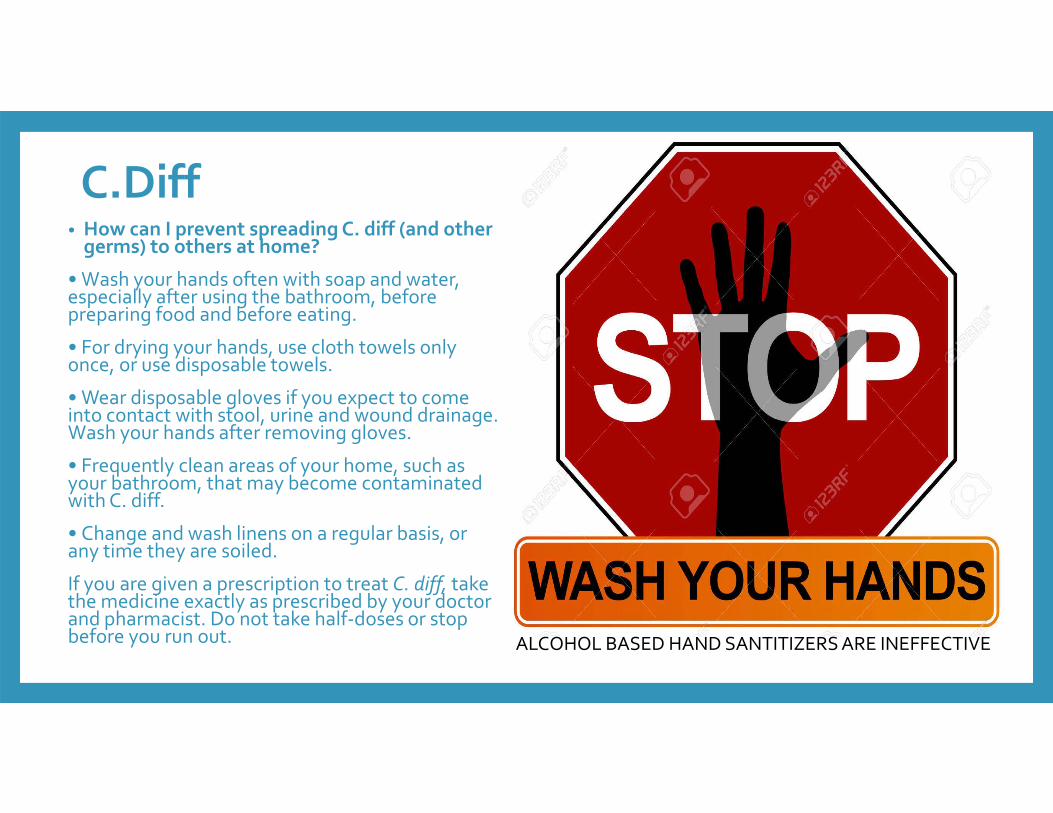
C.Diff• How can I prevent spreading C. diff (and other germs) to others at home?
• Wash your hands often with soap and water, especially after using the bathroom, before preparing food and before eating.
• For drying your hands, use cloth towels only once, or use disposable towels.
• Wear disposable gloves if you expect to come into contact with stool, urine and wound drainage. Wash your hands after removing gloves.
• Frequently clean areas of your home, such as your bathroom, that may become contaminated with C. diff.
• Change and wash linens on a regular basis, or any time they are soiled.
If you are given a prescription to treat C. diff, take the medicine exactly as prescribed by your doctor and pharmacist. Do not take half‐doses or stop before you run out. ALCOHOL BASED HAND SANTITIZERS ARE INEFFECTIVE
Baltimore
QUESTIONS???Discuss 4 or more components of a comprehensive skin/wound
assessment.Differentiate 3 or more interventions and associated wound characteristics
that support wound healing..
Brenda Hensley RN,MSN,CWOCN
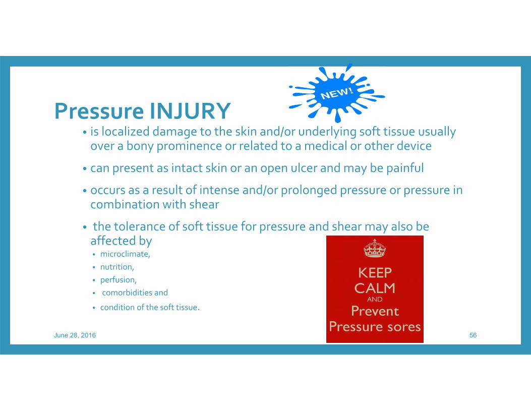
Pressure INJURY• is localized damage to the skin and/or underlying soft tissue usually over a bony prominence or related to a medical or other device
• can present as intact skin or an open ulcer and may be painful
• occurs as a result of intense and/or prolonged pressure or pressure in combination with shear
• the tolerance of soft tissue for pressure and shear may also be affected by• microclimate, • nutrition, • perfusion,• comorbidities and
• condition of the soft tissue.
June 28, 2016 56
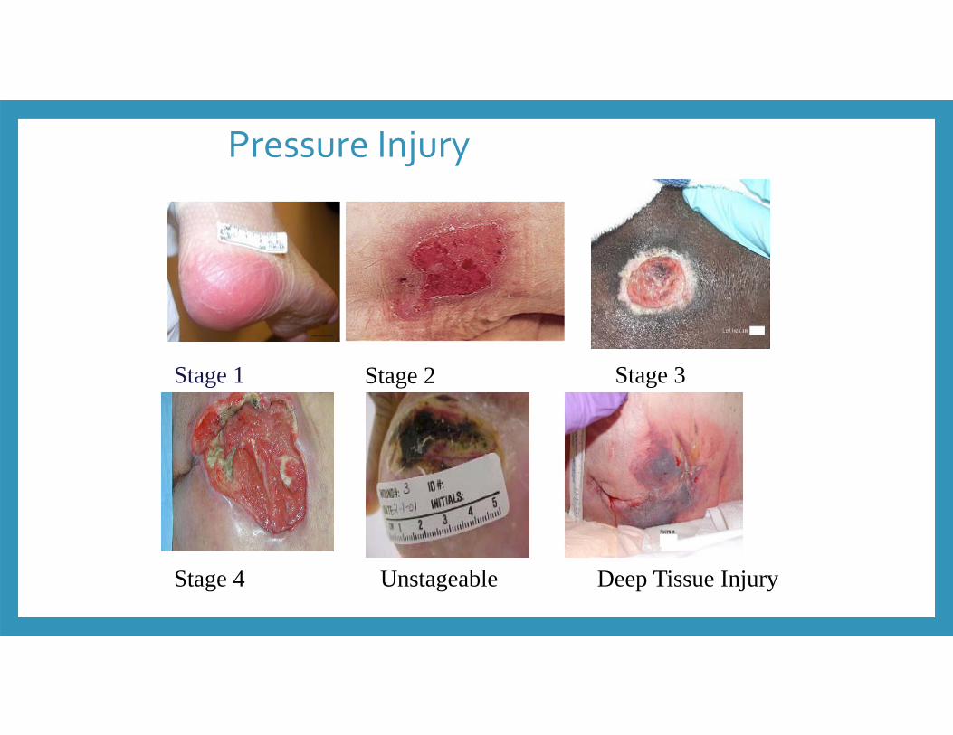
Stage 1 Stage 2 Stage 3
Stage 4 Unstageable Deep Tissue Injury
Pressure Injury
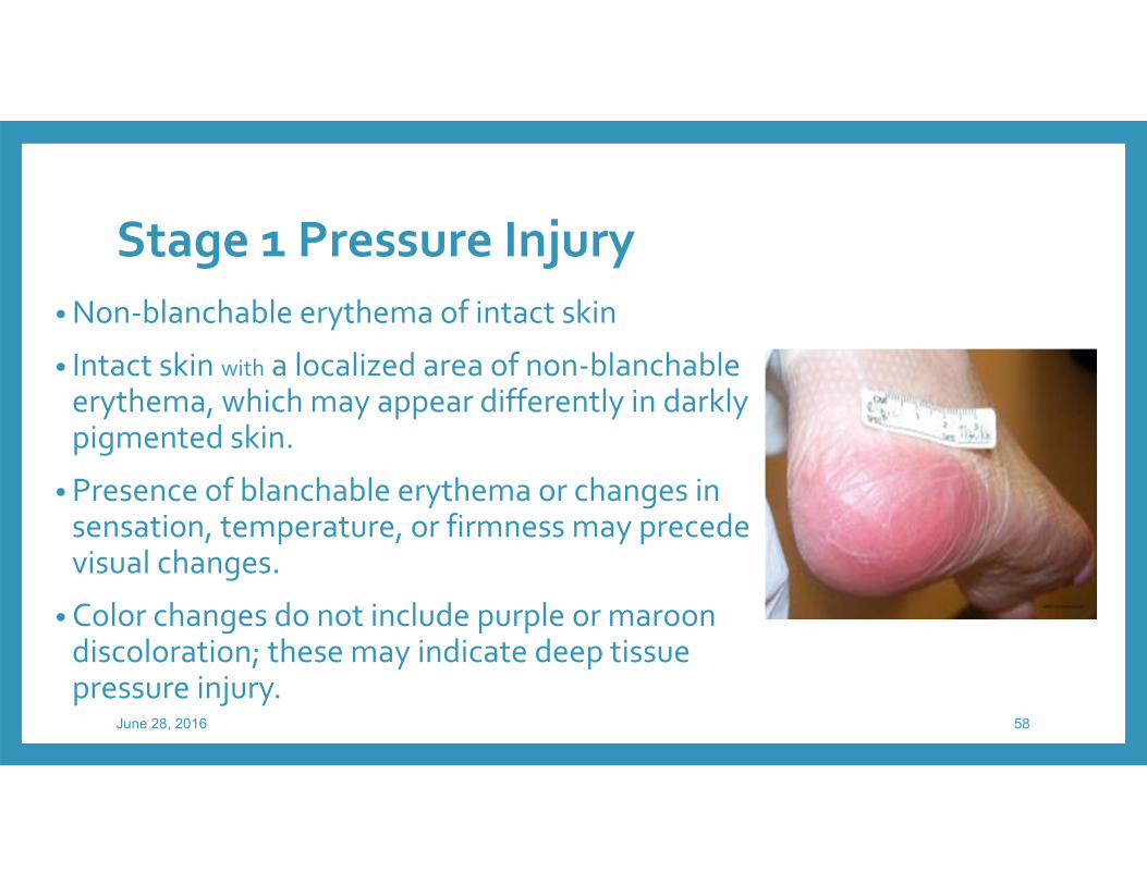
Stage 1 Pressure Injury• Non‐blanchable erythema of intact skin
• Intact skin with a localized area of non‐blanchableerythema, which may appear differently in darkly pigmented skin.
• Presence of blanchable erythema or changes in sensation, temperature, or firmness may precede visual changes.
• Color changes do not include purple or maroon discoloration; these may indicate deep tissue pressure injury.
June 28, 2016 58
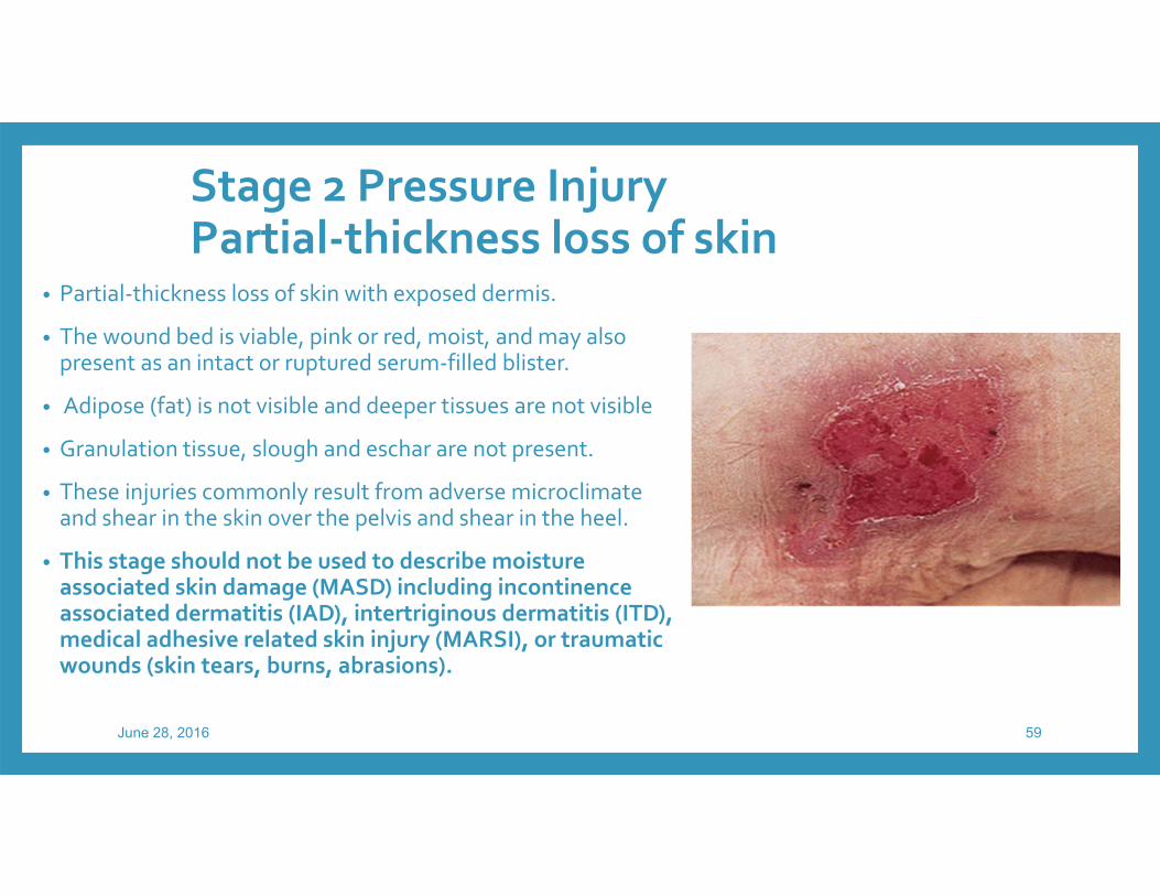
Stage 2 Pressure InjuryPartial‐thickness loss of skin
• Partial‐thickness loss of skin with exposed dermis.
• The wound bed is viable, pink or red, moist, and may also present as an intact or ruptured serum‐filled blister.
• Adipose (fat) is not visible and deeper tissues are not visible
• Granulation tissue, slough and eschar are not present.
• These injuries commonly result from adverse microclimate and shear in the skin over the pelvis and shear in the heel.
• This stage should not be used to describe moisture associated skin damage (MASD) including incontinence associated dermatitis (IAD), intertriginous dermatitis (ITD), medical adhesive related skin injury (MARSI), or traumatic wounds (skin tears, burns, abrasions).
June 28, 2016 59
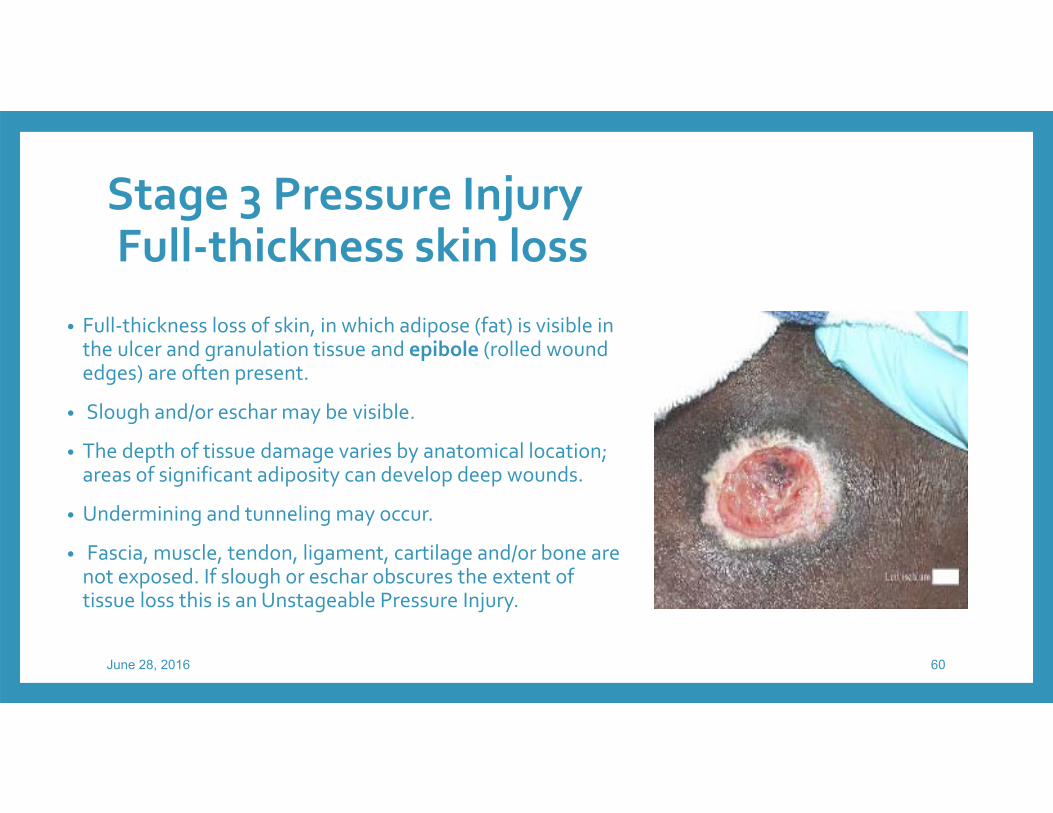
Stage 3 Pressure InjuryFull‐thickness skin loss
• Full‐thickness loss of skin, in which adipose (fat) is visible in the ulcer and granulation tissue and epibole (rolled wound edges) are often present.
• Slough and/or eschar may be visible.
• The depth of tissue damage varies by anatomical location; areas of significant adiposity can develop deep wounds.
• Undermining and tunneling may occur.
• Fascia, muscle, tendon, ligament, cartilage and/or bone are not exposed. If slough or eschar obscures the extent of tissue loss this is an Unstageable Pressure Injury.
June 28, 2016 60
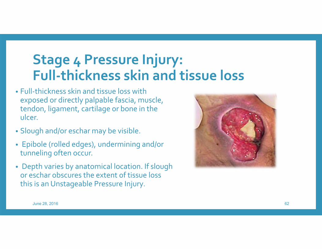
Stage 4 Pressure Injury: Full‐thickness skin and tissue loss
• Full‐thickness skin and tissue loss with exposed or directly palpable fascia, muscle, tendon, ligament, cartilage or bone in the ulcer.
• Slough and/or eschar may be visible.
• Epibole (rolled edges), undermining and/or tunneling often occur.
• Depth varies by anatomical location. If slough or eschar obscures the extent of tissue loss this is an Unstageable Pressure Injury.
June 28, 2016 62
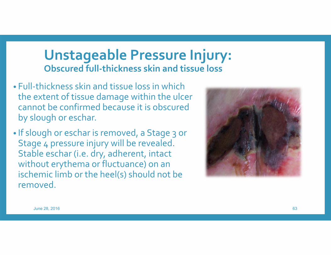
Unstageable Pressure Injury: Obscured full‐thickness skin and tissue loss
• Full‐thickness skin and tissue loss in which the extent of tissue damage within the ulcer cannot be confirmed because it is obscured by slough or eschar.
• If slough or eschar is removed, a Stage 3 or Stage 4 pressure injury will be revealed. Stable eschar (i.e. dry, adherent, intact without erythema or fluctuance) on an ischemic limb or the heel(s) should not be removed.
June 28, 2016 63
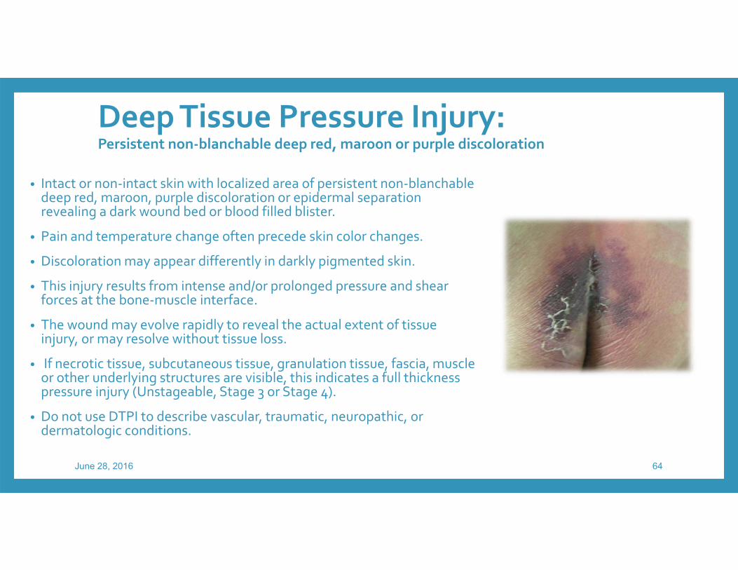
Deep Tissue Pressure Injury: Persistent non‐blanchable deep red, maroon or purple discoloration
• Intact or non‐intact skin with localized area of persistent non‐blanchabledeep red, maroon, purple discoloration or epidermal separation revealing a dark wound bed or blood filled blister.
• Pain and temperature change often precede skin color changes.
• Discoloration may appear differently in darkly pigmented skin.
• This injury results from intense and/or prolonged pressure and shear forces at the bone‐muscle interface.
• The wound may evolve rapidly to reveal the actual extent of tissue injury, or may resolve without tissue loss.
• If necrotic tissue, subcutaneous tissue, granulation tissue, fascia, muscle or other underlying structures are visible, this indicates a full thickness pressure injury (Unstageable, Stage 3 or Stage 4).
• Do not use DTPI to describe vascular, traumatic, neuropathic, or dermatologic conditions.
June 28, 2016 64
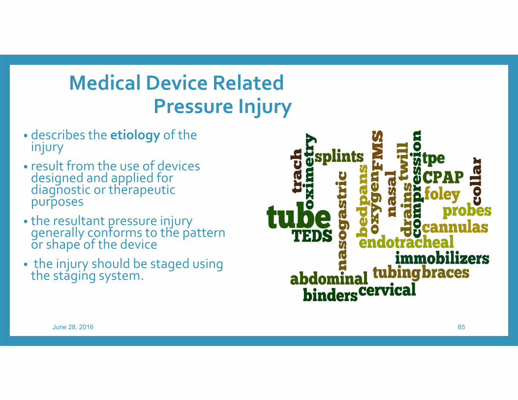
Medical Device Related Pressure Injury
• describes the etiology of the injury
• result from the use of devices designed and applied for diagnostic or therapeutic purposes
• the resultant pressure injury generally conforms to the pattern or shape of the device
• the injury should be staged using the staging system.
June 28, 2016 65
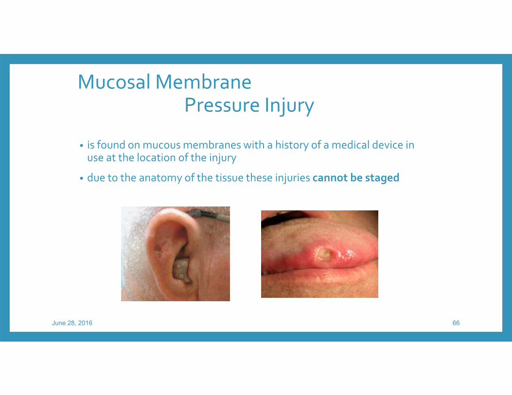
Mucosal Membrane Pressure Injury
• is found on mucous membranes with a history of a medical device in use at the location of the injury
• due to the anatomy of the tissue these injuries cannot be staged
June 28, 2016 66
Not all wounds are pressure injury
6/28/2016
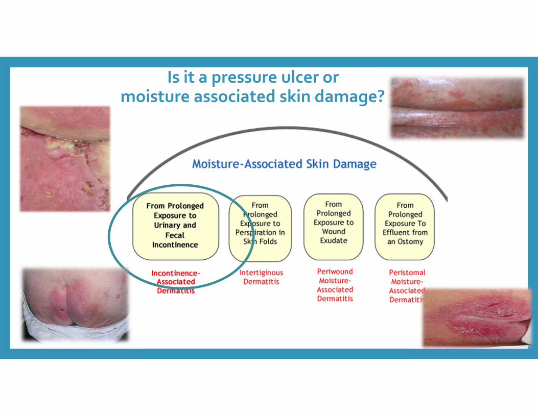
Is it a pressure ulcer or moisture associated skin damage?
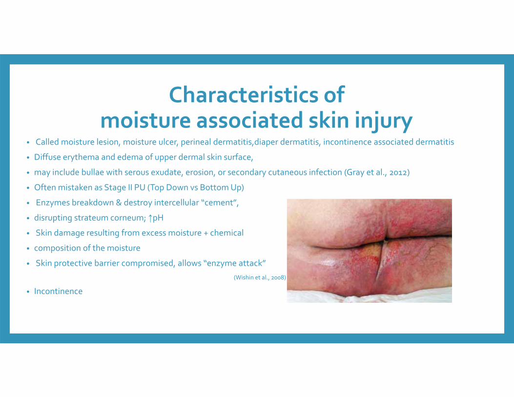
Characteristics of moisture associated skin injury
• Called moisture lesion, moisture ulcer, perineal dermatitis,diaper dermatitis, incontinence associated dermatitis
• Diffuse erythema and edema of upper dermal skin surface,
• may include bullae with serous exudate, erosion, or secondary cutaneous infection (Gray et al., 2012)
• Often mistaken as Stage II PU (Top Down vs Bottom Up)
• Enzymes breakdown & destroy intercellular “cement”,
• disrupting strateum corneum; ↑pH• Skin damage resulting from excess moisture + chemical
• composition of the moisture
• Skin protective barrier compromised, allows “enzyme attack”(Wishin et al., 2008)
• Incontinence
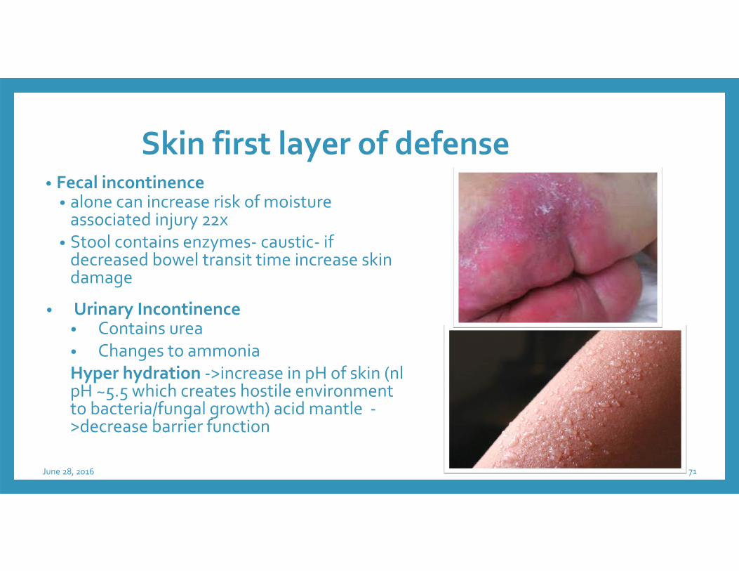
Skin first layer of defense• Fecal incontinence
• alone can increase risk of moisture associated injury 22x
• Stool contains enzymes‐ caustic‐ if decreased bowel transit time increase skin damage
• Urinary Incontinence• Contains urea• Changes to ammoniaHyper hydration ‐>increase in pH of skin (nlpH ~5.5 which creates hostile environment to bacteria/fungal growth) acid mantle ‐>decrease barrier function
71June 28, 2016
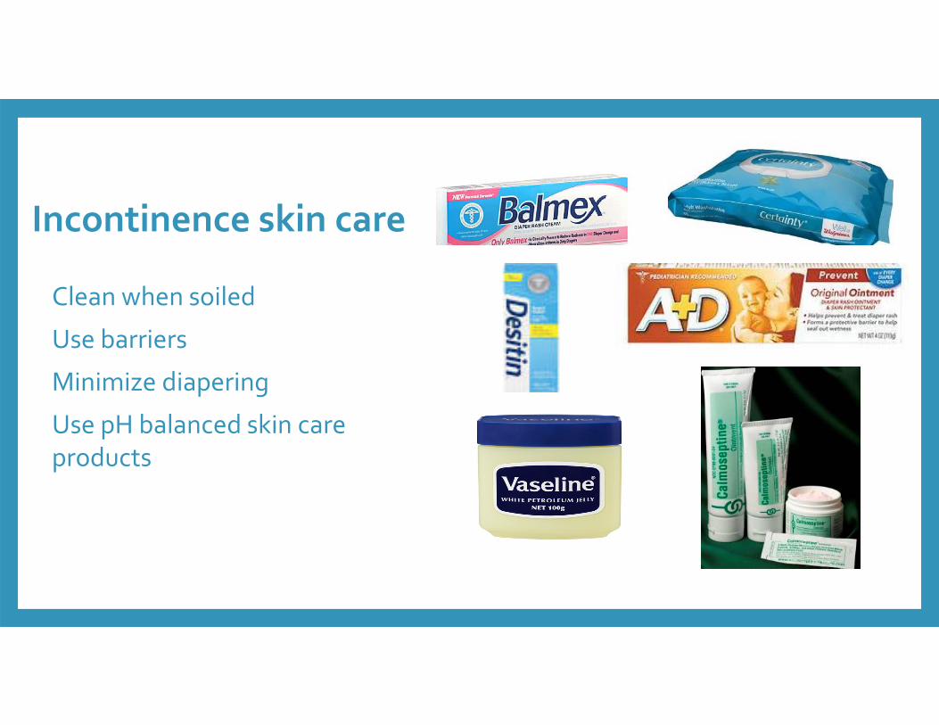
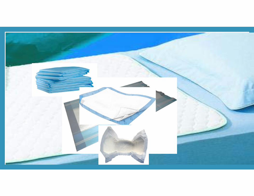
Incontinence skin care
Clean when soiled
Use barriers
Minimize diapering
Use pH balanced skin care products
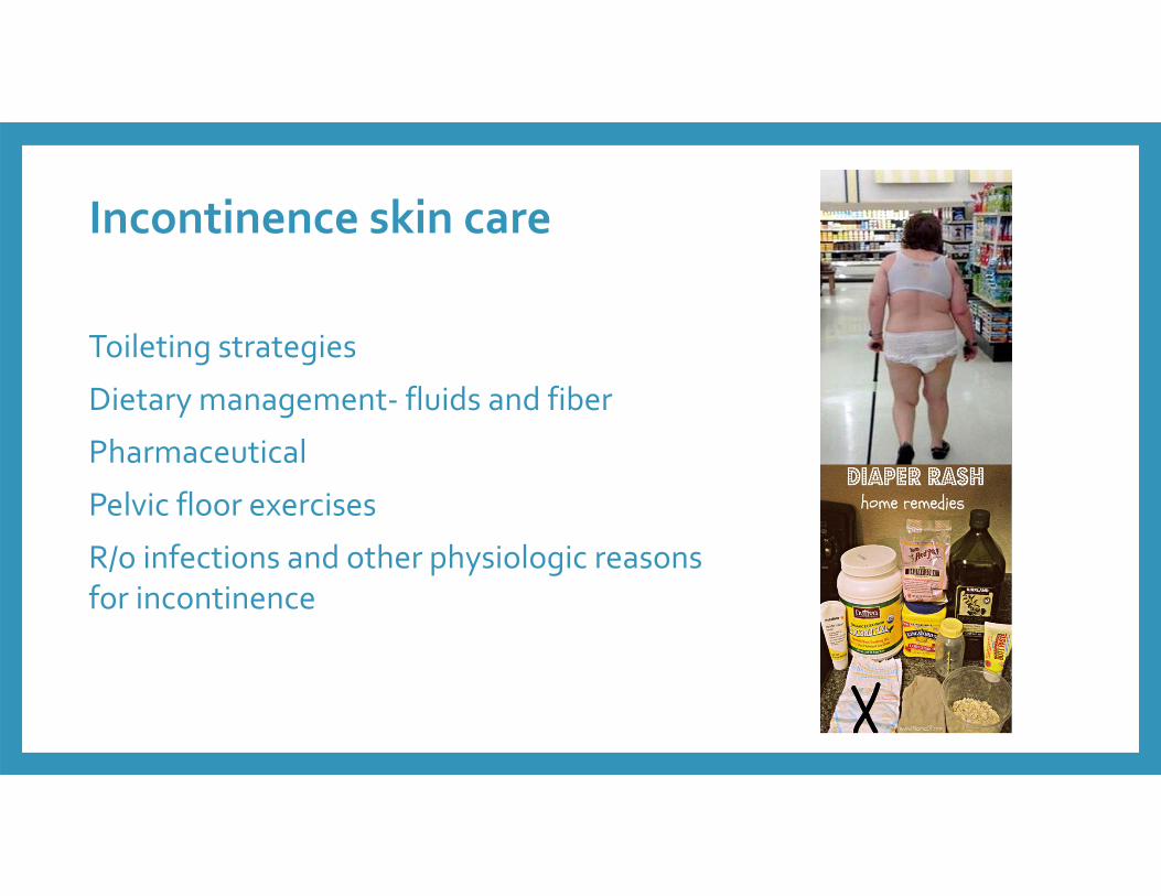
Incontinence skin care
Toileting strategies
Dietary management‐ fluids and fiber
Pharmaceutical
Pelvic floor exercises
R/o infections and other physiologic reasons for incontinence
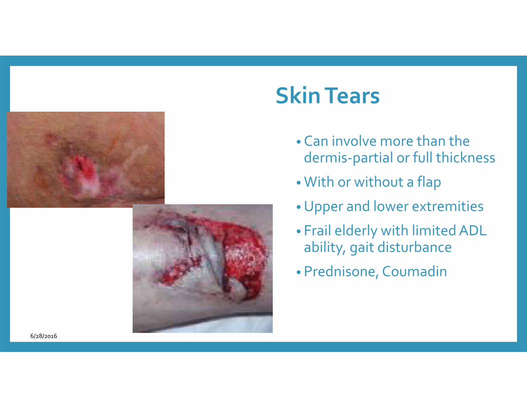
Skin Tears
• Can involve more than the dermis‐partial or full thickness
•With or without a flap
•Upper and lower extremities
• Frail elderly with limited ADL ability, gait disturbance
• Prednisone, Coumadin
6/28/2016
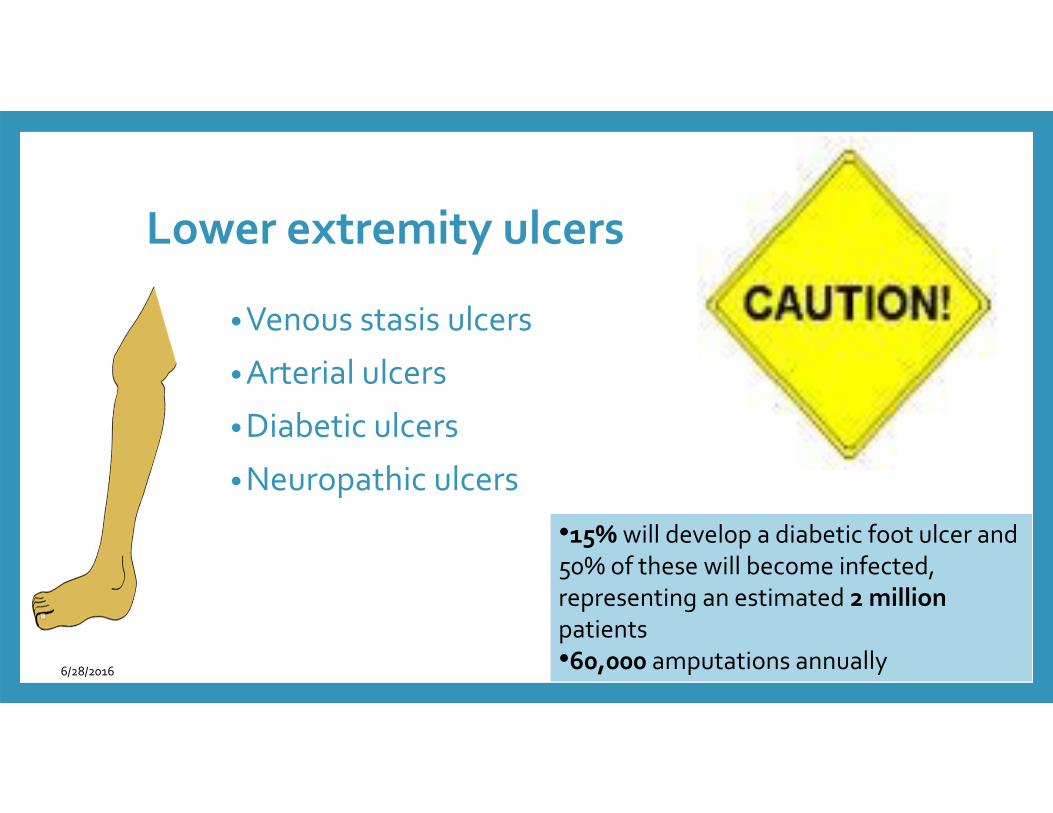
Lower extremity ulcers
•Venous stasis ulcers
•Arterial ulcers
•Diabetic ulcers
•Neuropathic ulcers
6/28/2016
•15% will develop a diabetic foot ulcer and 50% of these will become infected, representing an estimated 2 million patients•60,000 amputations annually
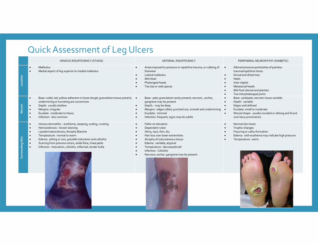
Quick Assessment of Leg UlcersVENOUS INSUFFICIENCY (STASIS) ARTERIAL INSUFFICIENCY PERIPHERAL NEUROPATHY (DIABETIC)
Loca
tion
Malleolus Medial aspect of leg superior to medial malleolus
Areas exposed to pressure or repetitive trauma, or rubbing of footwear
Lateral malleolus Mid tibial Phalangeal heads Toe tips or web spaces
Altered pressure points/sites of painless trauma/repetitive stress
Dorsal and distal toes Heels Inter‐digital Metatarsal heads Mid‐foot (dorsal and plantar) Toe interphalangeal joints
Wou
nd
Base: ruddy red; yellow adherent or loose slough; granulation tissue present, undermining or tunneling are uncommon
Depth: usually shallow Margins: irregular Exudate: moderate to heavy Infection: less common
Base: pale; granulation rarely present; necrosis , eschar, gangrene may be present
Depth : may be deep Margins: edges rolled, punched out, smooth and undermining Exudate: minimal Infection: frequent; signs may be subtle
Base: pink/pale; necrotic tissue variable Depth: variable Edges well defined Exudate: small to moderate Wound shape: usually rounded or oblong and found
over bony prominence
Surrou
nding Sk
in
Venous dermatitis – erythema, weeping, scaling, crusting Hemosiderosis – brown staining Lipodermatosclerosis; Atrophy Blanche Temperature: normal to warm Edema: pitting or non, possible induration and cellulitis Scarring from previous ulcers, ankle flare, tinea pedis Infection: Induration, cellulitis, inflamed, tender bulla
Pallor on elevation Dependent rubor Shiny, taut, thin, dry Hair loss over lower extremities Atrophy of subcutaneous tissue Edema: variable; atypical Temperature: decreased/cold Infection: Cellulitis Necrosis, eschar, gangrene may be present
Normal skin tones Trophic changes Fissuring or callus formation Edema: with erythema may indicate high pressure Temperature: warm
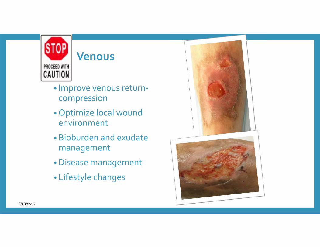
Venous
• Improve venous return‐compression
•Optimize local wound environment
• Bioburden and exudate management
•Disease management
• Lifestyle changes
6/28/2016
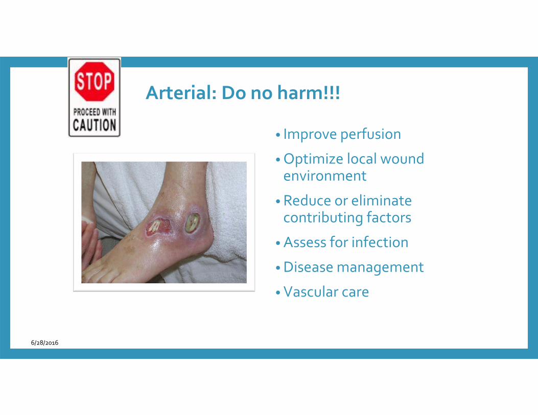
Arterial: Do no harm!!!
• Improve perfusion
•Optimize local wound environment
• Reduce or eliminate contributing factors
• Assess for infection
•Disease management
• Vascular care
6/28/2016
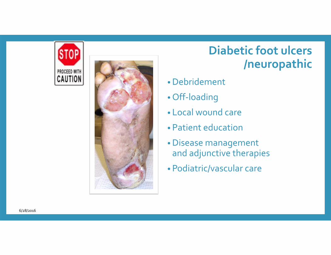
Diabetic foot ulcers/neuropathic
• Debridement
•Off‐loading
• Local wound care
• Patient education
•Disease management and adjunctive therapies
• Podiatric/vascular care
6/28/2016
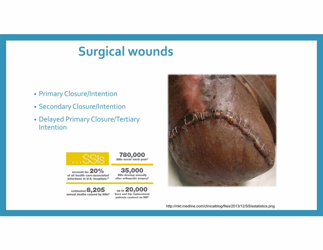
• Primary Closure/Intention
• Secondary Closure/Intention
• Delayed Primary Closure/Tertiary Intention
Surgical wounds
http://mkt.medline.com/clinicalblog/files/2013/12/SSIsstatistics.png
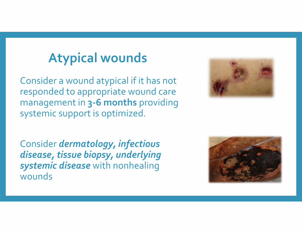
Atypical wounds
Consider a wound atypical if it has not responded to appropriate wound care management in 3‐6 months providing systemic support is optimized.
Consider dermatology, infectious disease, tissue biopsy, underlying systemic disease with nonhealingwounds
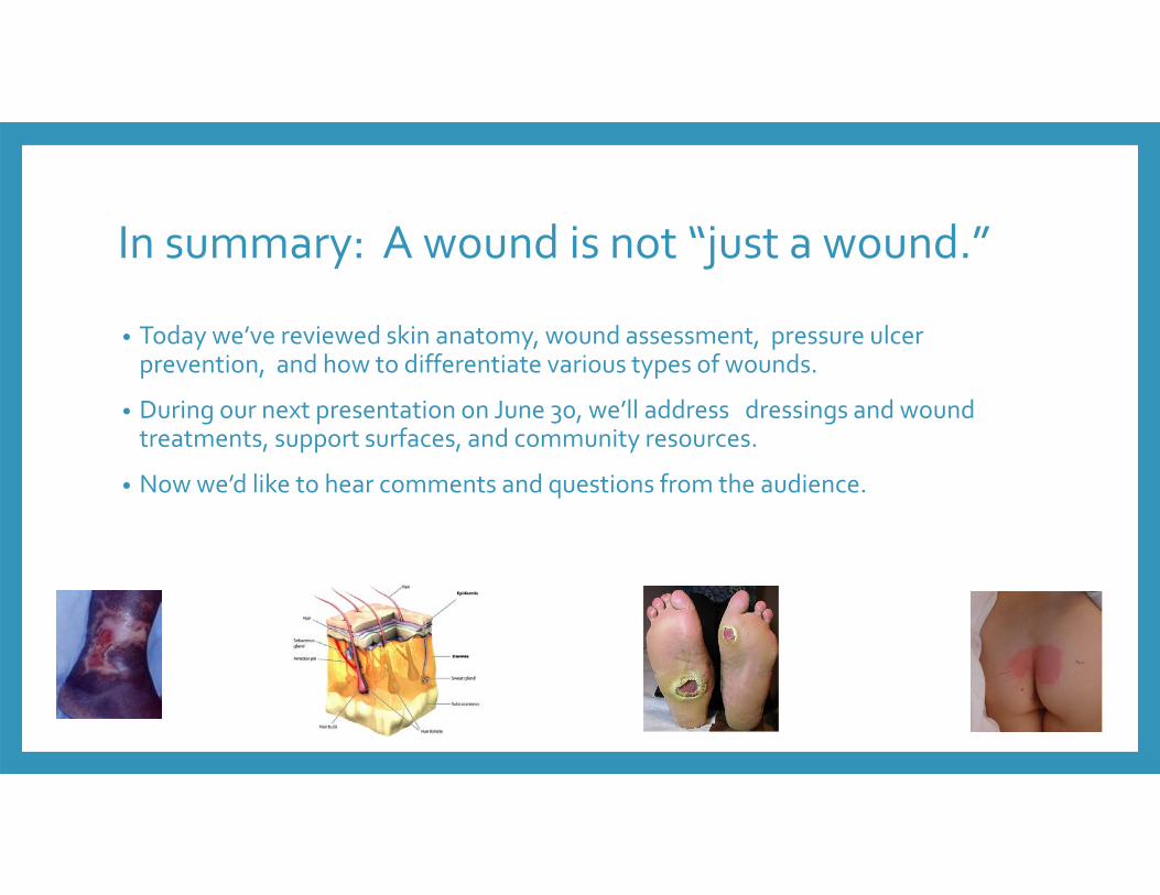
In summary: A wound is not “just a wound.”
• Today we’ve reviewed skin anatomy, wound assessment, pressure ulcer prevention, and how to differentiate various types of wounds.
• During our next presentation on June 30, we’ll address dressings and wound treatments, support surfaces, and community resources.
• Now we’d like to hear comments and questions from the audience.
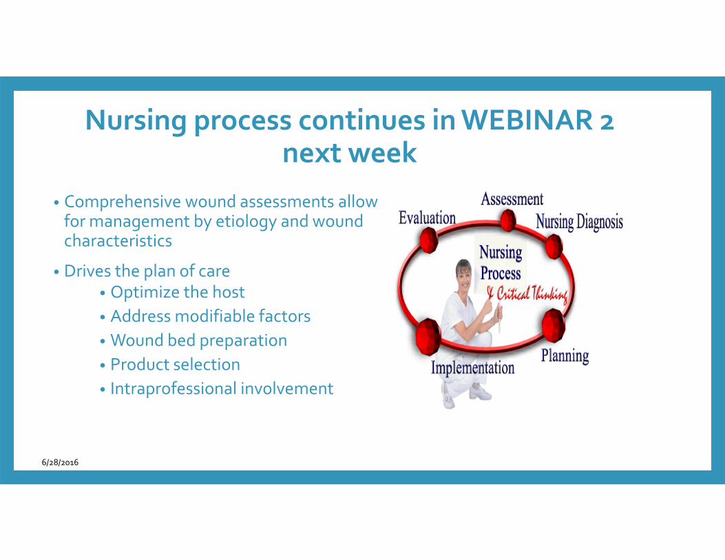
Nursing process continues in WEBINAR 2 next week
• Comprehensive wound assessments allow for management by etiology and wound characteristics
• Drives the plan of care• Optimize the host• Address modifiable factors• Wound bed preparation• Product selection• Intraprofessional involvement
6/28/2016
Select References• Chambers, H. G., & Chambers, J. A. (2015). Effects of Caregiving on the Families of Children and Adults with Disabilities.
Physical medicine and rehabilitation clinics of North America, 26(1), 1-19.
• Department of Disabled Adults. Viewed 05/01/2016 from http://dda.dhmh.maryland.gov/Pages/health_and_nursing.aspx
• Doughty, D., & McNichol, L. (2015). Wound, Ostomy and Continence Nurses Society® Core Curriculum: Wound Management. Lippincott Williams & Wilkins.
• Hopf, HW. Wound Repair and Regeneration. Official publication of the Wound Healing Society [and] the European Tissue Repair Society. 2006;14(1):55‐60
• NPUAP, (2016). Pressure Injury Staging. Retrieved 04/13/16 fromhttp://www.npuap.org/national‐pressure‐ulcer‐advisory‐panel‐npuap‐announces‐a‐change‐in‐terminology‐from‐pressure‐ulcer‐to‐pressure‐injury‐and‐updates‐the‐stages‐of‐pressure‐injury/
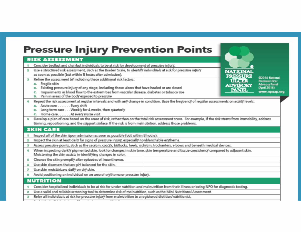
• NPUAP, (2016). Pressure Injury Prevention Points, Retrieved 05/01/2016 from http://www.npuap.org/national‐pressure‐ulcer‐advisory‐panel‐npuap‐announces‐a‐change‐in‐terminology‐from‐pressure‐ulcer‐to‐pressure‐injury‐and‐updates‐the‐stages‐of‐pressure‐injury/

YOUR QUESTIONS &
COMMENTS?


![2019-04-27 OnTarget First Aid Training Flyer [print]...Gun Range/Gunshot Wound Basic First Aid* Topics Covered: Gunshot Wound Basics: Tactical Treatment Ballistics and the Effects](https://static.fdocuments.us/doc/165x107/5f4849042f6f1a4a393115fb/2019-04-27-ontarget-first-aid-training-flyer-print-gun-rangegunshot-wound.jpg)