


Working up the Patient with Chest Pain
130
How Doctors Think in the ED Working up the Patient with Chest Pain Daniel R. Martin, MD, MBA Vice Chair of Education Program Director EM IM Program Ohio State University
Transcript of Working up the Patient with Chest Pain
How Doctors Think in the ED Working up the Patient with Chest PainDaniel R. Martin, MD, MBA
Vice Chair of Education
Program Director EM IM Program
Ohio State University


Objectives
• Share my story of 31 years at OSU
• Hear and eventually solve a case of chest pain (William Morgan)
• Learn how doctors think and the two types of thinking they use
• Learn the 6 immediately life threatening chest pain diagnosis
• Describe the usual work-up of chest pain in the ED
• Describe how we diagnose these 6 causes of chest pain

DISCLOSURES
• No other monetary benefits from any pharmaceutical companies
• NIH funding in the past
• >50 Jerry Garcia ties and 15 are Christmas ties
• Bought a new sport coat just for cruises from Macy’s
• Favorite drink is the Classic frozen Margarita with Grand Marnier or Blackberry Bourbon from Molly Woos
• Favorite TV shows: Seinfeld, Curb Your Enthusiasm, Law and Order
• Most recent movie, “Get Out” by Jordan Peele

My Story – Vulnerability Trust
• 31 years at OSU
• Early years
• Married 1980
• Education
• OSU 1988
• Education
• Research
• Leadership (VCE)
• MBA 2016

The Real Story - (Type 1 and a Pacer) • 23 yo weight loss and lots of urination
Eye doctor appointment Friday before start of medical school
Needed glasses but vision remained blurry
Back to eye doctor on Tuesday positive urine 4+ glucose
Type 1 Diabetes (on insulin)
• 59 yo just noticed that when I ran just became tired running
My internist checked labs and a stress test
During stress test recovery skipping beats
Wife (RN) noted heart rate in the 30s
Third degree heart block, diagnosis of cardiac sarcoid
Pacemaker – defibrillator = Heart block with a pacer

Questions?
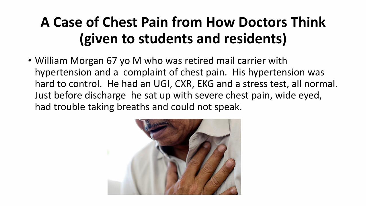
A Case of Chest Pain from How Doctors Think(given to students and residents)
• William Morgan 67 yo M who was retired mail carrier with hypertension and a complaint of chest pain. His hypertension was hard to control. He had an UGI, CXR, EKG and a stress test, all normal. Just before discharge he sat up with severe chest pain, wide eyed, had trouble taking breaths and could not speak.
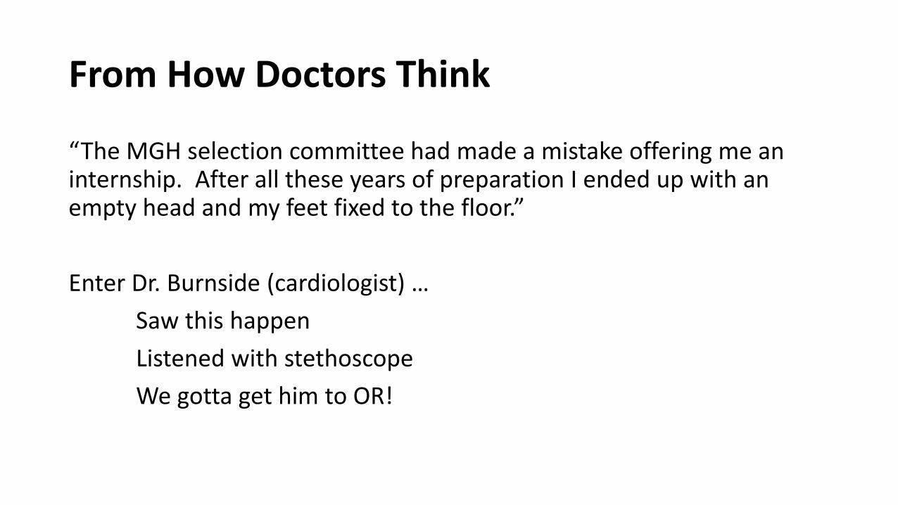
From How Doctors Think
“The MGH selection committee had made a mistake offering me an internship. After all these years of preparation I ended up with an empty head and my feet fixed to the floor.”
Enter Dr. Burnside (cardiologist) …
Saw this happen
Listened with stethoscope
We gotta get him to OR!
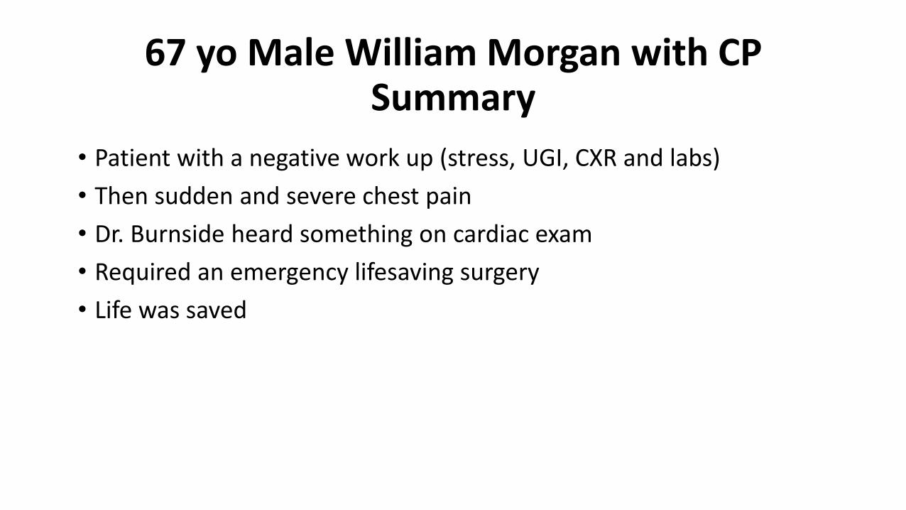
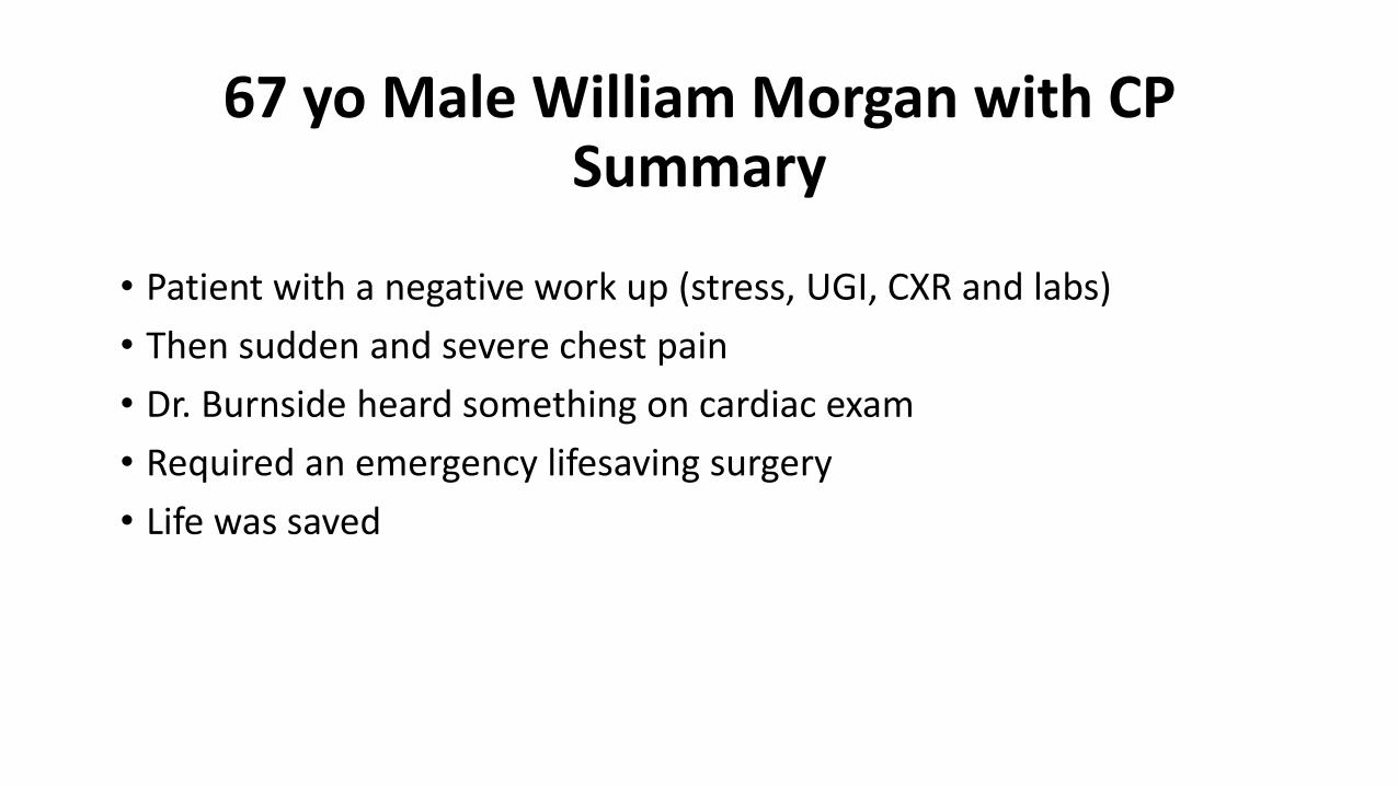
67 yo Male William Morgan with CP Summary
• Patient with a negative work up (stress, UGI, CXR and labs)
• Then sudden and severe chest pain
• Dr. Burnside heard something on cardiac exam
• Required an emergency lifesaving surgery
• Life was saved
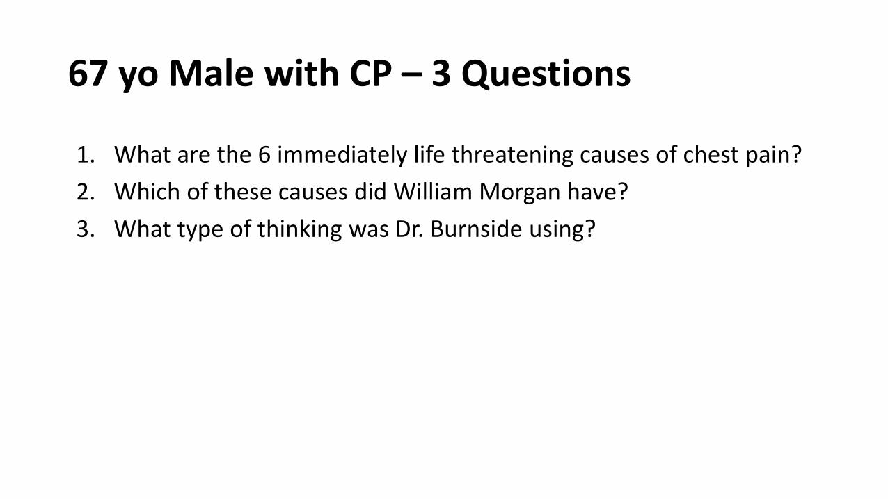
67 yo Male with CP – 3 Questions
1. What are the 6 immediately life threatening causes of chest pain?
2. Which of these causes did William Morgan have?
3. What type of thinking was Dr. Burnside using?


How Do Doctors Think?
• Catch-as-catch-can; trainees observe senior physicians and assimilate the approach to diagnosis and treatment (apprenticeship; see one, do one, teach one)
• Decision trees; preset algorithms and practice guidelines • What if vague complaints? Multiple complaints?• Do they expand knowledge or constrain it?
• Evidence based medicine• Too much reliance on numbers?• At best EBM can compliment decisions
• Bayesian analysis
• Normative; compile a list, determine likelihood, consequences of making or missing each dx + use Bayesian probability to narrow the list (seldom used in practice)

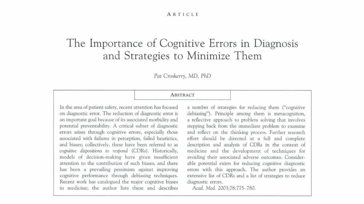
The Importance of Cognitive Errors in Diagnosis and Strategies to Minimize Them
Pat Croskerry, MD
Halifax, Nova Scotia
Academic Medicine 2003;78:775-780

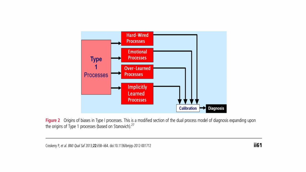
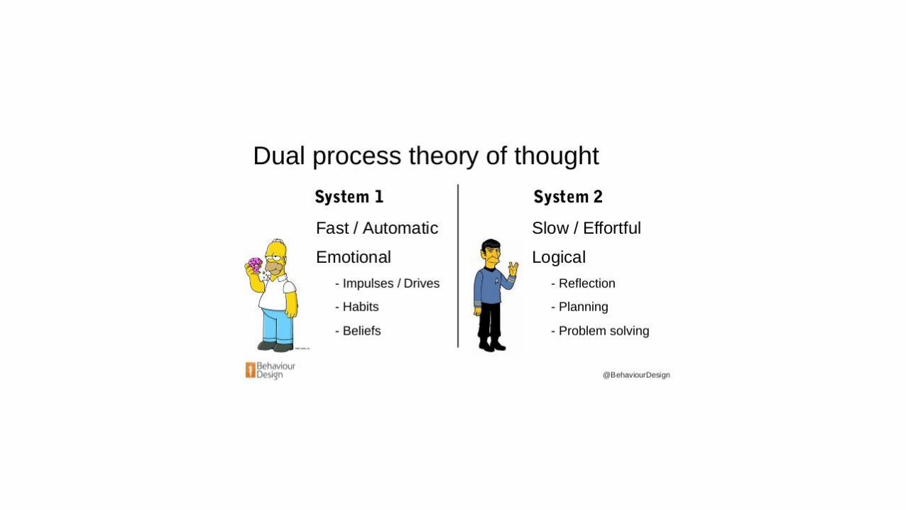


Facts about Dual Process Theory
• Verified by functional MRI, brain glucose utilization, studies of patients with brain lesions (Acad Med 2019;94:187-194)
• Novices and Trainees spend more time in the analytical mode and experienced clinicians spend more time in the intuitive mode. Experts have largest repertoire of cases, few errors and instant dx.
• Experts have more experience and more deliberate practice and this may take 10 years or 10,000 hours. (Anders Ericksen and Malcolm Gladwell)
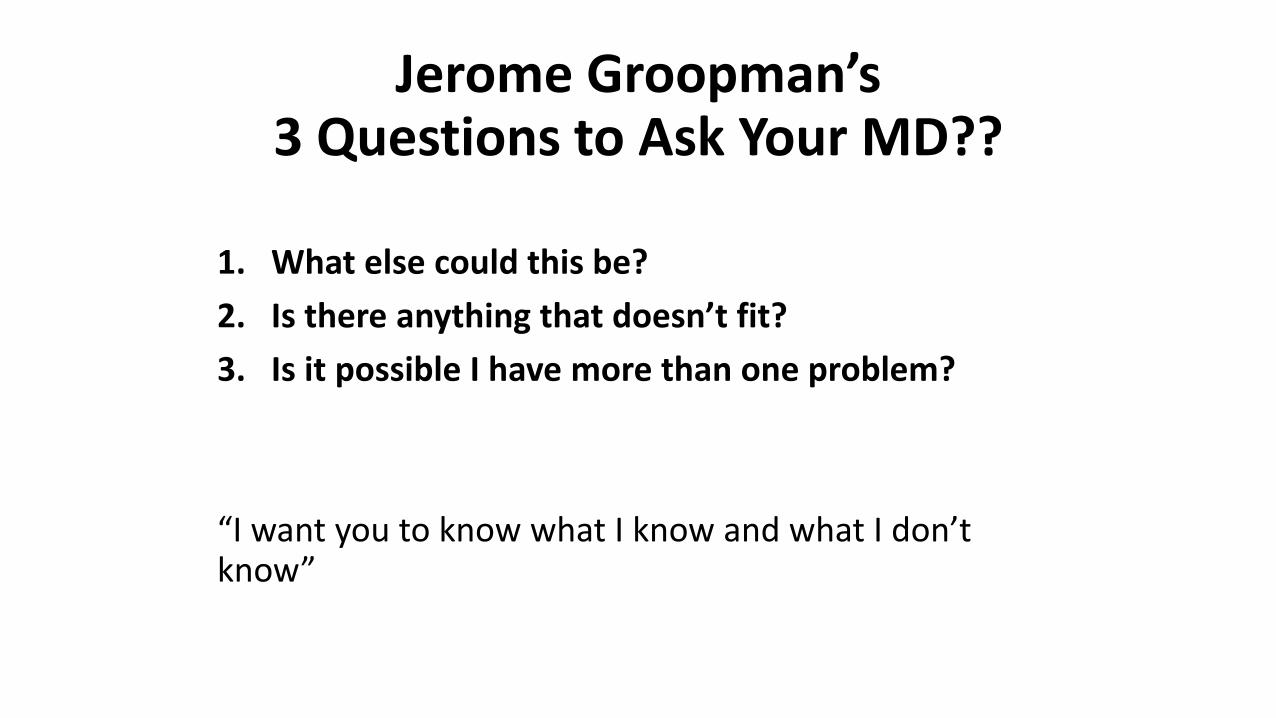
Jerome Groopman’s3 Questions to Ask Your MD??
1. What else could this be?
2. Is there anything that doesn’t fit?
3. Is it possible I have more than one problem?
“I want you to know what I know and what I don’t know”

So far…
• Learned a lot about me
• Case of chest pain (William Morgan)
• How doctors think
• Dual process theory
• Six immediately life-threatening chest pain
• Describe the usual work-up of chest pain in the ED
• Describe how we diagnose these 6 causes of chest pain
Chest Pain
What are the six immediately life threatening causes of chest pain??
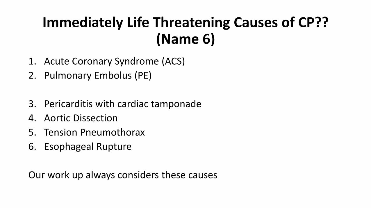
Immediately Life Threatening Causes of CP??(Name 6)
1. Acute Coronary Syndrome (ACS)
2. Pulmonary Embolus (PE)
3. Pericarditis with cardiac tamponade
4. Aortic Dissection
5. Tension Pneumothorax
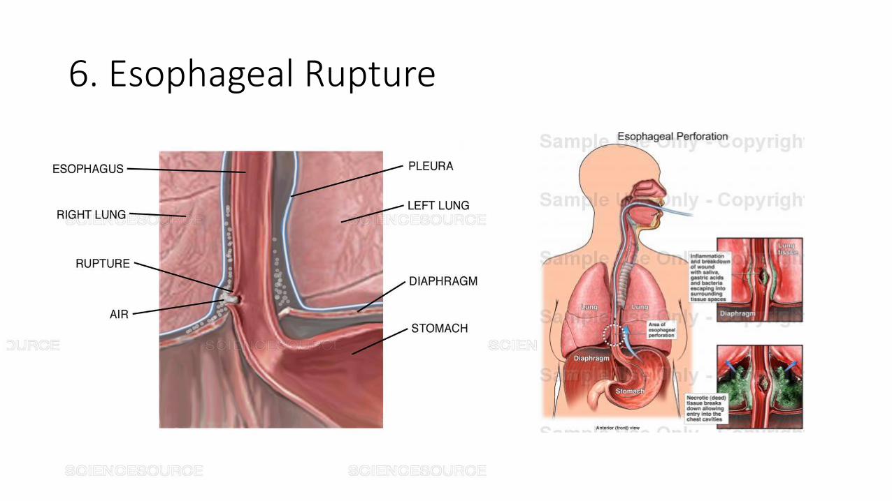
6. Esophageal Rupture
Our work up always considers these causes

1. Acute Coronary Syndrome (ACS)

1. Acute Coronary Syndrome

2. Pulmonary Embolus

3. Pericardial Tamponade

4. Aortic Dissection

4. Aortic Dissection

Dissection versus an Aneurysm (AAA)
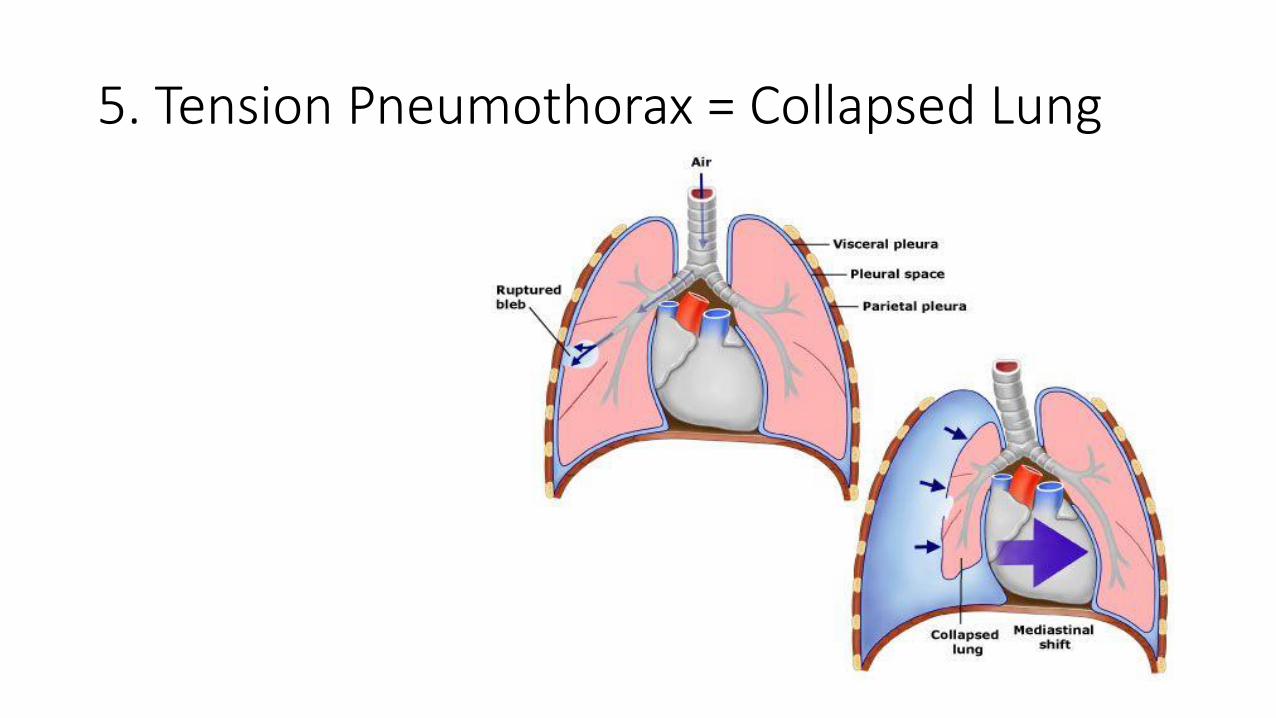
5. Tension Pneumothorax = Collapsed Lung
6. Esophageal Rupture

Mangiamo’s in Hilton Head

So far…
• Learned a lot about me
• Case of chest pain (William Morgan)
• How doctors think
• Dual process theory
• Six immediately life-threatening chest pain
• Describe the usual work-up of chest pain in the ED
• Describe how we diagnose these 6 causes of chest pain

ED Work Up of the Patient with Chest Pain
• Very common complaint (nearly 6 million ED visits per year)
• There are at least six immediately life threatening causes
• We can order these causes by likelihood
• History and physical exam can help
• Diagnostic work up considers life threats

Chest Pain and Work Up
• Vital Signs
• Cardiac Monitor
• H and P
• EKG (within 10 minutes of arrival)
• CXR
• Troponin

Chest Pain and Work Up
• Vital Signs
Blood pressure (systolic over diastolic)
Heart Rate (60 – 100 beats per minute)
Temperature
Respiratory Rate
Pulse Oximetry

Chest Pain and Work Up
• Cardiac Monitor
Continuous monitoring of the heart
Too fast or too slow
Irregular
Pauses or asystole
Fibrillation (atrial or ventricular)
Chest Pain and Work Up
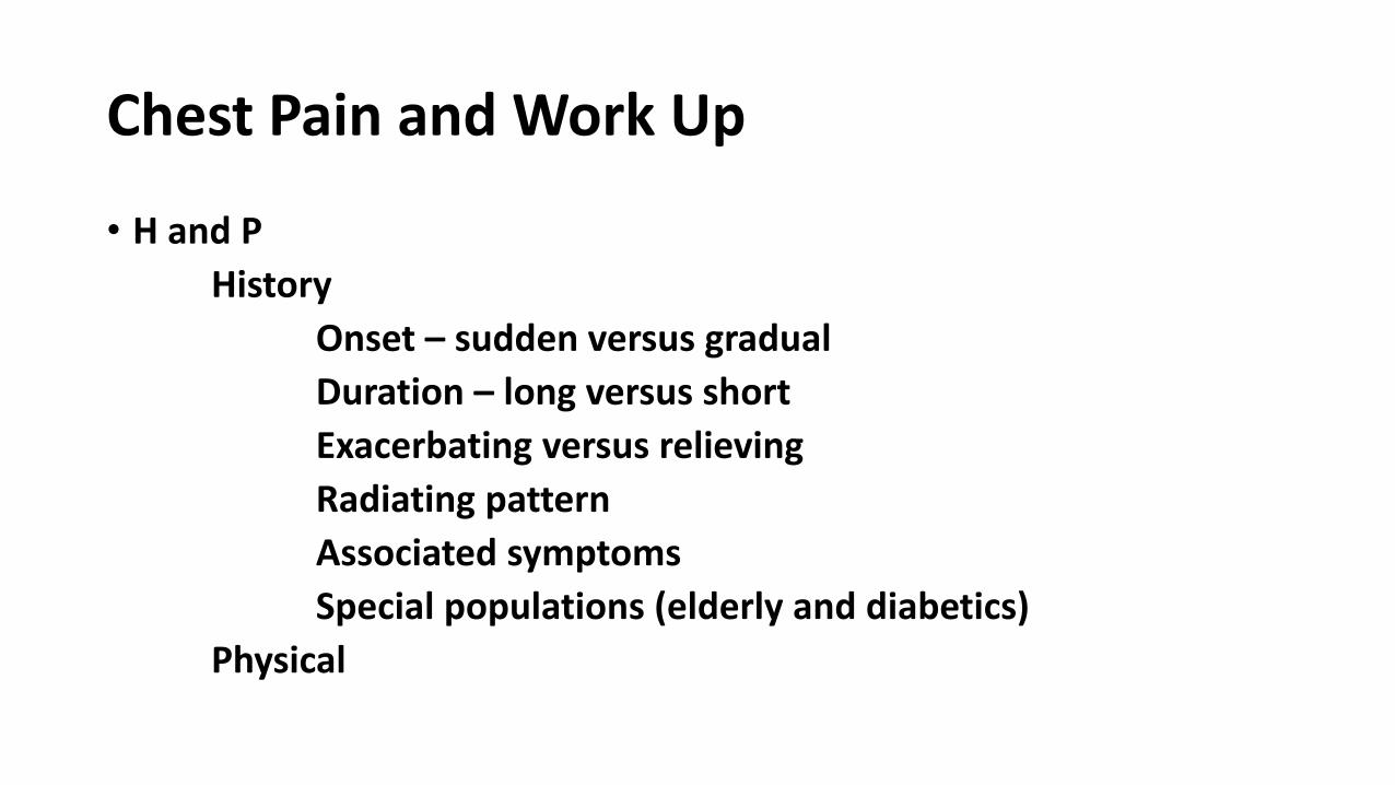
• H and P
History
Onset – sudden versus gradual
Duration – long versus short
Exacerbating versus relieving
Radiating pattern
Associated symptoms
Special populations (elderly and diabetics)
Physical

Chest Pain and Work Up
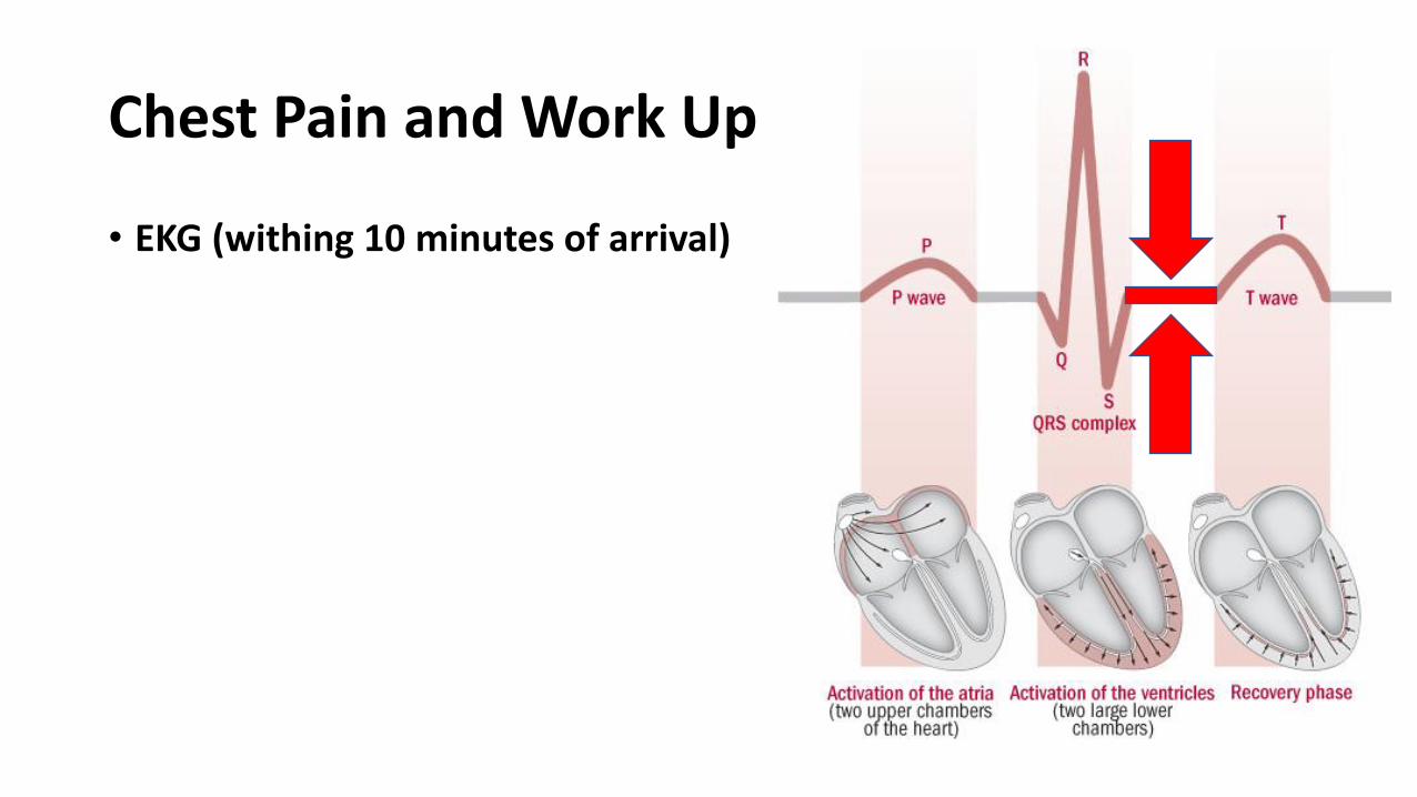
• EKG (withing 10 minutes of arrival)
Chest Pain and Work Up
• EKG (withing 10 minutes of arrival)

Chest Pain and Work Up
• EKG (withing 10 minutes of arrival)
Chest Pain and Work Up
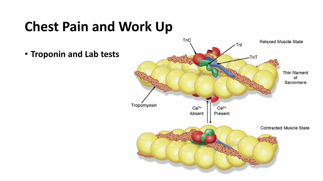
• Troponin and Lab tests
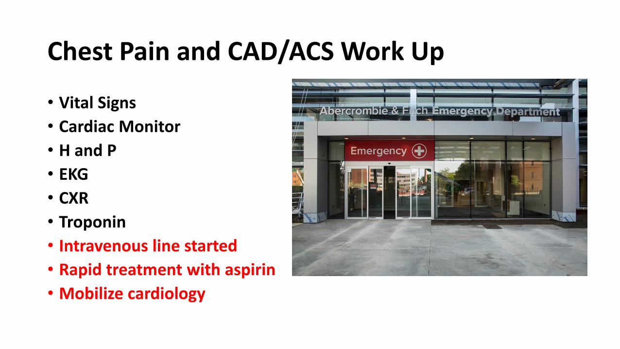
Chest Pain and CAD/ACS Work Up
• Vital Signs
• Cardiac Monitor
• H and P
• EKG
• CXR
• Troponin
• Intravenous line started
• Rapid treatment with aspirin
• Mobilize cardiology

History for ACS • Pain description
• Increase likelihood of ACS/CAD: • CP radiating to both arms and shoulders• Diaphoresis with CP• Nausea and vomiting with CP• CP with exertion
• Decreased likelihood of ACS/CAD: • Pain made worse by inspiration (pleuritic)• Positional • Sharp or stabbing• Palpable CP
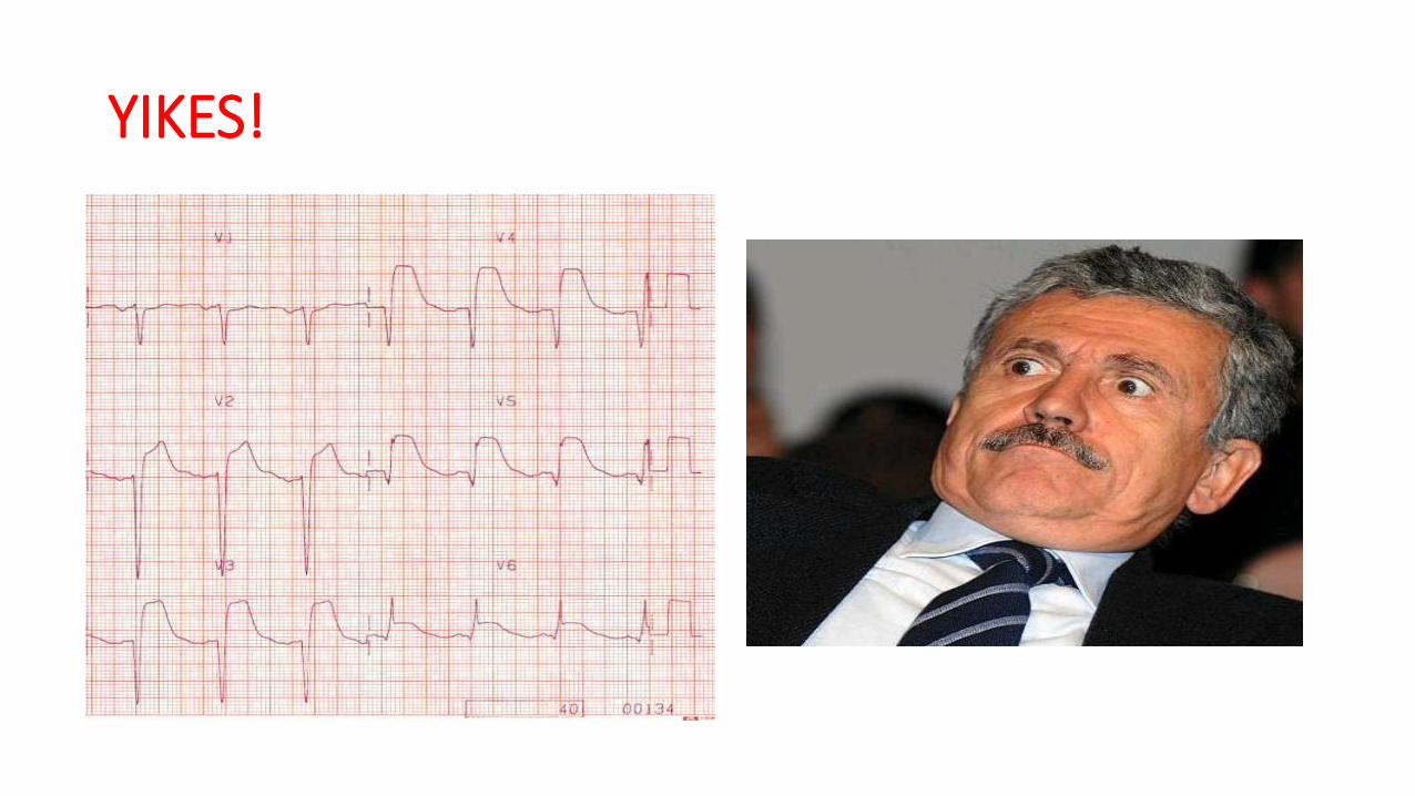
YIKES!
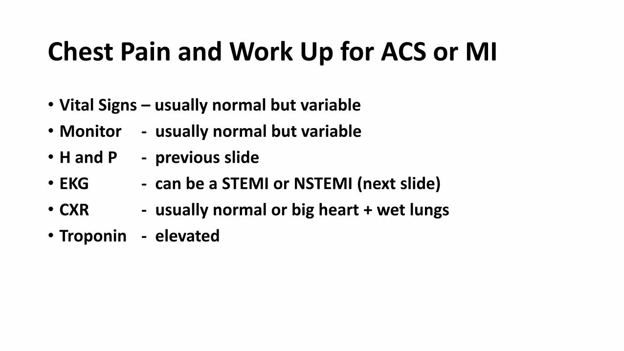
Chest Pain and Work Up for ACS or MI
• Vital Signs – usually normal but variable
• Monitor - usually normal but variable
• H and P - previous slide
• EKG - can be a STEMI or NSTEMI (next slide)
• CXR - usually normal or big heart + wet lungs
• Troponin - elevated

Chest Pain and Work Up for Significant CAD
• STEMI = Abnormal EKG and ST elevation emergent cath to try and restore perfusion
• NSTEMI = Elevated troponin but EKG with no ST elevation may also go to cath
• Most do not have these EKG or troponin changes but need to ask one major question:
Does this person have significant CAD and need some intervention?

Chest Pain and Work Up for Significant CAD
• Can use scoring systems
HEART
TIMI

HEART SCORE and MACE (Major Adverse Cardiac Events)
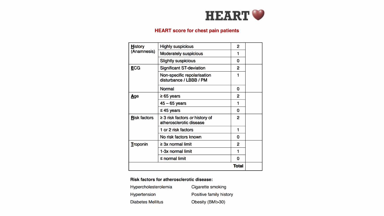
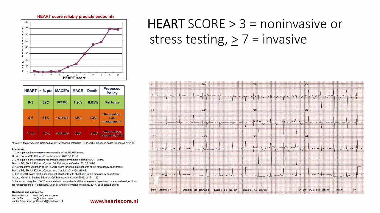
HHEART SCORE > 3 = noninvasive or stress testing, > 7 = invasive


What about ACS/CAD risk?
• STEMI to the cath lab
• Very low risk CP can be discharged
• Intermediate risk?
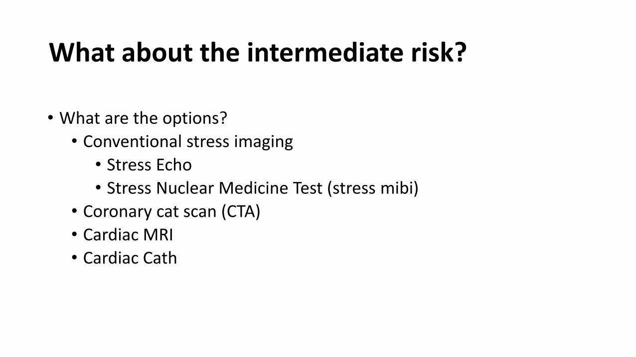
What about the intermediate risk?
• What are the options?
• Conventional stress imaging
• Stress Echo
• Stress Nuclear Medicine Test (stress mibi)
• Coronary cat scan (CTA)
• Cardiac MRI
• Cardiac Cath

Stress Testing Summary
Test Sensitivity Specificity LR-
Treadmill ECG 30% 93% 0.75
Stress Echo 85% 85%-90% 0.18
Stress Mibi 90% 80%-90% 0.12
Coronary CTA 92% 96% 0.11
CMRI (stress CMR) 90% 94% 0.10

Stress Testing
• Bruce Protocol (1963) • Increased workload = heart squeezes faster and harder • Coronaries dilate to increase flow (perfusion) • IF coronaries unable to help because of blockages (CAD)
Chest PainEKG changes Wall motion or perfusion abnormalities
• A positive stress test is usually followed by a cardiac catheterization to restore blood flow (perfusion)

Stress Testing
Strengths • Great functional test
• Imaging helps
• Don’t have to walk
• Monitors rhythm and BP
• Great effort = predictive
• Positivity at low work loads
• Accessible, Inexpensive
Weaknesses • Limited by effort (need 85%)
• Limited by rhythm
• Accuracy questionable
• Medications can affect results
• Body habitus
• Preexisting illnesses
• Gender differences

Higher pretest probability or unable to stress?
• Coronary CTA?
• Cardiac MRI?
• Or just admit and cath?

• Anatomical test
• Is there CAD present?
• Does not address likelihood of placque rupture
• Does not address impact of moderate stenosis
• Can overestimate severity
Coronary CTA
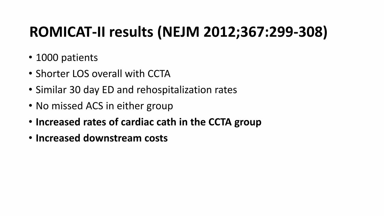
ROMICAT-II results (NEJM 2012;367:299-308)
• 1000 patients
• Shorter LOS overall with CCTA
• Similar 30 day ED and rehospitalization rates
• No missed ACS in either group
• Increased rates of cardiac cath in the CCTA group
• Increased downstream costs
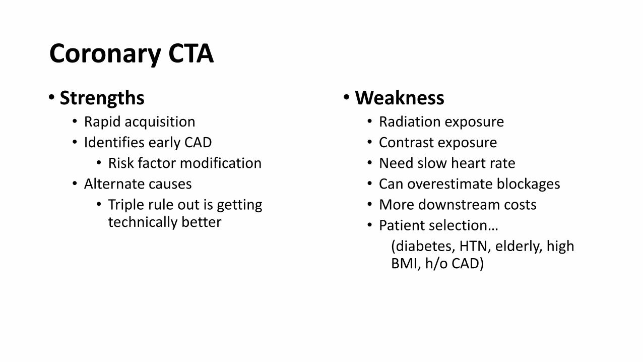
• Strengths• Rapid acquisition
• Identifies early CAD
• Risk factor modification
• Alternate causes
• Triple rule out is getting technically better
• Weakness• Radiation exposure
• Contrast exposure
• Need slow heart rate
• Can overestimate blockages
• More downstream costs
• Patient selection…
(diabetes, HTN, elderly, high BMI, h/o CAD)
Coronary CTA

CCTA - summary
• Helpful if negative and safe to DC or look for other causes
• Risk of poor images in patients with the most concern
• Difficult to interpret the intermediate result
• Increases resource utilization downstream
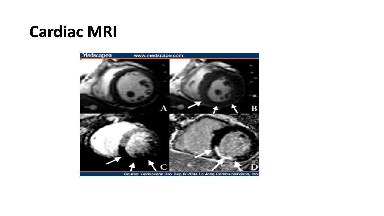
Cardiac MRI
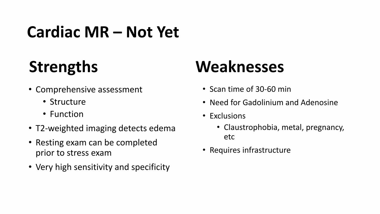
• Scan time of 30-60 min
• Need for Gadolinium and Adenosine
• Exclusions
• Claustrophobia, metal, pregnancy, etc
• Requires infrastructure
• Comprehensive assessment
• Structure
• Function
• T2-weighted imaging detects edema
• Resting exam can be completed prior to stress exam
• Very high sensitivity and specificity
Cardiac MR – Not Yet
Strengths Weaknesses

Chest Pain and Work Up for ACS or MI
• Vital Signs – usually normal but variable • Monitor - usually normal but variable • H and P - previous slide • EKG - can be a STEMI or NSTEMI• CXR - usually normal or big heart + wet lungs • Troponin - elevated, normal then rising or normal times two • Scores - HEART • Stress Testing (echo, perfusion, with meds) – timing?• CCT or CMR• Cardiac Cath


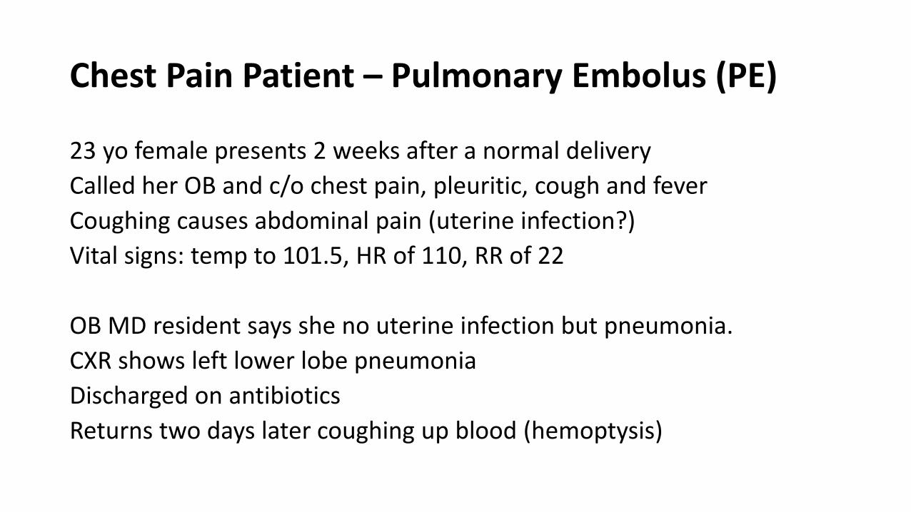
Chest Pain Patient – Pulmonary Embolus (PE)
23 yo female presents 2 weeks after a normal delivery
Called her OB and c/o chest pain, pleuritic, cough and fever
Coughing causes abdominal pain (uterine infection?)
Vital signs: temp to 101.5, HR of 110, RR of 22
OB MD resident says she no uterine infection but pneumonia.
CXR shows left lower lobe pneumonia
Discharged on antibiotics
Returns two days later coughing up blood (hemoptysis)

Chest Pain Patient
• Diagnosis = PE with lung injury (infarction)
• Admitted with long course due to an pulmonary infarct with infection and cavitation

Unexplained SOB HemoptysisSymptoms
Unilateral leg swelling Tachycardia
Hypoxemia (SaO2 < 95%)Signs
Limb/Body Immobility Recent Surgery (past 4w) Thrombophilia
Cancer* Age > 50Risk Factors
Obvious Factors Independently Associated with PE
*Adenocarcinoma and Brain Tumor (GBM)
When Should I Consider PE?

Interesting Factors
• Sudden onset?
• Reproducible CP?
• Comfortable?
PE (+) 39% had sudden onset
PE (-) 51% had sudden onset
Reproduction Of Chest Pain By Palpation: Diagnostic Accuracy In Suspected Pulmonary Embolism: Le Gal, G., et al, Br Med J 330:452, February 26, 2005
19.9% of patients PE (+) had reproducible CP
PE (+) 60% Comfortable
PE (+) 27% Mod Discomfort
PE (+) 13% Severe Distress
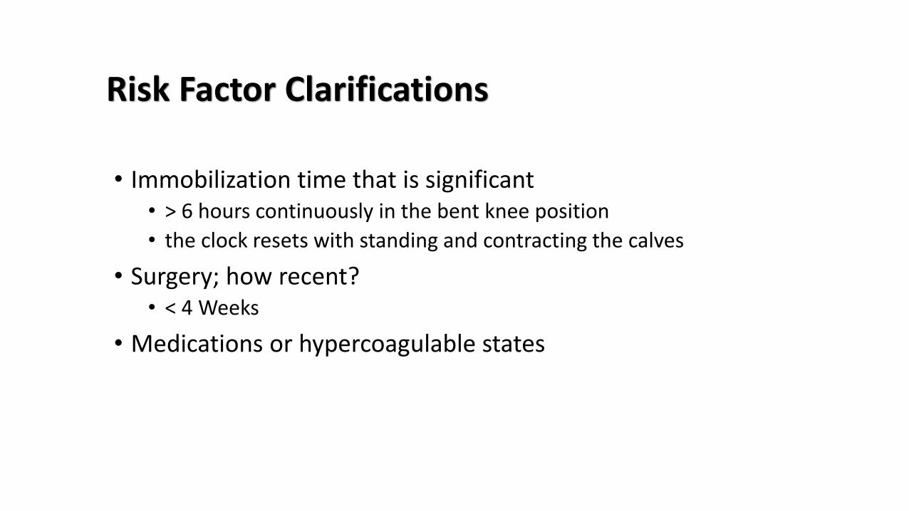
Risk Factor Clarifications
• Immobilization time that is significant • > 6 hours continuously in the bent knee position
• the clock resets with standing and contracting the calves
• Surgery; how recent? • < 4 Weeks
• Medications or hypercoagulable states
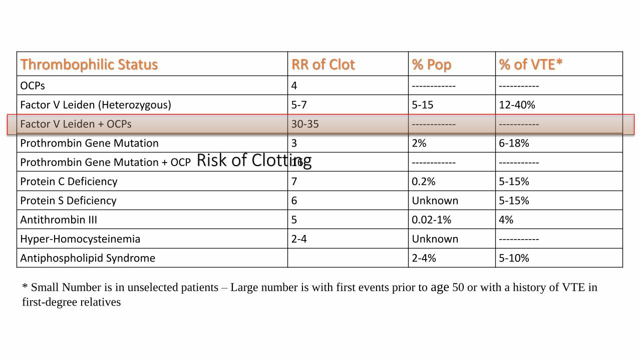
Risk of Clotting
Thrombophilic Status RR of Clot % Pop % of VTE*
OCPs 4 ------------ -----------
Factor V Leiden (Heterozygous) 5-7 5-15 12-40%
Factor V Leiden + OCPs 30-35 ------------ -----------
Prothrombin Gene Mutation 3 2% 6-18%
Prothrombin Gene Mutation + OCP 16 ------------ -----------
Protein C Deficiency 7 0.2% 5-15%
Protein S Deficiency 6 Unknown 5-15%
Antithrombin III 5 0.02-1% 4%
Hyper-Homocysteinemia 2-4 Unknown -----------
Antiphospholipid Syndrome 2-4% 5-10%
* Small Number is in unselected patients – Large number is with first events prior to age 50 or with a history of VTE in
first-degree relatives

What % of Patients With PE Have No, “as of yet” DX Risk Factors?
33%
Kline, et al PE Database Query: n=7720

The 2 groups of patients in whom PE is most likely to be missed:
-Older patients with a good alternative diagnosis-The most common wrong alternative dx is bronchitis
-Young, otherwise healthy, obese, hemodynamically stable women on
Estrogen
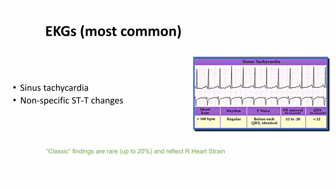
EKGs (most common)
• Sinus tachycardia
• Non-specific ST-T changes
“Classic” findings are rare (up to 20%) and reflect R Heart Strain

Some “Classic” EKGs showing strain on the right heart
S1Q3T3
RBBBP PulmonaleRV Strain
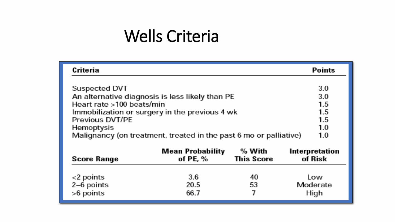
Wells Criteria

But Wait…Is There Anyone Who Needs No Evaluation?
Low PTP (< 15%) +
PERC RuleAge < 50 Heart rate <100 (at any time in ED) Room air SaO2 >94% No prior PE/DVT No recent surgery (< 4w) No HemoptysisNo exogenous estrogen (Tamoxifen)No DVT findings clinically
Will have a PTP <2%
and therefore will not
benefit from an
evaluation for PE

D Dimer to Diagnose a Pulmonary Embolus(byproduct of the clotting cascade)

Using the D-Dimer
• Most helpful if negative D-dimer (very sensitive)
• Lots of reasons for false positives
• If pre-test probability is very low may not help
• If pre-test probability is high may not help
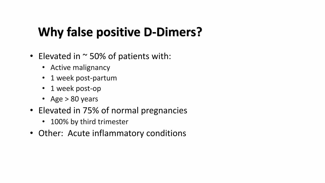
Why false positive D-Dimers?
• Elevated in ~ 50% of patients with:• Active malignancy
• 1 week post-partum
• 1 week post-op
• Age > 80 years
• Elevated in 75% of normal pregnancies • 100% by third trimester
• Other: Acute inflammatory conditions
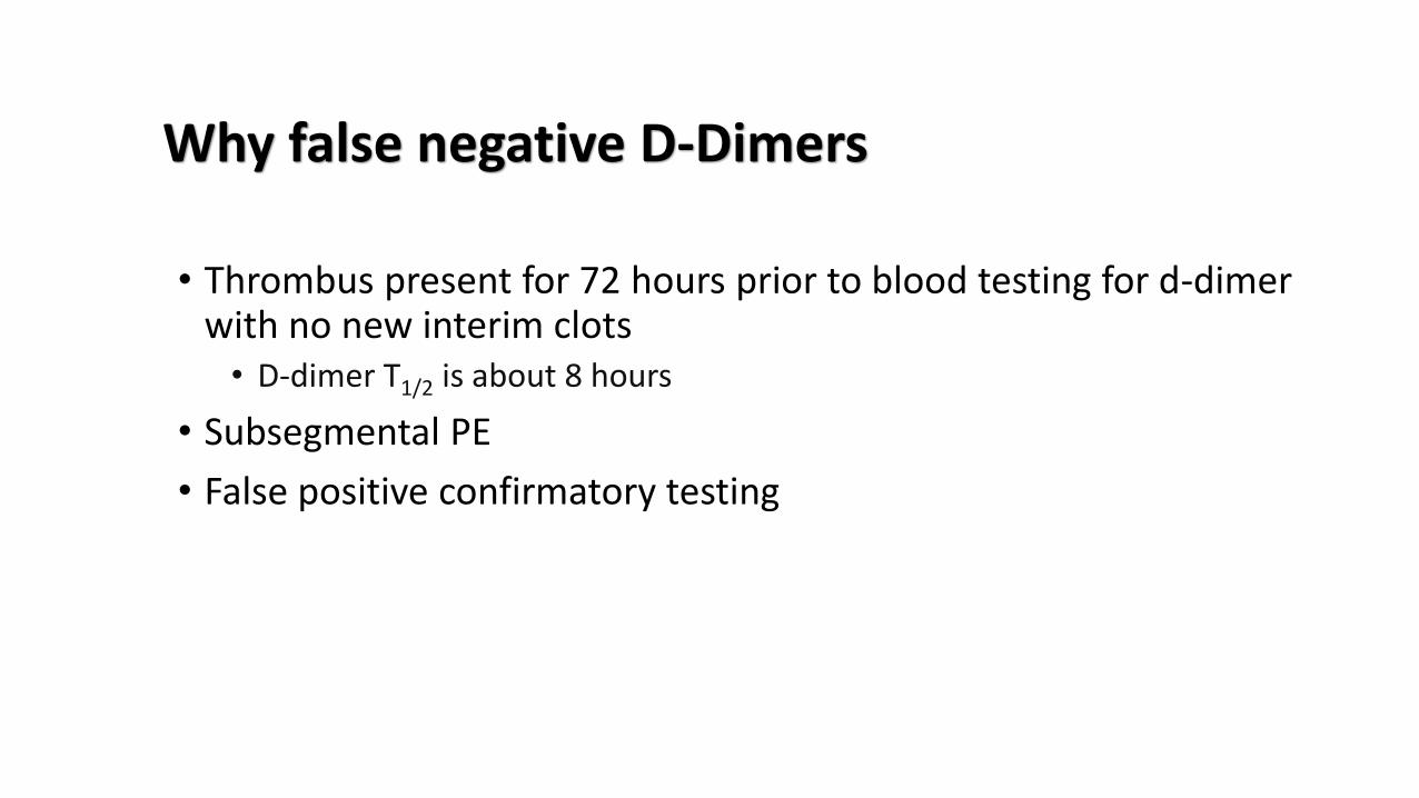
Why false negative D-Dimers
• Thrombus present for 72 hours prior to blood testing for d-dimer with no new interim clots
• D-dimer T1/2 is about 8 hours
• Subsegmental PE
• False positive confirmatory testing

Chest Pain and Work Up for PE
• Vital Signs – BP low or normal, oxygen low or normal
• Monitor - tachycardia but variable
• H and P - chest pain/SOB, risk factors present or leg swelling
• EKG - tachycardia or right heart strain
• CXR - usually normal
• Troponin - can be elevated in a PE
• D-Dimer - usually elevated
• Rules - Wells
• Echo - look for right heart strain
• CTPE or VQ - ?
• Dopplers - ?
• Angiogram - ?

Confirming a pulmonary embolus
• CTPE
• VQ
• MR
• Dopplers
• Angiogram
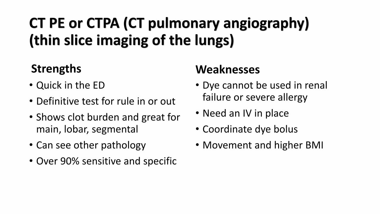
CT PE or CTPA (CT pulmonary angiography)(thin slice imaging of the lungs)
Strengths
• Quick in the ED
• Definitive test for rule in or out
• Shows clot burden and great for main, lobar, segmental
• Can see other pathology
• Over 90% sensitive and specific
Weaknesses • Dye cannot be used in renal
failure or severe allergy
• Need an IV in place
• Coordinate dye bolus
• Movement and higher BMI
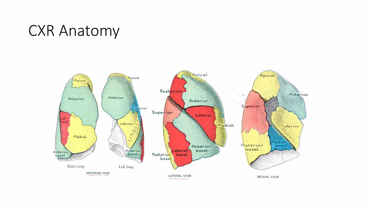
CXR Anatomy

Ventilation-Perfusion Scan (V/Q)
• Nuclear Medicine Scan matches areas of ventilated lung to perfused lung• Looks for matched and unmatched “defects”
V/Q is MUCH SAFER in pregnancy than an undiagnosed PE

VQ for PE
Strengths
• Less radiation
• Can use in renal patients
• Can use in allergy to dye
• Can be diagnostic
• Shows lung involved
Weaknesses • Usually have to leave the ED
• Takes time
• Need an IV in place
• Preexisting lung disease
• “Indeterminate” or “intermediate probability”
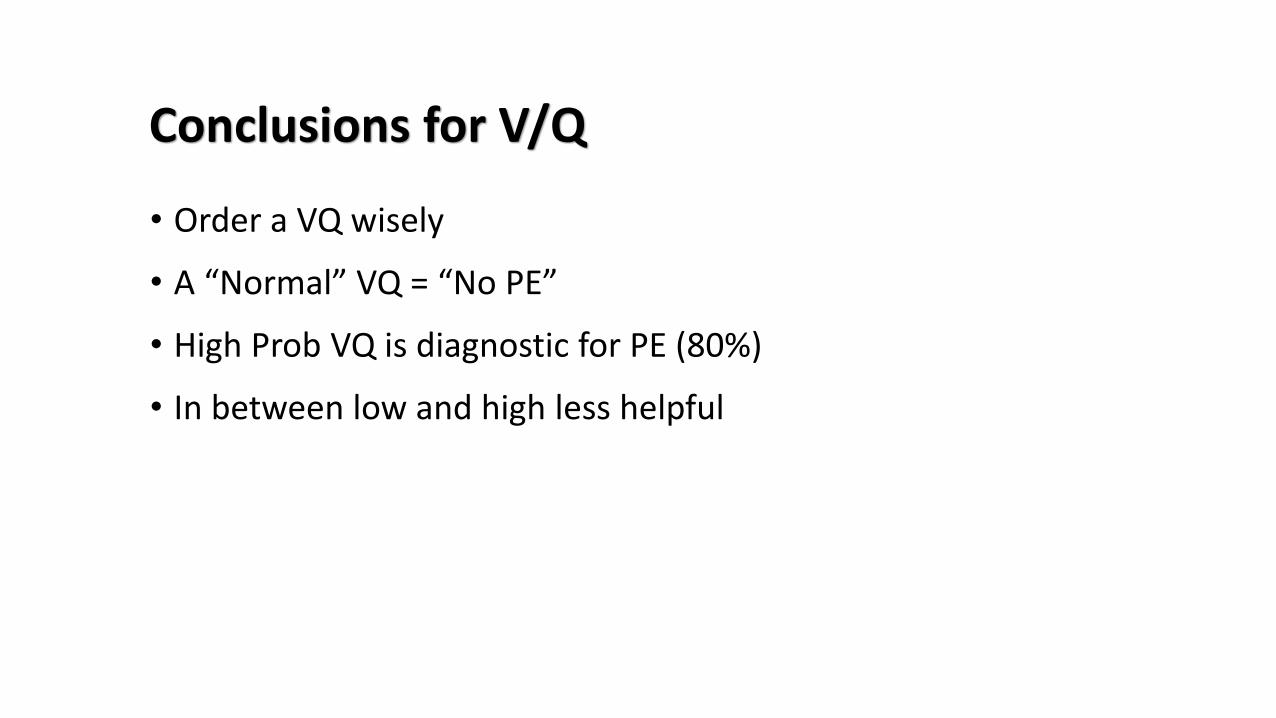
Conclusions for V/Q
• Order a VQ wisely
• A “Normal” VQ = “No PE”
• High Prob VQ is diagnostic for PE (80%)
• In between low and high less helpful
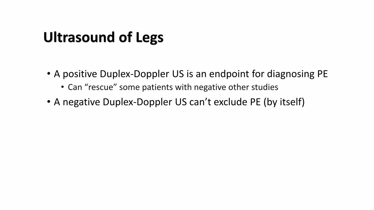
Ultrasound of Legs
• A positive Duplex-Doppler US is an endpoint for diagnosing PE• Can “rescue” some patients with negative other studies
• A negative Duplex-Doppler US can’t exclude PE (by itself)
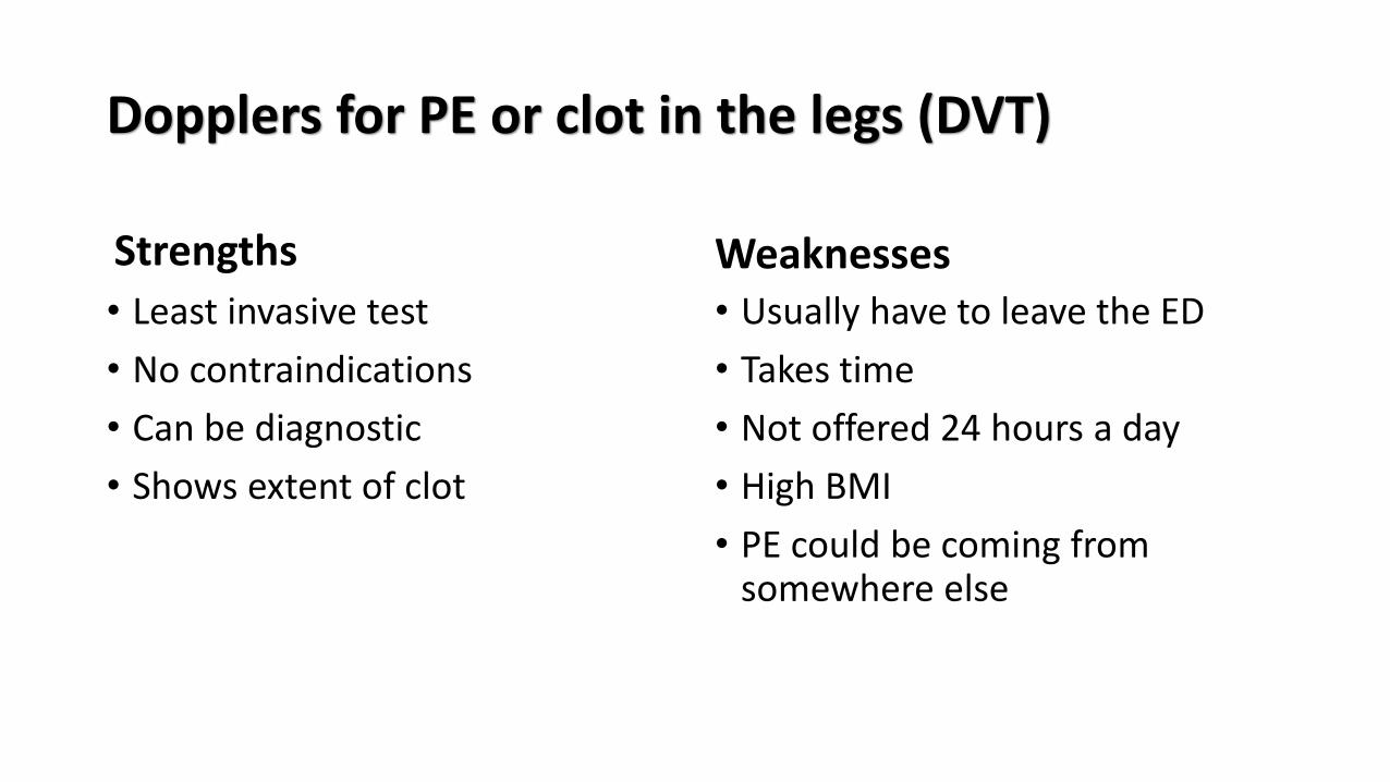
Dopplers for PE or clot in the legs (DVT)
Strengths
• Least invasive test
• No contraindications
• Can be diagnostic
• Shows extent of clot
Weaknesses • Usually have to leave the ED
• Takes time
• Not offered 24 hours a day
• High BMI
• PE could be coming from somewhere else
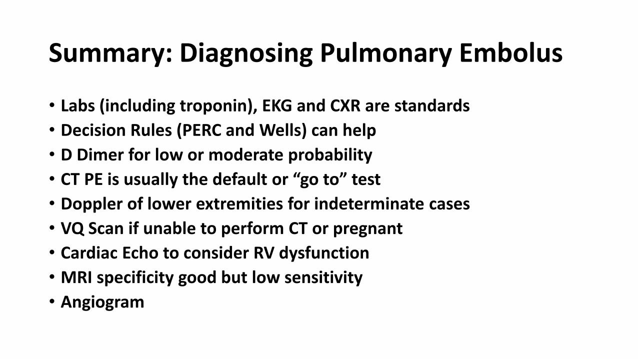
Summary: Diagnosing Pulmonary Embolus
• Labs (including troponin), EKG and CXR are standards
• Decision Rules (PERC and Wells) can help
• D Dimer for low or moderate probability
• CT PE is usually the default or “go to” test
• Doppler of lower extremities for indeterminate cases
• VQ Scan if unable to perform CT or pregnant
• Cardiac Echo to consider RV dysfunction
• MRI specificity good but low sensitivity
• Angiogram
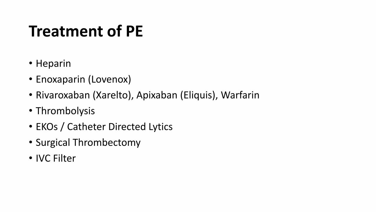
Treatment of PE
• Heparin
• Enoxaparin (Lovenox)
• Rivaroxaban (Xarelto), Apixaban (Eliquis), Warfarin
• Thrombolysis
• EKOs / Catheter Directed Lytics
• Surgical Thrombectomy
• IVC Filter

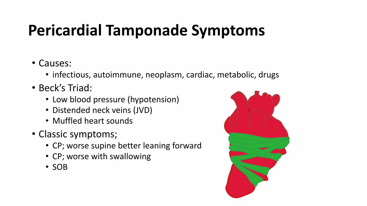
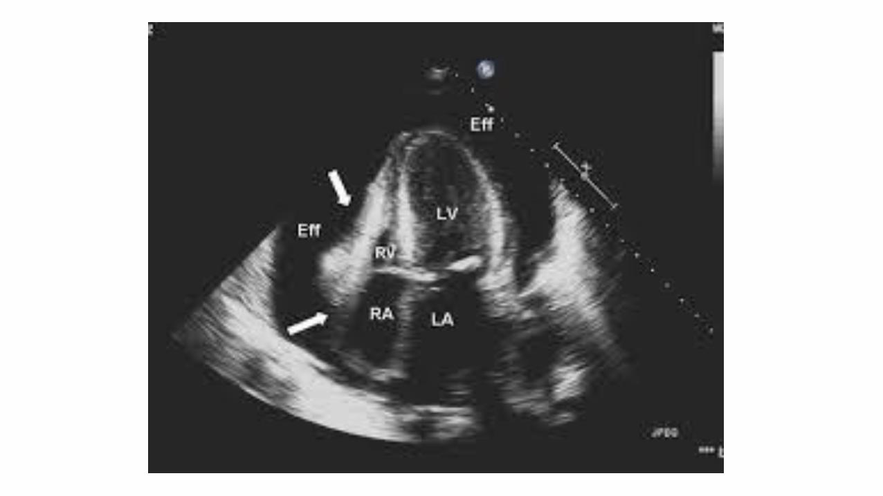
Pericardial Tamponade Symptoms
• Causes: • infectious, autoimmune, neoplasm, cardiac, metabolic, drugs
• Beck’s Triad: • Low blood pressure (hypotension)• Distended neck veins (JVD)• Muffled heart sounds
• Classic symptoms;• CP; worse supine better leaning forward • CP; worse with swallowing • SOB

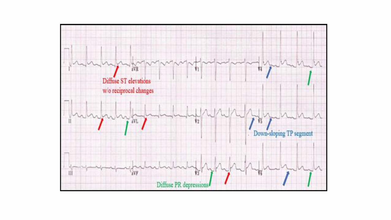
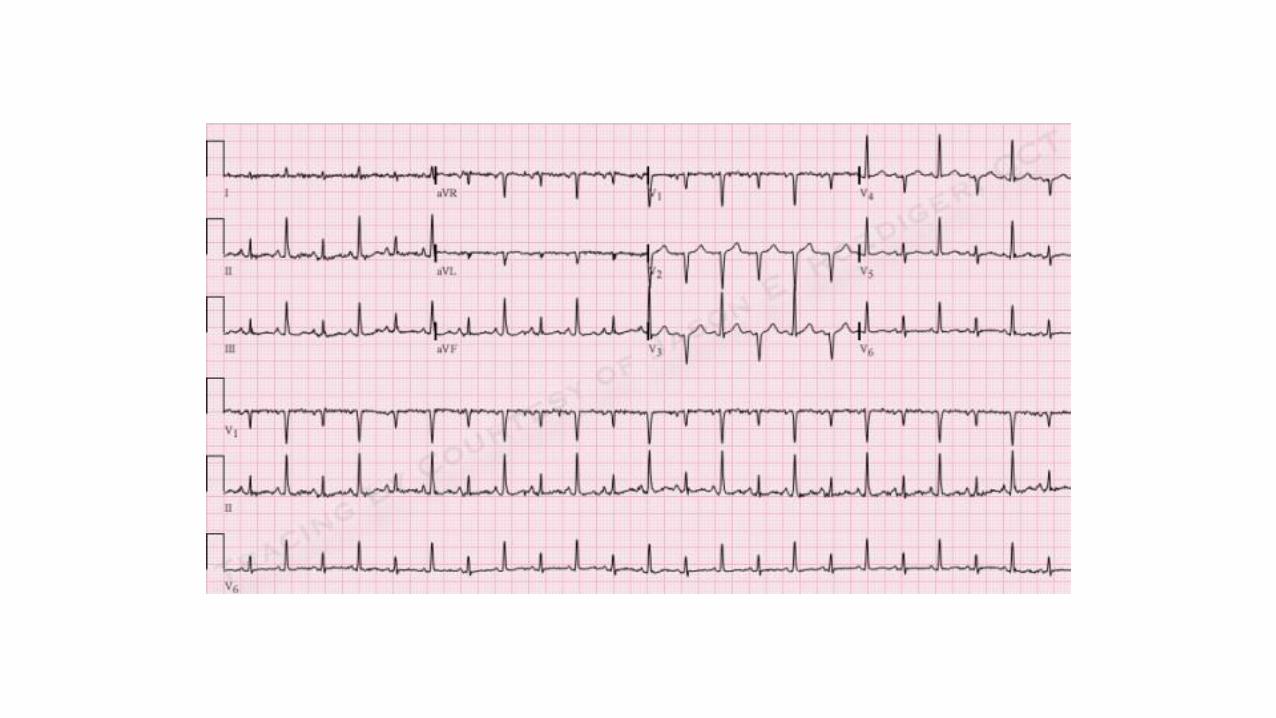
Pericardial Tamponade
• EKG: • Tachycardia
• Low voltage
• Can look like a STEMI
• T wave changes
• Electrical alternans
• CXR: BIG HEART (cardiomegaly)
• Bedside cardiac echo
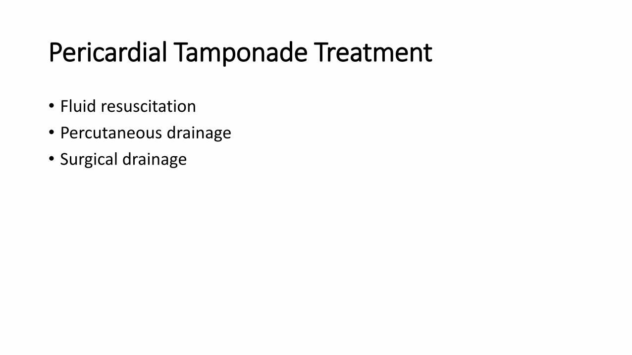
Pericardial Tamponade Treatment
• Fluid resuscitation
• Percutaneous drainage
• Surgical drainage
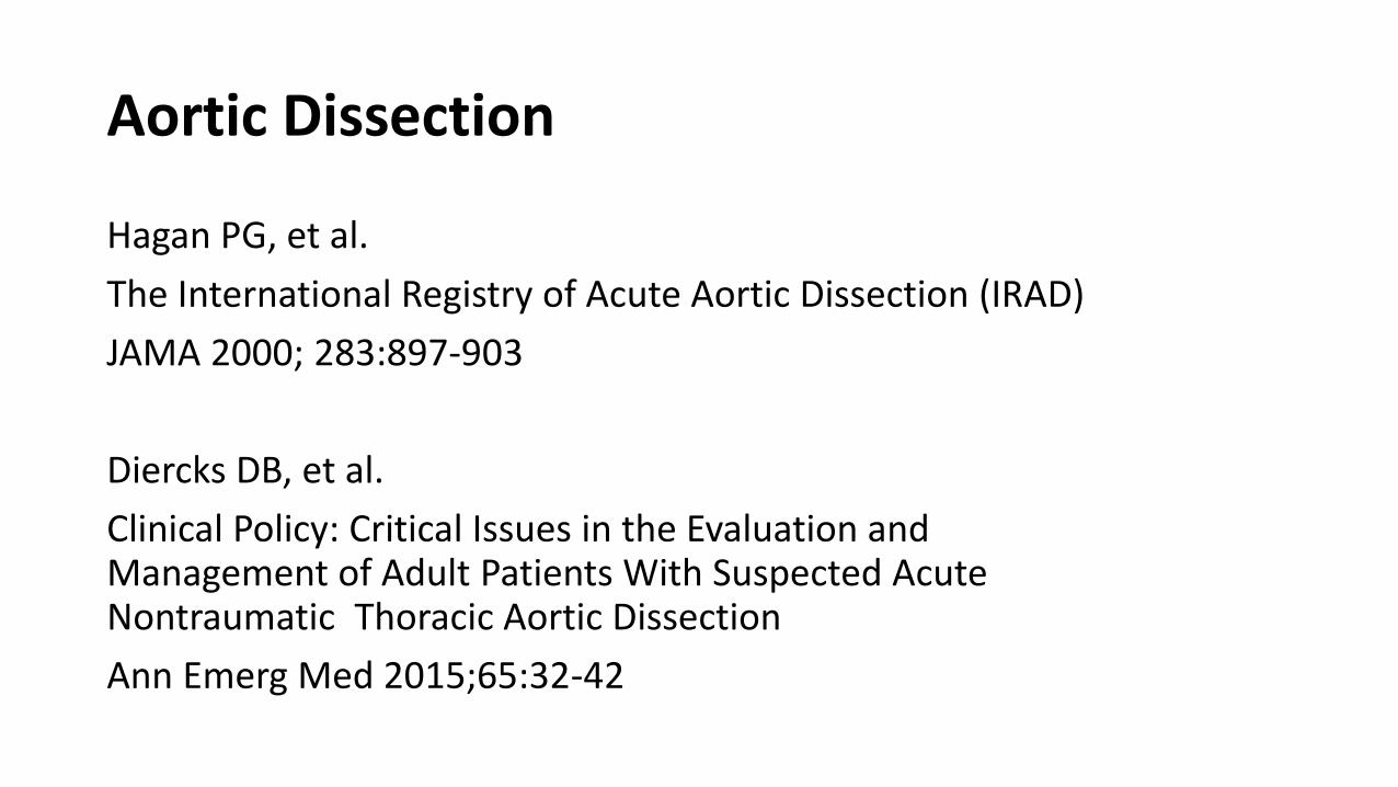
Aortic Dissection
Hagan PG, et al.
The International Registry of Acute Aortic Dissection (IRAD)
JAMA 2000; 283:897-903
Diercks DB, et al.
Clinical Policy: Critical Issues in the Evaluation and Management of Adult Patients With Suspected Acute Nontraumatic Thoracic Aortic Dissection
Ann Emerg Med 2015;65:32-42

Dissection of the Aorta
• Marfan’s is the usual culprit in the young!
• So Marfan’s Characteristics…• Pectus excavatum• Arachnodactyly• Arm span > height• High-arched palat• Kyphosis • MVP• Myopia• Lens Dislocation

Dissection of the Aorta
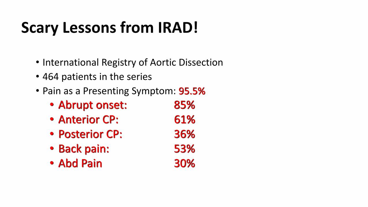
Scary Lessons from IRAD!
• International Registry of Aortic Dissection
• 464 patients in the series
• Pain as a Presenting Symptom: 95.5%
• Abrupt onset: 85%• Anterior CP: 61%• Posterior CP: 36%• Back pain: 53%• Abd Pain 30%

Scary Lessons from IRAD!
• Quality of Pain• Sharp: 64%• Tear/rip: 51%• Radiating: 28%• Migrating: 17%
• Other Signs• Elevated BP: 50%• Normal BP: 35%• New Murmur: 31%• Pulse Deficit: 15%
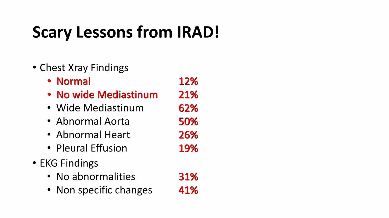
Scary Lessons from IRAD!
• Chest Xray Findings• Normal 12%• No wide Mediastinum 21%• Wide Mediastinum 62%• Abnormal Aorta 50%• Abnormal Heart 26%• Pleural Effusion 19%
• EKG Findings• No abnormalities 31%• Non specific changes 41%

Scary Lessons
• Use Gestalt to help
• Beware of false “normal findings”
• IRAD in-hospital mortality was 27.4%
• Type A mortality not fixed 58% and fixed 26%

Diagnosing Aortic Dissection
• D Dimer
• CTA (gold standard)
• Echo (TEE)
• MRI
• Aortography

Diagnosing Aortic Dissection
Test Sensitivity Specificity LR+ LR-
• D Dimer 95% 60% 2.4 .08
• CTA 93%-100% 98%-100%
• TEE 88%-98% 95%
• MRI 98%-100% 98%
• Aortography Gold Standard but rarely used
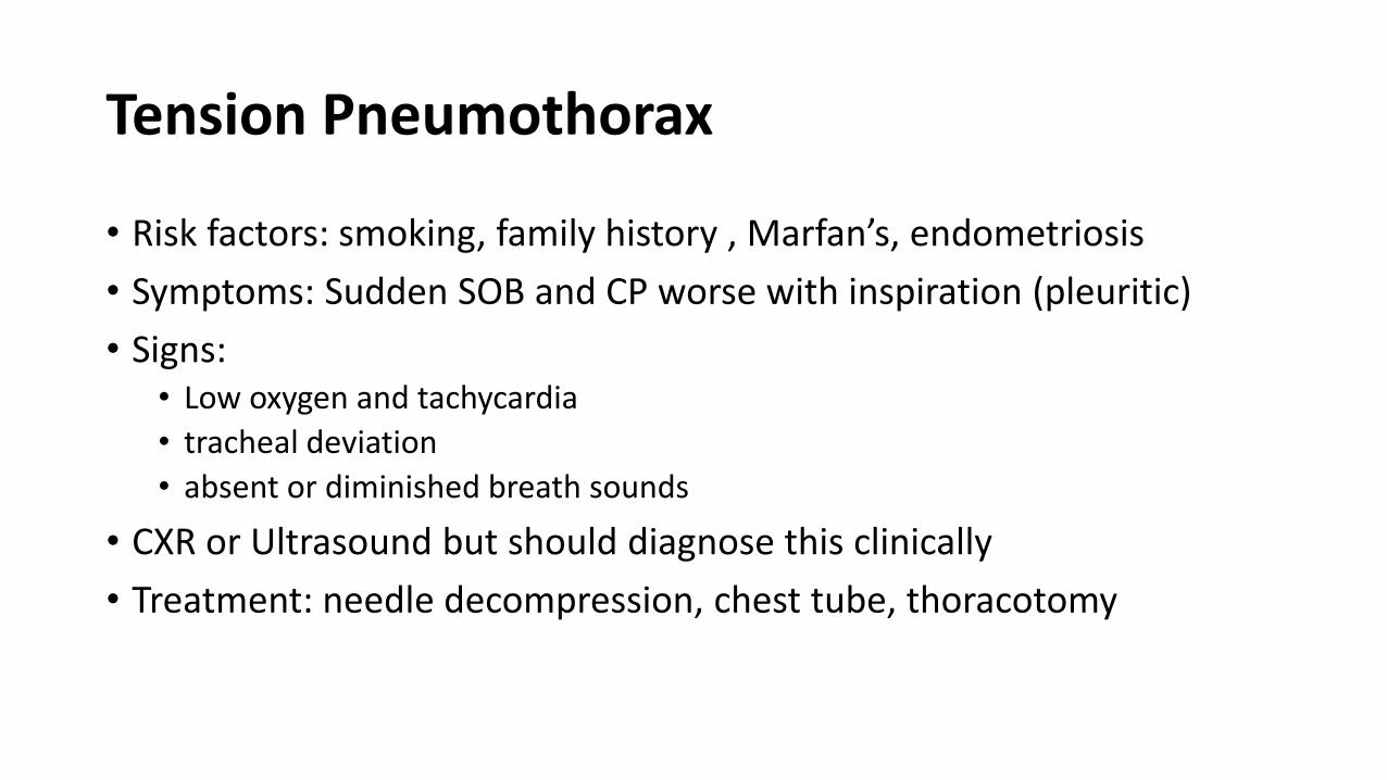
Tension Pneumothorax
• Risk factors: smoking, family history , Marfan’s, endometriosis
• Symptoms: Sudden SOB and CP worse with inspiration (pleuritic)
• Signs: • Low oxygen and tachycardia
• tracheal deviation
• absent or diminished breath sounds
• CXR or Ultrasound but should diagnose this clinically
• Treatment: needle decompression, chest tube, thoracotomy
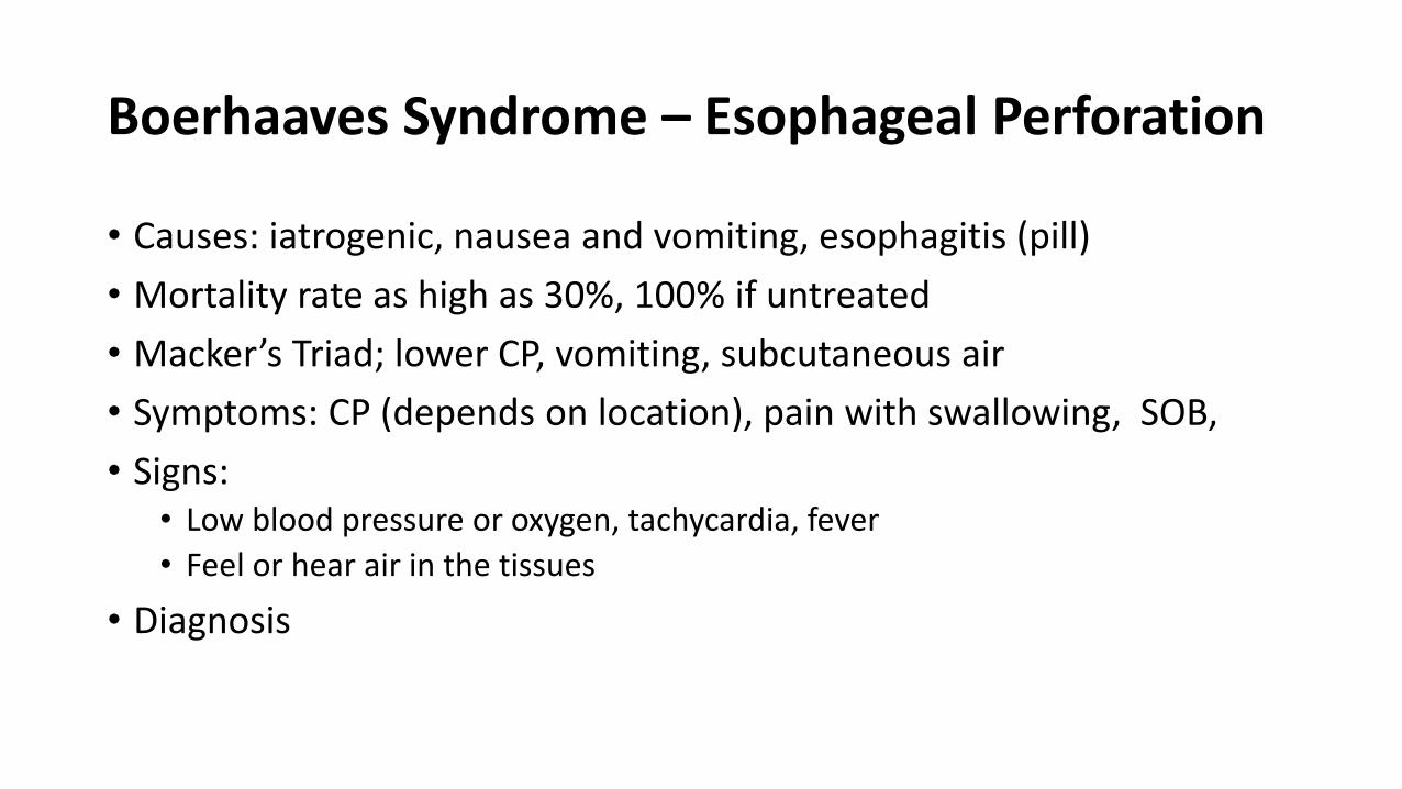
Boerhaaves Syndrome – Esophageal Perforation
• Causes: iatrogenic, nausea and vomiting, esophagitis (pill)
• Mortality rate as high as 30%, 100% if untreated
• Macker’s Triad; lower CP, vomiting, subcutaneous air
• Symptoms: CP (depends on location), pain with swallowing, SOB,
• Signs: • Low blood pressure or oxygen, tachycardia, fever
• Feel or hear air in the tissues
• Diagnosis

Diagnosis of Esophageal Perforation
• CXR • Subcutaneous air (emphysema)• Air in the mediastinum (pneumomediastinum) • Free air under the diaphragm
• Definitive Diagnosis • Gastrografin swallow (like an Upper GI) • CT with contrast • EGD
• Treatment • NPO and resuscitation • Antibiotics and meds to decrease acid • Drainage and debridement

67 yo Male William Morgan with CP Summary
• Patient with a negative work up (stress, UGI, CXR and labs)
• Then sudden and severe chest pain
• Dr. Burnside heard something on cardiac exam
• Required an emergency lifesaving surgery
• Life was saved

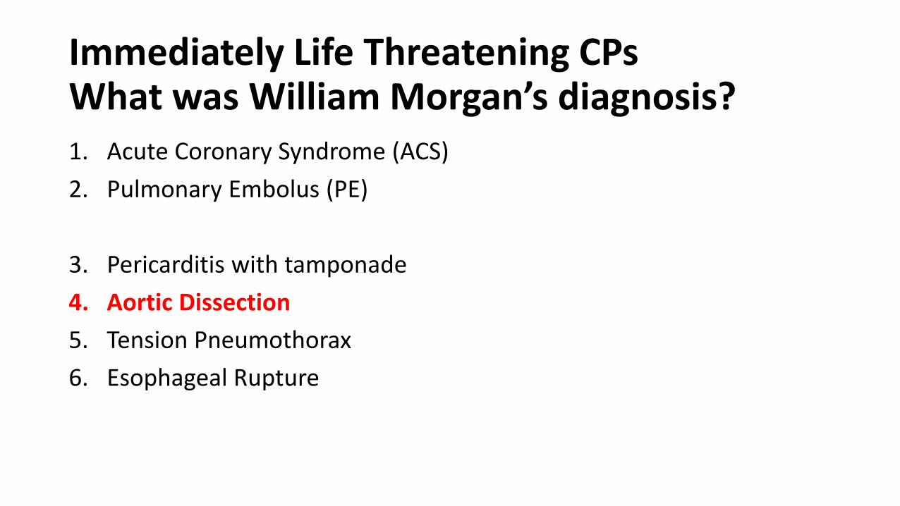
Immediately Life Threatening CPsWhat was William Morgan’s diagnosis?1. Acute Coronary Syndrome (ACS)
2. Pulmonary Embolus (PE)
3. Pericarditis with tamponade
4. Aortic Dissection
5. Tension Pneumothorax
6. Esophageal Rupture
Immediately Life Threatening CPsWhat was William Morgan’s diagnosis?1. Acute Coronary Syndrome (ACS)
2. Pulmonary Embolus (PE)
3. Pericarditis with tamponade
4. Aortic Dissection
5. Tension Pneumothorax
6. Esophageal Rupture
Takeaways from Today’s Discussion
• We all used dual process forms of thinking/decision making.
• Groopman’s 3 questions from his epilogue: What else? Anything that does not fit? More that one diagnosis?
• We learned the 6 immediately life-threatening causes of chest pain how to suspect them and how to diagnosis them.
• We can read EKGs and diagnose a STEMI.
• Scoring systems help but diagnostic testing has limitations
• Finally, we solved a case of chest pain.