Work-Related Neurogenic Thoracic Outlet Syndrome ... I. GUIDELINE SUMMARY Review Criteria for the...
14
Effective Date October 1, 2010; hyperlink and formatting update September 2016 Page 1 Work-Related Neurogenic Thoracic Outlet Syndrome: Diagnosis and Treatment* TABLE OF CONTENTS I. GUIDELINE SUMMARY II. INTRODUCTION III. ESTABLISHING WORK-RELATEDNESS IV. MAKING THE DIAGNOSIS A. Symptoms and Signs B. Electrodiagnostic Testing C. Other Diagnostic Tests V. TREATMENT A. Conservative Treatment B. Surgical Treatment VI. RETURN TO WORK (RTW) A. Early Assessment B. Returning to Work following Surgery VII. ELECTRODIAGNOSTIC WORKSHEET References *This guideline does not apply to severe or acute traumatic injury of the upper extremities, nor to vascular categories of TOS.
Transcript of Work-Related Neurogenic Thoracic Outlet Syndrome ... I. GUIDELINE SUMMARY Review Criteria for the...
Effective Date October 1, 2010; hyperlink and formatting update September 2016 Page 1
Work-Related Neurogenic Thoracic Outlet Syndrome:
Diagnosis and Treatment*
TABLE OF CONTENTS
I. GUIDELINE SUMMARY
II. INTRODUCTION
III. ESTABLISHING WORK-RELATEDNESS
IV. MAKING THE DIAGNOSIS
A. Symptoms and Signs
B. Electrodiagnostic Testing
C. Other Diagnostic Tests
V. TREATMENT
A. Conservative Treatment
B. Surgical Treatment
VI. RETURN TO WORK (RTW)
A. Early Assessment
B. Returning to Work following Surgery
VII. ELECTRODIAGNOSTIC WORKSHEET
References
*This guideline does not apply to severe or acute traumatic injury of the upper extremities, nor to
vascular categories of TOS.

2
I. GUIDELINE SUMMARY
Review Criteria for the Diagnosis and Treatment of
Work-Related Neurogenic Thoracic Outlet Syndrome (nTOS) CLINICAL FINDINGS
CONSERVATIVE
TREATMENT
SURGICAL
TREATMENT SUBJECTIVE
(Symptoms)
OBJECTIVE
(Signs)
DIAGNOSTIC
AND AND
Pain, paresthesias, or
weakness affecting the
upper extremity (most
commonly affecting the
ring or small finger)
Tenderness
Scalene
Trapezius
Anterior chest wall
Brachial plexus
Weakness
Loss of finger dexterity
Atrophy
Electrodiagnostic studies (EDS) are required to objectively
confirm the diagnosis of nTOS.
EDS criteria are as follows:
1. Absent or reduced amplitude (< 12 uV) of the ulnar SNAP
OR
Absent or reduced amplitude (< 10 uV) of the medial
antebrachial cutaneous nerve (MABC) SNAP with normal
amplitude of the MABC SNAP in the contralateral (unaffected)
extremity
AND
2. Absent or reduced amplitude (< 5 mV) of the median CMAP
OR
Absent or prolonged minimum latency (>33 msec) of the ulnar F-
wave (with or without abnormalities of the median F-wave), and
with normal F-waves in the contralateral (unaffected) upper
extremity
OR
Needle electromyography (EMG) showing denervation (e.g.
fibrillation potentials, positive sharp waves) in at least one
muscle supplied by each of two different nerves from the
lower trunk of the brachial plexus, with normal EMG of the
cervical paraspinal muscles and at least one muscle supplied
by a nerve from the middle or upper trunk of the brachial
plexus AND
3. Normal amplitude (≥ 15uV) of the median nerve SNAP
AND
4. Normal conduction velocity (≥ 50m/s) of the ulnar motor
nerve across the elbow
Modify job activities
that exacerbate
symptoms
AND/OR
Physical therapy with
strengthening and
stretching, postural
exercises
AND/OR
Anti-inflammatory drug
therapy
Surgical treatment should only
be considered if:
1. The patient has met the
diagnostic criteria under
Section III
AND
2. The condition interferes with
work or activities of daily
living
AND
3. The condition does not
improve despite conservative
treatment
Without confirmation of
brachial plexus compression by
both objective clinical
findings and abnormal EDS,
surgery will not be authorized.
Effective Date October 1, 2010; hyperlink and formatting update September 2016 Page 3
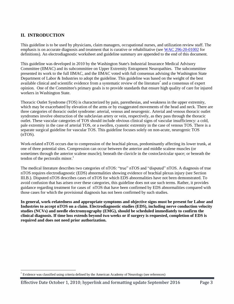
II. INTRODUCTION This guideline is to be used by physicians, claim managers, occupational nurses, and utilization review staff. The emphasis is on accurate diagnosis and treatment that is curative or rehabilitative (see WAC 296-20-01002 for definitions). An electrodiagnostic worksheet and guideline summary are appended to the end of this document.
This guideline was developed in 2010 by the Washington State's Industrial Insurance Medical Advisory
Committee (IIMAC) and its subcommittee on Upper Extremity Entrapment Neuropathies. The subcommittee
presented its work to the full IIMAC, and the IIMAC voted with full consensus advising the Washington State
Department of Labor & Industries to adopt the guideline. This guideline was based on the weight of the best
available clinical and scientific evidence from a systematic review of the literature* and a consensus of expert
opinion. One of the Committee's primary goals is to provide standards that ensure high quality of care for injured
workers in Washington State. Thoracic Outlet Syndrome (TOS) is characterized by pain, paresthesias, and weakness in the upper extremity, which may be exacerbated by elevation of the arms or by exaggerated movements of the head and neck. There are three categories of thoracic outlet syndrome: arterial, venous and neurogenic. Arterial and venous thoracic outlet syndromes involve obstruction of the subclavian artery or vein, respectively, as they pass through the thoracic outlet. These vascular categories of TOS should include obvious clinical signs of vascular insufficiency: a cold, pale extremity in the case of arterial TOS, or a swollen, cyanotic extremity in the case of venous TOS. There is a separate surgical guideline for vascular TOS. This guideline focuses solely on non-acute, neurogenic TOS (nTOS). Work-related nTOS occurs due to compression of the brachial plexus, predominantly affecting its lower trunk, at
one of three potential sites. Compression can occur between the anterior and middle scalene muscles (or
sometimes through the anterior scalene muscle); beneath the clavicle in the costoclavicular space; or beneath the
tendon of the pectoralis minor.1
The medical literature describes two categories of nTOS: “true” nTOS and “disputed” nTOS. A diagnosis of true
nTOS requires electrodiagnostic (EDS) abnormalities showing evidence of brachial plexus injury (see Section
III.B.). Disputed nTOS describes cases of nTOS for which EDS abnormalities have not been demonstrated. To
avoid confusion that has arisen over these categories, this guideline does not use such terms. Rather, it provides
guidance regarding treatment for cases of nTOS that have been confirmed by EDS abnormalities compared with
those cases for which the provisional diagnosis has not been confirmed by such studies. In general, work-relatedness and appropriate symptoms and objective signs must be present for Labor and Industries to accept nTOS on a claim. Electrodiagnostic studies (EDS), including nerve conduction velocity studies (NCVs) and needle electromyography (EMG), should be scheduled immediately to confirm the clinical diagnosis. If time loss extends beyond two weeks or if surgery is requested, completion of EDS is required and does not need prior authorization.
* Evidence was classified using criteria defined by the American Academy of Neurology (see references)

Effective Date October 1, 2010; hyperlink and formatting update September 2016 Page 4
III. ESTABLISHING WORK-RELATEDNESS
Work-related activities may cause or contribute to the development of nTOS.2,3
Because simply identifying an
association with workplace activities is not, in itself, adequate evidence of a causal relationship, establishing
work-relatedness requires all of the following:
1. Exposure: Workplace activities that contribute to or cause nTOS, and2. Outcome: A diagnosis of nTOS that meets the diagnostic criteria under Section III, and3. Relationship: Generally accepted scientific evidence, which establishes on a more probable than not basis
(greater than 50%) that the workplace activities (exposure) in an individual case contributed to thedevelopment or worsening of the condition (outcome).
When the Department receives notification of an occupational disease, the Occupational Disease & Employment
History form is mailed to the worker, employer or attending provider. The form should be completed and
returned to the insurer as soon as possible. If the worker’s attending provider completes the form, provides a
detailed history in the chart note, and gives an opinion on causality, he or she may be paid for this (use billing
code 1055M). Additional billing information is available in the AP Resource Center.
Symptoms of nTOS may be exacerbated by certain work-related activities, usually involving elevation or
sustained use of the arms. Such activities may include but are not limited to the following4:
Lifting overhead Holding tools or objects above shoulder level Reaching overhead Carrying heavy weights
Several occupations have been associated with nTOS. This is not an exhaustive list and is meant only as a guide in the consideration of work-relatedness:
Dry wall hanger or plasterer Assembly line inspector Welder Shelf stocker Beautician Dental hygienist
IV. MAKING THE DIAGNOSIS
A. SYMPTOMS AND SIGNS
A case definition of confirmed nTOS includes appropriate symptoms, objective physical findings ("signs"),
and abnormal EDS. A provisional diagnosis of nTOS may be made based upon appropriate symptoms and
objective signs, but confirmation of the diagnosis requires abnormal EDS.
Classic symptoms of nTOS include pain, paresthesias, or weakness in the upper extremity. Paresthesias most commonly affect the ring and small fingers.
5 Symptom severity tends to increase after certain activities and
worsens at the end of the day or during sleep.
Signs on examination may include tenderness to palpation over the brachial plexus, the scalene muscles, the trapezius muscles, or the anterior chest wall. Although tenderness may be a useful objective finding, it cannot support the diagnosis of nTOS alone. Advanced cases of nTOS are characterized by objective signs of weakness of the hand, loss of dexterity of the fingers, and atrophy of the affected muscles.
Provocative tests have been described that may help corroborate the diagnosis of nTOS. These tests are based on creating maximal tension on the anatomical sites of constriction. Studies have found a high false-positive rate for these tests in healthy subjects as well as carpal tunnel syndrome patients.
6 Although they are described for

Effective Date October 1, 2010; hyperlink and formatting update September 2016 Page 5
completeness, the sensitivity and specificity of these tests for nTOS have not been established, and these tests cannot replace confirmatory EDS testing (see Section III.B). Provocative tests include:
The elevated arm stress test (EAST or Roos test)- the patient places the affected arm in full abduction and external rotation and then opens and closes the hands slowly for 3 minutes. This test constricts the costoclavicular space. It is considered abnormal if typical symptoms are elicited and the patient cannot sustain this activity for the full 3 minutes.
The Adson test- the patient extends the neck and rotates the head toward the involved extremity, which is held extended at the side. This test constricts the interscalene triangle. It is considered abnormal if a change in the radial pulse is detected when the patient inhales deeply and holds their breath
The Wright test- the patient sits or stands with the arm in full abduction and external rotation. This test constricts the costoclavicular space. It is considered abnormal if typical symptoms are elicited and a change in pulse is detected.
The costoclavicular test- the examiner depresses the patient’s shoulder. This test constricts the costoclavicular space and creates tension across the pectoralis minor. It is considered abnormal if typical symptoms are elicited.
Every effort should be made to objectively confirm the diagnosis of nTOS before considering surgery. A
differential diagnosis for nTOS includes musculoskeletal disease (e.g. arthritis, tendinitis) of the cervical spine,
shoulder girdle or arm, cervical radiculopathy or upper extremity nerve entrapment7, idiopathic inflammation of
the brachial plexus (aka Parsonage-Turner syndrome), and brachial plexus compression due to an infiltrative
process or space-occupying mass (e.g. Pancoast tumor of the lung apex).
B. ELECTRODIAGNOSTIC STUDIES (EDS)
EDS abnormalities are required to objectively confirm the diagnosis of nTOS. Given the uncertainties in
diagnostic assessment of nTOS, EDS should be obtained as soon as the diagnosis is considered. EDS may help
gauge the severity of injury.8-10
Importantly, EDS can help exclude conditions that may mimic nTOS, such as
ulnar nerve entrapment or cervical radiculopathy.11
EDS evidence that confirms a diagnosis of nTOS requires:
1. Absent or reduced amplitude (< 12 uV) of the ulnar antidromic sensory nerve action potential (SNAP)
Or
Absent or reduced amplitude (< 10 uV) of the medial antebrachial cutaneous nerve (MABC) antidromic SNAP,
with normal amplitude of the MABC SNAP in the contralateral (unaffected) extremity
AND
2. Absent or reduced amplitude (<5 mV) of the median nerve compound motor action potential (CMAP)
Or
Absent or prolonged minimum latency (>33 msec) of the ulnar F-wave (with or without abnormalities of the
median F-wave), and with normal F-waves in the contralateral (unaffected) upper extremity
Or
Needle electromyography (EMG) showing denervation (e.g. fibrillation potentials, positive sharp waves) in at
least one muscle supplied by each of two different nerves from the lower trunk of the brachial plexus, with normal
EMG of the cervical paraspinal muscles and at least one muscle supplied by a nerve from the middle or upper
trunk of the brachial plexus.
AND
To exclude the presence of other focal neuropathies or polyneuropathy as a cause for the abnormalities described
above, the following must also be shown:
3. Normal amplitude (≥ 15 uV) of the median nerve antidromic SNAP.
AND
4. Normal conduction velocity (≥ 50 m/s) of the ulnar motor nerve across the elbow.

Effective Date October 1, 2010; hyperlink and formatting update September 2016 Page 6
C. OTHER DIAGNOSTIC TESTS Arterial or venous vascular studies may be helpful in the diagnosis of suspected arterial or venous TOS. However,
these tests have poor specificity for nTOS, and there is no substantial evidence that vascular studies can reliably
confirm the diagnosis of nTOS. Therefore, vascular studies conducted as a diagnostic tool for nTOS will not be
authorized. Some have suggested that magnetic resonance imaging (MRI) neurography may be helpful in the diagnosis of nTOS. However, these services will not be authorized for this condition because the clinical utility of these tests has not yet been proven. While the Committee recognizes that these tests may be useful in unusual circumstances where EDS results are normal but there are appropriate clinical symptoms, the Committee believes that at this time the use of these tests is investigational and should be used only in a research setting. Anterior scalene muscle (ASM) blocks have been used in the evaluation of suspected nTOS.
12,13 However, this
test has poor specificity for nTOS, and there is no substantial evidence that ASM can reliably confirm the diagnosis of nTOS. Therefore, ASM blocks conducted as a diagnostic tool for nTOS will not be authorized. X-rays of the chest may be useful to evaluate the possibility of an infiltrative process or space-occupying mass (e.g. Pancoast tumor of the lung apex) compressing the brachial plexus.
V. TREATMENT
Non-surgical therapy may be considered for cases in which a provisional diagnosis of nTOS has been made.
Surgical treatment should be provided only for cases in which the diagnosis of nTOS has been confirmed by
abnormal EDS. Under these circumstances, the potential benefits of brachial plexus decompression may outweigh
the risks of surgery.
A. CONSERVATIVE TREATMENT
Conservative treatment for nTOS has been described in narrative reviews, case reports, and retrospective case
series.14-16
No randomized controlled trials have been conducted to measure the efficacy of conservative
treatments for nTOS. No specific method of conservative treatment has been proven to be most effective due to a
lack of comparative studies.14
However, an observational study (n=50), showed that strengthening and stretching
exercises reduced pain among 80% of patients after 3 months and among 94% of patients after 6 months15
, and a
2007 systematic review of the available literature concluded that conservative treatment appears to be effective in
reducing symptoms, improving function, and facilitating return to work.14
Examples of conservative treatment
include modification of activities that exacerbate symptoms, education, postural exercises, physical therapy, and
anti-inflammatory drug therapy.
Because surgical outcomes are poor in many situations, conservative interventions, such as stretching and
strengthening exercises, should be considered first. If the initial response to conservative treatment is incomplete,
modifying or changing the approach should be considered. If there is no response to conservative treatment within
six weeks, or if time loss extends longer than 2 weeks, specialist consultation should be obtained.
Although Botulinum toxin (Botox) injections of the scalene muscles have been reported to relieve nTOS
symptoms17
, preliminary results of a randomized trial showed no clear clinical improvement related to this
treatment.18
In addition, it appears that there are substantial technical challenges and potentially severe adverse
effects from this procedure.Therefore, Botox injections conducted as a diagnostic tool or for treatment of nTOS
will not be authorized.
When feasible, job modifications that reduce the intensity of manual tasks may prevent progression and promote
recovery from nTOS.16
If symptoms persist despite appropriate treatment, permanent job modifications may still

Effective Date October 1, 2010; hyperlink and formatting update September 2016 Page 7
allow the patient to remain at work. Patients do not usually need time off from work activities prior to surgery,
unless they present with objective weakness or sensory loss in the upper extremity that limits work activities or
poses a substantial safety risk. B. SURGICAL TREATMENT
Surgical treatment for nTOS has been described in narrative reviews, case reports, and retrospective case
series.4,19-34
Surgery should include exploration of the brachial plexus throughout its course in the thoracic outlet
in order to decompress it by resecting any compressive and/or constrictive structures. These may include any of
the three sites of compression mentioned earlier. No specific method of surgical treatment has been proven to be
most effective.
Surgical treatment should only be considered if:
1. The patient has met the diagnostic criteria under Section III, and
2. The condition interferes with work or activities of daily living, and
3. The condition does not improve despite conservative treatment.
Without confirmation of nTOS by both objective clinical findings and abnormal EDS, surgery will not be
authorized. VI. RETURN TO WORK (RTW)
A. EARLY ASSESSMENT
Timeliness of the diagnosis can be a critical factor influencing RTW. Among workers with upper extremity
disorders, 7% of workers account for 75% of the long-term disability.35
A large prospective study in the
Washington State workers’ compensation system identified several important predictors of long-term disability:
low expectations of return to work (RTW), no offer of a job accommodation, and high physical demands on the
job.36
Identifying and attending to these risk factors when patients have not returned to work within 2-3 weeks of
the initial clinical presentation may improve their chances of RTW.
Washington State workers diagnosed accurately and early were far more likely to RTW than workers whose
conditions were diagnosed weeks or months later. Early coordination of care with improved timeliness and
effective communication with the workplace is also likely to help prevent long-term disability.
A recent quality improvement project in Washington State has demonstrated that delivering medical care
according to occupational health best practices similar to those listed in Table 1 can substantially prevent long-
term disability. Findings can be viewed at:
Centers of Occupational Health & Education.

Effective Date October 1, 2010; hyperlink and formatting update September 2016 Page 8
Table 2. Occupational Health Quality Indicators for Neurogenic Thoracic Outlet Syndrome (nTOS)
Clinical care action Time-frame*
1. Identify physical stressors from both work and non-
work activities;
2. Screen for presence of nTOS
3. Determine work-relatedness
4. Recommend ergonomic improvements or other
appropriate job modifications
1st health care visit
Communicate with employer regarding return to work
(RTW) using
1. Activity Prescription Form (or comparable RTW
form)
and/or
2. Phone call to employer
Each visit while work restrictions exist
1. Assess impediments for RTW
2. Request specialist consultation
If > 2 weeks of time-loss occurs or if there is no clinical
improvement within 6 weeks of conservative treatment
Specialist consultation Performed ASAP, within 3 weeks of request
Electrodiagnostic studies If the diagnosis of nTOS is being considered, schedule
studies immediately.
These tests are required if time-loss extends beyond 2
weeks, or if surgery is requested.
Surgical decompression Performed ASAP, within 4-6 weeks of determining need
for surgery
*“Time-frame” is anchored in time from 1st provider visit related to nTOS symptoms.
B. RETURNING TO WORK FOLLOWING SURGERY
How soon a patient can return to work depends on the type of surgery performed and when rehabilitation
begins. Most patients can return to light duty work within 4-6 weeks and regular duty within 10-12 weeks
of surgery.

Effective Date October 1, 2010; hyperlink and formatting update September 2016 Page 9
VII. ELECTRODIAGNOSTIC WORKSHEET
Claim Number:
Claimant Name:
PURPOSE AND INSTRUCTIONS
The purpose of this worksheet is to help interpret electrodiagnostic studies (EDS) done for an injured
worker. The worksheet should be used only when the main purpose of the study is to evaluate neurogenic
thoracic outlet syndrome (nTOS). It should accompany but not replace the detailed report normally
submitted to the insurer.
Electrodiagnostic Worksheet for Work-Related Neurogenic Thoracic Outlet Syndrome (nTOS)
Electrodiagnostic criteria for Work-Related nTOS are met if all four boxes are “Yes”.
Check the
correct box
Yes No
1. Ulnar SNAP* < 12 uV or absent?
OR
Medial antebrachial cutaneous nerve (MABC) SNAP* amplitude < 10 uV or absent,
with normal amplitude of the MABC SNAP* in the contralateral (unaffected) extremity?
AND 2. Median nerve CMAP amplitude < 5 mV or absent?
OR
Ulnar F-wave (with or without abnormalities of the median F-wave) minimum latency >
33 msec or absent, with normal F-waves in the contralateral (unaffected) upper extremity?
OR
Needle electromyography (EMG) showing denervation (e.g. fibrillation potentials, positive
sharp waves) in at least one muscle supplied by each of two different nerves from the lower
trunk of the brachial plexus, with normal EMG of the cervical paraspinal muscles and at
least one muscle supplied by a nerve from the middle or upper trunk of the brachial plexus?
AND 3. Normal amplitude (> 15uV) of the median nerve SNAP*?
AND 4. Normal conduction velocity (> 50 m/s) of the ulnar motor nerve across the elbow?
*Antidromic
Additional Comments:
Signed Date
Effective Date October 1, 2010; hyperlink and formatting update September 2016 Page 10
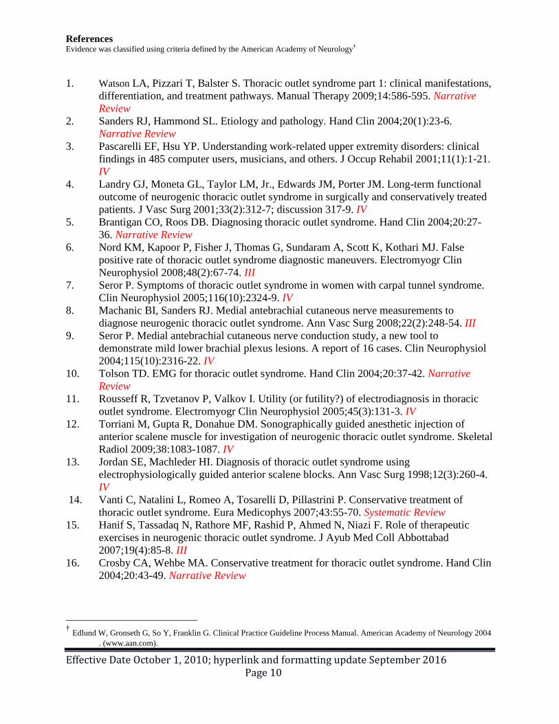
References Evidence was classified using criteria defined by the American Academy of Neurology†
1. Watson LA, Pizzari T, Balster S. Thoracic outlet syndrome part 1: clinical manifestations,
differentiation, and treatment pathways. Manual Therapy 2009;14:586-595. Narrative
Review
2. Sanders RJ, Hammond SL. Etiology and pathology. Hand Clin 2004;20(1):23-6.
Narrative Review
3. Pascarelli EF, Hsu YP. Understanding work-related upper extremity disorders: clinical
findings in 485 computer users, musicians, and others. J Occup Rehabil 2001;11(1):1-21.
IV
4. Landry GJ, Moneta GL, Taylor LM, Jr., Edwards JM, Porter JM. Long-term functional
outcome of neurogenic thoracic outlet syndrome in surgically and conservatively treated
patients. J Vasc Surg 2001;33(2):312-7; discussion 317-9. IV
5. Brantigan CO, Roos DB. Diagnosing thoracic outlet syndrome. Hand Clin 2004;20:27-
36. Narrative Review
6. Nord KM, Kapoor P, Fisher J, Thomas G, Sundaram A, Scott K, Kothari MJ. False
positive rate of thoracic outlet syndrome diagnostic maneuvers. Electromyogr Clin
Neurophysiol 2008;48(2):67-74. III
7. Seror P. Symptoms of thoracic outlet syndrome in women with carpal tunnel syndrome.
Clin Neurophysiol 2005;116(10):2324-9. IV
8. Machanic BI, Sanders RJ. Medial antebrachial cutaneous nerve measurements to
diagnose neurogenic thoracic outlet syndrome. Ann Vasc Surg 2008;22(2):248-54. III
9. Seror P. Medial antebrachial cutaneous nerve conduction study, a new tool to
demonstrate mild lower brachial plexus lesions. A report of 16 cases. Clin Neurophysiol
2004;115(10):2316-22. IV
10. Tolson TD. EMG for thoracic outlet syndrome. Hand Clin 2004;20:37-42. Narrative
Review
11. Rousseff R, Tzvetanov P, Valkov I. Utility (or futility?) of electrodiagnosis in thoracic
outlet syndrome. Electromyogr Clin Neurophysiol 2005;45(3):131-3. IV
12. Torriani M, Gupta R, Donahue DM. Sonographically guided anesthetic injection of
anterior scalene muscle for investigation of neurogenic thoracic outlet syndrome. Skeletal
Radiol 2009;38:1083-1087. IV
13. Jordan SE, Machleder HI. Diagnosis of thoracic outlet syndrome using
electrophysiologically guided anterior scalene blocks. Ann Vasc Surg 1998;12(3):260-4.
IV
14. Vanti C, Natalini L, Romeo A, Tosarelli D, Pillastrini P. Conservative treatment of
thoracic outlet syndrome. Eura Medicophys 2007;43:55-70. Systematic Review
15. Hanif S, Tassadaq N, Rathore MF, Rashid P, Ahmed N, Niazi F. Role of therapeutic
exercises in neurogenic thoracic outlet syndrome. J Ayub Med Coll Abbottabad
2007;19(4):85-8. III
16. Crosby CA, Wehbe MA. Conservative treatment for thoracic outlet syndrome. Hand Clin
2004;20:43-49. Narrative Review
† Edlund W, Gronseth G, So Y, Franklin G. Clinical Practice Guideline Process Manual. American Academy of Neurology 2004
. (www.aan.com).

Effective Date October 1, 2010; hyperlink and formatting update September 2016 Page 11
17. Jordan SE, Ahn SS, Gelabert HA. Combining ultrasonography and electromyography for
botulinum chemodenervation treatment of thoracic outlet syndrome: comparison with
fluoroscopy and electromyography guidance. Pain Physician 2007;10(4):541-6. IV
18. Travlos A. Treatment of thoracic outlet syndrome with botulinum toxin Injection: a
double-blind, randomized controlled trial. . University of British Columbia, 2007.
Available at: http://clinicaltrials.gov/ct2/show/study/NCT00444886?view=results. Not
yet published.
19. Povlsen B, Belzberg A, Hansson T, Dorsi M. Treatment for thoracic outlet syndrome
(review). Cochrane Database of Systematic Reviews 2010(1):Art. No.: CD007218. DOI:
10.1002/14651858.CD007218.pub2. III
20. Chang DC, Rotellini-Coltvet LA, Mukherjee D, De Leon R, Freischlag JA. Surgical
intervention for thoracic outlet syndrome improves patient's quality of life. J Vasc Surg
2009;49(3):630-5; discussion 635-7. IV
21. Chang DC, Lidor AO, Matsen SL, Freischlag JA. Reported in-hospital complications
following rib resections for neurogenic thoracic outlet syndrome. Ann Vasc Surg
2007;21(5):564-70. Observational Study
22. Abdellaoui A, Atwan M, Reid F, Wilson P. Endoscopic assisted transaxillary first rib
resection. Interact Cardiovasc Thorac Surg 2007;6(5):644-6. IV
23. Colli BO, Carlotti CG, Jr., Assirati JA, Jr., Marques W, Jr. Neurogenic thoracic outlet
syndromes: a comparison of true and nonspecific syndromes after surgical treatment.
Surg Neurol 2006;65(3):262-71; discussion 271-2. IV
24. Krishnan KG, Pinzer T, Schackert G. The transaxillary approach in the treatment of
thoracic outlet syndrome: a neurosurgical appraisal. Zentralbl Neurochir 2005;66(4):180-
9. IV
25. Altobelli GG, Kudo T, Haas BT, Chandra FA, Moy JL, Ahn SS. Thoracic outlet
syndrome: pattern of clinical success after operative decompression. J Vasc Surg
2005;42(1):122-8. IV
26. Sanders RJ, Hammond SL. Supraclavicular first rib resection and total scalenectomy:
technique and results. Hand Clin 2004;20(1):61-70. Narrative Review
27. Samarasam I, Sadhu D, Agarwal S, Nayak S. Surgical management of thoracic outlet
syndrome: a 10-year experience. ANZ J Surg 2004;74(6):450-4. IV
28. Nannapaneni R, Marks SM. Neurogenic thoracic outlet syndrome. Br J Neurosurg
2003;17(2):144-8. IV
29. Maxey TS, Reece TB, Ellman PI, Tribble CG, Harthun N, Kron IL, Kern JA. Safety and
efficacy of the supraclavicular approach to thoracic outlet decompression. Ann Thorac
Surg 2003;76(2):396-9; discussion 399-400. IV
30. Bhattacharya V, Hansrani M, Wyatt MG, Lambert D, Jones NA. Outcome following
surgery for thoracic outlet syndrome. Eur J Vasc Endovasc Surg 2003;26(2):170-5. IV
31. Balci AE, Balci TA, Cakir O, Eren S, Eren MN. Surgical treatment of thoracic outlet
syndrome: effect and results of surgery. Ann Thorac Surg 2003;75(4):1091-6; discussion
1096. IV
32. Sharp WJ, Nowak LR, Zamani T, Kresowik TF, Hoballah JJ, Ballinger BA, Corson JD.
Long-term follow-up and patient satisfaction after surgery for thoracic outlet syndrome.
Ann Vasc Surg 2001;15(1):32-6. IV
33. Athanassiadi K, Kalavrouziotis G, Karydakis K, Bellenis I. Treatment of thoracic outlet
syndrome: long-term results. World J Surg 2001;25(5):553-7. IV

Effective Date October 1, 2010; hyperlink and formatting update September 2016 Page 12
34. Franklin GM, Fulton-Kehoe D, Bradley C, Smith-Weller T. Outcome of surgery for
thoracic outlet syndrome in Washington state workers' compensation. Neurology
2000;54(6):1252-7. III
35. Hashemi L, Webster B, Clance E, Courtney T. Length of disability and cost of work-
related musculoskeletal disorders of the upper extremity. . J Occup Environ Med
1998;40:261-269. Descriptive Study
36. Turner J, Franklin G, Fulton-Kehoe D. Early predictors of chronic work disability
associated with carpal tunnel syndrome: a longitudinal workers’ compensation cohort
study. Am J Ind Med 2007;50:489-500. II

Effective Date October 1, 2010; hyperlink and formatting update September 2016 Page 13
Definitions for Classification of Evidence
Rating of Therapeutic Article
Rating of Diagnostic Article Rating of Prognostic Article Rating of Screening Article
Class I: Prospective, randomized,
controlled
clinical trial with masked outcome
assessment,
in a representative population. The
following are required:
a) primary outcome(s) clearly defined
b) exclusion/inclusion criteria clearly
defined
c) adequate accounting for drop-outs
and cross-overs with numbers
sufficiently low to have minimal
potential for bias
d) relevant baseline characteristics are
presented and substantially equivalent
among treatment groups or there is
appropriate statistical adjustment for
differences.
Class I: Evidence provided by a
prospective study in a broad
spectrum of persons with the
suspected condition, using a
reference (gold) standard for
case definition, where test is
applied in a blinded evaluation,
and enabling the assessment of
appropriate tests of diagnostic
accuracy. All patients
undergoing the diagnostic test
have the presence or absence of
the disease determined.
Class I: Evidence provided by a
prospective study of a broad
spectrum of persons who may be
at risk for developing the outcome
(e.g. target disease, work status).
The study measures the predictive
ability using an independent gold
standard for case definition. The
predictor is measured in an
evaluation that is masked to
clinical presentation and, the
outcome is measured in an
evaluation that is masked to the
presence of the predictor. All
patients have the predictor and
outcome variables measured.
Class I. A statistical,
population based sample of
patients studied at a
uniform point in time (usually
early) during the course of the
condition. All patients undergo
the intervention of interest.
The outcome, if not objective,
is determined in an evaluation
that is masked to the patients’
clinical presentations.
Class II: Prospective matched group
cohort study in a representative
population with masked outcome
assessment that meets a-d above OR a
RCT in a representative population that
lacks one criteria a-d.
Class II: Evidence provided by
a prospective study of a narrow
spectrum of persons with the
suspected condition, or a well
designed retrospective study of
a broad spectrum of persons
with an established condition
(by “gold standard”) compared
to a broad spectrum of controls,
where test is applied in a
blinded evaluation, and
enabling the assessment of
appropriate tests of diagnostic
accuracy.
Class II: Evidence provided by a
prospective study of a narrow
spectrum of persons at risk for
having the condition, or by a
retrospective study of a broad
spectrum of persons with the
condition compared to a broad
spectrum of controls. The study
measures the prognostic accuracy
of the risk factor using an
acceptable independent gold
standard for case definition. The
risk factor is measured in an
evaluation that is masked to the
outcome.
Class II. A statistical, non-
referral clinic-based sample of
patients studied at a uniform
point in time
(usually early) during the
course of
the condition. Most patients
undergo the intervention of
interest. The outcome, if not
objective, is
determined in an evaluation
that is
masked to the patients’ clinical
presentation.
Class III: All other controlled trials
(including well-defined natural history
controls or patients serving as own
controls) in a representative population,
where outcome is independently
assessed, or independently derived by
objective outcome measurement.**
Class III: Evidence provided
by a retrospective study where
either persons with the
established condition or
controls are of a narrow
spectrum, and where the
reference standard, if not
objective, is applied by
someone other than the person
that performed the test.
Class III: Evidence provided by a
retrospective study where either
the persons with the condition or
the controls are of a narrow
spectrum. The study measures the
predictive ability using an
acceptable
independent gold standard for
case definition. The outcome, if
not objective, is determined by
someone other than the person
who measured the predictor.
Class III. A sample of patients
studied during the course of
the condition. Some patients
undergo the intervention of
interest. The outcome, if not
objective, is determined in an
evaluation by someone other
than the treating physician.
Class IV: Evidence from uncontrolled
studies, case series, case reports, or
expert opinion.
Class IV: Any design where
test is not applied in an
independent evaluation OR
evidence provided by expert
opinion alone or in descriptive
case series (without controls).
Class IV: Any design where the
predictor is not applied in an
independent evaluation OR
evidence provided by expert
opinion or case series without
controls.
Class IV. Expert opinion, case
reports or any study not
meeting criteria for class I to
III.

Effective Date October 1, 2010; hyperlink and formatting update September 2016 Page 14
Acknowledgements
Acknowledgement and gratitude go to all subcommittee members, clinical experts, and
consultants who contributed to this important guideline:
IIMAC Committee Members
Gregory T. Carter MD MS- Chair
Dianna Chamblin MD
G.A. DeAndrea MD MBA
Jordan Firestone, MD PhD MPH
Andrew Friedman MD
Walter Franklin Krengel III MD
Robert G.R. Lang MD
Subcommittee Clinical Experts Michel Kliot MD
Mark H. Meissner MD Lawrence R. Robinson MD
Thomas E. Trumble MD
Michael D. Weiss MD
Consultants:
Terrell Kjerulf MD
Ken O’Bara MD
Department staff who helped develop and prepare this guideline include:
Gary M. Franklin MD, MPH, Medical Director
Lee Glass MD, JD, Associate Medical Director
Simone P. Javaher BSN, MPA, Occupational Nurse Consultant
Reshma N. Kearney MPH, Epidemiologist