Wolff-Parkinson-White Syndrome - Af-Ablation · Electrophysiologic Evaluation in Patients with...
16
Wolff-Parkinson-White Syndrome Introduction and epidemiology Supraventricular tachycardias (SVTs) denote all tachyarrhythmias that originate from supraventricular tissue or require it to be a part of the re-entrant circuit. These arrhythmias are frequently encountered in otherwise healthy patients without structural heart disease. The prevalence of SVT is estimated to be 570,000 in the US population with about 89,000 new cases diagnosed annually. The three most common cause of paroxysmal SVT include atrioventricular nodal re-entrant tachycardia (AVNRT) (56%), followed by atrioventricular re- entrant tachycardia (AVRT) (27%) and atrial tachycardia (AT) (17%). Most SVTs are due to either abnormal automaticity or reentry. Automatic SVTs are relatively uncommon, except in acutely ill patients (metabolic disturbances, myocardial ischemia, acute exacerbations of chronic lung disease, acute alcohol ingestion and electrolyte disturbances). The vast majority of SVTs seen in ambulatory patients are due to reentry and are seen in patients who are not acutely ill and free of chronic heart disease. The reentrant substrate for supraventricular arrhythmias tends to be congenital. Thus, the typical patient with reentrant supraventricular tachycardia is young and healthy. Mechanism of SVT also appears to be influenced by age and gender: SVT using accessory pathway (AP) are most common in the paediatric population; the age of onset of symptoms in patients with AVNRT is almost a decade later than those with AP-mediated tachycardias. Incidence and prevalence of SVTs increases with advancing age; for all age groups, women are twice more likely to develop SVT than men. Wolff-Parkinson-White syndrome tends to be more frequent in males than females, although the difference is reduced with increasing age due to the loss of pre- excitation. In contrast, both AVNRT and AT are more frequent in females than males. In 1930, Drs Wolff, Parkinson and White reported on 11 young and healthy patients with peculiar ECG findings that included short PR interval and bundle branch block. These patients suffered from paroxysms of SVT. This eventually became known as the Wolff-Parkinson-White (WPW) syndrome. The wide QRS patterns were initially thought to be related to a short PR and bundle branch block; extranodal atrio-ventricular connections accounting for ventricular pre-excitation were first proposed by Kent. A major reason for the interest in the WPW syndrome and AVRTs over the years is their associated morbidity and mortality. There is a well-established relationship between the presence of symptoms and the risk of sudden death (SD). In asymptomatic WPW patients the SD rate is low and is estimated to be about 1 per 1,000 patient-years. In a symptomatic young patient with WPW syndrome, the lifetime incidence of SD has been estimated to be approximately 3% to 4%. In some patients, ventricular fibrillation was the first manifestation of this syndrome. While electrophysiologic (EP) study and AP ablation is well established in symptomatic patients with WPW syndrome, the approach to asymptomatic patients is less clear. The improved safety of EP study and catheter-based ablation techniques provide an impetus for prophylactic pathway ablation. To support this, randomized studies have shown that in experienced arrhythmia centres prophylactic ablation in asymptomatic patients who are found to be at high risk for arrhythmias reduces the risk for life-threatening arrhythmias. Whether this proactive approach can be applied to all asymptomatic patients is not clear. Based on recommendations from the 2003 ACC/AHA/ESC guidelines, asymptomatic pre-excitation is associated with a class IIa indication for catheter ablation.
Transcript of Wolff-Parkinson-White Syndrome - Af-Ablation · Electrophysiologic Evaluation in Patients with...

Wolff-Parkinson-White Syndrome
Introduction and epidemiology
Supraventricular tachycardias (SVTs) denote all tachyarrhythmias that originate from
supraventricular tissue or require it to be a part of the re-entrant circuit. These arrhythmias are
frequently encountered in otherwise healthy patients without structural heart disease.
The prevalence of SVT is estimated to be 570,000 in the US population with about 89,000 new
cases diagnosed annually. The three most common cause of paroxysmal SVT include
atrioventricular nodal re-entrant tachycardia (AVNRT) (56%), followed by atrioventricular re-
entrant tachycardia (AVRT) (27%) and atrial tachycardia (AT) (17%).
Most SVTs are due to either abnormal automaticity or reentry.
Automatic SVTs are relatively uncommon, except in acutely ill patients (metabolic disturbances,
myocardial ischemia, acute exacerbations of chronic lung disease, acute alcohol ingestion and
electrolyte disturbances). The vast majority of SVTs seen in ambulatory patients are due to reentry
and are seen in patients who are not acutely ill and free of chronic heart disease. The reentrant
substrate for supraventricular arrhythmias tends to be congenital. Thus, the typical patient with
reentrant supraventricular tachycardia is young and healthy.
Mechanism of SVT also appears to be influenced by age and gender: SVT using accessory pathway
(AP) are most common in the paediatric population; the age of onset of symptoms in patients with
AVNRT is almost a decade later than those with AP-mediated tachycardias. Incidence and
prevalence of SVTs increases with advancing age; for all age groups, women are twice more likely
to develop SVT than men. Wolff-Parkinson-White syndrome tends to be more frequent in males
than females, although the difference is reduced with increasing age due to the loss of pre-
excitation. In contrast, both AVNRT and AT are more frequent in females than males.
In 1930, Drs Wolff, Parkinson and White reported on 11 young and healthy patients with peculiar
ECG findings that included short PR interval and bundle branch block. These patients suffered from
paroxysms of SVT. This eventually became known as the Wolff-Parkinson-White (WPW)
syndrome. The wide QRS patterns were initially thought to be related to a short PR and bundle
branch block; extranodal atrio-ventricular connections accounting for ventricular pre-excitation
were first proposed by Kent.
A major reason for the interest in the WPW syndrome and AVRTs over the years is their associated
morbidity and mortality. There is a well-established relationship between the presence of symptoms
and the risk of sudden death (SD). In asymptomatic WPW patients the SD rate is low and is
estimated to be about 1 per 1,000 patient-years. In a symptomatic young patient with WPW
syndrome, the lifetime incidence of SD has been estimated to be approximately 3% to 4%. In some
patients, ventricular fibrillation was the first manifestation of this syndrome.
While electrophysiologic (EP) study and AP ablation is well established in symptomatic patients
with WPW syndrome, the approach to asymptomatic patients is less clear. The improved safety of
EP study and catheter-based ablation techniques provide an impetus for prophylactic pathway
ablation. To support this, randomized studies have shown that in experienced arrhythmia centres
prophylactic ablation in asymptomatic patients who are found to be at high risk for arrhythmias
reduces the risk for life-threatening arrhythmias. Whether this proactive approach can be applied to
all asymptomatic patients is not clear. Based on recommendations from the 2003 ACC/AHA/ESC
guidelines, asymptomatic pre-excitation is associated with a class IIa indication for catheter
ablation.

Clinical presentation
The WPW syndrome is associated with numerous medical conditions, including various forms of
congenital heart disease. Numerous reports delineate the association of Ebstein anomaly with
WPW syndrome with up to 5% affected with both conditions in some case series. In adults, an
association with the left-sided bypass tracts and mitral valve prolapse has been noted. Left-sided
bypass tracts appear to be the most common, however, and because mitral valve prolapse is also
common in the patient population, their association may in fact represent the coexistence of two
relatively common conditions.
The WPW pattern refers to ventricular preexcitation on the ECG, while the WPW syndrome is
the association of symptoms with the WPW pattern.
Of all patients who show signs of preexcitation on the surface ECG, only about 50% develop
symptoms during their life. Since the preexcitation syndrome is characterized precisely by the
association of electrocardiographic changes and arrhythmias, it can be said that only 45-50% of
patients presenting preexcitation on 12-Leads ECG is suffering from WPW syndrome.
Pathophysiology
Preexcitation exists when, in relation to atrial events, all or some part of the ventricular muscle is
activated by the atrial impulse sooner than would be expected if the impulse reached the ventricles
only by way of the normal atrioventricular (A-V) conduction system.
The preexcitation syndromes previously were classified on the basis of proposed anatomic
connections described by the eponyms Kent fibers, James fibers, and Mahaim fibers. Subsequently,
the European Study Group for Preexcitation devised a new classification of the preexcitation
syndromes based on their proposed anatomic connections (1). These connections are (a) A-V
bypass tracts forming direct connections between the atria and ventricles, (b) nodoventricular fibers
connecting the A-V node to the ventricular myocardium, (c) fasciculoventricular connections from
the His-Purkinje system to the ventricular myocardium, and (d) A-V nodal bypass tracts, direct
communications from the atrium to the His bundle or from the atrium to the lower A-V node via a
specialized internodal tract or via specialized intranodal tracts with rapid conduction. Subsequently,
connections defined pathophysiologically as atriofascicular and nodofascicular (i.e., from the atrium
or A-V node to the right bundle branch (RBB) or adjacent myocardium) have been described and
distinguished from nodoventricular fibers. Physiological (to be distinguished from anatomic) A-V
connections produce the classic Wolff-Parkinson-White (WPW) syndrome; nodoventricular,
atriofascicular, nodofascicular, and fascicular-ventricular connections (which were frequently
referred to as “Mahaim fibers”) produce WPW variants, and A-V nodal bypass tracts may produce
the so-called Lown-Ganong-Levine syndrome. Of note is that many of the fibers actually described
by Mahaim have been demonstrated to exist anatomically in the absence of electrophysiologic
function (2). Hence, anatomic descriptors will be retained but applied only when the accessory
pathways demonstrate electrophysiologic function. Because these pathways appear to represent
developmental abnormalities, it is not surprising that multiple types of accessory pathways may
exist in any individual patient.

Electrophysiologic Evaluation in Patients with Wolff-Parkinson-White
Syndrome
Electrophysiologic studies in patients with WPW are useful for confirming the diagnosis, studying
the mode of initiation of tachycardias, localizing the bypass tract, demonstrating that the bypass
tract participates in the tachycardias, evaluating the refractoriness of the bypass tract and its
implication for risk of life-threatening arrhythmias, terminating tachycardias and aiding the
development of pharmacologic, pacing, or ablative therapy for arrhythmias associated with WPW
syndrome.
The typical AP (bundle of Kent) is a muscle fiber that bridges the atrio-ventricular groove
providing electrical continuity between the atrium and ventricle in parallel to the AV node-His-
Purkinje axis.
APs are composed of microscopic strands of morphologically normal myocardium that are located
along the cardiac annulus or septum. More than 50% of APs are located at the left free wall, 20% to
30% at the posteroseptum, 10% to 20% at the right free wall, and 5% to 10% at the anteroseptum.
It can conduct antegradely, retrogradely or bidirectionally. Antegradely conducting APs show
ventricular pre-excitation on the 12-lead ECG and are, therefore, “manifest” (AP conducts more
rapidly in the antegrade direction than the AV node, resulting in a discernible delta wave on the
surface ECG). In sinus rhythm, there are some secondary repolarization changes in pre-excitation,
represented by negative asymmetric (secondary) T waves, whose direction is opposite to that of the
delta wave; this evident when pre-excitation is maximal. There are other T wave changes that occur
in “intermitten” pre-excitation. When pre-excitation disappears after being present for a long
period, narrow complexes, representing normal intraventricular conduction, show evident T wave
abnormalities. The direction of the T wave vector during normal conduction is the same as that of
the delta wave during pre-excitation. For example, when there is a posteroseptal accessory pathway,
at the end of pre-excitation, there will be negative T waves in D2, D3 and a VF, as during pre-
excitation delta waves were negative in those leads (Fig.1). This is due to the “electric memory”
effect, which is also evident in the intermittent left bundle brunch block, electric ventricular
stimulation and ventricular tachycardia. If APs conduct only in the retrograde direction, they do not
cause pre-excitation and are, therefore, called “concealed” (AP only conduct in the retrograde
direction). When the AP is concealed, there is no delta wave on the ECG at baseline, in response to
atrial decremental or extrastimuli pacing, or whit vagal maneuvers. “Latent” APs are those that have
the capability to conduct in the antegrade direction. Latent pathways are most often far left lateral
pathway where the condution time to the AV node is much shorter than to the AP. Pacing closer to
the pathway may elicit pre-excitation. In some patients, the absence of overt retrograde conduction
over AP may be due a low resting sympathetic tone. Restoration of retrograde conduction with
isoproterenol is not unusual and is most likely to be seen in those individuals with clinically
documented SVT related to exercise. Adenosine can be a useful diagnostic tool: it can be used to
unmask ventricular pre-excitation in patients with a minor degree of pre-excitation. Episodes of re-
entrant tachycardia can start from early infancy, but their onset is more common during adolescenze
or adulthood; paroxysmal atrial fibrillation, on the other hand, appears almost exclusively in adults.
It is not rare to see ECG pre-excitation signs at birth, which disappear after a few time, as the
accessory pathway degenerates and fibrosis takes place, becoming unable to conduct; however the
disappearance of ECG signs of pre-excitation does not necessarily mean that there is a complete
elimination of conduction over Kent’s bundle, since both non manifest and concealed pre-excitation
do not associate with the typical ECG pattern (delta wave, short P-R interval, etc.) but are both able
to induce arrhythmias.
AV re-entrant paroxysmal tachycardia causes typical symptoms such as palpitations, dyspnea,
hypotension and even syncope; the latter is probably neuromediated, rather than due to the low
cardiac output secondary to the high heart rate (3). An extremely low percentage of patients with
pre-excitation dies suddenly due to ventricular fibrillation. The mechanism is almost certainly an
atrial fibrillation with a high ventricular response, which then degenerates into a ventricular
fibrillation due to the high ventricular frequency. This is a dramatic event that can occur even in
asymptomatic subjects, with an incidence of 1 subject out of 1000 per years. It was observed that all
patients with pre-excitation that were resuscitated from a cardiac arrest had a short (<250 ms)
antegrade refractory period of the accessory pathway; based on these data it was proposed to
consider ‘at risk’ patients with this electrophysiologic finding (4). However the positive predictive
value of this parameter is very low, since about 20% of the subjects who undergo an EP study have
these features and should therefore be considered at risk, while the actual incidence of sudden death
is considerably lower.
LOCALIZATION OF ACCESSORY PATHWAYS
Accessory pathways can be found anywhere along the tricuspid or mitral annuli except at the aorto-
mitral continuity. The ECG clues to identify the approximate location of an AP are 1) delta-wave
axis during manifest preexcitation and 2) P-wave axis during orthodromic AVRT.
Delta-Wave Axis
The horizontal QRS transition in the anterior precordium (V1-V4) differentiates left-, septal-, and
right-sided APs, while the vertical delta-wave axis determines its anterior or posterior location
along the annulus. Early QRS transition (at or before V1) indicates a left-sided AP because initial
ventricular forces are directed anteriorly toward the right ventricle. Negative delta waves in the
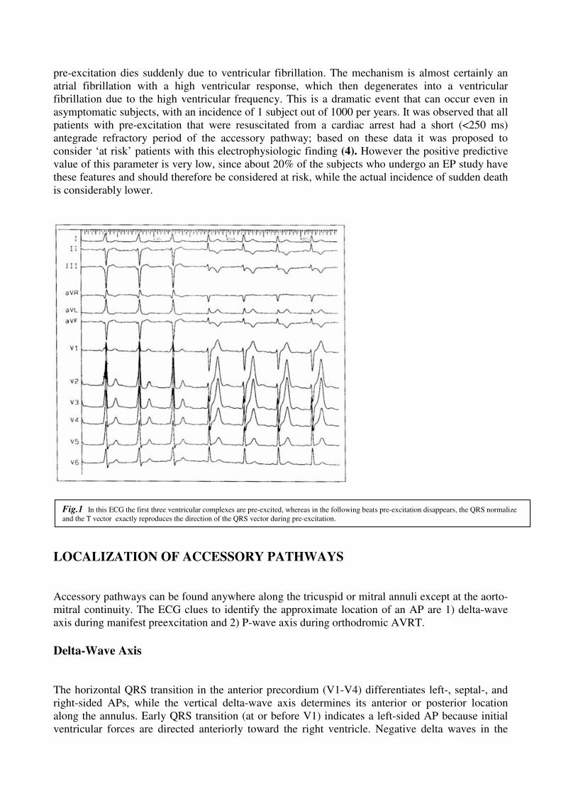
Fig.1 In this ECG the first three ventricular complexes are pre-excited, whereas in the following beats pre-excitation disappears, the QRS normalize
and the T vector exactly reproduces the direction of the QRS vector during pre-excitation.

lateral (I, aVL) or inferior leads identify a left free wall or left posterior AP, respectively.
Horizontal transition at V2, which overlies the interventricular septum, indicates a right postero- or
mid-septal AP. Late transition (at or beyond V3) indicates a right-sided AP. Anteroseptal APs show
a “Left Bundle Brunch Block (LBBB)” type pattern transitioning before V4, sum of delta wave
polarities in the inferior leads (II, III, aVF) ≥ +2, and a frontal QRS axis +30 to +120 degrees (Fig.2
).
P-Wave Axis
P-wave polarities during orthodromic AVRT also differentiate left-, septal-, and right-sided APs . A
rightward axis (positive in aVR, negative in aVL) indicates eccentric atrial activation arising from a
left-sided AP, while a leftward axis (positive in aVL, negative in aVR) identifies eccentric
activation from a right-sided AP. Postero- and mid-septal APs generate a midline, superior axis
(equally positive in aVL and aVR), while anteroseptal APs produce positive P waves inferiorly.
Fig.2 Localization of accessory pathways: the six figures show different horizontal QRS transition in the anterior precordium (V1-V4) and different
vertical delta-wave axis used to differentiate left-, septal-, and right-sided APs, and to determines its anterior or posterior location along the annulus.
The numbers identify following regions: 1. left free wall; 2. left posteroseptal region; 3. right posteroseptal region¸4. right free wall; 5. right anteroseptal
region.
Errors in AP localization are usually due to minimal pre-excitation on the surface ECG, multiple
APs (2-20% of patients) and thoracic deformity or congenital cardiac disease. Thus, detailed
intracardiac mapping is required for successful pathway ablation.
The intracardiac hallmark of preexcitation is a short (<35 ms) or negative HV interval. Because
atrio-ventricular APs originates above the His bundle, His extrasystoles conduct to the ventricle
without preexcitation. A preexcited His bundle extrasystole identifies an AP originating below the
His bundle (fasciculo-ventricular AP).
Endocavitary mapping
The ECG can help in localizing the AP, however the electrophysiologic study gives precise data on
its position. In order to define the exact location of the Kent bundle, mapping of the AV annulus is
performed in order to find the point with the shortest A-V interval during antegrade conduction over
the Kent bundle or the shortest VA interval during ventricular stimulation or orthodromic
tachycardia. This type of mapping can be performed using bipolar or unipolar recordings and it’s
based on the principle that earliest chamber activation (ventricular during antegrade conduction,
atrial during retrograde conduction) allows to localize the AP insertion in the chamber. Therefore
the ablator catheter needs to be positioned on the right or left AV annulus, in contact with the
endocardium, and then moved around until the shortest conduction interval is found. The position of
the catheter is confirmed by the fluoroscopy and by the recorded potential which is composed of
two deflections, the atrial and ventricular one. If the catheter is on the Kent bunde it’s easy to record
almost fused A and V waves, indicating the extremely short conduction time. Sometimes it’s even
possible to record the Kent bundle potential, seen as a rapid deflection of short duration, between
the A and the V, expressing the depolarization of the AP: the A and V waves and the Kent potential
are continuous, and the different components are hard to separate. The identification of such
continuous electrical activity strongly indicates the presence of an accessory pathway.
Antegrade mapping
The earliest site of ventricular activation during manifest preexcitation (prexcited sinus rhythm,
antidromic AVRT) identifies the ventricular insertion site of the AP. Target site criteria for ablation
during antegrade mapping include: 1) AP potential (Kent potential), 2) earliest local ventricular
activation relative to onset of the delta wave (pre-delta), and 3) fusion of atrial and ventricular
electrograms. Accessory pathway potentials reflect rapid local activation of the AP and are sharp,
high frequency deflections between atrial and ventricular elctrograms that precede onset of the delta
wave. The earlier the local ventricular electrogram on the ablation catheter precedes onset of the
delta wave, the higher the probability of success.
Relying on AV fusion alone, however, can be misleading, especially with slanting (oblique) and
slowly conducting APs where fusion might be absent at the site recording an AP potential.
Retrograde mapping
The earliest site of atrial activation during retrograde conduction over the AP (ventricular pacing,
orthodromic AVRT) identifies its atrial insertion site. A limitation of mapping during ventricular
pacing, however, is the possibility that retrograde conduction over the AV node can interfere with
identifying the earliest site of atrial activation over the AP (particularly, septal APs). Potential
solutions include pacing at a faster rate (to cause decrement or block in the AV node),
administration of drugs that slow AV nodal conduction, or mapping during orthodromic AVRT
(where retrograde conduction occurs only over the AP). Target site criteria for ablation include: 1)
AP potentials, 2) earliest site of atrial activation, and 3) fusion of annular (A and V) elctrograms.
Ablation
Electrogram stability is an important determinant of success during radiofrequency (RF) delivery.
After ablation, adenosine-induced AV and VA block provides further confirmation of success. If an
AP is resistant to a particular ablation strategy, options include changing 1)mapping criteria
(antegrade versus retrograde), 2) annular approach (e.g. transeptal versus transaortic) and 3) type of
ablation catheter.
Atypical accessory pathways
Permanet form of junctional reciprocating tachycardia (PJRT)
PJRT is a nearly incessant type of orthodromic AVRT utilizing a concealed, slowly conducting and
decremental AP. The AP is usually (but not always) located in the posteroseptal region and is
mapped by identifying the earliest site of atrial activation during ventricular pacing or tachycardia.
Fusion of atrial and ventricular electrograms is absent because AP conduction is slow and
decremental. Successful target sites might show a fragmented atrial electrogram or possibly an AP
potential.
Mahaim fibers
Mahaim fibers refer collectively to variant APs generally with decremental, antegrade-only
conduction, originating or inserting into or near the right bundle. Atrio-fascicular APs can be
targeted at its atrial insertion site by mapping along the antero- to posterolateral tricuspid annulus
and identifying the site demonstrating 1) Mahaim potentials between atrial and ventricular
electrograms (“His bundle-like” electrograms), 2) shortest stimulus-delta wave interval during
constant atrial pacing, 3) longest coupled AVJ-refractory APD that preexcites the ventricle during
antidromic tachycardia, and 4) susceptibility to mechanical block with catheter manipulation.
Nodo-fascicular (or nodo-ventricular) APs can be targeted by selective ablation of the slow AV
nodal pathway in the midseptum.
Unusual locations
Some posteroseptal APs course subepicardially and require ablation within the coronary venous
system (middle cardiac vein, coronary sinus diverticulum). 12-Lead ECG features suggesting a
posteroseptal AP within the coronary sinus are 1) negative delta wave in lead II, 2) steep (≥45
degree) positive delta wave in lead aVR, and 3) deep S wave (R≤S) in lead V6. Successful ablation
sites can show relatively large AP potentials (AP/A and/or AP/V amplitude ratios ≥1). The presence
of fused atrial and ventricular alectrograms can be misleading and may not coincide with a
successful ablation site. RF delivery should start with low energy (≤30 Watts) and titrated upwards
gradually (“low and slow”).
Some APs are anteroseptal in location where RF delivery can damage the His bundle. Parahisian
APs are defined as APs associated with a His bundle potential ≥0.1mV at its atrial or ventricular
insertion site. 12-Lead ECG features suggesting a parahisian AP are 1) positive delta waves in leads
I, II and aVF, and 2) negative delta waves in leads V1, V2, and sometimes V3. Anteroseptal APs
are prone to mechanical block with catheter manipulation suggesting that they course superficially
in the subendocardium in contrast to the deeper penetrating His bundle within the central fibrous
body. Recording an AP potential and the smallest possible His bundle potential are important target
site criteria for successful ablation without creating AV block. RF delivery with low (initially 5-7
Watts) incremental energy is important and should be terminated immediately with onset of a
junctional rhythm or persistence of AP conduction after 10 sec.

Multiple accessory pathways
In about 10-15% of subjects with pre-excitation, multiple APs are present. Histopathologic data
show a higher frequency of multiple APs than those observed clinically.
The presence of APs increases the incidence of symptoms and is associated with a higher risk of
sudden death due to atrial fibrillation degenerating into ventricular fibrillation. Patients with pre-
excitation resuscited from sudden death had a higher incidence of multiple APs than the control
group that did not have a cardiac arrest (5). Diagnosis of multiple APs on the ECG is possible but
not in all cases. The following criteria should be used:
a) two different pre-excitation morphologies during atrial fibrillation. In this case atrial frequency
is high and the two APs compete between themselves and the AV node-His pathway to conduct the
impulse to the ventricles. The variation of the pre-excited QRS complex shows that atrial impulse
reaches the ventricles over different paths. The diagnosis of a double AP requires two different
complexes, each with typical features of a Kent bundle location. Sometimes it’s also possible to
observe beats with an intermediate morphology, which express a fusion of the two types of
ventricular activation. Variability of the pre-excited QRS morphology by itself is not sufficient for
the diagnosis of multiple APs, as this phenomenon can also be caused by a fusion between a single
AP and the His bundle;
b) the lack of correspondence between the site of the AP determined by the polarity of the P waves
during an orthodromic tachycardia and the one determined by the QRS morphology during sinus
rhythm or atrial fibrillation or pre-excited tachycardia;
c) the presence of two different P waves during different episodes (or even during the same episode)
of orthodromic tachycardia;
d) the sudden change, from an orthodromic tachycardia to an antidromic tachycardia or from an
antidromic tachycardia to a different antidromic tachycardia;
e) the presence of an antidromic tachycardia. Its presence does not necessarily mean that multiple
APs are present, however in a high percentage of cases an orthodromic tachycardia uses another AP
for the retro-conduction of the impulse rather than the AV node-His pathway.
ccessory pathway-mediated tachycardias
The most common tachycardias associated with the WPW syndrome are the circus movement
tachycardias, 95% of which are orthodromic; that is, they conduct antegradely down the normal A-
V conducting system and retrogradely up the bypass tract. The relationship of conduction and
refractoriness of the normal A-V conducting system and the bypass tract, as well as the site of
stimulation, determine both the ability to initiate circus movement tachycardia and, theoretically,
the type of circus movement tachycardia. Conduction and refractoriness of the bypass tracts in most
cases behave like working muscle; therefore bypass tracts demonstrate rapid conduction, and have
refractory periods that tend to shorten at decreasing pacing cycle lengths (PCLs). The WPW
syndrome allows one to actually see all the requirements for a reentrant rhythm: (a) two anatomic or
functionally determined pathways of conduction; (b) unidirectional block in one of the pathways (in
this instance, either in the accessory pathway or in the A-V nodal His pathway); (c) sufficient
slowing in a part of the circuit to overcome refractoriness ahead of the circulating impulse; and (d)
conduction time of the impulse must exceed the longest effective refractory period of any
component in the circuit. Both antegrade and retrograde refractory periods of the accessory pathway
are major determinants of (a) the ability to initiate and sustain circus movement SVT, and (b) the
ventricular responses to atrial tachyarrhythmias (e.g., atrial fibrillation, atrial flutter, and atrial
tachycardia).

AVRT is a re-entrant arrhytmias and is categorized into orthodromic and antidromic variants.
During orthodromic tachycardia, the antegrade limb is the AV node-His-Purkinje system and the
retrograde limb is the AP. Conversely, during antidromic tachycardia, the antegrade limb is the AP
and the retrograde limb is the normal conduction system.
Orthodromic AVRT constitutes approximately 95% of spontaneous and laboratory-induced
AVRTs. For tachycardia initiation, an atrial premature complex (APC), either spontaneous or
induced by pacing, blocks at the AP and travels down the AV node-His-Purkinje system. The
conducted impulse reaches the ventricle and travels back up to the atrium over the AP, which has
now recovered its excitability. The impulse then reenters the AV node-His-Purkinje system,
perpetuating the tachycardia. Orthodromic tachycardia can also be initiated by a premature
ventricular complex (PVC). In this case, the PVC blocks the His-Purkinje system but travels over
the AP up to the atrium. If the AV node-His-Purkinje system has recovered excitability, the impulse
then travels down the node and reenters the ventricle and orthodromic tachycardia is started (Fig.3)
Bundle Branch Block (BBB)
Orthodromic AVRT involves the shortest circuit capable of sustained reentry and, therefore,
incorporates the bundle branch ipsilateral to the AP as an integral part of its circuit. Spontaneous or
induced bundle branch block during orthodromic AVRT can also provide important diagnostic
Fig.3 Induction of orthodromic AVRT by atrial premature complex (a) or by ventricular premature complex
(b).

clues. Lengthening of the tachycardia cycle length is seen in cases of AVRT when bundle branch
block occurs ipsilateral to the free wall pathway. During tachycardia, development of BBB
ipsilateral to the AP forces antegrade conduction over the controlateral bundle and enlarges the
circuit, with resulting increase in VA interval and tachycardia cycle length (6). The degree of VA
interval increase depends on the location of the AP. Accessory pathways located along the free
wall increase the VA interval >35 ms; in contrast, septal APs increase the VA interval <25 ms.
Development of BBB controlateral to the AP does not affect orthodromic AVRT, as the
controlateral bundle is a bystander to the tachycardia circuit (Fig.4).
Pacing maneuvers
The diagnosis of a narrow complex tachycardia (NCT) is facilitated by pacing maneuvers delivered
from the ventricles. Preexcitation of the atrium without a change in the atrial activation pattern by
His refractory ventricular premature depolarizations (VPDs) confirm the presence of an AP and, if
the tachycardia is reset or terminated, the AP is likely required for the tachycardia circuit. In this
case, retrograde conduction can occur only via an AP as the His bundle is refractory.
Spontaneous and laboratory-induced atrial fibrillation (AF) has been reported to occur in up to 32%
to 52% of patients with the WPW syndrome. Several mechanism have been proposed: 1) premature
atrial complex (PAC)-induced degeneration of AVRT to AF; 2) PVC-induced atrial depolarization
during the atrial vulnerable period leading to AF; 3) a reentrant circuit within the atrial branching
insertion sites of the AP fibers. In patients without structural heart disease, susceptibility to
subsequent AF is low (6% to 10%) after successful AP ablation. However, despite successful
pathway ablation, some patients still have recurrence of AF. The characteristics of these patients
include older age (>50 years old), a history of paroxysmal AF and presence of structural heart
disease, no antegrade conduction in the AP, slow ventricular response during AF and inducible AF
after AP ablation.
Risk of sudden death
Unlike the AV node, APs do not demonstrate rate-dependent, decremental conduction that slows
with faster atrial rates. The following features identify a low risk AP: 1) intermittent preexcitation,
2) exercise-induced AP block, 3) shortest preexcited RR interval during AF >250 ms, and 4) loss of
preexcitation with procainamide, ajmaline or disopyramide (7). Intermittent preexcitation
Fig.4 Effect of Bundle Branch Block on
orthodromic AVRT cycle length.

demonstrates that AP is incapable of substaining 1:1 conduction during sinus rhythm, and,
therefore, cannot conduct rapidly during AF. Similarly, abrupt loss of preexcitation during exercise
demonstrates that the AP is incapable of substaining 1:1 conduction during exercise-induced sinus
tachycardia. During exercise, abrupt loss of preexcitation (rate-dependentAP block) should be
differenziated from gradual loss of preexcitation (pseudonormalization) that is due to enhanced AV
nodal conduction. During pseudonormalization, the AP continues to conduct antegradely, but the
delta wave slowly disappears as the contribution to ventricular activation by the AV node-His-
Purkinje system increases. Because the antegrade effective refractory period (ERP) correlates with
the shortest preexcited RR interval during AF, an antegrade AP ERP or shortest atrial pacing cycle
length maintaining 1:1 AP conduction >250 ms is a reasonable but not ideal surrogate to the
shortest preexcited RR interval when AF is absent. Finally, the ability to alter AP conduction with
Na channel blocking drugs suggests a low risk AP although this is controversial.
Therapy of the pre-excitation syndrome
The therapy of pre-excitation has four different objectives:
1. To cure the symptoms; 2. To prevent the risk of sudden death; 3. To prevent or cure, in case of a chronic tachycardia, the worsening of the ventricular function; 4. Allow subjects with pre-excitation to carry out all the activities that are otherwise forbidden by
law when pre-excitation is present on the ECG. In the other cases, a therapy is not indicated: particularly in asymptomtic subjects, who only have
ECG anomaly, do not need any treatment, except from rare case, since their risk of developing
ventricular pre-excitation is very limited.
There are three different kinds of therapeutic approaches: antiaahythnic drugs, catheter ablation,
surgical ablation of the AP. Electrical therapy (cardioversion, pacing) is considered separately.
Pharmacologic treatment
In orthodromic AVRT, the AV node is the weak link and drugs that prolong AV nodal
refractoriness or depress its conduction can lead to block in the node resulting in tachycardia
termination. Vagal maneuvers terminate tachycardia by causing block in the node. First-line drugs
that are effective in acute termination of orthodromic AVRT include I.V. administration of
adenosine, verapamil or diltiazem or beta-blockers. I.V. digoxin is less effective due to its delayed
onset of action. I.V. procainamide is a viable alternative: it depresses conduction, prolongs
refractoriness in most cardiac tissue (i.e. atrium, ventricle and His-Purkinje system) and also blocks
conduction in the AP. Oral class Ic drugs are more efficacious than class Ia drugs in blocking AP
conduction; however, they should be avoided in patients with structural heart disease. Amiodarone
has various electrophysiologic effects but is not more effective than class Ic drugs used alone or in
combination with beta-blockers. In general, amiodarone should be reserved for those who are drug-
refractory, elderly and not suitable candidates for ablative therapy. Sotalol can be effective in
preventing tachycardia, although it is associated with a 4% risk of torsades de pointes, especially in
those with significant structural heart disease and congestive heart failure. Oral digoxin is not
effective as monotherapy for orthodromic AVRT and, by its direct effects on the AP, this drug may
actually accelerate conduction over the AP during atrial fibrillation. Therefore, digoxin should
never be used for the treatment of patients with pre-excitation.

In antidromic AVRT, retrograde AV nodal conduction may be the weak link. I.V. calcium-channel
blockers, beta-blockers and adenosine can be used for acute termination of tachycardia. I.V.
procainamide is the drug of choice in the acute treatment of antidromic AVRT. Even this drug does
not terminate the tachycardia, it may slow the tachycardia rate. In the absence of contraindications,
class Ic drugs are the drugs of choice for long-term oral treatment of antidromic tachycardia.
Catheter ablation
Catheter-based ablation is the procedure of choice for patients with symptomatic WPW syndrome
and for those who respond poorly to medical therapy. In most experienced centers, the success rate
is 95% to 97% with a recurrence rate of 6%.
Successful ablation is critically dependent on accurate localization of the AP. Preliminary pathway
localization can be obtained from delta wave and QRS morphologies. When pre-excitation is not
maximal, rapid atrial pacing or I.V. adenosine can be used to obtain full pre-excitation so as to
improve localization accuracy. This is especially useful in left lateral APs in which pre-excitation
may be enhanced with left atrial pacing (from the coronary sinus, CS, catheter). Intracardiac
electrogram criteria (8) used to identify appropriate target sites for ablation of manifest pathways
include presence of an AP potential (Fig.5), early onset of local ventricular activation relative to the
delta wave onset, electrogram stability and antegrade continuous electrical activity (fused atrial and
ventricular electrograms). Electrogram criteria have also been used to identify appropriate target
sites for ablation of concealed pathway and include retrograde AP potential, retrograde continuous
electrical activity with ventricular pacing or during tachycardia and electrogram stability.
Left free wall pathways constitute the majority of APs. Ablation can be guided by the CS catheter
that is used to bracket the pathway′s location. It can be ablated via either a transeptal or a retrograde
transaortic approach depending on the operator′s experience and preference. In the absence of a
PFO, the transeptal approach involves the puncture across the fossa ovalis. With the transaortic
approach, the tip of the ablation catheter is curved into a “pigtail” to avoid damaging the coronary
arteries, advanced retrogradely across the aortic valve into the left ventricle and positioned along
the mitral annulus using posterior and counterclockwise torque.
Catheter ablation is associated with a very high success rate. Successful ablation of right free wall
pathways requires detailed mapping of the lateral tricuspid annulus. The overall success rate for
right free wall pathway ablation is the lowest of any of the AP′s with an average of 90% and a
recurrence rate of 14%. Reasons for reduced success rate include catheter instability and lack of a
right-sided CS structure that parallel the tricuspid annulus to aid in mapping.
Ablation of anteroseptal and midseptal pathways can be challenging due to their proximity to the
AV node and His bundle; nevertheless it is associated with overall success rate of 95% to 98% and
a 1% to 3% risk of permanent AV block.
Ablation of posteroseptal pathways can be challenging due to the complex anatomy at the
posteroseptum. Most posteroseptal pathways can be ablated from the right side, although in up to
20% of cases a left-sided approach is needed (Fig.6). ECG and EP clues that suggest a left-sided
approach include a positive delta wave or a positive QRS complex in V1, earliest retrograde atrial
activation
at the CS ostium and increase in VA interval with LBBB during orthodromic tachycardia. Between
5% to 17% of posteroseptal and left posterior APs are located epicardially and ablation in the CS
(most commonly the middle cardiac vein) is needed. A manifest AP that may require ablation
within the CS is suggested by a negative delta wave in lead II.
It appears that a small percentage of APs are epicardial. This is suggested by the finding of a small
or no pathway potentials during endocardial mapping and large pathway potentials in the CS. Left-
sided pathways can be successfully ablated within the CS at sites with large AP potentials.
However, successful ablation of APs at other epicardial sites may require a percutaneous epicardial
approach, as an alternative to cardiac surgery.
Overall, ablation of AP is associated with a complication rate of 1% to 4% and a procedure related
death rate of approximately 0.2%. The complication of complete AV block occurs in about 1% of
patients and is seen most frequently in patients undergoing ablation of septal pathways. Autonomic
dysfunction and inappropriate sinus tachycardia are rare complications of radiofrequency ablation
of AP and are less frequent than that seen in slow pathway ablation for AVNRT.
Today, advances in catheter design, energy delivery systems, mapping systems and remote
navigation systems have made catheter ablation the therapy of choice for a majority of SVTs.
Surgical ablation
The elective surgical treatment of WPW has basically been abandoned. Until 1980’s several
patients underwent surgical interventions in order to interrupt conduction over the AP, but since
catheter ablation became available it was universally accepted that the risk/benefit ratio of such
surgical intervention was unacceptable, since better results were obtained using simpler and less
traumatic methods.
Elecrical therapy
Electrical therapy of pre-excitation is based on cardioversion, that is used in case of pre-excited
atrial fibrillation and rarely for AVRT, and on atrial or ventricular pacing in case of a re-entrant
tachycardia. Atrial stimulation can be performed via the endocavitary or transesophageal route,
while ventricular stimulation only via the endocavitary one. This kind of approach is advisable in
subjects in whom drug administration is not possible, or an AVRT doesn’t cease after vagal
maneuvers and it’s not well tolerated.

Fig.5 From top to bottom are leads DI, DIII, V1 and electrograms from the proximal His bundle area (HBE p),
proximal to distal coronary sinus (from CS 7-8 to CS 1-2), proximal and distal Ablator (ABLp and ABLd) and right
ventricular apex ( RV Ap): the distal Ablator records a rapid potential (Kent potential, K) between atrial (A) and
ventricular (V) electrograms.

Fig.6 Ablation of a manifest left posteroseptal AP by retrograde transaortic approach (LAO
projection). The ablation catheter is positioned along the posteroseptal mitral annulus where
it records an AP potential between atrial and ventricular electrograms. Application of RF
energy at this site caused loss of preexcitation in few seconds. The CS catheter provides a
useful reference landmark to the mitral annulus.

Bibliography
1 . Anderson RH, Becker AE, Brechenmacher C, e t a l . Vent r i cu la r p reexc i ta t ion. A proposed nomenc lature
f o r i t s subs t ra tes . Eur J Card io l 1975;3:27–36.
2 . Anderson RH, Bouton J , Burro w CT, e t a l . Sudden dea th in i n fancy : a s tudy of ca rd iac spec ia l i zed t issue. Br Med J 1974 ;2 :135–139 .
3 . Le i t h JW , Kle in GJ , Yee R e co l l . Syncope assoc ia ted wi t h supravent r icu lar t achycard ia . An express ion
o f tachycard ia ra te or vasomotor response? C i rcu la t ion 1992;85:1064.
4 . K le in GJ , Bashore TM, Se l lers TD e co l l . Vent r icu la r f i b r i l l a t i on in the W ol f f -Park inson-W h i te syndrome. N Eng l J med 1979; 301:1080-5.
5 . Teo W S.K le in GJ , Gu i raudon GM e co l l . Mu l t i p le accessory path way in t he Wo l f f -Park inson-W hi te syndrome as a r isk fac to r fo r vent r icu la r f i br i l l a t ion. Am J Card io l 1991;15 :989.
6 . Coume l P , A t t ue l P. Rec ip rocat ing t achyard ia in over t and la tent p reexc i ta t i on. I n f l uence o f f unc t ional
bund le b ranch b lock on t he ra te of t he t achycard ia . Eur J Card io l 1974;1:423–436.
7 . Sharma AD, Yee R, Gu i raudon G, K le in GJ. Sens i t iv i t y and spec i f i c i t y o f i nvas ive and non invas ive t es t ing f o r r i sk o f sudden dea th in W ol f f -Park inson-W h i te syndrome. J Am Col l Card io l 1987;10 :373-381.
8 . Ca lk ins H, K im YN, Schma l tz S , e t a l . E lec t rog ram c r i t er ia f o r ident i f icat ion of appropr ia te ta rge t s i tes f o r rad io f requency catheter ab la t ion of accessory a t r i ovent r icu la r connec t ions . C i rcu la t ion . Feb 1992;85 ﴾2 ﴿ : 565-573.