WLECUTAR FORMUlA, CONFIGURATION AND … CONFIGURATION AND CONFORMATION BY X-RAY ANALYSIS Isabella L....
16
Pure and Appi. Chem., Vol. 49, pp. 1291—1306. Pergamon Press, 1977. Printed in Great Britain. WLECUTAR FORMUlA, CONFIGURATION AND CONFORMATION BY X-RAY ANALYSIS Isabella L. Kane Labonatony for the Structure of Matter, Naval Research Laboratory, Washington, D.C. 20375, U.S.A. Abstract - In the realm of natural products, the more important objectives of crystallography are the elucidation of the structural formula, particularly when many asyninetric centers are present or when unusual linkages occur, and the determination of the preferred conformation of flexible molecules. The former objective is illustrated by the unusual formulas established by single crystal x-ray diffraction analysis for the potent toxins secreted in the skins of tropical American frogs. Sons of these toxins are steroidal alka- bids, while others contain novel allenic and acetylenic substituents on a spiro ring system or on cis-decahydroquinoline. For the latter objective, studies on cyclic peptides have established the existence of stable multiple conformations, a variety of intramolecular hydrogen bonds, and intricate conformational changes upon complexation. The analysis of crystal structures by x-ray crystallography has become indispensable in a large number of diverse scientific disciplines ranging from metallurgy and mineralogy, to molecular biology. The objectives extend from the very practical, i.e. establishing which atom is bonded to which in a particular substance, to the very fundamental, e.g., the distribution of electron density in covalent bonds. For the chemist dealing with natural products and other organic substances, one of the more important uses of crystallographic investigations is the elucidation of the structural formula of a natural product, a reaction intermediate or a photorearrangement product, especially when the substance has unusual or previously unknown chemical linkages or a large number of asymmetric centers. In such instances, other means of analyses often cannot establish the structural formula or distinguish among several equally plausible possibilities. Another objective of crystal structure analysis is the determination of the preferred con- formation of flexible molecules such as oligopeptides. Conformation is often the key to reaction mechanisms and reaction rates. As a bonus, from a single crystal structure analy- sis, in addition to the structural formula, stereoconfiguration and conformation, fairly exact values are obtained for the bond lengths, bond angles and torsion angles with standard deviations of 0.O1A, 0.50 and 1.00, respectively. Such values serve as norms for postulat- ing models and conformations for amorphous substances, substances in solution and otherwise non-crystalline materials. Some 14,000 crystal structures of organic and metal organic molecules have been described in the literature. Most of these structures have been reported in the last few years (Ref.l). The great proliferation is due mainly to four factors: the introduction of high speed com- puters, the development of automatic diffractometers for rapid data collection, the direct method for phase determination particularly for substances containing only light atoms, or many heavy atoms, and the appreciation that vast amounts of chemical and physical informa- tion become available with the expenditure of relatively little effort. A primary requirement for a crystal structure analysis is a single crystal of sufficiently good quality to diffract a beam of x-rays to reasonably high scattering angles so that a large number of reflections can be measured. Each reflection is characterized by four values, h, k, 1 and I, where h, k and 1 are the indices of the plane in the crystal from which the diffraction occurs and I is the intensity associated with the reflection. A typical diffraction pattern recorded photographically is illustrated in Fig.l. Currently most x-ray data are collected on automatic diffractometers with counters rather than film. These data are used in the electron density function P (XYZ) = Fhkl e _2n i(hX + kY + lZ) (1) 1291
Transcript of WLECUTAR FORMUlA, CONFIGURATION AND … CONFIGURATION AND CONFORMATION BY X-RAY ANALYSIS Isabella L....

Pure and Appi. Chem., Vol. 49, pp. 1291—1306. Pergamon Press, 1977. Printed in Great Britain.
WLECUTAR FORMUlA, CONFIGURATION AND CONFORMATION BY X-RAY ANALYSIS
Isabella L. Kane
Labonatony for the Structure of Matter, Naval Research Laboratory,Washington, D.C. 20375, U.S.A.
Abstract - In the realm of natural products, the more important objectivesof crystallography are the elucidation of the structural formula, particularlywhen many asyninetric centers are present or when unusual linkages occur, andthe determination of the preferred conformation of flexible molecules. Theformer objective is illustrated by the unusual formulas established by singlecrystal x-ray diffraction analysis for the potent toxins secreted in theskins of tropical American frogs. Sons of these toxins are steroidal alka-bids, while others contain novel allenic and acetylenic substituents on aspiro ring system or on cis-decahydroquinoline. For the latter objective,studies on cyclic peptides have established the existence of stable multipleconformations, a variety of intramolecular hydrogen bonds, and intricate
conformational changes upon complexation.
The analysis of crystal structures by x-ray crystallography has become indispensable in alarge number of diverse scientific disciplines ranging from metallurgy and mineralogy, tomolecular biology. The objectives extend from the very practical, i.e. establishing whichatom is bonded to which in a particular substance, to the very fundamental, e.g., thedistribution of electron density in covalent bonds. For the chemist dealing with naturalproducts and other organic substances, one of the more important uses of crystallographicinvestigations is the elucidation of the structural formula of a natural product, areaction intermediate or a photorearrangement product, especially when the substance hasunusual or previously unknown chemical linkages or a large number of asymmetric centers. Insuch instances, other means of analyses often cannot establish the structural formula or
distinguish among several equally plausible possibilities.
Another objective of crystal structure analysis is the determination of the preferred con-formation of flexible molecules such as oligopeptides. Conformation is often the key toreaction mechanisms and reaction rates. As a bonus, from a single crystal structure analy-sis, in addition to the structural formula, stereoconfiguration and conformation, fairlyexact values are obtained for the bond lengths, bond angles and torsion angles with standarddeviations of 0.O1A, 0.50 and 1.00, respectively. Such values serve as norms for postulat-ing models and conformations for amorphous substances, substances in solution and otherwisenon-crystalline materials.
Some 14,000 crystal structures of organic and metal organic molecules have been described inthe literature. Most of these structures have been reported in the last few years (Ref.l).The great proliferation is due mainly to four factors: the introduction of high speed com-puters, the development of automatic diffractometers for rapid data collection, the directmethod for phase determination particularly for substances containing only light atoms, ormany heavy atoms, and the appreciation that vast amounts of chemical and physical informa-tion become available with the expenditure of relatively little effort.
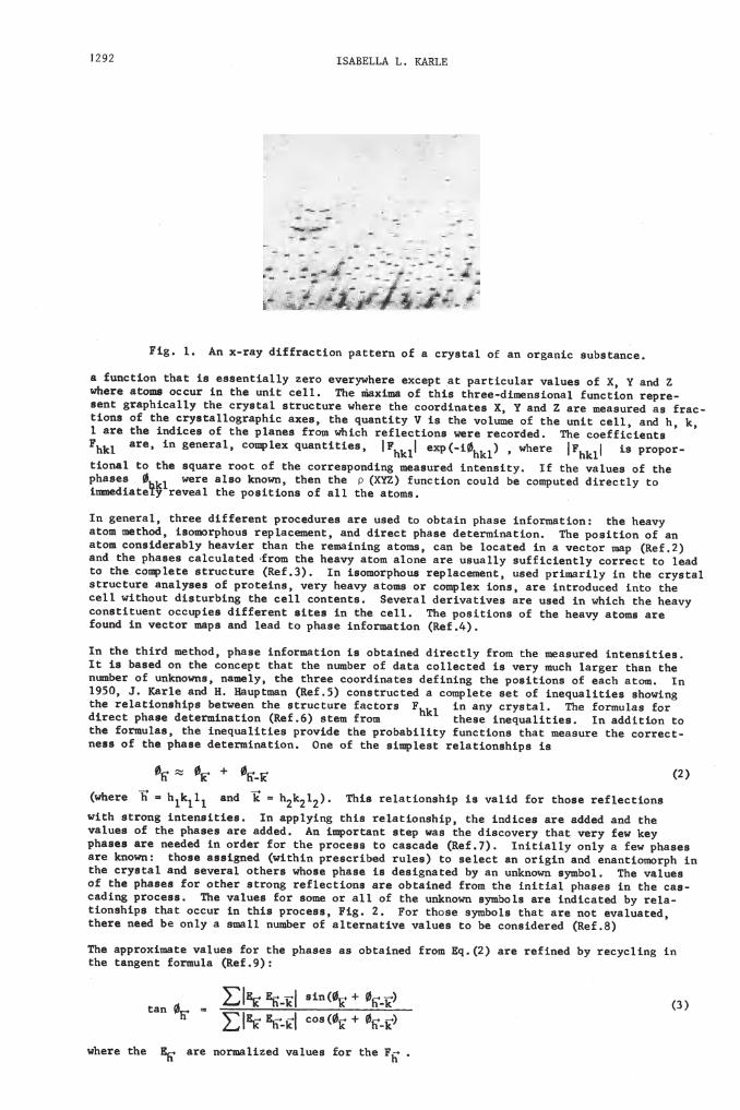
A primary requirement for a crystal structure analysis is a single crystal of sufficientlygood quality to diffract a beam of x-rays to reasonably high scattering angles so that alarge number of reflections can be measured. Each reflection is characterized by fourvalues, h, k, 1 and I, where h, k and 1 are the indices of the plane in the crystal fromwhich the diffraction occurs and I is the intensity associated with the reflection. Atypical diffraction pattern recorded photographically is illustrated in Fig.l. Currentlymost x-ray data are collected on automatic diffractometers with counters rather than film.These data are used in the electron density function
P (XYZ) = Fhkl e _2n i(hX + kY + lZ)(1)
1291
MJJ6L6 $JJ6 6L6 UOLW6J6 A6]116 tOL JJ6
- E'4J coa( +-
V aru(E + 14E) ()
ip6 f6ugGzJc tLWflJ6 (gjps 6bbLoxTu6 iwjnaa ox p6 bp6aea 66 op16u6q tr.ow d() 6L6 r.Gu6q p) ic)cjtu u
1116L6 uGeq P6 ourA 6 8W911 WIW6L O 6IGLU9tA6 ço 6 couaqeq (g6'9)touaptba occar. ru pra br.oceaa j or. cpoae aAwpora ipui 61.6 uoi 6Au7nu16q
c6qu br.ocaa JJJG ujna or. aozue ox. j ot p6 nujcuot.w a)mpoja 61.6 uqcucq pA x.u-
o 36 bpiaa OL oJJ6r. açx.ou L66CçOU8 ur.6 opr6ueq r.ow çps bpsa p6 ca-çp cr.Aacuj uq 6A6iJ ocpr.a z'qJoa6 bpa a q6aU916q pA uu nujcuot'us aAwpoj Jp6 /ja6291.6 jcuoz.w: cpoa 6aau6q (tqcpu br.eecr.peq Lnrea) ço aereci ati or.ri iJq Gu9ucrowoLbp ru
bp6aea 91.6 usqeq u or.qer. for. cpe br.ocsaa io cuacuqe (t)' Iutit9ITA ouiA et bpuaeaernea ot cpe bpcsaea er.e eqqeq yu rwbor.ceuc eieb z'68 1JJ6 qacOA6LA flJ6i AGLA Gtt jceA
AtP ar.ou U6L79TT68. gbbAu pa x.eeqouapb' fpG uqcsa 61.6 uqqeq auq qJ6
(z.per.e p = aiiq j = PiCr) jpa r.erecrouaprb ra arq for. ipoae LGtreCctOua
ic. + ()
ueaa Ot pS bJJaa6 q6161.wrueflou O6 o çjis apLIbGaI r.eteCtOuapIba ra
LJJ6 toLwnrea çJ16 tuednajtltea br.oAq6 rpe br.opaptitA tnucqoua IJ9I UJ698flL6 Lp6 COLL6C-
qc1 bp9ae q6c6LwIJ9qou (rce) a6W GLOW JJ686 TU6cIn9TtT6a ru uqqou ioqiG x.ejeqouaprba p6LG6u Lp6 2fLflCflL6 t9C10L6 U 9UA cLA619T £JJG oLXøflJ66 for.
1 KLTG aUg w janbiwau (ge) couar.ncceq a cowbrece a61 o ru6dnarrr6a apoi.ru
unWpGr. o nuJCUObWa uawA JJ6 1J11.66 COOLqtIJ9IG9 qeuu cpe boarroua O 69CJ1 OW1c ra peaeq ou pe coucebç JJaç pe unmpex. o qaca coec1Gq ra Aer.A macp rer.ez cpau JJG
ru rpe pr.q weipoq bpaae rutor.wairou ra opçaueq qr.ecijA r.ow JJ6 WGaatILGg pJ6uac6a'
onuq r A6COr. weba aLYq jeaq o bpaae rutor.mairou (rec)'coualrlneuc occnbrea qr6L6c açea p.r pe cerr jp6 boaqoua o cpe p6aiA afOW2 61.6
ceri zqpon qainr.pp çpe ceri coIJç6IJa 26A6L9 q6qAaqA6a 91.6 naeq ru tprcp (JJ6 pG9AA
ar.ncnr.e auarAaea ot br.oierua AGLA pG9AA aowa or. cowbj6x roua 91.6 ucr.oqnceq ruo 1p6
ço pecowbiee acx.accnx.e (jçe3) ru raowor.bpona x.ebjaceweui rraeq bx.rwax.rrA ru q16 cr.Aagar
auq cpe bpaaea cacnja1Gq r.ow cpe p69AA açom 9J0U6 61.6 nanaTIA anrcr6ucrA cor.r.ec o jeaqavow couaqer.ap7A pesArex. cpau cpe x.ewaruru aowa C6LI p6 ocaeq ru a ecçor. wab (ge)açow wecpoq raowor.bpoaa r.ebjaceweu auq qr.ec bpaae qeex.muaou jpe boap.ou o 61Jir pr.6e qrer.eu bx.oceqar.ea ar.e naeq o opcaru bpaae rutozwarou: pe pG9AA
nweqaceA L6A69J cpe boarcroua o aj 11J6 910W8
bpaaea tex.e ajao Jcuo1uf çpeu pe b (x) tnuccrou conq pe cowbni6q qr.ecijA io
lrouar o çpe adnar.e r.ooç o qie cor.x.eabouqu weaaar.eg ruc6uarA r cpe iaa6a o cpe
LPICI ru gu9J) cozabrex dnaucrcrea' IrC1 exb(r') tee rP( ra br.obox.-
r ar.e qis uqcea o ipe b7auea tzom tprcp r.etreccrouat'er.e r.ecox.qeq jpe coeurcreua
iroua o cpe cr.Aacarror.abprc axea' pe dnauriA A ra qJG ornwe O qi6 nurc cejr aug JJa jC
aexr r.abpcajA cpe cr.Aacal ar.ncqir.e b6L6 p6 coox.qua6a x' aug 91.6 meaanr.eq aa r.ac-iper.e aowa occur. p.3pe aup cejj jpe waxrwa ot cpr cpL6Gqw6uaTouar taucrou r.ebr.e-a tnucrou pa ra eaaeuirarjA xer.o eA6LAtper.6 exceb aç bar.rcarar. A9fl66 O A aug
j yu x-.r.aA qLacqou baçer.u o a cx.Aagaj o au oLaurc anpaiauce'
ivrrv r
L
-- ::: ::-

Molecular configuration and conformation by X—ray analysis 1293
1 1/\ /\ //\/\ /\//\ /1 /\\ L±Fig. 2. Schematic representation of the cascading process in phasedetermination. The pairs of arrowheads enclosed within a circle represent a reflection whose phase value has been determined from two differentpaths. Such occurrences lead to algebraic relations among the symbols.
Let us now consider a variety of problems to which x-ray diffraction analysis has beenapplied. The examples are taken mostly from the research program at the Naval Research
Laboratory.
PWTOREARRANGEMENTS
Ring closure had been effected in N-chloroacetyl tryptophanelby eliminating hydrogenchloride as a result of exposure to ultraviolet light (Ref.lO). This preparative method hadbeen applied to other aromatic amino acids and pharmacodynamic amines without changing the
original chromophore. In contrast, upon UV irradiation of N-chloroacetyl 3,4-dimethoxy-phenethylamine , ring closure occurred to yield two isomeric compounds 4 and 5 and a thirdisomeric compound 6 of completely unknown structure which was unyielding to n.m.r., massspectra and other spectroscopic means of analysis. Hence an x-ray diffraction analysis ofa single crystal of 6 was carried out.
RING CLOSURE
0CH3O H
m 194°
CH300.CH2CH2NHCCH2C2 _i-' + CH3ODmp 1500
+ C2H15NO3 mp 123°6
Phases for the Fhkl values were obtained directly from the measured x-ray intensities by
means of relationships (2) and (3). The calculated electron density function (I) contained16 maxima which are plotted in the left portion of Fig. 3. The numbers next to the dotsindicate the third coordinate. Connections were made between dots separated by 1.2 to l.5Ato reveal the geometry and structural formula of the molecule (right portion of Fig. 3)(Ref.ll). This unusual product has two five-membered rings fused to two four-memberedrings. The atoms can be identified as C, N or 0 either by chemical considerations or byweight and thermal factor analysis from the x-ray data. An electron density map computedfrom the differences between the observed structure factors and those calculated on the

1294 ISABELLA L. KARLE
66. •86 •93
•52 .91
Fig. 3. Left portion represents the positions of the maxima of a three-dimensional electron density function calculated from experimental data.Numbers adjacent to the dots represent the third coordinate (XlOO). In theright portion, dots separated by l.2-l.6A have been connected and thestructure of the molecule is revealed.
basis of the known C, N and 0 atoms reveals the positions of the hydrogen atoms, Fig. 4.
Fig. 4. Electron density contours, obtained from a difference map, forhydrogen atoms for molecule in Fig. 3 (Ref.ll).
Thus the structural formula of a completely unknown substance has been established quitedirectly without any assumptions or prior knowledge and without the introduction of a heavyatom marker. At the same time, much other information is obtained at no extra effort, suchas the stereoconfiguration about the four asymmetric carbon atoms, the bond lengths, bond
angles, conformational angles, hydrogen bonds, if they exist, packing modes, etc.
The changes that took place upon irradiation are shown schematically as follows:
CH3Ohv
CH3OyCH2CH2NHCCH2CI -0
3
From such information it is possible to postulate reaction mechanisms.S
Substances differing only in the number of methoxy groups on the phenethylamine producedrastically different products upon irradiation under identical conditions (Ref.12 & 13).If a hydroxyl group is present instead of methoxy, two different dimeric substances 8 and9 are produced in addition to simple ring closure (Ref .14):
•58.99
/2
a
.100
•52
.70 .100.81
.48
•68
.67
C 1/2 C
6

Molecular configuration and conformation by X—ray analysis 1295
2HO
261 2-"N--COCH Cl5
R
4h.v
7
8
9
Again, the structural formulas of both 8 and 9 were established by crystal structureanalyses (Ref.l5). As indicated, dimer 9 is more stable than dimer 8 since the reaction isnot reversible, although the cage in dimer 8 is more symmetrical than the cage in dimer 9.A possible mechanism for the rearrangement from the four-membered ring in 8 to the three-membered ring in 9 is as follows:
0
15$ LI I"!
vØ: 0II 0
152
I 1.53
+ ,2 CYCLOREVERSION, ORNORRISH TYPE I CLEAVAGE
An interesting observation is that the bond lengths in the area of rearrangement are muchlarger than normal in the intermediate, less stable dimer and are reduced to more normalvalues of l.52-l.53A after rearrangement.
BIOSYNTHESIS
Biosynthesis of the inesembrine alkaloids occurring in Sceletium strictum, a South African
succulent, begins with phenylalanine and tyrosine. It proceeds through many steps, some ofwhich are indicated here, and finally produces the four congeners 10, fl, 12 and 13 (Ref.l6).All the formulae were established by chemical and spectroscopic means except for product 13which occurred in a very small amount. The formula remained in doubt until a minute singlecrystal was grown and subjected to x-ray diffraction analysis (Ref.17). As a bonus, theorientation of the dimethoxyphenyl moiety with respect to the fused rings was revealed, aswell as the mode of puckering in the five- and six-membered rings, as shown in Fig. 5. Thefive-membered ring is in the classic envelope conformation with four coplanar atoms and thefifth atom 0.62A out of the plane, while the six-membered ring which contains one unsatur-ated bond and a carbonyl carbon also assumes an envelope conformation with five atomsnearly in a plane and the sixth atom out of the plane.

1296 ISABELLA L. KARLE
Fig. 5. Structure and conformation of the nesembrine alkaloid
N—dimethyl—N--formylrnesenibrenone, 13 (Ref. 17).
PH ENY LA L AN NE
+TYROSINE
OH
OMeOMe
0+
OH
Me
12
+
Me10: R1, R2OII: R10H, R2H
3
Molecular configuration and conformation by X—ray analysis 1297
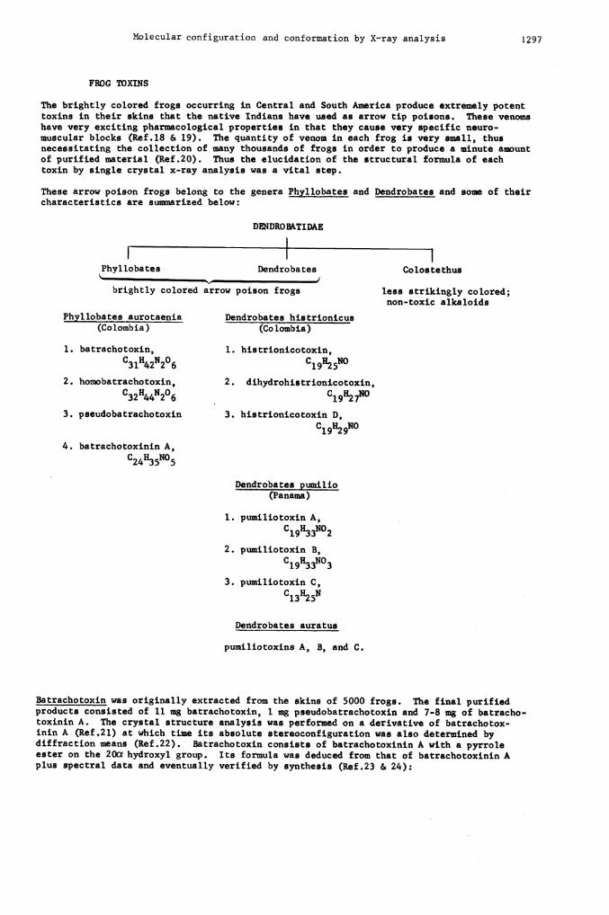
FROG TOXINS
The brightly colored frogs occurring in Central and South America produce extremely potenttoxins in their skins that the native Indians have used as arrow tip poisons. These venoms
have very exciting pharmacological properties in that they cause very specific neuro-muscular blocks (Ref.l8 & 19). The quantity of venom in each frog is very small, thusnecessitating the collection of many thousands of frogs in order to produce a minute ameuntof purified material (Ref.20). Thus the elucidation of the structural formula of each
toxin by single crystal x-ray analysis was a vital step.
These arrow poison frogs belong to the genera Phyllobates and Dendrobates and some of theircharacteristics are summarized below:
DENDROBkTIDAE
I 1 1Phyllobates Dendrobates Cobs tethus
brightly colored arrow poison frogs less strikingly colored;non-toxic alkaloids
Phyl lobates aurotaenia Dendrobates his trionicus
(Colombia) (Colombia)
1. batrachotoxin, 1. histrionicotoxin,
C31H42N206 C19}125N0
2. homobatrachotoxin, 2. dihydrohistrionicotoxin,
C32H44N206 C19H27N0
3. pseudobatrachotoxin 3. histrionicotoxin D,
C19H29N0
4. batrachotoxinin A,
C24H35N05
Dendrobates pumilio(Panama)
I. pumiliotoxin A,
C19H33N02
2. pumiliotoxin B,
C19H.33N03
3. pumiliotoxin C,
C13H25N
Dendrobates auratus
pumiliotoxins A, B, and C.
Batrachotoxin was originally extracted from the skins of 5000 frogs. The final purifiedproducts consisted of 11 ing batrachotoxin, I mg pseudobatrachotoxin and 7-8 mg of batracho'toxinin A. The crystal structure analysis was performed on a derivative of batrachotox-mm A (Ref .21) at which time its absolute stereoconfiguration was also determined bydiffraction maans (Ref.22). Batrachotoxin consists of batrachotoxinin A with a pyrroleester on the 20a hydroxyl group. Its formula was deduced from that of batrachotoxinin Aplus spectral data and eventually verified by synthesis (Ref .23 & 24):

This natural substance presented a number of novel features that were observed for the firsttime; namely, the 9cr-OH, the 3cr—9cx oxide, the 3—hemiketal, the 7—membered l3—l4 hetero-cyclic ring encompassing N-'methylaminoethanol (a precursor of choline) and the 16
unsaturation. The stereochemistry of the cis A/B and cis C/U ring junctions is the same asin the cardenolide glycosides and bufodienolides.
Unlike the steroidal alkaloids present in the Phyllobates species, the alkaloids from theDendrobates species are quite different. Unprecedented structural features have occurred inthe histrionicotoxin 15 (Ref.25), dihydrohistrionicotoxin 16 (Ref .26) and histrionicotoxin D17 (Ref .27):
\\\
CH15
067 269O8
Fig. 7. Bond lengths and bond angles in dihydrohistrionicotoxin (Ref.26).
1298 ISABELLA L. KARLE
HO
14
OH
HC
HC
\C
H—cH\
CH
C
CHH\\\
CH16
CH
C
IIICH
These are the first examples of athe animal kingdom. The absoluteshown in Figs. 6 and 7.
17
spiro-ring system and of acetylenic and allenic bonding instereoconfiguration and bond lengths and angles in 16 are
Fig. 6. Conformation and absolute configuration of dihydrohistrionicotoxin
(Ref.26).

Molecular configuration and conformation by X—ray analysis 1299
The formula of pumiliotoxin C 18 (Ref.28), reminiscent of conine (from a plant source),
H7
contains a cia ring junction, as has been also found in 17. The structural formulas of
pumiliotoxins A and B have not yet been elucidated and thus far it has not been possible toprepare crystals of these substances. Since some species of Dendrobates contain both his-tionicotoxins and pumiliotoxins, it is believed that both classes of alkaloids have the same
precursor.
These alkaloids from the poison arrow frogs have become valuable tools for studying neuro-transmission events in synapse and muscle. The batrachotoxin is associated with an irrevers-ible increaseof membrane permeability to Na+ ions and is an antagonist to tetrodotoxin
activity (Ref.18).
CYCLIC PEPTIDES
The emphasis in this lecture thus far has been upon elucidating the structural formula andconfiguration. For many natural products, the molecular formula is readily derived fromspectral data and/or chemical activity. The challenge lies in establishing the conforma-tion, particularly for flexible molecules such as the cyclic peptides. These substancesoccur in nature and many of them have antibiotic, ion transport and other useful physiologi-cal properties that are associated with the manner of folding of the peptide backbone andside chains.
Coincidentally, cyclic hexaglycyl, the first cyclic peptide for which the crystal structureand conformation were determined (Ref.7), was also one of the first substances whose struc-ture was determined by the direct method of phase determination. This crystal proved to beparticularly interesting since there were four independent molecules in the cell, notrelated by symeetry. Thus, there were 98 atoms (aside from hydrogen atoms) that needed tobe located. It proved to be a very unusual crystal because the molecules assumed fourdifferent conformations, side-by-side, in the same unit cell. Figure 8 illustrates thepacking of the molecules in the unit cell while the four different conformers are shownindividually in Fig. 9. This determination established that molecules can exist in morethan one stable conformation, thus indicating that there is very little difference inenergy between a number of different conformers.
Fig. 8. Packing of cyclic hexaglycyl molecules in a unit cell (Ref.7).
Fig.9. Four conformers of cyclic hexaglycyl occurring in the unit cell inFig. 8.

1300 ISABELLA L. KARLE
The conformer illustrated at the left of Fig. 9 has two intramolecular hydrogen bonds,often called a—turns, n-bends or 4—-l bonds. This type of folding is a common feature inmany.linear peptides and proteins and this determination provided the parameters needed formodel building, minimum energy calculations, comparison with spectroscopic data, etc.Figure 10 contains another view of hexaglycyl with intramolecular hydrogen bonds where itis being compared with the conformations of a linear tetrapeptide (Ref.29), a linear
(a) (b)
(c) (d)
Fig. 10. Comparison of the conformations of: a) bromobenzoate of a linear
tetrapeptide, b) cyclic hexaglycyl, c) bromobenzoate of a linear penta-peptide, d) backbone of the ring in Ferrichrome A. The dotted lines indi-cate intramolecular NH. • bonds (Ref.30).
pentapeptide (Ref.30) and the cyclic hexapeptide ring of Ferrichrome A (Ref.3l). Thehydrogen bonds in the two linear molecules have almost identical conformational parametersto those of c-hexaglycyl, while the 4—-l bond in Ferrichrome A is different, particularlywith respect to the middle peptide unit where the C group is directed upward instead ofdownward. It has been established that the c-hexaglycyl form occurs for peptide residuesthat are both levo, both dextro, or both Gly at the corners of the bend (called Type I)and that the Ferrichrome A form (called Type II) occurs for D,L or L,D or L,Glysequences (Ref.32 & 33).
Scale drawings of the three types of p-bends found in oligopeptides, i.e. types I and II,already mentioned, and a bend in which the middle peptide groups has a cis conformation(Ref .34) are shown in Fig. 11. The numbers adjacent to the corner bonds indicate typicalvalues for the torsional twist about the bonds in order to make the fold. (For a fullyextended chain in the trans conformation, the angular values would be 180°.) The 4—.-ltype of hydrogen bond contains 10 atoms in the ring.
Pig. 11. Three types of 4—el intramolecular hydrogen bonds. The numbersshown near atoms C and Ca indicate typical experimental values found forthe rotations about the boJs to C and C . An extended chain in thetrans conformation would have angular values of 1800.
Intramolecular hydrogen bonds of a 3—0-I type, containing only 7 atoms in the ring, havebeen discussed in the literature for a number of years but have been observed in moleculesin the crystalline state only recently. The conformation of dihydrochlamydocin (Ref.35),a metabolite from Diheterospora chlamydosporia (Ref.36), is illustrated in Fig. 12.This cyclic tetrapeptide contains two intramolecular 3—-l hydrogen bonds and the detailsfor one of these bonds are shown in Fig. 13. The N. • .0 separations are of the order of2.8 - 2.9A and the H.. .0 separations are 2.1 - 2.2A. The all trans tetrapeptide ringin dihydrochiamydocin is of further interest since it has been demonstrated geometricallythat four planar peptide units, each in the trans conformation, cannot form a closed
ring (Ref.37). Although peptide units, C' a whether cis or trans, areN-C
4—+ trans(I) 4—b trans (U) 4—' I cis

Molecular configuration and conformation by X—ray analysis 1301
Fig. 12. Conformation of dihydrochlamydocin. The fine lines indicate thetwo 3—a-i intramolecular NM.. .OC bonds (Ref.35).
Fig. 13. Details of a 3—a-i intramolecular hydrogen bond. The numericalvalues near C indicate the angular twists about N2—C and
generally quite planar, an accommodation has been made in this substance so that ringclosure could be effected. A rotation has taken place about each C'-N bond from 1800(trans, planar) to values of +162°, _i660 +156° and _l640.
Another example of a 3—k-i hydrogen bond has occurred in the synthetic cyclic pentapeptidec-Gly-Pro-Giy-D-Ala-Pro (Ref.37) shown in Fig. 14. Four of the peptide units are trans
L-PRO
Fig. 14. Conorination of a cyclic pentapeptide containing both a 3—b-iand a 4—a-i intramolecular hydrogen bond (Ref.38).
and planar while the fifth one is trans and non-planar with a rotation of 1600 about the
C-N1 bond, comparable to the non-planar peptide units in dihydrochiainydocin. The
c-pentapeptide also contains a 4 —a-i, type II, hydrogen bond.
In the folding of the backbone of cyclic peptides, 5—.-1 intramolecular hydrogen bonds,encompassing 13 atoms, have also been observed. Figure 15 shows the details of suchbonding in complexed valinomycin (Ref.39 & 40), where the peptide units are L, L and D.A comparable 5—a-i hydrogen bond, where all the residues are levo has also been observedin uncomplexed antamanide, which will be discussed later in the manuscript. Meanwhile,Fig. 16 shows a view of the conformation of valinomycin. The molecule contains a 36-membered ring that loops up and down in a sinusoidal fashion and that is stabilized byfour 4 —a-i and two 5 1 hydrogen bonds. The latter two bonds transverse theflattened ring.
PAACVOL 49-9—E
0-PRO
3— I
GLY
GLY D-AL
L-PRO

1302 ISABELLA L. KARLE
Fig. 15. Details of a 5—.1 intramolecular hydrogen bond occurring invalinomycin (Ref .40).
A number of different arrangements for intramolecular hydrogen bonds that stabilize foldsin peptide backbones have already been demonstrated. There is. still another possibility,and that is the absence of intramolecular hydrogen bonding. The conformation of cyclic-
L-Leu-L-Tyr-ô-Avaler-6-Avaler (Ref.4l), a synthetic inhibitor of chymotrypsin (Ref.42),Fig. 17, is very similar to one of the four conformers of cyclic hexaglycyl, Fig. 9. Theparticular point of interest is not only the absence of a hydrogen bond across the ring,the N(3). . .0(4) distance is 4.59A, but also that the proton on N(3) does not participate inhydrogen bond formation, even though it is quite accessible and not blocked by side chains.The lack of hydrogen bond formation by NH groups is of fairly frequent occurrence in cyclicoligopeptides.
Fig. 17. Conformation of cyclic-L-Leu-L-Tyr-ô-Avaler-ã-Avaler, amolecule without any intramolecular hydrogen bonds (Ref.4l).
Fig. 16. Conformation of valinomycin looking into the cavity. Themolecule contains four 4—.-1 hydrogen bonds and two 5—.-l hydrogen bonds(across the flattened ring) (Ref.39).
Molecular configuration and conformation by X—ray analysis 1303
It would be useful to establish certain norms of conforniational behaviour for peptidemolecules. Thus far, however, each new structure determination reveals some unexpectedfeatures such as a new configuration for internal hydrogen bonds, nonplanar peptide units,
peptide groups or unusual folding or unfolding of the peptide chain.
CONPLEXED AND UNCOMPLEXED ANTANANIDE
Antamanide, a cyclic decapeptide found in the poisonous green mushroom Amanita phalloides,has the following sequence:
8 9 10 1 2Pro—Phe—Phe—Val—Pro
I I (all L—).Pro—Phe—Phe—Ala-Pro
Wieland and coworkers have isolated, sequenced, synthesized, and characterized thebiological activity of this peptide (Ref.43). It acts as an antidote to the toxinphalloidine that occurs in much larger quantities in the same mushroom. The mode ofaction of antatnanide is not yet clearly understood but there are strong indications thatthe antamanide acts upon the membranes of liver cells to prevent the toxin from enteringthe liver.
Antamanide forms alkali—metal complexes with a strong preference for Na+ over K+ ions.The alkali metal complex is most stable in a lipophilic environment. The chief objectiveof the present research was to establish the conformation of the unconiplexed antamanideand the changes in the folding or unfolding of the peptide backbone that accompany thealkali complex formation. Knowledge of the conformation and changes in conformationaccompanying complexation or change in the polarity of the solvent may aid in the under-standing of the binding to receptor sites and may serve as a model for ion carriers.
The conformation of the Na+ complex of a biologically active synthetic analog of antamanideis shown in Fig. 18 (Ref.44). There are a number of unique features to notice: (1) Thepeptide backbond (darkened bonds) is folded into a shape resembling the periphery of asaddle. (2) Peptide units have usually been found to be planar and to have the transconformation. Here, eight of the peptide units are trans, but two of them have the ciaconformation (see e.g. the cis amide bond between the Ccs(7) and Cc(8); similarly forC0(2) and C°(3)). A survey of peptides containing cia amide bonds (Ref.32) indicatesthat the cis bond occurs only in those peptide units that have a substituted N atom,e.g., insarcosineor proline. However, peptide units with a substituted N atom do notnecessarily have the cia conformation, as is illustrated in this molecule, where twoprolyl residues occur in the cis conformation and two other prolyl residues occur in thetrans conformation. (3) The Na+ resides in a cup formed by the folding of the peptidechain. It is liganded to four carbonyl oxygen atoms of the peptide. A fifth ligand isformed with the 0 atom of a solvent molecule, ethyl alcohol in this case. Thus the Na+is pentacoordinate. The polar cavity is plugged with a solvent molecule and thelipophilic end of the solvent molecule completes the lipophilic exterior surface.(4) The side groups, particularly the phenyl groups, are folded up against the molecule,to make a compact spheroidal shape. Two phenyl groups on two phenylalanine residues areomitted from this drawing for clarity. They fold up against the molecule in front and inback. (5) There are six NH groups available for hydrogen bonding. Two of them enterinto intramolecular hydrogen bonds (indicated by the dashed lines) of the 4l typepreviously described. Twoothers participate in hydrogen bonds between molecules, and twoNH groups do not participate in any hydrogen bonds at all.
Antamanide forms complexes with Li+ as well as Na+. Fig.l8 also shows a comparison ofthe two different alkali complexes, Li+ antamanide.CH3CN (Ref.45 & 46) andNa+[Phe4Val6]antamanide.C2H5OH (Ref.44). Even though the alkali ions are different, thesolvent molecules are different, side chains in residues 4 and 6 are different, and thecrystallographic packing is different, the two complexes are isostructural, thusindicating that the conformation of the alkali complex of antamanide is independent ofits environment.

1304 ISABELLA L. KARLE
+Fig.l8. Upper diagrams: Two views of Li antamanide.CH3CN. Lowerdiagrams: Two views of Na+[Phe'fVal6]antamanide.C2H5OH. The alkali metalion is represented by a black dot. For clarity the solvent molecules havebeen omitted from the views on the left and two phenyl groups that fold upagainst the middle of the molecule have been omitted from the views on theright (Ref.44, 45 & 46).
Now we turn our attention to uncomplexed antamanide. From spectroscopic evidence insolution, it is known that the conformation is quite sensitive to the polarity of thesolvent (Ref. 47). The crystal used for the x—ray diffraction analysis was grown from amixture of n—hexane and methyl acetate, a very nonpolar solvent. In Fig.19 (Ref.48), itis immediately obvious that the ring in antamaiiide has become unfolded; in fact, it iscompletely elongated. The only features of the conformation that are similar to thealkali complex are the pairs of adjacent prolyl groups, one trans, one cis.
Fig. 19. Two views of the conformation of antamanide crystallized fromnonpolar solvents. Three molecules of water, designated by W, arecontained in the molecular cavity (Ref.48).

Molecular configuration and conformation by X—ray analysis 1305
Two NH groups make hydrogen bonds with carbony]. oxygens across the ring. Again, this is adifferent node of hydrogen bonding than any that has been observed earlier. It is a 51type with all L—residues with the middle peptide unit in the cis conformation. A veryinteresting feature of this structure are the three H20 molecules residing in the interior.They form hydrogen bonds with four of the six NH groups of the molecule and account for therelative rigidity of the 30—membered peptide ring. These water molecules appear to be anintegral part of the peptide molecule since they have been retained even after extensivedrying of the antamanide and the use of an extremely nonpolar solvent. This molecule canindeed be considered as an H20—complex.
The three water molecules reside in a shallow cup with the hydrophobic side chains forminga wall around them. Again all the side groups fold up against the peptide ring to make avery compact globular entity. It seems unlikely that the uncomplexed molecule in thisconformation recognizes a Na+ ion, rapidly releases its 1120 and folds up to engulf the Na+ion. Antatnanide crystals have now been prepared from various polar solvents for anx-ray diffraction examination. We shall have to wait for a while to see whether theconformation obtained from polar solvents will be closer to that of the alkali metalcomplex.
REFERENCES
1. 0. Kennard and D.G. Watson, Molecular Structures and Dimensions, Bibliography,
Vols.l—7, Oosthoek, Scheltema and Holkema, iJtrecht (1970—1975).2. A.L. Patterson, Phys. Rev. 46, 372—376(1934); Zeits. P. Kristallog. 90, 517—542 (1935).3. See e.g. G.H. Stout and L.H. Jensen, X—Ray Structure Determination, pp.270—299,
Macmillan, London (1968).4. See e.g. B.W. Matthews, "X—Ray Structure of Proteins" in The Proteins, 3rd Ed.Vol.III,
Eds. H. Neurath and R.L. Hill, Academic Press, New York (in press).5. J. Karle and H. Hauptnan, Acta Cryst. 3, 181—187 (1950).6. See e.g. J. Kane and I.L. Karle, Acta Cryst. 21, 849—859 (1966).7. I.L. Karle and J. Karle, Acta Cryst. 16, 969—975 (1963).8. I.L. Karle and J. Kane, Acta Cryst. 17, 835—841 (1964).9. J. Karle and H. Hauptinan, Acta Cryst. 9, 635—651 (1956).10. 0. Yonemitsu, P. Cerutti and B. Witkop, J. Amer. Chem. Soc. 88, 3941—3945 (1966).11. I.L. Karle, J.W. Gibson and J. Karle, Acta Crjst. B25, 2034—2039 (1969);
0. Yonemitsu, Y. Okuno, Y. Kanaoka, I.L. Kane and B. Witkop, J. Amer. Chem. Soc. 90,6522—6523 (1968).
12. 0. Yonemitsu, B. Witkop and I.L. Kanle, J. Amer. Chem. Soc. 89, 1039—1040 (1967);I.L. Karle, J. Kane and J.A. Estlin, Acta Cryst. 23, 494—500 (1967).
13. 0. Yonemitsu, H. Nakai, Y. Kanaoka, I.L. Karle and B. Witkop, J. Amer. Chem. Soc. 92,5691—5700 (1970); I.L. Kane and J. Karle, Acta Cryst. B26, 1276—1282 (1970).
14. T. Iwakuma, H. Nakai, 0. Yonemitsu, D. Jones, I.L. Kanle and B. Witkop, J. Amer. Chem.Soc. 94, 5136—5139 (1972).
15. D.S. Jones and I.L. Kane, Acta Cryst. B30, 617—623 (1974).16. Jean M. Karle, Ph.D. Dissertation, Duke University, Durham (1976). Also see
P.W. Jeff s, H.F. Campbell, D.S. Farrier, G. Ganguli, N.H. Martin and G. Molina,
Pytochem. 13, 933—945 (1974).17. Jean M. Kane, Acta Cryst., in press18. E.X. Albuquerque, J'.W. Daly and B. Witkop, Science 172, 995—1002 (1971).19. J. Daly and B. Witkop, Aidrichimica Acta 3, No.4 3—6 (1970).20. T. Tokuyama, J. Daly, B. Witkop, I.L. Kane and J. Karle, J. Amer. Chen. Soc. 90,
1917—1918 (1968).,21. I.L. Karle and J. Kane, Acta Cryst. B25, 428—434 (1969).22. R.D. Gilardi, Acta Cyst. B26, 440—441 (1970).23. E. Gossinger, W. Graf, R. Imliof and H. Wehrli, Helv. Chim. Acta 54, 2785—2793 (1971)
and references therein.24. T. Tokuyama, J. Daly and B. Witkop, J. Amer. Chen. Soc. 91, 3931—3938 (1969).25. J.W. Daly, I. Kanle, C.W. Myers, T. Tokuyama, J.A. Waters and B. Witkop,
Proc. Nat. Acad. Sd. USA, 68, 1870—1875 (1971).26. I.L. Karle, J. Amen. Chem. Soc. 95, 4036—4040 (1973).27. I.L. Kanle, J.W. Daly, T. Tokuyama and B. Witkop, Manuscript in preparation.28. J .W. Daly, T. Tokuyama, G. Habenmehl, I.L. Karle and B. Witkop, Liebigs Ann. Chem.
729, 198—204 (1969).29. T. L.ki, T. Ashida, M. Kakudo, Y. Sasada and Y. Katsube, Acta Ciyt. B25, 1840—1849
(1969).30. T. Ueki, S. Bando, T. Ashida and M. Kakudo, Acta Cryst. B27, 2219—2231 (1971).31. A. Zalkin, J.D. Forrester and D.H. Templeton, J. Amer. Chem. Soc. 88, 1810—1814 (1966).32. See e.g. I.L. Kane, "The State of the Art of X—Ray Crystallography of Peptides" in
Peptides: Chemistry Structure and Bio1oy, eds. R. Walter and J. Meienhofer,Ann Arbor Science, Ann Arbor, pp.61—84 (1975).

1306 ISABELLA L. KARLE
33. C.M. Venkatachalam, Biopolymers 6, 1425—1436 (1968).34. Y. litaka, H. Nakamura, K. Takada and T. Takita, Acta Cryst. B30, 2817—2825 (1974).35. J.L. Flippen and I.L. Kane, Biopolymers 15, 1081—1092 (1976).36. A. Closse and R. Huguenin, Helv. Chim. Acta 57, 533—545 (1974).37. N. Co and H.A. Scheraga, Macromolecules 3, 178—187 (1970).38. I.L. Kane, L. Pease and C. Watson. Manuscript in preparation.39. W.L. Duax, H. Hauptman, C.M. Weeks and D.A. Norton, Science J1J 911—914 (1972).40. I.L. Karle, J. Amer. Chem. Soc. 97, 4379—4386 (1975).41. I.L. Karle, Macromolecules 9, 61—66 (1976).42. V.1. Tsetlin, E.N. Shepel, V.T. Ivanov and Yu. A. Ovchinnikov, Bior. Khim. 1,
407—415 (1975).43. See e.g. Th. Wieland in Chemistry and Biology of Pptides, Ed. J. Meienhofer,
Ann Arbor Science, Ann Arbor, pp.377—396 (1972).44. I.L. Karle, Biochem. 13, 2155—2162 (1974).45. I.L. Karle, J. Karle, Th. Wieland, W. Burgermeister, H. Faulstich and B. Witkop,
Proc. Nat. Acad. Sci. USA 70, 1836—1840 (1973).46. I.L. Karle, J. Amer. Chem. Soc. 96, 4000—4006 (1974).47. See e.g. Yu. A. Ovchinnikov and V.T. Ivanov, Tetrahedron 31, 2177—2209 (1975).48. I.L. Karle, J. Karle, Th. Wieland, W. Burgermeister and B. Witkop, Proc. Nat. Acad.
Sci. USA, 73, 1782—1785 (1976).