with Sigmoid Volvulus: A Review Assessment of Marfan...
6
Received 04/02/2016 Review began 04/06/2016 Review ended 05/15/2016 Published 05/21/2016 © Copyright 2016 Inayat et al. This is an open access article distributed under the terms of the Creative Commons Attribution License CC-BY 3.0., which permits unrestricted use, distribution, and reproduction in any medium, provided the original author and source are credited. Imaging, Endoscopic and Genetic Assessment of Marfan Syndrome Presenting with Sigmoid Volvulus: A Review Faisal Inayat , Abu Hurairah , Faiq Shaikh 1. Department of Medicine, New York-Presbyterian Hospital, Weill Cornell Medical College, New York City, NY, USA 2. Division of Gastroenterology, Department of Medicine, SUNY Downstate Medical Center, Brooklyn, NY, USA 3. Imaging Informatics, University of Pittsburgh Medical Center, Pittsburgh, PA. 4. Molecular Imaging, Cellsight Technologies, Inc., San Francisco, CA. Corresponding author: Faisal Inayat, [email protected] Disclosures can be found in Additional Information at the end of the article Abstract The Marfan syndrome (MFS) is a pleiotropic, autosomal dominant disorder of connective tissue with highly variable clinical manifestations. It primarily involves the skeletal, cardiovascular, and ocular systems; however, gastrointestinal complications are rare. Herein, we describe the case of a 31-year-old male who initially presented with acute abdominal pain for one day. His imaging features revealed a dilated sigmoid colon, consistent with sigmoid volvulus that was immediately decompressed. Surgical resection was recommended to treat the sigmoid volvulus. Preceding the treatment, the patient underwent an extensive workup, including an echocardiography that revealed aortic root dilatation. His clinical history, physical exam, and echocardiographic findings raised the suspicion for MFS. Subsequently, the diagnosis of MFS was confirmed on genetic testing. This is a case that highlights the multidisciplinary (clinical, radiological, endoscopic, molecular/genetic) approach to diagnose a patient with MFS who presented with symptomatic sigmoid volvulus. As this presentation may be a harbinger of more severe manifestations of MFS, it is important to identify it as such in order to accomodate for timely management. Categories: Gastroenterology, Radiology Keywords: sigmoid, volvulus, marfan, imaging, endoscopic, genetic Introduction Marfan syndrome (MFS) is a connective tissue disorder caused by the mutation of fibrillin-1 gene. While this syndrome is not uncommon, it is rare for it to present with gastrointestinal involvement. Sigmoid volvulus (SV) is an emergent gastrointestinal condition associated with high mortality. The risk factors for SV include chronic constipation, colonic dysmotility, old age, colon cancer, and Hirschsprung’s disease. However, SV in association with MFS is a rare clinicopathologic entity. A review of the literature suggests that only one such case has been reported [1]. Case Presentation A 31-year-old male presented to the emergency department of our medical center with a one- day history of acute abdominal pain, which was constant, diffuse, non-radiating, stabbing in nature, and aggravated with movement. His last regular bowel motion was one week ago and he had been passing only mucus since. His past medical history was noncontributory and he had 1 2 34 Open Access Case Report DOI: 10.7759/cureus.619 How to cite this article Inayat F, Hurairah A, Shaikh F (May 21, 2016) Imaging, Endoscopic and Genetic Assessment of Marfan Syndrome Presenting with Sigmoid Volvulus: A Review. Cureus 8(5): e619. DOI 10.7759/cureus.619
-
Upload
truongthuan -
Category
Documents
-
view
222 -
download
2
Transcript of with Sigmoid Volvulus: A Review Assessment of Marfan...
Received 04/02/2016 Review began 04/06/2016 Review ended 05/15/2016 Published 05/21/2016
© Copyright 2016Inayat et al. This is an open accessarticle distributed under the terms ofthe Creative Commons AttributionLicense CC-BY 3.0., which permitsunrestricted use, distribution, andreproduction in any medium,provided the original author andsource are credited.
Imaging, Endoscopic and GeneticAssessment of Marfan Syndrome Presentingwith Sigmoid Volvulus: A ReviewFaisal Inayat , Abu Hurairah , Faiq Shaikh
1. Department of Medicine, New York-Presbyterian Hospital, Weill Cornell Medical College, New YorkCity, NY, USA 2. Division of Gastroenterology, Department of Medicine, SUNY Downstate MedicalCenter, Brooklyn, NY, USA 3. Imaging Informatics, University of Pittsburgh Medical Center, Pittsburgh,PA. 4. Molecular Imaging, Cellsight Technologies, Inc., San Francisco, CA.
Corresponding author: Faisal Inayat, [email protected] Disclosures can be found in Additional Information at the end of the article
AbstractThe Marfan syndrome (MFS) is a pleiotropic, autosomal dominant disorder of connective tissuewith highly variable clinical manifestations. It primarily involves the skeletal, cardiovascular,and ocular systems; however, gastrointestinal complications are rare. Herein, we describe thecase of a 31-year-old male who initially presented with acute abdominal pain for one day. Hisimaging features revealed a dilated sigmoid colon, consistent with sigmoid volvulus that wasimmediately decompressed. Surgical resection was recommended to treat the sigmoid volvulus.Preceding the treatment, the patient underwent an extensive workup, including anechocardiography that revealed aortic root dilatation. His clinical history, physical exam, andechocardiographic findings raised the suspicion for MFS. Subsequently, the diagnosis of MFSwas confirmed on genetic testing. This is a case that highlights the multidisciplinary (clinical,radiological, endoscopic, molecular/genetic) approach to diagnose a patient with MFS whopresented with symptomatic sigmoid volvulus. As this presentation may be a harbinger of moresevere manifestations of MFS, it is important to identify it as such in order to accomodate fortimely management.
Categories: Gastroenterology, RadiologyKeywords: sigmoid, volvulus, marfan, imaging, endoscopic, genetic
IntroductionMarfan syndrome (MFS) is a connective tissue disorder caused by the mutation of fibrillin-1gene. While this syndrome is not uncommon, it is rare for it to present with gastrointestinalinvolvement. Sigmoid volvulus (SV) is an emergent gastrointestinal condition associated withhigh mortality. The risk factors for SV include chronic constipation, colonic dysmotility, oldage, colon cancer, and Hirschsprung’s disease. However, SV in association with MFS is a rareclinicopathologic entity. A review of the literature suggests that only one such case has beenreported [1].
Case PresentationA 31-year-old male presented to the emergency department of our medical center with a one-day history of acute abdominal pain, which was constant, diffuse, non-radiating, stabbing innature, and aggravated with movement. His last regular bowel motion was one week ago and hehad been passing only mucus since. His past medical history was noncontributory and he had
1 2 3 4
Open Access CaseReport DOI: 10.7759/cureus.619
How to cite this articleInayat F, Hurairah A, Shaikh F (May 21, 2016) Imaging, Endoscopic and Genetic Assessment of MarfanSyndrome Presenting with Sigmoid Volvulus: A Review. Cureus 8(5): e619. DOI 10.7759/cureus.619
no pre-existing gastrointestinal disturbances.
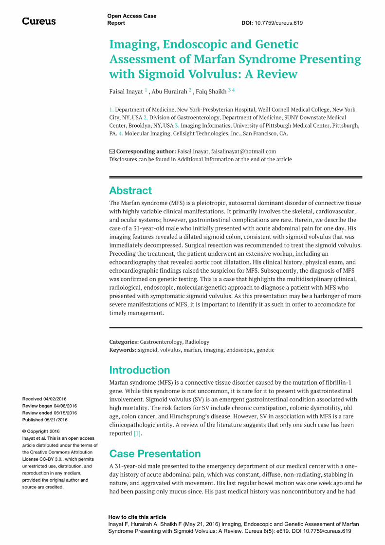
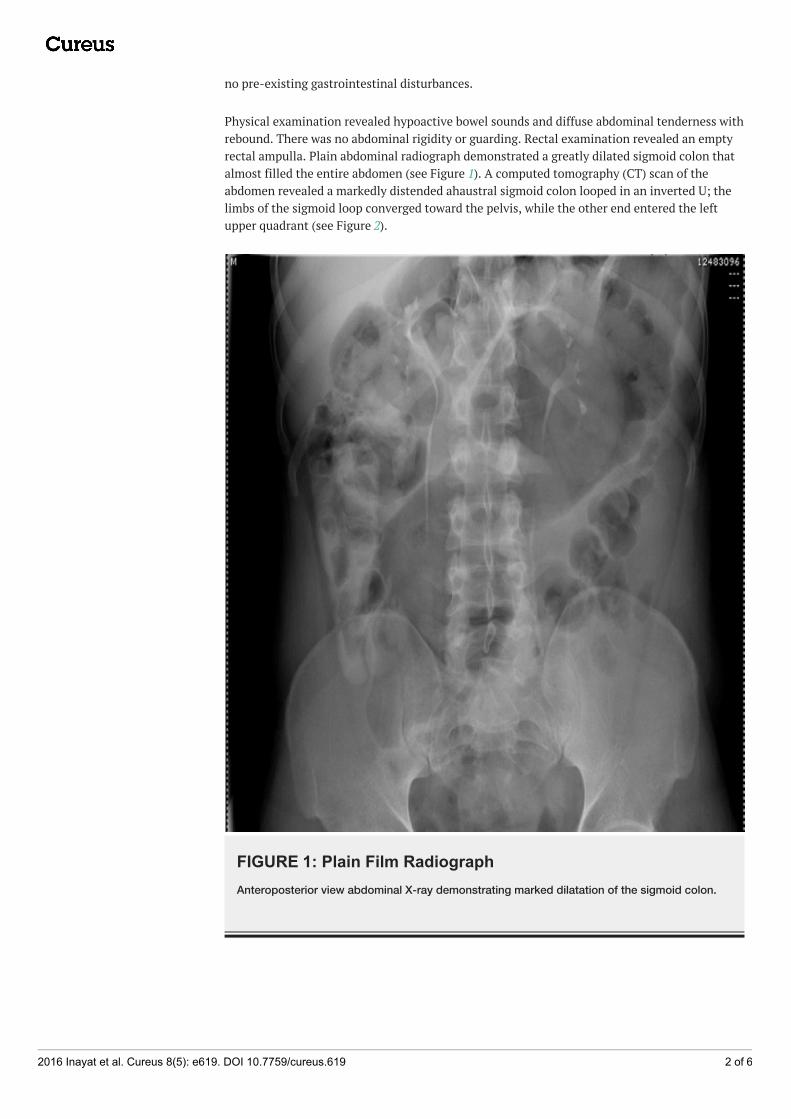
Physical examination revealed hypoactive bowel sounds and diffuse abdominal tenderness withrebound. There was no abdominal rigidity or guarding. Rectal examination revealed an emptyrectal ampulla. Plain abdominal radiograph demonstrated a greatly dilated sigmoid colon thatalmost filled the entire abdomen (see Figure 1). A computed tomography (CT) scan of theabdomen revealed a markedly distended ahaustral sigmoid colon looped in an inverted U; thelimbs of the sigmoid loop converged toward the pelvis, while the other end entered the leftupper quadrant (see Figure 2).
FIGURE 1: Plain Film RadiographAnteroposterior view abdominal X-ray demonstrating marked dilatation of the sigmoid colon.
2016 Inayat et al. Cureus 8(5): e619. DOI 10.7759/cureus.619 2 of 6
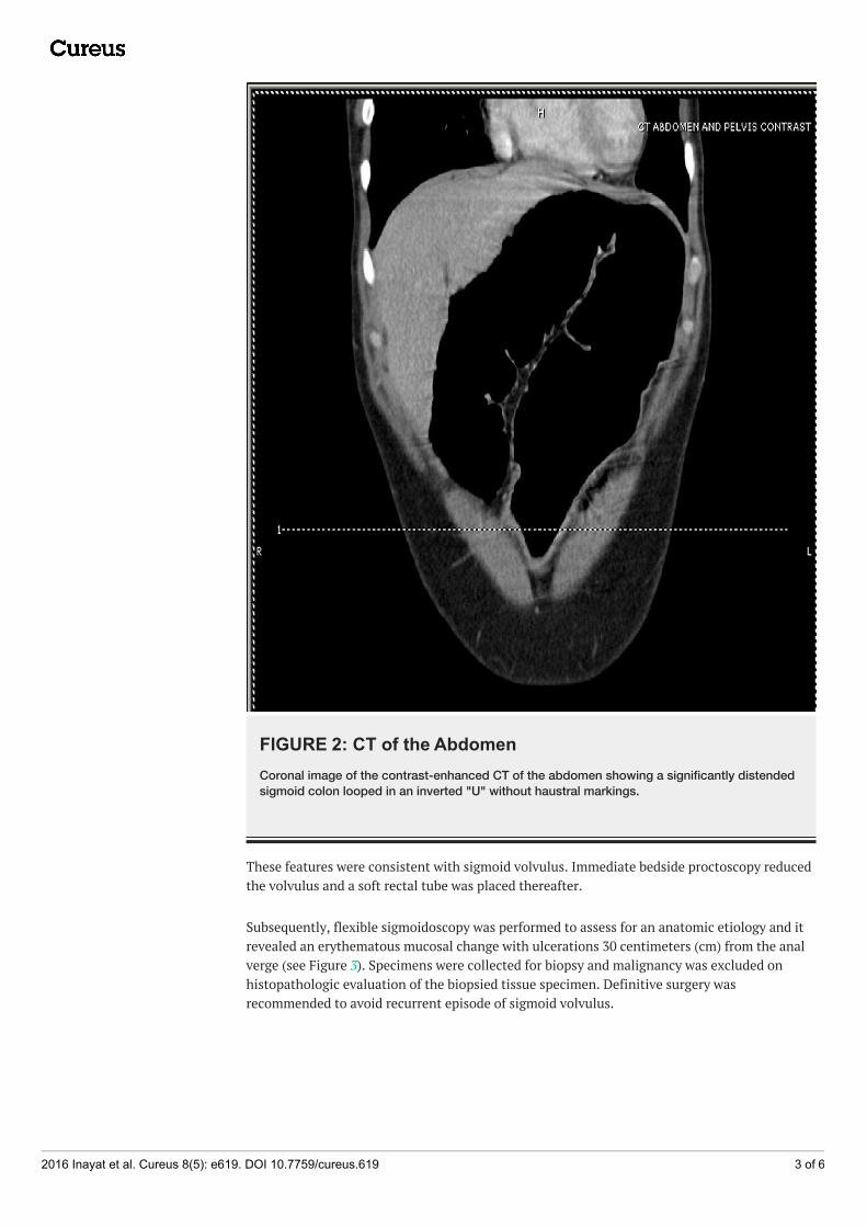
FIGURE 2: CT of the AbdomenCoronal image of the contrast-enhanced CT of the abdomen showing a significantly distendedsigmoid colon looped in an inverted "U" without haustral markings.
These features were consistent with sigmoid volvulus. Immediate bedside proctoscopy reducedthe volvulus and a soft rectal tube was placed thereafter.
Subsequently, flexible sigmoidoscopy was performed to assess for an anatomic etiology and itrevealed an erythematous mucosal change with ulcerations 30 centimeters (cm) from the analverge (see Figure 3). Specimens were collected for biopsy and malignancy was excluded onhistopathologic evaluation of the biopsied tissue specimen. Definitive surgery wasrecommended to avoid recurrent episode of sigmoid volvulus.
2016 Inayat et al. Cureus 8(5): e619. DOI 10.7759/cureus.619 3 of 6
FIGURE 3: SigmoidoscopySigmoidoscopy showing erythematous mucosal change with ulcerations 30 cm from the analverge.
During the preoperative workup, the patient was found to be 189 cm tall with an arm span of204 cm. He had an arm span/height ratio of 1.08. His face appeared narrow and thin, his teethovercrowded, and he was found to have myopic vision on the visual acuity test. AlthoughMarfan syndrome had not been clearly documented in the patient’s family, it was revealed thathis father died at a young age due to a sudden cardiac arrest. Furthermore, he reported ofMarfanoid appearance with arachnodactyly in one of his brothers. Laboratory evaluation was
notable only for leukocyte count of 10.3 x 10⁹/L. The patient had no signs and symptoms ofsepsis and his lactic acid level was within normal limits. Echocardiography showed evidence ofaortic root dilatation with mild aortic regurgitation.
Subsequently, a molecular genetic testing was performed and that showed a c.1948 C>T(p.Arg650Cys) mutation in fibrillin-1 gene, confirming the diagnosis of MFS. Genetic screeningwithin the family revealed that his brother was also a carrier of the same mutation.Subsequently, our patient underwent an uneventful subtotal colectomy. His recovery wasunremarkable and he was discharged from the hospital. Informed consent was obtained fromthe patient for this study.
DiscussionSigmoid volvulus (SV) occurs when there is torsion of the sigmoid colon over its vascularpedicle, causing obstruction or ischemia [1-2]. Risk factors for sigmoid volvulus include anelongated and redundant sigmoid colon, chronic constipation, obstipation, colonic dysmotility,old age, pregnancy, colon cancer, Parkinson’s, and Hirschsprung’s disease [3]. However, it is
2016 Inayat et al. Cureus 8(5): e619. DOI 10.7759/cureus.619 4 of 6
extremely rare to find SV secondary to MFS.
MFS is a multisystem connective tissue disorder. It follows an autosomal dominant pattern ofinheritance with around 30% of cases showing sporadic mutations [4]. It is caused by mutationof the gene encoding glycoprotein fibrillin-1 (FBN-1), which is the main constituent ofmicrofibrils in the extracellular matrix. Abnormal FBN-1 protein leads to laxity and weakness ofthe tissue structure [5]. MFS demonstrates clinical variability with predominant involvement ofskeletal, cardiovascular, and ocular systems. A variety of skeletal manifestations may be seendue to bone overgrowth and laxity of joints [6]. The cardiovascular complications include aorticroot dilatation, aortic dissection, mitral valve prolapse, and enlarged proximal pulmonaryartery. These are the major contributors in morbidity and mortality in patients with MFS.Myopia is the most common ocular feature. The ocular hallmark is ectopia lentis that is usuallybilateral. Pulmonary complications include bullae and spontaneous pneumothorax; striaeatrophicae and incisional hernias are the major skin problems [4].
The gastrointestinal involvement in MFS is rare. There are a few case reports on bowelobstruction due to malrotation of gut, gastroesophageal reflux disease (GERD), small hiatushernia, gastric volvulus, congenital band, diverticulitis, and Zenker’s diverticula in the adultpatient population [7-8]. There is only one previous report of SV with MFS described byJunpaparp et al. [1]. However, no case describing SV in a young patient with concomitant MFShas been reported. Despite the fact that patients with MFS are known to have long sigmoid andcecal mesenteries with possible structural intestinal defects that predispose them to developinga volvulus, such cases are extremely rare. Admittedly, it is difficult to establish this associationwithout a population study, but it would be unusual for a genetic disease that occurs in 1/3000individuals to be associated with a condition that is not that infrequent as SV.
The new diagnostic criteria for MFS put more weight on the cardiovascular manifestations ofthe disorder. Aortic root aneurysm and ectopia lentis are now cardinal features. In the absenceof any family history, the presence of these two features is sufficient for the unequivocaldiagnosis of MFS. In the absence of one of these two cardinal features, the presence of an FBN-1 gene mutation of positive systemic score is required [6]. Diagnosis of MFS was stronglysuspected in our patient on the basis of Marfanoid habitus, suggestive family history,echocardiographic evidence of aortic root dilatation and regurgitation, and intraoperativeevidence of large redundant sigmoid colon. Family assessment followed by the genetic testingfurther helped establish the diagnosis.
In MFS patients, the life span is generally reduced, mainly due to the cardiovascularcomplications. The medical treatment is β-blockers, which decreases the systolic aortic pulsepressure, thereby delaying aortic dilatation [5]. Therefore, MFS patients are also advised not toindulge in strenuous exercise [9].
A multidisciplinary approach, as applied in our case, using clinical, radiologic (CT, plain filmradiography), endoscopic, and genetic testing is required for early and accurate diagnosis,which then allows for comprehensive care of such patients, including application of screeningmeasures and interventions to address or prevent serious complications.
ConclusionsIn conclusion, sigmoid volvulus as a presentation of Marfan syndrome is unusual. Cliniciansshould maintain a high index of suspicion in these patients as prompt diagnosis is life-saving inthose with underlying or subsequent cardiovascular complications. Further population-basedclinical studies are warranted to broaden the scope of our knowledge on this association and toframe guidelines to standardize the care of such patients.
2016 Inayat et al. Cureus 8(5): e619. DOI 10.7759/cureus.619 5 of 6
Additional InformationDisclosuresHuman subjects: Consent was obtained by all participants in this study.
References1. Junpaparp P, Chayanupatkul M, Buppajarntham S, et al.: Sigmoid volvulus: is it related to
Marfan syndrome?. Int J Colorectal Dis. 2014, 29:771-772. 10.1007/s00384-014-1866-22. Hiltunen KM, Syrjä H, Matikainen M: Colonic volvulus. Diagnosis and results of treatment in
82 patients. Eur J Surg. 1992, 158:607-611.3. Sarioğlu A, Tanyel FC, Büyükpamukçu N, et al.: Colonic volvulus: a rare presentation of
Hirschsprung's disease. J Pediatr Surg. 1997, 32:117-118. 10.1016/S0022-3468(97)90113-54. Ha HI, Seo JB, Lee SH, et al.: Imaging of Marfan syndrome: multisystemic manifestations .
Radiographics. 2007, 27:989-1004. 10.1148/rg.2740651715. Fattori R, Reggiani LB, Pepe G, et al.: Magnetic resonance imaging evaluation of aortic elastic
properties as early expression of Marfan syndrome. J Cardiovasc Magn Reson. 2000, 2:251-256.
6. Loeys BL, Dietz HC, Braverman AC, et al.: The revised Ghent nosology for the Marfansyndrome. J Med Genet. 2010, 47:476-485. 10.1136/jmg.2009.072785
7. Thakur S, Jhobta A, Sharma B, et al.: Unusual presentation of adult Marfan syndrome as acomplex diaphragmatic hiatus hernia. Asian J Surg. 2014, 16:S1015-9584(14)00052-9.10.1016/j.asjsur.2014.04.003
8. Thomas GP, Purkayastha S, Athanasiou T, et al.: General surgical manifestations of Marfan'ssyndrome. Br J Hosp Med (Lond). 2008, 69:270-274. 10.12968/hmed.2008.69.5.29359
9. Dormand H, Mohiaddin RH: Cardiovascular magnetic resonance in Marfan syndrome . JCardiovasc Magn Reson. 2013, 15:33. 10.1186/1532-429X-15-33
2016 Inayat et al. Cureus 8(5): e619. DOI 10.7759/cureus.619 6 of 6