WHSC1, a 90 kb SET domain-containing gene, expressed in early
12
1998 Oxford University Press 1071–1082 Human Molecular Genetics, 1998, Vol. 7, No. 7 ARTICLE WHSC1, a 90 kb SET domain-containing gene, expressed in early development and homologous to a Drosophila dysmorphy gene maps in the Wolf–Hirschhorn syndrome critical region and is fused to IgH in t(4;14) multiple myeloma Ingrid Stec 1 , Tracy J. Wright 2 , Gert-Jan B. van Ommen 1 , Piet A. J. de Boer 3 , Arie van Haeringen 4 , Antoon F. M. Moorman 3 , Michael R. Altherr 2 and Johan T. den Dunnen 1,4, * 1 MGC-Department of Human Genetics and 4 Department of Clinical Genetics, Leiden University Medical Center, Leiden, The Netherlands, 2 Genomics Group, Los Alamos National Laboratory, Los Alamos, NM, USA and 3 Institute of Anatomy and Embryology, Academic Medical Center, Amsterdam, The Netherlands Received March 3, 1998; Revised and Accepted April 21, 1998 Wolf–Hirschhorn syndrome (WHS) is a malformation syndrome associated with a hemizygous deletion of the distal short arm of chromosome 4 (4p16.3). The smallest region of overlap between WHS patients, the WHS critical region, has been confined to 165 kb, of which the complete sequence is known. We have identified and studied a 90 kb gene, designated as WHSC1, mapping to the 165 kb WHS critical region. This 25 exon gene is expressed ubiquitously in early development and undergoes complex alternative splicing and differential polyadenylation. It encodes a 136 kDa protein containing four domains present in other developmental proteins: a PWWP domain, an HMG box, a SET domain also found in the Drosophila dysmorphy gene ash-encoded protein, and a PHD-type zinc finger. It is expressed preferentially in rapidly growing embryonic tissues, in a pattern corresponding to affected organs in WHS patients. The nature of the protein motifs, the expression pattern and its mapping to the critical region led us to propose WHSC1 as a good candidate gene to be responsible for many of the phenotypic features of WHS. Finally, as a serendipitous finding, of the t(4;14) (p16.3;q32.3) translocations recently described in multiple myelomas, at least three breakpoints merge the IgH and WHSC1 genes, potentially causing fusion proteins replacing WHSC1 exons 1–4 by the IgH 5′-VDJ moiety. INTRODUCTION Wolf–Hirschhorn syndrome (WHS; OMIM 194190) ( 1,2) is a multiple malformation syndrome caused by the loss of one copy of a distal segment of chromosome 4p. The main features of WHS patients are a prominent forehead with widely spaced eyes (known as ‘Greek helmet’) and a divergent strabism, brain anomalies causing mental retardation and growth retardation. Frequently, patients suffer from heart defects and several deficiencies of midline fusion e.g. cleft lip/palate, colobomata (iris defects), hernia diaphragmatica, omphalocele and hypo- spadia (1–4). WHS is allelic with the milder Pitt–Rogers–Danks syndrome (PRDS; OMIM 262350) (5–8). Patients affected by PRDS suffer from multiple congenital anomalies, overlapping in part with the symptoms of WHS. Up to now, only patients with a phenotype of WHS who have a microscopically detectable 4p deletion are considered to be affected by this syndrome, hence the cytogenetic description 4p– is used synonymously. Patients without an obvious deletion often remain ambiguous or undiagnosed. To date, no cases of typical WHS phenotype without deletion have been described. One undeleted phenotypic PRDS patient has been described by Donnai (9). Due to the large size of the deletions, which usually span several megabases, and the high variability of involvement of inner organs, WHS may well be a contiguous gene syndrome *To whom correspondence should be adressed. Tel: +31 71 5276105; Fax: +31 71 5276075; Email: [email protected]
Transcript of WHSC1, a 90 kb SET domain-containing gene, expressed in early
1998 Oxford University Press 1071–1082Human Molecular Genetics, 1998, Vol. 7, No. 7
ARTICLE
WHSC1, a 90 kb SET domain-containing gene,expressed in early development and homologous toa Drosophila dysmorphy gene maps in theWolf–Hirschhorn syndrome critical region and isfused to IgH in t(4;14) multiple myeloma Ingrid Stec 1, Tracy J. Wright 2, Gert-Jan B. van Ommen 1, Piet A. J. de Boer 3, Arie van Haeringen 4, Antoon F. M. Moorman 3, Michael R. Altherr 2 and Johan T. den D unnen 1,4,*
1MGC-Department of Human Genetics and 4Department of Clinical Genetics, Leiden University Medical Center,Leiden, The Netherlands, 2Genomics Group, Los Alamos National Laboratory, Los Alamos, NM, USA and3Institute of Anatomy and Embryology, Academic Medical Center, Amsterdam, The Netherlands
Received March 3, 1998; Revised and Accepted April 21, 1998
Wolf–Hirschhorn syndrome (WHS) is a malformation syndrome associated with a hemizygous deletion of thedistal short arm of chromosome 4 (4p16.3). The smallest region of overlap between WHS patients, the WHS criticalregion, has been confined to 165 kb, of which the complete sequence is known. We have identified and studieda 90 kb gene, designated as WHSC1, mapping to the 165 kb WHS critical region. This 25 exon gene is expressedubiquitously in early development and undergoes complex alternative splicing and differential polyadenylation.It encodes a 136 kDa protein containing four domains present in other developmental proteins: a PWWP domain,an HMG box, a SET domain also found in the Drosophila dysmorphy gene ash-encoded protein, and a PHD-typezinc finger. It is expressed preferentially in rapidly growing embryonic tissues, in a pattern corresponding toaffected organs in WHS patients. The nature of the protein motifs, the expression pattern and its mapping to thecritical region led us to propose WHSC1 as a good candidate gene to be responsible for many of the phenotypicfeatures of WHS. Finally, as a serendipitous finding, of the t(4;14) (p16.3;q32.3) translocations recently describedin multiple myelomas, at least three breakpoints merge the IgH and WHSC1 genes, potentially causing fusionproteins replacing WHSC1 exons 1–4 by the IgH 5 ′-VDJ moiety.
INTRODUCTION
Wolf–Hirschhorn syndrome (WHS; OMIM 194190) (1,2) is amultiple malformation syndrome caused by the loss of one copyof a distal segment of chromosome 4p. The main features of WHSpatients are a prominent forehead with widely spaced eyes(known as ‘Greek helmet’) and a divergent strabism, brainanomalies causing mental retardation and growth retardation.Frequently, patients suffer from heart defects and severaldeficiencies of midline fusion e.g. cleft lip/palate, colobomata(iris defects), hernia diaphragmatica, omphalocele and hypo-spadia (1–4). WHS is allelic with the milder Pitt–Rogers–Dankssyndrome (PRDS; OMIM 262350) (5–8). Patients affected by
PRDS suffer from multiple congenital anomalies, overlapping inpart with the symptoms of WHS.
Up to now, only patients with a phenotype of WHS who havea microscopically detectable 4p deletion are considered to beaffected by this syndrome, hence the cytogenetic description 4p–is used synonymously. Patients without an obvious deletion oftenremain ambiguous or undiagnosed. To date, no cases of typicalWHS phenotype without deletion have been described. Oneundeleted phenotypic PRDS patient has been described byDonnai (9).
Due to the large size of the deletions, which usually spanseveral megabases, and the high variability of involvement ofinner organs, WHS may well be a contiguous gene syndrome
*To whom correspondence should be adressed. Tel: +31 71 5276105; Fax: +31 71 5276075; Email: [email protected]

Human Molecular Genetics, 1998, Vol. 7, No. 71072
Figure 1. Gene structure of WHSC1. Top: 4p16.3 markers are shown from telomere to centromere. The WHSCR is located between the FGFR3 gene locus and theHuntington disease (HD) gene. Middle: cosmid contig overlapping the WHSCR. Smallest region of overlap determined by two deleted WHS patients [cell lines CM andLGL7447 (13)] (deleted region dotted line, WHSCR shadowed, WHSC1 bold line). WHSC1 covers cosmids cl96a2, cl19h1, cl190b4 and cl184d6. Bottom: exon–intronorganization of WHSC1. Boxes represent exons, lines represent introns, primers in exons are given in upper case letters. Exon 4 contains the translational start codon ATG,exon 25 contains the translational stop codon TAG. The ORF is shadowed in light grey. Lightning symbols inidicate the breakpoints of the (4;14) translocations in the multiplemyeloma cell lines in WHSC1. Alternative splicing is found in at least three different regions indicated by superpositioned exons. Different polyadenylation sites ending ina poly(A) tail are indicated by poly(A) stretches. Four domains are detected, demonstrated below the exon numbering: PWWP domain, HMG box, PHD-type zinc fingerdomain and SET domain. cDNA length scale and the corresponding length of genomic sequence are shown at the very bottom of the figure.
caused by the loss of more than one gene. In ∼85% of WHS cases,a de novo deletion of 4p16 in one of the parents’ germ cells hasoccurred. In ∼15%, the deletion is the result of an unbalancedtranslocation in the affected child transmitted by one of theparents carrying a balanced 4p translocation. During embryonicdevelopment, haploinsufficiency of genes influencing mor-phogenesis often has profound effects and may lead to a broadrange of phenotypic features such as found in a number of (micro)deletion syndromes, e.g. Rubinstein–Taybi syndrome (OMIM180849; deletion of 16p13.3) (10), Smith–Magenis syndrome(SMS; OMIM 182290; deletion of 17p11.2) (11) or velo-cardio-facial syndrome (VCFS; OMIM 192430; deletion of22q.11.2) (12). This may apply to WHS as well. If locus controlregions are deleted, gene expression may be disturbed over verylarge distances, i.e. position effects on the boundaries ofcoordinately expressed genes.
The gene defect(s) underlying both WHS and PRDS is (are)unknown. Based on the minimal overlap of deletions of differentWHS patients (Fig. 1), the WHS critical gene region (WHSCR)has been confined to only 165 kb (13). This region has beensequenced completely during the search for the Huntingtondisease gene (14). Wright et al. reported different transcription
units within this candidate gene region. In the present report, wedescribe a novel developmental gene two-thirds of which mapsin the distal part of the WHSCR. We have designated this gene asWHSC1 for Wolf–Hirschhorn syndrome candidate 1. WHSC1was identified initially due to the high similarity of the translationproduct of an expressed sequence tag (EST) contig Hs.110457,located in the 165 kb WHSCR and previously not reported, withthe so-called SET domain of the Drosophila protein ASH1(absent, small or homeotic discs) (15). The SET domain [forsuppressor of variegation, enhancer of zeste and Trithorax (16),see Discussion] is found in proteins which are involved inembryonal development (15). WHSC1 merges two of thetranscripts in the 165 kb region, HFBEP10 and 194164, reportedpreviously by Wright et al. (13). Its expression profile, especiallyin the tissues affected in WHS and PRDS, and its deducedfunction make it an excellent candidate gene for causal involve-ment in the phenotype of both syndromes.
RESULTS
Our strategy to complete WHSC1 (Fig. 1) utilized databasecomparisons, primarily dbEST and gene prediction programs, to

1073
Nucleic Acids Research, 1994, Vol. 22, No. 1Human Molecular Genetics, 1998, Vol. 7, No. 71073
Figure 2. RT-PCR analysis in WHSC1 on RNA. (a) Lane 1, 850 bp, primers L/K spanning exons 1, 4, 5 and 6 (on blood-derived RNA); lane 2, 780 bp, primersCW2/CW3 spanning exons 5–8 (blood); lane 3, 880 bp, primers H/B spanning exons 9, 10, 11 and 13–16 (testis); lane 4, 820 bp, primer combination V/F spanningexons 7–11 and 13 and 14 (testis); lane 5, 720 bp, primers A/3R spanning exons 16–20 (testis); lane 6, 1600 bp, primer combination A/mR spanning exons 16–25(blood); lane 7, 550 bp, primers G/1R spanning exons 24 and 25 (blood); lane 8, 1320 bp, primers 1F/1R spanning exons 20–25 (blood). (b) Alternative splicing. Lanes9 (blood), 10 (heart) and 11 (brain), primers S/F; 550/370 bp spanning exons 12b and 13 and exons 12a and 13 respectively. (c) Lanes 12 and 13, primers L/W: lane12, brain, 400 bp spanning exons 1, 2, 3 and 4; lane 13, heart, 230 bp spanning exons 1 and 4. The 100 bp and 1 kb ladders are used as size markers (left and/or rightof lanes).
design primers for RT-PCR analysis. Using overlapping RT-PCRsets, starting from the 3′ end, we extended the gene in the 5′direction until we failed to obtain 4p16.3-derived amplificationproducts. Transcription covering the entire gene could bedetected in adult blood, brain and heart. After a nested PCRreaction, transcription could also be detected in adult testis andpancreas. All RT-PCR products were sequenced and comparedwith the genomic sequence to identify the exon–intronboundaries (Fig. 1). Finally, we used 5′ and 3′ RACE, northernblotting and in situ hybridization on mouse and human embryosections to characterize the transcription and expression profile ofWHSC1.
Characterization of WHSC1
The entire WHSC1 gene measures 90 kb, is transcribed fromtelomere to centromere and extends for 60 kb into the telomericend of the 165 kb critical region. It contains 25 exons, togetherencoding an 8 kb cDNA. The ATG translation start codon lies inexon 4 preceded by a 5′-untranslated region (UTR) of >400 bp(Fig. 1, Table 1). The exons vary between 82 and 3565 bp, theintrons between 132 and 13 718 bp. Exon 14 does not fulfil theGT rule at the 5′-splice donor site. It contains a GC dinucleotide,a donor site which occasionally has been found in other genes,e.g. in exon 30 of the Duchenne muscular dystrophy gene (17).The splice donor site of exon 14 is functional, since RT-PCRcovering this exon consistently yields unique products includingthis exon (Fig. 2a, lane 4).
The most 5′ dbEST match of WHSC1 is EST 27266 (Table 2).Using a forward primer designed on more upstream EST matches(k3378, k3397 and j3435 in cl75b9a) in combination with severalreverse primers located in exons 4 (Fig. 1, W and M) and 6(Fig. 1, K), we have not been able to extend WHSC1 further in the5′ direction. Some amplification products were obtained, but theirsequence showed that they were artefactual products, not derivedfrom 4p16.3. Similarly, 5′ RACE using human fetal brain andadult testis cDNA (Marathon Ready kits; Clontech) with a rangeof primers spread throughout WHSC1 failed to extend the cDNAsequence upstream of exon 1. Only after nested PCR were somePCR products obtained, but these where unspliced genomicproducts, not extending WHSC1 in the 5′ direction.
Alternative splicing was found for exons 2 and 3 (Figs 1 and2c). RT-PCR between exons 1 and 4 (primers L/W) produces amajor 280 bp product on blood, pancreas, testis and heart RNA,and a 400 bp product on brain material (Fig. 2c, lanes 12 and 13).The sequence of the 400 bp product contains exons 1–4, while the280 bp product lacks exons 2 and 3. The EST database containsone clone, EST 27266, which only lacks exon 2 (Table 2). Thus,at least three different splice forms of WHSC1 exist in this region.
WHSC1 shows dbEST matches for the region spanning exons1–12a and 20–25. Exons 13–19 are not represented in dbEST(Table 2). The exon 1–12a matches are with sequences derivedmostly from random-primed cDNA libraries of human, murineand rat origin, and they do not form a single contig. Another groupof EST matches is found with genomic sequences starting and/orending in intronic sequences, often flanking oligo(dT) oroligo(dA) stretches. Some of these are derived from the oppositetranscriptional strand (Table 2). These ESTs probably representunspliced RNAs or DNA-primed products.
Transcription in the exon 10–13 region is very complex. Sometranscripts contain either one of two alternatively spliced exons12, while others terminate here using either of two alternativepoly(A) addition sites. Exon 12, identified through EST zr01a04,uses two splice donor sites yielding exons of 94 and 227 bprespectively (12a and 12b, Fig. 1, Table 1). Expression of exon 12is only detectable using a primer located in this exon (Fig. 2b,lanes 9–11). In contrast, PCR across exon 12, e.g. from exon 7 to14 [V/F (Fig. 2a, lane 4)], yields only products without this exon.
An alternatively spliced exon was found by sequence analysisof cDNA clones HFBEP10 and zv63h03 which terminate in‘intron 11’. HFBEP10 contains exons 7 (in part), 8, 9 and 10spliced to 11b, and has a total size of 3613 bp. Another clone, ESTzv63h03, ends in a poly(A) tail 646 bp downstream of exon 11,preceded 37 bp upstream by a perfect AATAAA poly(A) additionsignal (Fig. 1, Tables 1 and 2). This polyadenylation site could beconfirmed by 3′ RACE PCR on heart and brain mRNA. To findout whether the ‘intronic’ exon 11b is evolutionary conserved, aSouthern blot with DNA from a range of species was hybridizedwith HFBEP10. Human DNA shows two hybridizing bands of4.8 kb (prominent) for exon 11b and 3.8 kb (weak) for the exon7 segment (345 bp). Cross-hybridization was detected in cow,dog, mouse, rat and primate (Fig. 3).

Human Molecular Genetics, 1998, Vol. 7, No. 71074
Table 1. Sequences and positions of exon–intron boundaries in WHSC1
Exon no. Intron (3′) Exon (5′) Exon (3′) Intron (5′) Size (bp) Cosmid Position
1 TAACTG gtaatt (≥40) cl184d6 143 239–14 200
2 ttgcag AGACAA CACGAG gtgggc 180 cl184d6 14 938–14 759
3 cctcag CTGTCT TCAGAG gtcagg 175 cl184d6 7888–7714
4 ccatag TGTTCT AAAAAG gtattt 626 cl184d6 6485–5858
5 tttcag ATTCCA TTAAAG gtattg 163 cl184d6 2893–2731
6 aactag GTCAGA ATTAAG gtgata 167 cl190b4 19 885–19 719
7 tcccag CTATTG GATGAG gtcagt 483 cl190b4 18 615–18 133
8 ccacag GTGGTA ACTCTG gtaaac 145 cl190b4 6129–5985
9 tccaag GTAATG CGGAAG gtaatt 119 cl190b4 1610–1492
10 taatag AGAGAC AGGCAG gtaatg 82 cl19h1 38 351–38 270
11 ttacag CAACGA AATGAG gtaaaa 125 cl19h1 37 148–37 024
11a ttacag CAACGA 227 cl19h1 37 148–36 922
11b ttacag CAACGA 2922 cl19h1 37 148–34 227
12a taacag CTTTTG CTGCAG gtggcg 94 cl19h1 34 463–34 370
12b taacag CTTTTG ACCAAG gtaaga 227 cl19h1 34 463–34 237
13 ctctag GTCTCG TGCCAG gtgagg 132 cl19h1 25 731–25 600
14 ccgcag CTGTGT CCTCAG gcaagt 124 cl19h1 24 719–24 596
15 tgttag GGATTC CAAAAG gtacag 200 cl19h1 23 502–23 303
16 cttcag GTAAAA CCAAAG gtgagg 180 cl19h1 21 666–21 487
17 ttgcag GGGGGA CTACAG gtgtga 157 cl19h1 21 134–20 978
18 taatag ATGGTG AAAACG gtacgg 206 cl19h1 20 844–20 639
19 ttttag CACTGC ATCAAG gtggcg 104 cl19h1 18 894–18 791
20 ctgcag GTGAAT AGAAAG gtatgt 270 cl19h1 17 356–17 087
21 ttccag GGAGAA GACAAG gtaatg 117 cl19h1 15 792–15 676
22 ttacag GACCGT CTGCAG gtacaa 142 cl19h1 1956–1815
23 tcccag GGACGG CCAAAG gtaagg 107 cl19h1 1525–1419
24 ctctag ACCTCG CCTTCG gtgggt 205 cl19h1 344–140
25 ttgcag GGAAGT 1159 cl96a2 33 362–32 204
25a ttgcag GGAAGT 3565 cl96a2 33 362–29 797
For every exon–intron boundary, six nucleotides each are given.
Also at its extreme 3′ end, WHSC1 appears to utilize twoalternative poly(A) addition sites (Table 2), represented by twoEST contigs. The first EST contig (Hs.110457) spans exons20–25, and ends at a polyadenylation site located at bp 32 204 incl96a2. This site, not preceded by an obvious consensus poly(A)addition signal, is found in five human and five murine ESTs(Table 2). The second EST contig [Hs.19416, containingtranscript 194164 of Wright et al. (13)] ends 2.4 kb furtherdownstream, at bp 29 796 in cl96a2, and is preceded by anAATAAA poly(A) signal 31 bp upstream. This contig contains 22human and three murine transcripts and spans 2.3 kb. It is notinterrupted by introns, has no coding potential, detects nosimilarities in the various databases and misses overlapping withthe first contig by 684 bp. Since EST contigs rarely span >2 kb
of cDNA, it is not surprising that no clone connecting the twocontigs was present. Only 500 bp 3′ of WHSC1, at bp 29 277 ofcl96a2, maps the 3′ end of another gene, transcribed from theopposite strand [EST contig Hs.21771, containing transcripts53282 and 267784 of Wright et al. (13)].
Between exons 4 and 25, WHSC1 contains a 4095 bp openreading frame (ORF) encoding a putative protein of 136 kDafollowed by a 3′ UTR of 3294 bp. Alternative splicing of exons2 and 3 does not result in additional translation products.Transcripts using the alternative poly(A) addition sites in exon11a and 11b (Tables 1 and 2) would contain a translational stopat cDNA bp 1887 (6 bp of ‘intron 11’), yielding a 62 kDa protein,transcripts containing exon 12 would end at bp 1941 (60 bp ofexon 12), encoding a 64 kDa protein with 20 novel amino acids.

1075
Nucleic Acids Research, 1994, Vol. 22, No. 1Human Molecular Genetics, 1998, Vol. 7, No. 71075
Table 2. EST matches in WHSC1
Exons ESTs Remarks
1, 3, 4 EST27266
4 HBMSF1D9
4–11 zv63h03 (758357) internal deletion exon 4–7, 3′ poly(A) 646 bp downstream of exon 11
5–7 EST111365 rat (reverse)
i5 ys11f05 27616–26811, ends in genomic dA stretch
6–7 45h5 (W28349) starts in intron 5, ends in intron 7
6–7 zr01a04 unspliced, 5′ end in intron 5, 3′ in intron 7 dA stretch
7 EST05750 HMG homology?
7–8 EST28510
7–9 ztd05d09.r1
i7 yv67d10 intronic, ends in genomic dA stretch
9–12a mw14h01.r1 murine, exon 2561 bp downstream in intron 11
10 zs25f05 antisense, primed at intron 9 dT and intron 10 dA stretch
11–12a HUMEST6H1
20–25 zo77g11, zm24f01, zm13b05, zo78d05, zo65g02.r1, zf12c11 ending in poly(A) tail at 32 205, 31 bp 5′ preceded by AATATA
20–22 mh09c05.r1 murine
22–23 ml55h10.r1 murine
25 mg09f12.r1, mi51h05.r1 murine
25 zs47g04.s1, aa61g04.s1 3′ end antisense, 5′ end not on chromosome 4a
25a EST17737, EST36779, EST60207, EST64437, EST70993,nf65b08.s, nf68c05.s, nh82c06.s, nk39a02.s, nn40a11.s,zd48f12.s,zn34b11, ye76b09, yg46e03.r, ym59a12.r, yp61c04.r,yp84g03, yr11g06, yr13c09.r, yr25c04, yr72b04.r, yr76b04
covers 32 203–29 796: ending in poly(A) tail, 31 bp 5′preceded by ATTAAA
aOn the opposite strand, bp 32530–33021 of cl96a2, exon 25 has an exact match with the 3′ end of ESTs zs47g04 and aa61g04. These ESTs, part of a large EST contig,seem not to originate from chromosome 4 since their 5′ sequences show no homology with any of the 2 Mb chromosome 4 sequences known from this region.
Northern blot analysis
Northern blot analysis with different RT-PCR probes dispersedover WHSC1 using human adult and fetal multiple tissue blots(Clontech) revealed a complex transcription pattern (Fig. 4).Main transcripts of 9 and 6 kb were recognized with all probes andin most tissues. The size of the 9 kb transcript indicates that the8 kb WHSC1 cDNA sequence described here misses some0.5–1 kb of untranslated sequence, probably 5′. Similarly, thetranscripts initiated from the same promoter but ending in ‘intron11’ (i.e. using exons 11a or 11b) would have sizes of 5.2 and2.6 kb respectively. Using the exon 1–4 probe (L/M, 500 bpproduct derived from blood RNA), transcripts of ∼2.5 kb weredetected in all tissues, whereas the 9 kb transcript was observedonly in heart.
Expression is most diverse in fetal tissues (Fig. 4b). At least fivetranscripts were detected, ranging in size from 3.7 to >10 kb usingan exon 7–11b cDNA probe. Fetal brain contained the mostcomplex pattern, with transcripts of 3.7, 4.5, 9, 10 kb and a unique>10 kb transcript. Adult brain transcripts appear to be a subset ofthose in fetal brain. The major 9 kb transcript was detected in allfetal and adult tissues examined, the 6 kb transcript in fetal liverand kidney only (Fig. 4c).
Hybridization of the exon 20–25 probe (1F/1R, 1300 bpproduct derived from testis RNA) uniquely revealed a 3.5 kbRNA in all tissues examined (Fig. 4a). Expression of alltranscripts was very prominent in skeletal muscle and heart and
Figure 3. Zoo blot analysis with HFBEP10. Zoo blot containing 4 µg of DNAdigested with EcoRI. The filter is hybridized non-radioactively with the 3.6 kbHFBEP10 cDNA clone, the signal was detected using an anti-fluoresceinalkaline phosphatase conjugate. C, cow; D, dog; M, mouse; R, rat; P, primate;H, human.
clearly detectable in tissues of the inner organs such as kidney,lung, pancreas and liver.
To exclude that transcripts were derived from other, homolo-gous genes, a chromosomal hybrid mapping panel was hybrid-ized with the exon 7–11b probe. The results show a 100%concordance, with a unique localization on chromosome 4.

Human Molecular Genetics, 1998, Vol. 7, No. 71076
Figure 4. Expression of WHSC1: northern blot analysis. Northern blots containing 2 µg of poly(A)+ RNA from various human adult or fetal tissues were hybridizedwith different RT-PCR products and the cDNA probe indicated. (a) Adult tissue blot hybridized with RT-PCR probe 1F/1R spanning exons 20–25. (b) Fetal tissueblot hybridized with cDNA clone HFPEB10 spanning exons 7–11b. (c) Adult tissue blot hybridized with cDNA clone HFPEB10 spanning exons 7–11b. Br, brain;H, heart; Ki, kidney; Li, liver; Lu, lung; Pa, pancreas; Pl, placenta; Sm, skeletal muscle.
Furthermore, we can exclude the existence of homologous geneselsewhere on chromosome 4 since only the known WHSC1-hybridizing fragments were detected (Fig. 3, lane H).
RNA in situ hybridization on embryonic mouse sections
To analyse the developmental pattern of expression of WHSC1,we performed in situ hybridization on mouse embryo sections ofdifferent developmental stages (E10.5, 12.5, 13.5 and 16.5 p.c.)and human embryos (developmental days 52–56). We used35S-labelled antisense and sense RNA probes covering differentregions of WHSC1. Identical expression patterns were obtainedusing a human probe containing exons 1, 4, 5 and 6 (L/K, 850 bp),murine exon 9–12a probe (H/S, 280 bp) and murine exon 20–25aprobe (mF/mR, 1 kb) on mouse sections (Figs 1 and 5).Hybridization of the human probe L/K to human embryonicsections displayed an essentially similar but weaker signal. Thenegative control with human probe L/K to human sectionsyielded a weak signal in the outflow region of the heart.
Figure 5a1 shows the expression pattern using the murine exon9–12a RNA probe at developmental day 13.5. It stronglyhybridizes specifically to brain, ganglia and neural tube, to theanlage region of the jaw, to the frontal face region including thedeveloping upper and lower lip, to intestinal and lung epithelium,to liver and to the adrenals and the urogenital system. Figure 5c–eshows detailed pictures of the expression pattern in brain (c),ganglia, adrenal and epithelium of the intestine (d), and in theliver and the lung epithelium (e). At day E10.5, expression ishighly specific throughout the developing nervous system (Fig.5b). The negative (sense) RNA controls showed no signal withany probe–section combination (Fig. 5a2). Some tissues, such asthe liver, the dorsal ganglia and the fifth ganglion in the brain,show relatively even expression, while other tissues, e.g.epithelium of the gut and lung, show a marked inward–outwardgradient, suggesting that rapidly growing layers in these tissueshave a higher expression than their surroundings.
DISCUSSION
WHS is typically caused by large deletions of the distal short armof one of the chromosomes 4. The critical region previously hasbeen confined to 165 kb, and nine potential transcriptional unitshave been described in this interval (13). Several genes mayjointly contribute to the WHS phenotype. We have characterizeda novel gene (WHSC1) covering 60 kb of the 165 kb criticalregion and merging two of the transcription units reportedrecently in the WHSCR, HFBEP10 and 194164. Its location, thenature of the motifs in the encoded protein, and the expressionprofile in human and mouse embryos imply that hemizygousdeletions of this gene might be responsible for many of thephysical and neurological features in WHS.
Our results indicate that the expression of the WHSC1 gene iscomplex, showing, apart from the major consistent transcripts of9 and 6 kb, many different transcripts in different tissues.Alternative splicing, both between and within tissues, affectsexons 2, 3, 11 and 12. Since the length of exons 2, 3 and 12 israther small, 100–230 bp, it is unlikely that their alternativesplicing yields transcripts of lengths which can be discriminatedon northern blots. However, alternative polyadenylation of exons11a and 11b should yield transcripts differing some 2.2 kb in size.Similarly, the two polyadenylation sites used for exon 25 shouldyield differently sized transcripts depending on the respectivepoly(A) addition site used. Finally, based on the complexexpression patterns observed on fetal and adult northern blots,WHSC1 expression is most likely also driven by more than onepromoter.
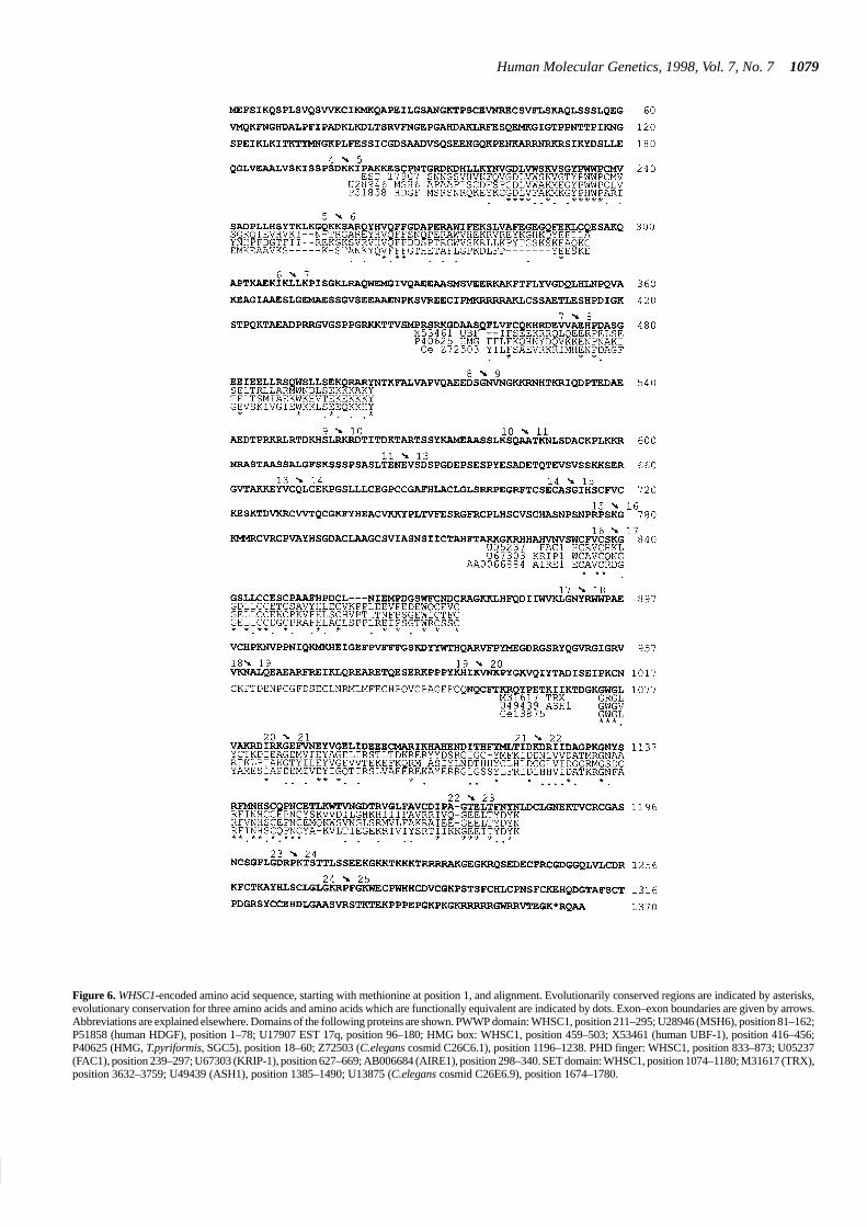
The WHSC1-encoded protein reveals homologies to membersof several protein families. This suggests that WHSC1 product isa DNA-binding protein, most likely a transcription factor orco-regulator. At amino acid position 211–295, the WHSC1-encoded protein contains a highly conserved region which wehave designated the PWWP domain (for the proline–tryptophan–tryptophan–proline motif in the consensus amino acid sequence)(see Fig. 6). This domain has been noted but not described(ProDom database, ID14260). It is also present in the G/T DNAmismatch repair gene MSH6 product causing non-polyposis

1077
Nucleic Acids Research, 1994, Vol. 22, No. 1Human Molecular Genetics, 1998, Vol. 7, No. 71077
Figure 5. RNA in situ hybridization on sagittal mouse embryonic sections. (a) Hybridization of murine antisense (a1) and sense (a2) RNA probes generated withprimers H/S (exons 9–12a, Fig. 1) to mouse sections of developmental day 13.5 p.c. Expression is very prominent in neuronal cells as in the brain, in the fifth ganglion(ganglion trigeminale) and optical nerve. Enhanced expression is also detected in the margin of the frontal facial region, the region forming the lip, the region of thedeveloping jaw, the epithelium of lung and gut, the liver, the genitourinary system and the adrenals. (b) Hybridization of a murine antisense RNA probe generatedwith primers mF/mR (exons 21–25) to mouse sections of developmental day 10.5 p.c. It demonstrates specific expression in brain and the neural tube. In detail: specificexpression in the brain (c) and epithelium of the intestine and lung (d and e). a, adrenal; ie, inner ear; glt, ganglion trigeminale; gt, genital tubercle; h, heart; j, jaw;lu, lung; li, liver; ll, lower lip; lu, lung; mg, midgut; on, optical nerve; re, rhombencephalon; spgl, spinal ganglia; te, telencephalon; ub, urinary bladder; ul, upper lip;umg, umbilical midgut; arrows indicate the developing region of the frontal face.

Human Molecular Genetics, 1998, Vol. 7, No. 71078
colorectal cancer (HNPCC) (18), in the hepatoma-derivedgrowth factor (HDGF) (19) and in a 17q-derived EST expressedin lymphocytes (20).
Amino acids 459–503 show homology with the HMG box 2.HMG proteins are grouped in three families, HMG-1/-2,HMG-14/-17 and HMG-I(Y). HMG boxes facilitate DNAinteractions and have been found in many proteins includingRNA polymerase I transcription factor UBF (upstream bindingfactor), the mammalian testis-determining factor SRY and twomitochondrial transcription factors (21). HMG boxes are DNA-binding domains, causing a 130� bend of the double helix. HMGproteins are positively correlated to cell proliferation. Highhomology (36% identity, 52% similarity) is observed between theWHSC1-encoded protein and the HMG2 box in BF1_humannucleolar transcription factor 1 (UBF-1, autoantigen NOR-90)(22), TETPY (Tetrahymena pyriformis) HMG non-histonechromosomal protein (23) and a gene product of Caenorhabditiselegans, C26C6.1 (24).
The PHD-type zinc finger (plant-homeodomain) is located atposition 833–875 of the WHSC1 protein. This domain has beenfound in a series of proteins of different species: human ATRXprotein (alpha-thalassaemia mental retardation syndrome, X-linked,with genital abnormalities and facial dysmorphism), human FAC-fetal protein (fetal Alz-50-reactive clone 1 (25), humanKRIP-1(KRAB-A interacting protein; KRAB-A: Krüppel-associated box A) (26), human Mi-2 immune autoantigen, a nuclearprotein (27), AIRE (autoimmune regulator), a recently cloned generesponsible for APECED syndrome (28,29), human TIF1 (trans-criptional intermediary factor 1) and C.elegans gene C44B9.4.ATRX is thought to influence gene expression by affectingchromatin (30). KRIP-1 is a member of a group of proteins whichare known to play important roles in differentiation, oncogenesis andsignal transduction. It probably interacts, like TIF1, with proteinsessential for transcription regulation. The function of the TIFproteins appears to be as a mediator of ligand-dependent activationwhich acts on the basal transcription machinery (31).
The SET domain (16) at position 1074–1180 consists of ∼140amino acids and has been identified in a large range oftranscriptional/developmental proteins from diverse organisms(15): human ALL1 (acute lymphocytic leukaemia) protein (32),in ORFKG1T, a hypothetical human protein (24), in threeASH1-related Drosophila proteins TRX (Trithorax) (33),SUVAR 9 (suppressor of variegation 9) and enhancer ofzeste/polycomb [E(z)] (16,34,35), in HRX (human homologue ofTRX, human and murine) (36,37), in two predicted C.elegansgenes (24) and two yeast ORFs (38,39). Highest homology (44%identity, 64% similarity) of the WHSC1 SET domain occurs withASH1 of Drosophila melanogaster. Mutations in the ash1 genecause misdevelopment in a number of organs in Drosophila, e.g.transformation of the halters to wings, genitalia to legs, first legto second leg, etc. (40). Nilsow et al. (41) created a null mutantin the fission yeast SET1 gene displaying morphological abnor-malities, cell growth and sporulation defects. In Drosophila, the‘enhancer of zeste’ gene has a negative regulator function ofsegment identity genes of the Antennapedia and Bithoraxcomplexes (34,35). The SET domain genes are a highlyconserved family encoding proteins which influence transcrip-tion by changing chromatin-mediated regulating mechanismsleading to secondary effects on developmental programmes. Incombination with the expression profile of WHSC1, the proteindomain homologies, in particular with the SET domain, strongly
suggest that WHSC1 encodes a protein which plays a substantialrole in transcription regulation of genes involved in embryonicdevelopment.
We have collected a set of seven patients with many featurescharacteristic for WHS or PRDS. To date, we have not been ableto detect any gross deficiencies nor microdeletions or pointmutations focused on the domains recognized in the proteinsequence. A silent third base change from C to T was found in oneof the patients in exon 22. Since most WHS/PRDS patients carryextensive 4p16 deletions and lack this gene completely, thequestion arises if small mutations in this single gene wouldalready yield the characteristic WHS phenotypical spectrum orwhether the deletion of more genes is required.
Apart from the facial hallmarks, WHS patients suffer frommultiple malformations which arise during the first trimester ofdevelopment. These malformations affect the CNS (micro-cephaly, hypotonia, seizures), eyes (widely spaced, round), ears(low set), mouth (cleft lip/palate, fused teeth), extremities(overlapping toes), genitourinary system (hypospadia, cryptorch-ism) and heart. The results of the in situ hybridization on murineembryonic sections demonstrate expression in many of thesetissues as early as day 10.5. The temporal and spatial multi-organic expression pattern provides independent support for theinvolvement of WHSC1 in the development of the WHSphenotype, besides its location. The brain, jaw and genitourinarysystem show gene expression corresponding to affected organs inWHS. For example, brain anomalies cause developmental delay,while midline fusion defects of the developing jaw and genito-urinary region may lead to cleft lip/palate and hypospadia. Alsothe distinct inward–outward gradients observed in several tissuesand the overall abundance in rapidly growing, not yet differen-tiated structures suggest that WHSC1 may well be involved indetermining as yet unknown morphogenetic gradients, as aremany other transcription factors. Typically, the actions and effectsof such proteins are highly dosage sensitive, which would beconsistent with a haploinsufficiency model for the protein(s)affected in WHS. The complex pattern of tissue-specific tran-scription initiation, splicing and polyadenylation would present asignificant potential source of further inter-individual geneticdiversity at the transcript and protein level. Indeed, this variabilitywould present an attractive mechanistic explanation for thegenerally observed phenotypic variation in haploinsufficiency(deletion) syndromes. This would link the natural geneticdiversity of the expression levels of genes on the non-affectedchromosome to clinical variation caused by deletion on thehomologous chromosome.
Finally, an apparently unrelated recent finding is well worthnoting. Patients with multiple myeloma (MM) suffer from tumourswhich destroy bone marrow structures especially in the skull,clavicles, sternum and vertebrae. Recent reports describe t(4;14)(p16.3;q32.3) translocations in a significant fraction of primarytumours and tumour-derived cell lines of MM patients located <100kb centromeric of the FGFR3 gene in 4p16.3. Several of these mapin cosmids cl75b9, cl184d6 and cl190b4 (42,43). As deduced fromthe cloned and sequenced breakpoints, we conclude that three of thebreakpoints disrupt the WHSC1 gene: two in intron 4 (i.e. cell linesNCI-H929 at bp 4127 and JIM3 at bp 3491 in cosmid cl184d6) andone in intron 5 (cell line OPM2 at bp 23 665 in cosmid cl190b4). Inthese cases, transcripts are expected which fuse either the 5′ regionof the IgH locus (14q32, switch region of the heavy chain gene) withnearly the entire WHSC1 gene [der(4)] or the 5′ WHSC1 gene region
1079
Nucleic Acids Research, 1994, Vol. 22, No. 1Human Molecular Genetics, 1998, Vol. 7, No. 71079
Figure 6. WHSC1-encoded amino acid sequence, starting with methionine at position 1, and alignment. Evolutionarily conserved regions are indicated by asterisks,evolutionary conservation for three amino acids and amino acids which are functionally equivalent are indicated by dots. Exon–exon boundaries are given by arrows.Abbreviations are explained elsewhere. Domains of the following proteins are shown. PWWP domain: WHSC1, position 211–295; U28946 (MSH6), position 81–162;P51858 (human HDGF), position 1–78; U17907 EST 17q, position 96–180; HMG box: WHSC1, position 459–503; X53461 (human UBF-1), position 416–456;P40625 (HMG, T.pyriformis, SGC5), position 18–60; Z72503 (C.elegans cosmid C26C6.1), position 1196–1238. PHD finger: WHSC1, position 833–873; U05237(FAC1), position 239–297; U67303 (KRIP-1), position 627–669; AB006684 (AIRE1), position 298–340. SET domain: WHSC1, position 1074–1180; M31617 (TRX),position 3632–3759; U49439 (ASH1), position 1385–1490; U13875 (C.elegans cosmid C26E6.9), position 1674–1780.

Human Molecular Genetics, 1998, Vol. 7, No. 71080
Figure 7. (4;14) (p16.3;q32.3) translocation in multiple myelomas. Top: WHSC1 gene on chromosome 4p. Middle: IgH locus on chromosome 14q. Bottom: at theleft derivative chromosome 4; at the right derivative chromosome 14. 3′ E, 3′ enhancer; 5′ E, 5′ enhancer; V-D-J, variable region, diversity genes, joining segments;Cµ, isoform of the constant region; Sµ, switch region on the IgH locus.
with the 3′ IgH locus [der(14)] (Fig. 7). Depending on thetranslational consequences, these rearrangements are capable ofgenerating fusion proteins. Most of the other breakpoints lie 10–30kb upstream, probably disrupting an as yet undetected 5′ end ofWHSC1 and/or its transcription regulatory region. Up-regulation ofthe FGFR3 gene, present 70 kb distally on the der(14), was observedin seven out of the eight (4;14) MM translocations. This led to theproposal that the FGFR3 gene should be a candidate oncogene,controlled by the 3′ Cµ enhancer. Our data indicate that fusion ofIgH and WHSC1 genes and their untimely expression in the myeloidlineage driven from the 5′ IgH enhancer might be another event withsimilar profound consequences, e.g. involvement of WHSC1-en-coded proteins in the clinical heterogeneity of MM.
MATERIALS AND METHODS
RT-PCR
Based on database homologies and computer prediction, wedesigned >40 primers spread over WHSC1. RNA of adult brain,blood, heart, testis, spleen and/or kidney was used for first strandcDNA synthesis by incubating 1–3 µg of total RNA and 1 µg ofrandom primer at 65�C for 10 min. MMLV reverse transcriptase,1× MMLV reverse transcriptase buffer, 10 mM dithiothreitol(DTT), 1 mM dNTPs (Pharmacia) and 40 U of RNAsin(Promega) were added and incubated at 42�C for 1 h.
For PCR, we used 2 µl of cDNA in an PCR end volume of16.6 µl, 1× Amplitaq buffer, 200 µM dNTPs, 1.3 mM MgCl2 and0.7 U of Amplitaq Taq polymerase. Briefly, 30 cycles each of 30 sat 94�C, 400 s at an annealing temperature between 53 and 60�Cand an extension time depending on the size of the expected PCRfragment between 30 s and 3 min were carried out. First delay at94�C for 1 min, last delay at 72�C for 5 min was used. RACEPCR with Marathon ready cDNA (Clontech) was carried outaccording to the protocol of Lung et al. (44).
The following primers were used (F, f: forward; R, r: reverse,given in the 5′ to 3′ direction. Tm: annealing temperature; ISH: insitu hybridization): LF, CCTAGAACCACTGGTAACTC, Tm51.3�C; LF(ISH), [T3f]-ACCTAGAACCACTGGTAACTCTT;WR, CATCCAGCCCAGATGCTTCCGTTC, Tm 71.8�C; MR,CAGCAGCACTGTCACCACAAATGG, Tm 69�C; KR, CTG-TACGTGATACTGGCGTGCACTC, Tm 67.1�C; K(ISH),[T7r]-CTGTACGTGATACTGGCGTGCACTC; VF, [M13f]-ATGCAGCATCCCAGTTTTTG, Tm 61�C; HF, GGACGGA-CAAGCACAGTCTT, Tm 60.3�C; H(ISH), [T7r]-AGRRCR-GAYAAGCACAGTCTT; SF(ISH), [T3f]-ACTTGACTGRT-GTGGGCTCC; SF, [M13f]-ACTTGACTGRTGTGGGCTCC;SR, [M13r]-ACTTGACTGGTGTGGGCTCC, Tm 62.5�C; FR,[M13r]-CGCTGCAGGTGAACCTCCCTTCTGG, Tm 70�C;BR, GAAGCACCAGCTCACGTTGA, Tm 60�C; AF, AACAG-CATCATCTGCACTGC, Tm 60�C; 3R, TCCTGTTCAGA-

1081
Nucleic Acids Research, 1994, Vol. 22, No. 1Human Molecular Genetics, 1998, Vol. 7, No. 71081
CACTCCGAA, Tm 60�C; 1F,CAGATGAGAATCCTTGTGGC,Tm 60�C; 1R, AAATTCAAGTGTGGCGGTAA, Tm 60�C; mF,GAGTGTCTGAACAGGA, Tm: 60 oC; mR, GTCATGCTCA-CAGCAGTAG, Tm 60�C; GF, GGCAGCTGGTGCTGTGTG-AC, Tm 60�C.
Primer extensions: M13f, GACGTTGTAAAACGACGGC-CAGT; M13r, CAGGAAACAGCTATGACCATGA; T3f, CA-CAATTAACCCTCACTAAAGGG; T7r, CACTAATACGACT-CACTATAGGG.
RNA in situ hybridization
RNA probes were labelled as described by Moorman et al. (45).Fixation of freshly isolated embryos was done overnight at 4�Cin 4% (para) formaldehyde in phosphate-buffered saline (PBS)with agitation. The embryos were embedded in paraffin toprepare serial 7 µm sections. Hybridization was done with35S-labelled WHSC1 cRNAs as described in detail previously(45,46). Image analysis software (Adobe Photoshop) was used tochange the black–white picture into pink–blue.
Zoo blot analysis
Zoo blots containing DNA digested with EcoRI from a numberof species (human, primate, rat, mouse, dog and cow) werepurchased from Bios. The exon 7–11b probe was labellednon-radioactively using fluorescein-11-dUTP (Vistra; MolecularDynamics/Amersham). Following hybridization, washing anddetection using an anti-fluorescein alkaline phosphatase conju-gate, the blot was analysed using a Storm 840 (MolecularDynamics).
Northern blotting
Northern blots containing poly(A)+ RNA from adult and fetalhuman tissues were purchased from Clontech (fetal blot: no.7765-1, adult blots: nos 7760-1 and 7759-1). Northern blots werehybridized with labelled RT-PCR probes and DNA from cDNAclone HFBEP10 spanning different exonic regions of WHSC1.Blots were washed at 0.1–0.3× SSC, 0.05–0.1% SDS at 65�C andexposed for 1–5 days at –70�C.
Database searches/exon prediction programs
The BLAST algorithm (47) was used to search for homologies ofnucleic acids and protein sequences. Exon prediction wasperformed using the X-Grail program (48).
Accession numbers (Genbank/Swissprot/EMBL)
Cell line NCI-H929, U73662; cell line JIM3, U73660; cell lineOPM2, AF006657; G/T mismatch-binding protein, U28946;HDGF, P51858; 17q-derived EST: U17907-1; human ATRX,U72937; human FAC-fetal protein, U05237; human KRIP-1,U67303; human Mi-2 immune autoantigen, X86691; AIRE1,AA006684; human TIF1, S78219 and S78221; gene productC44B9.4 of C.elegans, Z73424; human ALL1, Q03164;ORFKG1T, D31891; TRX, M31617; SUVAR 9, EMBLX80070,P45975; [E(z)], P42124; human HRX, L04284; murine HRX,L17069; two predicted C.elegans genes: U13875 and U37430;two yeast ORFs, P38827 and P46995; ASH1 protein, U49439;human BF_1 UBF-1, autoantigen NOR, X53461; HMG_TETPY
HMG protein, P40625; and gene product C26C6.1 of C.elegans,Z72503. Further numbers refer to Figure 6.
ACKNOWLEDGEMENTS
We wish to thank Dian Donnai, Marie Croquette and AnnickToutain for their readiness and efforts to provide us with patientmaterial. We are especially grateful for the willingness of allparents and patients to cooperate in this study. We further wish tothank Cees Hersbach and Jaco Hagoort for their expert technicalsupport in scanning of mouse embryo sections after hybridiza-tion, Johan van Triest for his expert photographical assistence,Tom Peat for his valuable comments during gene analysis, and theGenome Technology Center (University Leiden) for help insequencing and Ilo. I.S. is supported by the Deutsche For-schungsgemeinschaft (DFG) (Ste802/1-1). T.J.W. and M.R.A.are supported by the US Department of Energy under contract no.W-7405-ENG-36 and the Los Alamos National LaboratoryDirected Research and Development Fund.
REFERENCES
1. Wolf, U., Reinwein, H., Porsch, R., Schroter, R. and Baitsch, H. (1965)Defizienz an den kurzen Armen eines Chromosoms nr. 4. Humangenetik, 1,397–413.
2. Hirschhorn, K., Cooper, H.L. and Firschein, I.L. (1965) Deletion of shortarms of chromosome 4–5 in a child with defects of midline fusion.Humangenetik, 1, 479–482.
3. Estabrooks, L.L., Rao, K.W., Driscoll, D.A., Crandall, B.F., Dean, J.C.S.,Ikonen, E., Korf, B. and Aylsworth, A.S. (1995) Preliminary phenotypic mapof chromosome 4p16 based on 4p deletions. Am. J. Med. Genet., 57, 581–586.
4. Wilson, M.G., Towner, J.W., Coffin, G.S., Ebbin, A.J., Siris, E. and Brager, P.(1981) Genetic and clinical studies in 13 patients with the Wolf–Hirschhornsyndrome. Hum. Genet., 59, 297–307.
5. Pitt, D.B., Rogers, J.G. and Danks, D.M. (1984) Mental retardation, unusualface, and intrauterine growth retardation: a new recessive syndrome? Am. J.Med. Genet., 19, 307–313.
6. Clemens, M., Martsolf, J.T., Rogers, J.G., Mowery-Rushton, P., Surti, U. andMcPherson, E. (1996) Pitt–Rogers–Danks syndrome: the result of a 4pmicrodeletion. Am. J. Med. Genet., 66, 95–100.
7. Kant, S.G., Van Haeringen, A., Bakker, E., Stec, I., Donnai, D., Mollevanger,P., Beverstock, G.C., Lindemann-Kusse, M.C. and Van Ommen, G.J.B.(1997) Pitt–Rogers–Danks syndrome and Wolf–Hirschhorn syndrome arecaused by a deletion in the same region on chromosome 4p16.3. J. Med.Genet., 34, 569–572.
8. Altherr, M.R., Denison, K., Clemens, M., Quarrel, O. and Wright, T.J. (1996)Molecular overlap in Wolf–Hirschhorn and Pitt–Rogers–Danks syndromes.Am. J. Hum. Genet., 59, Suppl., A23.
9. Donnai, D. (1986) Brief clinical report: a further patient with the Pitt–Rogers–Danks syndrome of mental retardation, unusual face, and intrauterine growthretardation. Am. J. Med. Genet., 24, 29–32.
10. Rubinstein, J.H. and Taybi, H. (1963) Broad thumbs and toes and facialabnormalities. Am. J. Dis. Child., 105, 588–608.
11. Smith, A.C.M., McGavran, L., Robinson, J., Waldstein, G., Macfarlane, J.,Zonona, J., Reiss, J., Lahr, M., Allen, L. and Magenis, E. (1986) Interstitialdeletion of (17)(p11.2p11.2) in nine patients. Am. J. Med. Genet., 24,393–414.
12. Shprintzen, R.J., Goldberg, R.B., Young, D. and Wolford, L. (1981) Thevelo-cardio-facial syndrome: a clinical and genetic analysis. Pediatrics, 67,167–172.
13. Wright, T.J., Ricke, D.O., Denison, K., Abmayr, S., Cotter, P.D., Hirschhorn,K., Keinaenen, M., McDonald-McGinn, Somer, M., Spinner, N., Yang-Feng,T., Zackai, E. and Altherr, M.R. (1997) A transcript map of the newly defined165 kb Wolf–Hirschhorn syndrome critical region. Hum. Mol. Genet., 6,317–324.
14. Baxendale, S., MacDonald, M.E., Mott, R., Francis, F., Lin, C., Kirby, S.F.,James, M., Zehetner, G., Hummerich, H., Valdes, J., Collins, F.S., Deaven,L.J., Gusella, J.F., Lehrach, H. and Bates, G.P. (1993) A cosmid contig andhigh resolution restriction map of the 2 megabase region containing theHuntington’s disease gene. Nature Genet., 4, 181–186.

Human Molecular Genetics, 1998, Vol. 7, No. 71082
15. Tripoulas, N., LaJeunesse, D., Gildea, J. and Shearn, A. (1996) TheDrosophila ash1 gene product, which is localized at specific sites on polytenechromosomes, contains a SET domain and a PHD finger. Genetics, 143,913–928.
16. Tschiersch, B., Hofmann, A., Krauss, V., Dorn, R., Korge, G. and Reuter, G.(1994) The protein encoded by the Drosophila position-effect variegationsuppressor gene Su(var)3-9 combines domains of antagonistic regulators ofhomeotic gene complexes. EMBO J., 13, 3822–3831.
17. Roberts, R.G., Coffey, A.J., Borrow, M. and Bentley, D.R. (1993) Exonstructure of the human dystrophin gene. Genomics, 16, 536–538.
18. Miyaki, M., Konishi, M., Tanaka, K., Kikuchi-Yanoshita, R., Muraoka, M.,Tasuno, M., Igari, T., Koike, M., Chiba, M. and Mori, T. (1997) Germlinemutation of MSH6 as the cause of hereditary nonpolyposis colorectal cancer.Nature Genet., 17, 271–272.
19. Nakamura, H., Izumoto, Y., Kambe, H., Kuroda, T., Mori, T., Kawamura, K.,Yamamoto, H. and Kishimoto, T. (1994) Molecular cloning of complemen-tary DNA for a novel human hepatoma-derived growth factor. Its homologywith high mobility group-1 protein. J. Biol. Chem., 269, 25143–25149.
20. Friedman, L.S., Ostermeyer, E.A., Lynch, E.D., Welcsh, P., Szabo, C.I.,Meza, J.E., Anderson, L.A., Dowd, P., Lee, M.K. and Rowell, S.E. (1995) 22genes from chromosome 17q21: cloning, sequencing, and characterization ofmutations in breast cancer families and tumors. Genomics, 25, 256–263.
21. Baxevanis, A.D. and Landsman, D. (1995) The HMG-1 box protein family:classification and functional relationships. Nucleic Acids Res., 11,1604–1613.
22. Jantzen, H.M., Admon, A., Bell, S.P. and Tjian, R. (1990) Nucleolartranscription factor hUBF contains a DNA-binding motif with homology toHMG proteins. Nature, 344, 830–836.
23. Hayashi, T., Hayashi, H. and Iwai, K. (1989) Tetrahymena HMG nonhistonechromosomal protein. Isolation and amino acid sequence lacking the N- andC-terminal domains of vertebrate HMG 1. J. Biochem., 105, 577–581.
24. Wilson, R., Ainscough, R., Anderson, K., Baynes, C., Berks, M., Bonfield, J.,Burton, J., Connell, M., Copsey, T., Cooper, J., Coulson, A., Craxton, M.,Dear, S., Du, Z., Durbin, R., Favello, A., Fulton, L., Gardner, A., Green, P.,Hawkins, T., Hillier, L., Jier, M., Johnston, L., Jones, M., Kershaw, J., Kirsten,J., Laister, N., Latreille, P., Lightning, J., Lloyd, C., McMurray, A.,Mortimore, B., O’Callaghan, M., Parsons, J., Percy, C., Rifken, L., Roopra,A., Saunders, D., Shownkeen, R., Smaldon, N., Smith, A., Sonnhammer, E.,Staden, R., Sulston, J., Thierry-Mieg, J., Thomas, K., Vaudin, M., Vaughan,K., Waterston, R., Watson, A., Weinstock, L., Wilkinson-Sproat, J. andWohldman, P. (1994) 2.2 Mb of contiguous nucleotide sequence fromchromosome III of C.elegans. Nature, 368, 32–38.
25. Bowser, R. (1996) Assignment of the human FAC1 gene to chromosome17q24 by fluorescence in situ hybridization. Genomics, 38, 455–457.
26. Kim, S.S., Chen, Y.M., O’Leary, E., Witzgall, R., Vidal, M. and Bonventre,J.V. (1996) A novel member of the RING finger family, KRIP-1, associateswith the KRAB-A transcriptional repressor domain of zinc finger proteins.Proc. Natl Acad. Sci. USA, 24, 15299–15304.
27. Seelig, H.P., Moosbrugger, I., Ehrfeld, H., Renz, M. and Genth, E. (1995) Themajor dermatomyositis-specific Mi-2 autoantigen is a presumed helicaseinvolved in transcriptional activation. Arthritis Rheum., 38, 1389–1399.
28. Nagamine, K., Peterson, P., Scott, H.S., Kudoh, J., Minoshima, S., Heino, M.,Krohn, K.J.E., Lalioti, M.D., Mullis, P.E., Antonarakis, S.E., Kawasaki, K.,Asakawa, S., Ito, F. and Shimuzu (1997) Positional cloning of the APECEDgene. Nature Genet., 17, 393–398.
29. Finnish–German APECED consortium (1997) An autoimmune disease,APECED, caused by mutations in a novel gene featuring two PHD-type zincfinger domains. Nature Genet., 17, 399–403.
30. Gibbons, R.J., Bachoo, S., Picketts, D.J., Aftimos, S., Asenbauer, B.,Bergoffen, J.A., Berry, S.A., Dahl, N., Fryer, A., Keppler, K., Kurosawa, K.,Levin, M.L., Masuno, M., Neri, G., Pierpont, M.A., Slaney, S.F. and Higgs,D.R. (1997) Mutations in transcription regulator ATRX establish thefunctional significance of a PHD-like domain. Nature Genet., 17, 147–148.
31. Le Douarin, B., Zechel, C., Garnier, J.-M., Lutz, Y., Tora, L., Pierrat, B.,Heery, D., Gronemeyer, H., Chambon, P. and Losson, R. (1995) TheN-terminal part of TIF1, a putative mediator of the ligand dependentactivation function (AF-2) of nuclear receptors, is fused to B-raf in theoncogenic protein T18. EMBO J., 14, 2020–2033.
32. Gu, Y., Nakamura, T., Adler, H., Prasad, R., Canaani, O., Cimino, G., Croce,C.M. and Canaani, E. (1992) The t(4;11) chromosome translocation ofhuman acute leukemias fuses the ALL-1 gene, related to Drosophila trithorax,to the AF-4 gene. Cell, 71, 701–708.
33. Mazo, A.M., Huang, D.-H., Mozer, B.A. and David, I.B. (1990) The trithoraxgene, a trans-acting regulator of the bithorax complex in Drosophila, encodes aprotein with zinc-binding domains. Proc. Natl Acad. Sci. USA, 87, 2112–2116.
34. Abel, K.J., Brody, L.C., Valdes, J.M., Erdos, M.R., Mc Kinley, D.R., Castilla,R.H., Merajver, S.D., Couch, F.J., Friedman, L.S., Ostermeyer, E.A., Lynch,E.D., King, M.-C., Welcsh, P.L., Osborne-Lawrence, S., Spillman, M.,Bowcock, A.M., Collins, F.S. and Weber, B.L. (1996) Characterization ofEZH1, a human homolog of Drosophila enhancer of zeste near BRCA1.Genomics, 37, 161–171.
35. Jones, R.S. and Gelbart, W.M. (1993) The Drosophila Polycomb-group geneEnhancer of zeste contains a region with sequence similarity to trithorax.Mol. Cell. Biol., 13, 6357–6366.
36. Tkachuk, D.C., Kohler, S. and Cleary, M.L. (1992) Involvement of a homologof Drosophila trithorax by 11q23 chromosomal translocations in acuteleukemias. Cell, 13, 691–700.
37. Djabali, M., Selleri, P., Parry, P., Bower, M., Young, B. and Evans, G.A.(1992) A trithorax-like gene is interrupted by chromosome 11q23 transloca-tions in acute leukaemias. Nature Genet., 1, 113–118.
38. Johnston, M., Andrews, S., Brinkman, R., Cooper, J., Ding, H., Dover, J., Du,Z., Favello, A., Fulton, L., Gattung, S., Geisel, C., Kirsten, J., Kucaba, T.,Hillier, L., Jier, M., Johnston, L., Langston, Y., Latreille, P., Louis, E.J., Macri,C., Mardis, E., Menezes, S., Mouser, L., Nhan, M., Rifkin, L., Riles, L., StPeter, H., Trevaskis, K., Vaughan, K., Vignati, D., Wilcox, L., Wohldman, P.,Waterston, R., Wilson, R. and Vaudin, M. (1994) Complete nucleotidesequence of Saccharomyces cerevisiae chromosome VIII. Science, 265,2077–2082.
39. Obermaier, B., Gassenhuber, J., Piravandi, E. and Domdey, H. (1995)Sequence analysis of a 78.6 kb segment of the left of Saccharomyceschromosome II. Yeast, 15, 1103–1112.
40. Shearn, A., Hersberger, E. and Hersbeger, G. (1987) Genetic studies ofmutations at two loci of Drosophila melanogaster which cause a wide varietyof homeotic transformations. Wilhelm Roux’s Arch. Dev. Biol., 196, 231–242.
41. Nislow, C., Ray, E. and Pillus, L. (1997) SET1, a yeast member of theTrithorax family, functions in transcriptional silencing and diverse cellularprocesses. Mol. Biol. Cell, 8, 2421–2436.
42. Richelda, R., Ronchetti, D., Baldini, L., Cro, L., Viggiano, L., Marzella, R.,Rocchi, M., Otsuki, T., Lombardi, L., Maiolo, A.T. and Neri, A. (1997) Anovel chromosomal translocation t(4;14) (p16.3;q32) in multiple myelomainvolves the fibroblast growth-factor receptor 3 gene. Blood, 90, 4062–4070.
43. Chesi, M., Nardini, E., Brents, L.A., Schrock, E., Ried, T., Kuehl, W.M. andBergsagel, P.L. (1997) Frequent translocation t(4;14) (p16.3;q32) in multiplemyeloma is associated with increased expression and activating mutations offibroblast growth factor receptor 3. Nature Genet., 16, 260–264.
44. Lung, C.-C. and Chan, E.K.L. (1996) Simplifying 5′ RACE in the hunt forfull-length cDNAs. Trends Genet., 12, 389–391.
45. Moorman, A.F.M., Vermeulen, J.L.M., Koban, M.U., Schwartz, K., Lamers,W.H. and Boheler, K.R. (1995) Patterns of expression of sarcoplasmicreticulum calcium ATPase and phospholamba mRNAs during rat heartdevelopment. Circ. Res., 76, 616–625.
46. Moorman, A.F.M., de Boer, P.A.J, Vermeulen, J.L.M. and Lamers, W.H.(1993) Practical aspects of radio-isotopic in situ hybridization on RNA.Histochem. J., 25, 251–266.
47. Altschul, S.F., Gish, W., Miller, W., Meyers, E.W. and Lipman, D. (1990)Basic local alignment search tool. J. Mol. Biol., 215, 403–410.
48. Uberbacher, E.C. and Mural, R.J. (1991) Locating protein-coding regions inhuman DNA sequences by a multiple sensor–neural network approach. Proc.Natl Acad. Sci. USA, 88, 11261–11265.
NOTE ADDED IN PROOF
Recent publications (Cui et al., Nature Genet., 18, 331–337;Cardoso et al., Hum. Mol. Genet., 7, 679–684) implicate the SETdomain as the key interaction site with dual-specific proteinphosphatases and antiphosphatases. The phosphorylation statusof SET domain proteins is proposed to be involved in regulatingthe balance between differentiation and transformation, throughstable chromatin modification, while their involvement in fusiongenes leads to oncogenic transformation. These observationsgreatly strengthen the case for direct involvement of WHSC1 indevelopment and of IgH/WHSC1 fusion gene products int(4:14)-based multiple myeloma.