Whole rDNA analysis reveals novel and endophytic fungi in ... · Whole rDNA analysis reveals novel...
22
Fungal Diversity 101 Whole rDNA analysis reveals novel and endophytic fungi in Bletilla ochracea (Orchidaceae) Tao, G. 1,2,3 , Liu, Z.Y. 1,2* , Hyde, K.D. 4 , Liu, X.Z. 5 and Yu, Z.N. 1 1 College of Life Science & Technology, Huazhong Agricultural University, Wuhan 430070, P.R. China 2 Guizhou Key Laboratory for Agricultural Biotechnology, Guiyang 550006, P.R. China 3 Guizhou Institute of Plant Protection, Guiyang 550006, P.R. China 4 School of Science, Mae Fah Lunag University, Chaing Mai, Thailand 5 Key Laboratory of Systematic Mycology and Lichenology, Institute of Microbiology, Chinese Academy of Sciences, Beijing 100101, P.R. China Tao, G., Liu, Z.Y., Hyde, K.D. Lui, X.Z. and Yu, Z.N. (2008). Whole rDNA analysis reveals novel and endophytic fungi in Bletilla ochracea (Orchidaceae). Fungal Diversity 33: 101-122. Endophytes within leaf and root tissues of Bletilla ochracea (Orchidaceae) were investigated using DGGE and random cloning analysis. Eighteen operational taxonomic units (OTUs) of endophytic fungi from leaves and ten taxa from roots were revealed. Two dominant ascomycete OTUs were Mycosphaerella species (41%) (Mycosphaerellaceae). An unknown Ascomycete sp. 2 (13.6%) and an Alternaria sp. (9%) were also common. One Sebacina sp. (Sebacinaceae, Basidiomycota) (46%), two Fusarium species (30.7%) and a Nectria sp. (13.4%) (Nectriaceae) were common in the orchid roots. The diversity within leaves (H′, 2.354) was higher than that within roots (H′, 1.560). Fungal communities within leaf and root tissues were significantly different. Key words: DGGE, fungal diversity, endophytes, ITS, Orchidaceae, phylogenetic analysis Article Information Received 15 July 2008 Accepted 1 October 2008 Published online 30 November 2008 *Corresponding author: Zuo-Yi Liu; e-mail: [email protected] Introduction The term ‘endophyte’ is commonly defined as all organisms, including bacteria (Kobayashi and Palumbo, 2000), fungi (Stone et al., 2000), algae (Peters, 1991), and insects (Feller, 1995), that grow inside living plant tissues without causing disease symptoms (Petrini, 1991; Mostert et al., 2000; Stone et al., 2000; Sanchez-Márquez et al., 2007). Endophytic fungi can be latent pathogens (Brown et al., 1998; Jumpponen, 2001; Photita et al., 2004), mutualists, for example mycorrhizal fungi (Sieber, 2002), and/or saprobes (Gardes, 2002; Promputtha et al., 2007), but should be detected within the tissues of healthy host plants (Mostert et al., 2000; Schulz and Boyle, 2005). Fungal endophytes play important roles in ecosystem processes such as decomposition and nutrient cycling, and have beneficial symbiotic relationships with roots of many plants (Christensen, 1989). The Orchidaceae is one of the largest plant families, including almost 10% of all flowering plant species (Jones, 2006). They are fasci-nating plants for researchers and have beautiful flowers and special mycorrhizal symbiosis (Griesbach, 2002; Zettler et al., 2004). Orchids are usually divided into two groups, the epiphytic orchids and the terrestrial orchids based on their photosynthetic ability (Bidar-tondo, 2005; Zettler et al., 2004). Most studies of orchid fungal associations have focused on terrestrial photosynthetic orchids (Otero et al., 2002; McCormick et al., 2004, 2006; Shef-ferson et al., 2005; Irwin et al., 2007). Studies have often shown associations between specific mycorrhizal fungi and Orchid species (Zettler et al., 2004; Otero et al., 2007; Shefferson et al., 2008;). Orchid mycorrhizae have often been characterized as belonging to several anamorphic genera: Epulorhiza, Ceratorhiza, and Moniliopsis (Warcup, 1981a; Moore, 1988; Ma et al., 2003; Pereira et al.,
-
Upload
hoangkhanh -
Category
Documents
-
view
218 -
download
2
Transcript of Whole rDNA analysis reveals novel and endophytic fungi in ... · Whole rDNA analysis reveals novel...

Fungal Diversity
101
Whole rDNA analysis reveals novel and endophytic fungi in Bletilla ochracea
(Orchidaceae)
Tao, G.
1,2,3, Liu, Z.Y.
1,2*, Hyde, K.D.
4 , Liu, X.Z.
5 and Yu, Z.N.
1
1College of Life Science & Technology, Huazhong Agricultural University, Wuhan 430070, P.R. China 2Guizhou Key Laboratory for Agricultural Biotechnology, Guiyang 550006, P.R. China 3Guizhou Institute of Plant Protection, Guiyang 550006, P.R. China
4School of Science, Mae Fah Lunag University, Chaing Mai, Thailand
5Key Laboratory of Systematic Mycology and Lichenology, Institute of Microbiology, Chinese Academy of Sciences,
Beijing 100101, P.R. China
Tao, G., Liu, Z.Y., Hyde, K.D. Lui, X.Z. and Yu, Z.N. (2008). Whole rDNA analysis reveals novel and endophytic
fungi in Bletilla ochracea (Orchidaceae). Fungal Diversity 33: 101-122.
Endophytes within leaf and root tissues of Bletilla ochracea (Orchidaceae) were investigated using DGGE and random
cloning analysis. Eighteen operational taxonomic units (OTUs) of endophytic fungi from leaves and ten taxa from roots
were revealed. Two dominant ascomycete OTUs were Mycosphaerella species (41%) (Mycosphaerellaceae). An
unknown Ascomycete sp. 2 (13.6%) and an Alternaria sp. (9%) were also common. One Sebacina sp. (Sebacinaceae,
Basidiomycota) (46%), two Fusarium species (30.7%) and a Nectria sp. (13.4%) (Nectriaceae) were common in the
orchid roots. The diversity within leaves (H′, 2.354) was higher than that within roots (H′, 1.560). Fungal communities
within leaf and root tissues were significantly different.
Key words: DGGE, fungal diversity, endophytes, ITS, Orchidaceae, phylogenetic analysis
Article Information Received 15 July 2008
Accepted 1 October 2008
Published online 30 November 2008
*Corresponding author: Zuo-Yi Liu; e-mail: [email protected]
Introduction
The term ‘endophyte’ is commonly
defined as all organisms, including bacteria
(Kobayashi and Palumbo, 2000), fungi (Stone
et al., 2000), algae (Peters, 1991), and insects
(Feller, 1995), that grow inside living plant
tissues without causing disease symptoms
(Petrini, 1991; Mostert et al., 2000; Stone et
al., 2000; Sanchez-Márquez et al., 2007).
Endophytic fungi can be latent pathogens
(Brown et al., 1998; Jumpponen, 2001; Photita
et al., 2004), mutualists, for example
mycorrhizal fungi (Sieber, 2002), and/or
saprobes (Gardes, 2002; Promputtha et al.,
2007), but should be detected within the tissues
of healthy host plants (Mostert et al., 2000;
Schulz and Boyle, 2005). Fungal endophytes
play important roles in ecosystem processes
such as decomposition and nutrient cycling,
and have beneficial symbiotic relationships
with roots of many plants (Christensen, 1989).
The Orchidaceae is one of the largest
plant families, including almost 10% of all
flowering plant species (Jones, 2006). They are
fasci-nating plants for researchers and have
beautiful flowers and special mycorrhizal
symbiosis (Griesbach, 2002; Zettler et al.,
2004). Orchids are usually divided into two
groups, the epiphytic orchids and the terrestrial
orchids based on their photosynthetic ability
(Bidar-tondo, 2005; Zettler et al., 2004). Most
studies of orchid fungal associations have
focused on terrestrial photosynthetic orchids
(Otero et al., 2002; McCormick et al., 2004,
2006; Shef-ferson et al., 2005; Irwin et al.,
2007). Studies have often shown associations
between specific mycorrhizal fungi and Orchid
species (Zettler et al., 2004; Otero et al., 2007;
Shefferson et al., 2008;). Orchid mycorrhizae
have often been characterized as belonging to
several anamorphic genera: Epulorhiza,
Ceratorhiza, and Moniliopsis (Warcup, 1981a;
Moore, 1988; Ma et al., 2003; Pereira et al.,

102
2003, 2005), other studies have revealed
teleomorph genera (Ceratobasidium,
Oliveonia, Sebacina, Thana-tephorus and
Tulasnella) as well as several genera of
Basidiomycota (Warcup and Talbot, 1966,
1971; Currah et al., 1997; Taylor et al., 2003;
Zettler et al., 2004).
Apart from mycorrhizal fungi within
Orchid roots, many of the endophytic fungi are
not mycorrhizal, and studies on these endo-
phytic fungi are lacking (e.g. see Rasmussen,
2002; Dearnaley, 2007), especially in leaf
tissues. Based on knowledge of endophytes in
other plants (Guo et al., 2000, 2001, 2003;
Schulz and Boyle, 2005; Li et al., 2007), it is
likely that all orchids contain a large com-
munity of fungal endophytes which are an
important component of fungal biodiversity. To
understand potential symbiosis with distinctive
endophytes and thus to elucidate adaptive
significance of the Orchid plant, it is essential
to gain insight on fungal endophytes and their
genetic diversity.
Traditional approaches for revealing
fungal endophytes involve isolation proce-
dures, sterilization techniques, cultural condi-
tions and sporulation of isolates (Taylor et al.,
1999; Guo et al., 1998; Koide et al., 2005;
Ganley and Newcombe, 2006; Hyde and
Soytong, 2007). Endophyte isolations com-
monly result in a considerable number of
sterile mycelia (sensu Lacap et al., 2003), and
these fungi can not be identified due to lack
morphological characters. Molecular tech-
niques have been successfully employed in
phylogenetic analysis for the identification of
morphospecies by applying rDNA sequences
(Guo et al., 2000, 2001, 2003; Promputtha et
al., 2005, 2007; Wang et al., 2005). The
problem with these methods is that many
endophytes do not grow out on the artificial
media and are not isolated (Hyde and Soytong,
2007). Allen et al. (2003) concluded that
unculturable Sebacina-like basidiomycete
endophytes were present in the Gaultheria
shallon (Ericaceae) roots and represented a
significant component of the root endophyte
communities, but that they were absent from
cultured endophytes.
DNA-based techniques have the
advantage of allowing direct identification of
dominant fungi within plant tissues and are not
limited by culturability or affected to conta-
minants (Duong et al., 2006). PCR with
fungus-specific primers from the genomic
DNA extracted directly from natural samples,
coupled with separation methods such as
random cloning, denaturing gradient gel
electrophoresis (DGGE), terminal restriction
fragment length polymorphism (T-RFLP)
analysis and amplified ribosomal DNA
restriction analysis (ARDRA) can reveal
hidden taxa (Anderson and Cairney, 2004;
Duong et al., 2006; Seena et al., 2008) and
have been applied in mycorrihiza (Bougoure
and Cairney, 2005), endophyte and saprobe
studies (Duong et al., 2006).
In the present study, we used combined
ITS-PCR, random cloning, DGGE and phylo-
genetic analysis to investigate the fungal
communities within roots and leaves of the
terrestrial Orchid Bletilla ochracea in south-
west China. The main purpose of this study
was to compare endophytic diversity between
roots and leaves of the same plants, and to
establish whether there was community con-
sistency within different organs of a single
host. This study is a preliminary step towards
determining relationships between orchids and
their endophytes, and towards a more compre-
hensive knowledge of orchid endophytes in
nature.
Materials and methods
Sampling sites and treatments Bletilla ochracea, a species of orchid,
were collected from their native habitat from a
mountain near Guiyang City in Guizhou
Province, China (26°30′24.1
″N, 106°27
′43.3
″E)
in August 2006. The altitude was ca. 1310m
above sea level, mean annual temperature
15.3ºC, and mean annual precipitation 1100-
1200 mm. These samples were taken to the
laboratory together with the soil, and replanted
for the further experiments. The sample plants
were treated as follows to remove the
microorganism on the plant surface. Healthy
leaves and roots were cut from experimental
plants, and debris or soil on the surface was
removed by careful rinsing under gently
running tap water. Roots were examined at 5-
10 mm intervals using a microscope, and those
with the hyphal pelotons or coils within the
cortical cells were selected for further DNA

Fungal Diversity
103
extraction. Adult leaves were cut into six 2-4
cm diameter discs. Root pieces and leaf discs
were surface-sterilized in a sequence of 75%
ethanol for 1 minute, 0.1% HgCl2 for 3.5
minutes, and finally rinsed in five changes of
sterile distilled water. Genomic DNA was
extracted from the sample at once, or place in
sterile paper bag, and stored at -70°C until
further analysis.
DNA extraction Total DNA was extracted from samples
using a modified protocol of CTAB (Doyle and
Doyle, 1987; Guo et al., 2001). Approximately
500 mg of root or 1g of leaf tissues were placed
in a mortar with liquid nitrogen and ground
into fine powder for 5-10 minutes. The powder
was immediately placed into 1.5 mL Eppendorf
microcentrifuge tubes, and the protocol of
DNA extraction was as described previously
(Doyle and Doyle, 1987; Guo et al., 2001). The
DNA pellet was washed with 70% ethanol
twice or more and allowed to air dry, and then
resuspended in 200-500 µl TE buffer (10mM
Tris–HCl, 1mM EDTA, pH 8.0) and measured
by a fluorometer (Beckman, DU-800) with
approximately 100 ng/µl of DNA. The total
DNA samples were stored at -20°C for PCR
amplification.
ITS amplification, and cloning The fungal ITS regions, including the
intervening 5.8S rDNA and flanking ITS1 and
ITS2, were amplified with universal primers of
ITS1 (5′-TCCGTAGGTGAACCTGCGG-3′)
and ITS4 (5′-TCCTCCGCTTATTGATATGC-
3′) (White et al., 1990), and with the fungal
specific primers ITS1F (5′-CTTGGTCATTTA
GAGGAAGTAA-3′) (Gardes and Bruns, 1993)
and ITS4 directly from the total DNA of
samples. The reactions of ITS1-ITS4 primer
pair were carried out as same as described in
Yang and Liu (2005). The protocol of ITS1F-
ITS4 primer amplification was as follows: 3
minutes initial denaturation at 95°C, followed
by 35 cycles of 50 seconds denaturation at
94°C, 50 seconds primer annealing at 56°C, 1
minute extension at 72°C, and a final 10
minutes extension at 72°C. PCR products were
electrophoresed in 1.2% (w/v) agarose gels,
stained with ethidium bromide and checked for
size and purity under UV light.
Primary PCR products with multiple
bands were excised together from the agarose
gels with a sterile scalpel, and directly purified
with PCR Product Purification Kit (Tiangen,
China) according to the manufacturer’s
protocol. Purified PCR products of multiple
bands were cloned into pMD18-T vector
(Takara) with an overnight ligation reaction at
16°C, and transformed into the cells of E. coli,
DH5α (Tiangen) by the protocol provided by
the manufacturer. Recombinants were
identified by blue-white screening, and as
many as possible clones on Luria-Bertani
medium were confirmed and selected by PCR
with primers ITS1 and ITS4 for further DGGE
analysis. Recombinant colonies with inserts
were incubated overnight at 37°C with shaking
at 200 rpm in 3-5 mL of Luria-Bertani broth
(Difco, Detroit, Mich.) added to 100 ng/mL of
ampicillin. The cultured clones above
supplemented with 16% glycerol were stored at
-70°C for further sequencing.
ITS amplification for DGGE Preparation for DGGE: ITS amplification
(ITS1-ITS4) of each recombinant was carried
out directly from single clone of DH5α colony.
ITS amplification of ITS1F-ITS4 primer clones
was also performed with the same procedure of
ITS1-ITS4 and with the same primers of ITS1
and ITS4, because the site of ITS1 was located
at the inner region of ITS1F. Secondly, the
additional 40 bp GC-rich sequences (Sheffield
et al., 1989) were introduced into the fungal
ITS sequences by the PCR. This GC clamp
stabilized the melting behavior of the DNA
fragment, and made it suitable for analysis by
DGGE. These PCR products were subjected to
DGGE to examine the variation of different
fungal taxa and within a single species.
The primers of PCR for DGGE analysis
were forward primer ITS1 and the reverse
primer ITS4-GC clamp (CGCCCGCCGCGCG
CGGCGGGCGGGGCGGGGCACGGGTCCT
CCGCTTATTGATATG) [the GC clamp
sequence is underlined], and reaction con-
ditions were as following protocol: 3 minutes
at 94°C (1 cycle); 40 seconds at 94°C, 40
seconds at 57°C, and 60 seconds at 72°C (34
cycles); and finally 10 minutes at 72°C (1
cycle). PCR products were analyzed by 1.2%
agarose gel electrophoresis. Amplifications

104
were performed on a My Cycler Thermo-cycler
(Bio-Rad).
DGGE analysis and sequencing DGGE was performed by a DCode
Universal Mutation Detection System instru-
ment and a gradient former model 475
according to the manufacturer’s instructions
(Bio-Rad). Electrophoresis was performed with
1 mm thick 7.5% polyacrylamide gels with a
ratio of acrilamide to bisacrilamide of 37.5:1,
and a vertical denaturing gradient of urea and
formamide from 30% to 50%. The running
buffer was 1 × TAE (40mM Tris, 40 mM acetic
acid, 1mM EDTA, pH 7.4). Approximately
50ng of PCR products for DGGE were mixed
with the same volume of loading buffer dye
(2% bromophenol blue, 2% xylene cyanol,
100% glycerol) and added to individual wells.
Gels were run at a constant temperature of
58°C for 14-16 hours at 80V, stained with
ethidium bromide (50ug/ml) by gently shaking
for 15 minutes and visualized under UV
illumination. Gel images were stored by using
the Bio Imaging Systems (Syngene).
The ITS clones from leaf and root tissues
were analysed by DGGE, and grouped into
different OTUs. One clone of the every same
OTU was sequenced by using the vector M13
primers with an ABI automated Sequencer
(ABI 3730) (Perkin Elmer). These determined
ITS sequences were submitted to phylogenetic
analysis.
Phylogenetic analysis ITS sequences were initially aligned by
using the program package Clustal X 1.81
(Thompson et al., 1997) under the default
settings (multiple alignment parameters: gap
opening 10.00 and gap extension 0.20) and was
followed by manual adjustments by using
BioEdit version 5.0.6 (Hall, North Carolina
State University, Raleigh, NC).
All analyses were conducted in PAUP
4.0b 10 (Swofford, 1998). Topology was
determined by maximum parsimony (MP)
analysis and neighbour-joining (NJ) analysis
for the ITS sequences. Robustness of clades
was estimated by bootstrap analysis
(Felsenstein, 1985) with 1000 replications.
Maximum parsimony (MP) analyses were
performed with heuristic searches consisting of
1000 random sequence addition replicates with
tree bisection-reconnection (TBR) branch
swapping. All characters were equally
weighted and unordered, and gaps were treated
as missing data. Neighbour-joining method by
genetic distance analysis among different
sequences was also used to generate trees with
qualitatively identical results. The phylogenic
tree was edited by Treeview (Page, 1996).
To establish the general placement of the
clone sequences, known taxa sequences of
Eumycota and Plantae for comparison were
obtained through a BLAST search from
GenBank for phylogenetic analysis (Table 1).
Because the ITS regions were highly variable
in nucleotides and in length, the alignment of
these regions among distantly taxa was not
reliable. The ITS regions were therefore
excluded from the data set for this analysis, and
only the less variable 5.8S gene sequences
among distant groups were used in the initial
phylogenetic analysis. To further identify these
sequences to as low taxonomic level as
possible, both the 5.8S gene and the ITS
regions were used in the subsequent analysis,
which contained more closely related taxa. Fungal diversity analysis
The Shannon-Weiner diversity index (H′)
was employed to evaluate and compare the
diversity of fungal communities between
different tissues of Bletilla ochracea plant, and
H′ was calculated according to the formula k
H΄= -Σpi ×ln pi i=1
where k is the total clone of fungal
species, and pi is the proportion of individuals
that species i contributes to the total (Pielou,
1975). Results
PCR amplification and ITS cloning Total DNA extracted from living surface-
sterilized leaves and roots of Bletilla ochracea
contained genomic DNA of endophytic fungi.
ITS sequences including the 5.8S region of
fungi and plant were amplified from total DNA
with universal primers of ITS1 and ITS4, and
the fungal specific primers ITS1F and ITS4
Fungal Diversity
105
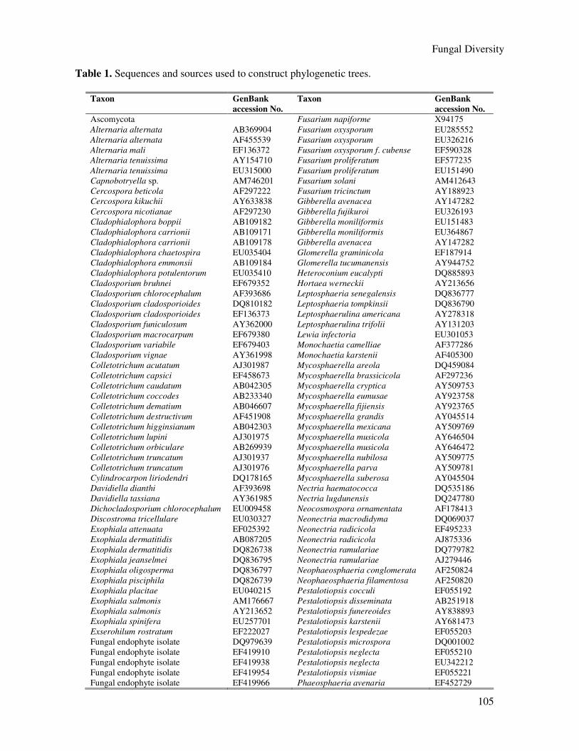
Table 1. Sequences and sources used to construct phylogenetic trees.
Taxon GenBank
accession No.
Taxon GenBank
accession No.
Ascomycota Fusarium napiforme X94175
Alternaria alternata AB369904 Fusarium oxysporum EU285552
Alternaria alternata AF455539 Fusarium oxysporum EU326216
Alternaria mali EF136372 Fusarium oxysporum f. cubense EF590328
Alternaria tenuissima AY154710 Fusarium proliferatum EF577235
Alternaria tenuissima EU315000 Fusarium proliferatum EU151490
Capnobotryella sp. AM746201 Fusarium solani AM412643
Cercospora beticola AF297222 Fusarium tricinctum AY188923
Cercospora kikuchii AY633838 Gibberella avenacea AY147282
Cercospora nicotianae AF297230 Gibberella fujikuroi EU326193
Cladophialophora boppii AB109182 Gibberella moniliformis EU151483
Cladophialophora carrionii AB109171 Gibberella moniliformis EU364867
Cladophialophora carrionii AB109178 Gibberella avenacea AY147282
Cladophialophora chaetospira EU035404 Glomerella graminicola EF187914
Cladophialophora emmonsii AB109184 Glomerella tucumanensis AY944752
Cladophialophora potulentorum EU035410 Heteroconium eucalypti DQ885893
Cladosporium bruhnei EF679352 Hortaea werneckii AY213656
Cladosporium chlorocephalum AF393686 Leptosphaeria senegalensis DQ836777
Cladosporium cladosporioides DQ810182 Leptosphaeria tompkinsii DQ836790
Cladosporium cladosporioides EF136373 Leptosphaerulina americana AY278318
Cladosporium funiculosum AY362000 Leptosphaerulina trifolii AY131203
Cladosporium macrocarpum EF679380 Lewia infectoria EU301053
Cladosporium variabile EF679403 Monochaetia camelliae AF377286
Cladosporium vignae AY361998 Monochaetia karstenii AF405300
Colletotrichum acutatum AJ301987 Mycosphaerella areola DQ459084
Colletotrichum capsici EF458673 Mycosphaerella brassicicola AF297236
Colletotrichum caudatum AB042305 Mycosphaerella cryptica AY509753
Colletotrichum coccodes AB233340 Mycosphaerella eumusae AY923758
Colletotrichum dematium AB046607 Mycosphaerella fijiensis AY923765
Colletotrichum destructivum AF451908 Mycosphaerella grandis AY045514
Colletotrichum higginsianum AB042303 Mycosphaerella mexicana AY509769
Colletotrichum lupini AJ301975 Mycosphaerella musicola AY646504
Colletotrichum orbiculare AB269939 Mycosphaerella musicola AY646472
Colletotrichum truncatum AJ301937 Mycosphaerella nubilosa AY509775
Colletotrichum truncatum AJ301976 Mycosphaerella parva AY509781
Cylindrocarpon liriodendri DQ178165 Mycosphaerella suberosa AY045504
Davidiella dianthi AF393698 Nectria haematococca DQ535186
Davidiella tassiana AY361985 Nectria lugdunensis DQ247780
Dichocladosporium chlorocephalum EU009458 Neocosmospora ornamentata AF178413
Discostroma tricellulare EU030327 Neonectria macrodidyma DQ069037
Exophiala attenuata EF025392 Neonectria radicicola EF495233
Exophiala dermatitidis AB087205 Neonectria radicicola AJ875336
Exophiala dermatitidis DQ826738 Neonectria ramulariae DQ779782
Exophiala jeanselmei DQ836795 Neonectria ramulariae AJ279446
Exophiala oligosperma DQ836797 Neophaeosphaeria conglomerata AF250824
Exophiala pisciphila DQ826739 Neophaeosphaeria filamentosa AF250820
Exophiala placitae EU040215 Pestalotiopsis cocculi EF055192
Exophiala salmonis AM176667 Pestalotiopsis disseminata AB251918
Exophiala salmonis AY213652 Pestalotiopsis funereoides AY838893
Exophiala spinifera EU257701 Pestalotiopsis karstenii AY681473
Exserohilum rostratum EF222027 Pestalotiopsis lespedezae EF055203
Fungal endophyte isolate DQ979639 Pestalotiopsis microspora DQ001002
Fungal endophyte isolate EF419910 Pestalotiopsis neglecta EF055210
Fungal endophyte isolate EF419938 Pestalotiopsis neglecta EU342212
Fungal endophyte isolate EF419954 Pestalotiopsis vismiae EF055221
Fungal endophyte isolate EF419966 Phaeosphaeria avenaria EF452729

106
Table 1 (continued). Sequences and sources used to construct phylogenetic trees.
Taxon GenBank
accession No. Taxon
GenBank
accession No.
Fungal endophyte sp. EF495231 Sebacina incrustans AF490395
Phaeosphaeria halima AF422971 Sebacina sp. AF440664
Phaeosphaeria halima AF422991 Serendipita vermifera DQ520096
Phaeosphaeria nodorum AF181708 Sebacina vermifera DQ983814
Phaeosphaeria phragmitis AJ496631 Tremellales sp. EF060917
Phaeosphaeria pontiformis AJ496632 Tulasnella albida AY373294
Phaeococcomyces catenatus AF050277 Tulasnella calospora DQ388045
Phaeococcomyces chersonesos AJ507323 Tulasnella calospora EF393622
Rhynchosporium secalis AF384682 Tulasnella danica AY373297
Strelitziana africana DQ885895 Tulasnella eichleriana AY373292
Truncatella angustata EU342216 Tulasnella pruinosa DQ457642
Xanthoria elegans AF278756 Tulasnella tomaculum AY373296
Uncultured endophytic fungus EF504333 Tulasnella violea AY373293
Uncultured endophytic fungus EF504576 Uncultured endophytic fungus EF504366
Uncultured endophytic fungus EF505438 Uncultured mycorrhiza AY634132
Uncultured endophytic fungus EF505583 Uncultured Sebacinales EF127237
Basidiomycota Plantae
Cryptococcus anemochorus DQ830986 Bletilla striata AF273334
Cryptococcus arboriformis AB260936 Bletilla striata AF461466
Cryptococcus bhutanensis EU266557 Bletilla striata EU100762
Cryptococcus cellulolyticus AF444442 Coelogyne cristata AF302742
Cryptococcus diffluens AF444374 Coelogyne dayana AF281126
Cryptococcus dimennae EU266559 Coelogyne harana AF302749
Cryptococcus flavescens AM176643 Coelogyne plicatissima AF281125
Cryptococcus laurentii AF410468 Coelogyne rhabdobulbon AF281127
Cryptococcus rajasthanensis AM262981 Coelogyne veitchii AF302759
Dioszegia aurantiaca EU266500 Coelogyne virescens AF281122
Dioszegia crocea AJ581078 Diuris punctata DQ904024
Dioszegia fristingensis EU070925 Michelia chapensis DQ234270
Dioszegia hungarica EU252552 Phalaenopsis amabilis AY391519
Dioszegia takashimae DQ003332 Pleione albiflora AY101967
Epulorhiza sp. EF393629 Pleione bulbocodioides EU100770
Epulorhiza sp. AJ31344 Pleione chunii AY008471
Kwoniella mangroviensis EF215528 Pleione formosana EU100756
Piriformospora indica AF019636 Pleione grandiflora AF461476
Sebacina aff. epigaea AF490393 Pleione hookeriana AF461469
Sebacina allantoidea AF490396 Pleione pleionoides AF461480
Sebacina epigaea AF490397 Pleione x confusa AF461479
Pestalotiopsis olivacea EF055215
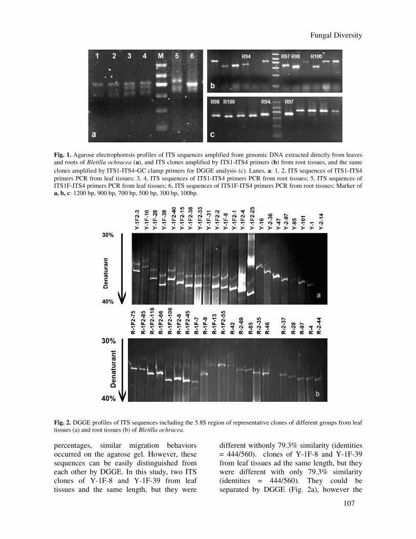
(Fig. 1a). The PCR products contained dif
ferent ITS sequences of fungal taxa.PCR
products with multiple bands were cloned into
E. coli, DH5α. recombinants and a single ITS
sequence of the fungus or plant was obtained
(Fig. 1b). Random ITS clones were selected for
further DGGE analysis (Fig. 1c). We obtained
203 clones and 211 clones from leaf tissues and
root tissues respectively.
DGGE analysis PCR products of ITS clones with addi-
tional 40-bp GC-rich sequences were subjected
to DGGE to elucidate the diversity of fungal
interspecies as well as intraspecies. All ITS
clones of endophytic fungi from the leaf tissues
grouped into 18 different ITS clone sequences
(Fig. 2a) and 10 from the root tissues (Fig. 2b)
with a total of 203 clones (leaf tissues) and 211
clones (root tissues) based on DGGE and
sequence analysis. In addition, we also
obtained 26 ITS clones and 25 ITS clones of
Bletilla ochracea respectively from the leaf and
root tissues. All ITS clones amplified from leaf
and root tissues migrated in the range of 35%
to 40% denaturant concentration (Fig. 2). In
cases where ITS sequences had similar lengths,
but large differences in G+C
Fungal Diversity
107
Fig. 1. Agarose electrophoresis profiles of ITS sequences amplified from genomic DNA extracted directly from leaves
and roots of Bletilla ochracea (a), and ITS clones amplified by ITS1-ITS4 primers (b) from root tissues, and the same
clones amplified by ITS1-ITS4-GC clamp primers for DGGE analysis (c). Lanes, a: 1, 2, ITS sequences of ITS1-ITS4
primers PCR from leaf tissues; 3, 4, ITS sequences of ITS1-ITS4 primers PCR from root tissues; 5, ITS sequences of
ITS1F-ITS4 primers PCR from leaf tissues; 6, ITS sequences of ITS1F-ITS4 primers PCR from root tissues; Marker of
a, b, c: 1200 bp, 900 bp, 700 bp, 500 bp, 300 bp, 100bp.
Fig. 2. DGGE profiles of ITS sequences including the 5.8S region of representative clones of different groups from leaf
tissues (a) and root tissues (b) of Bletilla ochracea.
percentages, similar migration behaviors
occurred on the agarose gel. However, these
sequences can be easily distinguished from
each other by DGGE. In this study, two ITS
clones of Y-1F-8 and Y-1F-39 from leaf
tissues and the same length, but they were
different withonly 79.3% similarity (identities
= 444/560). clones of Y-1F-8 and Y-1F-39
from leaf tissues ad the same length, but they
were different with only 79.3% similarity
(identities = 444/560). They could be
separated by DGGE (Fig. 2a), however the

108
two ITS sequences had the same position on
the agarose gel (data not shown).
Taxonomic placement of endophytic fungi
from leaf tissues by sequence analysis DGGE, sequence and phylogenetic
analysis revealed 18 OTUs of endophytic
fungi and one kind of plant clone from the
leaf tissues.
Phylogenetic analysis of 5.8S gene
sequences. In order to establish the general
taxonomic placement, neighbor-joining
phylogenetic trees of the 65 aligned 5.8S gene
sequences (42 references and 23 clones) (Fig.
3) was constructed with 1000 bootstrap repli-
cations, and Michelia chapensis, a plant in the
family of Magnoliaceae, was used as out-
group. The data resulted in two main clades
Fig. 3. Neighbor-joining phylogenetic tree showing the relationship between endophytic fungi and orchid plant from
leaf tissues of Bletilla ochracea and related fungi and plants based on the sequences of 5.8S of rDNA. The tree was
rooted with Michelia chapensis (DQ234270). Bootstrap values > 50% (1000 replicates) are shown at the branches.

Fungal Diversity
109
the family of Magnoliaceae, was used as
outgroup. The data resulted in two main clades
(A and B), representing the fungal group and
orchid plant group. Clade A contained three
subclades (subclades Aa, Ab and Ac, with
66%, 93% and 100% bootstrap support values
(BSV) respectively). These comprised the main
fungal groups of Ascomycota and Basidio-
mycota. Subclade Aa of Clade A was a large
group of Ascomycota including 20 clones. The
highly conserved 5.8S gene could be used for
identifying distant groups to family or higher
Fig. 4. Neighbor-joining phylogenetic tree showing the relationship between endophytic fungi and related fungi based
on the sequences of 5.8S gene and ITS regions of nuclear rDNA. The tree was rooted with Plectosphaerella
cucumerina. Bootstrap values > 50% (1000 replicates) are shown at the branches.
level. Among these clones, Y25 and Y-2-94,
Y-1F2-2, Y-1F2-3 and Y-2-36, Y-2-24, Y-2-
97, and Y-1F-8 clustered with Phyllacho-
raceae, Nectriaceae, Herpotri-chiellaceae,
Mycosphaerellaceae, Amphisphae-riaceae and
Davidiellaceae. Y-1F-28, Y-1F-31 and Y-1F2-
1 clustered with Chaetothyriales and
Pleosporales. However, Y8, Y95, Y-1F-39, Y-
1F2-15, Y-2-64, Y-1F2-38 could only be
placed at the higher taxonomic level of Asco-
Fig. 5. Neighbor-joining phylogenetic tree showing the relationship between endophytic fungi and related fungi based
on the sequences of ITS1-5.8S-ITS2 of rDNA. Bootstrap values > 50% (1000 replicates) are shown at the branches.

110
mycota because of lack of close phylogenetic
taxa in this NJ tree. Subclade Ab including Y-
1F2-4 and Y-1F2-25 was a main clade of Clade
A, and consisted of species of Tremellales
(93% BSV) of Basidiomycota. Y47 clustered
with the species of Tulasnellaceae (Subclade
Ac, 100% BSV) belonging to Cantharellales, a
different order to the Tremellales. Subclade Ab
was more closely related to subclade c than
subclade Ac. Y13 and Y-1F-10 belonged to
Orchidaceae in Clade B, and hence not within
the scope of this study.
Phylogenetic analysis of ITS regions. The
ITS regions (ITS1-5.8S-ITS2) were used to
further identify these fungi.
Phyllachorales. Y25 and Y-2-94, Y-1F2-
15 were identified as Phyllachoraceae and
other Ascomycota in 5.8S NJ tree (Fig. 3), and
the NJ tree based on ITS regions of the 20
aligned sequences indicated that Y25 and Y-2-
94 belonged to Colletotrichum (Phylla-
choraceae) (Fig. 4). Because the ITS regions of
Y25 and Y-2-94 were the same size with 99.3%
similarity (identities = 573/577), Y25 and Y-2-
94 were only slightly variable within 1 OTU.
The clone of Y-1F2-15 could be a species of
Colletotrichum as it clustered with C.
boninense (DQ286160) with 74% BSV, but
Fig. 6. Maximum-parsimony tree showing the relationship between endophytic fungi and related fungi based on the
sequences of ITS1-5.8S-ITS2 of rDNA. The tree was rooted with Mycosphaerella suberosa (TL=375, CI=0.6800,
HI=0.3200, RI=0.7853, RC=0.5340). Bootstrap values > 50% (1000 replicates) are shown at the branches.
interestingly it had a distant phylogenetic rela-
tionship with Y25 and Y-2-94.
Pleosporales. Y-2-18 and Y-1F2-1 were
placed in genus Alternaria and Leptosph-
aerulina within Pleosporaceae (Pleosporales),
because they clustered with the species of these
genus with the strong BSV (100% and 97%)
(Fig. 5). In Fig. 9, Y-1F-31 clustered with 6
species of genus Phaeosphaeria within Phaeo-
sphaeriaceae (Pleosporales) and other 5 refer-
ences, and was identified to genus Phaeo-
sphaeria.
Capnodiales. Further analysis of the
taxonomic levels of Y-2-24, Y-1F2-38 and Y-
2-64 based on the maximum-parsimony tree
was shown in Fig. 6, Y-2-24 was in a subclade
clustered with Mycosphaerella, belonged to the
family Mycosphaerellaceae (Capnodiales),
species with a 98% BSV, and was formed a
terminal cluster with M. fori with a 98% BSV
and 98.5% sequence similarity (identities
=530/538). Y-1F2-38 and Y-2-64 clustered in
the other subclade with Mycosphaerella
species with a 81% BSV, but had distant
relationship with Y-2-24. Because Y-1F2-38
and Y-2-64 had the same size of ITS regions
with 99.8% similarity (identities = 555/556),
they both were just one single OTU. Y-1F-8
had been identified to Davidiellaceae (Capno-
diales) according to 5.8S sequence, and

Fungal Diversity
111
combined with sequences of ITS regions (Fig.
8) to further reveal that Y-1F-8 was a species
of Cladosporium (Davidiellaceae), and is very
close relationship with C. cladosporioides
(DQ810182) of highly ITS similarity of 100%
(identities = 551/551).
Fig. 7. Neighbor-joining phylogenetic tree showing the relationship between endophytic fungi and related fungi based
on the sequences of ITS1-5.8S-ITS4. The tree was rooted with Truncatella angustata. Bootstrap values > 50% (1000
replicates) are shown at the branches.
Fig. 8. Maximum-parsimony tree showing the relationship between endophytic fungi and references based on the
sequences of ITS1-5.8S-ITS2 of nuclear rDNA. The tree was rooted with Dichocladosporium chlorocephalum (TL=96,
CI=1.0000, HI=0.0000, RI=1.0000, RC=1.0000). Bootstrap values > 50% (1000 replicates) are shown at the branches.

112
Fig. 9. Neighbor-joining phylogenetic tree showing the relationship between endophytic fungi and related fungi based
on the sequences of ITS1-5.8S-ITS2 of rDNA. Bootstrap values > 50% (1000 replicates) are shown at branches.
Fig. 10. Neighbor-joining phylogenetic tree showing the relationship between endophytic fungi and related fungi based
on the sequences of ITS1-5.8S-ITS2 of rDNA. The tree was rooted with Kwoniella mangroviensis. Bootstrap values >
50% (1000 replicates) are shown at the branches.
Amphisphaeriaceae. Y-2-97 belonged to
the family Amphisphaeriaceae (Xylariales)
(Fig. 3), and further analyses showed that Y-2-
97 belonged to the genus Pestalotiopsis (Fig.
7). Y-2-97 was closely related to P. microspora
with a 93% BSV and 99.5% similarity
(identities = 603/606).
Nectriaceae. Y-1F2-2 together with some
clones from root tissues were further identified
to the species of Gibberella and its anamorph
of Fusarium of Nectriaceae (Hypocreales)
(Fig. 13), and Y-1F2-2 clustered with G.
avenacea with 99% BSV and 99.8% similarity
(identities = 560/561).
Tulasnellaceae. Y47 belonged to Tula-
snellaceae (Cantharellales) of Basidiomycota
(Fig. 3), and further identifications based on
ITS regions showed that Y47 was species of
genus Epulorhiza (Fig. 11).
Tremellales. Y-1F2-25 and Y-1F2-4
belonged to Tremellales of Basidiomycota(Fig.
3), and further identified to the species of

Fungal Diversity
113
Fig. 11. Neighbor-joining phylogenetic tree showing the relationship between endophytic fungi and related fungi based
on the sequences of ITS1-5.8S-ITS2 of rDNA. The tree was rooted with Tulasnella tomaculum. Bootstrap values > 50%
(1000 replicates) are shown at the branches.
Fig. 12. Neighbor-joining phylogenetic tree showing the relationship between endophytic fungi from root tissues of
Bletilla ochracea, orchid plant and references based on the sequences of 5.8S of rDNA. Bootstrap values > 50% (1000
replicates) are showed at the branches. Accession numbers of GenBank nucleotide database are given for all sequences.

114
genus Dioszegia and Cryptococcus, respect-
tively from the NJ tree (Fig.10).Y-1F2-25 had
a distant phylogenetic relationship with other
species of Dioszegia, Y-1F2-4, however, had a
very strong BSV (100%) and 100% sequence
similarity (identities = 529/529) in its subclade
with Cryptococcus flavescens (Fig. 10).
Chaetothyriales. Y-1F2-3 and Y-2-36
were placed at family Herpotrichiellaceae
(Chaetothyriales) (Fig. 14), although Y-1F2-3
and Y-2-36 were grouped with species of
Fig. 13. Neighbor-joining phylogenetic tree showing the relationship between endophytic fungi and related fungi based
on ITS1, 5.8S and ITS2 sequences. The tree was rooted with Neocosmospora ornamentata. Bootstrap values > 50%
(1000 replicates) are shown at the branches.
Phaeococcomyces with 92% BSV, they could
not form a terminal cluster with them. And
because Y-1F2-3 and Y-2-36 had higher
sequence similarity of 99.4% (identities =
681/685), we regarded Y25 and Y-2-94 as one
OTU.
Based on phylogenetic analysis of 5.8S in
Fig. 3, Y-1F-28, Y-1F-39, Y8 and Y95 were
identified to the order or higher levels. And
because there are not enough molecular data in
GenBank to construct phylogenetic trees, these
clones could not be identified to a lower
taxonomic level.
As a result, we combined the 5.8S gene
and the ITS regions to identify the fungal ITS
clones from the leaf tissues of Bletilla
ochracea by phylogenetic analysis to 18 taxa
of genus or higher level listed in Table 2.
Taxonomic placement of endophytic
fungi from root tissues by sequence analysis
On the basis of DGGE and phylogenetic
analysis of ITS regions, 10 OTUs of endophy-
tic fungi and one kind of plant clone sequence
from the root tissues were grouped.
Phylogenetic analysis of sequences based
on 5.8S gene. Phylogeny generated from 5.8S
gene sequences resulted in two main clades of
A and B with 100% and 85% BSV (Fig. 12),
respectively, representing fungal group and
plant group of orchid. Clade A includes two
subclades (Aa and Ab, 73% and 87% BSV) of
large fungal groups of Ascomycota and Basi-
diomycota. Subclade Aa was a group of Asco-
mycota including 14 clones.
Among these clones, R-2-8, R-2-37, R-
1F-9, R-1F2-66, R38, R81, R-1F-7, R4, R-2-
46, R-1F2-106, R-1F2-9 clustered with the
family Nectriaceae, and R-2-17, R-2-35, R46
were placed at the family Mycosphaerellaceae
and Herpotrichiellaceae, respectively. Sub-
clade Ab including R85, R-1F2-75 and R-1F2-

Fungal Diversity
115
85 was consisted of species in Tremellales of
Basidiomycota. R85, R-1F2-75 and R-1F2-85
were formed a terminal cluster with the
Sebacinaceae (95% BSV). R42, R-2-47 and R-
2-55 belonged to Orchidaceae plant in Clade
B, and not within the scope of this study.
Phylogenetic analysis of sequences based
on ITS regions. The diversity of these fungal
clones was further investigated based on ITS
regions. The phylogeny revealed two main
clades (I, II) with 100% and 89% BSV (Fig.
13), containing sequences from Neonectria and
its anamorph Cylindrocarpon, Fusarium and its
telemorphs of Gibberella and Nectria, and
fungal clones from root tissues.
Fig. 14. Neighbor-joining phylogenetic tree showing the relationship between endophytic fungi and related fungi based
on ITS1, 5.8S, ITS2 sequences. The tree was rooted with Heteroconium eucalypti. Bootstrap values > 50% (1000
replicates) are shown at the branches.
Clade I included subclade Ia consisting of
fungal clones of R4, R-2-46, R-1F2-106, R-
1F2-9, and R38 and subclade Ib consisting of
R81 and R-1F-7. Fungal clones from subclade
Ia grouped together with reference species of
Fusarium and its telemorph Gibberella
(Nectriaceae). Among these clones, R4, R-2-46
and R-1F2-106 showed 98.7-98.9% sequence
similarity (identities = 537/544-538/544)
among them, so we regarded them as one OUT.
R-1F2-9 was placed in a terminal group with
Gibberella moniliformis and showed high

116
sequence similarity (99.3%, identities =
554/558). R38 had a distant relationship with
other clones because of forming a single cluster
with no any references in this group. However,
clones from subclade Ib, R81, clustered with
Cylindrocarpon liriodendri, and R-1F-7,
clustered with Neonectria radicicola, were
supported with high bootstrap values (Fig. 13).
In clade II, fungal clones of R-2-37, R-2-
8, R-1F-9 and R-1F2-66 were regarded as a
single OTU of genus Nectria (Nectriaceae)
since they all clustered with Nectria
haematococca (89% BSV) and had 98.6-99.6%
sequence similarity (identities = 564/572-
570/572) among them (Fig. 13).
In the Fig. 14, R-2-35 and R46 clustered
with 15 references of species of family
Herpotrichiellaceae. R-2-35 was identified to
genus Exophiala on the basis of 100% BSV
with species of Exophiala in the subcluster.
R46 was placed at family level of
Herpotrichiellaceae because no references
formed the terminal cluster with it, and had
distant relationship with Exophiala and Clado-
phialophora. R-2-17 was further identified to
genus Cercospora, anamorph of Myco-
sphaerella (Mycosphaerellaceae) based on
phylogentic analysis in Fig. 6, and it was
formed a subclade with Cercospora kikuchii
(AY633838) with 99.8% similarity (identities =
535/536).
Further analysis of R85, R-1F2-75 and R-
1F2-85 based on the maximum parsimony tree
indicated they all had strong similarities to
each other (99.5-99.7%) and all clustered with
Sebacina species (Fig. 15). They were the same
OUT of Sebacina (Sebacinaceae) in
Sebacinales of Basidiomycota. However,
interestingly, they were not closely claded with
Sebacina vermifera which is the classic Orchid
endophyte (Milligan and Williams, 1988).
As a result, we identified the fungal
clones within the roots based on 5.8S gene and
the ITS regions to 10 taxa of genus or family
level listed in Table 3.
Fig. 15. Maximum-parsimony tree showing the relationship between endophytic fungi from roots and related fungi
based on the sequences of ITS1-5.8S-ITS2. The tree was rooted with Piriformospora indica (TL=931, CI=0.6853,
HI=0.3147, RI=0.6601, RC=0.4524). Bootstrap values > 50% (1000 replicates) are shown at the branches.
Fungal community composition and fungal
diversity All the ITS clones were grouped into 18
OTUs of endophytic fungi from leaf tissues
(Table 2) and 10 OTUs from root tissues
(Table 3) by DGGE and phylogentic analysis.
Within the leaves, the groups of
endophytic fungi were consisted of Asco-
mycota (91% to total clones) and Basidio-
mycota (9%). Of all the 18 OTUs, 15 taxa
belonged to Ascomycota, and the other 3 taxa
of Basidiomycota. The 15 taxa of Ascomycota
included 2 taxa of Mycosphaerella (41%) in
Mycosphaerellaceae, 2 taxa of Alternaria (9%)
and Leptosphaerulina (5.7%) in Pleospo-
raceae, 2 taxa of Colletotrichum (6.2%) in
Phyllachoraceae, 1 taxon of Gibberella (4.5%)
in Nectriaceae, 3 taxa of lower proportion
(3.4%, sum to total) of Cladosporium,
Pestalotiopsis and Phaeosphaeria, 2 taxa of

Fungal Diversity
117
order Chaetothyriales (5.1%) and 3 taxa of not
identifying to lower level in Ascomycota
(15.8%). The 3 taxa of Basidiomycota
consisted of 2 yeast taxa of Dioszegia and
Cryptococcus of order Tremellales (7.9% to
total clones) and 1 mycorrhizal fungus of
Epulorhiza (1.1%). Of all the 18 taxa, 2 taxa of
Mycosphaerella were the dominant species
within the leaves and Ascomycota sp.2 and
Alternaria sp. were also main groups.
Correspondingly, the 10 fungal taxa
within roots contained 9 taxa of Ascomycota
(54%) and 1 taxon of Basidiomycota (46%).
The 9 taxa of Ascomycota consisted of 6 taxa
of Fusarium (30.7%) and its telemorph
Gibberella (4.3%), Nectria (13.4%),
Neonectria (1.1%) and Cylindrocarpon (0.5%)
in Nectriaceae, 2 taxa of family Herpotrichiell-
aceae (2.2%) and 1 taxon of Cercospora
(1.6%) in Mycosphaerellaceae. Of the 9 taxa, 2
taxa of Fusarium were dominant species, and
species of family Nectriaceae were the main
groups within root tissues. The only 1 taxon of
Sebacina in Basidiomycota from roots was also
Table 2. Fungal diversity within leaf tissues.
OTU Taxon No. of clones Proportion to total
Y-2-18 Alternaria sp. 16 9.04%
Y-1F-39 Ascomycete sp.1 3 1.70%
Y8 Ascomycete sp.2 24 13.56%
Y95 Ascomycete sp.3 1 0.57%
Y-1F-28 Chaetothyriales sp. 7 3.96%
Y-1F-8 Cladosporium sp. 2 1.13%
Y-1F2-15 Colletotrichum sp.1 7 3.96%
Y25,Y-2-94 Colletotrichum sp.2 4 2.26%
Y-1F2-4 Cryptococcus sp. 4 2.26%
Y-1F2-25 Dioszegia sp. 10 5.65%
Y47 Epulorhiza sp. 2 1.13%
Y-1F2-2 Gibberella sp.1 8 4.52%
Y-1F2-3,Y-2-36 Herpotrichiellaceae sp.1 2 1.13%
Y-1F2-1 Leptosphaerulina sp. 10 5.65%
Y-2-24 Mycosphaerella sp.1 50 28.25%
Y-1F2-38, Y-2-64 Mycosphaerella sp. 2 23 12.99%
Y-2-97 Pestalotiopsis sp. 2 1.13%
Y-1F-31 Phaeosphaeria sp. 2 1.13%
Table 3. Fungal diversity within root tissues.
OTU Taxon No. of clones Proportion to total
R-2-17 Cercospora sp. 3 1.61%
R81 Cylindrocarpon sp. 1 0.54%
R-2-35 Exophiala sp. 2 1.08%
R-2-37, R-2-8,R-1F-9, R-1F2-66 Nectria sp. 25 13.44%
R38 Fusarium sp.1 19 10.22%
R4,R-2-46, R-1F2-106 Fusarium sp.2 38 20.43%
R-1F2-9 Gibberella sp.2 8 4.30%
R46 Herpotrichiellaceae sp.2 2 1.08%
R-1F-7 Neonectria sp. 2 1.08%
R-1F2-75,R-1F2-85, R85 Sebacina sp. 86 46.24%
the dominant species with 46% proportion to
total.
A broad fungal spectrum above showed
very high diversity within leaves and roots. The
Shannon-Weiner diversity index (H′) of fungi
within leaves and roots were 2.354 and 1.560,
respectively. The results indicated that fungal
diversity in leaf tissues were higher than that in
root tissues, and also revealed that fungal
communities within leaves and roots were
significantly different to each other

118
Discussion This is the first report on the fungal diversity and their phylogenetic relationships within leaves and roots of terrestrial orchids in China using combined molecular methods especially DGGE.
Comparison of fungal communities with
previous studies. The mycorrhizal fungi (Sebacina sp.,
Epulorhiza sp.), and some dominant species of
Ascomycota, non-mycorrhizal fungi, e.g.
Mycosphaeella (41%), Alternaria (9%) in
leaves, Fusarium (30.7%) and its telemorph
Gibberella (4.3%), Nectria (13.4%) in roots
were detected in this study. Several studies had
revealed diversity of endophytic fungal
communities, including mycorrhizal fungi and
non-mycorrhizal fungi in tropical orchid plants
(Bayman et al., 1997; McCormick et al., 2004; Porras-Alfaro and Bayman, 2007; Richardson
and Currah, 1995). These studies however
revealed the presence of limited fungal
communities within tropical orchids as they
used traditional isolation methods only (see
Hyde and Soytong, 2007; Shefferson et al.,
2008). Bayman et al. (1997) isolated
endophytic fungi within leaf and root tissues of
epiphytic Lepanthes plants. Comparing with
them, we observed a higher diversity and
significant differences within leaf and root
tissues of adult plants in our present study. To
date, little is known of roles which these fungi
play in distribution, population size, and
genetic diversity of orchid plants (Bayman et
al., 2008), especially of the genus Bletilla. and
hence the studies of the only mycorrhizal fungi
or special groups of endophytes for diversity
maybe miss critical fungi for orchid
establishment. So it is necessary to wholely
investigate on fungal communities within plant.
Are fungi within Orchid tissues specialists or
generalists.
Previous studies concerning host
specificity of orchid mycorrhizae using in vitro
and in situ approaches have often lead to
conflicting results (Masuhara and Katsuya,
1994). However, this confusion may be
because isolation techniques are inherently
biased by choice of and response to growth
medium (Allen et al., 2003; McCormick et al.,
2004). Some studies have shown that
mycorrhizal are often host specific in
nonphotosynthetic and photosynthetic orchids
(Otero et al., 2007; Shefferson et al., 2005;
Taylor et al., 2003). McCormick et al. (2004)
found unrelated photosynthetic orchids to
support a range of mycorrhizal fungi; some
were specific to hosts while others were not.
Otero et al. (2002, 2004) studied mycorrhizal
associations of some tropical epiphytic orchids
and found they comprised generalists. On the
other hand, mycorrhizal fungi have been found
in a wide variety of orchid species around the
world (McCormick et al., 2000; Otero et al.,
2002; Warcup, 1981). The mycorrhizae present
may also change during the development of
individual plants of some orchids. In Gastrodia
ellata, Mycena osmundicola was mycorrhizal
in the protocorm stage but was replaced by
Armillaria mellea in subsequent stages (Xu and
Mu, 1990). In the present study, we detected
only 1 species of mycorrhizae (Sebacina sp.)
within roots, and this was the dominant species
as found within Caladenia carnea (Bougoure
et al., 2005), hence Sebacina sp. seems to be a
mycorrhizae which is specific to Bletilla
ochracea. We also isolated many other
dominant species of non-mycorrhizal fungi
(shown in Table 2 and 3). These taxa cannot be
ignored even though we do not know their
roles in mycorrhizal ecology. So the facts
above implied that fungal specificity to orchids
may be narrow and temporary during their
special life stages, and fungal diversity through
their whole life cycles is universe and affected
by the factors of field sites and environment,
even different host plants.
Fungal diversities within leaves and roots. It was surprising that in this study com-
munities of mycorrhizal and non-mycorrhizal
fungi of leaf and root tissues differed
significantly. It was also surprising that we also
found a mycorrhizal fungus (Epulorhiza sp.)
within the leaves. The 10 OTUs extracted from
roots consisted of of one OTU of Sebacina
(Sebacinales, Basidio- mycota), but also six
OTUs of Nectriaceae (Hypocreales) and 3
OTUs of 2 families of Mycosphaerellaceae and
Herpotrichiellaceae. The fungal diversity
within leaves (18 OTUs) were higher than
within the roots, and the 18 OUTs were

Fungal Diversity
119
distributed amongst 9 different orders
(Capnodiales (3 OTUs), Cantharellales,
Chaethyriales, Chaetothyriales, Hypocreales,
Phyllachorales, Pleosporales, Tremellales and
Xylariales) of Ascomycota, 3 OTUs of
Ascomycete, whose phylogenetic placement
could not be resolved and 3 OUTs of
Basidiomycota. Bayman et al. (1997) used
traditional isolation techniques and found that
Xylaria spp. (Xylariales) and Rhizoctonia-like
taxa (Basidiomycota) comprised the majority
of endophytes within epiphytic orchids, and the
fungal communities within the leaves and roots
were surprising similar.
Comparisons of methodology with previous
studies DGGE has been used extensively for
examination of fungal communities in different
ecological systems such as grass, wheat, wood,
soil (Smit et al., 1999; van Elsas et al., 2000;
Vainio and Hantula, 2000) by using PCR
amplification of the nuclear ribosomal RNA
genes. In comparison with the more conserved
coding regions of the rRNA genes, e.g. SSU
rDNA and LSU rDNA, the variable ribosomal
DNA (rDNA) internally transcribed spacer
(ITS) regions generally provide greater
taxonomic resolution (Anderson et al., 2003;
Lord et al., 2002). Additionally, ITS data are
considered useful for the relative ease with
which ITS data can be recovered and the
abundance of ITS data in GenBank (21,075
fungal ITS sequences before 2004, Lutzoni et
al., 2004). There have been studies of
endophytes using similar approaches. Guo et
al. (2001) and Duong et al. (2006) studied
endophyte disversity within plants using
molecular approaches. Guo et al. (2001)
developed a technique using direct ampli-
fication of ITS sequences extracted from frond
tissues of Livistona chinensis followed by
cloning, sequencing and phylogenetic analysis
to identify endophytic fungi, however, they
only obtained 6 phylotypes. The most common
endophytic taxa occurring in Livistona
chinensis, such as Guignardia, Pseudospiropes
and Xylaria species (Guo et al., 2000),
however, were not detected. Duong et al.
(2006) used a molecular method based on
DGGE coupled with sequence analysis of the
18S rRNA gene to assess fungal diversity
within leaves of Magnolia liliifera, and
recovered 14 OTUs distributed among 6
different orders and 2 unknown taxa. This
method, however, failed to reveal any Xylaria
species, the common endophytes from leaves
of most plants (Arnold et al., 2003, 2007).
These molecular methods can overcome the
main limitations of previous studies of possibly
unculturable or slow growing fungi on artificial
media. Duong et al. (2006) could detect more
abundant endophytes and higher diversities
than that of Guo et al. (2001). In our study, we
used random cloning, combining with DGGE
and phylogenetic analysis to investigate the
fungal communities within roots and leaves. In
the step of random cloning, we can obtained
plentiful ITS clones of endophytes, and used
DGGE to group the different ITS sequences of
different G+C% even if they are the same
length, so we can obtain the more abundant
fungal information (data shown in results)
within the plant than that of previous studies
(Guo et al., 2001; Duong et al., 2006).
This study has significance for orchid
biology. The population size of Bletilla
ochracea is decreasing and is at risk of
extinction because of our human activities. It is
possible that availability of endophytic fungi is
one of the limiting factors for establishment of
new plants and populations. So further research
should elaborate on the possible connections
between endophytes and plants within single
organs, among organs of a single plant, and
possibly among host species of different field
sites.
Acknowledgements
We thank Dr. E.C. Yang and Dr. B.D. Sun from
the Graduate University of Chinese Academy of Science,
Beijing, for precious suggestions and phylogenetic
analysis in this paper. We also thank Dr. P.P. Than, from
Maejo University, Chiangmai, Thailand, for comments
on the manuscript. Support was provided by Scientific
Foundation of Guizhou Academy of Agricultural
Science and Guizhou Province (QianKeHe J Zi (2008) No. 2095), China.
References
Allen, T.R., Millar, T., Berch, S.M. and Berbee, M.L.
(2003). Culturing and direct DNA extraction find

120
different fungi from the same ericoid mycorrhizal
roots. New Phytologist 160: 255-272.
Anderson, I.C. and Cairney, J.W.G. (2004). Diversity
and ecology of soil fungal communities:
increased understanding through the application
of molecular techniques. Environment Micro-
biology 6: 769-779.
Anderson, C., Campbell, C.D. and Prosser, J.I. (2003).
Potential bias of fungal 18S rDNA and internal
transcribed spacer polymerase chain reaction
primers for estimating fungal biodiversity in soil.
Applied and Environmental Microbiology 5: 36-
47.
Arnold, A.E., Henk, D.A., Eells, R.L., Lutzoni, F.E. and
Vilgalys, R. (2007). Diversity and phylogenetic
affinities of foliar fungal endophytes in loblolly
pine inferred by culturing and environmental
PCR. Mycologia 99: 185-206.
Arnold, A.E., Mejia, L.C., Kyllo, D., Rojas, E.I.,
Maynard, Z., Robins, N. and Herre, E.A. (2003).
Fungal endophytes limit pathogen damage in a
tropical tree. Proceedings of the National
Academy of Sciences of the United States of
America 100: 15649-15654.
Bayman, P., Lebron, L.L., Tremblay, R.L. and Lodge,
D.J. (1997). Variation in endophytic fungi from
roots and leaves of Lepenthes (Orchidaceae).
New Phytologist 135: 143-149.
Bayman, P., Porras-Alfaro, A. and Otero, J. (2008).
Diversity and distribution of Ceratobosidium and
Thanatephorus: What orchid mycorrhizal fungi
can tell us. Phytopathology 98: S19. Bidartondo, M.I. (2005). The evolution of mycohe-
terotrophy. New Phytologist 167: 335-352.
Bougoure, J.J., Bougoure, D.S., Cairney, J.W.G. and
Dearnaley, J.D.W. (2005). ITS-RFLP and
sequence analysis of endophytes from Acianthus,
Caladenia and Pterostylis (Orchidaceae) in
southeastern Queensland. Mycological Research
109: 452-460.
Bougoure, D.S. and Cairney J.W.G. (2005). Assem-
blages of ericoid mycorrhizal and other root
associated fungi from Epacris pulchella
(Ericaceae) as determined by culturing and direct
DNA extraction from roots. Environmental
Microbiology 7: 819-827.
Brown, K.B., Hyde, K.D. and Guest, D.I. (1998).
Preliminary studies on endophytic fungal com-
munities of Musa acuminata species complex in
Hong Kong and Australia. Fungal Diversity 1:
27-51.
Christensen, M. (1989). A view of fungal ecology.
Mycologia 81: 1-19.
Currah, R.S., Zelmer, C.D., Hambleton, S., and
Richardson, K.A. (1997). Fungi from orchid
mycorrhizas. In: Orchid Biology: Reviews and
Perspectives. VII. (eds. J. Arditti and A.M.
Pridgeon) Kluwer Academic Publishers,
Dordrecht. 117-170.
Dearnaley, J.D.W. (2007). Further advances in orchid
mycorrhizal research. Mycorrhiza 17: 475-486.
Doyle, J.J. and Doyle, J.L. (1987). A rapid DNA
isolation procedure for small quantities of fresh
leaf tissue. Phytochemical Bulletin 19: 11-15.
Duong, L. M., Jeewon, R., Lumyong, S. and Hyde, K.D.
(2006). DGGE coupled with ribosomal DNA
gene phylogenies reveal uncharacterized fungal
phylotypes. Fungal Diversity 23: 121-138.
van Elsas Duarte, G.F., Keijzer-Wolters, A. and Smit, E.
(2000). Analysis of the dynamics of fungal
communities in soil via fungal-specific PCR of
soil DNA followed by denaturing gradient gel
electrophoresis. Journal of Microbiological
Methods 43: 133-151.
Feller, I.C. (1995). Effects of nutrient enrichment on
growth and herbivory of dwarf red mangrove
(Rhizophora mangle). Ecological Monographs
65: 477-505.
Felsenstein, J. (1985). Confidence limits on phylogenies:
an approach using the bootstrap. Evolution 39:
783-791.
Ganley, R.J. and Newcombe, G. (2006). Fungal
endophytes in seeds and needles of Pinus
monticola. Mycological Research 110: 318-327.
Gardes, M. (2002). An orchid-fungus marriage: physical
promiscuity, conflict and cheating. New
Phytologist 154: 4-7.
Gardes, M. and Bruns, T.D. (1993). ITS primers with
enhanced specificity for basidiomycetes:
application to the identification of mycorrhiza and
rusts. Molecular Ecology 2: 113-118.
Griesbach, R.J. (2002). Development of Phalaenopsis
orchids for the mass-market. In: Trends in New
Crops and New Uses (eds. J. Janick and A.
Whipkey). ASHS, Alexandria, USA.
Guo, L.D., Hyde, K.D. and Liew, E.C.Y. (1998). A
method to promote sporulation in palm
endophytic fungi. Fungal Diversity 1: 109-113.
Guo, L.D., Hyde, K.D. and Liew, E.C.Y. (2000).
Identification of endophytic fungi from Livistona
chinensis (Palmae) using morphological and
molecular techniques. New Phytologist 147: 617-
630.
Guo, L.D., Hyde, K.D. and Liew, E.C.Y. (2001).
Detection and identification of endophytic fungi
within frond tissues of Livistona chinensis based
on rDNA sequence. Molecular Phylogenetics and
Evolution 20: 1-13.
Guo, L.D., Huang, G.R., Wang, Y., He, W.H., Zheng,
W.H. and Hyde, K.D. (2003). Molecular
identification of white morphotype strains of
endophytic fungi from Pinus tabulaeformis.
Mycological Research 107: 680-688.
Hall, T. (1997-2001). BioEdit. http://www.mbio. ncsu.
edu/RNaseP/info/programs/BIOEDIT/bioedit.htm
l.
Hyde, K.D. and Soytong, K. (2007). Understanding
microfungal diversity – a critique. Cryptogamie
Mycologie 28: 1-9.
Irwin, M.J., Bougoure, J.J. and Dearnaley, J.D.W.
(2007). Pterostylis nutans (Orchidaceae) has a
specific association with two Ceratobasidium
Fungal Diversity
121
root associated fungi across its range in Eastern
Australia. Mycoscience 48: 231-239.
Jones, D.L. (2006). A complete guide to native orchids of
Australia including the Island Territories. Reed
New Holland, Sydney.
Jumpponen, A. (2001). Dark septate endophytes – are
they mycorrhizal? Mycorrhiza 11: 207-211.
Kobayashi, D.Y. and Palumbo, J.D. (2000). Bacterial
endophytes and their effects on plants and uses in
agriculture. In: Microbial Endophytes (eds. C.W.
Bacon and J.F. White). Marcel Dekker, New
York: 199-236.
Koide, K., Osono, T. and Taked, A.H. (2005).
Colonization and lignin decomposition of
Camellia japonica leaf litter by endophytic fungi.
Mycoscience 46: 280-286.
Lacap, D.C., Hyde, K.D. and Liew, E.C.Y. (2003). An
evaluation of the fungal ‘morphotype’ concept on
ribosomal DNA sequence. Fungal Diversity 12:
53-66
Li, W.C., Zhou, J., Guo, S.Y. and Guo, L.D. (2007).
Endophytic fungi associated with lichens in
Baihua mountain of Beijing, China. Fungal
Diversity 25: 69-80.
Lord, N.S., Kaplan, C.W., Shank, P., Kitts, C.L. and
Elrod, S.L. (2002). Assessment of fungal
diversity using terminal restriction fragment
pattern analysis: comparison of 18S and ITS
ribosomal regions. FEMS Microbiology Ecology
42: 327-337.
Lutzoni, F., Kauff, F., Cox, C., McLaughlin, D., Celio,
G. and Dentiger, B. (2004). Assembling the
Fungal Tree of Life: progress, classification and
the evolution of subcellular traits. American
Journal of Botany 91: 1446-1480.
Ma, M., Tan, T.K., and Wong, S.M. (2003)
Identification and molecular phylogeny of
Epulorhiza isolates from tropical orchids.
Mycological Research 107: 1041-1049.
Masuhara, G. and Katsuya, K. (1994). In situ and in vitro
specificity between Rhizoctonia spp. &
Spiranthes sinensis (Persoon) Ames. var. amoena
(M. Bieberstein) Hara (Orchidaceae). New
Phytologist 127: 711-718.
McCormick, M.K., O’Malley, K.L., Whigham, D.F. and
O’Neill, J.P. (2000). Specialization and species
distribution in orchid-fungal symbioses. Pro-
ceedings of the Ecological Society of America
85th Annual Meeting, Snowbird, Utah, USA.
McCormick, M.K., Whigham, D.F. and O’Neill, J.
(2004). Mycorrhizal diversity in photosynthetic
terrestrial orchids. New Phytologist 163: 425-438.
McCormick, M.K., Whigham, D.F., Sloan, D.,
O’Malley, K. and Hodkinson, B. (2006). Orchid-
fungus fidelity: a marriage meant to last? Ecology
87: 903-911.
Milligan, M.J. and Williams, P.G. (1988). The
mycorrhizal relationship of multinucleate
rhizoctonias from non-orchids with Microtis
(Orchidaceae). New Phytologist 108: 205-209.
Moore, R.T. (1988). The genera Rhizoctonia-like fungi:
Ascorhizoctonia, Ceratorhiza gen. Nov.,
Eupulorhiza gen. nov., Moniliopsis, and
Rhizoctonia. Mycotaxon 29: 91-99.
Mostert, L., Crous, P.W. and Petrini, O. (2000).
Endophytic fungi associated with shoots and
leaves of Vitis vinifera, with specific reference to
the Phomopsis viticola complex. Sydowia 52:
46-58.
Otero, J.T., Ackerman, J.D. and Bayman, P. (2002).
Diversity and host specificity of endophytic
Rhizoctonia-like fungi from tropical orchids.
American Journal of Botany 89: 1852-1858.
Otero, J.T., Ackerman, J.D. and Bayman, P. (2004).
Differences in mycorrhizal preferences between
two tropical orchids. Molecular Ecology 13:
2393-2404.
Otero, J.T., Flanagan, N.S., Herre, E.A., Ackerman, J.D.
and Bayman, P. (2007). Widespread mycorrhizal
specificity correlates to mycorrhizal function in
the neotropical, epiphytic orchid Ionopsis
utricularioides (Orchidaceae). American Journal
of Botany 94: 1944-1950.
Page, R.D.M. (1996). An application to display
phylogenetic trees on personal computers.
Comparative Applied Bioscience 12: 357-358.
Pereira, O.L., Kasuya, M.C.M., Borges, A.C. and de
Araújo, E.F. (2005). Morphological and
molecular characterization of mycorrhizal fungi
isolated from neotropical orchids in Brazil.
Canada Journal of Botany 83: 54-65.
Pereira, O.L., Rollemberg, C.L., Borges, A.C.,
Matsuoka, K. and Kasuya, M.C.M. (2003).
Epulorhiza epiphytica sp. nov. isolated from
mycorrhizal roots of epiphytic orchids in Brazil.
Mycoscience 44: 153-155.
Peters, A.F. (1991). Field and culture studies of
Streblonema-Macrocystis new species Ectocar-
pales Phaeophyceae from Chile, a sexual
endophyte of giant kelp. Phycologia 30: 365-377.
Petrini, O. (1991). Fungal endophytes of tree leaves. In:
Microbiol Ecology of Leaves. (eds. J Andrews
and S. Hirano). Springer Verlag, New York: 179-
197.
Photita, W., Lumyong, S., Lumyong, P., McKenzie, E.H.
C. and Hyde, K.D. (2004). Are some endophytes
from Musa acuminata latent pathogens? Fungal
Diversity 16: 131-140.
Pielou, E.C. (1975). Ecological Diversity. John Wiley
and Sons Inc.
Porras-Alfaro, A. and Bayman, P. (2007). Mycorrhizal
fungi of Vanilla: diversity, specificity and effects
on seed germination and plant growth. Mycologia
99: 510-525. Promputtha, L., Jeewon, R., Lumyong S., Mckenzie,
E.H.C. and Hyde, K.D. (2005). Ribosomal DNA
fingerprinting in the identification of non
sporulating endophytes from Magnolia liliifera
(Magnoliaceae). Fungal Diversity 20: 167-186.
Promputtha, L., Lumyong, S., Dhanasekaran, V.,
McKenzie, E.H.C., Hyde, K.D and Jeewon, R.

122
(2007). A phylogenetic evaluation of whether
endophytes become saprotrophs at host
senescence. Microbial Ecology 53, 579-590.
Rasmussen, H.N. (2002). Recent developments in the
study of orchid mycorrhiza. Plant Soil 244:149-
163
Richardson, K.A. and Currah, R.S. (1995). The fungal
community associated with the roots of some
rainforest epiphytes of Costa Rica. Selbyana 16:
49-73.
Sánchez Márquez, S., Bills, G.F. and Zabalgogeazcoa, I.
(2007). The endophytic mycobiota of the grass
Dactylis glomerata. Fungal Diversity 27: 171-
195.
Schulz, B. and Boyle, C. (2005). The endophytic
continuum. Mycological Research 109: 661-687.
Seena, S., Wynberg, N. and Barlocher, F. (2008). Fungal
diversity during leaf decomposition in a stream
assessed through clone libraries. Fungal Diversity
30: 1-14.
Shefferson, R.P., Kull, T. and Tali, K. (2008).
Mycorrhizal interactions of orchids colonizing
Estonian mine tailings hills. American Journal of
Botany 95: 156-164.
Shefferson, R.P., Weiß, M., Kull, T. and Taylor, D.L.
(2005). High specificity generally characterizes
mycorrhizal association in rare lady’s slipper
orchids, genus Cypripedium. Molecular Ecology
14: 613-626.
Sheffield, V.C., Cox, D.R., Lerman, L.S., and Myers,
R.M. (1989). Attachment of a 40-base pair G+C-
rich sequence (GC-clamp) to genomic DNA
fragments by the polymerase chain reaction
results in improved detection of single-base
changes. Proceedings of the National Academy of
Sciences of the United States of America 86: 232-
236.
Sieber, T.N. (2002). Fungal root endophytes. In: The
Hidden Half (eds. Y. Waisel, A. Eshel and U.
Kafkafi). Marcel Dekker, New York: 887-917.
Smit, E., Leeflang, P., Glandorf, B., van Elsas J.D.a nd
Wernars, K. (1999). Analysis of fungal diversity
in the wheat rhizophere by sequencing of cloned
PCR-amplfied genes encoding 18S rRNA and
temperature gradient gel electrophoresis. Applied
and Environmental Microbiology 65: 2614-2621.
Stone, J.K., Bacon, C.W. and White, J.F. (2000). An
overview of endophytic microbes: endophytism
defined. In: Microbial Endophytes (eds. C.W.
Bacon and J.F. White). Dekker, New York: 3-30.
Swofford, D.L. (1998). Phylogenetic Analysis Using
Parsimony (PAUP) (*and other methods).
Version 4 Sinauer Associates, Sunderland,
Massachusetts, USA.
Taylor, D.L., Bruns, T.D., Szaro, T.M. and Hodges, S.A.
(2003). Divergence in mycorrhizal pecialization
within Hexalectris spicata (Orchidaceae), a
nonphotosynthetic desert orchid. American
Journal of Botany 90: 1168-1179.
Taylor, J.E., Hyde, K.D., and Jones, E.B.G. (1999).
Endophytic fungi associated with the temperate
palm, Trachycarpus fortunei, within and outside
its natural geographic range. New Phytologist
142: 335-346.
Thompson, J.D., Gibson, T.J., Plewniak, F., Jeanmougin,
F. and Higgins, D.G. (1997). The Clustal X
windows interface: flexible strategies for multiple
sequence alignment aided by quality analysis
tools. Nucleic Acids Research 24: 4876-4882.
Vainio, E.J. and Hantula, J. (2000). Direct analysis of
wood-inhabiting fungi using denaturing gradient
gel electrophoresis of amplified ribosomal DNA.
Mycological Research 104: 927-936.
Wang, Y., Guo, L.D. and Hyde, K.D. (2005). Taxonomic
placement of sterile morphotypes of endophytic
fungi from Pinus tabulaeformis (Pinaceae) in
northeast China based on rDNA sequences.
Fungal Diversity 20: 235-260.
Warcup, J.H. (1981). The mycorrhizal relationships of
Australian orchids. New Phytologist 87: 371-381.
Warcup, J.H. (1981a). Orchid mycorrhizal fungi. In:
Orchid Society of New South Wales. Proceedings
of the Orchid Symposium, Held as a Satellite
Function of the 13th International Botanical
Congress. Sydney, Australia: Harbour Press: 57-
63.
Warcup, J.H., and Talbot, P.H.B. (1966). Perfect states
of some Rhizoctonia. Transactions of the British
Mycological Society 49: 427-435.
Warcup, J.H., and Talbot, P.H.B. (1971). Perfect states
of Rhizoctonia associated with orchids III. New
Phytologist 70: 35-40.
White, T.J., Bruns, T.D., Lee, S., and Taylor, J.W.
(1990). Amplification and direct sequencing of
fungal ribosomal RNA genes for phylogenetics.
In PCR Protocols: A Guide to Methods and
Applications (eds. M.A. Innis, D.H. Gelfand, J.S.
Sninsky and T.J. White) Academic Press, New
York: 315-322.
Xu, J.T., Mu, C. (1990). The relation between growth of
Gastrodia elata protocorms and fungi. Acta
Botanica Sinica 32:26-31.
Yang, Y. and Liu X.Z. (2005). Dactylella coccinella sp.
nov., an anamorphic species. Mycotaxon 91: 127-
132.
Zettler, L.W., Sharma, J. and Rasmussen, F. (2004).
Mycorrhizal diversity. In: Orchid Conservation
(eds. K. Dixon, P. Cribb, S. Kell and R. Barrett)
Natural History Publications, Kota Kinabalu,
Sabah, Malaysia: 185-203.