Whole Body Periodic Acceleration Reduces Levels of Delayed Onset Muscle Soreness After
52
University of Miami Scholarly Repository Open Access Dissertations Electronic eses and Dissertations 2010-05-14 Whole Body Periodic Acceleration Reduces Levels of Delayed Onset Muscle Soreness Aſter Eccentric Exercise Daniel H. Serravite University of Miami, [email protected] Follow this and additional works at: hps://scholarlyrepository.miami.edu/oa_dissertations is Open access is brought to you for free and open access by the Electronic eses and Dissertations at Scholarly Repository. It has been accepted for inclusion in Open Access Dissertations by an authorized administrator of Scholarly Repository. For more information, please contact [email protected]. Recommended Citation Serravite, Daniel H., "Whole Body Periodic Acceleration Reduces Levels of Delayed Onset Muscle Soreness Aſter Eccentric Exercise" (2010). Open Access Dissertations. 650. hps://scholarlyrepository.miami.edu/oa_dissertations/650
Transcript of Whole Body Periodic Acceleration Reduces Levels of Delayed Onset Muscle Soreness After
University of MiamiScholarly Repository
Open Access Dissertations Electronic Theses and Dissertations
2010-05-14
Whole Body Periodic Acceleration Reduces Levelsof Delayed Onset Muscle Soreness After EccentricExerciseDaniel H. SerraviteUniversity of Miami, [email protected]
Follow this and additional works at: https://scholarlyrepository.miami.edu/oa_dissertations
This Open access is brought to you for free and open access by the Electronic Theses and Dissertations at Scholarly Repository. It has been accepted forinclusion in Open Access Dissertations by an authorized administrator of Scholarly Repository. For more information, please [email protected].
Recommended CitationSerravite, Daniel H., "Whole Body Periodic Acceleration Reduces Levels of Delayed Onset Muscle Soreness After Eccentric Exercise"(2010). Open Access Dissertations. 650.https://scholarlyrepository.miami.edu/oa_dissertations/650
UNIVERSITY OF MIAMI
WHOLE BODY PERIODIC ACCELERATION REDUCES LEVELS OF DELAYED ONSET MUSCLE SORENESS AFTER ECCENTRIC EXERCISE
By
Daniel H. Serravite
A DISSERTATION
Submitted to the Faculty of the University of Miami
in partial fulfillment of the requirements for the degree of Doctor of Philosophy
Coral Gables, Florida
May 2010

©2010 Daniel H. Serravite All Rights Reserved
UNIVERSITY OF MIAMI
A dissertation submitted in partial fulfillment of the requirements for the degree of
Doctor of Philosophy
WHOLE BODY PERIODIC ACCELERATION REDUCES LEVELS OF DELAYED ONSET MUSCLE SORENESS AFTER ECCENTRIC EXERCISE
Daniel H. Serravite Approved: ________________ _________________ Joseph Signorile, Ph.D. Terri A. Scandura, Ph.D. Prof. of Exercise and Sport Science Dean of the Graduate School ________________ _________________ Arlette Perry, Ph.D. Kevin A. Jacobs, Ph.D. Chair Person and Prof. of Exercise and Sport Science Prof. of Exercise and Sport Science ________________ ________________ Jose A. Adams, M.D. Marvin A Sackner, M.D. Division of Neonatology Division of Pulmonary Disease Mount Sinai Medical Center Miami Mount Sinai Medical Center Miami
SERRAVITE, DANIEL H. (Ph.D., Exercise Physiology)
Whole Body Periodic Acceleration Reduces (May 2010) Levels of Delayed Onset Muscle Soreness After Eccentric Exercise Abstract of a dissertation at the University of Miami. Dissertation supervised by Professor Joseph Signorile. No. of pages in text. (41)
Several recovery strategies have been used, with limited effectiveness, to reduce the
muscle discomfort or pain and the diminished muscle performance following a bout of
unaccustomed physical activity, a condition known as delayed onset of muscle soreness
(DOMS). Muscle damage in this condition is associated with mechanical disruption of
the muscle and connective tissue and inflammation and increased oxidative stress. Low
frequency, low intensity, whole body periodic acceleration (WBPA) that increases nitric
oxide (NO) release from vascular endothelium into the circulation through increased
pulsatile shear stress offers a potential solution. This is because endothelial derived nitric
oxide has anti-inflammatory, antioxidant and anti-nociceptive properties. The purpose of
this study was to examine the effects of WBPA on the pain and diminished muscle
performance associated with DOMS induced by unaccustomed eccentric arm exercise in
young male subjects. Seventeen active men, 23.4 ± 4.6 yr of age, made six visits to the
research facility over a two-week period. On day one, the subject performed a 1RM
elbow flexion test and was then randomly assigned to the WBPA or control group.
Criterion measurements were taken on Day 2, prior to and immediately following
performance of the eccentric exercise protocol (10 sets of 10 repetitions using 120% of
1RM) and after the recovery period. During all subsequent sessions (24, 48, 72, and 96
h) these data were collected before the WBPA or passive recovery was provided.
Variables including isometric strength (MVC), blood markers (CPK, MYO, IL-6, TNF-α
and Uric Acid), soreness, pain, circumference, and range of motion (ROM) were
examined in their relation to the recovery protocol. Significantly higher MVC values
were seen for the WBPA group across the entire 96 h recovery period. Additionally,
within group differences were seen in CPK, MYO, IL-6, soreness, pain, circumference,
and ROM showing a smaller impact and more rapid recovery by the WBPA group. It was
concluded the application of WBPA hastens recovery from DOMS after eccentric
exercise. Given the lack of other potential mechanisms, these effects appear to be
mediated by the increased NO release with WBPA.

DEDICATION
I dedicate this dissertation to my sons, Luca and Nico, and my wife Jenny who
supported me throughout the Ph.D. journey.
Thank you to my parents Humberto and Sara who gave up everything to provide
me the best education and always supported me to pursue my dreams.
iii
ACKNOWLEDGEMENT
The authors would like to thank Tom E. Abdenour, Head Trainer for the Golden State
Warriors Basketball Team of the National Basketball Association for the suggestion to
undertake this investigation. We would also like to thank the students who helped with
the data collection including Laura Quirola, Kyle Raynolds and Brian Brookman-Jones.
iv

TABLE OF CONTENTS
Page
LIST OF FIGURES .................................................................................................... vi LIST OF TABLES ....................................................................................................... vii Chapters: 1 INTRODUCTION ............................................................................................... 1 2 METHODS ............................................................................................................. 3 3 RESULTS ........................................................................................................... 11 4 DISCUSSION . ..................................................................................................... 14 Appendix: Figures .............................................................................................................. 21 Tables .............................................................................................................. 31 Bibliography .............................................................................................................. 34
v
LIST OF FIGURES
Figure 1: STUDY DESIGN .................................................................................... 21 Figure 2: MOTION PLATFORM ..................................................................... 22 Figure 3: MVC 90° ........................................................................................... 23 Figure 4: MVC 150° .......................................................................................... 24 Figure 5: CRETINE PHOSPHOKINASE ......................................................... 25 Figure 6: MYOGLOBIN ................................................................................... 26 Figure 7: SORENESS ....................................................................................... 27 Figure 8: PAIN QUESTIONNAIRE ............................................................... 28 Figure 9: CIRCUMFERENCE .......................................................................... 29 Figure 10: RANGE OF MOTION ....................................................................... 30
vi

LIST OF TABLES
Table 1: PHYSICAL CHARACTERISTICS OF SUBJECT .......................... 31 Table 2: BLOOD MARKERS ......................................................................... 32 Table 3: SORENESS AND PAIN MARKERS DESIGN ................................ 33
vii
CHAPTER 1: INTRODUCTION
A delayed perception of skeletal muscle discomfort or pain that is termed delayed
onset of muscle soreness (DOMS) is commonly experienced following a bout of
unaccustomed physical activity (4, 14, 27). DOMS is usually associated with soreness,
decreased strength, localized swelling, stiffness, and reduced range of motion (4, 14, 27).
The soreness from DOMS usually follows an inverted U-shape curve over time in which
the level of discomfort increases during the first 24 hours following the cessation of
exercise, peaks between 24 to 72 hours, then subsides and eventually disappears by 5-7
days post-exercise (10, 13, 39). Further, the contour of the inverted U-shape may vary
depending on the intensity, volume, and type of activity inducing DOMS (73). The
decrement in muscle performance can parallel the course of the soreness or be
independent of it (14). Eccentric exercises are the preferred procedure for evoking
DOMS since they produce greater torque, causing greater reductions in strength (74) and
generating more muscle damage than concentric or isometric contractions (48). This is
especially true in sedentary or novice recreational athletes (49) performing unaccustomed
eccentric exercises using small muscles, such as arm muscles (35), at high movement
velocities (12).
A number of different mechanisms have been used to explain DOMS, and
therefore, several methods have been employed to counteract the soreness of DOMS with
varying degrees of success. These include ice water immersion, stretching, anti-
inflammatory drugs, ultrasound, transcutaneous electric nerve stimulation (TENS),
massage compression garments, acupuncture, hyperbaric oxygen therapy, oral
antioxidants such as vitamins C and E, exercise (8, 14, 17, 21, 31, 33, 45). Whole body
vibration (WBV) has been shown to reduce calf and gluteal pain and decrease
1
2
inflammation after downhill running (9) and when combined with stretching (58)
produces a decrease in pain that ranges from 22-61% compared to stretching alone.
These positive effects may be explained by the enhanced local blood flow immediately
after vibration training (36) and/or by the potentiation of pain inhibition through
increased proprioceptive feedback (41). These studies, however, did not measure
recovery of performance.
Whole body periodic acceleration (WBPA), which produces low frequency, low
intensity vibrations, increases nitric oxide (NO) release from vascular endothelium into
the circulation through increased pulsatile shear stress. The latter is produced by the
addition of small pulses superimposed upon the natural pulse; the number of pulses is a
function of platform frequency (3, 34, 71). Since endothelial derived nitric oxide has
anti-inflammatory, antioxidant and anti-nociceptive properties (1, 23, 28, 42), the purpose
of this study is to determine whether WBPA could hasten the recovery from DOMS
resulting from exposure of a sample of fit young volunteers to strenuous eccentric
exercise by the elbow flexors.
CHAPTER 2: METHODS
Subjects
Seventeen healthy young men volunteered for this study. Their mean (± SD) age,
height, and body weight was 23.4 ± 4.6 years, 176.0 ± 6.2 cm, and 79.1 ± 11.4 kg,
respectively. A power analyses performed using muscle CPK as the dependent variable
following massage indicated that ten subjects were required (F (5, 10) = 3.32, p = 0.05)
(75). All volunteers provided written informed consent prior to participation in the study.
The investigation was approved by the University of Miami’s Institutional Review Board
and conducted in conformity to the Declaration of Helsinki for Medical Research
involving Human Subjects. Individuals who reported participation in competitive sports
in the prior 12 months; cardiovascular, endocrine, or neuromuscular disorders; orthopedic
problems that would limit or be aggravated by either isoinertial arm curl exercise or the
performance of a standard isometric test; or other chronic medical conditions that might
affect performance were excluded from participating in the study. In addition, subjects
taking anti-inflammatory agents, nutritional supplements, or any other medications that
could affect neuromuscular performance, as well as L-Arginine supplements, were not
allowed to participate in this study. Subjects were required to avoid any formal physical
activity, such as strength or endurance training, for 48 h before the initiation of the study
and throughout its duration.
Experimental Design and Procedures
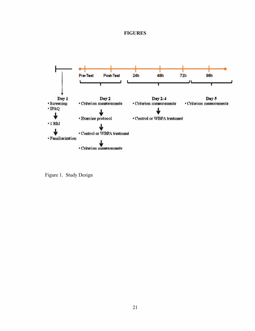
Figure 1 presents a schematic of the study protocol. Participants were randomly
assigned to a control or WBPA condition. For 2 weeks prior to data collection, and
during the protocol period, subjects were instructed to continue their normal eating
3
4
habits, but to refrain from the use of dietary supplementation. Subjects were also
instructed to abstain from exercise for 48 h before and for the duration of the study.
Criterion measurements of pain, anthropometry, ROM, soreness, blood-borne markers,
and isometric strength were taken at baseline and immediately, 24 h, 48 h, 72 h and 96 h
after performing the lifting protocol.
On day 1, the participant completed a health status questionnaire (HSQ) to
confirm eligibility. Upon acceptance into the study he completed the international
physical activity questionnaire (19). A maximum 1-repetition strength test (1RM),
performed using the National Strength and Conditioning Association protocol, was used
to determine the maximum amount of weight that the participant could lift in a single
repetition during an elbow flexion exercise using the dominant arm (5). The day
concluded with the participant being familiarized with the lifting and WBPA protocols.
The time required for the visit was approximately one hour. On day 2, prior to the
participant’s arrival, he was randomly assigned to the control or WBPA group. Criterion
measurements were made before he performed the lifting protocol and after the treatment
(WBPA or passive recovery). On days 3 through 5 criterion measurements were made
first, followed by the treatment. During the last visit, on day 6, only the criterion
measurements were made with no further treatment.
Exercise Protocol
After performing the criterion measurement on day 2, the participant performed
the lifting protocol consisting of 10 sets of 10 lengthening muscle contractions using a
dumbbell loaded at 120% of his dominant elbow flexor 1 RM. The lifts were performed
on a standard seated arm curl (preacher’s) bench. The dumbbell was lowered from 75°
5
flexion to 180° (full extension) at a rate of 5 seconds per repetition controlled by the
participant matching the sound of a metronome. When a participant showed difficulty in
controlling the movement velocity, minimal spotting was provided by the investigator.
After each eccentric movement, the participant had a 2s pause while the researcher lifted
the dumbbell to the starting position to prepare the participant for the next repetition. The
exercise protocol was followed by 30 min of passive recovery. Subsequent to the 30 min
recovery, the participant underwent a 45-minute WBPA or a passive recovery bout
depending on his group assignment.
Whole Body Periodic Acceleration Protocol
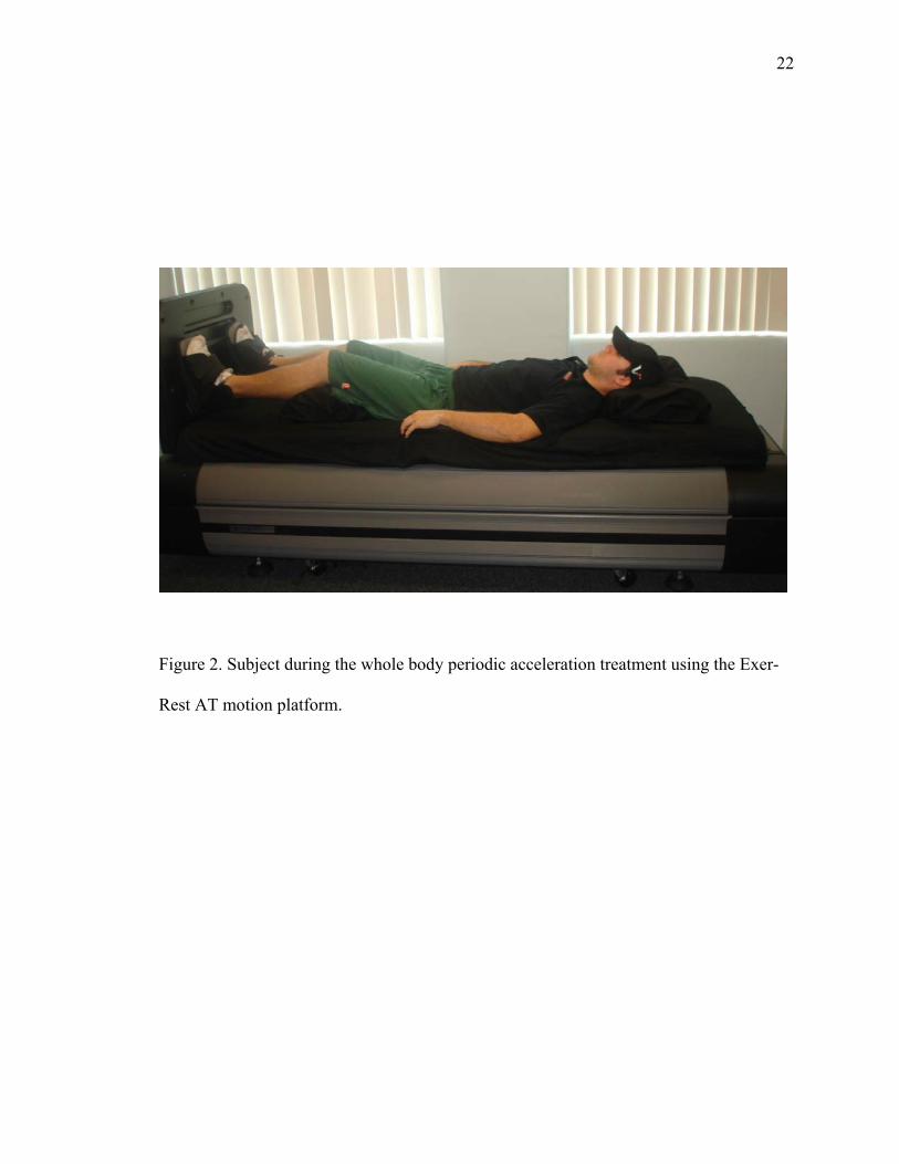
WBPA was provided using a motion platform that has the appearance of a single
size bed with a mattress. The platform is driven by a digitally controlled servo motor
assembly (Exer-Rest®, Non-Invasive Monitoring Systems, Inc) (see Figure 2). This
apparatus has a handheld wireless controller that regulates its speed in cycles per minute,
travel distance in mm and time of the treatment in minutes. The participant lies supine on
the mattress and is coupled to the motion platform via sandals connected to a footboard.
The platform moves 16 mm in a repetitive sinusoidal head-to-foot direction at 140 times
per minute, thereby applying approximately ±0.22 g to the participant for the 45-minute
WBPA period. These settings have been shown to release NO into the circulation of
healthy adults and patients with inflammatory diseases (59).
Criterion Measurements
Muscle Strength
Each subject performed a unilateral isometric maximal voluntary contraction
(MVC) using his dominant arm. During the testing, the elbow was positioned at two
6
separate angles, 90° (MVC90) and 150° (MVC150). Data were collected using a digital
force gauge (Chatillon, DFS Series, Ametek, FL). The MVC testing protocol began with
a warm-up consisting of two submaximal contractions (80% of perceived maximum) and
one maximal contraction. The participant was then instructed to apply maximal force by
pulling the handle of a load cell unit towards his body for five seconds. Strong verbal
encouragement was provided by the investigator throughout the strength testing. Three
maximal contractions were performed with a 2-minute rest period between trials. The
peak force was recorded for each trial. The trial producing the highest force value was
considered the MVC.
Blood Measurements.
Approximately 20 ml of whole blood were collected from the antecubital vein
using a standard venipuncture technique at baseline and immediately, 24 h, 48 h, 72 h and
96 h after performing the lifting protocol. The 24 h, 48 h, 72 h and 96 h samples were
taken prior to the WBPA or passive recovery sessions. The blood was collected into
serum tubes and centrifuged for 15 min to obtain serum. Samples were stored at −80°C
until analyzed for serum creatine kinase (CPK), myoglobin (MYO), IL-6 and TNF-α
using a microplate reader (Thermo Multiskan Spectrum, Vantaa, Finland).
Assay Procedure- CPK: CPK was quantified using an EnzyChrom Creatine
Kinase Assay Kit (ECPK-100; Bioassay Systems, CA). Each reaction well contained a
mixture of 10 μL Substrate Solution, 100 μL Assay Buffer, and 1 μL Enzyme Mix.
Subsequently, 10 μL samples were transferred into each well and 100 μL of reconstituted
reagent were added. The contents of the plate were then mixed by tapping. The reaction
was incubated at 37°C. CPK is fully activated within 10 min by glutathione provided in
7
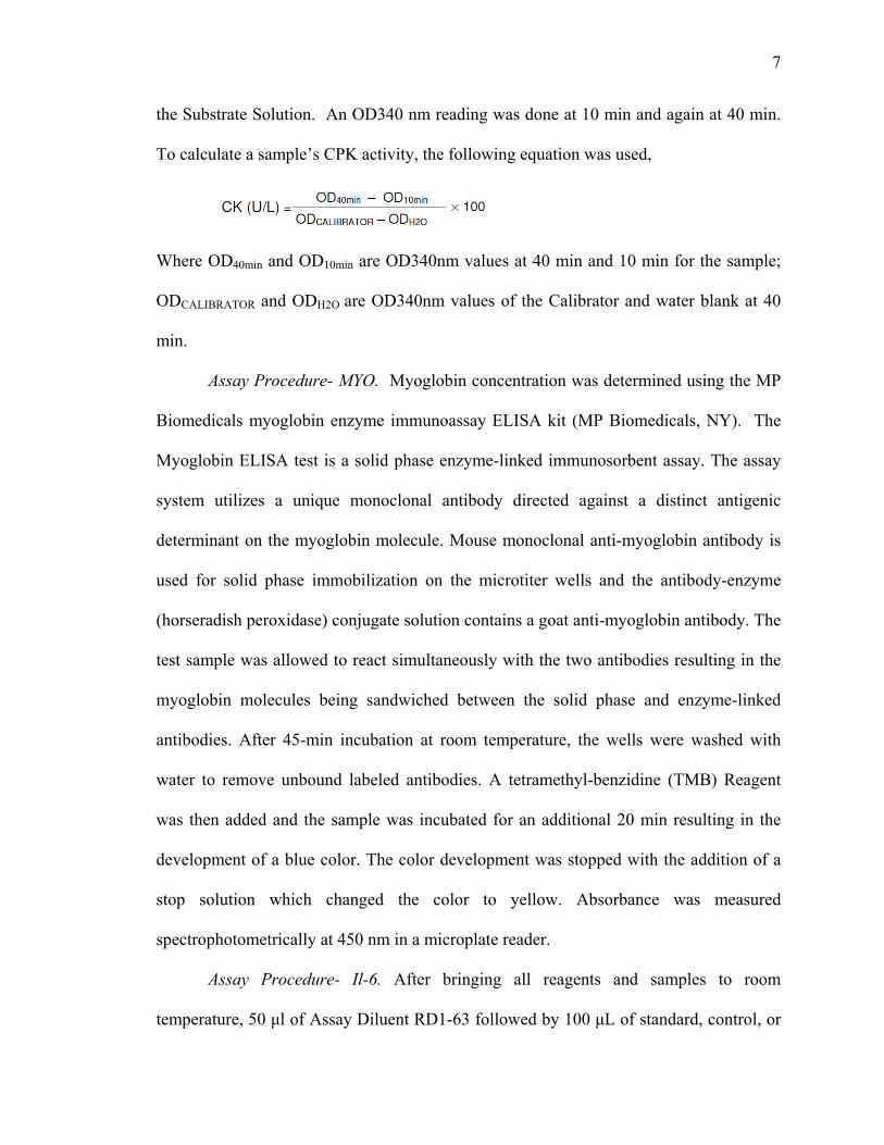
the Substrate Solution. An OD340 nm reading was done at 10 min and again at 40 min.
To calculate a sample’s CPK activity, the following equation was used,
Where OD40min and OD10min are OD340nm values at 40 min and 10 min for the sample;
ODCALIBRATOR and ODH2O are OD340nm values of the Calibrator and water blank at 40
min.
Assay Procedure- MYO. Myoglobin concentration was determined using the MP
Biomedicals myoglobin enzyme immunoassay ELISA kit (MP Biomedicals, NY). The
Myoglobin ELISA test is a solid phase enzyme-linked immunosorbent assay. The assay
system utilizes a unique monoclonal antibody directed against a distinct antigenic
determinant on the myoglobin molecule. Mouse monoclonal anti-myoglobin antibody is
used for solid phase immobilization on the microtiter wells and the antibody-enzyme
(horseradish peroxidase) conjugate solution contains a goat anti-myoglobin antibody. The
test sample was allowed to react simultaneously with the two antibodies resulting in the
myoglobin molecules being sandwiched between the solid phase and enzyme-linked
antibodies. After 45-min incubation at room temperature, the wells were washed with
water to remove unbound labeled antibodies. A tetramethyl-benzidine (TMB) Reagent
was then added and the sample was incubated for an additional 20 min resulting in the
development of a blue color. The color development was stopped with the addition of a
stop solution which changed the color to yellow. Absorbance was measured
spectrophotometrically at 450 nm in a microplate reader.
Assay Procedure- Il-6. After bringing all reagents and samples to room
temperature, 50 μl of Assay Diluent RD1-63 followed by 100 μL of standard, control, or
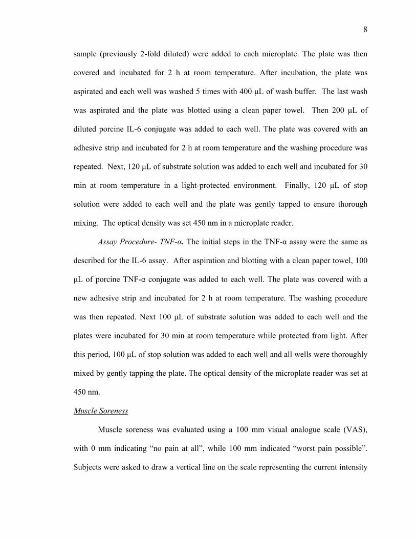
8
sample (previously 2-fold diluted) were added to each microplate. The plate was then
covered and incubated for 2 h at room temperature. After incubation, the plate was
aspirated and each well was washed 5 times with 400 μL of wash buffer. The last wash
was aspirated and the plate was blotted using a clean paper towel. Then 200 μL of
diluted porcine IL-6 conjugate was added to each well. The plate was covered with an
adhesive strip and incubated for 2 h at room temperature and the washing procedure was
repeated. Next, 120 μL of substrate solution was added to each well and incubated for 30
min at room temperature in a light-protected environment. Finally, 120 μL of stop
solution were added to each well and the plate was gently tapped to ensure thorough
mixing. The optical density was set 450 nm in a microplate reader.
Assay Procedure- TNF-α. The initial steps in the TNF-α assay were the same as
described for the IL-6 assay. After aspiration and blotting with a clean paper towel, 100
μL of porcine TNF-α conjugate was added to each well. The plate was covered with a
new adhesive strip and incubated for 2 h at room temperature. The washing procedure
was then repeated. Next 100 μL of substrate solution was added to each well and the
plates were incubated for 30 min at room temperature while protected from light. After
this period, 100 μL of stop solution was added to each well and all wells were thoroughly
mixed by gently tapping the plate. The optical density of the microplate reader was set at
450 nm.
Muscle Soreness
Muscle soreness was evaluated using a 100 mm visual analogue scale (VAS),
with 0 mm indicating “no pain at all”, while 100 mm indicated “worst pain possible”.
Subjects were asked to draw a vertical line on the scale representing the current intensity
9
of their pain under three different conditions; resting, after a palpating test, and during
passive flexion and extension. With the subject in a seated position and the exercised arm
resting on a table, the investigator palpated the biceps brachii at three sites: mid-belly and
3 cm above and below the mid belly, by applying firm pressure using the index and
middle fingers. The participant was then asked to rate his level of soreness as the
researcher extended and flexed his elbow joint while the participant relaxed his arm.
All the soreness measurements were performed by the same investigator.
McGill Pain Questionnaire
Muscle pain was assessed using the short-form of the McGill pain questionnaire
(MPQ) (44). The short-form McGill pain questionnaire (SF-MPQ) contains 15 major
descriptors (11 sensory; 4 affective) which are rated on an intensity scale from 0-3 (0 =
none, 1 = mild, 2 = moderate and 3 = severe). Three pain scores were derived from the
sum of the intensity rank values of the words chosen for sensory, affective, and total
descriptors.
Upper Arm Circumference
Anthropometric measurements were made according to the International Society
of Advancement in Kynanthropometry (ISAK). Upper arm circumference was assessed
using a constant tension tape while the arm was maintained in a relaxed and hanging
position at the participant’s side. The measurement was taken at a point equidistant
between the superior and lateral border of the acromion process and the proximal and
lateral border of the head of the radius.
10
Range of Motion (ROM)
Passive range of motion (PROM) of the elbow joint was evaluated using a plastic
goniometer while the participant lay supine on an examination table. ROM was defined
as the difference between the extended and flexed angles. The lateral epicondyle of the
humerus, the proximal apex of the deltoid muscle, and the styloid process of the radius
were used as landmarks to measure elbow joint angles. The participant’s arm was
passively flexed and extended using a very low angular velocity from a full flexed
position towards a full extension position.
Statistical Procedures
The responses of the dependent variables (VAS, pain, strength, ROM,
circumference, CPK, MYO, IL-6 and TNF-α) before and after exercise (immediately
after, 24 h, 48 h, 72 h and 96 h after) were compared between conditions. A series of 6
(time point) x 2 (treatment group) repeated measure ANOVA were performed. If
significant differences or interactions were found, LSD post hoc analyses were used to
determine the sources of these differences. Analyses were performed using the statistical
software package SPSS (version 17.0), with a significance level set a priori at p < 0.05.
Data from subjects who dropped out of the study were not included in the
analyses unless one full trial was completed. Also, if a subject did not remain inactive
during the testing period his data were not included in the analysis. All instruments were
calibrated prior to each use. To increase intra- and inter-rater reliability, every attempt
was made to use the same examiner for each test and a detailed examiner protocol with
systematic instructions was provided and followed for each test throughout the
investigative period.
CHAPTER 3: RESULTS
No significant differences were found between the WBPA and control groups in
physical characteristics or for baseline values for any of the criterion measures. Data are
presented as means ± SD relative to pre-intervention values unless otherwise stated.
Muscle Strength
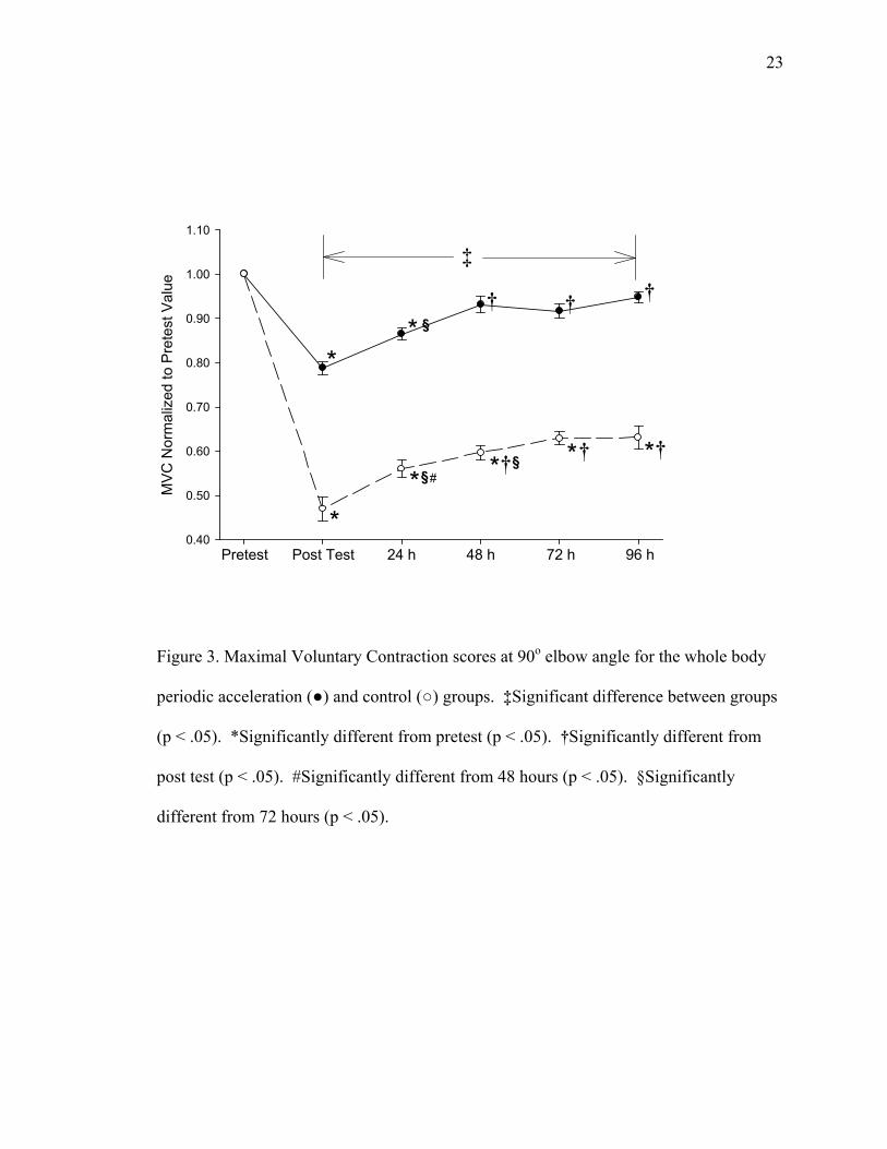
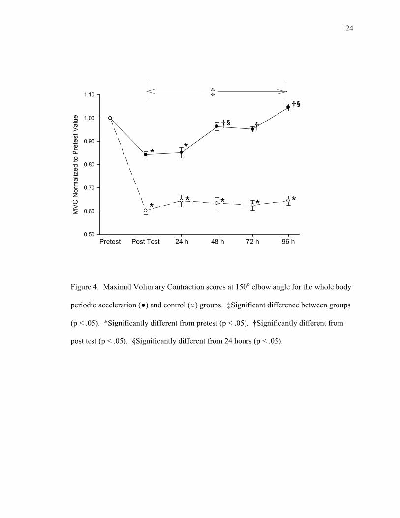
Significant differences were seen between the WBPA and control groups in
MVC90 and MVC150 from post test through 96 h (p < 0.05). The patterns of change in
MVC90 and MVC150 for the WBPA and control groups also differed across the 96 h
recovery; however, the patterns of change for both strength measures were consistent
within experimental groups (see Figures 3 and 4). Both groups showed the greatest
strength decreases immediately after exercise; however, the WBPA group recovered to
baseline values by 48 h post-exercise, while MVC values for the control group remained
depressed throughout the 96 h recovery period.
Blood Measurements
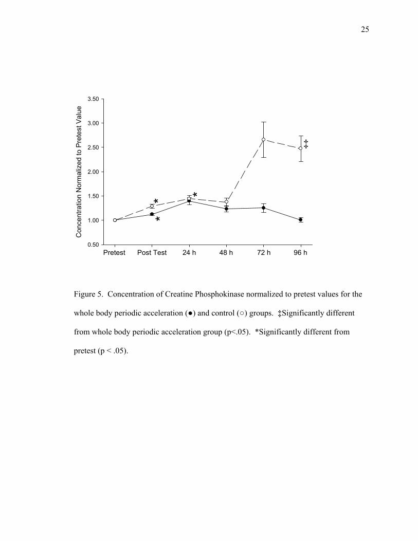
CPK. As shown in Figure 5, significantly higher CPK values were seen for the
controls compared to the WBPA group (p < 0.05). Both groups showed increases in CPK
post-test values (p < 0.05). For the control group CPK levels remained elevated at 24 h
and increased at 72 h and 96 h; however, only the increases at 24 h reached statistical
significance (p < 0.05).
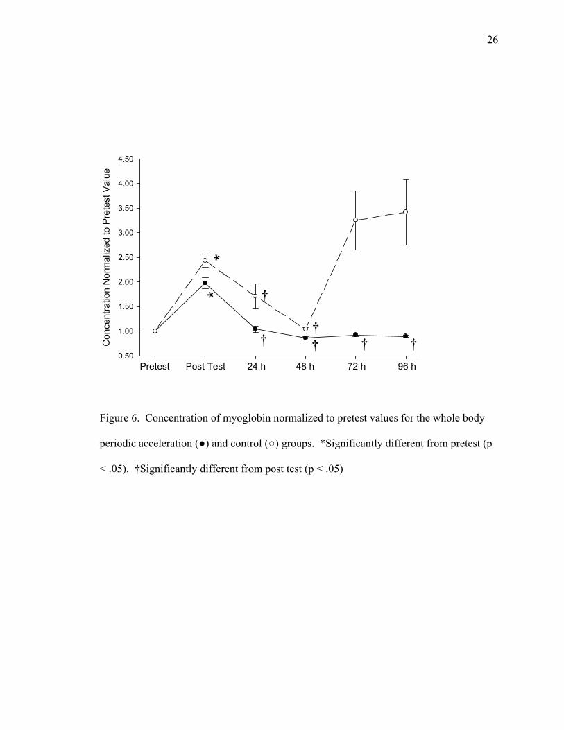
MYO. No significant differences between the WBPA and control groups were
seen in MYO concentration. Both groups showed a peak at the post test time point (1.97
± 0.99 and 2.43 ± 1.08 respectively) (p < 0.05) and returned to baseline during the 4 days
of recovery (Fig 6).
11
12
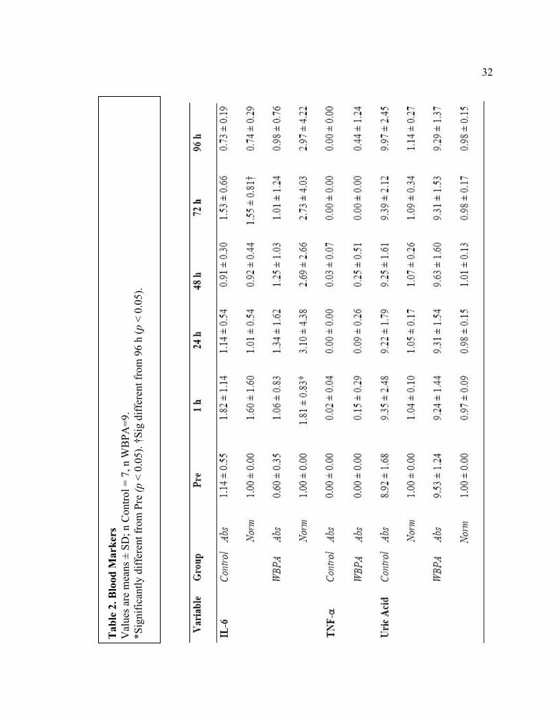
IL-6, TNF-α and Uric acid. Changes in inflammatory markers are shown in Table
2. There were no significant differences between groups. When normalized to pretest
value, however, IL-6 showed a significant increase post-test in the WBPA group (1.81 ±
0.83).
Muscle Soreness
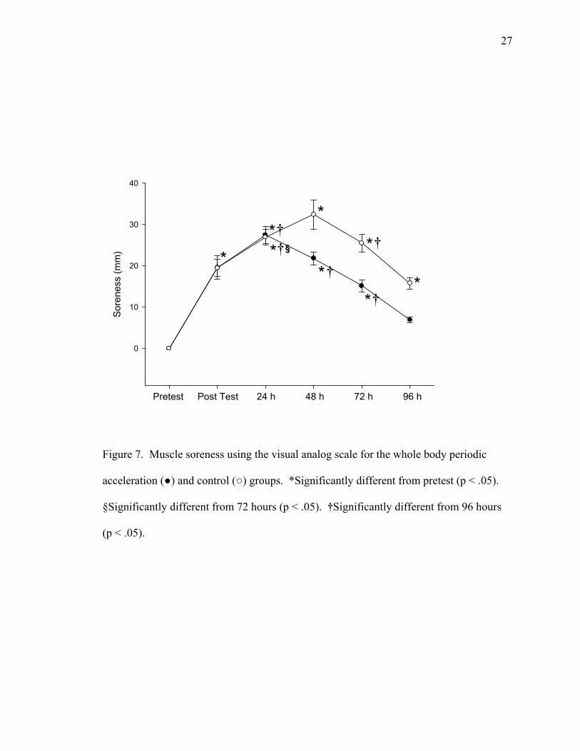
Ratings of muscle soreness are shown in Figure 7. While there were no significant
differences between groups, there were significant within group differences across the 4
recovery days. In the WBPA group, the rating of soreness peaked at 24 h (27.4 ± 18.0
mm) and was significantly different from pretest values from 24 h to 72 h post exercise (p
< 0.05). In contrast, the control group soreness score peaked at 48 h (32.4 ± 28.8 mm)
and was significantly elevated from posttest through the recovery period. Differences in
ratings of muscle soreness after the palpating test, and during flexion and extension are
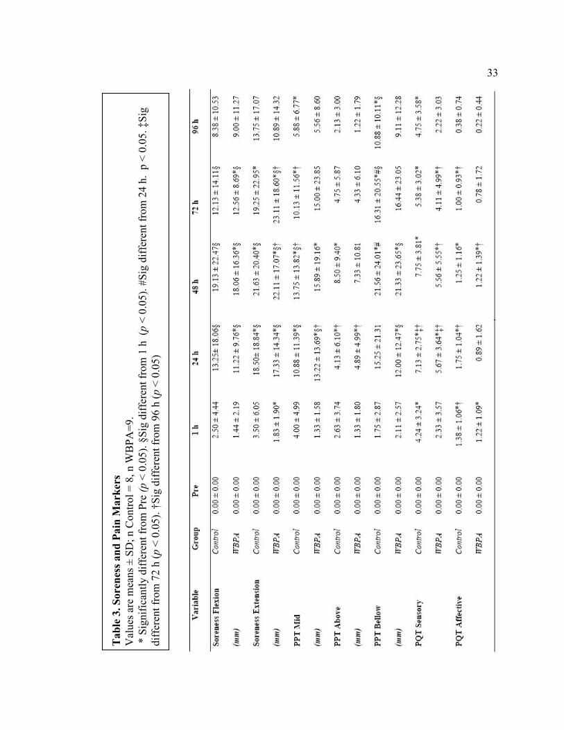
shown in Table 3.
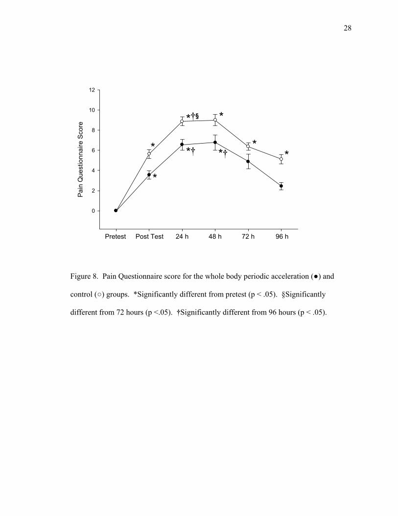
McGill Pain Questionnaire
Figure 8 summarizes the within group differences in the McGill pain scores since
no significant differences between groups were seen. The WBPA and control groups
showed significant increases from baseline at post-test (3.6 ± 3.8 and 5.6 ± 3.6
respectively). The WBPA scores remained high at the 24 and 48 h time points, while the
control group scores were elevated above post-test scores for the entire 96 h testing
period (p < 0.05). The sensory and affective dimensions of the pain questionnaire test are
summarized in Table 3.
13
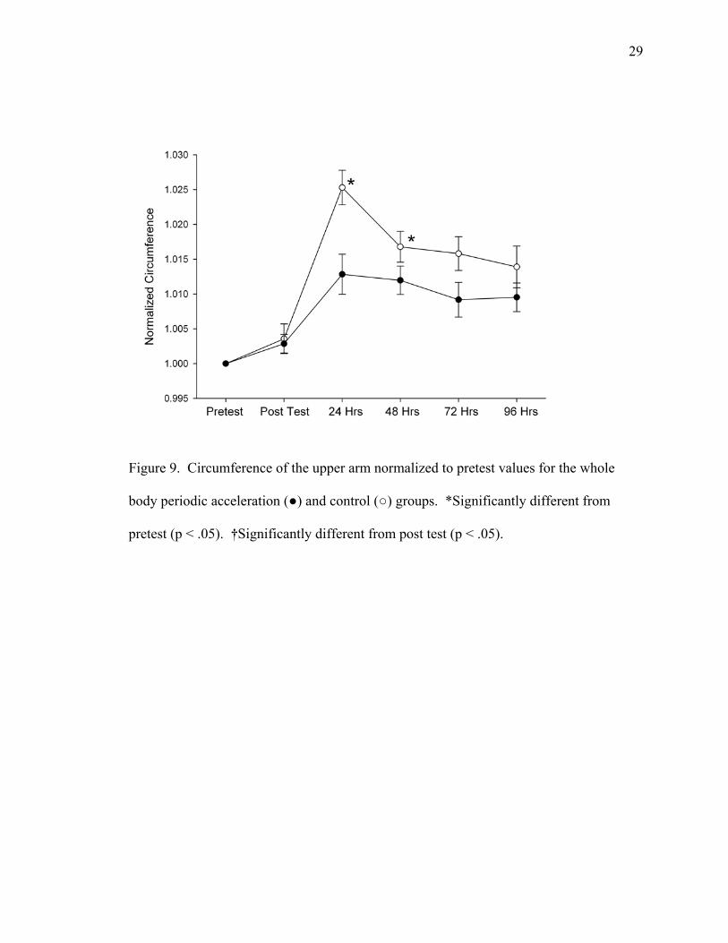
Circumference
Changes in upper arm circumference are shown in Figure 9. While no significant
differences in circumference were seen at any point in the recovery period for the WBPA
group, the control group had a significant increase in circumference at 24 h (33.4 ± 3.5
cm) that remained elevated at the 48 h test point (p < 0.05).
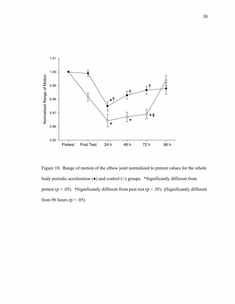
Range of Motion
As shown in Figure 10, there were no significant differences between groups
during the recovery days; however, both groups showed significant reductions in ROM at
24 h (0.97 ± 0.03 and 0.96 ± 0.04 for the WBPA and control groups, respectively) (p <
0.05). The deficits in ROM for the control group remained significant through 72 h post-
exercise.
CHAPTER 4: DISCUSSION
The purpose of the present study was to examine the effectiveness of WBPA as a
recovery method to treat DOMS resulting from a bout of unaccustomed eccentric elbow
flexion exercise. The major finding was that application of WBPA during the recovery
period after an eccentric exercise bout enhanced muscle performance and diminished
the negative impacts of DOMS in a young, fit male population sample.
To our knowledge this is the first study to show a more rapid strength recovery
using WBPA compared to passive recovery. In contrast, other interventions such as
cryotherapy (24, 32), active recovery (43, 76), stretching (27), electrical stimulation (38,
43), massage (75), and NSAIDs (63, 69), hyperbaric oxygen therapy (45), acupuncture
(33), and compression garments (21) have failed to show a significant positive impact on
strength performance during recovery after eccentric exercise. However, contrast water
therapy (CWT) was shown to enhance strength recovery at 24h, 48h and 72h when
compared to passive recovery (72, 73). It should be noted that differences in damage
protocol, targeted muscle and performance measures exist between Vaile et al and the
present study. In comparison to the CWT protocol, a greater decrease in muscle
performance was seeing immediately following the first treatment in both, control and
WBPA groups, indicating a disparity in the damage protocol effectiveness. Although
both damage protocols used similar intensities, a lower volume in a bilateral leg press
rather than unilateral arm curl were used in Vaile et al. Additionally, the positive
response in strength recovery with CWT, typically took 24h, while WBPA produced
improvements immediately after exposure which lasted throughout the 96 h evaluation
period.
14
15
The mechanisms that may have contributed to the improved strength performance
during the recovery period when applying WBPA are not clear. If reduced levels of
structural damage are responsible for the increased force production seen with WBPA,
the impact of NO should be considered. Although changes in intramuscular NO as a
result of diffusion of NO from eNOS cannot readily be examined because of its rapid
metabolism, its effects offer a plausible explanation for the increases in performance seen
with WBPA since NO reduces muscular damage from inflammation and oxidative stress
(6, 23, 25, 57).
Our results showing a significant difference in CPK concentration between the
WBPA and control groups reflect to some extent those seen with other recovery methods.
For example, in two separate studies using sport massage (64) and electro-membrane
microcurrent therapy (37) CPK concentrations at 96 h after eccentric exercise were
significantly lower than in controls. In contrast to our study, however, the use of
microcurrent produced no improvement in strength recovery, while the sports massage
study did not measure strength recovery. Additionally, while the results from studies
using cryotherapy as a recovery modality are equivocal (24, 30, 32), those showing a
positive impact (24, 32) reported reduced plasma CPK concentrations at 72 h post
exercise, but failed to have a positive impact on strength. Finally, in contrast to our
results, the use of light concentric exercise (76) or vitamin C supplementation (15) have
produced no impact on CPK concentration or strength during recovery. Moreover,
administration of vitamin C actually may be detrimental in DOMS (17).
16
The attenuated response of CPK in the WBPA group towards the end of the
recovery period, along with improved recovery of strength, may indicate reductions in the
levels of muscle damage with this intervention. However, it should be recognized CPK
cannot be considered a direct measure of the degree of muscle damage due to the large
variability in its response to eccentric exercise in similarly exercised individuals (16, 40,
52). While some recovery studies have shown CPK peaking within the first 24h after
exercise (11, 22, 69), others have confirmed that the highest CPK concentration is found
at day 4-5 of the recovery (37, 65). In addition, studies using different recovery methods
have reported variations in the patterns of change in CPK concentration. In the current
study, differences in CPK between the WBPA and control group may partially be
explained by the NO release with WBPA (60); NO could modulate the CPK efflux by
influencing the activation and accumulation of neutrophils (11, 65), as well as
counteracting the appearance of reactive oxygen species (25) after eccentric exercise.
Along with CPK, MYO is commonly used to measure muscle damage. Even
though there were no significant differences in MYO concentrations between groups, the
earlier peak in MYO compared to CPK agrees with previous studies (72). Additionally,
the increased MYO levels seen at 72 and 96 h post-exercise reflect the results reported by
Beck et al (7) and Howatson et al (30) when providing a protease supplement or an ice
massage, respectively. The differences in the time course of the appearance of MYO and
CPK can be explained by MYO’s smaller size and more direct route of delivery into the
blood (62). Unlike MYO, due to its larger size CPK is delivered into the bloodstream via
the lymphatic system which delays its appearance in the bloodstream following acute
muscle damage.
17
Our current findings, which show significant increases in IL-6 by the WBPA
group during the post-test assessment, have not been previously reported to our
knowledge. Although IL-6 may be expected to increase with exercise (26), the lack of
response in our controls is not without precedent (29). Results of previous studies
indicate that the lack of significant increases in IL-6 concentration in the control group
may have been due to the age and previous levels of conditioning of our participants (26,
53). Moreover, the timing of blood collection may blunt increases in IL-6 since peak
concentrations have been shown between 6h to 12h post eccentric exercise (46, 70). In
addition, our use of an exercise protocol involving a single arm may have limited the IL-6
response due to the low volume of muscle mass activated (24, 51).
The increase in IL-6 seen with WBPA may have been the result of NO increases
at the muscle level. NO has been shown to upregulate the pretranslational signaling
events leading to muscle IL-6 production (67). The increase in IL-6 can be considered a
positive response since it has been proposed to stimulate the production of anti-
inflammatory cytokines and may indirectly inhibit pro-inflammatory cytokines (50, 54-
56, 66). Moreover, IL-6 also increases satellite cell proliferation and muscle regeneration
(25). The lack of significant differences in systemic levels of TNF-α in our study reflects
the results reported by other researchers who examined inflammatory markers following
an acute bout of exercise.
Even though no significant differences in soreness and pain scores were seen
between groups, the shorter durations of the soreness and pain responses in the WBPA
group demonstrates an enhanced recovery pattern within this group. The positive
impact of WBPA on pain is not without precedent; in a comparable study with

18
fibromyalgia patients, 45 minutes of WBPA produced significant reductions in pain
within one to three treatments (61). The potential for NO to reduce inflammation and
edema may explain, in part, the more rapid recovery from soreness and pain seen in the
WBPA group due to reduced activation of the pain afferent fibers. The role of NO in
pain relief has recently been confirmed by the work of Cunha et al (20), who found that
the analgesic effects of morphine are achieved by stimulating a nNOS/NO/KATP channel
antinociceptive pathway, this pathway appears to cause a hyperpolarization of
nociceptive neurons, counteracting their enhanced excitability during the inflammatory
process. Also, NO counteracts the effects of endothelin-1 that also plays a role in pain
perception (28, 42).
The lack of significant increase in circumference in the WBPA compared to the
increase at 24 and 48 h in controls is indicative of the capacity of WBPA to reduce the
localized swelling commonly associated with DOMS. This positive impact is further
supported by the more rapid recovery of ROM in the WBPA group compared to controls.
The impact of WBPA on circumference and ROM may be explained by an increased
muscle blood flow or a decrease in inflammation. While the possibility that localized
blood flow served as a mechanism to reduce swelling and increase ROM in the present
study is questionable given the results reported by Adams et al (2) showing no significant
increase in muscle blood flow in pigs exposed to WBPA; however, muscle blood flow
may still be considered a possible mechanism since these researchers reported a 158%
increase (albeit not significant) in muscle blood flow with a WBPA exposure lasting 10
minutes rather than the 45 minutes used in the current study. The information supporting
reductions in inflammation as a possible mechanism is supported by the positive impacts
19
of NO on skeletal muscle inflammation, due to reduced neutrophil-mediated lysis and
decrease superoxide concentration (68).
LIMITATIONS
A limitation of this study was the failure to quantify leukocytes during the
recovery period. However, the quantification of cytokines provided a reliable indicator of
inflammation. Additionally, the quantification of glutathione could further help to explain
changes in some of the criterion measurement since it has been previously shown that
subjects with low total plasma glutathione levels had a smaller plasma CPK and MYO
response, and faster recovery from eccentric exercise compared with subjects with higher
levels (40). Moreover, lack of hydration control during the recovery period should also
be considered a limitation since hydration status can affect lymph flow and therefore
impact the levels of CPK following exercise (62).
Finally controlling diet during the recovery period may have also provided a more
stable testing environment (18), although the impact of nutrition on DOMS, as well as
inflammation, is equivocal (8, 47).
CONCLUSION
The use of WBPA as a recovery method after high-intensity eccentric resistance
exercise improved strength recovery and had a positive impact on DOMS symptoms.
These benefits were most likely the result of the enhanced release of NO through WBPA.
Future research should investigate the effects of WBPA within skeletal muscle through
quantification of inflammatory and oxidative stress markers as well as ultrastructural
damage using electron microscopy. Finally, the use of WBPA as a pre-conditioning
20
method to reduce levels of DOMS and potentially increase subsequent cardiovascular and
neuromuscular performance should be the subject of future investigations.
FIGURES
Figure 1. Study Design
21
22
Figure 2. Subject during the whole body periodic acceleration treatment using the Exer-
Rest AT motion platform.
23
M
VC N
orm
aliz
ed to
Pre
test
Val
ue
0.40
0.50
0.60
0.70
0.80
0.90
1.00
1.10
Pretest Post Test 24 h 48 h 72 h 96 h
**
*
** * *
‡
†††
§
§§
#
† † †
Figure 3. Maximal Voluntary Contraction scores at 90o elbow angle for the whole body
periodic acceleration (●) and control (○) groups. ‡Significant difference between groups
(p < .05). *Significantly different from pretest (p < .05). †Significantly different from
post test (p < .05). #Significantly different from 48 hours (p < .05). §Significantly
different from 72 hours (p < .05).
24
MVC
Nor
mal
ized
to P
rete
st V
alue
0.50
0.60
0.70
0.80
0.90
1.00
1.10
*
** * *
‡§
§† †
†
*
*
Pretest Post Test 24 h 48 h 72 h 96 h
Figure 4. Maximal Voluntary Contraction scores at 150o elbow angle for the whole body
periodic acceleration (●) and control (○) groups. ‡Significant difference between groups
(p < .05). *Significantly different from pretest (p < .05). †Significantly different from
post test (p < .05). §Significantly different from 24 hours (p < .05).
25
Con
cent
ratio
n N
orm
aliz
ed to
Pre
test
Val
ue
0.50
1.00
1.50
2.00
2.50
3.00
3.50
*
Pretest Post Test 24 h 48 h 72 h 96 h
*
*‡
Figure 5. Concentration of Creatine Phosphokinase normalized to pretest values for the
whole body periodic acceleration (●) and control (○) groups. ‡Significantly different
from whole body periodic acceleration group (p<.05). *Significantly different from
pretest (p < .05).
26
C
once
ntra
tion
Nor
mal
ized
to P
rete
st V
alue
0.50
1.00
1.50
2.00
2.50
3.00
3.50
4.00
4.50
**
Pretest Post Test 24 h 48 h 72 h 96 h
†
†† † † †
Figure 6. Concentration of myoglobin normalized to pretest values for the whole body
periodic acceleration (●) and control (○) groups. *Significantly different from pretest (p
< .05). †Significantly different from post test (p < .05)
27
So
rene
ss (m
m)
0
10
20
30
40
**
Pretest Post Test 24 h 48 h 72 h 96 h
**
*
**
*
†† †
†
†
§
Figure 7. Muscle soreness using the visual analog scale for the whole body periodic
acceleration (●) and control (○) groups. *Significantly different from pretest (p < .05).
§Significantly different from 72 hours (p < .05). †Significantly different from 96 hours
(p < .05).
28
Pain
Que
stio
nnai
re S
core
0
2
4
6
8
10
12
**
Pretest Post Test 24 h 48 h 72 h 96 h
** ** *
*
†
††
§
Figure 8. Pain Questionnaire score for the whole body periodic acceleration (●) and
control (○) groups. *Significantly different from pretest (p < .05). §Significantly
different from 72 hours (p <.05). †Significantly different from 96 hours (p < .05).
29
Figure 9. Circumference of the upper arm normalized to pretest values for the whole
body periodic acceleration (●) and control (○) groups. *Significantly different from
pretest (p < .05). †Significantly different from post test (p < .05).
30
Nor
mal
ized
Ran
ge o
f Mot
ion
0.95
0.96
0.97
0.98
0.99
1.00
1.01
**
††
†
Pretest Post Test 24 h 48 h 72 h 96 h
*
*
§
Figure 10. Range of motion of the elbow joint normalized to pretest values for the whole
body periodic acceleration (●) and control (○) groups. *Significantly different from
pretest (p < .05). †Significantly different from post test (p < .05). §Significantly different
from 96 hours (p < .05).
31
TABLES
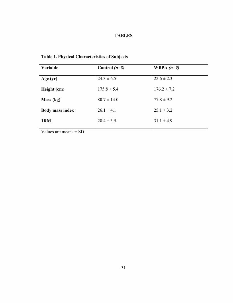
Table 1. Physical Characteristics of Subjects
Variable Control (n=8) WBPA (n=9)
Age (yr) 24.3 ± 6.5 22.6 ± 2.3
Height (cm) 175.8 ± 5.4 176.2 ± 7.2
Mass (kg) 80.7 ± 14.0 77.8 ± 9.2
Body mass index 26.1 ± 4.1 25.1 ± 3.2
1RM 28.4 ± 3.5 31.1 ± 4.9
Values are means ± SD
32
Tab
le 2
. Blo
od M
arke
rs
Val
ues a
re m
eans
± S
D; n
Con
trol =
7, n
WB
PA=9
. *S
igni
fican
tly d
iffer
ent f
rom
Pre
(p <
0.0
5). †
Sig
diff
eren
t fro
m 9
6 h
(p <
0.0
5).
33
Tab
le 3
. Sor
enes
s and
Pai
n M
arke
rs
Val
ues a
re m
eans
± S
D; n
Con
trol =
8, n
WB
PA=9
. *
Sig
nific
antly
diff
eren
t fro
m P
re (p
< 0
.05)
. §Si
g di
ffer
ent f
rom
1 h
(p
< 0.
05).
#Sig
diff
eren
t fro
m 2
4 h.
p <
0.0
5. ‡
Sig
diff
eren
t fro
m 7
2 h
(p <
0.0
5). †
Sig
diff
eren
t fro
m 9
6 h
(p <
0.0
5)
BIBLIOGRAPHY
1. Abraham WM, Ahmed A, Serebriakov I, Lauredo IT, Bassuk J, Adams JA and Sackner MA. Whole-body periodic acceleration modifies experimental asthma in sheep. Am.J.Respir.Crit.Care Med. 174: 7: 743-752, 2006.
2. Adams JA, Mangino MJ, Bassuk J, Kurlansky P and Sackner MA. Regional blood flow during periodic acceleration. Crit.Care Med. 29: 10: 1983-1988, 2001.
3. Adams JA, Moore JE,Jr, Moreno MR, Coelho J, Bassuk J and Wu D. Effects of periodic body acceleration on the in vivo vasoactive response to N-omega-nitro-L-arginine and the in vitro nitric oxide production. Ann.Biomed.Eng. 31: 11: 1337-1346, 2003.
4. Armstrong RB. Mechanisms of exercise-induced delayed onset muscular soreness: a brief review. Med.Sci.Sports Exerc. 16: 6: 529-538, 1984.
5. Baechle TR, Earle RW and National Strength & Conditioning Association. Essentials of strength training and conditioning. Champaign, IL: Human Kinetics, 2008, p. 641.
6. Barreiro E and Hussain SN. Protein carbonylation in skeletal muscles: impact on function. Antioxid.Redox Signal. 12: 3: 417-429, 2010.
7. Beck TW, Housh TJ, Johnson GO, Schmidt RJ, Housh DJ, Coburn JW, Malek MH and Mielke M. Effects of a protease supplement on eccentric exercise-induced markers of delayed-onset muscle soreness and muscle damage. J.Strength Cond Res. 21: 3: 661-667, 2007.
8. Best TM, Hunter R, Wilcox A and Haq F. Effectiveness of sports massage for recovery of skeletal muscle from strenuous exercise. Clin.J.Sport Med. 18: 5: 446-460, 2008.
9. Broadbent S, Rousseau JJ, Thorp RM, Choate SL, Jackson FS and Rowlands DS. Vibration therapy reduces plasma IL-6 and muscle soreness after downhill running. Br.J.Sports Med. 2008.
34
35
10. Byrnes WC and Clarkson PM. Delayed onset muscle soreness and training. Clin.Sports Med. 5: 3: 605-614, 1986.
11. Cannon JG, Orencole SF, Fielding RA, Meydani M, Meydani SN, Fiatarone MA, Blumberg JB and Evans WJ. Acute phase response in exercise: interaction of age and vitamin E on neutrophils and muscle enzyme release. Am.J.Physiol. 259: 6 Pt 2: R1214-9, 1990.
12. Chapman D, Newton M, Sacco P and Nosaka K. Greater muscle damage induced by fast versus slow velocity eccentric exercise. Int.J.Sports Med. 27: 8: 591-598, 2006.
13. Chapman DW, Newton M, McGuigan M and Nosaka K. Effect of lengthening contraction velocity on muscle damage of the elbow flexors. Med.Sci.Sports Exerc. 40: 5: 926-933, 2008.
14. Cheung K, Hume P and Maxwell L. Delayed onset muscle soreness : treatment strategies and performance factors. Sports Med. 33: 2: 145-164, 2003.
15. Childs A, Jacobs C, Kaminski T, Halliwell B and Leeuwenburgh C. Supplementation with vitamin C and N-acetyl-cysteine increases oxidative stress in humans after an acute muscle injury induced by eccentric exercise. Free Radic.Biol.Med. 31: 6: 745-753, 2001.
16. Clarkson PM and Ebbeling C. Investigation of serum creatine kinase variability after muscle-damaging exercise. Clin.Sci.(Lond) 75: 3: 257-261, 1988.
17. Close GL, Ashton T, Cable T, Doran D, Holloway C, McArdle F and MacLaren DP. Ascorbic acid supplementation does not attenuate post-exercise muscle soreness following muscle-damaging exercise but may delay the recovery process. Br.J.Nutr. 95: 5: 976-981, 2006.
18. Cockburn E, Hayes PR, French DN, Stevenson E and St Clair Gibson A. Acute milk-based protein-CHO supplementation attenuates exercise-induced muscle damage. Appl.Physiol.Nutr.Metab. 33: 4: 775-783, 2008.
19. Craig CL, Marshall AL, Sjostrom M, Bauman AE, Booth ML, Ainsworth BE, Pratt M, Ekelund U, Yngve A, Sallis JF and Oja P.
36
International physical activity questionnaire: 12-country reliability and validity. Med.Sci.Sports Exerc. 35: 8: 1381-1395, 2003.
20. Cunha TM, Roman-Campos D, Lotufo CM, Duarte HL, Souza GR, Verri WA,Jr, Funez MI, Dias QM, Schivo IR, Domingues AC, Sachs D, Chiavegatto S, Teixeira MM, Hothersall JS, Cruz JS, Cunha FQ and Ferreira SH. Morphine peripheral analgesia depends on activation of the PI3Kgamma/AKT/nNOS/NO/KATP signaling pathway. Proc.Natl.Acad.Sci.U.S.A. 107: 9: 4442-4447, 2010.
21. Davies V, Thompson KG and Cooper SM. The effects of compression garments on recovery. J.Strength Cond Res. 23: 6: 1786-1794, 2009.
22. Donnelly AE, Maughan RJ and Whiting PH. Effects of ibuprofen on exercise-induced muscle soreness and indices of muscle damage. Br.J.Sports Med. 24: 3: 191-195, 1990.
23. Espey MG, Miranda KM, Thomas DD, Xavier S, Citrin D, Vitek MP and Wink DA. A chemical perspective on the interplay between NO, reactive oxygen species, and reactive nitrogen oxide species. Ann.N.Y.Acad.Sci. 962: 195-206, 2002.
24. Eston R and Peters D. Effects of cold water immersion on the symptoms of exercise-induced muscle damage. J.Sports Sci. 17: 3: 231-238, 1999.
25. Filippin LI, Moreira AJ, Marroni NP and Xavier RM. Nitric oxide and repair of skeletal muscle injury. Nitric Oxide 21: 3-4: 157-163, 2009.
26. Fischer CP. Interleukin-6 in acute exercise and training: what is the biological relevance? Exerc.Immunol.Rev. 12: 6-33, 2006.
27. Gulick DT, Kimura IF, Sitler M, Paolone A and Kelly JD. Various Treatment Techniques on Signs and Symptoms of Delayed Onset Muscle Soreness. J.Athl Train. 31: 2: 145-152, 1996.
28. Hans G, Schmidt BL and Strichartz G. Nociceptive sensitization by endothelin-1. Brain Res.Rev. 60: 1: 36-42, 2009.
29. Hirose L, Nosaka K, Newton M, Laveder A, Kano M, Peake J and Suzuki K. Changes in inflammatory mediators following eccentric exercise of the elbow flexors. Exerc.Immunol.Rev. 10: 75-90, 2004.
37
30. Howatson G, Gaze D and van Someren KA. The efficacy of ice massage in the treatment of exercise-induced muscle damage. Scand.J.Med.Sci.Sports 15: 6: 416-422, 2005.
31. Howatson G, Goodall S and van Someren KA. The influence of cold water immersions on adaptation following a single bout of damaging exercise. Eur.J.Appl.Physiol. 105: 4: 615-621, 2009.
32. Howatson G and Van Someren KA. Ice massage. Effects on exercise-induced muscle damage. J.Sports Med.Phys.Fitness 43: 4: 500-505, 2003.
33. Hubscher M, Vogt L, Bernhorster M, Rosenhagen A and Banzer W. Effects of acupuncture on symptoms and muscle function in delayed-onset muscle soreness. J.Altern.Complement.Med. 14: 8: 1011-1016, 2008.
34. Hutcheson IR and Griffith TM. Release of endothelium-derived relaxing factor is modulated both by frequency and amplitude of pulsatile flow. Am.J.Physiol. 261: 1 Pt 2: H257-62, 1991.
35. Jamurtas AZ, Theocharis V, Tofas T, Tsiokanos A, Yfanti C, Paschalis V, Koutedakis Y and Nosaka K. Comparison between leg and arm eccentric exercises of the same relative intensity on indices of muscle damage. Eur.J.Appl.Physiol. 95: 2-3: 179-185, 2005.
36. Kerschan-Schindl K, Grampp S, Henk C, Resch H, Preisinger E, Fialka-Moser V and Imhof H. Whole-body vibration exercise leads to alterations in muscle blood volume. Clin.Physiol. 21: 3: 377-382, 2001.
37. Lambert MI, Marcus P, Burgess T and Noakes TD. Electro-membrane microcurrent therapy reduces signs and symptoms of muscle damage. Med.Sci.Sports Exerc. 34: 4: 602-607, 2002.
38. Lattier G, Millet GY, Martin A and Martin V. Fatigue and recovery after high-intensity exercise. Part II: Recovery interventions. Int.J.Sports Med. 25: 7: 509-515, 2004.
39. Lavender AP and Nosaka K. Changes in markers of muscle damage of middle-aged and young men following eccentric exercise of the elbow flexors. J.Sci.Med.Sport 11: 2: 124-131, 2008.
38
40. Lee J and Clarkson PM. Plasma creatine kinase activity and glutathione after eccentric exercise. Med.Sci.Sports Exerc. 35: 6: 930-936, 2003.
41. Lundeberg T, Abrahamsson P, Bondesson L and Haker E. Vibratory stimulation compared to placebo in alleviation of pain. Scand.J.Rehabil.Med. 19: 4: 153-158, 1987.
42. Marasciulo FL, Montagnani M and Potenza MA. Endothelin-1: the yin and yang on vascular function. Curr.Med.Chem. 13: 14: 1655-1665, 2006.
43. Martin V, Millet GY, Lattier G and Perrod L. Effects of recovery modes after knee extensor muscles eccentric contractions. Med.Sci.Sports Exerc. 36: 11: 1907-1915, 2004.
44. Meissner JE. McGill-Melzack pain questionnaire. Nursing 10: 1: 50-51, 1980.
45. Mekjavic IB, Exner JA, Tesch PA and Eiken O. Hyperbaric oxygen therapy does not affect recovery from delayed onset muscle soreness. Med.Sci.Sports Exerc. 32: 3: 558-563, 2000.
46. Miles MP, Andring JM, Pearson SD, Gordon LK, Kasper C, Depner CM and Kidd JR. Diurnal variation, response to eccentric exercise, and association of inflammatory mediators with muscle damage variables. J.Appl.Physiol. 104: 2: 451-458, 2008.
47. Miles MP, Pearson SD, Andring JM, Kidd JR and Volpe SL. Effect of carbohydrate intake during recovery from eccentric exercise on interleukin-6 and muscle-damage markers. Int.J.Sport Nutr.Exerc.Metab. 17: 6: 507-520, 2007.
48. Newham DJ, McPhail G, Mills KR and Edwards RH. Ultrastructural changes after concentric and eccentric contractions of human muscle. J.Neurol.Sci. 61: 1: 109-122, 1983.
49. Newton MJ, Morgan GT, Sacco P, Chapman DW and Nosaka K. Comparison of responses to strenuous eccentric exercise of the elbow flexors between resistance-trained and untrained men. J.Strength Cond Res. 22: 2: 597-607, 2008.
39
50. Nielsen S and Pedersen BK. Skeletal muscle as an immunogenic organ. Curr.Opin.Pharmacol. 8: 3: 346-351, 2008.
51. Nosaka K and Clarkson PM. Changes in indicators of inflammation after eccentric exercise of the elbow flexors. Med.Sci.Sports Exerc. 28: 8: 953-961, 1996.
52. Nosaka K and Clarkson PM. Variability in serum creatine kinase response after eccentric exercise of the elbow flexors. Int.J.Sports Med. 17: 2: 120-127, 1996.
53. Peake J, Nosaka K and Suzuki K. Characterization of inflammatory responses to eccentric exercise in humans. Exerc.Immunol.Rev. 11: 64-85, 2005.
54. Pedersen BK. Edward F. Adolph distinguished lecture: muscle as an endocrine organ: IL-6 and other myokines. J.Appl.Physiol. 107: 4: 1006-1014, 2009.
55. Pedersen BK and Febbraio MA. Muscle as an endocrine organ: focus on muscle-derived interleukin-6. Physiol.Rev. 88: 4: 1379-1406, 2008.
56. Pedersen BK and Fischer CP. Beneficial health effects of exercise--the role of IL-6 as a myokine. Trends Pharmacol.Sci. 28: 4: 152-156, 2007.
57. Phillips L, Toledo AH, Lopez-Neblina F, Anaya-Prado R and Toledo-Pereyra LH. Nitric oxide mechanism of protection in ischemia and reperfusion injury. J.Invest.Surg. 22: 1: 46-55, 2009.
58. Rhea MR, Bunker D, Marin PJ and Lunt K. Effect of iTonic whole-body vibration on delayed-onset muscle soreness among untrained individuals. J.Strength Cond Res. 23: 6: 1677-1682, 2009.
59. Sackner MA, Gummels E and Adams JA. Effect of moderate-intensity exercise, whole-body periodic acceleration, and passive cycling on nitric oxide release into circulation. Chest 128: 4: 2794-2803, 2005.
60. Sackner MA, Gummels E and Adams JA. Nitric oxide is released into circulation with whole-body, periodic acceleration. Chest 127: 1: 30-39, 2005.
40
61. Sackner MA, Gummels EM and Adams JA. Say NO to fibromyalgia and chronic fatigue syndrome: an alternative and complementary therapy to aerobic exercise. Med.Hypotheses 63: 1: 118-123, 2004.
62. Sayers SP and Clarkson PM. Short-term immobilization after eccentric exercise. Part II: creatine kinase and myoglobin. Med.Sci.Sports Exerc. 35: 5: 762-768, 2003.
63. Sayers SP, Knight CA, Clarkson PM, Van Wegen EH and Kamen G. Effect of ketoprofen on muscle function and sEMG activity after eccentric exercise. Med.Sci.Sports Exerc. 33: 5: 702-710, 2001.
64. Smith LL. Acute inflammation: the underlying mechanism in delayed onset muscle soreness? Med.Sci.Sports Exerc. 23: 5: 542-551, 1991.
65. Smith LL, Keating MN, Holbert D, Spratt DJ, McCammon MR, Smith SS and Israel RG. The effects of athletic massage on delayed onset muscle soreness, creatine kinase, and neutrophil count: a preliminary report. J.Orthop.Sports Phys.Ther. 19: 2: 93-99, 1994.
66. Starkie R, Ostrowski SR, Jauffred S, Febbraio M and Pedersen BK. Exercise and IL-6 infusion inhibit endotoxin-induced TNF-alpha production in humans. FASEB J. 17: 8: 884-886, 2003.
67. Steensberg A, Keller C, Hillig T, Frosig C, Wojtaszewski JF, Pedersen BK, Pilegaard H and Sander M. Nitric oxide production is a proximal signaling event controlling exercise-induced mRNA expression in human skeletal muscle. FASEB J. 21: 11: 2683-2694, 2007.
68. Tidball JG. Inflammatory processes in muscle injury and repair. Am.J.Physiol.Regul.Integr.Comp.Physiol. 288: 2: R345-53, 2005.
69. Tokmakidis SP, Kokkinidis EA, Smilios I and Douda H. The effects of ibuprofen on delayed muscle soreness and muscular performance after eccentric exercise. J.Strength Cond Res. 17: 1: 53-59, 2003.
70. Tomiya A, Aizawa T, Nagatomi R, Sensui H and Kokubun S. Myofibers express IL-6 after eccentric exercise. Am.J.Sports Med. 32: 2: 503-508, 2004.
41
71. Uryash A, Wu H, Bassuk J, Kurlansky P, Sackner MA and Adams JA. Low-amplitude pulses to the circulation through periodic acceleration induces endothelial-dependent vasodilatation. J.Appl.Physiol. 106: 6: 1840-1847, 2009.
72. Vaile J, Halson S, Gill N and Dawson B. Effect of hydrotherapy on the signs and symptoms of delayed onset muscle soreness. Eur.J.Appl.Physiol. 102: 4: 447-455, 2008.
73. Vaile JM, Gill ND and Blazevich AJ. The effect of contrast water therapy on symptoms of delayed onset muscle soreness. J.Strength Cond Res. 21: 3: 697-702, 2007.
74. Walsh LD, Hesse CW, Morgan DL and Proske U. Human forearm position sense after fatigue of elbow flexor muscles. J.Physiol. 558: Pt 2: 705-715, 2004.
75. Zainuddin Z, Newton M, Sacco P and Nosaka K. Effects of massage on delayed-onset muscle soreness, swelling, and recovery of muscle function. J.Athl Train. 40: 3: 174-180, 2005.
76. Zainuddin Z, Sacco P, Newton M and Nosaka K. Light concentric exercise has a temporarily analgesic effect on delayed-onset muscle soreness, but no effect on recovery from eccentric exercise. Appl.Physiol.Nutr.Metab. 31: 2: 126-134, 2006.