White Dot Syndromes - specialtyeyeinstitute.com fileVision can be 20/20 to LP. 3/13/2017 6...
19
3/13/2017 1 White Spot Syndromes SURENDAR S. PUROHIT, MD White Dot Syndromes Inflammation and dysfunction of: Outer retina Retinal pigment epithelium Choroid Vitritis / iritis not a necessary finding Etiology Unknown Autoimmune cause hypothesized for some WDS Several of these my be related / sprectrum of same process Common characteristics Blurred vision Photopsias Visual field changes Floaters Change in contrast sensitivity Symptoms can be present prior to clinical findings White spots can be subtle or prominent One or both eyes Can be asymmetric
-
Upload
nguyenkhue -
Category
Documents
-
view
212 -
download
0
Transcript of White Dot Syndromes - specialtyeyeinstitute.com fileVision can be 20/20 to LP. 3/13/2017 6...

3/13/2017
1
White Spot SyndromesSURENDAR S. PUROHIT, MD
White Dot Syndromes
Inflammation and dysfunction of: Outer retina
Retinal pigment epithelium
Choroid
Vitritis / iritis not a necessary finding Etiology
Unknown
Autoimmune cause hypothesized for some WDS
Several of these my be related / sprectrum of same process
Common characteristics
Blurred vision Photopsias Visual field changes Floaters Change in contrast sensitivity Symptoms can be present prior to clinical findings White spots
can be subtle or prominent
One or both eyes
Can be asymmetric

3/13/2017
2
White spot syndromes
Differential diagnosis: Granulomatous diseases (sarcoidosis, sympathetic ophthalmia, TB-)
Masquerade syndromes (syphilis, lymphoma)
Infectious diseases (toxoplasmosis, POHS, Behcet’s disease)
Degenerative conditions (drusen)
40 y.o. white femaleCC: metamorphopsia os
Vision: OD: 20/20 -2.50
OS: 20/40 -3.75
IOP 15/18 Ant Seg: no cells / flare OU Vitreous: no cells OU
3/13/2017
3

3/13/2017
4
Work upSyphilis serologies: negativeBartonella Henslae titers: negativeViral titres (HSV, Varizella, CMV): negative Toxoplasma titres: negativeACE: 24ANA: positive 1:160 homogenous pattern
3/13/2017
5
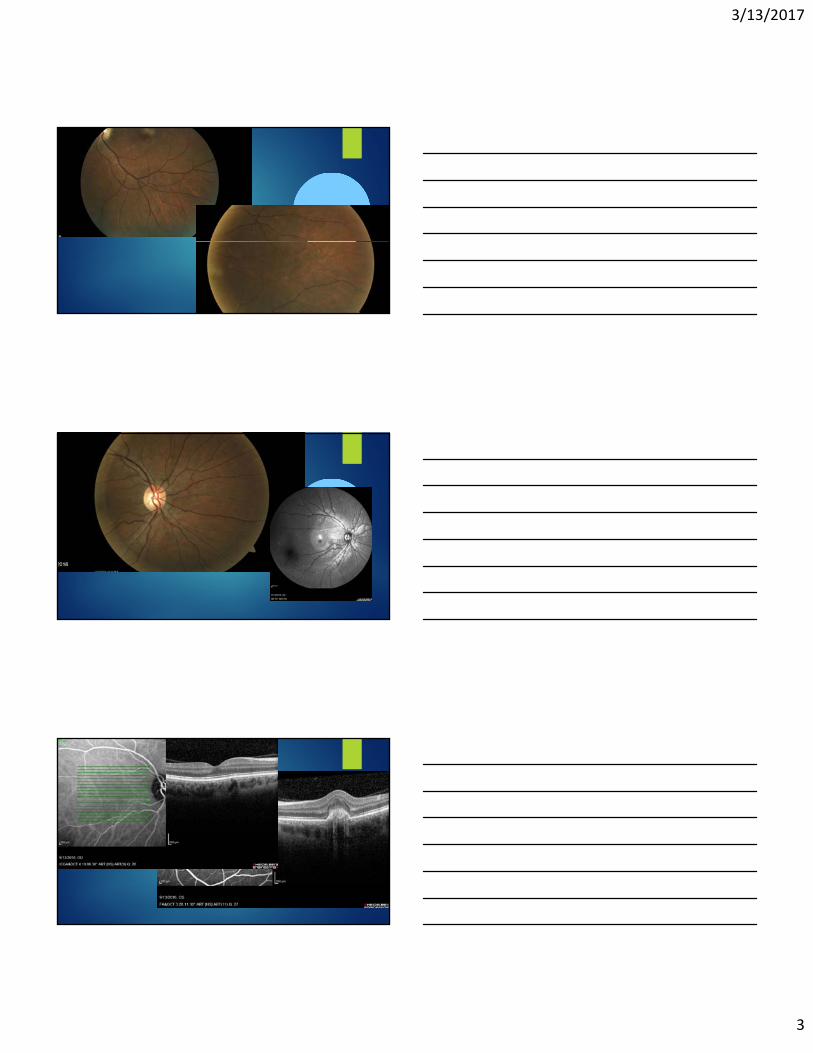
Punctate inner choroidopathy (PIC)
Healthy myopic women (90%) Mean age: 30 Blurred vision Paracentral scotomas Small yellow/white lesions of inner choroid / RPE Overlying SRF Change to atrophic scars Usually unilateral (85%) No anterior or posterior seg inflammation
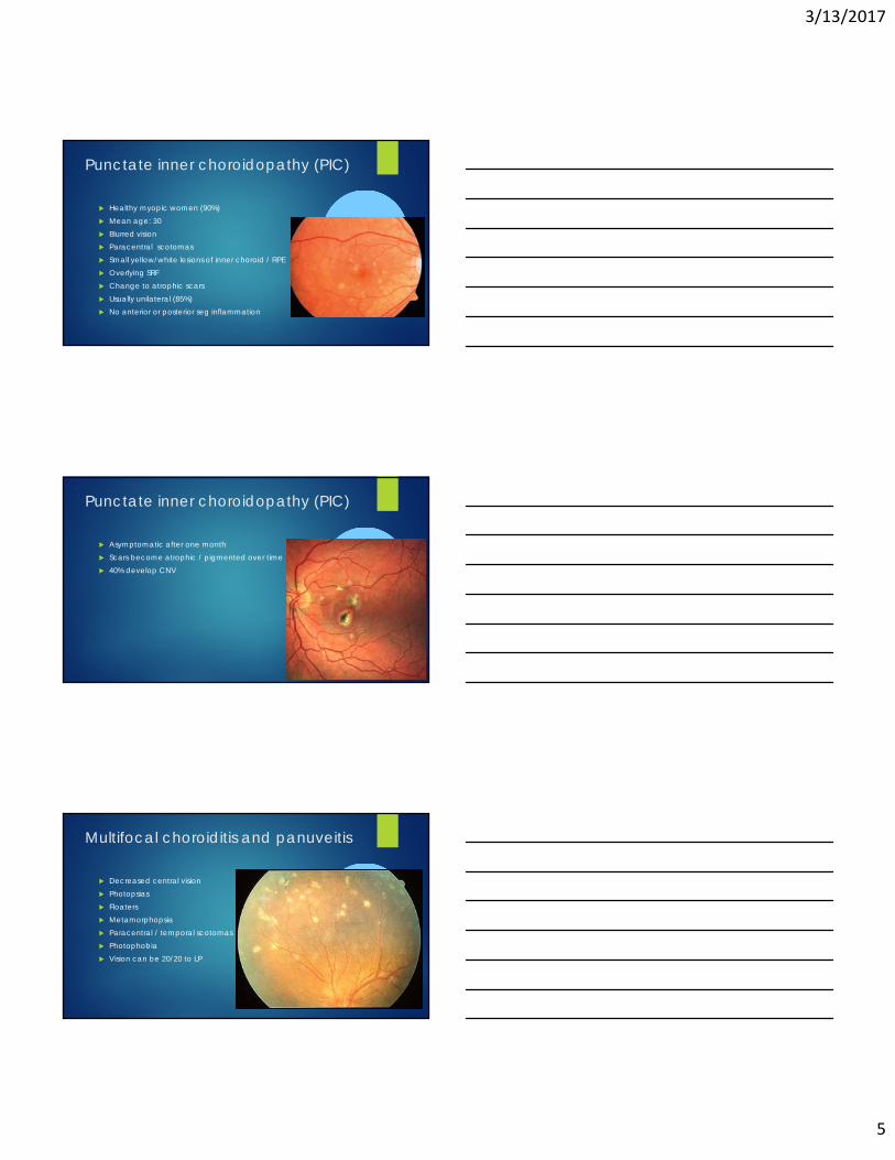
Punctate inner choroidopathy (PIC)
Asymptomatic after one month Scars become atrophic / pigmented over time 40% develop CNV
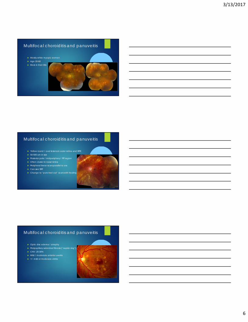
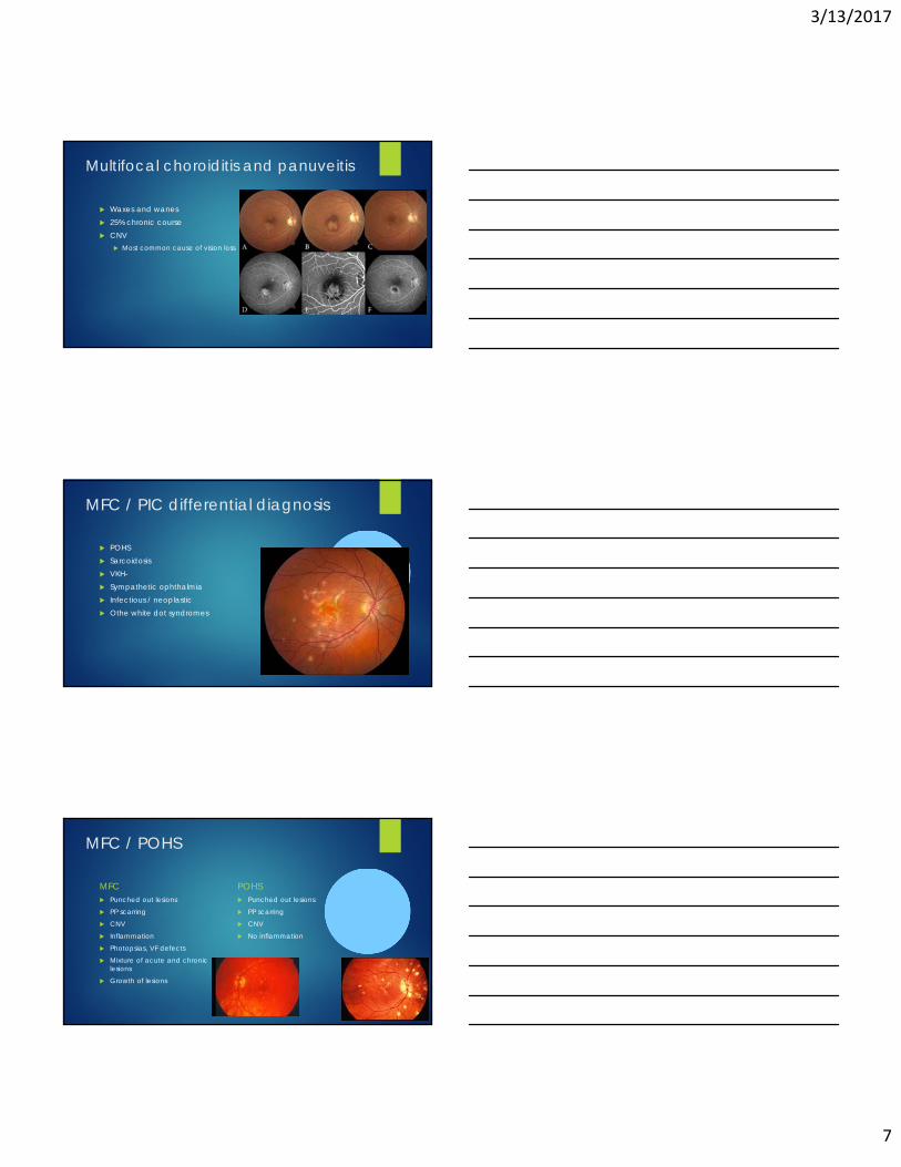
Multifocal choroiditis and panuveitis
Decreased central vision Photopsias Floaters Metamorphopsia Paracentral / temporal scotomas Photophobia Vision can be 20/20 to LP
3/13/2017
6
Multifocal choroiditis and panuveitis
Mostly white myopic women Age 20-60 Most in their 30s
Multifocal choroiditis and panuveitis
Yellow round / oval lesions in outer retina and RPE 50-500 um in size Posterior pole / midperiphery / PP region Often cluster in nasal retina Peripheral linear scars parallel to ora Can see SRF Change to “punched out” scars with healing
Multifocal choroiditis and panuveitis
Optic disc edema / atrophy Peripapillary subretinal fibrosis (“napkin ring”) CNV: 25-30% Mild / moderate anterior uveitis +/- mild or moderate vitritis
3/13/2017
7
Multifocal choroiditis and panuveitis
Waxes and wanes 25% chronic course CNV
Most common cause of vision loss
POHS Sarcoidosis VKH- Sympathetic ophthalmia Infectious / neoplastic Othe white dot syndromes
MFC / PIC differential diagnosis
MFC / POHS
MFC Punched out lesions
PP scarring
CNV
Inflammation
Photopsias, VF defects
Mixture of acute and chronic lesions
Growth of lesions
POHS Punched out lesions
PP scarring
CNV
No inflammation

3/13/2017
8
MFC / PIC
MFC Median age: 45
CNV: 28%
More inflammation
PIC Median age: 29
CNV: 77%
MFC and PIC treatment
Corticosteroids Immunosuppressives Anti-VEGF PDT
Multiple evanescent white dot syndrome (MEWDS)
Acute multifocal unilateral retinopathy Unknown cause Young myopic females (75%) Multiple white dots level of outer retina or RPE

3/13/2017
9
Multiple evanescent white dot syndrome (MEWDS)
Acute onset of blurred vision Temporal field loss (enlarged blind spot) Photopsias Flu like symptoms Vision: 20/20 – 20/300 Usually returns to normal Duration: 6 weeks
Multiple evanescent white dot syndrome (MEWDS)
Numerous small paramacular white spots level of RPE or deep retina Spare the fovea “granularity” to macula Spots disappear over times Left with mild RPE mottling Mild iritis +/- vitritis
Multiple evanescent white dot syndrome (MEWDS)
FA: Early and late hyperfluorescence of white dots
“wreath” like pattern
ICG: numerous hypofluorescent spots Usually no treatment required

3/13/2017
10
Birdshot Chorioretinopathy
Chronic bilateral inflammatory dx Healthy men or women : 30-60 yrs old Symptoms:
Vitreous floaters
Decreased vision (especially at night)
Birdshot Chorioretinopathy
Blurred vision, floaters, photopsias Vision usally > =20/40 No pain or redness Severe nyctalopia Changes in color or visual fields Purely ocular disease Mean age: 53 Predominately white patients Bilateral disease
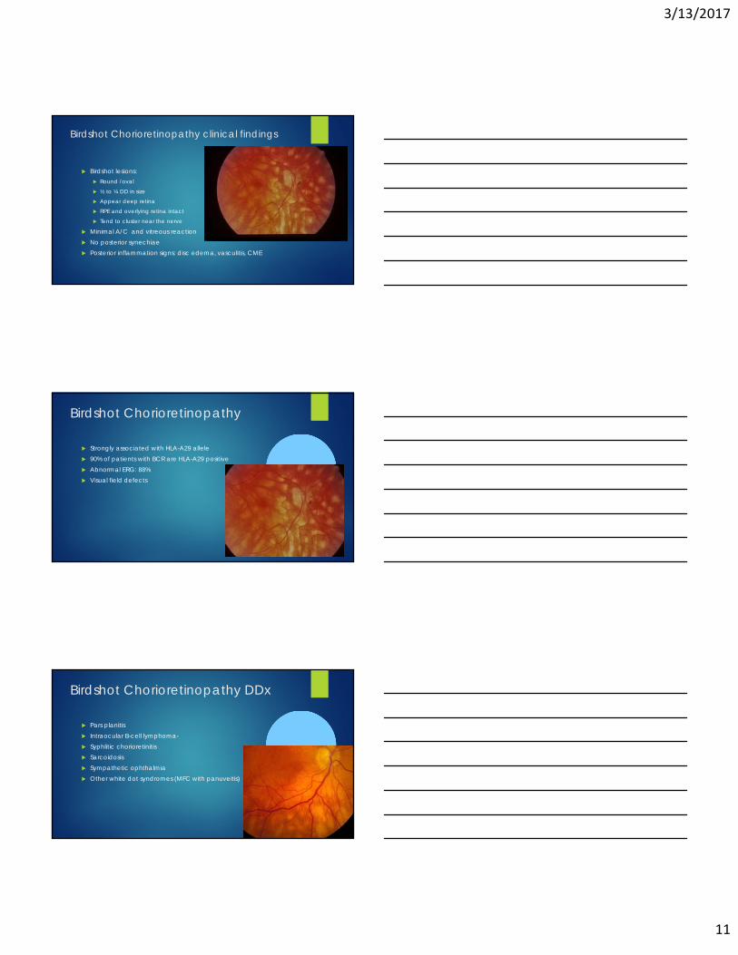
Birdshot Chorioretinopathy
Multiple small cream-colored lesions Scattered around the optic nerve towards the equator “shotgun pattern”
3/13/2017
11
Birdshot Chorioretinopathy clinical findings
Birdshot lesions: Round /oval
½ to ¼ DD in size
Appear deep retina
RPE and overlying retina intact
Tend to cluster near the nerve
Minimal A/C and vitreous reaction No posterior synechiae Posterior inflammation signs: disc edema, vasculitis, CME
Birdshot Chorioretinopathy
Strongly associated with HLA-A29 allele 90% of patients with BCR are HLA-A29 positive Abnormal ERG: 88% Visual field defects
Birdshot Chorioretinopathy DDx
Pars planitis Intraocular B-cell lymphoma- Syphlitic chorioretinitis Sarcoidosis Sympathetic ophthalmia Other white dot syndromes (MFC with panuveitis)

3/13/2017
12
Birdshot Chorioretinopathy Treatment
Corticosteroids Immunosuppressive therapy Anti-VEGF if there is CNVM Yearly ERG, visual field
Placoid Diseases
Acute posterior multifocal placoid pigment epithelipathy (APMPPE) Serpiginous choroiditis Relentless placoid chorioretinitis Persistent placoid maculopathy
“placoid lesions”
Acute posterior multifocal placoid pigment epitheliopathy (APMPPE)
Rapid onset of central vision loss Can see central / paracentral scotomas
Photopsia
metamorphopsia
Unilateral or bilateral (more common) If unilateral, fellow eye can become involved in a few weeks
Headache, stiff neck malaise
Hx of antecedent viral syndrome
Young adults

3/13/2017
13
APMPPE fundus findings
multiple round cream colored flat lesions Level of RPE
Indistinct margins Lesions not found anterior to equator Usually bilateral Fresh lesions can develop
Lesions of differing ages can be seen
Lesions clear gradually leaving Hypopigmentation
Pigment clumping
APMPPE
Localized serous RD over the lesions Vitritis not common Sometimes can see AC rxn
APMPPE: clinical course
Improves over 2-4 weeks Good prognosis Foveal involvement good predicator of final vision

3/13/2017
14
APMPPE: imaging
FA: early hypofluorescence, late hyperfluorescence ICG: shows more lesions that clinical exam
APMPPE: systemic associations
CNS: Cerebral vasculitis
Meningo-encephalitis
Stroke
CN VI palsy
Transient hearing loss
Headache
Systemic vasculitis
APMPPE: etiology unclear
Etiology: unclear HLA-B7 and HLA DR2 association
Possible viral cause
Found post various vaccinations
Differential Dx: Other white dot syndromes
Harada’s disease
TB, sarcoid, syphilis
Choroidal metastsis, lymphoma --

3/13/2017
15
APMPPE: treatment
Observation Corticosteroids / immunosuppressives
If CNS involvement
Serpiginous choroiditis
Bilateral progressive chronic inflammatory chorioretinitis Usually one eye active at a time
Ages 30-70 Unknown cause
Serpiginous choroiditis: clinical findings
Classic appearance (80%) Geographic patches of gray / creamy yellow placoid lesions
Peripapillary
Progresses in centrifugal manner
Fingerlike / serpentine projections

3/13/2017
16
Serpiginous choroiditis: clinical findings
Edematous outer retina Serous RD can be seen As active lesions heal:
Extensive RPE / choriocapillaris atrophy
Recurrences can occur at edge of old scars Symptomatic when fovea becomes involved Macular serpiginous choroiditis
Serpiginous choroiditis: clinical findings
white eye 1/3 have fine vitreous cells A/C reaction not common Multiple recurrences over months / years 75% patients develop central vision loss Final vision
<20/200 in 25% regardless of treatment
Serpiginous choroiditis: differential dx.
APMPPE Younger pts
Lesion scattered throughout posterior pole
Recurrences rare
Other white dot syndromes Tuberculous serpiginous choroiditis - Sarcoidosis, syphilis, toxoplasmosis, posterior scleritis etc

3/13/2017
17
Serpiginous choroiditis: treatment
Corticosteroids immunosuppressives
Acute zonal occult outer retinopathy (AZOOR)
Idiopathic inflammatory disorder Young healthy women Photopsia and acute progressive VF loss
VF starts as enlargement of blind spot
Due to damage to broad zones of outer retina Unilateral initially Can progress to bilaterality
Acute zonal occult outer retinopathy (AZOOR)
Fundus initially appears normal Retinal atrophy and mottling Attenuated arterioles Often peripapillary involvement May resemble:
sectoral retinitis pigmentosa
Diffuse unitlateral subacute neuroretinitis (DUSN)
No a/c rxn; minimal vitreous cells
3/13/2017
18
Acute zonal occult outer retinopathy (AZOOR)
Usually active for 6 months then stabilize Can be chronic progressive disease Fundus autofluorescence:
Involved areas show hypoautofluorescence
Acute zonal occult outer retinopathy (AZOOR):
Differential dx: Hereditary retinal diseases (RP) -
Retinal dystrophies
Cancer associated retinopathy
Treatmet: No proven therapy
Acute macular neuroretinopathy
Young middle aged women Acute onset of mild decreased vision
one or both eyes
One/multiple paracental scotomas wedge shaped intraretinal lesions
Point to fovea
Flower petal arrangement
Outer retina
Spare RPE and retinal vessels

3/13/2017
19
Acute macular neuroretinopathy
Acute macular neuroretinopathy
Self limited Scotomas can persist No treatment proven effective