WHAT THE WEIZMANN INSTITUTE IS DOING we are... · WHAT THE WEIZMANN INSTITUTE IS DOING ... Medical...
20
r Cance about DOING IS TITUTE INS WEIZMANN THE T WHA rch r Resea r Cance e fo tut i ross Inst M.D. Mo
Transcript of WHAT THE WEIZMANN INSTITUTE IS DOING we are... · WHAT THE WEIZMANN INSTITUTE IS DOING ... Medical...

rCanceabout
D O I N GI ST I T U T EI N SW E I Z M A N NT H ETW H A
r c hr R e s e ar C a n c ee f ot u tir o s s I n s tM . D . M o

rCanceabout
D O I N GI ST I T U T EI N SW E I Z M A N NT H ETW H A
r c hr R e s e ar C a n c ee f ot u tir o s s I n s tM . D . M o

STEP Malignant Transformation
MALIGNANT TRANSFORMATION p. 5
ENVIRONMENTAL AND LIFESTYLE FACTORS p. 6Viruses p. 7Radiation p. 8DNA REPAIR p. 9Boosting natural defenses p. 10ONCOGENES p. 11Perilous partnerships p. 12Down with leukemia p. 13Outwitting a brainy gene p. 14TUMOR SUPPRESSOR GENES p. 15The cancer killer p. 16Death benefits p. 18Controlling the switch p. 19
H O W C A N C E R D E V E L O P S
a n d h o w t h e w e i z m a n n i n s t i t u t e
Reversing malignancy p. 13Making history p. 17Experimental leukemia p. 19
P I O N E E R I N G S T E P S
Data mining for cancer genes p. 55
S Y S T E M S B I O L O G Y
M O D E L S O F C A N C E R
TA K I N G AC T I O N AT S T E P 1
1
Yeast p. 56
Fruit flies p. 56
Mice p. 57
Humans p. 57

TUMOR GROWTH p. 20
BLOCKING ABNORMAL SIGNALS p. 21Killing the messenger p. 23Inside jobs p. 25Life-saving overdose p. 26ENCOURAGING CELL SUICIDE p. 27Drop-dead gear p. 28To die for the cause p. 29Fateful decisions p. 30The marrow of a therapy p. 30CUTTING OFF BLOOD SUPPLY p. 31Starving the tumor to death p. 32EARLY DETECTION AND DIAGNOSIS p. 34Tumors get a new look p. 35From electronics to breast cancer p. 35Electronic finger detects cancer p. 36Announce your intentions p. 36
METASTASIS p. 37
PREVENTING CELL MIGRATION p. 38Respect thy neighbor p. 39Good migrations p. 41KILLING THE TUMOR p. 42A green solution p. 43Tumors smell the danger p. 44An attractive therapy p. 45The hormone connection p. 46IMMUNE STRATEGIES p. 47A winning combination p. 48Cancer vaccine p. 49Killer schemes p. 50FIGHTING ADVANCED CANCER p. 51A therapy to the bone p. 52Mother Nature to the rescue p. 53Achilles' heel of drug resistance p. 54
i s w o r k i n g t o p r e v e n t i t
I N T H E B O D YSTEP Tumor Growth STEP Metastasis
TumorCell
NormalCell
NewCapillaries
Blood Vessel
Lymph Vessels
Blood Vessels
MetastaticTumors
Abnormal growth is promoted. “Good neighbor-hood”/adhesion rules are ignored. Cells “forget” to commit suicide and live forever.
The tumor feeds itself by promoting an abnormaldevelopment of blood vessels.
Malignant cells travel to distant locations via lymphand blood vessels. Metastatic tumors form at thenew sites.
TA K I N G AC T I O N AT S T E PTA K I N G AC T I O N AT S T E P 2 3
2 3

4
A new era is dawning in the battle against cancer. In themore than 30 years that have elapsed since U.S. PresidentRichard Nixon “declared war” on cancer in 1971, sci-entists have made gigantic strides in charting the courseof this affliction. We now know that cancer is a diseaseof damaged genes. We also know that it develops inmultiple stages and takes many years, even decades, tounfold. We have learned moreover that there are hun-dreds of different cancers, each caused by a specific setof genetic defects, which is one of the major reasonsthat malignancy is so difficult to treat.
Thanks to this new understanding, the entire manage-ment of cancer promises to change. Genetic testing willmake it possible to identify groups of people who aremost prone to develop cancer and who must thereforetake preventive measures. Non-invasive DNA analysisand improved imaging techniques will allow physiciansto detect and diagnose cancer earlier. Finally, therapieswill become more selective and precise. Rather thandestroying tissues in bulldozer fashion as do the tradi-tional chemotherapy and radiation treatments, they willserve as finer tools, carving out the tumor while sparinghealthy tissues. It may even become possible to designtailor-made treatments to suit the patient’s individualtraits and genetic profile.
Cancer is a healthcare issue of vast scope. In the UnitedStates, for example, the various cancers combined claimmore than half a million lives annually, a death toll second only to that caused by heart disease. Globally, cancer kills some six million people every year, according to the World Health Organization. Ironically,the rise in the incidence of cancer stems indirectly from
our progress in science and technology: people in modern society live longer lives, but this prolonged life expectancy increases the risk of developing cancer.Fortunately, scientific progress also offers brighterprospects for beating cancer.
At the Weizmann Institute, several dozen research teams are attacking the problem from different directions; their studies are conducted within the framework of several research centers whose activitiesare coordinated by Weizmann’s M.D. Moross Institutefor Cancer Research. Important achievements arealready on record: Weizmann scientists were the first toclone p53, a gene involved in more than half of allhuman cancers; basic research conducted at theInstitute provided the foundation for the developmentof Glivec, the first in the upcoming generation of molecular drugs; and a number of cancer therapiesdeveloped at Weizmann are currently being tested inclinical trials.
In this publication, we describe the Institute's multi-faceted cancer research targets. Our ultimategoal: to understand what causes cells to become cancerous and what can be done to intervene in the progression of malignancy at its different stages.
Introduction
Prof. Yoram GronerDirector, M.D. Moross Institute for Cancer Research

5
Contrary to popular belief, getting cancer is not so easy. Although genesconstantly sustain damage, in mostcases this damage does not lead to malignancies: many mutations –”spelling mistakes” in the letters of thegenetic code – are successfully repaired;if the repair fails, the defective cell isoften commanded to self-destruct. For a cell to undergo a malignant transfor-mation – in other words, startdividing uncontrollably – up to 10 mutations must accumulate in thegenetic code. That's why cancer is mostly a disease of old age: it may take several decades for the necessary number of mistakes to add up in thegenes of a single cell. Most people die of other causes before cancer has achance to unfold.
But how do spelling mistakes occur inthe genetic code in the first place?Some are copying errors, “typos” thatappear when a cell duplicates its genesbefore dividing into two daughter cells.Others occur when the cell suffers abuse
from the environment, such as thatinflicted by viruses, chemical carcinogensor radiation; or from internal aggressors,such as the highly reactive moleculescalled free radicals. Mutations can alsobe inherited; a parent can pass on tooffspring a mutated form of a gene.Such unfortunate inheritance explainswhy cancer sometimes runs in families:several family members may carry themutation that predisposes them to thedisease. It also explains why some cancers occur early in life: the presenceof inherited mutations speeds up malignant transformation.
Two types of genes play a major role in malignant transformation: oncogenes and tumor suppressor genes. When present in their normalform, these genes escort the cellthrough the healthy processes of growth and division. But when boththese gene types contain fatal mistakes,the cell can embark upon a malignantcourse: a destructive, uncheckedgrowth.
Malignant TransformationTaking Action at Step 1

6
Cancer is largely a preventable disease. Hundredsof thousands of lives could be saved annually ifpeople avoided risky behaviors and exposure tocarcinogens that push cells toward malignancy.Smoking tops the list, accounting for one-thirdof all cancer deaths in developed countries. Ahigh proportion of fatal cancers can be blamed
on dietary practices and lack of exercise. About 15 percent of cancers worldwide are caused byknown infectious agents, including viruses, bacteria and parasites. Exposure to radiation and to cancer-causing chemicals can also damage genes, thus contributing to malignanttransformation.
Environmental and Lifestyle Factors

7
Viruses account for a relatively small percentage ofmalignancies, but it was largely thanks to these infectious organisms that the roots of cancer were firstuncovered. In the 1970s, scientists studying cancer-causing viruses showed that a virus could trigger tumorgrowth by turning normal genes into cancer-causinggenes. This research touched off a hunt for latent cancer genes that continues to this day. Currently,research into the viral origins of cancer holds potentialin two major areas: prevention of malignancies causedby viruses and, on a broader scale, gaining a betterunderstanding of cancer mechanisms.
How does a virus manage to evade a cell's defensesagainst foreign invaders? What strategies does it use to take over the cellular machinery? These questionsare addressed in the laboratory of Prof. Yosef Shaulof the Molecular Genetics Department. Shaul has discovered how hepatitis B virus, a major contributorto liver cancer, sabotages a cell's growth machinery:the virus has a regulatory gene that acts as a “molecularbridge” allowing it to reproduce inside the cell. The same mechanism may explain how hepatitis B triggers malignancy: this gene apparently allows thevirus to shut down tumor suppressor genes in livercells, causing these cells to undergo a malignant transformation. While Shaul continues to study the linkbetween hepatitis B and liver cancer, his research hasalready led to the development of a hepatitis B vaccinetested in clinical trials by an Israeli biotechnology company. An estimated 350 million people in Africaand Asia suffer from hepatitis B, making this ailment a health problem of global proportions. If these infections were eradicated, a significant proportion of liver cancers could be prevented.
More recently, the study of viruses has led Shaul to the discovery of an important tumor suppressor mechanism. He focused his attention on one of thegenes with which hepatitis B interacts – a major regulator of cell growth called c-Abl. Working withother Weizmann Institute researchers, Shaul found that c-Abl normally safeguards the cell against cancer:when DNA damage occurs, c-Abl recruits a tumor suppressor gene, and together they either prevent thecell from dividing or command it to commit suicide.But when either gene is mutated – by, say, exposure toa virus or to radiation – the risk of cancer soars.
Viruses can wear two hats when it comes to cancer.While some contribute to malignant transformation,others exert an opposite, protective effect, selectivelydestroying cancer cells but leaving healthy cellsuntouched. Veteran Weizmann Institute researcherProf. Emeritus Ernest Winocour of the MolecularGenetics Department spent more than three decadesstudying the connection between viruses and cancer.He has clarified important aspects of viral behavior in malignancy while focusing his attention on members of the parvovirus family. In particular, he has beenstudying the adeno-associated virus, or AAV, which isunique in its ability to bind to a specific site on ahuman chromosome rather than, as other viruses do,simply invading the host cell in a general way.Winocour has clarified the mechanism by which AAVtargets its specific site and suggested a way for incorporating this mechanism into gene therapy, including gene therapy of cancer. �
Prof. Yosef Shaul holds the Oscar and Emma Getz Professorial Chair
Prof. Ernest Winocour
IIff hheeppaattiittiiss BB iinnffeeccttiioonnss wweerree eerraaddiiccaatteedd,, aa ssiiggnniiffiiccaanntt pprrooppoorrttiioonn ooff lliivveerr ccaanncceerrss ccoouulldd bbee pprreevveenntteedd
Viruses

8
Medical X-rays can detect small cancerous tumors,improving the patient's chances for survival. But excessive radiation can itself cause cancer by damagingthe DNA. What are the healthy limits of medicalscreening? And what should be the standards for maximum radiation exposure in the workplace?Weizmann Institute researchers led by Prof. AmosBreskin of the Particle Physics Department, workingwith colleagues from the United States, have designed a novel radiation detector that makes it possible toaddress these questions. The detector, which is 100 to1,000 times more accurate than any previous system,has for the first time allowed scientists to assess radiation effects on the living cell, down to the scale of DNA. At this level of accuracy, scientists can predicthow much radiation will severely damage a cell'sgenetic material.
In addition to setting more precise limits to radiationexposure, this research may help improve radiationtherapy for cancer, in which a tumor is destroyed by
streams of protons, or gamma rays. The difficulty hereis to deliver just the right amount of radiation withoutexposing the person to unnecessary tissue damage.Breskin's research may enable physicians to adjust radiation to levels at which it is just sufficient to do its job.
Another Institute study, performed with the help of X-ray crystallography, has furnished a tool for developing pharmaceuticals that would protect the bodyagainst high-dose radiation. A team led by Prof. JoelSussman of the Structural Biology Department andProf. Israel Silman of the Neurobiology Department,working with scientists from the Netherlands andFrance, has for the first time produced a three-dimensional “movie” revealing how molecules breakapart when exposed to X-rays. The Weizmann team andits European collaborators plan to examine the anti-radiation potential of various substances that could beapplied on a conventional basis or in an emergencysuch as the Chernobyl nuclear power plant failure. �
Prof. Amos Breskin holds the Walter P. Reuther Professorial Chair ofResearch in Peaceful Uses of Atomic Energy
TThhiiss rreesseeaarrcchh mmaayy hheellpp iimmpprroovvee rraaddiiaattiioonntthheerraappyy ffoorr ccaanncceerr,, iinn wwhhiicchh aa ttuummoorr iissddeessttrrooyyeedd bbyy ssttrreeaammss ooff pprroottoonnss
Prof. Israel Silman holds theBernstein-Mason Professorial Chair of Neurochemistry
Prof. Joel Sussman holds the Morton and Gladys PickmanProfessorial Chair of Structural Biology
Radiation

9
Every cell in the human body daily sustains thousands of attacks, yet cellular DNA survivesthe assaults relatively intact. The secret of thisresilience: DNA is being regularly patrolled byvigilant “maintenance crews” responsible for its integrity. These crews are molecular repair mechanisms that correct or erase DNA
damage, preventing it from perpetuating mutations during cell division. If it weren't forDNA repair, cancer would be rampant; in fact, if DNA repair ceased completely, life on earthwould be impossible because the DNA would sustain severe damage and mutations would eventually go haywire.
DNA Repair

01
Prof. Zvi Livneh of the Biological ChemistryDepartment studies DNA repair mechanisms and theirrole in cancer. In most cases, DNA repair systems operate on an all-or-nothing basis: when unable to precisely correct the damage, they stop working. Thestoppage leads to gaps in the DNA, which preventgenetic replication from being completed. Livneh,however, identified a new group of enzymes thatendow DNA repair systems with the ability to compromise: they are able to fill in the gaps in theDNA, but while doing so they may introduce mutations. Thus these enzymes, called bypass DNApolymerases, ensure the cell's continued existence but also pose a certain risk; imbalances in their activity may increase mutations to a level at which they cause cancer.
Livneh has also discovered a mechanism that restrainsthe mutational activity of the new enzymes. It involves”heroic” proteins that throw themselves on the damagedsites in DNA, preventing them from being copied,much like demonstrators throwing themselves down onthe road to block traffic. Livneh is currently studyingvarious aspects of this protein valor, which provides asecond line of defense against the mutation and givesthe cell a crucial second chance at healthy growth.
In addition, Livneh's team is studying the possibility of identifying people whose bodies aren’t as effective at repairing DNA as those of others. One day scientistsmay be able to warn people who are more susceptibleto genetic defects induced by a particular exposure: forexample, people more susceptible to damage caused by ultraviolet radiation may be warned against excessive exposure to sunlight.
An important study conducted by Livneh and his colleagues has already made it possible to identify people who are predisposed to lung cancer due toreduced capacity to repair DNA damage caused by,among other factors, tobacco smoke. (Only 10% ofheavy smokers develop lung cancer, suggesting a personal genetic susceptibility.) The scientists focusedon a DNA repair enzyme called OGG1 (8-oxoguanine
DNA glycosylase 1); it repairs DNA parts damaged bytoxic molecules called oxygen radicals, which arefound in tobacco smoke. When OGG1 is not sufficient-ly active, the person's ability to repair DNA isdecreased. The researchers discovered that low OGG1activity results in high susceptibility to cancer, increas-ing the risk 5- to 10-fold compared with normal OGG1activity. Smoking further escalates the risk, since itcauses more damage for DNA repair enzymes, including OGG1, to fix. Smokers who have a low level of OGG1 activity were estimated to be around120 times more likely to get lung cancer than non-smokers with regular levels of OGG1 activity. A simpleblood test based on these findings will be able to detectsmokers who are at an especially high risk of developing lung cancer.
Apart from providing personalized guidelines foravoiding risky exposures, understanding DNA repairmay one day help prevent cancer on a molecular level.If we can fully clarify how DNA repair occurs, we may be able to turn it on as required in order to avertmalignant transformation. �
Prof. Zvi Livneh holds the Maxwell Ellis Professorial Chair in Biomedical Research
AA ssiimmppllee bblloooodd tteesstt wwiillll bbee aabbllee ttoo ddeetteeccttssmmookkeerrss wwhhoo aarree aatt aann eessppeecciiaallllyy hhiigghh rriisskk ooff ddeevveellooppiinngg lluunngg ccaanncceerr
Boosting natural defenses

11
Healthy, unmutated oncogenes (also known asproto-oncogenes) take part in stimulating normalcell growth and division. But when an oncogenecontains one or more mutations, it may stay in theON position too long, continuously commanding
the cell to proliferate. (A cell with activated oncogenes is sometimes compared to a car with ajammed accelerator.) Future therapies would blockabnormal cell proliferation by shutting down themisbehaving oncogenes.
Oncogenes

21
Several types of leukemia – acute lymphocytic andacute myeloid leukemia in babies, and many cases oftherapy-related acute leukemia in older children andadults – are characterized by an abnormality at a particular site on chromosome 11. Prof. Eli Canaaniof the Molecular Cell Biology Department has succeeded in identifying and cloning the gene at thissite. This gene, called ALL-1, normally plays a keyrole in establishing the correct body pattern of a developing embryo by activating a series of regulatorygenes involved in this process. In a normal cell, ALL-1works by forming an unusually large complex withsome 30 other proteins, and together they activate target genes. But Canaani found that in acute leukemia,ALL-1 is defective; it abnormally fuses to any of itsmore than two dozen partner genes, resulting in theproduction of anomalous fused proteins that are oncogenic. Canaani now seeks to establish how theseunnatural fused proteins convert the normal cell into a malignant one. In one series of experiments, he is conducting comparisons between proteins that are physically associated with the normal as opposed toleukemic ALL-1. His aim is to reveal the features ofthe abnormal ALL-1 protein complex responsible forthe malignant transformation. Another research direction is to try to pinpoint the critical genes activated by the abnormal ALL-1 protein. Canaani pursues this goal by comparing the entire repertoire of genes activated in acute leukemias with and withoutdefective ALL-1, in order to identify the genes that areactive only in leukemias with the ALL-1 abnormality.
In the meantime, Canaani's basic research from the1980s provided the foundation for the development ofGlivec (known as Gleevec in the United States), thedrug that has won unusual praise and attention as thefirst therapy based on the molecular understanding of a specific cancer. Canaani had isolated two genes thatabnormally swap pieces of genetic material and lead tothe production of a fused protein that triggers chronicmyelogenous leukemia (CML). It was the first discoveryof cancer initiated by protein fusion. Using these find-ings, the pharmaceutical company Novartis developeda molecule that works by seeking out and destroying
cancer cells making the fused protein. Glivec wasapproved in 2001 by the U.S. Food and DrugAdministration for the treatment of CML and is nowroutinely prescribed around the world to patients withthis malignancy. �
Prof. Eli Canaani holds the Harry Kay Professorial Chair of Cancer Research
IInnssttiittuuttee rreesseeaarrcchh ffrroomm tthhee 11998800ss pprroovviiddeedd tthhee bbaassiiss ffoorr tthhee ddeevveellooppmmeenntt ooff GGlliivveecc,, tthheeffiirrsstt ddrruugg sstteemmmmiinngg ffrroomm aann uunnddeerrssttaannddiinngg ooff tthhee ggeenneettiicc oorriiggiinnss ooff ccaanncceerr
Perilous partnerships

31
The study of Down syndrome, which is caused by an extra copy of chromosome 21, has led to the discovery of a gene responsible for acute myelo-genous leukemia, or AML. Children with Down syndrome run an unusually high risk – up to 20 times higher than average – of developing this type of leukemia. Prof. Yoram Groner of theMolecular Genetics Department has explained thereason. His team has identified a gene, called AML-1,which is located on chromosome 21 and is normallyinvolved in a wide range of growth-related processes,from embryonic development to blood cell production.In Down syndrome, an extra copy of this gene resultsin the production of altered proteins, which may trigger malignancy. Groner's team is developing genetically modified mice that carry extra copies of the AML-1 gene and therefore serve as a researchmodel for the AML-1 involvement in Down syndrome.Studying these animals provides clues about the molecular mechanisms underlying the malignant transformation induced by the altered activity of AML-1. Groner's findings suggest that defects inAML-1 may also trigger leukemia in children without Down syndrome and in adults. �
Prof. Yoram Groner holds the Dr. Barnet Berris Professorial Chair of Cancer Research
TThhee ssttuuddyy ooff DDoowwnn ssyynnddrroommee hhaass lleedd ttoo tthhee ddiissccoovveerryy ooff aa ggeennee rreessppoonnssiibbllee ffoorr aaccuuttee mmyyeellooggeennoouuss lleeuukkeemmiiaa
Down with leukemia
In the 1960s, Prof. Leo Sachs of the MolecularGenetics Department was the first to show that cancer cells – in tissue cultures and in living organ-isms – can be made to revert to normal behavior.This finding led to the development of a new treatment, differentiation therapy, for restoring theability of cancer cells to grow and develop in abenign fashion. This approach is now being usedclinically in human promyelocytic leukemia and isbeing tested in other types of cancer. Differentiationtherapy grew out of Prof. Sachs’ landmark discoveryof proteins that control the growth and differentiationof blood cells. These proteins, now called colony-stimulating factors and interleukins, signal stem cellsto multiply and differentiate to different cell types,and they also prevent blood cells from dying prema-turely; in fact, one of the proteins that Sachs identified,the granulocyte colony-stimulating factor, is nowused to boost the production of disease-fighting whiteblood cells in cancer patients undergoing irradiation or chemotherapy. This colony-stimulating factor alsoinduces migration of stem cells from the bone marrowto the peripheral blood, so that peripheral bloodrather than bone marrow can now be used for stemcell transplantation. Sachs is currently carrying outstudies aimed at fine-tuning the use of colony-stimulating factors and interleukins in cancer. In parallel, he is studying the genetic and biochemicalchanges that make cancer cells outlive normal cells. �
Reversing malignancy
Prof. Leo Sachs holds the Otto Meyerhof Professorial Chair of Molecular Biology
Pioneering steps
41
The very first in the series of mutations causing colon cancer occurs in the beta-catenin gene; this gene is abnormally activated in about 90 percent ofcolorectal cancer patients, and in a much smaller percentage of people with almost every other type ofcancer. Beta-catenin plays a dual role in the cell: it promotes adhesion, or stickiness, between cells, andregulates the expression of genes in the nucleus.
Research conducted in the laboratory of Prof. AvriBen-Ze'ev of the Molecular Cell Biology Department suggests that in cancer, beta-catenin functions as anoncogene: when aberrantly activated, it spurs malignant transformation and causes the cell to proliferate abnormally. In one collaborative projectwith Institute colleagues, Ben-Ze'ev discovered that innormal cells, the p53 tumor suppressor gene keepsbeta-catenin in check, but in malignant cells p53 losesits grip on beta-catenin. In another collaborative project, a team led by Ben-Ze'ev isolated a short peptide (protein fragment) that blocks a vital portion of the beta-catenin molecule; the protein may thwartthe development of cancer by preventing beta-cateninfrom acting as an oncogene.
More recently, Ben-Ze'ev's team unraveled several crucial elements in the signaling chain unleashed bythe corrupt beta-catenin. One of these elements is Nr-CAM, a cell adhesion molecule not previouslyknown to play a role in cancer. In healthy people, theprotein made by the Nr-CAM gene is present only inthe brain and not at all in other tissues of the body, butthe Weizmann scientists showed that the Nr-CAM levels are dramatically elevated in colon cancer andmelanoma cells; in fact, the more advanced the tumor,the higher the Nr-CAM level. These findings couldlead to the screening of large populations and earlydetection of cancer, based on the detection of the protein made by the Nr-CAM gene: this protein is likely to be present only in people with cancer causedby overly activated beta-catenin. Moreover, since theprotein made by the Nr-CAM gene sticks out from thesurface of cells, it is a convenient target for cancer
therapy: by inactivating Nr-CAM, it may be possible tointerrupt the chain of signals released by beta-catenin,thereby suppressing the development of prevalentmalignancies such as melanoma and colon cancer.
Ben-Ze'ev's laboratory has also revealed that beta-catenin is involved in a key mechanism leading to the metastasis of colon cancer. By manipulating thismechanism, Ben-Ze'ev's team succeeded in reversingthe metastatic properties of colon cancer cells in vitro.This research raises hopes that a target-specific therapymight be devised to prevent, or reverse, the invasivebehavior of metastatic colon cancer cells. �
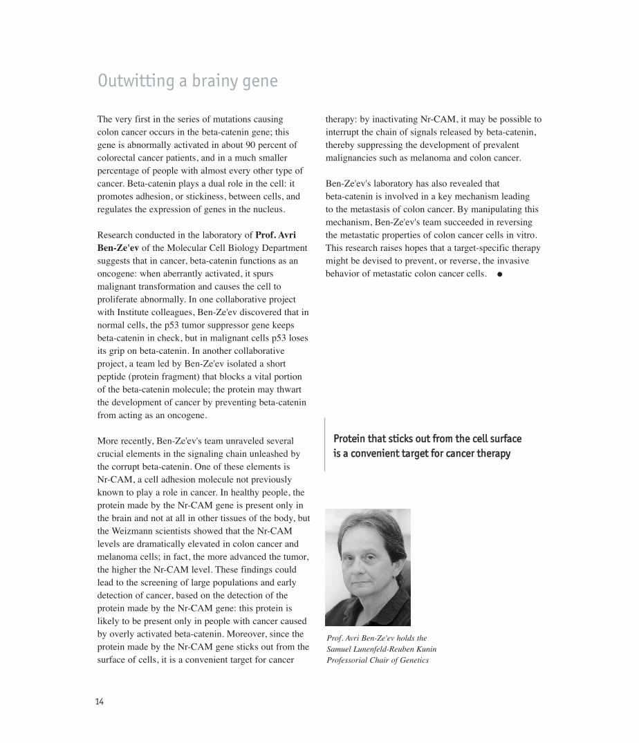
Prof. Avri Ben-Ze'ev holds the Samuel Lunenfeld-Reuben Kunin Professorial Chair of Genetics
PPrrootteeiinn tthhaatt ssttiicckkss oouutt ffrroomm tthhee cceellll ssuurrffaaccee iiss aa ccoonnvveenniieenntt ttaarrggeett ffoorr ccaanncceerr tthheerraappyy
Outwitting a brainy gene

51
Tumor Suppressor Genes
In healthy cells, tumor suppressor genes keepthe cell's growth machinery in check, releasingtheir grip on growth-promoting chemicals in acontrolled manner, only as needed. In cancer,however, these genes are not working, allowingthe tumor to grow without restraint. (To con-tinue the automobile metaphor, a cell without
properly working tumor suppressor genes is likea car without brakes.) In fact, when cancer runsin families, the hereditary defect is often in thesuppressor genes. Therapies compensating forthe lack or malfunction of these genes would beequivalent to fixing the brakes in order to stopthe developing cancer.

61
The most “glamorous” of all tumor suppressor genesis p53. Several years ago, it was pronouncedMolecule of the Year by Science; it has also been the subject of a Newsweek cover story headlined “The Cancer Killer.” The p53 gene owes its celebrity status to the fact that defective copies of it are foundin more than half of all human cancers, includingsuch major killers as cancers of the breast, lung, colon and prostate. A therapy compensating for a lack of properly functioning p53 copies would there-fore have enormous potential for combating cancer. In fact, a gene therapy that delivers active p53 totumor cells is already being tested in clinical trials in the United States and Europe.
Weizmann Institute scientists have made seminal contributions to understanding the role of p53 in normal and cancerous cells. It is now known that p53acts as the cell’s damage control and as a “guardian of the genome.” When genes are damaged by radiation, chemicals or other means, threatening to set the cell on a course toward malignant transformation, p53 senses the damage and its supplybuilds up. The p53 protein activates numerous genesthat prevent tumors from forming; by so doing iteither blocks the growth of damaged cells, allowingfor the correction of DNA damage, or commandsthese cells to commit suicide. But if the cell has nohealthy p53, the road to cancer remains open.
Prof. Moshe Oren of the Molecular Cell BiologyDepartment, together with the Weizmann Institute’sProf. Emeritus David Givol and Prof. Arnold Levine,then of Princeton University, was the first to clonep53 – in other words, to isolate the gene and deter-mine the sequence of its genetic letters – in 1983. Thep53 clone and its genetic sequence provided laborato-ries around the world with one of the most frequentlyused tools for studying cancer. Subsequently, Oren wasthe first to show that reactivation of p53 in cancer cells can prompt them to self-destruct, a principle that underlies the ongoing p53 gene therapytrials. Oren is now focusing on elucidating the
biological processes that allow this gene to functionas a tumor suppressor. Among the questions studied in his lab: How does p53 interact with other genes?How are the levels of p53 regulated in a cell? Usually,p53 is present in minute amounts, but its levels soarin response to DNA damage and other types of stress. Oren has discovered the role of a major regulator of p53 activity, called MDM2. He found that MDM2 is responsible for the elimination of p53,and he now seeks to clarify how exactly MDM2achieves this. Oren predicts that interfering withMDM2 will strengthen p53, thereby boosting the natural anti-cancer defense mechanisms.
Prof. Varda Rotter of the Molecular Cell BiologyDepartment was the first to develop antibodies against p53, laying the foundations for the study ofthis gene’s function. She also provided some of theearliest evidence that p53 is a tumor suppressor. Atpresent, Rotter is working toward two major goals: to decipher the function of p53 in the normal cell andto clarify the behavior of mutant p53 in tumor cells.In particular, she seeks to understand how p53 caninduce three different processes: blockage of growth,cell differentiation or cell death. She is working toclarify whether all three processes can occur in a single cell, or whether different cells respond differently to p53.
The state of a patient’s p53 may determine whether conventional cancer treatments are likely to be effective. Several years ago, oncologists made a surprising discovery: radiation therapy andsome chemotherapies, rather than directly killing cells as had previously been thought, in fact work by activating p53, which in turn orders cells toself-destruct. Therefore, patients with intact copies of p53 are more likely to be helped by these treatments. In the absence of p53, the cancer is resistant to chemotherapy. Prof. Emeritus DavidGivol of the Molecular Cell Biology Department has conducted several studies exploring the effects of p53 on different chemotherapies.
The cancer killer

71
Givol is now using DNA chips to determine which other genes are activated by p53. He discovered new cell suicide genes that
are turned on by p53 in response to DNA damage. If p53 is defective, alternative means may be used toactivate such genes. �
Prof. Moshe Oren
WWeeiizzmmaannnn sscciieennttiissttss wweerree tthhee ffiirrsstt ttoo cclloonnee tthheepp5533 ggeennee aanndd hhaavvee mmaaddee sseemmiinnaall ccoonnttrriibbuuttiioonnssttoo uunnddeerrssttaannddiinngg iittss ffuunnccttiioonn
Prof. Varda Rotter holds the Norman and Helen Asher Professorial Chair of Cancer Research
Prof. David Givol
In the 1950s, Weizmann Institute scientists were among the first to demonstrate that certain types of cancerous growths develop in a two-stage process and that cancer depends on the presence of multiple factors. Until then, it was believed that a single factor was sufficient for cancerto develop. The discovery – by
the late Prof. Isaac Berenblum,a foremost cancer researcher –improved the understanding of various scenarios that mightlead to malignancy. It also provided an explanation for thetime lapse, sometimes severaldecades, between exposure tocarcinogenic substances and thedevelopment of a tumor. �
Making history
The late Prof. Isaac Berenblum
Pioneering steps

81
Finding a defective gene can be likened to locating a burnt-out lightbulb somewhere in North Americawhile having no idea where to begin the search. Prof. Adi Kimchi of the Molecular GeneticsDepartment has developed a fast and convenientmethod for identifying tumor suppressor genes, whichuntil recently were particularly difficult to isolate. Themethod, called Technical Knockout, or TKO, works byselectively disabling various genes and monitoring theeffect of this disabling on the cell. In this manner, scientists can isolate out of 30,000 genes a single genethat possesses a particular function.
Kimchi and her team have successfully used thismethod to discover several completely new genescalled DAP (death-associated proteins) that are connected to death-inducing processes in cells. One of these genes, called DAP-kinase, falls into the category of tumor suppressor genes; it is responsiblefor destroying cells that have begun converting to acancerous state.
In a recent discovery, Kimchi found that DAP-kinaseis the trigger that activates the p53 gene, leading to the destruction of cells intent on undergoing a malignant transformation. A malfunction of DAP-kinase interferes with this destruction program, allowing a cancerous growth to develop. Another stagein cancer development controlled by DAP-kinase is metastasis: loss of this gene or a malfunction of itsprotein promotes the formation of metastatic tumors.DAP-kinase abnormalities have been detected inhuman cancers of the lung, breast, head and neck, aswell as in type B cell lymphoma. �
Prof. Adi Kimchi holds the Helena Rubinstein ProfessorialChair in Cancer Research
IInnssttiittuuttee sscciieennttiissttss hhaavvee ddeevveellooppeedd aa ffaasstt aanndd ccoonnvveenniieenntt mmeetthhoodd ffoorr iiddeennttiiffyyiinngg ttuummoorr ssuupppprreessssoorr ggeenneess
Death benefits

91
Dr. Doron Ginsberg of the Molecular Cell Biology Department is studying a family of molecular switches, called E2F, that control cellgrowth and division. These switches are normallyregulated by the RB tumor suppressor gene. (The RB gene owes its name to retinoblastoma, a malignant tumor of the retina in which it was originally found to be defective; however, RB hassince been found to be mutated in a variety of other cancers.) When RB is working properly, thegrowth switch is turned on only as needed. But when RB is missing or when it malfunctions, the E2F remains permanently turned on, promoting unchecked cellular growth. �
Dr. Doron Ginsberg holds the Recanati Career Development Chair of Cancer Research
WWhheenn tthhee RRBB ttuummoorr ssuupppprreessssoorr ggeennee iiss mmiissssiinngg,,tthhee rreessuulltt iiss uunncchheecckkeedd cceelllluullaarr ggrroowwtthh
Controlling the switch
Prof. Emeritus Nechama Haran-Gheraof the Immunology Department, a former student and then collaboratorof Prof. Berenblum, has continued andextended his studies in a career spanningfive decades. She produced severaltypes of leukemia in mice and providedinsights into the multiple phases ofblood cancer development. It emergedthat the development of the diseaseincludes an initial stage in whichbone marrow cells are transformedinto preleukemic clones; they mayremain dormant throughout an animal’s life. Haran-Ghera definedimmunological and other factors thatkeep preleukemia in check or, conversely, terminate the dormantstate and lead to a burst of overtleukemia. In related research sheshowed, in a mouse model of radia-tion-induced acute myeloid leukemia,that certain growth factors used in theclinic to boost the production and/or
maturation of blood cells can be hazardous because they augment theproliferation of preleukemic cells andmay therefore trigger overt leukemia.
More recently, Haran-Ghera collaborated with Israeli physicians on a study that may improve the treatment of multiple myeloma. Sheshowed that the hormone erythro-poietin, widely used to treat anemia in cancer patients, may be helpful in treating the cancer itself. She revealedthat erythropoietin affects the cancerindirectly, through the immune system;it was found to prolong survival,cause tumor regression and reducemortality in mice injected withmalignant myeloma cells. These findings have provided the scientificbasis for clinical trials of erythro-poietin in multiple myeloma and for testing the effects of this hormone on other types of cancer. �
Experimental leukemia
Prof. Nechama Haran-Ghera
RReeaasseerrcchh mmaayy iimmpprroovveetthhee ttrreeaattmmeenntt ooff mmuullttiippllee mmyyeelloommaa
Pioneering steps