What sports medicine practitioners should ... - SEMS-journal
17
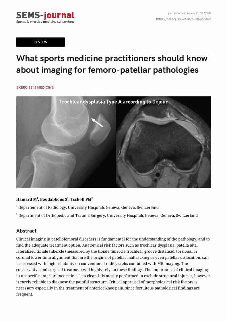
published online on 21.02.2020 https://doi.org/10.34045/SEMS/2020/2 REVIEW What sports medicine practitioners should know about imaging for femoro-patellar pathologies EXERCISE IS MEDICINE Hamard M 1 , Boudabbous S 1 , Tscholl PM 2 1 Departement of Radiology, University Hospitals Geneva, Geneva, Switzerland 2 Department of Orthopedic and Trauma Surgery, University Hospitals Geneva, Geneva, Switzerland Abstract Clinical imaging in patellofemoral disorders is fundamental for the understanding of the pathology, and to find the adequate treatment option. Anatomical risk factors such as trochlear dysplasia, patella alta, lateralized tibiale tubercle (measured by the tibiale tubercle trochlear groove distance), torsional or coronal lower limb alignment that are the origine of patellar maltracking or even patellar dislocation, can be assessed with high reliability on conventional radiographs combined with MR imaging. The conservative and surgical treatment will highly rely on these findings. The importance of clinical imaging in unspecific anterior knee pain is less clear. It is mostly performed to exclude structural injuries, however is rarely reliable to diagnose the painful structure. Critical appraisal of morphological risk factors is necessary especially in the treatment of anterior knee pain, since fortuitous pathological findings are frequent.
Transcript of What sports medicine practitioners should ... - SEMS-journal
published online on 21.02.2020
https://doi.org/10.34045/SEMS/2020/2
REVIEW
What sports medicine practitioners should knowabout imaging for femoro-patellar pathologies
EXERCISE IS MEDICINE
Hamard M1, Boudabbous S1, Tscholl PM2
1 Departement of Radiology, University Hospitals Geneva, Geneva, Switzerland2 Department of Orthopedic and Trauma Surgery, University Hospitals Geneva, Geneva, Switzerland
AbstractClinical imaging in patellofemoral disorders is fundamental for the understanding of the pathology, and tofind the adequate treatment option. Anatomical risk factors such as trochlear dysplasia, patella alta,lateralized tibiale tubercle (measured by the tibiale tubercle trochlear groove distance), torsional orcoronal lower limb alignment that are the origine of patellar maltracking or even patellar dislocation, canbe assessed with high reliability on conventional radiographs combined with MR imaging. Theconservative and surgical treatment will highly rely on these findings. The importance of clinical imagingin unspecific anterior knee pain is less clear. It is mostly performed to exclude structural injuries, howeveris rarely reliable to diagnose the painful structure. Critical appraisal of morphological risk factors isnecessary especially in the treatment of anterior knee pain, since fortuitous pathological findings arefrequent.
published online on 21.02.2020
https://doi.org/10.34045/SEMS/2020/2
RésuméUne connaissance clinique adéquate est nécessaire pour une prise en charge optimale du syndromefémoro-patellaire et pour l’interprétation de l’imagerie. Les anomalies anatomiques qui favorisent cesyndrome sont de nos jours bien investiguées par le couple radiographie conventionnelle-IRM. Cesanomalies sont nombreuses incluant la dysplasie trochléaire, la patella alta, une latéralisation dutubercule tibial (mesurée par la distance dite TAGT), anomalie d’alignement frontal ou de torsion desmembres inférieurs; toutes ces anomalies sont à l’origine d’une mobilité inadéquate de la patella voir uneluxation. Le traitement pourra être conservateur ou chirurgical et dépendra étroitement des trouvaillescliniques et radiologiques. En dehors de cette pathologie, le rôle clinco-radiologique est mal défini pourtoute douleur antérieure du genou. Il a pour objectif principal d’éliminer d’éliminer des anomaliesstructurelles sans toujours trouver une corrélation avec la douleur. Une analyse critique et synthétique estnécessaire aussi bien pour les anomalies anatomiques trouvées ainsi que pour le choix du traitement.
IntroductionAnterior knee pain (AKP) is one of the most common complaints in athletes, especially performing pivotingand jumping sports. Many structures around the patella can become symptomatic, mainly by structuraloveruse with or without patellar maltracking, poor muscle control or functional instability of the lowerlimb. The most frequently painful structures are the medial and lateral patellofemoral retinacula, theentheses of the patellar and less frequently the quadriceps tendon, and the intra-articular but extra-synovial fat pads. These structures, although visible on clinical imaging, might not always appearpathologic if painful, however are frequently associated with morphological risk factors. The analysis ofthe frequently painful structures and the anatomy predisposing to patellar maltracking requires asystematic approach on multi-modal imaging by radiologists and clinicians for the best understanding ofthe underlying causes of anterior knee pain.The indication, advantages and disadvantages of the different imaging modalities will be here discussed inthe context of anterior knee pain.
Patellofemoral dysplasia – radiological signs and measurementsConventional radiography (CR)CR allows detection and a rapid and global appreciation of the bony structure of the knee, the morphologyof the patellofemoral joint and subchondral degenerative changes. The evaluation of AKP requires aweight-bearing antero-posterior (AP) view to evaluate the femoro-tibial knee joint, a strict lateral (L) viewwith overlapping posterior medial and lateral femoral condyles and an axial imaging in 30° of knee flexion(Table 1).

published online on 21.02.2020
https://doi.org/10.34045/SEMS/2020/2
Table 1: Imaging modalities, measurements and practical hints for investigation of patellofemoral disorders
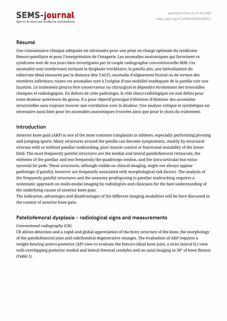
The most important morphological risk factors for patellar maltracking and patellar dislocation are basedon the studies by Henry [1,2] and David Dejour [3] and are measured on the strict L view (trochleardysplasia and patella alta). The tibial tubercle trochlear groove distance (TT-TG), initially measured onaxial CR [4] is currently exclusively measured on multi-slice imaging (CT or MRI).Trochlear dysplasia has according to Dejour et al [3] three radiographic features that are associated with aflat or even convex trochlea: the crossing sign (when the line of the trochlear groove crosses the highestpoint of the lateral trochlear facet), the double contour (when there is hypoplasia of the medial femoralcondyle and the medial trochlear facet) and the supratrochlear spur (Figure 1). Only strict L view permit toevaluate this classification. Important to note, that trochlear dysplasia cannot be diagnosed on axial CR [5].Patella alta is the second main morphological risk factor which can be visualized on the L view. Severalindices have been proposed to evaluate the position of the patella, whereby the Insall-Savati (InSa-I) [6]and Caton-Deschamps index (CD-I) [7] are the most frequently used on CR, CT and MRI (Figure 2A) [8].Patella alta is defined by a InSa-I of >1.3-1.4 or a CD-I by >1.3, although the cut-off might vary in literature.Furthermore, surgical correction might be indicated only if CD-I is above 1.4, hence relativizing the cut-offvalues of patella alta [8].Patellar dysplasia can be appreciated on axial CR according to the Wiberg classification. Patellae with avery short medial and therefore vertical facet (Wiberg type 3) are associated with trochlear dysplasia. Thisclassification, however, has only limited clinical relevance [9]. An infero-medial patellar spur is highlyassociated with trochlear dysplasia and can be a sign of patellar instability (Figure 3) [10].
published online on 21.02.2020
https://doi.org/10.34045/SEMS/2020/2
Long leg standing CR can be indicated to evaluate associated valgus, which has been found to be anadditional risk factor for patellar instability recently [11].
Figure 1: Trochlear dysplasia type A to type D according to David Dejour (1998) on strict L CR and axial multisliceimaging. The combination of the three radiographic signs on CR permits to classify trochlear dysplasia: crossing sign(white arrow), supratrochlear spur (white asterix) and double contour (small white arrows). On multi-slice imaging,
the trochlear groove is shallow (type A), flat (type B), convex (type C) or shows the «cliff-pattern» (type D).

published online on 21.02.2020
https://doi.org/10.34045/SEMS/2020/2
Figure 2: Patellar height measurement on strict L CR view and sagittal MRI. Insall-Salvati Index = A:B; Caton-Deschamps Index = C:D; Patello-Trochlear Index = e:d
Figure 3: Inferomedial patellar spur – a sign of distal patellar dysplasia on axialCR, highly associated with trochlear dysplasia and patellar instability.

published online on 21.02.2020
https://doi.org/10.34045/SEMS/2020/2
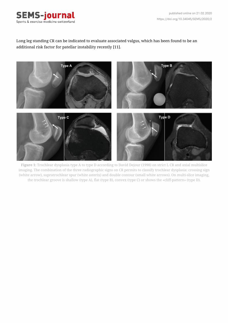
Multi-slices imaging modalities (MRI/CT)Computed tomography (CT) and magnetic resonance imaging (MRI) are both nowadays widely availableand permit multiplanar reconstructions in every planes. Whereas CT scan acquisition is much faster,irradiation has been minimized using cone-beam CT (CBCT) and the resolution of bony structure is moreaccurate, there is no irradiation with MRI and cartilage and soft-tissue are better visualized. MRI thereforeis the gold-standard in patellofemoral assessment, especially in the early evaluation after primary patellardislocation.Whereas both imaging modalities can detect the main predisposing factors for patellar instability, cut-offvalues may differ due to the different landmarks (bony on CT versus cartilaginous-tendinous landmarkson MRI for trochlear morphology) and to the different flexion angle of the knee during acquisition of theimages. Whereas CT are always performed in full extension, MRI are performed at about 10-15° of kneeflexion due to the position in the knee coil, highly influencing the TT-TG value or patellar tilt.Measuring trochlear dysplasia on 2D imaging has been introduced by David Dejour in 1998 on CT scan [3],and has led to the four type classification, from type A having a shallow trochlea to type D with a convextrochlea and the cliff pattern due to the hypoplastic medial trochlea and the absent trochlear groove(Figure 1). The classification of trochlear dysplasia on CT and MRI might differ due to the different osseousand cartilaginous trochlear surface, and also according the level of the axial image. Therefore, the trochleaneeds to be assessed from proximal to distal for complete appreciation of its morphology and not solely ona single cut [12]. There are several more measurement methods on axial imaging, such as the lateraltrochlear inclination angle [13], sulcus angle [14], trochlear depth [14] and trochlear facet/condyleasymmetry (14) all able to describe trochlear dysplasia, however with only limited clinical relevance.The most common patellar height ratios used on CR (InSa-I and CD-I) can also be measured on MRI (Figure2B), however require a marginal correction of 0.1 for InSa-I and CD-I according to a comparativeCR/CT/MRI study [15,16]. However, it might be difficult to find a single sagittal cut on CT or MRI whichrepresents the entire patellar tendon and the patella, making the measurement of the InSa-I sometimesimpossible [8]. More recently, the ratio of the overlap between the patellar and the trochlear cartilage hasbeen proposed by Biedert [17] and slightly modified by others [18,19]. This type of ratio however is highlydepending on knee flexion angle, and on patellar position and needs therefore to be analysed carefully.The patellar tendon length with a cut-off value of 52 mm has gained only little attention in literature,probably due to the high interpersonal variability.The tibial tubercle to trochlear groove (TT-TG) distance is a measurement for a distal lateral patellar vectorforce and represents the distance between the trochlear groove and the tibial tubercle and is increasedabove 15 mm and generally associated with trochlear dysplasia if above 20 mm [20]. It can nicely bemeasured on CT by superposing two axial slices (Figure 4) or measured with adequate tools on MRI.Improved inter-rater reliability has been shown using the centre of the patellar tendon on the tibialtubercle (PT-TG). Some difficulties may arise in high-grade trochlear dysplasia where the trochlear grooveis found only distally. Due to the distally lateral orientation of the trochlear groove, the TT-TG will beunderestimated in these cases [21]. The measurement results of CT and MRI cannot be compared. Theflexed knee position on MRI decreases the TT-TG significantly compared to the fully extended knee on CTwhich undergoes the “screw-home mechanism” close to extension (Figure 5A and B). Therefore, TT-TGmight vary up to 8-10 mm between CT and MRI. Due to the dynamic susceptibility of the TT-TG in fullextension and the improved interrater reliability using soft-tissue landmarks, MRI in a standard knee coil
published online on 21.02.2020
https://doi.org/10.34045/SEMS/2020/2
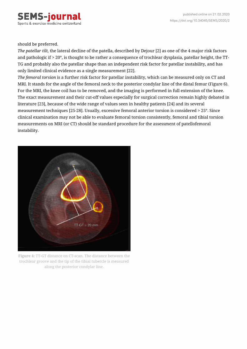
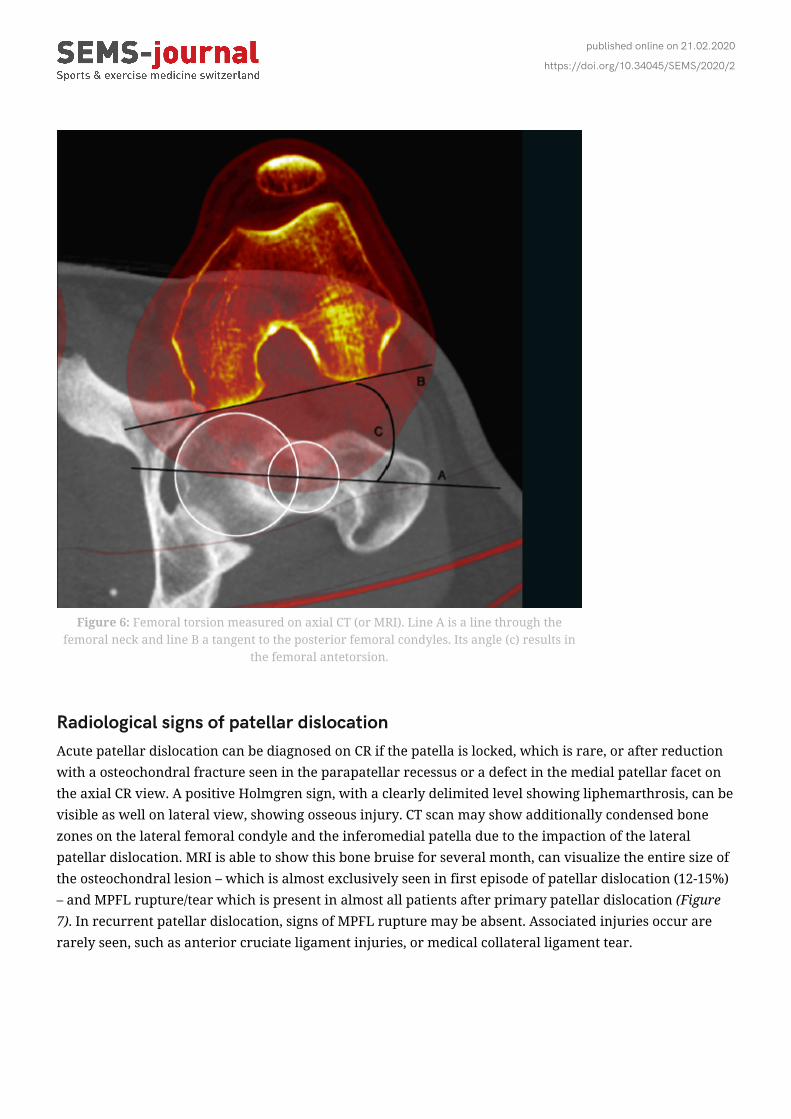
should be preferred.The patellar tilt, the lateral decline of the patella, described by Dejour [2] as one of the 4 major risk factorsand pathologic if > 20°, is thought to be rather a consequence of trochlear dysplasia, patellar height, the TT-TG and probably also the patellar shape than an independent risk factor for patellar instability, and hasonly limited clinical evidence as a single measurement [22].The femoral torsion is a further risk factor for patellar instability, which can be measured only on CT andMRI. It stands for the angle of the femoral neck to the posterior condylar line of the distal femur (Figure 6).For the MRI, the knee coil has to be removed, and the imaging is performed in full extension of the knee.The exact measurement and their cut-off values especially for surgical correction remain highly debated inliterature [23], because of the wide range of values seen in healthy patients [24] and its severalmeasurement techniques [25-28]. Usually, excessive femoral anterior torsion is considered > 25°. Sinceclinical examination may not be able to evaluate femoral torsion consistently, femoral and tibial torsionmeasurements on MRI (or CT) should be standard procedure for the assessment of patellofemoralinstability.
Figure 4: TT-GT distance on CT-scan. The distance between thetrochlear groove and the tip of the tibial tubercle is measured
along the posterior condylar line.

published online on 21.02.2020
https://doi.org/10.34045/SEMS/2020/2
Figure 5: TT-TG distance on A) CT and B) MRI. Note the difference of bony and cartilaginouslandmarks, as well as flexion angle influencing the TT-TG distance.
published online on 21.02.2020
https://doi.org/10.34045/SEMS/2020/2
Figure 6: Femoral torsion measured on axial CT (or MRI). Line A is a line through thefemoral neck and line B a tangent to the posterior femoral condyles. Its angle (c) results in
the femoral antetorsion.
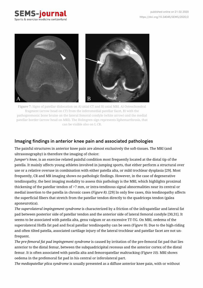
Radiological signs of patellar dislocationAcute patellar dislocation can be diagnosed on CR if the patella is locked, which is rare, or after reductionwith a osteochondral fracture seen in the parapatellar recessus or a defect in the medial patellar facet onthe axial CR view. A positive Holmgren sign, with a clearly delimited level showing liphemarthrosis, can bevisible as well on lateral view, showing osseous injury. CT scan may show additionally condensed bonezones on the lateral femoral condyle and the inferomedial patella due to the impaction of the lateralpatellar dislocation. MRI is able to show this bone bruise for several month, can visualize the entire size ofthe osteochondral lesion – which is almost exclusively seen in first episode of patellar dislocation (12-15%)– and MPFL rupture/tear which is present in almost all patients after primary patellar dislocation (Figure7). In recurrent patellar dislocation, signs of MPFL rupture may be absent. Associated injuries occur arerarely seen, such as anterior cruciate ligament injuries, or medical collateral ligament tear.
published online on 21.02.2020
https://doi.org/10.34045/SEMS/2020/2
Figure 7: Signs of patellar dislocation on A) axial CT and B) axial MRI. A) Osteochondralfragment (arrow head on CT) from the inferomedial patellar facet, B) with the
pathognomonic bone bruise on the lateral femoral condyle (white arrow) and the medialpatellar border (arrow head on MRI). The Holmgren sign represents liphemarthrosis, that
can be visible also on L CR.
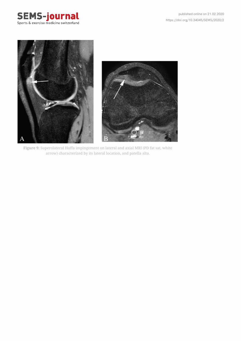
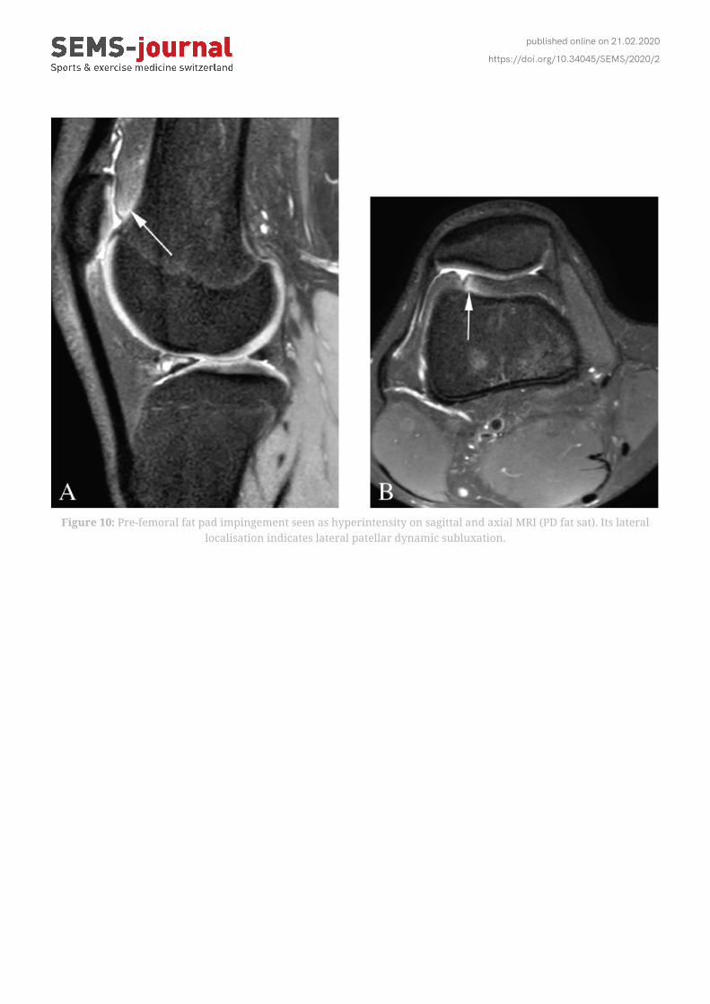
Imaging findings in anterior knee pain and associated pathologiesThe painful structures in anterior knee pain are almost exclusively the soft-tissues. The MRI (andultrasonography) is therefore the imaging of choice:Jumper’s knee, is an exercise related painful condition most frequently located at the distal tip of thepatella. It mainly affects young athletes involved in jumping sports, that either perform a structural overuse or a relative overuse in combination with either patella alta, or mild trochlear dysplasia [29]. Mostfrequently, CR and MR imaging shows no pathologic findings. However, in the case of degenerativetendinopathy, the best imaging modality to assess this pathology is the MRI, which highlights proximalthickening of the patellar tendon of >7 mm, or intra-tendinous signal abnormalities near its central ormedial insertion to the patella in chronic cases (Figure 8). [29] In only few cases, this tendinopathy affectsthe superficial fibers that stretch from the patellar tendon directly to the quadriceps tendon (galeaaponeurotica).The superolateral impingement syndrome is characterized by a friction of the infrapatellar and lateral fatpad between posterior side of patellar tendon and the anterior side of lateral femoral condyle [30,31]. Itseems to be associated with patella alta, genu valgum or an excessive TT-TG. On MRI, oedema of thesuperolateral Hoffa fat pad and focal patellar tendinopathy can be seen (Figure 9). Due to the high-ridingand often tilted patella, associated cartilage injury of the lateral trochlear and patellar facet are not un-frequent.The pre-femoral fat pad impingement syndrome is caused by irritation of the pre-femoral fat pad that liesanterior to the distal femur, between the subquadricipital recessus and the anterior cortex of the distalfemur. It is often associated with patella alta and femoropatellar maltracking (Figure 10). MRI showsoedema in the prefemoral fat pad in his central or inferolateral part.The mediopatellar plica syndrome is usually presented as a diffuse anterior knee pain, with or without

published online on 21.02.2020
https://doi.org/10.34045/SEMS/2020/2
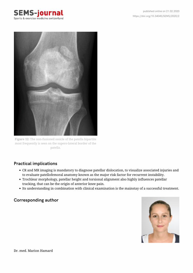
clicking or sometimes even a sensation of locking. It only can be visualized on MRI as a thin plicature ofthe capsule medial to the patella (Figure 11). There is no sign on imaging that proofs a conflict. Patient’shistory, clinical imaging and its size lead to the diagnosis. Consecutive cartilage damage of the medialpatellar facet is rare. The superopatellar plica or the intercondylar plica are other embryonic remnants,however much more rarely symptomatic.Degenerative cartilage lesions are very frequently seen on MR imaging, most often however asymptomatic.Since there are no nerve endings in the cartilage, the origin of pain most probably lies in the subchondralbone. MR imaging (or in some selected cases where arthro-CT scan might be necessary) can graduate andlocalize the extent of the lesion according to the Outerbridge classification.Patella bipartita describes the presence of an unfused ossicle usually found on the superolateral aspect ofthe patella (Figure 12), and can be found in 1-2% of the population. It usually is asymptomatic, howeverdirect trauma to the patella can induce pain at fibrous scar, which can be visible on MRI.
Figure 8: Jumper’s knee with structural intra-tendinous signal alterations on MRI (A) lateralplane PD, and B) axial plane PD fatsat). Thickening of the patellar tendon >7 mm (A; whiteline), and posterior signal abnormalities in the central or medial part of the tendon high
signal T2w or PD images (B; white arrow).
published online on 21.02.2020
https://doi.org/10.34045/SEMS/2020/2
Figure 9: Superolateral Hoffa impingement on lateral and axial MRI (PD fat sat, whitearrow) characterized by its lateral location, and patella alta.
published online on 21.02.2020
https://doi.org/10.34045/SEMS/2020/2
Figure 10: Pre-femoral fat pad impingement seen as hyperintensity on sagittal and axial MRI (PD fat sat). Its laterallocalisation indicates lateral patellar dynamic subluxation.

published online on 21.02.2020
https://doi.org/10.34045/SEMS/2020/2
Figure 11: The mediopatellar plica seen as a thin plicature of the knee capsule (whitearrow), in contact with the medial patellar facet (axial MRI, PD fat sat).
published online on 21.02.2020
https://doi.org/10.34045/SEMS/2020/2
Figure 12: The non-fusioned ossicle of the patella bipartitemost frequently is seen on the supero-lateral border of the
patella.
Practical implicationsCR and MR imaging is mandatory to diagnose patellar dislocation, to visualize associated injuries andto evaluate patellofemoral anatomy known as the major risk factor for recurrent instability.Trochlear morphology, patellar height and torsional alignment also highly influences patellartracking, that can be the origin of anterior knee pain.Its understanding in combination with clinical examination is the mainstay of a successful treatment.
Corresponding author
Dr. med. Marion Hamard

published online on 21.02.2020
https://doi.org/10.34045/SEMS/2020/2
Département de RadiologieHôpitaux Universitaires GenèveRue Gabrielle-Perret-Gentil 41205 Genè[email protected] �
BibliographyDejour H, Walch G. [Chronic posterior instabilities]. Orthopade. 1987;16(2):149-56.1.Dejour H, Walch G, Nove-Josserand L, Guier C. Factors of patellar instability: an anatomic2.radiographic study. Knee Surg Sports Traumatol Arthrosc. 1994;2(1):19-26.Reynaud P, Dejour D. [Patellar entrapment due to a force-impaction fracture of the lateral condyle.3.Apropos of a case]. Rev Chir Orthop Reparatrice Appar Mot. 1998;84(8):752-5.Goutallier D, Bernageau J, Lecudonnec B. [The measurement of the tibial tuberosity. Patella groove4.distanced technique and results (author’s transl)]. Rev Chir Orthop Reparatrice Appar Mot.1978;64(5):423-8.Salzmann GM, Weber TS, Spang JT, Imhoff AB, Schottle PB. Comparison of native axial radiographs5.with axial MR imaging for determination of the trochlear morphology in patients with trochleardysplasia. Arch Orthop Trauma Surg. 2010;130(3):335-40.Hinckel BB, Gobbi RG, Kihara Filho EN, Demange MK, Pecora JR, Camanho GL. Patellar Tendon-6.Trochlear Groove Angle Measurement: A New Method for Patellofemoral Rotational Analyses. OrthopJ Sports Med. 2015;3(9):2325967115601031.Caton J, Deschamps G, Chambat P, Lerat JL, Dejour H. [Patella infera. Apropos of 128 cases]. Rev Chir7.Orthop Reparatrice Appar Mot. 1982;68(5):317-25.Biedert RM, Tscholl PM. Patella Alta: A Comprehensive Review of Current Knowledge. Am J Orthop8.(Belle Mead NJ). 2017;46(6):290-300.Otto A, Tscholl PM, Paasuke R, Herbst E, Willinger L, Imhoff AB, et al. Neither lateral patellar facet9.nor patellar size are altered in patellofemoral unstable patients: a comparative magnetic resonanceimaging analysis. Knee Surg Sports Traumatol Arthrosc. 2019.Saragaglia D, Mader R, Blaysat M, Mercier N. Medial facet patelloplasty in patellar instability10.associated with patellofemoral dysplasia: a report of 26 cases. Orthop Traumatol Surg Res.2012;98(2):167-72.Frosch KH, Schmeling A. A new classification system of patellar instability and patellar maltracking.11.Arch Orthop Trauma Surg. 2016;136(4):485-97.Tscholl PM, Wanivenhaus F, Fucentese SF. Conventional Radiographs and Magnetic Resonance12.Imaging for the Analysis of Trochlear Dysplasia: The Influence of Selected Levels on MagneticResonance Imaging. Am J Sports Med. 2017;45(5):1059-65.Carrillon Y, Abidi H, Dejour D, Fantino O, Moyen B, Tran-Minh VA. Patellar instability: assessment on13.MR images by measuring the lateral trochlear inclination-initial experience. Radiology.2000;216(2):582-5.Pennock AT, Alam M, Bastrom T. Variation in tibial tubercle-trochlear groove measurement as a14.function of age, sex, size, and patellar instability. Am J Sports Med. 2014;42(2):389-93.Lee PP, Chalian M, Carrino JA, Eng J, Chhabra A. Multimodality correlations of patellar height15.measurement on X-ray, CT, and MRI. Skeletal Radiol. 2012;41(10):1309-14.Tscholl PM BD, Fucentese SF. Validation of patellar height measurements on conventional16.radiographs and MRI in patients with patellar instability. EFFORT congress 2017; Vienna2017.Biedert RM, Albrecht S. The patellotrochlear index: a new index for assessing patellar height. Knee17.Surg Sports Traumatol Arthrosc. 2006;14(8):707-12.

published online on 21.02.2020
https://doi.org/10.34045/SEMS/2020/2
Dejour D, Ferrua P, Ntagiopoulos PG, Radier C, Hulet C, Remy F, et al. The introduction of a new MRI18.index to evaluate sagittal patellofemoral engagement. Orthop Traumatol Surg Res. 2013;99(8Suppl):S391-8.Munch JL, Sullivan JP, Nguyen JT, Mintz D, Green DW, Shubin Stein BE, et al. Patellar Articular19.Overlap on MRI Is a Simple Alternative to Conventional Measurements of Patellar Height. Orthop JSports Med. 2016;4(7):2325967116656328.Diederichs G, Issever AS, Scheffler S. MR imaging of patellar instability: injury patterns and20.assessment of risk factors. Radiographics. 2010;30(4):961-81.Tscholl PM, Antoniadis A, Dietrich TJ, Koch PP, Fucentese SF. The tibial-tubercle trochlear groove21.distance in patients with trochlear dysplasia: the influence of the proximally flat trochlea. Knee SurgSports Traumatol Arthrosc. 2016;24(9):2741-7.Grelsamer RP, Saleh J, Gladstone J. Congruous versus incongruous patellar tilt-a preliminary study.22.Bull NYU Hosp Jt Dis. 2012;70(4):232-4.Diederichs G, Kohlitz T, Kornaropoulos E, Heller MO, Vollnberg B, Scheffler S. Magnetic resonance23.imaging analysis of rotational alignment in patients with patellar dislocations. Am J Sports Med.2013;41(1):51-7.Reikeras O. Patellofemoral characteristics in patients with increased femoral anteversion. Skeletal24.Radiol. 1992;21(5):311-3.Billing L. Roentgen examination of the proximal femur end in children and adolescents; a25.standardized technique also suitable for determination of the collum-, anteversion-, and epiphysealangles; a study of slipped epiphysis and coxa plana. Acta Radiol Suppl. 1954;110:1-80.Fabry G, MacEwen GD, Shands AR, Jr. Torsion of the femur. A follow-up study in normal and26.abnormal conditions. J Bone Joint Surg Am. 1973;55(8):1726-38.Sutter R, Dietrich TJ, Zingg PO, Pfirrmann CW. Femoral antetorsion: comparing asymptomatic27.volunteers and patients with femoroacetabular impingement. Radiology. 2012;263(2):475-83.Sutter R, Dietrich TJ, Zingg PO, Pfirrmann CW. Assessment of Femoral Antetorsion With MRI:28.Comparison of Oblique Measurements to Standard Transverse Measurements. AJR Am J Roentgenol.2015;205(1):130-5.Tscholl PM, Biedert RM, Wanivenhaus F, Fucentese SF. Patellar tendinopathy with intratendinous29.alteration on MRI may be related to patellofemoral dysplasia. Scand J Med Sci Sports.2018;28(4):1443-50.Campagna R, Pessis E, Biau DJ, Guerini H, Feydy A, Thevenin FS, et al. Is superolateral Hoffa fat pad30.edema a consequence of impingement between lateral femoral condyle and patellar ligament?Radiology. 2012;263(2):469-74.Ciriello V, Gudipati S, Tosounidis T, Soucacos PN, Giannoudis PV. Clinical outcomes after repair of31.quadriceps tendon rupture: a systematic review. Injury. 2012;43(11):1931-8.
ANTERIOR KNEE PAIN FAT PAD IMPINGEMENT SYNDROME FEMORO-PATELLAR INSTABILITY JUMPER’S
KNEE PATELLA ALTA PATELLAR DISLOCATION PATELLAR TILT TIBIAL TENDON-TROCHLEAR
GROOVE TROCHLEAR DYSPLASIA