What is radiation? - Research Service Centers is radiation? Radiation is energy that travels through...
37
What is radiation? Radiation is energy that travels through space or matter in the form of a particle or wave The effect radiation has on matter depends on the type of radiation and how much energy it has Energy is measured in electron volts ( eV ) 1 eV = 1.6 x 10 - 19 joules More common to see kilo electron volts used (1 k eV = 1,000 eV)
Transcript of What is radiation? - Research Service Centers is radiation? Radiation is energy that travels through...

What is radiation?
Radiation is energy that travels through space or matter in the form of a particle or wave
The effect radiation has on matter depends on the type of radiation and how much energy it has Energy is measured in electron volts (eV)
1 eV = 1.6 x 10⁻¹⁹ joules
More common to see kilo electron volts used (1 keV = 1,000 eV)

Types of radiation
2 main categories Particulate radiation: consists of particles that have mass and
energy, and may or may not have an electric charge Alpha particles and protons (positive charge) Beta particles (positive or negative charge) Neutrons (uncharged)
Electromagnetic radiation: consists of photons that have energy, but no mass or charge (just like light, but higher frequency) X rays Gamma rays

The Electromagnetic Spectrum
Type of Radiation
Effects
Source
wavelength

Ionizing Radiation
Radiation is called ionizing if it is capable of forming ion pairs in matter An ion pair is formed when an
electron is removed from an atom, leaving a free electron and a positively charged atom
The ability to ionize depends on factors including energy, mass, and charge
Most non-ionizing radiation is not harmful
Figure taken from:www.e-radiography.net/radsafety/rad_physics4.jpg

Radiation Quantities and Units

Radiation Measurement
Count rate Exposure Absorbed dose Equivalent dose
or dose equivalent Effective dose
or effective dose equivalent
Measured directly
Difficult to measure directly, usually calculated
Always calculated

Exposure
Exposure: the electric charge produced by photons (x rays or gamma rays) in a mass of air
Traditional unit is the Roentgen (R) SI unit is Coulombs/kg air 1 R = 2.58 X 10⁻⁴ C/kg air Can be measured as a total exposure or an exposure
rate

Absorbed Dose Absorbed dose: the energy deposited in a material by radiation
per unit mass Traditional unit is the rad (radiation absorbed dose) 1 rad = 0.01 J/kg In SI units, rad has been replaced by Gray (Gy)
1 Gy = 1 J/kg 100 rad = 1 Gy
You can convert exposure to dose in air using:
In tissue, 1 rad is approximately equal to 1 R
(R) (rad/R) 876.0(rad) XDair ×=

Equivalent Dose /Dose Equivalent
Takes into account that some kinds of radiation cause more biological harm than others
The traditional unit for this is the rem (stands for Roentgen Equivalent Man)
In SI units, the rem has been replaced by the sievert (Sv), where 1 Sv = 100 rem
Equivalent dose: H (rem) = Σ (D (rad) * wR ) wR = radiation weighting factor
Dose equivalent (pre-1990): H (rem) = Σ (D (rad) * Q ) Q = quality factor
Radiation Type Quality Factor Radiation Weighting Factor
x-rays, γ rays, or β particles 1 1
Neutrons (depends on energy) 2-11 5-20
Protons (high-energy) 10 2-5
Alpha particles 20 20

Effective Dose / Effective Dose Equivalent
Takes into account that some tissues and organs in the human body are more sensitive to radiation than others
Multiply the Equivalent Dose or Dose Equivalent to each organ/tissue by the tissue weighting factor (wT) for that organ/tissue and add them all together
Use equivalent dose and 1990 wT values – get effective dose Use dose equivalent and 1977 wT values – get effective dose
equivalent The unit is still either rem or Sv
∑ ×= TT wHEDE

Tissue Weighting Factors Tissue weighting factor: the
proportion of the risk of stochastic effects resulting from irradiation of an organ or tissue to the total risk of stochastic effects when the whole body is irradiated uniformly
Stochastic effect: A health effect that occurs randomly and for which the probability of the effect occurring, rather than its severity, is assumed to be a linear function of dose without threshold (example: getting cancer)
Tissue or OrganwT
(2007 recomm.)
wT(ICRP 60 -
1990)
wT(ICRP 23 -
1977)
Gonads 0.08 0.20 0.25
Bone marrow 0.12 0.12 0.12
Colon 0.12 0.12 N/A
Lung 0.12 0.12 0.12
Stomach 0.12 0.12 N/A
Bladder 0.04 0.05 N/A
Breast 0.12 0.05 0.15
Liver 0.04 0.05 N/A
Esophagus 0.04 0.05 N/A
Thyroid 0.04 0.05 0.03
Skin 0.01 0.01 N/A
Brain 0.01 N/A N/A
Salivary glands 0.01 N/A N/A
Bone surface 0.01 0.01 0.03
Remainder 0.12 0.05 0.30

Table of Radiation Units
Quantity Traditional Unit S.I. Unit
Activity Curie (Ci) Becquerel (Bq)
Exposure Roentgen (R) Coulomb/Kilogram (C/kg)
Absorbed Dose Rad Gray (Gy)
Equivalent Dose Rem Sievert (Sv)

Radiation Protection Philosophy
ALARA
As Low As Reasonably Achievable

Radiation Protection Principles
For External RadiationTimeDistanceShielding

Time
Reduce time in a radiation area, exposure will be reduced.
TimeRateDoseDose ×=
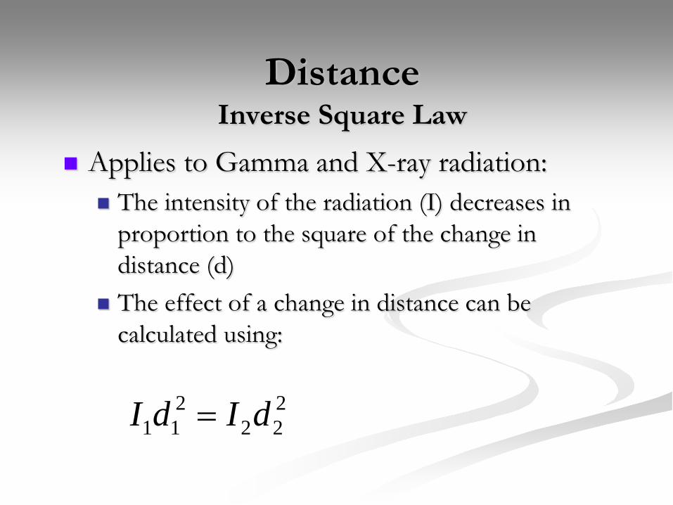
DistanceInverse Square Law
Applies to Gamma and X-ray radiation: The intensity of the radiation (I) decreases in
proportion to the square of the change in distance (d)
The effect of a change in distance can be calculated using:
222
211 dIdI =

Inverse Square Law

Shielding
Shielding material placed between the radiation source and personnel will reduce the radiation intensity by attenuation, and thus reduce the exposure received.
Attenuation: process by which a beam of radiation is reduced in intensity by absorption or scatter in the medium.

Shielding - Photons
Shielding equation for gamma and x-ray radiation:I = intensity after passing through shieldI0= initial intensity of sourceµ = constant related to ability of material to block radiation
x = thickness of shielding material
μxoeII −=

Half-Value Layer
Another way of determining shielding efficiency is by using the Half-Value Layer (HVL) HVL: The thickness of a shielding material required to reduce the intensity
of the radiation by one half. This is commonly used for x-ray sources in which the photons have a range
of energies Is related to µ by: HVL = 0.693/µ HVL equation:
where n = number of half-value layers
n
II2
0=

Dose Monitoring
Exposure Monitoring External radiation exposure is measured by personal
monitoring devices. Personal monitoring is required when it is likely that an individual will receive in 1 year, a dose that is in excess of 10% of the allowed dose.
Not used for H-3, C-14, or S-35

Dose Monitoring
At the University of Florida, whole body doses are determined using an optically stimulated luminescence dosimeter (Luxel). This badge shall be worn on the front part of the
body somewhere between the waist and the collar.

Dose Monitoring
Luxel Dosimeter:

Dose Limits
Maximum Permissible Exposure for Occupational Workers
Whole Body: 5.0 REM /yearEye: 15 REM /year
Skin or Extremity: 50 REM /year50 REM committed dose equivalent to any individual organ
or tissue /year

Dose Limits
Occupational Dose limit for individual members of the public: Total effective dose equivalent to individual
members of the public shall not exceed 0.1 rem in a year.

DNA is the primary target for biological damage

Radiation Damage Mechanisms1. Direct Action: Direct
ionization of the DNA molecule, which may result in genetic damage.
2. Indirect Action: Radiation ionizes water, which causes free radicals to form. Free radicals attack targets such as DNA. Much more common.

Possible Effects to Cells1. Radiation may pass
through cell without doing any damage.
2. Damage may occur but be repaired.
3. The damaged cell may reproduce in its damaged form.
4. The cell may die.

LONG TERM EFFECTS
Delayed effects due to previous acute high dose exposures or from chronic low dose exposure over many years. Cancer Embryological Effects Cataracts Life span shortening

Genetic Effects
• Genetic effects = heritable mutations to DNA• Seen in mammals but no convincing evidence in
humans• Very difficult to measure due to subtle effects, long
lifespans, uncertainties in background rate, and confounding factors
• Japanese bomb survivors– 77,000 births with no substantial evidence of genetic
effects

Human Evidence of Radiation Carcinogenesis
Radium dial painters Radiologists and dentists Uranium miners Atomic bomb survivors Patients receiving medical procedures

Cancer Risk from Chronic Exposure
From the NRC:
LINEAR - An increase in dose results in a proportional increase in risk
NO-THRESHOLD - Any dose, no matter how small, produces some risk
The risk does not start at 0 because there is some risk of cancer, even with no occupational exposure.
Exposure to radiation is not a guarantee of harm. However, because of the linear, no-threshold model, more exposure means more risk, and there is no dose of radiation so small that it will not have some effect.

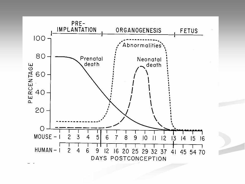
EFFECTS ON EMBRYO AND FETUS
Embryonic/fetal cells are rapidly dividing! High sensitivity Higher probability that damage will be
reproduced over a large number of cells
Effects depend on stage of gestation

REGULATIONS FOR PREGNANT WORKERS
1. Limit embryo/fetus dose equivalent to 500 mrem (0.5 rem) total.
2. Once a pregnancy becomes known limit embryo fetus dose equivalent to 50 mrem per month, excluding medical exposure
3. Wear two personnel monitors. Fetal monitor under apron at waist. Maternal, outside apron at collar.

FEDERAL GUIDELINES FEDERAL REGISTER 1/27/87
“The health protection objectives…for the unborn should be achieved in accordance with the provisions of Title VII of the Civil Rights Act of 1964…with respect to discrimination in employment practices.”
-VOLUNTARY declaration of pregnancy to employer as soon as soon as possible.

FEDERAL GUIDELINES FEDERAL REGISTER 1/27/87
Protection of the unborn is a joint responsibility of the employer and the worker.
Protection through:Use of protective equipment, worker self selection,
and temporary job rotation.