Weight before and after a diagnosis of breast cancer or ...
12
RESEARCH ARTICLE Open Access Weight before and after a diagnosis of breast cancer or ductal carcinoma in situ: a national Australian survey Carolyn Ee 1* , Adele Elizabeth Cave 1 , Dhevaksha Naidoo 1 , Kellie Bilinski 1 and John Boyages 2 Abstract Background: Overweight/obesity are strongly implicated in breast cancer development, and weight gain post- diagnosis is associated with greater morbidity and all-cause mortality. The aim of this study was to describe the prevalence of overweight/obesity and the pattern of weight gain after diagnosis of breast cancer amongst Australian women. Methods: We collected sociodemographic, medical, weight and lifestyle data using an anonymous, self- administered online cross-sectional survey between November 2017 and January 2018 from women with breast cancer living in Australia. The sample consisted mainly of members of the Breast Cancer Network Australia Review and Survey Group. Results: From 309 responses we obtained complete pre/post diagnosis weight data in 277 women, and calculated pre/post Body Mass Index (BMI) for 270 women. The proportion of women with overweight/obesity rose from 48.5% at diagnosis to 67.4% at time of survey. Most women were Caucasian with stage I-III breast cancer (n = 254) or ductal carcinoma in situ (DCIS) (n = 33) and mean age was 59.1 years. The majority of women (63.7%) reported they had gained weight after diagnosis with an average increase of 9.07 kg in this group. Of the women who provided complete weight data, half gained 5 kg or more, 17.0% gained > 20 kg, and 60.7% experienced an increase in BMI of >1 kg/m 2. Over half of the women rated their concern about weight as high. Of those women who gained weight, more than half reported that this occurred during the first year after diagnosis. Two-thirds (69.1%) of women aged 35–74 years gained, on average, 0.48 kg more weight per year than age-matched controls. Conclusions: Although the findings from this survey should be interpreted cautiously due to a limited response rate and self-report nature, they suggest that women in Australia gain a considerable amount of weight after a diagnosis of breast cancer/DCIS (in excess of age-matched data for weight gain) and report high levels of concern about their weight. Because weight gain after breast cancer may lead to poorer outcomes, efforts to prevent and manage weight gain must be prioritized and accelerated particularly in the first year after diagnosis. Keywords: Breast cancer, DCIS, Overweight, Obesity, Weight gain, Australian women, National survey, Prevalence © The Author(s). 2020 Open Access This article is distributed under the terms of the Creative Commons Attribution 4.0 International License (http://creativecommons.org/licenses/by/4.0/), which permits unrestricted use, distribution, and reproduction in any medium, provided you give appropriate credit to the original author(s) and the source, provide a link to the Creative Commons license, and indicate if changes were made. The Creative Commons Public Domain Dedication waiver (http://creativecommons.org/publicdomain/zero/1.0/) applies to the data made available in this article, unless otherwise stated. * Correspondence: [email protected] 1 NICM Health Research Institute, Western Sydney University, Locked Bag 1797, Penrith, NSW 2751, Australia Full list of author information is available at the end of the article Ee et al. BMC Cancer (2020) 20:113 https://doi.org/10.1186/s12885-020-6566-4
Transcript of Weight before and after a diagnosis of breast cancer or ...

RESEARCH ARTICLE Open Access
Weight before and after a diagnosis ofbreast cancer or ductal carcinoma in situ: anational Australian surveyCarolyn Ee1* , Adele Elizabeth Cave1, Dhevaksha Naidoo1, Kellie Bilinski1 and John Boyages2
Abstract
Background: Overweight/obesity are strongly implicated in breast cancer development, and weight gain post-diagnosis is associated with greater morbidity and all-cause mortality. The aim of this study was to describe theprevalence of overweight/obesity and the pattern of weight gain after diagnosis of breast cancer amongstAustralian women.
Methods: We collected sociodemographic, medical, weight and lifestyle data using an anonymous, self-administered online cross-sectional survey between November 2017 and January 2018 from women with breastcancer living in Australia. The sample consisted mainly of members of the Breast Cancer Network Australia Reviewand Survey Group.
Results: From 309 responses we obtained complete pre/post diagnosis weight data in 277 women, and calculatedpre/post Body Mass Index (BMI) for 270 women. The proportion of women with overweight/obesity rose from48.5% at diagnosis to 67.4% at time of survey. Most women were Caucasian with stage I-III breast cancer (n = 254)or ductal carcinoma in situ (DCIS) (n = 33) and mean age was 59.1 years. The majority of women (63.7%) reportedthey had gained weight after diagnosis with an average increase of 9.07 kg in this group. Of the women whoprovided complete weight data, half gained 5 kg or more, 17.0% gained > 20 kg, and 60.7% experienced anincrease in BMI of >1 kg/m2. Over half of the women rated their concern about weight as high. Of those womenwho gained weight, more than half reported that this occurred during the first year after diagnosis. Two-thirds(69.1%) of women aged 35–74 years gained, on average, 0.48 kg more weight per year than age-matched controls.
Conclusions: Although the findings from this survey should be interpreted cautiously due to a limited responserate and self-report nature, they suggest that women in Australia gain a considerable amount of weight after adiagnosis of breast cancer/DCIS (in excess of age-matched data for weight gain) and report high levels of concernabout their weight. Because weight gain after breast cancer may lead to poorer outcomes, efforts to prevent andmanage weight gain must be prioritized and accelerated particularly in the first year after diagnosis.
Keywords: Breast cancer, DCIS, Overweight, Obesity, Weight gain, Australian women, National survey, Prevalence
© The Author(s). 2020 Open Access This article is distributed under the terms of the Creative Commons Attribution 4.0International License (http://creativecommons.org/licenses/by/4.0/), which permits unrestricted use, distribution, andreproduction in any medium, provided you give appropriate credit to the original author(s) and the source, provide a link tothe Creative Commons license, and indicate if changes were made. The Creative Commons Public Domain Dedication waiver(http://creativecommons.org/publicdomain/zero/1.0/) applies to the data made available in this article, unless otherwise stated.
* Correspondence: [email protected] Health Research Institute, Western Sydney University, Locked Bag1797, Penrith, NSW 2751, AustraliaFull list of author information is available at the end of the article
Ee et al. BMC Cancer (2020) 20:113 https://doi.org/10.1186/s12885-020-6566-4

BackgroundBreast cancer is the most common cancer in womenworldwide and in Australia [1–3]. There were over 2million new cases of breast cancer (BC) globally in 2018,with this figure expected to rise to over 3 million by2040 [3], by which time Australia expects to diagnosemore than 25,000 new cases annually [2]. Obesity is aknown risk factor for BC, particularly for post-menopausal women [4]. Obesity after menopause (fromweight gain during either the premenopausal or post-menopausal years) is directly related to an increasedrelative risk of BC of 1.11 per 5 kg increase in weight [5].Obesity at diagnosis is associated with worse BC sur-
vival and all-cause mortality rates and may increase therisk of cancer recurrence by 30–40% [1, 6]. Furthermore,weight gain is common after BC diagnosis, and may in-crease the risk of disease recurrence and mortality andhave a negative impact on quality of life [1]. Weight gainafter BC diagnosis is thought to be multifactorial andmay be related to the use of systemic treatment, youngerage at diagnosis, as well as changes in lifestyle [1, 7].Given the growing population of BC survivors and thelink between weight gain and adverse health outcomes,research into weight after BC is of critical importance.The prevalence of weight gain after BC in Australia
has not been adequately quantified. One prospective co-hort study conducted in Queensland, in women whohad been diagnosed with early breast cancer, describedan increase in the proportion of women who were over-weight/obese from 57% at diagnosis to 68% over 6 years[8]. However, there is no national data currently avail-able and few population studies exist [9]. The aim of thisstudy was to describe the prevalence of self-reportedoverweight and obesity before and after a diagnosis ofBC or Ductal Carcinoma in Situ (DCIS).
MethodsStudy design and inclusion criteriaA cross-sectional self-administered anonymous surveywas conducted in Australia between November 2017and January 2018 using Qualtrics® online survey software[10]. Any woman living in Australia who self-identifiedas having BC was eligible to complete the survey. A copyof the Participant Information Sheet was provided elec-tronically via a link on the survey website prior to start-ing the survey, and women were informed that consentwas implied upon commencing the survey. This methodof consent was approved by the Human Research EthicsCommittee (See below for details). The sample includedmembers of the Breast Cancer Network Australia(BCNA) Review and Survey Group comprising BCNAmembers who had agreed to receive emails about re-search studies. Limiting research at BCNA to the Reviewand Survey group allows researchers to access women
who are engaged in the research process, while protect-ing other BCNA members from frequent research re-quests. The Review and Survey Group (n = 1857)represents approximately 2% of all BCNA members andis one of the largest breast cancer consumer groupsavailable for research in Australia, representing an im-portant source of feedback for the research community.The survey was emailed to 1835 members on Decem-
ber 5th, 2017 and a reminder email sent January 15th,2018 (Appendix). A smaller sample (n = 26) was alsodrawn from online communities (women’s healthorganization social media pages and online breast cancersupport groups in Australia) or through word of mouthduring November and December 2017. Ethics approvalfor this study was provided by the Human Research Eth-ics Committee, Western Sydney University (H12444,October 2017).
Survey instrumentThe survey was developed after reviewing previous lit-erature on weight after BC and was subsequently revisedto include feedback from six BCNA representatives andseveral health researchers. The 60-item survey includedquestions on the sociodemographic characteristics, med-ical details such as diagnosis and treatment, lifestylehabits, weight status, and weight management. Details ofthe survey questions are outlined in the Appendix. Inthis paper, we report on change in weight from time ofdiagnosis to time of the survey.
Weight after diagnosisWomen were asked to self-report their weight in kg atthe time of diagnosis, and current weight and height (inmeters). Body Mass Index (BMI) was calculated fromweight and height as weight/height2. The pattern ofweight since diagnosis was also assessed as “gainedweight overall”, “lost weight overall”, “weight stable” or“weight has fluctuated a great deal”. We devised an un-validated 11-point Likert scale to evaluate concern aboutweight (using the question “Please rate how concernedyou have been over your weight in the last 12 months”)ranging from 0 (not at all concerned) to 10 (very con-cerned). We further characterized these data into fourcategories according to the Likert score: No concern (0),A little concerned (1–3), Somewhat concerned (4–7),Very concerned (8–10). Weight at diagnosis was re-ported by 90% of total respondents (277 women) andcurrent weight by 95% of respondents (293 women).
Statistical analysisIBM SPSS® statistics package version 23 [11] and Stata®statistical software version 13.11 [12] were used toanalyze the data presented in this report. We used de-scriptive statistics to analyze diagnoses, treatments
Ee et al. BMC Cancer (2020) 20:113 Page 2 of 12
received, and health provider visits of respondents inpercentages. Women who did not self-report theirweight were excluded from analyses relating to weight.We calculated the percentage of women who were cur-rently overweight (BMI > 25 and < 30) or obese(BMI > 30) and compared this to the proportion whowere overweight/obese at time of diagnosis. Current andpre-cancer weight and BMI were reported as a meanand standard deviation. We calculated the number andpercentage of women whose BMI changed from healthy(< 25) to unhealthy (BMI > 25) from diagnosis to time ofsurvey, as well as women who reported an increase ofBMI of greater than 1 kg/m2. Tests for skewness andkurtosis for weight, BMI at diagnosis, current weightand BMI, and weight gain, indicated that our data had anormal distribution.We described the self-reported weight gain pattern as
percentage of body weight at diagnosis, the proportionof women who gained > 5 kg, and the proportion ofwomen who gained 5–10% and > 10% of body weight.We used a paired t-test to compare weight and BMI atdiagnosis and weight and BMI at time of survey, andFisher’s exact test to explore the association betweencurrent BMI classification and weight concern. To testthe relationship between weight gain and time sincediagnosis (and therefore the hypothesis that weight gainincreases with time) we performed a Pearson’s correl-ation. We also categorized time since diagnosis into 2.5year blocks and ran a one-way analysis of variance(ANOVA) exploring the relationship between time sincediagnosis and weight gain in the following groups ofwomen: women who reported gaining weight overall,and who had self-reported weight gain > 5%. We ex-plored the relationship between amount of weight gainand weight gain concern using the Pearson’s chi-squaredtest.We calculated the mean weight gain per year in our
sample as total weight gain divided by time since diagno-sis in years. We removed one outlier who reported gain-ing 10.5 kg per year over 2 years and reported rate ofweight gain across age groups in five-year brackets (seeFig. 3 and Table 5).
Comparing rate of weight gain against normative dataTo compare weight gain in our sample against norma-tive data in the Australian population, we used data theAusDiab study. The AusDiab study is a large national,longitudinal population-based study involving > 11,000adults aged 25 years and older. Baseline data collectionfor the AusDiab study occurred during 1999–2000, witha subsequent 5-year follow-up (during 2004–2005) [13].The AusDiab study reported the following mean weightgains per year at 5-year follow-up (2004–5): 700 g peryear for 25-34yo, 500 g for 35-44yo, 380 g for 45-54yo,
140 g for 55-64yo and 0 g for 65-74yo). For each re-spondent in our study for which we could calculate ayearly weight gain, we compared this weight gain withthe mean weight gain from AusDiab corresponding tothe age group of the respondent by subtracting the Aus-Diab weight gain from the weight gain reported by thatrespondent in our study. We used Pearson’s chi-squaredtesting to compare the numbers of women who gainedin excess of the rates reported in the AusDiab studyacross the age groups described above.
ResultsSurvey responseOf the 1857 BCNA members, 283 (15%) responded tothe survey. A further 26 women responded to the surveyfrom other channels giving a total of 309 responses.
Sample characteristicsDemographic characteristics of respondents are de-scribed in Table 1. The majority of women were Cauca-sian (92.5%, n = 285) with a mean age of 59.1 years(SD = 9.5, range 33–78, n = 298). Characteristics weresimilar across BCNA members and non-BCNA respon-dents with no differences between these groups on Pear-son’s Chi-squared test. The majority of women wereeither premenopausal (43%) or perimenopausal (12%) atthe time of diagnosis. Of the 145 women who were stillmenstruating at time of diagnosis, 68% were premeno-pausal and became postmenopausal, 18% were peri-menopausal and became postmenopausal, while asmaller number (13%) remained perimenopausal.
Clinical characteristicsDiagnosesClinical diagnoses of the respondents are summarized inTable 2. The majority of women (82%, n = 252) had beendiagnosed with non-metastatic BC. The mean time sincediagnosis of BC was 8.2 years (SD 5.12, range 1–32 years)and mean age at diagnosis was 50.9 years (SD = 9.02,range 29–74).
TreatmentsWomen reported receiving a range of BC treatments in-cluding surgery and/or radiation, and axillary, systemicand hormonal treatments, which are detailed in Table 2.The most commonly visited health care providers,within the last 12 months, were breast surgeons (n =172), physiotherapists (n = 124) and medical oncologists(for chemotherapy) (n = 119). On average, respondents(n = 247) had visited three health care providers in thelast 12 months (range, 1–10). For women with DCIS, 18(53%) had a mastectomy, 17 (50%) had received radi-ation and 19 (56%) had received hormonal treatment.
Ee et al. BMC Cancer (2020) 20:113 Page 3 of 12

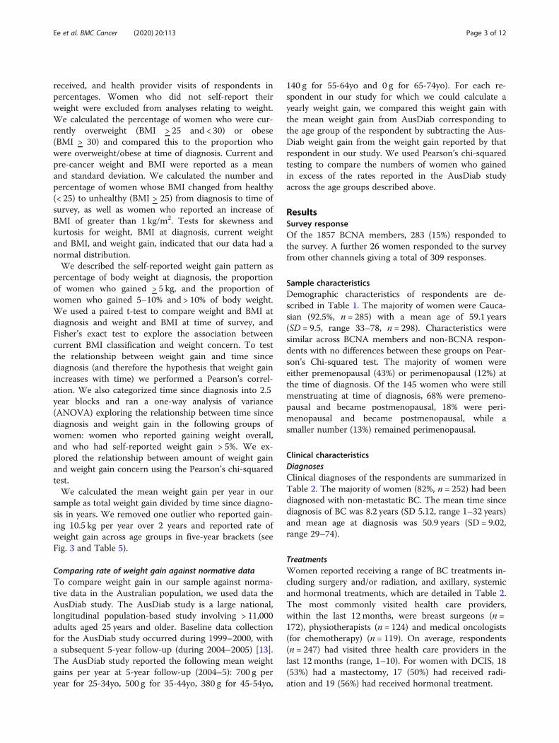
Weight changeTable 3 and Fig. 1 describe weight and BMI changepatterns in our respondents. Mean self-reportedweight at time of diagnosis was 71.24 kg (SD 14.01,range 47–158, n = 277) and at time of survey was76.08 kg (SD 15.37, range 46–150, n = 293). Mean
self-reported current BMI was 28.02 kg/m2 (SD = 5.88,n = 285) and mean pre-cancer BMI was 26.37 kg/m2
(SD = 5.92, n = 271). Just under half of women (48.5%)were overweight or obese at time of diagnosis, but bythe time of the survey this proportion had risen to67.3%. This increase was most marked for womenwho were obese, from 17.0% at diagnosis to 31.9% atthe time of the survey. Mean weight gain was 4.50 kg(SD 8.90, n = 277).
Table 1 Demographic characteristics of survey respondents
Description N(responses)
%
State (n = 309)
Australian Capital Territory 14 4.5%
New South Wales 91 29.5%
Northern Territory 0 0.0%
Queensland 48 15.5%
South Australia 28 9.1%
Tasmania < 5 1.0%
Victoria 95 30.7%
Western Australia 30 9.7%
Education (n = 307)
High school- year 10 30 9.8%
High school- year 12 35 11.4%
Vocational College 55 17.9%
Bachelor’s degree 90 29.3%
Postgraduate degree 97 31.6%
Ethnicity (n = 308)
European/Anglo Saxon/Caucasian 285 92.5%
Asian 5 1.6%
Oceanic (incl. Australian and New Zealand firstpeoples, Polynesian and Micronesian)
13 4.2%
North/South/Central American < 5 0.7%
Mixed ethnicity < 5 0.7%
Indian < 5 0.3%
Employment (n = 308)
Employee 140 45.5%
Self-employed 33 10.7%
Home duties/caring for children or family 15 4.9%
In education (going to school, university, etc.) < 5 1.3%
Doing voluntary work 10 3.3%
Unable to work because of illness 6 2.0%
Unable to work for other reasons < 5 0.3%
Retired 99 32.1%
Relationship Status (n = 309)
Single 39 12.6%
Married/de facto (living with partner) 230 74.4%
In a relationship (not living with partner) 7 2.3%
Divorced/separated 24 7.8%
Widowed 9 2.9%
Table 2 Diagnoses and treatments received
Description N % Missing n (%)
Diagnoses 1 (0.3%)
Ductal Carcinoma In Situ (DCIS) 33 10.7%
Localised breast cancer 252 81.8%
Metastatic breast cancer 14 4.6%
Inflammatory breast cancer <5 0.7%
Other including second primary 7 2.3%
Treatment to the Breast 2 (0.6%)
Lumpectomy alone <5 0.7%
Lumpectomy and radiation 129 42.0%
Mastectomy alone 74 24.1%
Mastectomy and radiation 71 23.1%
Lumpectomy and mastectomy alone 10 3.3%
Lumpectomy, mastectomy and radiation 16 5.2%
Double mastectomy 5 1.6%
Reconstruction after mastectomy (n = 164)
No 84 51.2%
Immediate 37 22.6%
Delayed 43 26.2%
Treatment to the Axilla (n = 167) 13 (7.8%)
Sentinel node biopsy only 24 15.6%
Axillary dissection +/− Sentinel nodebiopsy
56 36.4%
Axillary dissection +/− Sentinel nodebiopsy + radiation
72 46.8%
Radiation only <5 1.3%
Intravenous Systemic Therapy
Chemotherapy without Herceptin 164 53.1%
Herceptin only < 5 0.7%
Chemotherapy + Herceptin 46 14.9%
None/not reported 97 31.4%
Hormonal Treatments
Tamoxifen alone 58 18.8%
Other 146 47.3%
None 105 34.0%
Current use of hormone therapy
Yes 125 40.5%
Ee et al. BMC Cancer (2020) 20:113 Page 4 of 12

One fifth (54/270, 20.0%) of women went from beingin the healthy weight range at diagnosis (BMI <25), toan unhealthy weight range (BMI > 25), a further 14.8%moved from the overweight range into obesity, and60.7% (164/270) of women reported an increase in BMI
greater than 1 kg/m2 (Table 3). Of note, a small propor-tion of women lost weight whereby 5.6% of women wentdown at least one BMI category (Table 3).There was a statistically significant difference between
both weight and BMI at diagnosis and current weight
Table 3 Weight change patterns after diagnosis of breast cancer
Description N % Missing
N (%)
Self-reported weight gain pattern 17 (5.5%)
Weight gain 186 63.7%
Weight loss 38 13.0%
Stable 48 16.4%
Fluctuated 20 6.9%
Calculated % weight change from baseline 32 (10.4%)
Weight loss 62 22.4%
<5% weight gain 53 19.1%
5–10% weight gain 64 23.1%
>10% weight gain 98 35.4%
Calculated weight change 32 (10.4%)
Weight loss 62 22.4%
Weight gain up to 5 kg 75 27.1%
Weight gain ≥5 kg and < 10 kg 75 27.1%
Weight gain ≥10 kg and < 20 kg 18 6.5%
Weight gain >20 kg 47 17.0%
Timing of weight gaina (n = 186)
<6 months post diagnosis 47 25.3%
6–12months post diagnosis 60 32.3%
12–18 months post diagnosis 38 20.4%
18–24 months post diagnosis 16 8.6%
2–3 years post diagnosis 14 7.5%
>3 years post diagnosis 11 5.9%
Change in BMI classification from time of diagnosis to time of survey (n = 270)
Weight gain Healthy to overweight 49 18.2%
Overweight to obese 40 14.8%
Healthy to obese 5 1.9%
Underweight to healthy weight <5 1.5%
Underweight to overweight <5 0.4%
Stable Remained in healthy range 69 25.6%
Remained in overweight range 39 14.4%
Remained in obese range 40 14.8%
Remained in underweight range 8 3.0%
Weight loss Healthy weight to underweight 3 1.1%
Obese or overweight to healthy weight 6 2.2%
Overweight to underweight <5 0.4%
Obese to overweight 5 1.9%athis question was answered only if respondents had selected “gained weight overall”; BMI=Body Mass Index; Underweight = BMI < 20; Healthy weight = BMI > 20and < 25; Overweight = BMI > 25 and < 30; Obese = BMI > 30
Ee et al. BMC Cancer (2020) 20:113 Page 5 of 12

and BMI (mean difference 4.50 kg, CI 3.45–5.55,p = 0.00, n = 277 and 1.64 kg/m2, CI 1.24–2.04, p = 0.00,n = 270 respectively). The majority of respondents(63.7%) reported they had gained weight overall afterdiagnosis. This is consistent with the self-reportedweight gain in our study, where 58.5% of women gained> 5% of pre-diagnosis body weight. Half of the respon-dents had gained more than 5 kg, with 17.0% reportinggains of over 20 kg of weight.Of the women who reported gaining weight overall
and for whom we had complete weight data (n = 175),87.4% (153/175) gained ≥5 kg of weight, and 54.9%gained > 10% of pre-diagnosis body weight. Averageweight gain in this group was 9.07 kg. Women reportedthat weight gain predominantly occurred within the first
2 years of diagnosis (86.6%) with 57.5% reporting thatweight gain mostly occured within the first 12months.Weight gain was not correlated with time since diagnosis(n = 173, r = .114, p = 0.14). There was no difference in theamount of weight gain by time since diagnosis when thiswas examined in blocks of 2.5 years, in women who hadreported weight gain overall (n = 175, p = 0.26), and inwomen who self-reported weight gain of greater than 5%of diagnosis body weight (n = 162, p = 0.27). (Table 4).Three quarters (74.7%, n = 68/91) of women who
were currently obese reported very high levels of con-cern about their weight, compared to a quarter ofwomen in the healthy weight range (25.9%, n = 21/81)(p = 0.00). Women who had gained more weight weremore likely to express high levels of concern about
Fig. 1 Change in BMI classification after diagnosis of breast cancer. BMI=Body Mass Index
Table 4 Weight gain by time in years since diagnosis
Women who had gained >5% weight (n = 162) Women who reported weight gain pattern overall (n = 175)
Mean weight gain (kg) SD Freq. Mean weight gain (kg) SD Freq.
Time since diagnosis (years)
< 2.5 9.00 (6.51) 6 8.14 (6.36) 7
2.5–5 8.42 (4.57) 23 7.36 (4.73) 28
5–7.5 9.27 (5.27) 54 8.90 (5.47) 56
7.5–10 9.15 (5.42) 22 8.43 (5.64) 24
> 10 11.21 (7.14) 57 10.38 (7.37) 60
SD Standard Deviation, Freq Frequency
Ee et al. BMC Cancer (2020) 20:113 Page 6 of 12

their weight. Of the women who gained 5–10% ofweight and > 10% of weight, 54.8 and 78.4% reportedbeing very concerned about their weight respectively,compared with 22.5% of women who had gained lessthan 5% of their diagnosis weight (X2, (9, n = 263) =67.6137, p = 0.000). (Fig. 2).
Rate of weight gain, and comparison with normative dataOn average, women in our study gained 0.64 kg per year(n = 270, SD = 1.76, range − 8 to 10.5) (see Table 5). Forwomen aged 25–74 years (the age range for which wehave normative data), the mean weight gain in excess ofage-matched controls was 0.48 kg per year (n = 235, SD =1.67, range − 8.38 to 7.62). Overall, two thirds (69.8%) ofwomen in our sample gained in excess of normativeweight gain in the AusDiab study, including 25.1% ofwomen who gained > 1 kg per year in excess of norma-tive rates of weight gain. There was no difference be-tween age groups with regard to the number of womenwho gained in excess of normative weight gain (X2, (n =235) = 6.6929, p = 0.153). See Fig. 3 for mean weight gainin excess of normative data for each age group. Therewas only one woman in the 25–34 age group; to pro-tect confidentiality we did not include her data inTable 5 or Fig. 3.
DiscussionThis is the first national survey conducted in Australiato describe weight after breast cancer. The distributionof responses according to state and territory in our sur-vey is broadly consistent with the incidence of BC inthese regions [14] indicating our sample was nationallyrepresentative by location. We found that two-thirds ofour respondents were currently overweight or obese,with the majority of women reporting they had gainedweight after diagnosis, mostly within the first 12 monthsand at a substantial self-reported average of 9.07 kg. Ofnote, the proportion of women who were overweight orobese rose sharply from 48% at time of diagnosis to 67%at the time of the survey, with the proportion of womenwho were obese almost doubling from 17 to 32%. Themajority of women gained weight in excess of the ratesreported in age-matched controls without breast cancer.This equated to an average of an additional 2.42 kg over 5years. A very small proportion of women (5.6%) changedfrom a higher to lower BMI category. It would be of inter-est to explore such findings to enquire whether this is aresult of intended weight loss or treatment related effects.The proportion of women who were overweight or
obese in our study is consistent with those from a pro-spective study of 287 women conducted in Queensland,Australia which compared weight gain after diagnosis of
Fig. 2 Weight gain concern and current BMI classification (n = 285). BMI=Body Mass Index
Ee et al. BMC Cancer (2020) 20:113 Page 7 of 12

early BC. By 6 years, 68% of women in the cohort wereoverweight or obese, [8] which is remarkably similar toour findings. Median weight gain for study participantsbetween 6 and 72months was 0.7 kg, and mean BMI in-crease was 0.2 kg/m2. The authors of the cohort studycompared the weight gain in the BC cohort with age-matched controls and reported a significant difference,with only 50% of age-matched controls being overweightor obese. One other population study has been pub-lished from Shanghai on obesity and clinical outcomesof 4561 Chinese women [9]. In that study, mean weightgain at 18 months post-diagnosis was 1.7 kg. Meanweight gain in our study was significantly higher at 4.5kg which could be explained by the longer time sincediagnosis in our study. Further, the mean weight gain inwomen who had gained weight overall in our study issubstantially higher than what is reported in the Austra-lian cohort study (9.07 kg vs 5.3 kg) although we notethat the mean time since diagnosis in our study is 8.2years, while the cohort study used a 6-year follow-up.Our study provides additional data on weight gain afterBC in Australia, over a wider time frame and locationand with a larger sample size, and suggests that theproblem of weight gain after BC may be larger than pre-viously anticipated.A large international review found that 50–96% of
early stage BC patients experience weight gain duringtreatment in the range of 1.7 kg to 5.0 kg in the 18months following treatment [15]. Of those who gainedweight, 27% gained 2 kg to 5 kg and 24% gain 5 kg ormore in the 18 months following treatment. This com-pares to our study where 50.55% reported gaining 5 kgor more mainly in the first 18 months after treatment,again suggesting that weight gain after BC is a greaterproblem than previously thought.Our findings are of concern because weight gain pre-
and post- BC diagnosis have both been associated withincreased morbidity and mortality. Whilst those atheaviest weight at diagnosis appear to carry an increasedrisk, even those within the healthy weight range at diag-nosis face increased risk following weight gain [16]. Datafrom the Nurse’s Health Study in the USA showed therisk of cancer recurrence was increased by 40% following
a mean weight gain of 2.7 kg, and by 53% followingmean weight gain of 7.7 kg, [17] with the greatest in-creased risk in those of healthy weight at diagnosis. An-other observational study of 3993 women, each 5 kg gainin weight post-diagnosis was associated with a significant12% increase in all-cause mortality, 13% increase in BC-specific mortality, and 19% increase in cardiovascular-disease mortality (all p < 0.05) after an average 6.3 yearsfollow-up after diagnosis [18]. Extrapolating from theseresults indicate that approximately half of our cohortface a significant increase in cancer recurrence and mor-tality due to weight gain > 5 kg, and that efforts to pre-vent weight gain in women diagnosed with BC need tobe accelerated and prioritized.Our findings indicate high levels of concern about
weight, particularly in women who were currently over-weight or obese. Weight gain exacerbates the significantbody image concerns already faced by BC survivors, hasa negative impact on quality of life, and may be a causeof distress if it was unanticipated [19]. We did not ex-plore quality of life or levels of distress in our cohort,but additional research in this area appears to bewarranted.Although the proportion of overweight and obesity in
our survey is similar to national data for women aged45–64 (which ranges from 61 to 69%) [20], the majorityof our respondents were from a higher socioeconomicgroup with 60% having a Bachelor’s degree qualificationor higher, and 56% being employed or self-employed.National data indicate rates of overweight and obesityfor women in the highest socioeconomic group as low as48% [20] indicating that the proportion of overweightand obesity in our respondents is higher than would beexpected of women with similar demographic character-istics. Finally, when compared to age-matched controlsfrom the AusDiab study, 69.8% of women in our surveygained in excess of normative weight gain, indicatingthat the weight gain experienced within our sample isunlikely to be explained by weight gain that would nor-mally be experienced as women age and progressthrough the menopausal transition.This study also highlights the importance of treatment
teams being aware that weight gain, particularly in thefirst year after treatment, is an important issue, whichwould benefit from interventions such as diet and exer-cise. In this study, 186 of 292 patients (63.69%) gainedweight, 57% gained within the first 12 months and 77%within 18 months. The timing of weight gain within thefirst year of treatment has been reported by others [21,22]. Recently, the Clinical Oncological Society ofAustralia has strongly advocated for exercise to be em-bedded as part of standard practice in cancer care andadvised all members of the multidisciplinary cancer teamto promote physical activity, encourage patients to
Table 5 Mean weight gain per year in each age group, andproportion who gained in excessive of normative rates
Age (years) Mean weight gain per yearin kg in our study (SD)
% who gained in excessof AusDiab data
35–44 (n = 21) 1.59 (1.16) 81.0
45–54 (n = 72) 0.75 (2.35) 61.1
55–64 (n = 100) 0.50 (1.47) 76.0
65–74 (n = 42) 0.39 (0.94) 64.3
All (n = 235) 0.64 (1.76) 69.8
Ee et al. BMC Cancer (2020) 20:113 Page 8 of 12

adhere to exercise guidelines and refer patients to anaccredited exercise physiologist or physiotherapist withexperience in cancer care [23]. All people with cancershould progress towards and, once achieved, maintainparticipation in at least 150 min of moderate intensity or75 min of vigorous-intensity aerobic exercise (e.g. walk-ing, jogging, cycling, swimming) each week; and two tothree resistance exercise (i.e. lifting weights) sessionseach week involving moderate-to-vigorous-intensity ex-ercises targeting the major muscle groups. In womenwith breast cancer, there appears to be a window of op-portunity within the first 18 months to initiate weightmanagement interventions in order to prevent excessiveweight gain.Strengths of this survey include the higher than ex-
pected response rate from the BCNA Review and SurveyGroup. According to the Research and Evaluation Man-ager, BCNA (email communication 3 Oct 2017), the typ-ical response rate in this group is 10%, whereas theresponse to our survey was 15%. However, given that theReview and Survey Group represents only approximately2% of all BCNA members, the validity of our findings issomewhat limited but important to highlight particularlyto clinicians managing patients with breast cancer to en-sure they encourage and more importantly “prescribe”an exercise program after cancer treatment.We achieved a broadly nationally representative sample
according to location. The percentage of respondentsfrom each Australian State and Territory is similar to na-tional averages on BC incidence as described by the Aus-tralian Institute of Health and Welfare cancer data [14].Limitations of this survey included its self-report na-
ture. In general, people tend to underestimate their
weight and overestimate their height with self-reporting[24]. Social desirability bias and response bias may play apart in this inaccuracy. In our cross-sectional study, it ispossible that recall bias led to further underestimation ofpre-diagnosis weight, therefore inflating the reportedweight gain. Additionally, a small proportion of womenchose not to report their weight in this survey (10% forpre-diagnosis weight, and 5% for current weight). How-ever, using self-reported weight and height is simple andreadily accessible, and is considered less intrusive thanobjectively measured weight, therefore allowing us toconduct a nation-wide survey and increase the responserate. The true prevalence of weight gain after BC may bedifferent to that found in our survey as women who hadgained weight after BC may have been more likely to re-spond to our survey compared to women who had notgained weight. Nevertheless, the prevalence reported inour survey is remarkably similar to that in prospectivecohort studies, suggesting that our data is robust. There isan urgent need to further understand the predictors ofweight gain in women with BC. Further planned analysesfrom our data will include analysis of the predictors ofweight gain in our sample, including use of chemotherapy,hormonal therapy, and menopausal stage at diagnosis.We acknowledge that the inability to provide matched
controls in this survey is a limitation. However, we wereable to retrospectively match women by age to controlsfrom the 2005 AusDiab study and found that womengained in excess of normative data, although limitationsof our comparison is that we could not locate more re-cent data on normative rates of weight gain, and theduration of weight gain varied in our sample. Further-more, that our findings are remarkably similar to a
Fig. 3 Mean weight gain per year in excess of normative data, by age (n = 234)
Ee et al. BMC Cancer (2020) 20:113 Page 9 of 12

cohort study in the state of Queensland in Australia [8]we believe our findings are a reliable representation ofbreast cancer survivors. Additionally, it would be ofinterest to look at change in weight over time and ac-cording to menopausal status in matched controls. Assuch this will be examined in a future manuscript.We were unable to report on the proportion of fat
mass gained relative to muscle mass lost, know as sarco-penia. Sarcopenia is common in many women evenwithout body weight change, with 74% of women in-creasing total body fat relative to lean muscle, [25] withan increased risk from tamoxifen use [26] and commonafter reduced activity during chemotherapy. Suchchanges are associated with the development of comor-bidities such as diabetes and cardiovascular disease,thereby influencing long-term survival [27]. Excess adi-posity is also associated with poorer prognosis throughincreases in adipose derived circulating estrogens andvia increased circulating levels of insulin, insulin-likegrowth factor and leptin [28].Another potential weakness of our study is that the
vast majority of survey respondents were Caucasian,thereby limiting the generalizability of our data towomen from other ethnicities but provides an importantperspective over and above the Shanghai study wherepatients were less overweight or obese at diagnosis andwhose diet differed from a Western diet. Previous re-search from the United States, has shown when com-pared with non-Hispanic whites, Hispanic and blackwomen have higher rates of obesity (21.8%, comparedwith 29.4 and 39.2%, respectively), lower rates of meet-ing physical activity guidelines (19.0%, compared with12.5 and 17.5%, respectively), and lower intake of threeor more servings of fruit and vegetables per day (27.7%,compared with 19.7 and 21.9%, respectively). Under-standing this in the Australian context will be an im-portant component of future research [29].Additionally, although the response rate from the
BCNA Review and Survey Group was higher than whatis typically seen, this represented a very small proportionof all BCNA members, limiting the validity of our find-ings. Notwithstanding such limitations, the demograph-ics in our sample (who were predominantly well-educated and either employed or self-employed) are notinconsistent with national data indicating that the inci-dence of breast cancer is highest in the areas with high-est socioeconomic advantage [30]. Additionally, thedemographics of the BCNA respondents and non-BCNArespondents were similar, suggesting that our findingscan be extrapolated to other BCNA members.
ConclusionThis is the first national survey of Australian women todescribe weight gain after diagnosis of BC. Survey
respondents gained a subtantial amount of weight (meanof 9.07 kg), with a doubling of the proportion of womenliving with obesity. This is coupled with high rates ofconcern about weight after breast cancer. Given thatweight gain after BC may lead to poorer outcomes, thereis a need to prioritize and accelerate efforts to assistwomen to prevent and manage weight gain after BC,particularly during the first 12 months after diagnosis.
AppendixSpecific demographic, medical, menopausal andlymphoedema details requested in the surveyDemographic characteristics.State of residence, highest level of education, ethnicity,
employment status, relationship status, current age andage at diagnosis were included to describe the character-istics of women.Medical details.Women were asked about their diagnosis, treatments
received including treatments received to the axilla, thenumber of lymph nodes removed, whether they had areconstruction, use of hormonal treatments, menopausalstate (at diagnosis and current), presence of other med-ical conditions and symptoms such as hot flushes andthe presence and severity of lymphoedema.Women were asked to describe the type of breast can-
cer they were diagnosed with as either “ductal cancer in-situ (DCIS)”, “localised stage breast cancer (where yourbreast cancer is contained within your breast and/orlymph nodes), “metastatic breast cancer (breast cancerthat has spread beyond the breast tissue and lymphnodes to distant parts of the body, such as the bones,liver and lungs; also called advanced, secondary or stagefour) “ or “inflammatory breast cancer. “For conveni-ence, inflammatory breast cancer and metastatic breastcancer were then combined and referred to as advancedbreast cancer. Women were also asked to indicate thetreatments they received such as “Lumpectomy alone”,“Lumpectomy and radiation”, “mastectomy alone”,“mastectomy and radiation”, “removal of lymph nodes”,“chemotherapy”, “hormonal therapy”, “targeted therapy(Herceptin)”, and “other”. As chemotherapy is invariablynot provided to women with DCIS, we recoded the diag-nosis as “localised” if a woman indicated that she had re-ceived chemotherapy.Menopausal state at the time of diagnosis was assessed
as either “Premenopausal (regular periods with nomenopausal symptoms such as hot flushes)”, “Perimeno-pausal/in the menopausal transition (no periods for atleast 2 months, plus hot flushes)”, “Postmenopausal (noperiods for at least 12 months)” or “Previous surgicalmenopausal (both ovaries or uterus/womb had been re-moved).” Participants who indicated they were premeno-pausal or perimenopausal at the time of diagnosis were
Ee et al. BMC Cancer (2020) 20:113 Page 10 of 12

asked if they were having periods before breast cancertreatment and to describe what has happened to theirperiods now; “they have stopped”, “they stopped andthen started again”, “they have become more irregular”,“no change” or “other”.Lymphoedema severity was defined as either “no prob-
lem (no noticeable swelling)”, “mild (soft swelling that isnot obvious to others and comes and goes)”, “moderate(swelling with occasional hardness in some areas that isobvious to others and is always present)”, “severe (pro-fuse swelling with thickened skin, constant hardness,and a very large, heavy arm that is extremely obvious toothers and is always present) as described elsewhere [7].”Lifestyle habits.Women were asked if they had tried the following spe-
cific diets in the previous 12months: Atkins diet (lowcarbohydrate), 5:2 diet (eat what you want 5 days a week,send your body into starvation mode for 2 days),Paleolithic diet, Dukan diet (High-protein, low-carb),Vegetarian diet, Vegan diet, Weight Watchers diet, Rawfood diet, Ultra low-fat diet, Zon diet, Cambridge diet(very low calories), South Beach diet (low-GI), Other.They were asked if they ate at least the recommendedserves of fruit and vegetables a day (2 fruit, five vege-table) with answer options of Yes/No. Self-perceived dietquality was assessed as Excellent/Very Good/Good/Fair/Poor. Smoking was assessed as current cigarrete use(Never smoked/Ex smoker/Recently quit ex smoker(smoked in the last 3 months)/Current smoker) andcurrent smokers were asked to indicate the number ofcigarettes they smoked each day. Alcohol intake wasassessed as Non drinker/1–7 standard drinks a week/8–14 standard drinks a week/> 14 standard drinks a week)and a guide to standard drink sizes was provided. Thevalidated Weight Self Efficacy Scale (WEL-SF) [3] wasused to evaluate how confident women now felt aboutbeing able to successfully resist the desire to overeat ineight different situations on an 11-point Likert scalefrom 0 (not confident at all) to 10 (very confident). Wefurther dichotomised the responses into “Not confident”(0–4) and “Confident” (5–10). Physical activity levelswere calculated according to the number of 20-min ses-sions of less vigorous exercise or more vigorous exercisea week, given a weighting and described in terms ofMET (metabolic cost) minutes where MET minutes lessthan 80 were coded as no physical activity, 80 to 400 aslow, 400 to 560 as moderate and more than 560 as high.A value of 4 METs was given to moderate physical activ-ity and 7.5 to vigorous physical activity [26].Weight management.Experiences with a range of weight loss interventions
(Exercise, Diet – various: Intermittent fasting, etc.(please specify), Meal replacements e.g. shakes, Medica-tion, Weight loss supplements/products, Surgery (please
specify), Online program e.g. 12 week Body Transform-ation, Social support, Weight loss program e.g. JennyCraig, Psychological treatments such as CBT (CognitiveBehavioural Therapy) and the perceived effectiveness ofthe interventions on was described using a five-pointLikert scale from 1 (not at all effective) to 5 (very effect-ive). The responses were further dichotomized into 1 to2 (not effective) and 3 to 5 (effective). Women were alsoasked about perceived barriers and facilitators to suc-cessful weight loss and weight maintenance, and whatthey believed should be research priorities in this area.
AbbreviationsBC: Breast Cancer; BCNA: Breast Cancer Network Australia; BMI: Body MassIndex; DCIS: Ductal Carcinoma In Situ; GP: General Practitioner
AcknowledgementsWe thank the consumer representatives from Breast Cancer NetworkAustralia who provided feedback on the survey instrument used in thisstudy; Natalie Zakhary who assisted with formatting the online survey andKaren Monaghan who assisted with data cleaning.Participants in this research were recruited from Breast Cancer NetworkAustralia’s (BCNA) Review and Survey Group, a national, online group ofAustralian women living with breast cancer who are interested in receivinginvitations to participate in research. We acknowledge the contribution ofthe women involved in the Review and Survey Group who participated inthis project. Part of this manuscript has previously been presented at theAustralian New Zealand Obesity Society/Australasian Society for Lifestylemedicine and International Chair on Cardiometabolic Risk (ANZOS-ASLM-ICCR) Annual Scientific Meeting in 2019 (http://anzos-aslm-iccr-2019.p.asnevents.com.au/days/2019-10-18/abstract/65217).
Authors’ contributionsCE conceived of the study, designed the survey instrument, and collectedthe data. JB and KB contributed to design of the survey instrument andstudy. AEC led the data analysis. DN contributed significantly tointerpretation of the data, drafting of the manuscript, and critical revision forimportant intellectual content. CE, AEC, JB, KB and DN contributedsignificantly to the interpretation of the data, drafting the manuscript, criticalrevision of the manuscript for important intellectual content, and providedfinal approval for publication.
FundingThis study did not receive any funding. CE is supported by an endowmentfrom the Jacka Foundation of Natural Therapies.
Availability of data and materialsThe datasets used and/or analysed during the current study are availablefrom the corresponding author on reasonable request.
Ethics approval and consent to participateConsent was implied upon commencing the online anonymous survey.Ethics approval was provided by the Human Research Ethics Committee,Western Sydney University (H12444, Oct 2017).
Consent for publicationNot applicable.
Competing interestsThe authors declare that they have no competing interests.
Author details1NICM Health Research Institute, Western Sydney University, Locked Bag1797, Penrith, NSW 2751, Australia. 2ICON Cancer Centre, Sydney AdventistHospital, Wahroonga, NSW 2076, Australia.
Ee et al. BMC Cancer (2020) 20:113 Page 11 of 12

Received: 25 July 2019 Accepted: 21 January 2020
References1. Vance V, Mourtzakis M, McCargar L, Hanning R. Weight gain in breast
cancer survivors: prevalence, pattern and health consequences. Obes Rev.2011;12(4):282–94.
2. Cancer compendium: information and trends by cancer type [https://www.aihw.gov.au/reports/cancer/cancer-compendium-information-trends-by-cancer/report-contents/breast-cancer].
3. Bray FFJ, Soerjomataram I, Siegel RL, Torre LA, Jemal A. Global CancerStatistics 2018: GLOBOCAN estimates of incidence ad mortality worldwidefor 36 cancers in 185 countries. CA Cancer J Clin. 2018;68(6):394 in press.
4. Demark-Wahnefried W, Platz EA, Ligibel JA, Blair CK, Courneya KS,Meyerhardt JA, Ganz PA, Rock CL, Schmitz KH, Wadden T, et al. The role ofobesity in cancer survival and recurrence. Cancer Epidemiol Biomark Prev.2012;21(8):1244–59.
5. Moley KH, Colditz GA. Effects of obesity on hormonally driven cancer inwomen. Sci Transl Med. 2016;8:323.
6. Ewertz M, Jensen M-B. Gunnarsd√≥ttir KnÅ, H√∏jris I, Jakobsen EH, NielsenD, Stenbygaard LE, Tange UB, cold Sr: effect of obesity on prognosis afterearly-stage breast Cancer. J Clin Oncol. 2011;29(1):25–31.
7. Saquib N, Flatt SW, Natarajan L, Thomson CA, Bardwell WA, Caan B, Rock CL,Pierce JP. Weight gain and recovery of pre-cancer weight after breastcancer treatments: evidence from the women's healthy eating and living(WHEL) study. Breast Cancer Res Treat. 2007;105(2):177–86.
8. Vagenas D, DiSipio T, Battistutta D, Demark-Wahnefried W, Rye S, Bashford J,Pyke C, Saunders C, Hayes SC. Weight and weight change following breastcancer: evidence from a prospective, population-based, breast cancercohort study. BMC Cancer. 2015;15:28.
9. Chen X, Lu W, Zheng W, Gu K, Chen Z, Zheng Y, Shu XO. Obesity andweight change in relation to breast cancer survival. Breast Cancer Res Treat.2010;122(3):823–33.
10. Qualtrics: Qualtrics. In. Provo, Utah, USA; 2018.11. IBM Corp. IBM SPSS statistics for windows. 23rd ed. Armonk, NY: IBM Corp;
2015.12. StataCorp. Stata Statistical Software Series 13: Release 13. 13th ed. College
Station: StataCorp LP; 2013.13. Barr E, Magliano D, Zimmet P, Polkinghorne K, Atkins R, Dunstan D, Murray
S, Shaw J. AusDiab 2005: The Australian Diabetes, Obesity and LifestyleStudy. Melbourne: Tracking the Accelerating Epidemic: Its Causes andOutcomes; 2006.
14. Australian Institute of Health and Welfare. Cancer Data in Australia.Canberra: Australian Government; 2018.
15. Chen X, Lu W, Gu K, Chen Z, Zheng Y, Zheng W, Shu XO. Weight changeand its correlates among breast Cancer survivors. Nutr Cancer. 2011;63(4):538–48.
16. Calle EE, Rodriguez C, Walker-Thurmond K, Thun MJ. Overweight, obesity,and mortality from Cancer in a prospectively studied cohort of U.S. adults.N Engl J Med. 2003;348(17):1625–38.
17. Kroenke CH, Chen WY, Rosner B, Holmes MD. Weight, weight gain, andsurvival after breast cancer diagnosis. J Clin Oncol. 2005;23(7):1370–8.
18. Nichols HB, Trentham-Dietz A, Egan KM, Titus-Ernstoff L, Holmes MD, BerschAJ, Holick CN, Hampton JM, Stampfer MJ, Willett WC, et al. Body mass indexbefore and after breast cancer diagnosis: associations with all-cause, breastcancer, and cardiovascular disease mortality. Cancer Epidemiol BiomarkPrev. 2009;18(5):1403–9.
19. Makari-Judson G, Braun B, Jerry DJ, Mertens WC. Weight gain followingbreast cancer diagnosis: implication and proposed mechanisms. World JClin Oncol. 2014;5(3):272–82.
20. Australian Institute of Health and Welfare. A picture of overweight andobesity in Australia. Canberra: Australian Government; 2017.
21. Gandhi A, Copson E, Eccles D, Durcan L, Howell A, Morris J, Howell S,McDiarmid S, Sellers K, Gareth Evans D, et al. Predictors of weight gain in acohort of premenopausal early breast cancer patients receivingchemotherapy. Breast (Edinburgh, Scotland). 2019;45:1–6.
22. Heideman WH, Russell NS, Gundy C, Rookus MA, Voskuil DW. The frequency,magnitude and timing of post-diagnosis body weight gain in Dutch breastcancer survivors. Eur J Cancer (Oxford, England : 1990). 2009;45(1):119–26.
23. Cormie P, Atkinson M, Bucci L, Cust A, Eakin E, Hayes S, McCarthy S,Murnane A, Patchell S, Adams D. Clinical oncology Society of Australia
position statement on exercise in cancer care. Med J Aust. 2018;209(4):184–7.
24. Connor Gorber S, Tremblay M, Moher D, Gorber B. A comparison of directvs. self-report measures for assessing height, weight and body mass index:a systematic review. Obes Rev. 2007;8(4):307–26.
25. Irwin ML, McTiernan A, Baumgartner RN, Baumgartner KB, Bernstein L,Gilliland FD, Ballard-Barbash R. Changes in body fat and weight after abreast Cancer diagnosis: influence of demographic, prognostic, and lifestylefactors. J Clin Oncol. 2005;23(4):774–82.
26. Nissen MJ, Shapiro A, Swenson KK. Changes in weight and bodycomposition in women receiving chemotherapy for breast cancer. ClinBreast Cancer. 2011;11(1):52–60.
27. Caan BJ, Kwan ML, Hartzell G, Castillo A, Slattery ML, Sternfeld B, Weltzien E.Pre-diagnosis body mass index, post-diagnosis weight change, andprognosis among women with early stage breast cancer. Cancer CausesControl. 2008;19(10):1319–28.
28. Rose DP, Haffner SM, Baillargeon J. Adiposity, the metabolic syndrome, andbreast cancer in African-American and white American women. Endocr Rev.2007;28(7):763–77.
29. Greenlee HA, Crew KD, Mata JM, McKinley PS, Rundle AG, Zhang W, Liao Y,Tsai WY, Hershman DL. A pilot randomized controlled trial of a commercialdiet and exercise weight loss program in minority breast cancer survivors.Obesity (Silver Spring, Md). 2013;21(1):65–76.
30. Australian Institute of Health and Welfare (AIHW). BreastScreen Australiamonitoring report 2019. In: Cancer series, vol. 127. Canberra: AustralianGovernment; 2019.
Publisher’s NoteSpringer Nature remains neutral with regard to jurisdictional claims inpublished maps and institutional affiliations.
Ee et al. BMC Cancer (2020) 20:113 Page 12 of 12