NATIONAL INDUSTRIAL CHEMICALS NOTIFICATION ... Web viewFive male and five female CDR (SD)

Treatment Strategies and Prognostic Factors of Primary Gastric Diffuse Large B Cell
Lymphoma: A Retrospective Multicenter Study of 272 Cases from China Lymphoma Patient
Registry
Haiyan Yang 2, #; Meng Wu1, #; Ye Shen3; Tao Lei2; Lan Mi4; Xin Leng1; Lingyan Ping1; Yan Xie1;
Yuqin Song1; Xinan Cen3, *; Jun Zhu1, *
Author's Institutions:
# Haiyan Yang and Meng Wu contributed equally to this manuscript.
* Xinan Cen and Jun Zhu jointly supervised this work.
1 Key laboratory of Carcinogenesis and Translational Research (Ministry of Education), Department of
Lymphoma, Peking University Cancer Hospital & Institute, Postal address: No 52, Fucheng Road,
Haidian District, Beijing, China. 100142
2 Department of Lymphoma, Zhejiang Cancer Hospital, Postal address: No. 1, Bansan Road,
Hangzhou, Zhejiang, China
3 Department of Hematology, Peking University First Hospital, Postal address: No. 8, Xishiku Street,
Xicheng District, Beijing, China. 100034
4 Key laboratory of Carcinogenesis and Translational Research (Ministry of Education), Department of
outreach and industrial Affairs, Peking University Cancer Hospital & Institute, Postal address: No 52,
Fucheng Road, Haidian District, Beijing, China. 100142
*Corresponding Authors:
Xinan Cen:
E-mail address: [email protected]
Tel: +86-13501385729
Jun Zhu:
E-mail address: [email protected]
Tel: +86-010-8819-6109
Fax: +86-010-8819-6115
1

Abstract: Background: The respective and combinatorial roles of surgery, Rituximab and
chemotherapy in primary gastric diffuse large B cell lymphoma (PGDLBCL) therapy remained
unclear. The purpose of the study was to evaluate present treatment strategies and prognostic factors of
PGDLBCL. Methods: 272 cases (from 1994-1 to 2015-12) were retrospectively analyzed. According
to the therapy regimen, patients were classified into four groups: chemotherapy (C), chemotherapy +
surgery (C+S), Rituximab + chemotherapy (R+C), and Rituximab + chemotherapy + surgery (R+C+S).
Results: The 3-year progression-free survival (PFS) and 3-year overall survival (OS) of the entire
cohort were 77.0% and 81.2% respectively (median follow-up time: 44.3 months). Survival of surgery-
treated patients was superior to the survival of those receiving drug therapy alone (PFS: 82.6% vs.
74.7%, p=0.015; OS: 87.8% vs. 78.6%, p=0.036). Rituximab showed significant clinical benefit in OS
(87.1% vs. 75.0%, p=0.007), especially in advanced-stage or high risk (IPI 3-5) patients. Group C had
the lowest PFS and OS among the four groups, while the survival of other three groups were similar
(Group C vs. Group C+S vs. Group R+C vs. Group R+C+S: 3-year PFS: 67.2% vs. 81.4% vs. 81.2% vs.
81.8%, p=0.002; 3-year OS: 68.4% vs. 85.4% vs. 87.2% vs. 88.6%, p<0.001). Multivariate analysis
showed that IPI and therapy regimens were highly predictive for both PFS and OS. Conclusions: Our
results suggested that the combinations of chemotherapy and surgery, or chemotherapy and Rituximab,
are superior to other treatment strategies for PGDLBCL. IPI and therapy regimens are independent
predictors of outcomes. Future prospective trial is warranted.
Key words: Primary gastric diffuse large B cell lymphoma (PGDLBCL), Treatment strategies,
Prognostic factors, Retrospective multicenter analysis
Introduction
Diffuse large B cell lymphoma (DLBCL) is the most common NHL subtype, accounting for
approximately 32.5% of all NHL diagnosed annually.[1] Among all DLBCL patients, 32% presents
with extranodal primary sites, in which gastrointestinal tract (34%) is the most common primary site.
[2] In the primary gastrointestinal lymphoma, the most commonly affected site is stomach
(approximately 50-60%), followed by small intestines (30%) and large intestines (about 10%).[3]
Primary gastric lymphoma (PGL), following adenocarcinoma,[4] ranked as second most common
gastric malignant tumor (2-8%).[5] DLBCL is the main histological subtype of PGL, accounting for
2

38-59%.[3, 6-8] Gastrectomy used to be the first-line therapy in primary gastric DLBCL (PGDLBCL)
two decade ago. However, considering the efficacy and quality of life, more and more non-surgical
therapies, such as chemotherapy and radiotherapy, have been used in PGDLBCL patients, especially in
the Rituximab era. The optimal treatment for PGDLBCL still remains controversial. We thus
conducted a retrospective analysis of 272 cases to evaluate present treatment strategies and prognostic
factors of PGDLBCL.
The China Lymphoma Patient Registry included the following five Chinese institutions: Peking
University Cancer Hospital & Institute, Peking University First Hospital, Zhejiang Cancer Hospital,
Tianjin Medical University Cancer Institute and Hospital, and Harbin Medical University Cancer
Hospital, the former three institutions provided the data of PGDLBCL patients for this study.
Patients and Methods
Patients and therapy regimens
A total of 303 patients with previously untreated PGDLBCL at three centers of CLAP were
reviewed between January 1994 and December 2015. Their histopathologic data were identified by the
pathology departments according to “the 2008 WHO classification of tumors of hematopoietic and
lymphoid tissues”.[9] Imaging and endoscopic examination were used together to define stage and
evaluate therapy response. Stage was defined according to the Lugano Staging System for
Gastrointestinal Lymphomas.[10] Response criteria were defined according to the International
Working Group Recommendation.[11] The analysis was approved by the Medical Ethics Committee of
Peking University Cancer Hospital & Institute.
Twenty-four patients did not receive consecutive therapy from each institution, who were
excluded from analysis. Among the 279 patients, two patients died as a result of gastrointestinal
bleeding before any anti-lymphoma therapy could be administered. Five patients received palliative
surgery only (The only goal of palliative surgery was to alleviate obstruction), four of which died as a
result of progressive disease within three months after surgery, the fifth patient did not receive other
anti-lymphoma therapy after surgery, who was alive as the last follow-up reported (OS 155.9 months).
Excepted the seven patients above, the rest 272 patients received either chemotherapy (193 cases) or
the combination of chemotherapy and surgery (79 cases). In order to evaluate present treatment
strategies, the 272 patients were retrospectively analyzed at last. According to the therapy regimen,
3

patients were further classified into four groups: chemotherapy (C, 80 cases), chemotherapy + surgery
(C+S, 50 cases), Rituximab + chemotherapy (R+C, 112 cases), and Rituximab + chemotherapy +
surgery (R+C+S, 30 cases). All patients received CHOP-like (the main drugs in CHOP-like regimen
was cyclophosphamide + doxorubicin + vincristine ± prednisone) as chemotherapy regimen. The
combination of chemotherapy and surgery meant that the patients received a complete resection of
gastric tumor first followed by chemotherapy as described above. Radiotherapy was used as palliative
therapy only in this study (24 cases).
Statistic methods
All patients were followed up from inpatients, outpatients or telephone. Follow up time was
defined from the date of diagnosis until death or the last follow-up. Overall survival (OS) was
calculated from the date of diagnosis until death as a result of any cause. Progression-free survival
(PFS) was measured from the date of diagnosis to the first relapse, progressive disease, or last follow-
up. OS and PFS rates were estimated using the Kaplan-Meier method. To identify prognostic variables
for OS and PFS, univariate analysis was performed for the baseline clinical features, therapy regimens,
and complications. Log-rank tests and Cox regression models were used to analyze the univariate and
multivariate impacts of various prognostic factors. All data was analyzed with IBM SPSS Statistics,
version 22.0.
Results
Clinical features
The median age for the 272 patients of PGDLBCL was 56 years (range: 16-87 years), and the
male-to-female ratio was 0.97:1. More than half of patients (153/270) presented with local-stage
PGDLBCL. The patients with 0-1 score of International Prognostic Index (IPI) were accounted for
55.1% (146/265). The rest showed either 2 score (20.8%, 55/265), or 3 score (16.2%, 43/265), or 4-5
score (7.9%, 21/265). Other findings included B symptom 46.1% (125/269), anemia 48.5% (132/272),
gastrointestinal bleeding before treatment 31.5% (69/219), elevated Lactate dehydrogenase (LDH)
29.5% (78/264), elevated Erythrocyte Sedimentation Rate (ESR) 62.6% (109/174). The most common
lesion sites were the body (43.9%, 115/262) or antrum (40.1%, 105/262) of the stomach. Patient
baseline clinical features were listed in Table1.
4

Table 1 Patient Clinical Features in Different Therapy Groups
ALL
(N=272)
Chemo-
therapy
(N=80)
Chemo-
therapy
+Surgery
(N=50)
Rituximab+
Chemo-
therapy
(N=112)
Rituximab
+ Chemo-
therapy+
Surgery
(N=30)
N (%) N (%) N (%) N (%) N (%)
Sex
Male 134 (49.3%) 42 (52.5%) 28 (56.0%) 50 (44.6%) 14 (46.7%)
Female 138 (50.7%) 38 (47.5%) 22 (44.0%) 62 (55.4%) 16 (53.3%)
Age
≤60-year-old 169 (%) 48 (60.0%) 34 (68.0%) 68 (60.7%) 19 (63.3%)
>60-year-old 103 (%) 32 (40.0%) 16 (32.0%) 44 (39.3%) 11 (36.7%)
Lugano Stage
Stage I 61 (22.6%) 11 (13.9%) 14 (28.6%) 28 (25.0%) 8 (26.7%)
Stage II 92 (34.1%) 25 (31.6%) 21 (42.9%) 39 (34.8%) 7 (23.3%)
Stage IV 117 (43.3%) 43 (54.4%) 14 (28.6%) 45 (40.2%) 15 (50.0%)
Unknown 2 1 1 - -
B Symptom
No 146 (53.9%) 40 (50%) 27 (55.1%) 59 (52.7%) 20 (66.7%)
Yes 125 (46.1%) 40 (50%) 22 (44.9%) 53 (47.3%) 10 (33.3%)
Unknown 1 - 1 - -
ECOG PS
0-1255 (94.8%) 77 (97.5%)
45 (91.8%)106
(95.5%)27 (90.0%)
>1 14 (5.2%) 2 (2.5%) 4 (8.2%) 5 (4.5%) 3 (10.0%)
Unknown 3 1 1 1 -
LDH
Normal(≤240U/L) 186 (70.5%) 52 (65.8%) 34 (70.8%) 77 (71.3%) 23 (79.3%)
5

Elevated (>240U/L) 78 (29.5%) 27 (34.2%) 14 (29.2%) 31 (28.7%) 6 (20.7%)
Unknown 8 1 2 4 1
IPI
0-1 146 (55.1%) 39 (49.4%) 29 (63.0%) 63 (56.8%) 15 (51.7%)
2 55 (20.8%) 22 (27.8%) 9 (19.6%) 20 (18.0%) 4 (13.8%)
3 43 (16.2%) 14 (17.7%) 4 (8.7%) 19 (17.1%) 6 (16.2%)
4-5 21 (7.9%) 4 (5.1%) 4 (8.7%) 9 (8.1%) 4 (7.9%)
Unknown 7 1 4 1 1
HGB
≥120g/L (Male) or
≥110g/L (Female)140 (51.5%) 43 (53.8%) 20 (40.0%) 62 (55.4%) 15 (50.0%)
90≤HGB< 120g/L
(Male) or 90≤HGB
<110g/L (Female)
79 (29%) 20 (25.9%) 22 (44.0%) 28 (25.0%) 9 (30.0%)
<90g/L 53 (19.5%) 17 (21.3%) 8 (16.0%) 22 (9.6%) 6 (20.0%)
Gastrointestinal bleeding before treatment
No 150 (68.5%) 45 (69.2%) 21 (70.0%) 69 (69.0%) 15 (62.5%)
Yes 69 (31.5%) 20 (30.8%) 9 (30.0%) 31 (31.0%) 9 (37.5%)
Unknown 53 15 20 12 6
ESR
Normal (≤15mm/h) 65 (37.4%) 20 (33.3%) 4 (22.2%) 36 (45.0%) 5 (31.3%)
Elevated(>15mm/h) 109 (62.6%) 40 (66.7%) 14 (77.8%) 44 (55.0%) 11 (68.8%)
Unknown 98 20 32 32 14
ECOG PS: Eastern Cooperative Oncology Group performance status; LDH: Lactate dehydrogenase;
IPI: International Prognostic Index; HGB, Hemoglobin; ESR: Erythrocyte sedimentation rate.
Survival in different therapy regimens
With a median follow-up time of 44.3 months (range: 0.2-233.1), the 3-year PFS and 3-year OS
of the entire cohort were 77.0% and 81.2% respectively (Figure 1). In the entire cohort, surgery-treated
patients were superior to those receiving drug therapy alone in both PFS and OS (3-year PFS: 82.6%
6

vs. 74.7%, p=0.015; 3-year OS: 87.8% vs. 78.6%, p=0.036; Figure 2). Rituximab showed significant
clinical benefit in OS (87.3% vs. 79.3%, p=0.09). Patients receiving Rituximab had higher PFS,
although without statistical significance (81.0% vs. 77.0%, p=0.063; Figure 3). The inclusion of
radiotherapy during initial treatment (24 cases) in this cohort did not improve PFS (56.5% vs. 79.3%,
p=0.012) or OS (73.4% vs. 82.9%, p=0.178).
Figure 1. 272 patients Kaplan-Meier curve of overall survival and progression free survival
Figure 2. Progression free survival and overall survival depending on whether receiving surgery in 272
patients
7

Figure 3. Progression free survival and overall survival depending on whether receiving Rituximab in
272 patients
In the local-stage (Stage I and Stage II) cohort, the advantage of surgery was not significant in
either PFS or OS (3-year PFS: 91.4% vs. 90.2%, p=0.163; 3-year OS: 95.7% vs. 91.2%, p=0.088).
Patients received Rituximab did not show any statistically significant improvement compared to those
who did not receive Rituximab (3-year PFS: 93.3% vs. 88.0%, p=0.128; 3-year OS: 95.9% vs. 89.4%,
p=0.074). In advanced-stage (Stage IV) cohort, Rituximab showed significant clinical benefit in OS (3-
year PFS: 66.9% vs. 54.2%, p=0.210; 3-year OS: 75.9% vs. 57.5%, p=0.036). The PFS and OS of
patients after surgery were relative higher, even without statistically significance (3-year PFS: 67.2%
vs. 57.7%, p=0.103; 3-year OS: 74.5% vs. 64.6%, p=0.281).
The similar results were found in patients with different IPI score. In the low risk (IPI 0-2) cohort,
neither Rituximab nor surgery showed clinical benefit in PFS (p=0.199, p=0.063) and OS (p=0.091,
p=0.056). In the high risk (IPI 3-5) cohort, patients received Rituximab had better PFS and OS
compared to those who did not receive Rituximab (3-year PFS: 59.2% vs 34.6%, p=0.029; 3-year OS:
72.9% vs 34.6%, p=0.003). Patients that underwent surgery also had better PFS and OS, albeit the
differences were not statistically significant (3-year PFS: 58.2% vs 46.3%, p=0.123; 3-year OS: 65.5%
vs 53.6%, p=0.277).
In order to compare the effect of different therapy combinations, patients were further classified
into four therapy groups. In the entire cohort, patients received both surgery and Rituximab therapy
(Group R+C+S) failed to show survival benefit compared with those received either surgery (Group
8

C+S) or Rituximab therapy (Group R+C). Patients receiving only chemotherapy (Group C) had the
lowest PFS and OS among the four groups (Group C vs. Group C+S vs. Group R+C vs. Group R+C+S:
3-year PFS: 67.2% vs. 81.4% vs. 81.2% vs. 81.8%, p=0.003; 3-year OS: 68.4% vs. 85.4% vs. 87.2% vs.
88.6%, p<0.001; Figure 4). The survival of patients in Group C+S and Group R+C were similar.
Figure 4. Progression free survival and overall survival depending on therapy regimens in 272 patients
Complications affect survival
Treatment-related complications included gastrointestinal bleeding (21 cases), perforation (4
cases), and obstruction (4 cases), which correlated with poorer outcomes (3-year PFS: 50.9% vs.
80.2%, p<0.001; 3-year OS: 52.5% vs. 84.7%, p<0.001). All these complications were chemotherapy-
related. None of surgery-related complications had been observed in the 272 patients.
Prognostic parameters
Based upon univariate analysis (Table 2), factors that strongly correlated with both PFS and OS
included: age, Lugano stage, Eastern Cooperative Oncology Group performance status (ECOG), B
symptom, IPI, LDH, Hemoglobin (HGB), ESR, prior gastrointestinal bleeding, therapy regimens and
complications (p<0.05). In multivariate analysis, IPI (PFS: p=0.018; OS: p=0.004) and therapy
regimens (PFS: p=0.012; OS: p=0.003) were highly predictive for both PFS and OS (Table 3).
Table 2 Prognostic factors in univariate analysis
3-year PFS 3-year OS
9

N % P % P
Sex 0.774 0.145
Male 134 75.3 77.2
Female 138 78.6 85.3
Age 0.002 0.001
≤60-year-old 169 80.4 85.1
>60-year-old 103 71.2 74.9
Lugano Stage <0.001 <0.001
Stage I 61 92.3 95.9
Stage II 92 89.5 90.4
Stage IV 117 59.7 66.9
Unknown 2 - -
B Symptom <0.001 <0.001
No 146 87.4 91.7
Yes 125 65.1 69.6
Unknown 1 - -
ECOG PS 0.039 0.024
0-1 255 79.2 82.9
>1 14 38.5 53.8
Unknown 3 - -
LDH <0.001 <0.001
Normal (≤240U/L) 186 88.8 90.9
Elevated (>240U/L) 78 47.9 58.0
Unknown 8 - -
IPI <0.001 <0.001
0-1 146 88.2 92.4
2 55 81.0 82.7
3 43 55.7 60.7
4-5 21 34.3 45.5
Unknown 7 - -
10

HGB <0.001 <0.001
≥120g/L (Male) or
≥110g/L (Female)140 90.0 92.0
90≤HGB<120g/L (Male) or
90≤HGB<110g/L (Female)79 66.7 74.1
<90g/L 53 58.3 64.0
Gastrointestinal bleeding before
treatment<0.001 <0.001
No 150 82.8 86.1
Yes 69 57.3 60.5
Unknown 53 - -
ESR 0.004 0.008
Normal (≤15mm/h) 65 88.5 90.0
Elevated (>15mm/h) 109 68.8 73.4
Unknown 98 - -
Rituximab 0.063 0.009
No 130 77.0 79.3
Yes 142 81.0 87.3
Surgery 0.015 0.036
No 193 74.7 78.6
Yes 79 82.6 87.8
Radiotherapy 0.012 0.178
No 247 79.3 82.9
Yes 24 56.5 73.4
Unknown 1 - -
Therapy regimens 0.003 <0.001
Chemotherapy 80 67.2 68.4
Chemotherapy + Surgery 50 81.4 85.4
Rituximab + Chemotherapy 112 81.2 87.2
Rituximab + Chemotherapy + 30 81.8 88.6
11

Surgery
Complication <0.001 <0.001
No 243 80.2 84.7
Yes 29 50.9 52.5
ECOG PS: Eastern Cooperative Oncology Group performance status; LDH: Lactate dehydrogenase;
IPI: International Prognostic Index; HGB, Hemoglobin; ESR: Erythrocyte sedimentation rate.
Table 3. Prognostic factors in multivariate analysis
PFS OS
HR 95% CI p HR 95% CI p
B symptom 0.527 0.252-1.101 0.088 0.487 0.219-1.084 0.078
IPI 0.018 0.004
0-1 vs. 4-5 0.245 0.087-0.695 0.008 0.163 0.015-0.522 0.002
2 vs. 4-5 0.284 0.099-0.810 0.019 0.274 0.090-0.836 0.023
3 vs. 4-5 0.722 0.291-1.793 0.483 0.782 0.298-2.057 0.619
HGB 0.345 0.258
Male: ≥120g/L vs. <90g/L
Female: ≥110g/L vs. <90g/L1.065 0.439-2.587 0.889 1.752 0.655-4.696 0.264
Male: 90≤HGB<120g/L vs. <90g/L
Female: 90≤HGB<110g/L vs. <90g/L1.653 0.767-3.565 0.199 2.041 0.868-4.801 0.102
ESR 0.774 0.346-1.73 0.533 0.849 0.360-2.000 0.708
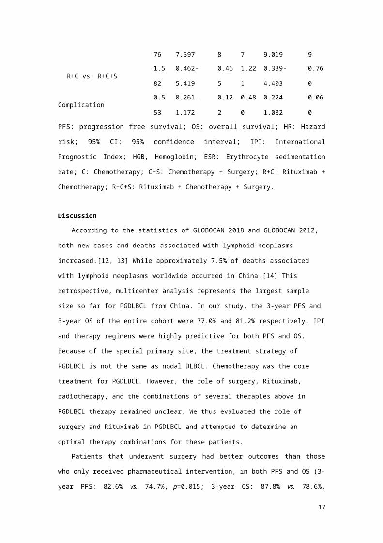
Therapy regimens 0.012 0.003
C vs. R+C+S 4.129 1.226-13.909 0.022 4.584 1.323-15.879 0.016
C+S vs. R+C+S 1.876 0.463-7.597 0.378 2.187 0.530-9.019 0.279
R+C vs. R+C+S 1.582 0.462-5.419 0.465 1.221 0.339-4.403 0.760
Complication 0.553 0.261-1.172 0.122 0.480 0.224-1.032 0.060
PFS: progression free survival; OS: overall survival; HR: Hazard risk; 95% CI: 95% confidence
interval; IPI: International Prognostic Index; HGB, Hemoglobin; ESR: Erythrocyte sedimentation rate;
C: Chemotherapy; C+S: Chemotherapy + Surgery; R+C: Rituximab + Chemotherapy; R+C+S:
Rituximab + Chemotherapy + Surgery.
12

Discussion
According to the statistics of GLOBOCAN 2018 and GLOBOCAN 2012, both new cases and
deaths associated with lymphoid neoplasms increased.[12, 13] While approximately 7.5% of deaths
associated with lymphoid neoplasms worldwide occurred in China.[14] This retrospective,
multicenter analysis represents the largest sample size so far for PGDLBCL from China. In our study,
the 3-year PFS and 3-year OS of the entire cohort were 77.0% and 81.2% respectively. IPI and therapy
regimens were highly predictive for both PFS and OS. Because of the special primary site, the
treatment strategy of PGDLBCL is not the same as nodal DLBCL. Chemotherapy was the core
treatment for PGDLBCL. However, the role of surgery, Rituximab, radiotherapy, and the combinations
of several therapies above in PGDLBCL therapy remained unclear. We thus evaluated the role of
surgery and Rituximab in PGDLBCL and attempted to determine an optimal therapy combinations for
these patients.
Patients that underwent surgery had better outcomes than those who only received pharmaceutical
intervention, in both PFS and OS (3-year PFS: 82.6% vs. 74.7%, p=0.015; 3-year OS: 87.8% vs.
78.6%, p=0.036). Rituximab could improve OS of PGDLBCL, especially in advanced-stage or high
risk (IPI 3-5) patients, while such advantage was not significant in local-stage or low risk (IPI 0-2)
cohorts. In previous studies, no difference had been observed between surgery combined with
chemotherapy and chemotherapy alone.[15-18] The value of Rituximab remained controversial. Some
of studies advocated for Rituximab in PGDLBCL,[19-22] while others held opposing views.[18, 23]
All these studies above discussed the value of either surgery or Rituximab in PGDLBCL. While, our
study for the first time compared all four commonly adopted regimens: chemotherapy alone,
chemotherapy + surgery, chemotherapy + Rituximab, and chemotherapy + surgery + Rituximab. We
found that the survival of patients in Group C+S and Group R+C were similar (3-year PFS: 81.4% vs.
81.2%, p=0.551; 3-year OS: 85.4% vs. 87.2%, p=0.948), while patients received both surgery and
Rituximab therapy (Group R+C+S) failed to show survival benefit compared to those who underwent
either surgery (Group C+S) or Rituximab therapy (Group R+C). These results suggested that
chemotherapy alone was not enough for PGDLBCL, and the clinical outcome could not be further
improved by adding either rituximab or surgery to the two combinations. Although the effect of
13

surgery and Rituximab were similar. The better therapy choice was varied from different patients.
Although we did not observe any post-surgery complications, Koch P reported that surgery increased
the risk of bleeding, perforation, and obstruction in perioperative period, leading to postpone of
chemotherapy.[24] Furthermore, late complications of surgery, such as malabsorption syndrome,
marginal ulcer, and alkaline reflux gastritis, may emerge several years later, which could cause severe
nutrition depletion and reduction in the quality of life. In light of these, our team is inclined to choose
chemotherapy combined with Rituximab as the first-line therapy for most of PGDLBCL. However,
according to our data, treatment-related complications correlated with poorer outcomes (3-year PFS:
50.9% vs. 80.2%, p<0.001; 3-year OS: 52.5% vs. 84.7%, p<0.001). If we predicted that the patients
had high risk of bleeding or perforation after receiving pharmaceutical intervention, receiving surgery
before chemotherapy maybe reduce treatment-related complications and improve survival. In that case,
chemotherapy combined with surgery could be an optimal choose.
In addition, we provided a little evidence to reveal the role of radiotherapy in PGDLBCL. In our
study, all the 24 patients receiving radiotherapy during initial treatment had at least one of the
following two situations: 1) existing residual lesion after chemotherapy ± surgery; 2) intolerance to
chemotherapy. As a result, the inclusion of radiotherapy failed to show the improvement of survival.
Besides using radiotherapy as salvage treatment, three cycles of CHOP followed by involved field
radiotherapy was used effectively and well-tolerated in localized PGDLBCL patients.[25, 26]
However, in Rituximab era, it remains unclear whether Rituximab can replace radiotherapy as a better
choice for local-stage PGDLBCL. For local-stage patients, who achieved complete remission after four
cycles of chemotherapy, consolidation radiotherapy could improve local control rate.[27] However,
this regimen did not lead to any improvement in PFS and OS. Furthermore, Japanese researchers
reported the correlation between PGDLBCL treated by radiotherapy ± chemotherapy and gastric
adenocarcinoma.[28] Randomized controlled trial was strongly needed to evaluate the necessity and
safety of radiotherapy in PGDLBCL.
Furthermore, immunotherapy has risen to the fifth main therapeutic modality for DLBCL,
following chemotherapy, targeted therapy, surgery, and radiotherapy. Monoclonal antibodies targeting
programmed death-1 (PD-1), programmed death-ligand 1 (PD-L1) and cytotoxic T-lymphocyte
antigen-4 (CTLA-4) had been used alone or in combination with anti-CD20 antibody in a subset of
14

DLBCL.[29] FDA had also approved two chimeric antigen receptor (CAR) T products for relapse and
refractory B cell lymphoma. Future administration of immunotherapy regimens will be more dependent
upon patients’ biomarkers, rather than on histological types and subtypes.[30] As a special type of
DLBCL, biomarkers unique to PGDLBCL should be actively explored, which may facilitate the
discovery of safer and more effective therapeutics for this disease.
In summary, our results suggested that the combinations of chemotherapy and surgery, or
chemotherapy and Rituximab, can raise the survival rate of PGDLBCL patients, compared to
chemotherapy alone. However, the clinical outcome could not be further improved by adding either
Rituximab or surgery to the two combinations. Rituximab showed clinical benefit in survival,
especially in advanced-stage or high risk (IPI 3-5) patients. The inclusion of radiotherapy during initial
treatment did not improve survival in our study. IPI and therapy regimens are independent predictors of
outcomes. The future prospective trial is warranted.
Acknowledgement
Jun Zhu, Xinan Cen, and Haiyan Yang designed the research; Meng Wu and Jun Zhu wrote the
article; and all authors provided study materials or patients and approved the article. We would like to
thank Medpion (Beijing) Medical Technology Co., Ltd. for their help in data collection and statistical
analysis.
Funding: This study was funded by the National Natural Science Foundation of China (No. 81870154,
81470368, 81670187, 81600164, 81700197), the Beijing Natural Science Foundation (No. 7172047,
7172046), Capital's Funds for Health Improvement and Research (No. 2018-1-2151), the Beijing
Municipal Administration of Hospitals' Ascent Plan (No. DFL20151001), the Beijing Medical and
Health Foundation (No.2221-16-157), the key topics fund of Beijing Municipal Science and
Technology Commission (No. Z141107002514017), Zhejiang medical and health science project (No.
2015KYB067), the key funding program of Zhejiang medical and health platform (No. 2015ZDA021).
Conflict of interest: The authors declare that they have no conflict of interest.
References
15

1. Al-Hamadani M, Habermann TM, Cerhan JR, Macon WR, Maurer MJ, Go RS. Non-Hodgkin
lymphoma subtype distribution, geodemographic patterns, and survival in the US: A longitudinal
analysis of the National Cancer Data Base from 1998 to 2011. Am J Hematol. 2015; 90: 790–5.
2. Castillo JJ, Winer ES, Olszewski AJ. Sites of extranodal involvement are prognostic in patients
with diffuse large B-cell lymphoma in the Rituximab era: an analysis of the Surveillance,
Epidemiology and End Results database. Am J Hematol. 2014; 89: 310-4.
3. Ferrucci PF, Zucca E. Primary gastric lymphoma pathogenesis and treatment: what has changed
over the past 10 years? Br J Haematol. 2007; 136: 521-38.
4. Aisenberg AC. Coherent view of non-Hodgkin's lymphoma. J Clin Oncol. 1995; 13: 2656.
5. Kuo SH, Cheng AL. Helicobacter pylori and mucosa-associated lymphoid tissue: what's new.
Hematology. 2013; 2013: 109.
6. Howell JM, Auergrzesiak I, Zhang J, Andrews CN, Stewart D, Urbanski SJ. Increasing incidence
rates, distribution and histological characteristics of primary gastrointestinal non-Hodgkin lymphoma
in a North American population. Can J Gastroenterol. 2012; 26: 452-6.
7. Ding W, Zhao S, Wang J, Yang Q, Sun H, Yan J, et al. Gastrointestinal Lymphoma in Southwest
China: Subtype Distribution of 1,010 Cases Using the WHO (2008) Classification in a Single
Institution. Acta Haematol. 2016; 135: 21-8.
8. Nakamura S, Matsumoto T, Iida M, Yao T, Tsuneyoshi M. Primary gastrointestinal lymphoma in
Japan: a clinicopathologic analysis of 455 patients with special reference to its time trends. Cancer.
2003; 97: 2462-73.
9. Swerdlow SH, Campo, E., Harris, N.L., Jaffe, E.S., Pileri, S.A., Stein, H., Thiele, J., Vardiman,
J.W. WHO Classification of Tumours of Haematopoietic and Lymphoid Tissues. 2008.
10. Rohatiner A, D'Amore OBOF, Coiffier B, Crowther D, Gospodarowicz M, Isaacson P, et al.
Report on a workshop convened to discuss the pathological and staging classifications of
gastrointestinal tract lymphoma. Ann Oncol. 1994; 5: 397.
11. Cheson BD, Horning SJ, Coiffier B, Shipp MA, Fisher RI, Connors JM, et al. Report of an
international workshop to standardize response criteria for non-Hodgkin's lymphomas. NCI Sponsored
International Working Group. J Clin Oncol. 1999; 17: 1244.
16
12. Bray F, Ferlay J, Soerjomataram I, Siegel RL, Torre LA, Jemal A. Global cancer statistics 2018:
GLOBOCAN estimates of incidence and mortality worldwide for 36 cancers in 185 countries. CA
Cancer J Clin. 2018; 68: 394-424.
13. Torre LA, Bray F, Siegel RL, Ferlay J, Lortet-Tieulent J, Jemal A. Global cancer statistics, 2012.
CA Cancer J Clin. 2015; 65: 87-108.
14. Liu W, Liu J, Song Y, Wang X, Zhou M, Wang L, et al. Mortality of lymphoma and myeloma in
China, 2004-2017: an observational study. J Hematol Oncol. 2019; 12: 22.
15. Willich NA, Reinartz G, Hiddemann W, Tiemann M, Kocik J, Koch P. Operative and
conservative management of primary gastrointestinal lymphoma: Interim results of a German
multicenter study. Int J Radiat Oncol Biol Phys. 2000; 46: 895-901.
16. Avilés A, Nambo MJ, Neri N, Huerta-Guzmán J, Cuadra I, Alvarado I, et al. The role of surgery
in primary gastric lymphoma: results of a controlled clinical trial. Ann Surg. 2004; 240: 44-50.
17. M B, A R-F, E L, C H, A D, P A, et al. Surgical resection plus chemotherapy versus
chemotherapy alone: comparison of two strategies to treat diffuse large B-cell gastric lymphoma. Ann
Oncol. 2003; 14: 1751-7.
18. Mehmet K, Sener C, Uyeturk U, Seker M, Tastekin D, Tonyali O, et al. Treatment modalities in
primary gastric lymphoma: the effect of Rituximab and surgical treatment. A study by the Anatolian
Society of Medical Oncology. Contemp Oncol (Pozn). 2014; 18: 273-8.
19. Avilés A, Castañeda C, Cleto S, Neri N, Huerta-Guzmán J, Gonzalez M, et al. Rituximab and
chemotherapy in primary gastric lymphoma. Cancer Biother Radiopharm. 2009; 24: 25-8.
20. Leopardo D, Lorenzo GD, Renzo AD, Federico P, Luponio S, Buonerba C, et al. Efficacy of
Rituximab in gastric diffuse large B cell lymphoma patients. World J Gastroenterol. 2010; 16: 2526.
21. Zhang J, Li G, Yang H, Liu X, Cao J. Rituximab in treatment of primary gastric diffuse large B-
cell lymphoma. Leuk Lymphoma. 2012; 53: 2175-81.
22. Chihara D, Oki Y, Ine S, Kato H, Onoda H, Taji H, et al. Primary gastric diffuse large B-cell
Lymphoma (DLBCL): analyses of prognostic factors and value of pretreatment FDG-PET scan. Eur J
Haematol. 2010; 84: 493-8.
23. Sohn BS, Kim SM, Yoon DH, Kim S, Lee DH, Kim JH, et al. The comparison between CHOP
and R-CHOP in primary gastric diffuse large B cell lymphoma. Ann Hematol. 2012; 91: 1731.
17

24. Koch P, Probst A, Berdel WE, Willich NA, Reinartz G, Brockmann J, et al. Treatment results in
localized primary gastric lymphoma: Data of patients registered within the German Multicenter Study
(GIT NHL 02/96). J Clin Oncol. 2005; 23: 7050-9.
25. Park YH, Lee SH, Kim WS, Bang SM, Ryoo BY, Yang SH, et al. CHOP followed by involved
field radiotherapy for localized primary gastric diffuse large B-cell lymphoma: Results of a multi
center phase II study and quality of life evaluation. Leuk Lymphoma. 2006; 47: 1253.
26. Ishikura S, Tobinai K, Ohtsu A, Nakamura S, Yoshino T, Oda I, et al. Japanese multicenter phase
II study of CHOP followed by radiotherapy in stage I-II, diffuse large B-cell lymphoma of the stomach.
Cancer Sci. 2005; 96: 349-52.
27. Li Q, Li W, Wang L, Wang W, Niu S, Bi X, et al. Consolidation Radiotherapy in Stage IE- IIE,
Non-Bulky Primary Gastric Diffuse Large B-Cell Lymphoma with Post-Chemotherapy Complete
Remission. PLoS One. 2015; 10: e0133469.
28. Inaba K, Kushima R, Murakami N, Kuroda Y, Harada K, Kitaguchi M, et al. Increased risk of
gastric adenocarcinoma after treatment of primary gastric diffuse large B-cell lymphoma. BMC
Cancer. 2013; 13: 499.
29. Ok CY, Young KH. Checkpoint inhibitors in hematological malignancies. J Hematol Oncol.
2017; 10: 103.
30. Li Z, Song W, Rubinstein M, Liu D. Recent updates in cancer immunotherapy: a comprehensive
review and perspective of the 2018 China Cancer Immunotherapy Workshop in Beijing. J Hematol
Oncol. 2018; 11: 142.
18


![Research Article Palliative Reconstructive Surgery ...downloads.hindawi.com/archive/2014/275215.pdf · care, communication, decision making, and end-of-life care [ , ]. Palliative](https://static.fdocuments.us/doc/165x107/602265810cdf4609f317cf2b/research-article-palliative-reconstructive-surgery-care-communication-decision.jpg)