WaterSHED STROKES - American Heart Association
31
WATERSHED STROKES KARIN OLDS MD SLMBNI STROKE MEDICAL DIRECTOR UMKC ASSISTANT PROFESSOR
Transcript of WaterSHED STROKES - American Heart Association

WATERSHED STROKES KARIN OLDS MD
SLMBNI STROKE MEDICAL DIRECTOR
UMKC ASSISTANT PROFESSOR

• I have no disclosures

BORDER ZONE OR WATERSHED INFARCTS ARE ISCHEMIC LESIONS THAT OCCUR IN CHARACTERISTIC LOCATIONS AT THE JUNCTION BETWEEN TWO MAIN ARTERIAL TERRITORIES. THEY COMPRISE APPROXIMATELY 10% OF ALL ISCHEMIC STROKES
Internal Border Zone Infarcts
• The internal or subcortical border zones are located at the junctions of the anterior, middle and posterior cerebral artery territories with the Recurrent Artery of Heubner, lenticulostriateand anterior choroidal artery territories
External Border Zone Infarcts
• The external or cortical border zones are located at the junctions of the anterior, middle and posterior cerebral artery territories

External border zones—light blueInternal border zones—red

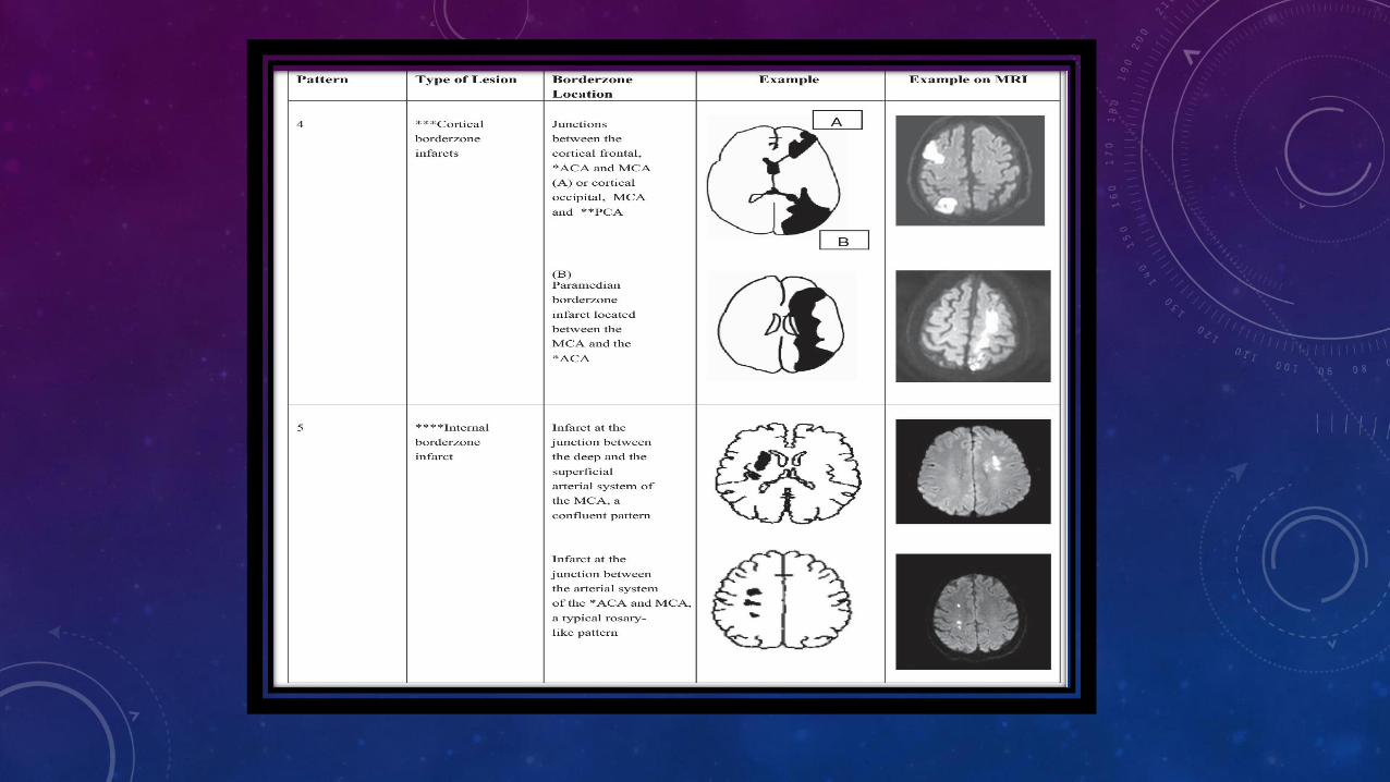
INTERNAL BORDER ZONE INFARCTS
• Often manifests as a series of 3 or more lesions, each with a diameter of 3 mm or more, arranged in a linear fashion parallel to the lateral ventricle in the centrum semiovale or corona radiata (rosary/bead)
• Can be confluent or partial; perhaps a briefer episode of hemodynamic compromise leads to a partial infarct, while a confluent infarct may be manifested by step-wise hemiplegia related to a longer period of hemodynamic compromise
• Caused mainly by arterial stenosis or occlusion, usually paired with hemodynamic compromise
• Thought to be much less likely than external border zone infarcts to be secondary to emboli
• Often associated with a more poor prognosis and clinical deterioration when compared with external border zone infarcts


EXTERNAL BORDER ZONE INFARCT
• Typically wedge shaped or ovoid shaped, size is variable because of anatomic variation and minimal/maximal distribution of each of the large vessel territories (MCA, ACA, PCA) from person to person
• Also may vary secondary to the development of leptomeningeal collateral vessels
• Often caused by hemodynamic compromise and may pair with moderate or severe narrowing of the carotid or proximal cerebral arteries
• As opposed to internal border zone infarcts, microemboli from the heart or atheroembolic events can propagate to the cortical border zones; these areas have a lower perfusion than more proximal regions, so less ability to “wash out” the microemboli
• Recent intraplaque hemorrhage/plaque rupture may play a role
• Patients often have a more benign clinical course and better prognosis than those with internal border zone infarcts, especially if unilateral or only in one border zone


BORDER ZONE INFARCTS IN THE CEREBELLUM
• These are usually < 2 cm in size and are seen at the borders of the AICA, SCA and PICA (and their branches)
• Due to stenosis or embolism. Embolic events can come from the heart or atherosclerotic disease
• May be see with vertebral dissections
• Typically not disabling in an of themselves, but often coexist with large territorial lesions
• They can be silent if small, or similar in presentation to larger cerebellar infarcts, with vertigo and ataxia


Red is AICADark Green is lateral SCALight Green is medial SCADark Blue is lateral PICALight Blue is medial PICA


STAGES OF HEMODYNAMIC IMPAIRMENT
• In stage I impairment, a decline in cerebral perfusion pressure leads to autoregulatory vasodilatation of resistive vessels of the brain
• This can be measured with various methods, including xenon-enhanced CT, CT perfusion, MR perfusion, SPECT and PET
• Mainly, the modalities measure cerebral blood flow, cerebral blood volume and mean transit time
• To measure cerebrovascular reactivity, cerebral blood flow measurements can be repeated after a vasodilatory challenge
• Normally, an increase in CBF is expected; however, with hemodynamic compromise, the CBF is often not increased because autoregularotion has already caused maximum vasodilatation in response to decreased CPP

STAGES OF HEMODYNAMIC IMPAIRMENT
• In stage II, further reduction in CPP causes inadequate autoregulatory vasodilation, and CBF decreases
• As blood flow decrease, the oxygen extraction fraction in the brain may increase, and can be measure with PET
• The increased oxygen extraction fraction in the ischemic region has been described as misery perfusion, where the metabolic demand of tissue is greater than its blood supply
• Patients with misery perfusion, which is a significant disturbance of cerebral autoregulation, have a significantly higher stroke recurrence ratio than patients without misery perfusion

STAGES OF HEMODYNAMIC IMPAIRMENT


WATERSHED STROKES AFTER CARDIAC SURGERY
• Watershed distribution strokes are seen more frequently in patients with postcardiac surgery stroke than in the general population (> 40% vs 2-5%)
• These patients are more likely to require long-term care than other postcardiac surgery stroke patients
• Probably involves a combination of hypoperfusion and embolization
• In cardiac surgery, global systemic hypoperfusion caused by severe intraoperative hypotension is known to be associated with poor outcomes
• One randomized trial showed improved neurological and cardiac outcome in patients with MAP maintained at 80 to 100 mm Hg vs 50 to 60 mm Hg

WATERSHED STROKES AFTER CARDIAC SURGERY
• One study done at John Hopkins followed patients who underwent cardiac surgery between 1998 and 2003 and developed a focal neurologic deficit post operatively. Data was reviewed for patients with watershed infarctions
• Intraoperative blood pressure was defined as blood pressure while on CPB, so patients who underwent off-pump CABG were excluded from the part of the study involving BP measurements
• Only those patients who had MRI/DWI imaging were included, although patients also underwent CT
• Standard MAP goal during CPB at John Hopkins is (was) 60 to 80 mm Hg
• 91 patients were included, with 5 patients excluded that underwent off-pump CABG and 2 that developed symptoms over 10 days post-operatively

WATERSHED STROKES AFTER CARDIAC SURGERY
• Patients with bilateral watershed infarctions were more likely to have undergone an aortic procedure and less likely to have undergone a simple or redo CABG
• Patients with bilateral watershed infarcts were 6.23 times as likely to be discharged to an ARF, 12. 46 times more likely to be discharged to subacute rehab or to SNF, and 17.28 times more likely to die in the hospital than be discharged home
• Patients with a length of stay > 14 days were more likely to have bilateral watershed infarcts than other stroke patterns
• Patients with a drop in MAP or at least 10 mm Hg were 4.06 times (adjusted OR: 95% CI: 1.05, 15.98) as likely to develop bilateral watershed strokes as those patienrs who had a smaller drop or no drop in bp
• Data showed that bilateral watershed strokes were more readily detected by DWI MRI (than CT) and were associated with poor short-term outcomes, and MAY have been related to a decrease in intraoperative blood pressure from a preoperative baseline

DEFICITS/FINDINGS RELATED TO LOCATION(S)
• Deficits are relative to stroke burden and locations
• Internal border zone infarcts, especially if more “cigar” shaped or multiple “beads on a string”, tend to do worse then isolated cortical border zone infarcts
• Bilateral infarcts also tend to do worse
• “Man in a Barrel” syndrome can occur if bilateral MCA/ACA cortical infarcts are extensive: proximal > distal UE weakness, inability to abduct arms; bilateral brachial weakness, can spare face, legs
• Balint’s syndrome can be caused by bilateral posterior MCA/PCA border zone infarcts
• 3 classic findings are simultanagnosia, optic ataxia and oculomotor apraxia—severe visual-spatial abnormalities


CASE STUDY
• 69 yo male presenting with chest pain radiating to left jaw, down left arm and increasing SOA x 1 week
• Chest pain not relieved with nitro; also, nitro caused N/V
• Further history revealed he had been sleeping in a recliner for 2 weeks
• Wife reported confusion, which was very unusual, as well as increasing fatigue and easy bruising
• PMH significant for ischemic cardiomyopathy, CAD, s/p STEMI, PAD, PVD, OSA/CSA, CKD, DM, hxtn, bilateral carotid artery disease, s/p stents bilaterally at OSH
• Initial labs revealed glucose 124, remainder of CMP normal, troponin 0.03, Hgb 6.9, hct 21, WBC 1.7, Plt37—significant thrombocytopenia

CASE STUDY
• Troponins elevated to 2, No ST elevation/depression
• Hgb worsened, pt transfused, nephrology, GI medicine, cardiology and Heme/onc all consulted
• Day 5, pt noted to be more somnolent, slurred speech, neurology consulted
• On exam, he was somnolent, clearly encephalopathic, no definite vision change, but right facial droop and subtle right sided weakness (superimposed on notable generalized weakness)
• Review of BPs over the previous 24 hours with SBP as low as 71, transiently, but often in the 90s
• MRI obtained. Pt continued to be markedly pancytopenic. Also ordered carotid Doppler/US
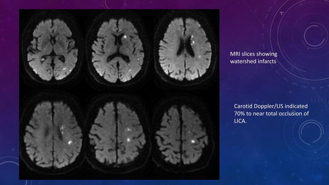
MRI slices showing watershed infarcts
Carotid Doppler/US indicated 70% to near total occlusion of LICA.

CASE STUDY
• The patient was recommended to transfuse to Hgb 9 or greater—both for neurologic and cardiologic reasons
• Recommend keep SBP > 110
• He underwent bone marrow biopsy prior to neuro consult—preliminary findings of myelodysplastic syndrome, 6-7% blasts
• He was improved significantly in regards to his encephalopathy and his right sided weakness within 2 days, but does continue to wax and wane in regards to his mental status. BP has mostly been > 110, and HGB > 8 with repeated transfusions

CONCLUSIONS
• Watershed or border zone infarcts are much more common than initially thought
• In addition to hemodynamic compromise, micro emboli likely play a role, especially in external or cortical border zone infarcts
• Internal border zone infarcts may be difficult to distinguish from other entities; chronically, WM lesions from demyelination, and when minimal, often thought to be “lacunar” infarcts
• Cerebellar border zone infarcts are also possible, and probably more common than initially recognized
• Hence, large vessel imaging is extremely important in ALL ischemic stroke work ups
• Longer times of hemodynamic compromise often leads to more severe stroke deficits
• Neurologic exams and, if focal deficits are found, MRI imaging are important post cardiac surgery, or any prolonged surgery with drop in MAP from baseline
• Quick hemodynamic support is imperative for good outcomes in cases where this is a problem

REFERENCES
• Google images
• Momijan-Mayor MD, Isabelle and Baron MD, FRCP, Jean-Claude, “The Pathophysiology of Watershed Infarction in Internal Carotid Artery Disease”, STROKE, 2005/https://doi.or/10.1161/01.STR.0000155727.82242.e1/Stroke. 2005; 36: 567-577.
• Mangla, MD, Rajiv, Kolar, MD, Balasubramanya, Almast, MD, Jeevak, Ekholm, MD, PhD, Sven E. “Border Zone Infarcts: Pathyophysiologic and Imaging Characteristics.” Radiographics.rsna.org. September-Octtober, 2011. 1201-1215.
• Gottesman, MD, Rebecca F., et al. “Watershed Strokes After Cardiac Surgery: Diagnosis, Etiology and Outcomes.” STROKE. DOI: 10.1161/01.STR.00002360424.68020.3a. 2306-2311.

Thank You!