Volume 6 Number 9 September 2015 Pages 5095–5312 …
9
Chemical Science www.rsc.org/chemicalscience ISSN 2041-6539 EDGE ARTICLE Ian F. Hermans, Gavin F. Painter et al. NKT cell-dependent glycolipid–peptide vaccines with potent anti-tumour activity Volume 6 Number 9 September 2015 Pages 5095–5312
Transcript of Volume 6 Number 9 September 2015 Pages 5095–5312 …

ChemicalSciencewww.rsc.org/chemicalscience
ISSN 2041-6539
EDGE ARTICLEIan F. Hermans, Gavin F. Painter et al.NKT cell-dependent glycolipid–peptide vaccines with potent anti-tumour activity
Volume 6 Number 9 September 2015 Pages 5095–5312

ChemicalScience
EDGE ARTICLE
Ope
n A
cces
s A
rtic
le. P
ublis
hed
on 2
5 Ju
ne 2
015.
Dow
nloa
ded
on 1
1/28
/202
1 2:
30:2
5 PM
. T
his
artic
le is
lice
nsed
und
er a
Cre
ativ
e C
omm
ons
Attr
ibut
ion-
Non
Com
mer
cial
3.0
Unp
orte
d L
icen
ce.
View Article OnlineView Journal | View Issue
NKT cell-depend
aThe Ferrier Research Institute, Victoria Univ
Hutt 5046, New Zealand. E-mail: gavin.painbMalaghan Institute of Medical Research,
Zealand. E-mail: [email protected] of Biological Sciences, The Universi
Central, 1142, New ZealanddMaurice Wilkins Centre for Molecular Bio
Symonds St, Auckland Central, 1142, New ZeDepartment of Chemistry, University of O
ZealandfDepartment of Pathology & Molecular Medi
ZealandgSchool of Biological Sciences, Victoria U
Wellington 6140, New Zealand
† Electronic supplementary informa10.1039/c4sc03599b
‡ These authors contributed equally to th
Cite this: Chem. Sci., 2015, 6, 5120
Received 20th November 2014Accepted 25th June 2015
DOI: 10.1039/c4sc03599b
www.rsc.org/chemicalscience
5120 | Chem. Sci., 2015, 6, 5120–5127
ent glycolipid–peptide vaccineswith potent anti-tumour activity†
Regan J. Anderson,‡a Benjamin J. Compton,‡a Ching-wen Tang,b Astrid Authier-Hall,b Colin M. Hayman,a Gene W. Swinerd,bg Renata Kowalczyk,c Paul Harris,c
Margaret A. Brimble,cd David S. Larsen,e Olivier Gasser,b Robert Weinkove,bf
Ian F. Hermans*bdg and Gavin F. Painter*a
It is known that T cells can eliminate tumour cells through recognition of unique or aberrantly expressed
antigens presented as peptide epitopes by major histocompatibility complex (MHC) molecules on the
tumour cell surface. With recent advances in defining tumour-associated antigens, it should now be
possible to devise therapeutic vaccines that expand specific populations of anti-tumour T cells. However
there remains a need to develop simpler efficacious synthetic vaccines that possess clinical utility. We
present here the synthesis and analysis of vaccines based on conjugation of MHC-binding peptide
epitopes to a-galactosylceramide, a glycolipid presented by the nonpolymorphic antigen-presenting
molecule CD1d to provoke the stimulatory activity of type I natural killer T (NKT) cells. The chemical
design incorporates an enzymatically cleavable linker that effects controlled release of the active
components in vivo. Chemical and biological analysis of different linkages with different enzymatic
targets enabled selection of a synthetic vaccine construct with potent therapeutic anti-tumour activity in
mice, and marked in vitro activity in human blood.
Introduction
With the recent regulatory approval of a cancer vaccine forprostate cancer1 and the clinical success of antibodies thatcause blockade of immune checkpoints in T cells,2 immuno-therapies that stimulate T cell function are now emerging ascredible treatment options for patients with advanced malig-nancies. However, despite cancer vaccines having the potentialto induce T cell responses with high specicity for denedtumour-associated antigens, in general they have not yet real-ised this promise, and issues remain around potency, lack of
ersity of Wellington, PO Box 33436, Lower
PO Box 7060, Wellington 6242, New
z
ty of Auckland, 3 Symonds St, Auckland
discovery, The University of Auckland, 3
ealand
tago, PO Box 56, Dunedin 9054, New
cine, University of Otago Wellington, New
niversity of Wellington, PO Box 600,
tion (ESI) available. See DOI:
is work.
clinical efficacy,3,4 manufacturing and cost. Therefore newscalable strategies for the production of efficacious cancervaccines are still needed. Synthetic vaccines based on antigenicpeptides are highly dened and easily manufactured. However,in order to generate a robust anti-tumour response it isimportant not only that peptide fragments are acquired byantigen presenting cells (APCs) and presented to T cells in thecontext of MHC molecules, but also that the APCs are in thecorrect activation state.5 This can be achieved by using adju-vants that target pattern-recognition receptors on APCs, such asthe Toll-like receptors (TLRs)6 or selected C-type lectin recep-tors.7 Advancing this idea further, conjugation of antigensdirectly to TLR ligands has been shown to achieve appropriatetargeting to APCs as well to induce their activation, leading toenhanced stimulation of peptide-specic cytotoxic T-lympho-cytes (CTLs).8,9 A less explored approach to activating APCs is tospecically stimulate accessory cells in the local environment.In animal models it has been shown that compounds thatstimulate innate-like T cells, a class of semi-activated T cellswith an invariant T cell receptor (TCR), can serve as powerfulimmune adjuvants when delivered with antigens.10,11 The moststudied innate-like T cells are the invariant natural killer T(NKT) cells, which recognise glycolipid antigens that bind to thelipid antigen-presenting molecule CD1d.12,13 A number ofnatural and synthetic CD1d ligands have been dened, of whichthe most studied is a-galactosylceramide (a-GalCer, KRN7000),a synthetic compound discovered through SAR studies on a
This journal is © The Royal Society of Chemistry 2015

Fig. 1 Candidate vaccine structures.
Scheme 1 Isolation of amine 1.
Edge Article Chemical Science
Ope
n A
cces
s A
rtic
le. P
ublis
hed
on 2
5 Ju
ne 2
015.
Dow
nloa
ded
on 1
1/28
/202
1 2:
30:2
5 PM
. T
his
artic
le is
lice
nsed
und
er a
Cre
ativ
e C
omm
ons
Attr
ibut
ion-
Non
Com
mer
cial
3.0
Unp
orte
d L
icen
ce.
View Article Online
class of glycolipid originally isolated from a marine sponge.14
Interestingly, similar compounds containing the a-galactosyllinkage have recently been found in mammalian tissue.15 Whenadministered with peptide antigens, a-GalCer provokes NKTcell-dependent activation of APCs, mediated by CD40-CD40Linteractions, which in turn licences the APCs to stimulatesuperior peptide-specic T cell responses.10 We, and others,hypothesised that stronger responses could be obtained byconjugation of peptides to a-GalCer, as this would providefocussed delivery of antigen and adjuvant to the same cell.16,17
Crucial to the strategy was inclusion of an enzymatically cleav-able linker to provide controlled release of the active compo-nents in vivo. Having recently described the rst example of avaccine of this type with an esterase-cleavable linker,16 and itsapplication in the suppression of allergic airway inammation,we now report that the same type of construct can be used topromote an immune response against established tumours inmice. We describe the synthesis and biological analysis of anexpanded series of conjugates with linkers that are susceptibleto either esterase or protease activity. The aim was to select avaccine construct that could be produced in a facile syntheticprocess and that has therapeutic anticancer activity. In addi-tion, we examined whether these conjugates activate humanNKT cells and enhance peptide-specic CTL responses inhuman blood in vitro.
A key consideration in the construction of conjugates wasthe site of conjugation of the antigen component to a-GalCer. Asdetailed below, we used a rearranged a-GalCer derivative with afree amino group to provide a synthetically selective and stablepoint of attachment. Also of consideration in conjugate designwas the type of self-immolative linker joining the two compo-nents, which would inuence serum stability and the release ofthe active components in vivo. This would largely be inuencedby the enzymatic activity to be exploited, which ideally would beenhanced within APCs. Of the many self-immolative linkersdescribed in the literature or used in approved pharmaceuti-cals, we chose to examine esterase-sensitive acyloxymethylcarbamates18 and protease-sensitive valine–citrulline–p-amino-benzyl (VC–PAB) carbamates19 in this series of conjugates. Wehave previously shown that conjugates incorporating the acy-loxymethyl carbamate group were able to focus activity on
This journal is © The Royal Society of Chemistry 2015
splenic CD8a+ dendritic cells (DCs), which are APCs that effi-ciently cross-present antigens to stimulate CTLs.16 The VC–PABgroup is a known substrate20,21 for lysosomal proteases,including cathepsin B which is abundant in APCs.22 A thirdconsideration in conjugate design was the choice of chemistrywith which to attach the peptide moiety, as this has been shownto have an impact on the immune response.23 We investigatedoxime ligation and copper(I)-catalyzed azide–alkyne cycloaddi-tion (CuAAC), both of which, although well-established,required considerable optimization in our system. For analysisof T cell responses, initial conjugates in this series contained animmunodominant CD8 epitope from ovalbumin (OVA257). Thiswas anked at the N-terminus by the cleavage sequence Phe–Phe–Arg–Lys, as the extra proteolytic step required for itsremoval has been shown to support cross-presentation ofpeptides onto MHC class I molecules for stimulation of CTLs.24
With these considerations in mind we undertook the synthesisof vaccine candidates 2–5 (Fig. 1).
Results and discussion
Our vaccine design stems from an unexpected nding during anearlier unpublished synthesis of a-GalCer. Hydrogenolytic
Chem. Sci., 2015, 6, 5120–5127 | 5121

Fig. 2 Vaccine design and in vivo metabolism.
Scheme 3 Synthesis of VC–PAB carbamates 17 and 18.
Chemical Science Edge Article
Ope
n A
cces
s A
rtic
le. P
ublis
hed
on 2
5 Ju
ne 2
015.
Dow
nloa
ded
on 1
1/28
/202
1 2:
30:2
5 PM
. T
his
artic
le is
lice
nsed
und
er a
Cre
ativ
e C
omm
ons
Attr
ibut
ion-
Non
Com
mer
cial
3.0
Unp
orte
d L
icen
ce.
View Article Online
deprotection of a late-stage intermediate,§ using Pd(OH)2/C inCHCl3/MeOH, led, in addition to the expected product a-GalCer,to signicant quantities of a more polar product that wasdetermined by 2D-NMR and mass spectral analysis to be amine1 (Scheme 1). Although intramolecular N / O migrations ofacyl groups are known in the literature they are usuallypromoted in strongly acidic media.25–27 We reasoned that, in thepresent case, HCl was produced from the solvent CHCl3 underthe hydrogenolytic conditions, leading to the observed migra-tion. A control experiment showed that, under similar reactionconditions but in the absence of a H2-atmosphere, a-GalCer didnot isomerise to amine 1. Although the formation of HCl fromCHCl3 by Pd-catalysed hydrogenolysis has been reported,28,29 itsuse (deliberate or otherwise) to isomerise amides to esters hasnot. Indeed, CHCl3 has been successfully used as a co-solvent inthe nal deprotection step (hydrogenolysis) of other synthesesof a-GalCer or analogues thereof, with no report of acyl-migra-tion side-reactions.30–35
Compound 1 was shown to readily revert to a-GalCer underaqueous conditions,16 suggesting that capping of the aminogroup with enzymatically-cleavable linkers could generate aninteresting new class of a-GalCer prodrugs, or “pro-adjuvants”.In particular, attachment of peptide antigens to 1 could lead to anew class of synthetic vaccines (structure (I), Fig. 2) that exploitthe action of NKT cells in stimulating an antigen-specicimmune response. Enzymatic cleavage of the self-immolativelinker would give metabolites 1 and (II); further enzymaticprocessing of the peptide (II) would provide an epitope that canbe presented on MHC class I, while O / N acyl transfer of 1would produce a-GalCer, which is presented on CD1d.
Scheme 2 Synthesis of acyloxymethyl carbamates 10 and 11.
5122 | Chem. Sci., 2015, 6, 5120–5127
Synthesis
We previously reported16 the synthesis of conjugate 2 howeverwe discuss here pertinent details of the linker synthesis andconjugation chemistry which were not previously disclosed,together with the synthesis of new targets 3–5. Conjugateswere constructed from three components: 1; a linker with areactive functional group for chemoselective ligation (either aketone for oxime ligation, or an azide for CuAAC); and asynthetic peptide with a complementary N-terminal functionalgroup. Compound 1 was efficiently obtained by the intentionalacid-catalysed migration of the cerotic acid moiety from thephytosphingosine nitrogen to the 40-OH group as reportedpreviously.16 The synthesis of linkers for attachment to amine1 is shown in Schemes 2 and 3. Acyloxymethyl carbonatelinkers 7 and 9, containing keto and azido groups, respec-tively, were produced by alkylation of the appropriate acid withiodomethyl carbonate 8. The silver salt of the acid was formedin situ by the inclusion of silver(I) oxide (0.6 equiv.) in thereaction (Scheme 2). The inclusion of molecular sieves in bothreactions improved product yields by preventing hydrolysis atthe ester and/or carbonate groups. Carbonates 7 and 9 werethen reacted with 1 in pyridine to afford ketone 10 (ref. 16) andazide 11, respectively. A small amount of a-GalCer was typi-cally formed in these reactions, which could be removed bycareful chromatography.
This journal is © The Royal Society of Chemistry 2015
Scheme 4 Synthesis of conjugate vaccines 2 and 3.
Edge Article Chemical Science
Ope
n A
cces
s A
rtic
le. P
ublis
hed
on 2
5 Ju
ne 2
015.
Dow
nloa
ded
on 1
1/28
/202
1 2:
30:2
5 PM
. T
his
artic
le is
lice
nsed
und
er a
Cre
ativ
e C
omm
ons
Attr
ibut
ion-
Non
Com
mer
cial
3.0
Unp
orte
d L
icen
ce.
View Article Online
The VC–PAB linkers were produced by acylation of knownamine 12 (ref. 20) with levulinic acid or 6-azidohexanoic acidemploying the mixed anhydride method (Scheme 3). Theresulting alcohols 13 and 14 were converted to their p-nitro-phenyl (pNP) carbonates 15 and 16, and subsequently reactedwith 1 in pyridine with triethylamine to afford ketone 17 andazide 18.
Although widely used, aminooxy-terminated peptides can bedifficult to obtain in pure form due to side reactions and puri-cation difficulties, as reported in the literature36–38 and illus-trated by our own difficulties encountered in trying to obtainsufficient quantities of peptide 19 from commercial sources.Instead, employing an Fmoc solid phase peptide synthesisstrategy utilising an in-house manufactured aminomethylpolystyrene resin gave sufficient quantities of 19 without inci-dent. Ketones 10 and 17were reacted with aminooxy-terminatedpeptide 19 containing the OVA257 epitope SIINFEKL (Scheme 4).The disparate solubility properties of the glycolipid and peptidereaction partners necessitated a peculiar solvent cocktail at highdilution (2 mM in ketone). Preliminary experiments gave verypoor conversion, even with catalysis by aniline (pH 4.7,20 mM).39 Quenching of the aminooxy group with solvent-derived low-Mw aldehydes/ketones was a problem at highdilution, which could be minimised by distilling solvents from2,4-dinitrophenylhydrazine before use, however reactions were
Scheme 5 Synthesis of conjugate vaccines 4 and 5.
This journal is © The Royal Society of Chemistry 2015
still unacceptably slow. Conversion of ketone 17 was 63% aer 1day at 25 �C, while in the case of 10, the major reaction pathwaywas hydrolysis of the levulinoyl ester, leading to the production of1 and formaldehyde, with consequent quenching of the amino-oxy group. Conversion of 17 improved to 92% when using 100mM aniline, and by reducing the pH to�4.3 the ester cleavage incompound 10 was almost completely eliminated. A small excessof peptide (0.3 equiv.) was sufficient to achieve >95% conversionof the ketone aer 2 days at 25 �C affording conjugates 2 (ref. 16)and 3 in >95% purity aer preparative HPLC.
Azides 11 and 18 were reacted with alkynyl-terminatedpeptide 20 in the presence of CuSO4, a reducing agent and tris[(1-benzyl-1H-1,2,3-triazol-4-yl)methyl]amine (TBTA) ligand(Scheme 5).40 Again, the solubility properties of the reactionpartners dictated an unusual solvent combination consisting of3 : 3 : 3 : 1 DMSO–chloroform–methanol–water. While no reac-tion was observed using the standard ascorbate reducing agent,a small piece of copper foil in the reaction mixture generatedsufficient Cu(I) for the reaction to proceed to completion aer24 h at 20 �C.41{ In the absence of TBTA, under otherwisesimilar conditions, conversion was approximately 30% aerthree days. The proportion of DMSO was kept at no more than30% due to its reported40 propensity to counteract the acceler-ating effect of TBTA at high concentration – a phenomenon wealso observed. Using an initial azide concentration of�3mM, atleast 1.5 equiv. of peptide was required to achieve full conver-sion within a reasonable timeframe (24 h). Longer reactiontimes as necessitated by using less peptide, removing TBTA orincreasing the DMSO concentration all resulted in formation ofclose-running impurities as observed by HPLC. Although wehave not been able to fully characterise these by-products,LCMSMS data suggests the occurrence of side-reactions withinthe peptide chain – an interesting nding in this normally verytolerant click reaction, especially considering that this peptidedoes not contain any of the usual reactive suspects (e.g. Cys,Met, Trp) in its sequence. Aer preparative HPLC, conjugates 4and 5 were obtained in 44% and 46% yields in high purity(>95%).
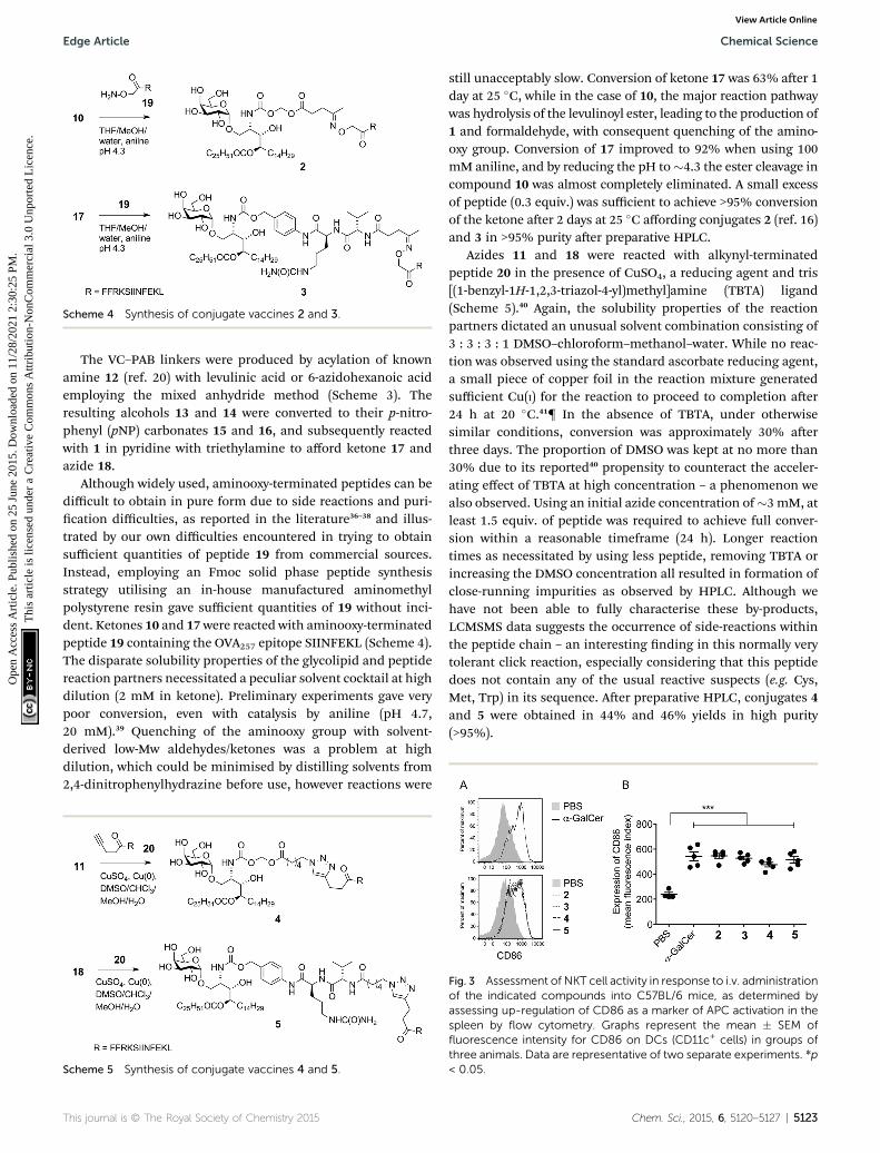
Fig. 3 Assessment of NKT cell activity in response to i.v. administrationof the indicated compounds into C57BL/6 mice, as determined byassessing up-regulation of CD86 as a marker of APC activation in thespleen by flow cytometry. Graphs represent the mean � SEM offluorescence intensity for CD86 on DCs (CD11c+ cells) in groups ofthree animals. Data are representative of two separate experiments. *p< 0.05.
Chem. Sci., 2015, 6, 5120–5127 | 5123

Chemical Science Edge Article
Ope
n A
cces
s A
rtic
le. P
ublis
hed
on 2
5 Ju
ne 2
015.
Dow
nloa
ded
on 1
1/28
/202
1 2:
30:2
5 PM
. T
his
artic
le is
lice
nsed
und
er a
Cre
ativ
e C
omm
ons
Attr
ibut
ion-
Non
Com
mer
cial
3.0
Unp
orte
d L
icen
ce.
View Article Online
To gauge the stability of the linkers in an aqueous medium,we developed an HPLC-MSMS method to detect and quantifylow-level formation of a-GalCer which would be expected aerany background hydrolysis leading to linker collapse. We havealready shown in previous work that once the phytosphingosineamino group is revealed formation of a-GalCer is facile in anaqueous system.16 Aqueous samples at pH 3, 5, 7 and 9 wereincubated at 20 �C and monitored periodically for a-GalCer overeight days. Compounds 2 and 4 generated low levels of a-GalCerat physiological and alkaline pH (Table 1, ESI†). No formation ofa-GalCer was detected (detection limit ¼ 0.05%) over this timefor conjugates 3 and 5 at all pH values indicating greaterstability of the VC–PAB carbamate-containing conjugates.
Biological analysis
To ensure that the different enzymatic cleavage sites enabledrelease of a CD1d-binding NKT cell agonist, we tested vaccines2–5 for their induction of NKT cell activity. By comparing thesefour compounds, we were able to independently assess theimpact of the enzymatic cleavage group (esterase-labile in 2 and4, versus protease-labile in 3 and 5) and the conjugationchemistry employed (oxime ligation in 2 and 3, versus CuAAC in4 and 5). The ability of the vaccines to activate NKT cells in micein vivo was determined indirectly by assessing NKT cell-medi-ated activation of DCs in the spleen by ow cytometry.11 Usingup-regulation of the activation marker CD86 as a readout, allfour vaccines induced marked DC activation, resulting insimilar levels of CD86 expression to that seen aer injection offree a-GalCer (Fig. 3). These results suggest that in vivo cleavageof the linkers in each case releases a-GalCer aer eliminationand rearrangement. While we cannot rule out some intrinsicagonist activity of the intact conjugates, or of cleaved
Fig. 4 Conjugated peptide vaccines stimulate peptide-specific cyto-toxic immune responses irrespective of linker design. Assessment ofthe cytotoxic response was assessed seven days after administration ofthe indicated compounds compared to administration of unconju-gated components (a-GalCer and OVA257 peptide). Cytolytic activitywas assessed on injected populations of fluorescent syngeneic sple-nocytes that had been loaded with different doses of the targetedOVA257 peptide. An additional population of fluorescent splenocyteswithout peptide served as internal negative control. Lysis was assessedby flow cytometry of blood, and is expressed as percent reduction inpeptide loaded cells relative to unloaded control. Data from five miceper group are shown with mean percentage of specific lysis � SEMindicated. (A): Assessment of 2–5; there were no significant differ-ences in activity between treatment groups. (B): Assessment in wild-type versus CD1d-deficient hosts. Mean percent lysis observed afterinjection of vaccines in wild-type hosts was significantly enhancedover injection of admixed components, regardless of peptide dose ontargets (p < 0.001).
5124 | Chem. Sci., 2015, 6, 5120–5127
intermediates, it is pertinent that the N-acetyl derivative of 1,which is unable to collapse to a-GalCer, does not activate NKTcells.16
We next tested vaccines 2–5 in an in vivo assay of cytotoxicactivity against peptide-loaded target cells. Each of the fourvaccines induced signicantly increased antigen-specic T cellcytotoxicity compared to the unvaccinated control, with nonotable differences in levels of cytolytic activity between thedifferent conjugates (Fig. 4A). These results indicate that all ofthe vaccines were able to efficiently release the MHC class I-binding peptide epitope and induce a peptide-specic CTLresponse, and the design of linker had little impact on biolog-ical activity. The importance of conjugation is further high-lighted by comparing these results to those obtained fromtreatment with the admixed unconjugated vaccine components(i.e. a-GalCer and OVA257 peptide) at equivalent molar concen-trations. Compared to each of the vaccines injection with theadmixture resulted in no detectable lysis of target cells (Fig. 4B).Additional evidence that CTL activity was mediated through theNKT cell pathway is provided by the observed loss of activity ofthe vaccines in CD1d-decient mice (Fig. 4B).
To assess the impact of conjugation of the glycolipid onantigen presentation, equivalent molar doses of the free peptideepitope OVA257 and N-terminally capped peptide 21 (see ESI†for structure and synthesis) were cultured with splenocytes fromOT-I mice, which express a transgenic TCR for the OVA257
epitope but harbour no NKT cells. Analysis of T cell prolifera-tion in this assay, where there is no adjuvant activity, provides asensitive measure of presentation of the minimal peptideepitope. Peptide 21 contains the Phe–Phe–Arg–Lys cleavagesequence used in all four vaccines and is therefore required tobe processed before MHC class I presentation, however it lacks
Fig. 5 Conjugation alters peptide presentation on MHC molecules invitro. (A): Graded doses of OVA257 peptide, peptide 21 or conjugates 2,3, 4 or 5were cultured with splenocytes fromOT-I mice, and then 72 hlater, proliferationwas assessed by uptake of 3H thymidine over the last8 h of culture. (B) and (C): Graded doses of OVA257 peptide orconjugate 5 were cultured with DC2114 dendritic cells and thenexpression of H-2Kb/OVA257 complexes determined on the cellsurface by flow cytometry 6, 12 and 24 h later. Representative flowcytometry histograms showing binding of anti-H-2Kb/OVA257 24 hafter pulsing with 5.7 mM OVA257 peptide, or 5.7 mM conjugate 5, areshown in (B), and graphs showing binding at the indicated times afterculture with graded doses are shown in (C).
This journal is © The Royal Society of Chemistry 2015

Edge Article Chemical Science
Ope
n A
cces
s A
rtic
le. P
ublis
hed
on 2
5 Ju
ne 2
015.
Dow
nloa
ded
on 1
1/28
/202
1 2:
30:2
5 PM
. T
his
artic
le is
lice
nsed
und
er a
Cre
ativ
e C
omm
ons
Attr
ibut
ion-
Non
Com
mer
cial
3.0
Unp
orte
d L
icen
ce.
View Article Online
any glycolipid moiety.24 This requirement for additional pro-cessing caused a reduction in peptide presentation relative tocultures containing free OVA257 (Fig. 5A), although it is not clearwhere this was occurring, and extracellular processing cannotbe ruled out. Interestingly, when conjugates 2–5 were examinedin this assay, presentation was reduced even further. Reducedpresentation of the T cell epitope was also seen when conjugate5 was cultured with dendritic cells, in this case using owcytometry to assess the appearance of H-2Kb/OVA257 complexeson the cell surface (Fig. 5B). Interestingly, in dendritic cellcultures containing free OVA257 the levels of expression of H-2Kb/OVA257 complexes decreased over time, whereas levels ofthese complexes increased in cultures containing conjugate 5over the same period (Fig. 5C). These data suggest that the extraprocessing steps enforced by modication of the minimalpeptide epitope have a signicant negative impact on theimmediate presentation of peptide on the cell surface, butnonetheless H-2Kb/OVA257 complexes do ultimately appear onthe cell surface with time. These delayed kinetics, which couldreect increased durability of the modied peptide fromextracellular degradation, or continued presentation fromintracellular pools, may be of more relevance in the in vivoenvironment, and may contribute to the strong CD8+ T cellresponses seen in the presence of NKT cell-mediated adjuvancy.However, further investigation is required to fully elucidate thein vivo signicance of these in vitro ndings.
Given that potent cytotoxic activity was induced by all fourvaccines, we tested them all as therapeutic agents against theaggressive mouse melanoma, B16.OVA.42 This transplantabletumour cell line has been modied to express ovalbumin as amodel of a tumour-specic antigen. To explore the impact ofconjugation, injection of admixed unconjugated components(OVA257 peptide and a-GalCer) was used as a control. Addi-tionally, a-GalCer was admixed with the N-terminally modiedpeptide, 21, to investigate whether any observed differencesbetween vaccines 2–5 and the unconjugated components weresimply due to peptide modication43 rather than conjugation tothe glycolipid. Once the tumours were engraed and palpable, asingle administration of each of the vaccines (500 pmol, ca. 1.5
Fig. 6 Conjugated peptide vaccines stimulate potent anti-tumouractivity. A single dose (500 pmol) of each of the conjugate vaccineswas administered intravenously five days after challenge with B16.OVAmelanoma cells (n ¼ 8 per group). Control groups received admixedpeptide and a-GalCer, or the N-terminally modified peptide 21 and a-GalCer. Mean tumour sizes per group � SEM over time are shown.Data are representative of two separate experiments.
This journal is © The Royal Society of Chemistry 2015
mg) resulted in marked anti-tumour activity that was signi-cantly superior to that seen in either group treated withadmixed components (Fig. 6). Importantly, although a numberof elegant studies have been undertaken with similarly well-dened lipopeptide vaccines, we are unaware of any that reportsuch pronounced single-agent activity in a therapeutic tumourmodel.9,44–48 Interestingly, despite vaccine 4 inducing similarlevels of cytolytic activity to the other vaccines (see Fig. 4), inrepeated experiments this conjugate induced less anti-tumouractivity. Therefore, it would appear that vaccines with thecombination of acyloxymethyl carbamate and triazole groups inthe linker induce a different quality of T cell response, whichrequires further investigation. While vaccines 2, 3 and 5 showedsimilar anti-tumour activity, the convenient chemistry used to
Fig. 7 Conjugate vaccine 6 activates NKT cells in a CD1d-dependentmanner, and enhances expansion of NLV peptide-specific CD8+ Tcells in human PBMCs in vitro. (A): PBMCs from an HLA-A*02 negativedonor were cultured for 18 hours in the presence of vaccine 6 (500 mgmL�1) and anti-CD1dmonoclonal antibody (20 mgmL�1) or IgG isotypecontrol on an ELISpot plate coated with IFN-g capture antibody.Experiments were conducted in triplicate, and numbers representmean number of IFN-g ELISpots per well. **p < 0.01, ***p < 0.001. (B):Typical flow cytometry plots showing expression of CD137 on NKTcells after 18 hours. Numbers represent the percentage of cells acti-vated. (C): PBMCs from an HLA-A*02 positive CMV seropositive donorwere cultured in the presence of vaccine 6 (500 mgmL�1), NLV peptideor a-GalCer or both, at equimolar concentrations. The frequency ofNLV-specific CD8+ T cells was determined by flow cytometry afterseven days. Typical flow cytometry plots are presented showing gatedNLV-specific cells (top panels), with numbers representing NLV-specific CD8+ T cell frequency as a percentage of all live T cells. Lowerpanels show expression of CD137 on these cells. (D): Summary of flowcytometry data from several donors, showing activated NLV-specificcells (CD137+) as percentage of all T cells.
Chem. Sci., 2015, 6, 5120–5127 | 5125

Chemical Science Edge Article
Ope
n A
cces
s A
rtic
le. P
ublis
hed
on 2
5 Ju
ne 2
015.
Dow
nloa
ded
on 1
1/28
/202
1 2:
30:2
5 PM
. T
his
artic
le is
lice
nsed
und
er a
Cre
ativ
e C
omm
ons
Attr
ibut
ion-
Non
Com
mer
cial
3.0
Unp
orte
d L
icen
ce.
View Article Online
make 5, together with the superior stability of the VC–PABcarbamate linkage it contains (Table 1, SI†), led us to select thisdesign for further analysis.
We next investigated whether conjugation would also resultin increased T cell activity in assays of human cells. Conjugatevaccine 6, (Fig. 1) incorporating peptide sequence NLVPMVATV(NLV), an HLA-A*02-restricted epitope from cytomegalovirus(CMV) pp65 protein, was synthesised using the experimentalconditions developed for vaccine 5. This peptide was chosenbecause it is possible to detect NLV-specic CD8+ T-cells inperipheral blood mononuclear cells (PBMC) of HLA-A2+ CMV-seropositive donors by ow cytometry using peptide-loadedMHC Class I dextramers.49 In addition, it is notable that CMVproteins are currently being considered as useful tumour-associated antigens for immunotherapy. They are reported to bepresent in glioblastoma (GBM) while absent from the normalbrain tissue, and patient-derived CMV pp65-specic T cells arecapable of recognizing and killing autologous GBM tumourcells.50 In initial experiments, the capacity of vaccine 6 tostimulate human NKT cells was assessed in an HLA-A2 negativedonor. By simply incubating 6 with PBMC in the presence orabsence of a blocking antibody for CD1d, we observed that theconjugate stimulated NKT cells to produce interferon-g (IFN-g)in a CD1d-dependent manner, as determined by ELISpot assay(Fig. 7A). Interestingly, signicantly fewer IFN-g-producing NKTcells were seen relative to cultures supplemented with free a-GalCer, which may reect the extra processing steps required torelease the NKT cell agonist from the conjugate. As additionalevidence of NKT cell activation in the cultures, analysis by owcytometry showed that the NKT cells had upregulated CD137, amarker of T cell activation, and this was also blocked byinclusion of anti-CD1d (Fig. 7B). Importantly, aer seven days ofculture in blood from HLA-A2+ CMV seropositive donors theproportion of NLV peptide-specic CD8+ T-cells was enhancedfollowing treatment with conjugate 6 as compared to peptidealone or admixed peptide and a-GalCer (Fig. 7C). This result wasseen in PBMCs from several donors (Fig. 7D). This enhancedproliferation of human peptide-specic T cells shows that theeffect of conjugation is not restricted to the mouse system, orindeed the choice of peptide, and that the NKT cell-dependentmechanism of action can be exploited in a human setting. Thefact that the result was reproducible in several donors is animportant nding given the lower abundance of NKT cells inhumans compared to mice,51 and supports the results of arecent study that showed NKT cell-mediated adjuvant activity ina high proportion of participants in a trial of a derivative of a-GalCer.52
Conclusions
In summary we have developed and rened NKT cell-dependentlipopeptide vaccines that show potent therapeutic activity in anaggressive melanoma model. All of the conjugates testedshowed superior biological activity to unconjugated admixedcontrols. While the choice of linker had only limited impact onvaccine potency, the increased stability of the VC–PAB carba-mate at physiological pH and ease of synthesis of the triazole
5126 | Chem. Sci., 2015, 6, 5120–5127
linkage are important attributes for future good manufacturingpractice development. Importantly, in vitro data suggest themechanism for the increased T cell activity works in the humansetting. The application of this synthetic vaccine technology toother relevant antigens, and other disease conditions, iscurrently under further investigation.
Acknowledgements
We thank the personnel of the Biomedical Research Unit of theMalaghan Institute of Medical Research for animal husbandry.This research was supported by the New Zealand Ministry ofScience and Innovation (C08X0808), Genesis Oncology Trust(GOT-1352-RPG), and New Zealand Health Research Council(Project 14-500).
Notes and references§ Synthesised via Gervay-Hague glycosylation methodology.
{ We have observed in subsequent experiments that the reaction proceeds simi-larly well in the absence of CuSO4, indicating that Cu(0) alone can act as a sourceof Cu(I).41
1 P. W. Kantoff, C. S. Higano, N. D. Shore, E. R. Berger,E. J. Small, D. F. Penson, C. H. Redfern, A. C. Ferrari,R. Dreicer, R. B. Sims, Y. Xu, M. W. Frohlich andP. F. Schellhammer, N. Engl. J. Med., 2010, 363, 411–422.
2 J. D. Wolchok, H. Kluger, M. K. Callahan, M. A. Postow,N. A. Rizvi, A. M. Lesokhin, N. H. Segal, C. E. Ariyan,R. A. Gordon, K. Reed, M. M. Burke, A. Caldwell,S. A. Kronenberg, B. U. Agunwamba, X. Zhang, I. Lowy,H. D. Inzunza, W. Feely, C. E. Horak, Q. Hong,A. J. Korman, J. M. Wigginton, A. Gupta and M. Sznol, N.Engl. J. Med., 2013, 369, 122–133.
3 Y. Hailemichael, Z. Dai, N. Jaffarzad, Y. Ye, M. A. Medina,X. F. Huang, S. M. Dorta-Estremera, N. R. Greeley, G. Nitti,W. Peng, C. Liu, Y. Lou, Z. Wang, W. Ma, B. Rabinovich,K. S. Schluns, R. E. Davis, P. Hwu and W. W. Overwijk, Nat.Med., 2013, 19, 465–472.
4 S. A. Rosenberg, J. C. Yang and N. P. Restifo, Nat. Med., 2004,10, 909–915.
5 S. G. Reed, M. T. Orr and C. B. Fox, Nat. Med., 2013, 19, 1597–1608.
6 J. T. van Dissel, S. M. Arend, C. Prins, P. Bang, P. N. Tingskov,K. Lingnau, J. Nouta, M. R. Klein, I. Rosenkrands,T. H. Ottenhoff, I. Kromann, T. M. Doherty andP. Andersen, Vaccine, 2010, 28, 3571–3581.
7 T. H. Ottenhoff, T. M. Doherty, J. T. van Dissel, P. Bang,K. Lingnau, I. Kromann and P. Andersen, Hum. Vaccines,2010, 6, 1007–1015.
8 T. H. Wright, A. E. Brooks, A. J. Didsbury, J. D. MacIntosh,G. M. Williams, P. W. Harris, P. R. Dunbar andM. A. Brimble, Angew. Chem., Int. Ed., 2013, 52, 10616–10619.
9 H. Cai, M. S. Chen, Z. Y. Sun, Y. F. Zhao, H. Kunz andY. M. Li, Angew. Chem., Int. Ed., 2013, 52, 6106–6110.
10 S. Fujii, K. Shimizu, C. Smith, L. Bonifaz andR. M. Steinman, J. Exp. Med., 2003, 198, 267–279.
This journal is © The Royal Society of Chemistry 2015

Edge Article Chemical Science
Ope
n A
cces
s A
rtic
le. P
ublis
hed
on 2
5 Ju
ne 2
015.
Dow
nloa
ded
on 1
1/28
/202
1 2:
30:2
5 PM
. T
his
artic
le is
lice
nsed
und
er a
Cre
ativ
e C
omm
ons
Attr
ibut
ion-
Non
Com
mer
cial
3.0
Unp
orte
d L
icen
ce.
View Article Online
11 I. F. Hermans, J. D. Silk, U. Gileadi, M. Salio, B. Mathew,G. Ritter, R. Schmidt, A. L. Harris, L. Old andV. Cerundolo, J. Immunol., 2003, 171, 5140–5147.
12 D. I. Godfrey and M. Kronenberg, J. Clin. Invest., 2004, 114,1379–1388.
13 A. Bendelac, P. B. Savage and L. Teyton, Annu. Rev. Immunol.,2007, 25, 297–336.
14 M. Morita, K. Motoki, K. Akimoto, T. Natori, T. Sakai,E. Sawa, K. Yamaji, Y. Koezuka, E. Kobayashi andH. Fukushima, J. Med. Chem., 1995, 38, 2176–2187.
15 L. Kain, B. Webb, B. L. Anderson, S. Deng, M. Holt,A. Constanzo, M. Zhao, K. Self, A. Teyton, C. Everett,M. Kronenberg, D. M. Zajonc, A. Bendelac, P. Savage andL. Teyton, Immunity, 2014, 41, 543–554.
16 R. J. Anderson, C. W. Tang, N. J. Daniels, B. J. Compton,C. M. Hayman, K. A. Johnston, D. A. Knight, O. Gasser,H. C. Poyntz, P. M. Ferguson, D. S. Larsen, F. Ronchese,G. F. Painter and I. F. Hermans, Nat. Chem. Biol., 2014, 10,943–949.
17 M. Cavallari, P. Stallforth, A. Kalinichenko, D. C. Rathwell,T. M. Gronewold, A. Adibekian, L. Mori, R. Landmann,P. H. Seeberger and G. De Libero, Nat. Chem. Biol., 2014,10, 950–956.
18 J. Alexander, R. Cargill, S. R. Michelson and H. Schwam, J.Med. Chem., 1988, 31, 318–322.
19 A. Younes, A. K. Gopal, S. E. Smith, S. M. Ansell,J. D. Rosenblatt, K. J. Savage, R. Ramchandren,N. L. Bartlett, B. D. Cheson, S. de Vos, A. Forero-Torres,C. H. Moskowitz, J. M. Connors, A. Engert, E. K. Larsen,D. A. Kennedy, E. L. Sievers and R. Chen, J. Clin. Oncol.,2012, 30, 2183–2189.
20 G. M. Dubowchik, R. A. Firestone, L. Padilla, D. Willner,S. J. Hofstead, K. Mosure, J. O. Knipe, S. J. Lasch andP. A. Trail, Bioconjugate Chem., 2002, 13, 855–869.
21 G. M. Dubowchik and R. A. Firestone, Bioorg. Med. Chem.Lett., 1998, 8, 3341–3346.
22 S. Guha, M. Rajani and H. Padh, Indian J. Biochem. Biophys.,2007, 44, 443–449.
23 W. Zeng, K. J. Horrocks, G. Robevska, C. Y. Wong,K. Azzopardi, M. Tauschek, R. M. Robins-Browne andD. C. Jackson, J. Biol. Chem., 2011, 286, 12944–12951.
24 J. B. Flechtner, K. P. Cohane, S. Mehta, P. Slusarewicz,A. K. Leonard, B. H. Barber, D. L. Levey and S. Andjelic, J.Immunol., 2006, 177, 1017–1027.
25 H. Baadsgaard and W. D. Treadwell, Helv. Chim. Acta, 1955,38, 1669–1679.
26 G. Drefahl and H.-H. Horhold, Chem. Ber., 1961, 94, 1641–1656.
27 S. K. Johansen, H. T. Kornø and I. Lundt, Synthesis, 1999,171, 177.
28 J. A. Secrist and M. W. Logue, J. Org. Chem., 1972, 37, 335–336.
29 W. W. Turner, R. N. Booher, S. E. Smits and A. Pohland, J.Med. Chem., 1977, 20, 1065–1068.
30 T. Tashiro, N. Hongo, R. Nakagawa, K. Seino, H. Watarai,Y. Ishii, M. Taniguchi and K. Mori, Bioorg. Med. Chem.,2008, 16, 8896–8906.
This journal is © The Royal Society of Chemistry 2015
31 J. J. Park, J. H. Lee, S. C. Ghosh, G. Bricard,M. M. Venkataswamy, S. A. Porcelli and S. K. Chung,Bioorg. Med. Chem. Lett., 2008, 18, 3906–3909.
32 J. M. H. Cheng, S. H. Chee, D. A. Knight, H. Acha-Orbea,I. F. Hermans, M. S. M. Timmer and B. L. Stocker,Carbohydr. Res., 2011, 346, 914–926.
33 P. Matto, E. Modica, L. Franchini, F. Facciotti, L. Mori, G. deLibero, G. Lombardi, S. Fallarini, L. Panza, F. Compostellaand F. Ronchetti, J. Org. Chem., 2007, 72, 7757–7760.
34 K. Murata, T. Toba, K. Nakanishi, B. Takahashi,T. Yamamura, S. Miyake and H. Annoura, J. Org. Chem.,2005, 70, 2398–2401.
35 S.-Y. Luo, S. S. Kulkarni, C.-H. Chou, W.-M. Liao andS.-C. Hung, J. Org. Chem., 2006, 71, 1226–1229.
36 I. P. Decostaire, D. Lelievre, H. Zhang and A. F. Delmas,Tetrahedron Lett., 2006, 47, 7057–7060.
37 C. Bure, D. Lelievre and A. Delmas, Rapid Commun. MassSpectrom., 2000, 14, 2158–2164.
38 L. E. Canne, A. R. Ferre- D'Amare, S. K. Burley andS. B. H. Kent, J. Am. Chem. Soc., 1995, 117, 2998–3007.
39 A. Dirksen, T. M. Hackeng and P. E. Dawson, Angew. Chem.,Int. Ed., 2006, 45, 7581–7584.
40 S. I. Presolski, V. Hong, S. H. Cho and M. G. Finn, J. Am.Chem. Soc., 2010, 132, 14570–14576.
41 V. V. Rostovtsev, L. G. Green, V. V. Fokin and K. B. Sharpless,Angew. Chem., Int. Ed., 2002, 41, 2596–2599.
42 D. M. Brown, T. L. Fisher, C. Wei, J. G. Frelinger andE. M. Lord, Immunology, 2001, 102, 486–497.
43 S. Zwaveling, S. C. Ferreira Mota, J. Nouta, M. Johnson,G. B. Lipford, R. Offringa, S. H. van der Burg andC. J. Melief, J. Immunol., 2002, 169, 350–358.
44 V. Lakshminarayanan, P. Thompson, M. A. Wolfert,T. Buskas, J. M. Bradley, L. B. Pathangey, C. S. Madsen,P. A. Cohen, S. J. Gendler and G. J. Boons, Proc. Natl. Acad.Sci. U. S. A., 2012, 109, 261–266.
45 B. L. Wilkinson, S. Day, L. R. Malins, V. Apostolopoulos andR. J. Payne, Angew. Chem., Int. Ed., 2011, 50, 1635–1639.
46 H. Cai, Z. Y. Sun, M. S. Chen, Y. F. Zhao, H. Kunz andY. M. Li, Angew. Chem., Int. Ed., 2014, 53, 1699–1703.
47 S. Ingale, M. A. Wolfert, J. Gaekwad, T. Buskas andG. J. Boons, Nat. Chem. Biol., 2007, 3, 663–667.
48 G. G. Zom, S. Khan, C. M. Britten, V. Sommandas,M. G. Camps, N. M. Loof, C. F. Budden,N. J. Meeuwenoord, D. V. Filippov, G. A. van der Marel,H. S. Overklee, C. J. Melief and F. Ossendorp, CancerImmunol. Res., 2014, 2, 756–764.
49 J. W. Gratama, J. W. van Esser, C. H. Lamers, C. Tournay,B. Lowenberg, R. L. Bolhuis and J. J. Cornelissen, Blood,2001, 98, 1358–1364.
50 S. K. Nair, J. H. Sampson and D. A. Mitchell,OncoImmunology, 2014, 3, e29289.
51 R. Weinkove, C. R. Brooks, J. M. Carter, I. F. Hermans andF. Ronchese, Haematologica, 2013, 98, 376–384.
52 J. N. Tet, S. Crabe, B. Orlandini, H. Nell, A. Bendelac,S. Deng, P. B. Savage, L. Teyton and V. Serra, Vaccine, 2014,32, 6138–6145.
Chem. Sci., 2015, 6, 5120–5127 | 5127