volume 5 Numbe 7 Julry 1978 Nucleic Acids Research ... histone proteolytic activity and found to be...
20
volume 5 Number 7 July 1978 Nucleic Acids Research Alteration in nucleosome structure induced by thermal denaturation V.L.Seligy and N.H.Poon Molecular Genetics Group, Division of Biological Sciences, National Research Council, Ottawa, K1A0R6 Canada Received 19 May 1978 ABSTRACT Mononucl eosomes prepared from goose erythrocyte nuclei exhibited limited heterogeneity with respect to number of electrophoretic components, histones and DNA composition. The components differ slightly in ionic strength induced self- association. Thermal denaturation of each component gave only two dominant, highly cooperative, melting transitions, T" and T"'. Urea and trypsin were used to establish the differential lability of these two transitions. Comparison of the morphologies of the mononucleosomes at various stages throughout the melting profile indicated that the 13.3 ± 1.5 nm diameter mononucleosomes start to disrupt only in the latter half of transition T" and do not unfold until after reaching T"'. The resultant, open ended (17.4 ± 2.2 nm diameter) toroids are still largely negatively staining and much more uniform in shape if fixed simultaneously with gluteraldehyde. INTRODUCTION Electron microscopy and thermal denaturation are two of many techniques which have been used extensively to probe chroma- tin and its major substructural component, the nucleosome (1) or v-body (2). The latter has been extensively characterized by physical and biochemical methods (see current reviews 3-6) and by electron microscopy using bright and dark field modes (1,2,6-15). A large number of attempts have been made to inter- pret the significance of the thermal melting transitions of chromatin and nucleosomes with respect to specific interactions of the DNA and protein moieties (16-35). Visualization of an altered nucleosome structure, such as found in the case for urea denatured nucleosomes (27,36), may be very useful for final interpretation. In the present study we correlated visual changes in © Information Retrieval Limited 1 Falconberg Court London W1V5FG England
Transcript of volume 5 Numbe 7 Julry 1978 Nucleic Acids Research ... histone proteolytic activity and found to be...

volume 5 Number 7 July 1978 Nucleic Acids Research
Alteration in nucleosome structure induced by thermal denaturation
V.L.Seligy and N.H.Poon
Molecular Genetics Group, Division of Biological Sciences, National Research Council, Ottawa,K1A0R6 Canada
Received 19 May 1978
ABSTRACT
M o n o n u c l e o s o m e s p r e p a r e d from g o o s e e r y t h r o c y t e nucleie x h i b i t e d l i m i t e d h e t e r o g e n e i t y w i t h r e s p e c t to n u m b e r o fe l e c t r o p h o r e t i c c o m p o n e n t s , h i s t o n e s and DNA c o m p o s i t i o n . Thec o m p o n e n t s d i f f e r s l i g h t l y in ionic s t r e n g t h i n d u c e d s e l f -a s s o c i a t i o n . T h e r m a l d e n a t u r a t i o n o f each c o m p o n e n t gave onlytwo d o m i n a n t , h i g h l y c o o p e r a t i v e , m e l t i n g t r a n s i t i o n s , T" andT " ' . U r e a and t r y p s i n w e r e used to e s t a b l i s h the d i f f e r e n t i a ll a b i l i t y o f t h e s e two t r a n s i t i o n s . C o m p a r i s o n of the m o r p h o l o g i e sof the m o n o n u c l e o s o m e s at v a r i o u s s t a g e s t h r o u g h o u t the m e l t i n gp r o f i l e i n d i c a t e d t h a t the 13.3 ± 1.5 nm d i a m e t e r m o n o n u c l e o s o m e ss t a r t to d i s r u p t o n l y in the l a t t e r h a l f o f t r a n s i t i o n T" and d onot u n f o l d until a f t e r r e a c h i n g T " ' . T h e r e s u l t a n t , o p e n ended( 1 7 . 4 ± 2.2 nm d i a m e t e r ) t o r o i d s a r e still l a r g e l y n e g a t i v e l ys t a i n i n g and m u c h m o r e u n i f o r m in s h a p e if fix e d s i m u l t a n e o u s l yw i t h g l u t e r a l d e h y d e .
I N T R O D U C T I O NE l e c t r o n m i c r o s c o p y and t h e r m a l d e n a t u r a t i o n are two of
many t e c h n i q u e s w h i c h have been used e x t e n s i v e l y to pr o b e c h r o m a -tin and its m a j o r s u b s t r u c t u r a l c o m p o n e n t , the n u c l e o s o m e (1) o rv- b o d y ( 2 ) . T h e l a t t e r has been e x t e n s i v e l y c h a r a c t e r i z e d byph y s i c a l and b i o c h e m i c a l m e t h o d s (see c u r r e n t r e v i e w s 3-6) andby e l e c t r o n m i c r o s c o p y u s i n g b r i g h t and d a r k f i e l d m o d e s( 1 , 2 , 6 - 1 5 ) . A l a r g e n u m b e r of a t t e m p t s h a v e been m a d e to i n t e r -pret the s i g n i f i c a n c e o f the t h e r m a l m e l t i n g t r a n s i t i o n s o fc h r o m a t i n and n u c l e o s o m e s w i t h r e s p e c t to s p e c i f i c i n t e r a c t i o n sof the DNA and p r o t e i n m o i e t i e s ( 1 6 - 3 5 ) . V i s u a l i z a t i o n o f anal t e r e d n u c l e o s o m e s t r u c t u r e , such as fou n d in the c a s e for uread e n a t u r e d n u c l e o s o m e s ( 2 7 , 3 6 ) , may be very useful for finali n t e r p r e t a t i o n .
In the p r e s e n t s t u d y we c o r r e l a t e d v i s u a l c h a n g e s in
© Information Retrieval Limited 1 Falconberg Court London W1V5FG England

Nucleic Acids Research
n u c l e o s o m a l m o r p h o l o g y w i t h the v a r i o u s t h e r m a l t r a n s i t i o n s of
t h e DNA a s s o c i a t e d w i t h n u c l e o s o m e s . C o n t r o l l e d , t h e r m a l l y
d e n a t u r e d m o n o n u c l e o s o m e s u n f o l d to y i e l d i m a g e s s i m i l a r to
t h o s e d e s c r i b e d f o r u r e a d e n a t u r e d n u c l e o s o m e s ( 2 7 , 3 6 ) . The
d a t a a r e c o n s i s t e n t w i t h an a r r a n g e m e n t of DNA s i m i l a r to t h a t
r e c e n t l y p r e d i c t e d f r o m n e u t r o n s c a t t e r i n g and X - r a y d i f f r a c t i o n
o b s e r v a t i o n s ( 3 , 6 , 1 5 , 3 7 ) .
M A T E R I A L S A N D M E T H O D S
P r e p a r a t i o n o f n u c l e o s o m e s : D i g e s t s o f i n t a c t g o o s e e r y t h r o c y t e s
( 1 4 ) o r c h r o m a t i n ( 3 8 ) w e r e c a r r i e d o u t w i t h m i c r o c o c c a l n u c l e a s e
( E . C . 3 . 1 . 4 . 7 , c o d e N F C P , W o r t h i n g t o n B i o c h e m i c a l C o r p . , a c t u a l
a s s a y 2 6 . 8 u n i t s p e r m i c r o g r a m ) at 2 3 ° . The e n z y m e w a s a s s a y e d
f o r h i s t o n e p r o t e o l y t i c a c t i v i t y and f o u n d to be nil up to 24 hrs
a t 3 7 ° . N u c l e a r d i g e s t s w e r e t e r m i n a t e d a f t e r a b o u t 1 5 % a c i d
s o l u b i l i t y by a d d i t i o n o f t h r e e v o l u m e s of ice c o l d 2 m M E D T A pH
8 . 0 . T h e d i l u t e d d i g e s t s , a b o u t 0.05 in i o n i c s t r e n g t h f o r n u c l e i ,
w e r e g e n t l y s t i r r e d w i t h a m i c r o m a g n e t i c b a r for 5 m i n u t e s at 0°C
b e f o r e e n r i c h i n g f o r c h r o m a t i n f r a g m e n t s ( r a n g e 3s < 5 0 s ) by
c e n t r i f u g a t i o n f o r 15 m i n u t e s a t 2000 xg in a s w i n g i n g b u c k e t
r o t o r . C h r o m a t i n f r a g m e n t s c o r r e s p o n d i n g to b r i g h t f i e l d e l e c t r o n
m i c r o s c o p i c a l l y d e f i n e d n u c l e o s o m e s ( 7 - 1 1 , 1 4 ) w e r e f u r t h e r i s o -
l a t e d by u l t r a c e n t r i f u g a t i o n ( 3 9 ) and ul trafi 1 t r a t i o n ( 1 4 ) . The
r e l a t i v e p u r i t y o f the n u c l e o p r o t e i n p r e p a r a t i o n s w a s a s s e s s e d
by c o m p a r i s o n o f e l e c t r o p h o r e t i c m o b i l i t i e s w i t h nucl e o p r o t e i n s
f r o m the o r i g i n a l d i g e s t and DNA e x t r a c t e d f r o m the p u r e m o n o -
n u c l e o s o m e f r a c t i o n s ( 1 4 , 4 0 , 4 1 ) . R e f e r e n c e DNA m a r k e r s w e r e from
X p h a g e and P M 2 DNA d i g e s t e d w i t h Hpa II and H a e I I I , r e s p e c t i v e l y .
P r o t e i n s w e r e a n a l y z e d o n 1 2 % and 2 0 % p o l y a c r y l a m i d e g e l s
( 4 2 , 4 3 ) u s i n g p u r i f i e d h i s t o n e s and c r o s s - l i n k e d m a r k e r s , 1 4 , 3 0 0
to 7 1 , 5 0 0 MW (8DH B i o c h e m i c a l s L t d . , P o o l e E n g l a n d ) .
P u r e l y o p h i l i z e d h i s t o n e s c o n s i s t i n g o f H 2 A , H 2 B , H3 and
H 4 ( 1 : 1 : 1 : 1 ) all f r o m m a t u r e g o o s e e r y t h r o c y t e s ( 4 4 ) w e r e w e i g h e d
o u t , d i s s o l v e d in g l a s s d i s t i l l e d w a t e r and a d j u s t e d to pH 7.0
w i t h a d d i t i o n o f s o d i u m p h o s p h a t e to 5 m M . T h e p r o t e i n s t o c k was
2.5 m g / m l .
T h e r m a l d e n a t u r a t i o n : S a m p l e s w e r e a n a l y z e d by c o m p a r i n g the
m e l t i n g c u r v e s a n d f i r s t d e r i v a t i v e p l o t s o b t a i n e d in 5 mM N a P O .
pH 7 . 0 . T h e r m a l d e n a t u r a t i o n w a s d o n e in a m o d i f i e d G i l f o r d 2 4 0
2234

Nucleic Acids Research
e q u i p p e d w i t h a 6 0 4 6 a n a l o g m u l t i p l e x e r and 2 5 2 7 t h e r m o p r o -
g r a m m e r . T h e t h e r m o c u v e t t e a s s e m b l y w a s e l e c t r o n i c a l l y h e a t e d .
T h e m e t h o d for d i g i t a l i z i n g the d a t a and c o m p u t e r a n a l y s i s has
be e n b r i e f l y d e s c r i b e d ( 2 4 ) . F o r t h e r m a l d e n a t u r a t i o n s t u d i e s
i n v o l v i n g e l e c t r o n m i c r o s c o p i c a n a l y s i s , the s a m p l e s w e r e h e a t e d
in a m a n n e r e q u i v a l e n t to the 2 5 2 7 p r o g r a m m e r s y s t e m . S p e c i f i c
e x p e r i m e n t a l d e t a i l s a r e g i v e n in f i g u r e d e s c r i p t i o n s .
E l e c t r o n m i c r o s c o p y : F o r r o u t i n e e l e c t r o n m i c r o s c o p i c w o r k
a l i q u o t s o f u n f i x e d or g l u t e r a l d e h y d e f i x e d n u c l e o s o m e s , h i s -
t o n e s or DNA w e r e p l a c e d on K o d a k P h o t o - f l o ( 0 . 1 % v / v ) p r e -
w a s h e d or UV (2 5 4 n m ) i r r a d i a t e d f o r m v a r c o a t e d g r i d s ( 1 4 ) an
eq u a l v o l u m e of e i t h e r uranyl a c e t a t e s t a i n or 4 % ( v / v ) g l u t a r a l -
d e h y d e . T h e s a m p l e g r i d s w e r e s u b s e q u e n t l y f l u s h e d w i t h f i l t e r e d
d i s t i l l e d w a t e r , s t a i n e d (in t h e c a s e o f f i x e d s a m p l e s ) , a i r
d r i e d , and s a n d w i c h e d b e t w e e n t h e f o r m v a r w i t h a c o a t i n g o f
e v a p o r a t e d c a r b o n ( 4 5 ) a t 5 x 10 m m H g . The n e g a t i v e s t a i n
w a s u s u a l l y 1% ( w / v ) u r a n y l a c e t a t e - 5% a c e t o n e ; v a r i a t i o n s
o v e r the r a n g e o f 0 . 0 0 5 % to 5% a q u e o u s uranyl a c e t a t e w e r e
e x a m i n e d . All i m a g e s w e r e r e c o r d e d on K o d a k E l e c t r o n I m a g e
P l a t e s u s i n g a S e i m e n s E l m i s k o p 1A at 8 0 K V . O p t i c a l e n l a r g e -
m e n t o f the o r i g i n a l i m a g e s w a s d e s c r i b e d p r e v i o u s l y u s i n g the
M a c r o s y s t e m and c o n v e n t i o n a l e n l a r g e r ( 1 4 ) . No c h r o m a t i c a b b e r a -
t i o n s o r i m a g e d i s t o r t i o n s w e r e d e t e c t e d . T h e s i z e o f an o b j e c t
w a s d e t e r m i n e d f r o m m e a s u r e m e n t s e i t h e r f r o m a c c u r a t e l y e n l a r g e d
p r i n t s , u s i n g n e u t r a l c o n t r a s t p a p e r , o r as we p r e f e r r e d , f r o m
t h e o r i g i n a l n e g a t i v e s v i e w e d by an o p t i c a l m i c r o s c o p e and an
ey e p i e c e w i t h a c a l i b r a t e d s c a l e . T h e l a t t e r m e t h o d is c o n -
s i d e r e d m o r e p r e c i s e b e c a u s e the o r i g i n a l p l a t e s c o n t a i n i n g t h e
p r i m a r y e l e c t r o n i m a g e s a r e i m m u n e to m e a s u r e m e n t l o s s in c o m -
p a r i s o n to the e n l a r g e d p r i n t s w h i c h c a n r e s u l t in as m u c h as
1 0 % d e v i a t i o n as th e p r i n t c o n t r a s t is i n c r e a s e d .
D e t e r m i n a t i o n o f t e m p e r a t u r e d i s c r e p a n c i e s b e t w e e n a c t u a l
s a m p l e h e a t i n g t e m p e r a t u r e a n d s a m p l e t e m p e r a t u r e a t i n c i p i e n t
t r a n s f e r to e l e c t r o n m i c r o s c o p e g r i d s w a s f a c i l i t a t e d b y u s e of
c u s t o m m a d e m i c r o t h e r m o c o u p l e s w h i c h w e r e g e o m e t r i c a l l y f i x e d
i n t o f i v e m i c r o l i t r e p i p e t t e s . T h e e x a c t t e m p e r a t u r e a n d d u r a -
t i o n in s e c o n d s w e r e r e c o r d e d s i m u l t a n e o u s l y u s i n g a M o d e l 7001
AM M o s e l y A u t o g r a p h X-Y r e c o r d e r ( H e w l e t t - P a c k a r d ) e q u i p p e d w i t h
2235

Nucleic Acids Research
a B i o E 3 3 0 pen p o s i t i o n a d a p t o r ; X - a x i s s w e e p w a s 0 . 2 5 s e c o n d sp e r c m ; Y - a x i s t e m p e r a t u r e of e x p a n s i o n w a s 2 . 1 3 ° C p e r c m . F o rg r i d t e m p e r a t u r e m e a s u r e m e n t s , m i c r o t h e r m o c o u p l e p r o b e s w e r eg e o m e t r i c a l l y f i x e d at 0.5 mm a b o v e the c e n t e r o f e a c h g r i d .T h i s d i s t a n c e a l l o w e d f o r full c o n t a c t w i t h f i v e m i c r o l i t r e o rg r e a t e r b u b b l e s of f i x a t i v e , s t a i n o r w a t e r p l a c e d on the g r i d sp r i o r t o a p p l i c a t i o n o f s a m p l e . T h e p r o c e d u r e w a s used to a v o i dd i r e c t c o n t a c t w i t h t h e p r o b e a n d g r i d a s s e m b l y . T h e g r i d s , int u r n , w e r e r i g i d l y h e l d in p o s i t i o n by t h e i r r i m s ( = 0 . 5 mm f r o mo u t e r e d g e ) w i t h D u m o n t N o . 5, s u p e r - f i n e , l o c k i n g t w e e z e r s .
R e s u l t s
C h a r a c t e r i z a t i o n o f n u c l e o s o m e s : M o n o n u c l e o s o m e s used f o rt h e r m a l a n d e l e c t r o n m i c r o s c o p y s t u d i e s a r e m o r e l i m i t e d inh e t e r o g e n e i t y t h a n r e c e n t l y r e p o r t e d ( 1 4 , 2 8 , 4 6 - 4 9 ) . D e n s i t o -m e t r i c s c a n s g a v e o n l y t w o m a j o r c o m p o n e n t s ( F i g . l a ) e q u i v a l e n tin m o b i l i t y to a b o u t 1 9 4 , 7 0 0 to 2 4 7 , 5 0 0 d a l t o n s o f DNA ( c a l i -b r a t e d w i t h H a e III P M 2 - D N A r e s t r i c t i o n f r a g m e n t s F to N ( F i g . l c )as a s s i g n e d in r e f . 5 0 ) . A w e i g h t a v e r a g e m o l e c u l a r w e i g h t o f9 8 , 3 4 0 d a l t o n s w a s c o m p u t e d for t h e D N A ( F i g . l b ) . E a c h c o m p o -n e n t c o n t a i n s h i s t o n e s H 2 A , H 2 B , H3 and H 4 . T h e d o m i n a n t s p e c i e s ,a b o u t 8 8 % of t h e s a m p l e , c o r r e s p o n d s to t h e " c o r e p a r t i c l e " byd e f i n i t i o n o f D N A b a s e p a i r n u m b e r ( 3 , 4 , 6 ) . We e a r l i e r n o t e d( 1 4 ) t h a t m o n o n u c l e o s o m e s , o b t a i n e d by d i g e s t i o n of n u c l e i h e l di n t a c t by p h y s i o l o g i c a l s a l i n e b u f f e r , w e r e l e s s h e t e r o g e n e o u st h a n f r o m n u c l e i or c h r o m a t i n d i g e s t e d in l o w i o n i c s t r e n g t hb u f f e r . S i n c e t h e D N A d i g e s t i o n p a t t e r n s a r e a b o u t t h e s a m e( 2 3 , 3 8 , 5 1 ) t h e h e t e r o g e n e i t y w e o b s e r v e d m a y be r e l a t e d tod i f f e r e n c e s in s e l f - a s s o c i a t i o n of n u c l e o s o m e s i n d u c e d by i n -c r e a s e d i o n i c s t r e n g t h ( 4 6 ) . W e e x a m i n e d t h i s p o s s i b i l i t y u s i n gc h r o m a t i n d i g e s t e d at l o w i o n i c s t r e n g t h a n d p u r i f i e d by s u c r o s eg r a d i e n t c e n t r i f u g a t i o n u s i n g l o w a n d h i g h i o n i c s t r e n g t h( 1 4 , 3 9 ) b u f f e r . F i g u r e 2A s h o w s t h e p r o p o r t i o n of c h r o m a t i nf r a g m e n t s o r p u r i f i e d n u c l e o s o m e s w h i c h do not a g g r e g a t e at t h ev a r i o u s i o n i c s t r e n g t h s , r e l a t i v e to 1 5 0 mM NaCl n u c l e o s o m e s . T h es o l u b i l i t y o f t h e l o w i o n i c s t r e n g t h d e r i v e d n u c l e o s o m e s iss i m i l a r t o t h a t o b s e r v e d by 01 ins et al ( 4 6 ) and W i t t i g andW i t t i g ( 3 2 ) . E l e c t r o p h o r e s i s o f t h e v a r i o u s p r e c i p i t a t e s a n d
2236

Nucleic Acids Research
Figure 1 . C h a r a c t e r i z a t i o n of m o n o n u c l e o s o m e s used in combinedthermal d e n a t u r a t i o n and e l e c t r o n m i c r o s c o p y s t u d i e s . D e n s i t o -m e t r i c scans ( 5 8 0 n m ) of p h o t o g r a p h i c n e g a t i v e s of ethidium bro-m i d e stained p o l y a c r y l a m i d e slabs (0.3cm x 2 0 c m ) were m a d e : (a)m o n o n u c l e o s o m e s (125 pg D N A ) , (b) DNA from n o m o n u c l e o s o m e s (50 yg]and (c) Hae III r e s t r i c t i o n f r a g m e n t s of PM2 DNA (15 pg, 3X charte x p a n s i o n ) . The 4% (w/v) p o l y a c r y l a m i d e gel slabs w e r e e l e c t r o -p h o r e s e d with c o n t i n u o u s buffer c i r c u l a t i o n . C a l i b r a t i o n of re-s t r i c t i o n f r a g m e n t s was based upon p r e - d e s i g n a t e d sizes ( 5 0 ) .(d) disc p o l y a c r y l a m i d e gel (0.5 cm x 10 c m ) showing amido blackstaining p r o t e i n s (Top to bottom, H 3 , H 2 B , H2A and H4) from mainn u c l e o s o m e band; the m i n o r c o m p o n e n t also c o n t a i n s the sameh i s t o n e s .
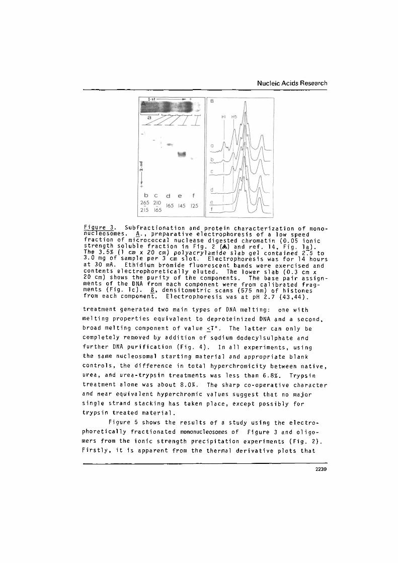
s o l u b l e f r a c t i o n s , shown in Fig. 2 B , i n d i c a t e s as ionic strengthi n c r e a s e s there is a preferen t i a l s e l e c t i o n of m o n o n u c l e o s o m e sover o l i g o m e r i c forms and a r e d u c t i o n in h e r e r o g e n e i t y of m o n o -n u c l e o s o m e s . To fu r t h e r qualify the s i g n i f i c a n c e of the indi-vidual m o n o n u c l e o s o m e c o m p o n e n t s w i t h r e s p e c t to p r o t e i n , DNAsize and thermal m e l t i n g b e h a v i o u r , n u c l e o s o m e s were p r e p a r a -t i v e l y e l e c t r o p h o r e s e d and individual c o m p o n e n t s eluted ande x a m i n e d . F i g u r e 3A shows that the n u c l e o p r o t e i n from thev a r i o u s b a n d s retain their r e l a t i v e n o b i l i t y , w h i c h is a p p r o x i -m a t e l y related to the computed DNA base pair n u m b e r . The v a r i -a t i o n s are c o n s i s t e n t w i t h other r e p o r t s ( 1 4 , 2 8 , 4 6 , 4 9 ) . Thecontent of n o n h i s t o n e is very low (<2%). This has been alreadynoted ( 1 4 ) . As shown in Figure 3 B , the m a j o r v a r i a t i o n in pro-tein c o n t e n t c e n t e r s on wh e t h e r HI an d / o r H5 are p r e s e n t or not;the 125 base pair f r a g m e n t may have m o r e H3 and H4 as s o c i a t e dwith it. D i f f e r e n t i a l s o l u b i l i t y of the m o n o n u c l e o s o m e popu-lation is a p p a r e n t l y related to HI and H5 (Fig. 2) and somen o n h i s t o n e p r o t e i n s ( 5 4 ) .
2237

Nucleic Acids Research
Fi gure 2. Effect of ionic strength on self-association or aggre-gation of nucleosomes. A, low gravity (2000 x g, 20 minutes)separation of aggregated nuceloprotein as a function of saltconcentration. (A) chromatin digested with micrococcal nuclease(4 x 1 0 " units/yg DNA) for 5 minutes at 23° (0.4% acid soluble( 3 8 ) , 8+ 3% insoluble chromatin in 1 mM EDTA pH 8.0 at 2000 x g20 m i n u t e s ) . Chromatin digested to 15 - 18% acid solubility inlow (38, A ) and high ( 1 4 , A ) ionic strength buffer. Nucleosomesassociated with (#) and without (O) histones HI and H5 (see Fig.3, c and d ) . Nucleosomes from 11.5 £ monomer peak from chroma-tin digested at low ionic strength (see A ) and fractionated by5-20% sucrose gradients in 1 mM EDTA pH 8.0 (•) or 100 mM NaCl-50 mM Tris-HCl-1 mM EDTA, pH 8.0 (D ) . B_. El ectrophoreti canalysis of DNA prepared from chromatin aggregates collected ine x p e r i m e n t , & ; a to j refer to precipitates in panel A_; h is the
150 mM NaCl soluble mononucleosome. Photograph is a reverseprint of a 3.5% polyacrylamide slab gel.
Melti ng profi1es: The derivative melting profiles of mononucleo-
somes treated or untreated with urea and/or trypsin are shown in
Figure 4. These data show that the melting profile of isolated
mononucleosomes is composed of two components, referred to here
as T" and T1" . This agrees with recent reports ( 2 2 , 2 3 , 2 7 - 3 5 ) .
Urea treatment, which has been shown to disrupt chromatin and
nucleosomes ( 1 6 , 2 3 , 2 7 ) , preferentially eliminates T"1 over T";
above 6M urea a DNA-like component (T°) occurs (Fig. 4 ) , which
represents about 8 to 1 2 % of the total hyperchromicity. In our
experiments urea neither caused a change in total hyperchromicity
of the components nor any noticeable increase in turbidity ( 2 7 ) .
Treatment of mononucleosomes with trypsin resulted in the
preferential elimination of T"' over T". This is also shown in
Figure 4. The extent of elimination of T" could be enhanced if
urea was included; urea added before or during proteolytic diges-
tion was very effective in this elimination. Trypsin and urea
2238
Nucleic Acids Research
b c265 2102F5 165
d e165 145
f
125
Figure 3. Subfractionation and protein characterization of mono-nucleosomes. A., preparative electrophoresis of a low speedfraction of micrococcal nuclease digested chromatin (0.05 ionicstrength soluble fraction in Fig. 2 (A) and ref. 1 4 , Fig. l a j .The 3.5% (1 cm x 20 cm) polyacrylamide slab gel contained 2.5 to3.0 mg of sample per 3 cm slot. Electrophoresis was for 14 hoursat 30 mA. Ethidium bromide fluorescent bands were exercised andcontents electrophoretically eluted. The lower slab (0.3 cm x20 cm) shows the purity of the components. The base pair assign-ments of the DNA from each component were from calibrated frag-ments (Fig. l c ) . J3, densitometric scans (575 nm) of histonesfrom each component. Electrophoresis was at pH 2.7 ( 4 3 , 4 4 ) .
treatment generated two main types of DNA melting: one withmelting properties equivalent to deproteinized DNA and a second,broad melting component of value <T". The latter can only becompletely removed by addition of sodium dodecylsulphate andfurther DNA purification (Fig. 4 ) . In all experiments, usingthe same nucleosomal starting material and appropriate blankcontrols, the difference in total hyperchromicity between native,urea, and urea-trypsin treatments was less than 6.8%. Trypsintreatment alone was about 8.0%. The sharp co-operative characterand near equivalent hyperchromic values suggest that no majorsingle strand stacking has taken place, except possibly fortrypsin treated material.
Figure 5 shows the results of a study using the electro-phoretically fractionated mononucleosomes of Figure 3 and oligo-mers from the ionic strength precipitation experiments (Fig. 2 ) .Firstly, it is apparent from the thermal derivative plots that
2239

Nucleic Acids Research
10.0
I 0
30 50 70 90Temperature °C
Fi gure 4. Derivative thermal denaturation profiles of mononucleo-somes before and after treatment with urea and/or trypsin. Theplots (24) were obtained with 1.5 x 10'1* M DNA-P in 5 mM Pd, atpH 7.0. Mononucl eosomes are from experiment in Fig. 1. a_, un-treated; b_, urea 6 M for two hours and c_, 8 M; d_, trypsin 0.003w/w at 23° for 1 hour at pH 7.0 and e_ trypsin with 6 M ureaadded during digestion, and £, DNA from nucleosomes in 6 M urea.Experiments were in duplicate; details are in Methods.
melting of nucleosome fragments are similar. Secondly, as DNA
fragment size is increased there is a slight shift of the T'"
transition and an appearance of minor low (<T") and high ( > T M I )
melting transitions. Thirdly, component T" is slightly augmented
and less co-opera;ive (measured by half peak height) in submono-
mer particles (the; 125 base pair u n i t ) ; it becomes increasingly
contiguous with T"' as DNA fragment size increases. Since some
fragments have either HI or H5 associated with them, the shift of
T" and T"1 may be due to these proteins. However, T" broadens and
both T" and T"' shift to lower values when 1 mM P 0 4 buffer is used
Controls for correlating visual and thermal studies: As major
controls we previously studied the patterns and images found in
the absence of sample, stability of sample to uranyl salt con-
centration and contamination due to free histone ( 1 4 ) . The
drying pattern with buffer alone prepared and stained in the
2240

Nucleic Acids Research
10.0 -
30 50 70 90Temperature °C
Figure 5. D e r i v a t i v e thermal d e n a t u r a t i o n p r o f i l e s of m o n o n u c l e o -s o m e s , d i n u c l e o s o m e s and o l i g o m e r i c f r a g m e n t s . Plots a_ and bc o r r e s p o n d to fragments o b t a i n e d from e x p e r i m e n t s in Fig. 2 T n d i -cated by symbols A and A ; c to f plots c o r r e s p o n d to b_, c_, d_, andf_ c o m p o n e n t s in Fig. 3.
same way as for s a m p l e s , is nei t h e r random nor the same as whensample is inc l u d e d . The d e p o s i t i o n pattern is altered as thec o n c e n t r a t i o n of sample per grid is increa s e d ( 1 4 ) . At higherm a g n i f i c a t i o n s no images c o m p a r a b l e to the ones for n u c l e o s o m e scould be de t e c t e d with b u f f e r a l o n e at any t e m p e r a t u r e . Thisp o s s i b i l i t y was e x t e n s i v e l y studied b e c a u s e transfer of heatedsample from thermal cuvette to individual e l e c t r o n m i c r o s c o p egrids could i n t r o d u c e a r t i f a c t s due to heat f l u c t u a t i o n s ofsample and grid. The v a r i a b l e s a s s o c i a t e d with sample t r a n s f e rwere q u a l i f i e d for each increase in t e m p e r a t u r e by use o f m i c r o -t h e r m o c o u p l e s inserted a p p r o p r i a t e l y in the c a p i l l a r i e s of thet e m p e r a t u r e e q u i l i b r a t e d m i c r o p i p e t t e s and onto the grid s u r f a c e s .A detail of this d e s c r i p t i o n is p r e s e n t e d in the M e t h o d s and inthe legend of Figure 6. From F i g u r e 6A and C it can be seenthat the a v e r a g e time required for sample t r a n s f e r and t r e a t m e n ton the grid is very short, 1.63 ± 0.3 seconds and 2.13 ± 0.2s e c o n d s , r e s p e c t i v e l y . Loss of heat d u r i n g t r a n s f e r is ch a r a c -
2241

Nucleic Acids Research
90
eo70
60
50
40
302 0
A
-.
—»50 70 90
Temperature BeforeTransfer (°C)
I 2 3Time (seconds)
I 2Duration (seconds)
Fi gure 6_. D e t e r m i n a t i o n of t e m p e r a t u r e d i f f e r e n t i a l s d u r i n gs p e c i m e n heating and t r a n s f e r to elect r o n m i c r o s c o p e g r i d s . A_.Ch a n g e in sample t e m p e r a t u r e during t r a n s f e r from thermal c u v e t t eto individual g r i d s . Vertical s h a d e d bar indicates all pointsw h e r e s a m p l e s were r e l e a s e d from 5 m i c r o l i t r e p i p e t t e s e q u i p p e dw i t h m i c r o - t h e r m o c o u p l e probes in the c a p i l l a r i e s . ]3. D i s c r e p -e n c y in sample t e m p e r a t u r e before t r a n s f e r and i n c i p i e n t r e l e a s eof s a m p l e onto e l e c t r o n m i c r o s c o p e g r i d s . Data was o b t a i n e dfrom g r a p h s in A using an averaged t r a n s f e r time of 1.63 +_ 0.30s e c , w h e r e the n u m b e r of r e p e t i t i o n s was 85. C_. Re s u l t a n ti n c r e a s e d grid t e m p e r a t u r e s f o l l o w i n g application of samples atv a r i o u s sample t r a n s f e r t e m p e r a t u r e s shown in A and B_. Verticalt e m p e r a t u r e drops i n d i c a t e points in time and Tinal grid t e m p e r a -t u r e s when w a s h i n g of grids with staining solutions or w a t e ro c c u r r e d . M a x i m u m grid t e m p e r a t u r e , w h i c h occurs at 9 5 ° C , was32 . 4 + 0.8°C.
t e r i s t i c for each s t a r t i n g t e m p e r a t u r e ; it does not become s i g n i -
f i c a n t until above 70°C (Fig. 6A and B ) . At 97°C the a v e r a g e
h e a t loss during t r a n s f e r was 2.8 ± 1.6°C. S i m i l a r l y it was
found that sample t r a n s f e r t e m p e r a t u r e below 50°C did not
s i g n i f i c a n t l y alter the original grid t e m p e r a t u r e ; m a x i m u m grid
t e m p e r a t u r e at 97°C was 8.4 + 0.8°C a b o v e a m b i e n t room t e m p e r a -
t u r e . Even at this t e m p e r a t u r e the e x p o s u r e was less than 1.5
se c o n d s after w h i c h the grids w e r e flushed with b u f f e r (Fig. 6 C ) .
By u s i n g the data in Figure 6 and j u d i c i o u s l y c o m p a r i n g grids
w i t h o u t s a m p l e , b e f o r e and after h e a t i n g , we co n c l u d e d that
e l e v a t e d t e m p e r a t u r e s a l o n e cannot generate m o r p h o l o g i c a l s t a i n -
ing a r t i f a c t s .
C o o l i n g of sa m p l e during t r a n s f e r could result in p o s s i b l e
r e n a t u r a t i o n of some denatured m a t e r i a l . To d e t e r m i n e to w h a t
e x t e n t this m i g h t occur c h r o m i c i t y c h a n g e s at 260 nm were m o n i -
tored as n u c l e o s o m e s a m p l e s were p r o g r a m heated to various
2242

Nucleic Acids Research
100
8 0
= 60
S
I 40X
20
-
- j1
J
^
if"f ---_-
|40 60 80Temperature CO
Figure 7. Analysis of nucleosomal-DNA denaturation and renatur-ation. Samples of nucleosomes (1.5 x 1 0 " 4 M D N A - P ) , in trip-licate, were melted to various extents (a, b and c ) , cooledrapidly to ambient temperature (23°) and re-heated by way of athermal program to 9 7 ° . Hyperchromicity of control s a m p l e , de-natured in one continuous phase was 3 3 . 7 % ( ) . Symbols (-<—)and (—>--) indicate cooling and re-heating p h a s e s , respectively.
temperatures, cooled to room temperature and program re-heated
to 97° (melting of all D N A ) . The results, presented in Figure 7,
are only for the beginnings and ends of the T" and T"' transi-
tions. Each of the transitions has some potential for hypo-
chromicity (after correcting for water e x p a n s i o n ) ; the beginning
of the T" transition is most affected and T"' the least.
Hyperchromicity due to re-heating the nucleosomal material is
almost totally recoverable for the former, but not for the
latter. Therefore combining the results in Figure 6A, B and C
with those of Figure 7, it is likely that only the material at
the beginning of transition T" might be affected by transfer of
sample during heating. H o w e v e r , at these temperatures sample
heat loss, prior to fixation on the grid s , is very s m a l l .
To determine to what extent the morphological and spectro-
scopic c h a n g e s , which occur during nucleosomal-DNA d e n a t u r a t i o n ,
may be attributable to free histone-histone interactions a l o n e ,
pure, nucleic acid f r e e , histones were heat treated and simul-
taneously monitored for mor p h o l o g y . The results of these
experiments can be briefly summarized as f o l l o w s : 1) a co-
operative heat induced hyperchromic change can be clearly
detected at 275 nm; 2) the major transition occurs about 10°C
2243

Nucleic Acids Research
Table 1. S ta t i s t i ca l measurements of native and denatured Mononucleosomes
Form N O.D. I.D. Contour Lengtha Percent B-Form(nm + s.d.) (nm + s.d.) Length
Native 550 13.3 + 1.5 1 .68+0.8
Denatured 405 17.4 + 2.2 9.8 + 2.0 42.9 + 6.3 86 + 4.8
C o m p u t e d by d i r e c t l e n g t h m e a s u r e m e n t o f l i n e a r f o r m s o r byf o r m u l a , TT((O . D . - I.D.)/2 + I . D . ) .C o m p u t e d f r o m w e i g h t a v e r a g e m o l e c u l a r w e i g h t 9 8 , 3 4 0 d +_ 3%.
a b o v e that o f p u r i f i e d DNA from n u c l e o s o m e s ; a n d 3) the m o r p h o l o g yo f t h e h i s t o n e s , w h i c h h a v e been d e s c r i b e d ( 1 4 ) , c h a n g e , but thei m a g e s do not r e s e m b l e t h o s e o b t a i n e d w i t h n u c l e o s o m e s at any o fth e e l e v a t e d t e m p e r a t u r e s .I m a g e and s t a i n i n g c h a r a c t e r i s t i c s o f n a t i v e a n d d e n a t u r e d n u c l e o -s o m e s : F i g u r e 8 s h o w s t h e general n e g a t i v e s t a i n i n g p r o p e r t i e so f m o n o n u c l e o s o m e s at 2 3 ° C , p r i o r to t h e r m a l d e n a t u r a t i o n . Alli m a g e s w e r e p a r t i c u l a t e , e i t h e r c i r c u l a r or p r o l a t e e l i p s o i d s .A s i z e a n a l y s i s o f the i n s i d e and o u t s i d e d i a m e t e r s is s u m m a r i z e din T a b l e I. T h e d i m e n s i o n s of the p a r t i c l e s a r e in go o d a g r e e m e n tw i t h o u r e a r l i e r d e t a i l e d study (14) and t h o s e o b t a i n e d from c h i c ke r y t h r o c y t e s (1,2,11). T h e bulk o f the n u c l e o s o m a l i m a g e s c h a n g e dv e r y l i t t l e in s h a p e or s t a i n a b i l i t y as the t e m p e r a t u r e is r a i s e df r o m a m b i e n t ( 2 3 ° C ) to 7 0 ° C . T h i s h o l d s w h e t h e r the s a m p l e isf i x e d in g l u t e r a l d e h y d e i m m e d i a t e l y p r i o r to s t a i n i n g or d i r e c t l ys t a i n e d w i t h o u t f i x a t i o n (Fig. 8 b , b ' ) . A s i g n i f i c a n t a m o u n t o fs w e l l i n g a n d t e n d e n c y o f t h e n u c l e o s o m e s to a g g r e g a t e w a s n o t i c e dat t h e l a t t e r p a r t o f t r a n s i t i o n T" ( F i g . 8 b , b ' ) . H o w e v e r , nof u r t h e r c h a n g e w a s n o t e d until T"' w a s r e a c h e d ; at thi s p o i n t ,t h e a v e r a g e n u c l e o s o m a l m o r p h o l o g y has d r a s t i c a l l y a l t e r e d andb o t h i n n e r a n d o u t e r d i a m e t e r s h a v e i n c r e a s e d ( F i g . 8 c , c ' ,T a b l e I ) , T h e a v e r a g e f i b e r l e n g t h f r o m p e r i m e t e r m e a s u r e m e n t sis l e s s than the l e n g t h c o m p u t e d f r o m t h e w e i g h t a v e r a g e d b a s ep a i r n u m b e r a s s u m i n g B - f o r m d u p l e x DNA ( F i g . 1) ( 1 , 2 7 ) .
A d e t a i l e d c o m p a r i s o n of the u n f i x e d and f i x e d m a t e r i a li n d i c a t e s t h a t the u n f i x e d m a t e r i a l y i e l d s m o r e l i n e a r and U-
2244

Nucleic Acids Research
a
Figure 8. Morphological changes in mononucleosomes at variousstages of heat denaturation. Duplicate samples removed fromthermal-regulated cuvette at 2°C intervals and transferred togrids. Procedures are described in methods and in Fig. 6. a_,a}, 23°; b^ b} and c_, c_l correspond to end of T" and T"' trans-itions shown in Fig. 4., d, d' are high magnifications of c,c'. Gl uteral dehyde was omitted in a', b', c1 and d_'.
2245

Nucleic Acids Research
shaped structures than open ended c i r c l e s . This is shown inthe high magnifications of Figure 8d,d'. Close s c r u t i n i z a t i o nof grids with samples from each temperature interval indicatedthat a minor c o m p o n e n t with similar morphological features seenin F i g u r e 8b to c also occurs at the beginning of T". H o w e v e r ,these ringed structures may have disintegrated as the temperaturewas i n c r e a s e d , b e c a u s e they could not be detected at higher tem-p e r a t u r e s .
DISCUSSIONThe composition of mono n u c l e o s o m e s used here s p e c i f i c a l l y
for combined melting and visualization studies is in good agree-m e n t with other reports ( 3 - 6 ) . We have shown that m o n o n u c l e o -some variation (14,27) can arise through differences in self-a s s o c i a t i o n of mono m e r or oligomer nucleosomes by increased ionics t r e n g t h and presence of HI and H5 associated p r o t e i n s . Thehetero g e n e i t y e s t a b l i s h e d by ele c t r o p h o r e t i c a l l y s u b f r a c t i o n a t e dm o n o n u c l e o s o m e s is in agreement with those from chicken ( 2 7 ) .The extremely low n o n h i s t o n e p r o t e i n content of the monomers isnot unusual b e c a u s e : 1) mature chicken or goose e r y t h r o c y t enuclei contain very low amounts of nonhistone in comparison tonuclei from immature cells or other tissues (52,53) (especiallyin classes below 68,000 d a l t o n s ) and 2) the bulk of the highm o l e c u l a r weight residual p r o t e i n s , which appear to be mostlya s s o c i a t e d with nuclear membranes o r ma t r i x , are relativelyi n s o l u b l e unless e x t e n s i v e l y digested with micrococcal nuclease( 5 4 ) . Since the initial preparation of this m a n u s c r i p t severalrelated reports have been published which further d o c u m e n t thedifferential s e l f - a s s o c i a t i o n o f monomers and oligomers (32,55-37)T h e s e studies provide a basis for interpreting the earliero b s e r v e d differential s e n s i t i v i t y o f erythrocyte chromatin( h e t e r o c h r o m a t i c and e u c h r o m a t i c s t r u c t u r e ) to both mono andd i v a l e n t cations (57-60) before and after sequential removal ofpr o t e i n s (.52,53,59).
The melting p r o f i l e s of electr o p h o r e t i c a l l y defined mono-n u c l e o s o m e s and select oligomeric fragments obtained by ionics t r e n g t h p r e c i p i t a t i o n methods clearly indicate that only twom a j o r transitions (T" and T"') are associated with m o n o n u c l e o -s o m e s . As shown here and in part elsewhere ( 2 2 , 2 3 , 2 7 , 2 8 , 3 0 - 3 4 ) ,
2246
Nucleic Acids Research
thermal t r a n s i t i o n s £ T" a n d > T " ' are pe c u l i a r to n u c l e o p r o t e i n s of
large DNA length (>_ 265 base p a i r s ) and inc r e a s e d protein complexity.
H o w e v e r they are not n e c e s s a r i l y due to prev i o u s d e g r a d a t i o n ( 2 1 ,
2 5 , 2 8 , 3 0 , 3 2 ) . A l l o w i n g for s o l v e n t d i f f e r e n c e s , our res u l t s are
very similar to those o b t a i n e d by Mandel and Fasman ( 2 2 ) , Lewis
( 2 8 ) , Wittig and Wit t i g ( 3 2 ) . They are also in e x c e l l e n t a g r e e -
ment with the highly c o m p l e m e n t a r y physical s t u d i e s by W e i c h e t and
co- w o r k e r s (68) that a p p e a r e d on su b m i s s i o n d a t e of our original
m a n u s c r i p t . The c o m b i n e d s t u d i e s clearly i n d i c a t e that the presence
of t r a n s i t i o n s T" and T"' c a n n o t be directly a c c o u n t e d for by
varia t i o n s in DNA l e n g t h , p r e s e n c e or a b s e n c e of l y s i n e - r i c h
h i s t o n e s ( H I , H 5 ) or n o n h i s t o n e p r o t e i n s ; b u t , c o - o p e r a t i v i t y and
resol u t i o n of these t r a n s i t i o n s p a r t i c u l a r l y T " , can be inf l u e n c e d
by the preceding p a r a m e t e r s as well as by d i f f e r e n c e s in ionic
strength of the m e l t i n g b u f f e r . We routinely used 5 mM N a P O . at
pH 7 . 0 , b e c a u s e much less visual u n f o l d i n g of the m o n o n u c l e o s o m e s
(14) takes p l a c e than at lower ionic s t r e n g t h s such as with 0.1 mM
EDTA,and T" and T"' are still d i s t i n c t . W e i c h e t et a l (68) have
also recently o b s e r v e d the la t t e r e f f e c t using sodium c a c o d y l a t e
b u f f e r . The thermal r e v e r s i b i l i t y of T" and d i f f e r e n t i a l s e n s i -
tivity of T"' with urea and trypsin t r e a t m e n t of n u c l e o s o m e s will
be discussed f u r t h e r in r e f e r e n c e to nu c l e o s o m e u n f o l d i n g .
Olins and c o - w o r k e r s (27) have s k i l l f u l l y shown by e l e c t r o n
m i c r o s c o p y that urea induces u n f o l d i n g of n u c l e o s o m e s . A s i m i l a r
claim has been m a d e by W o o d c o c k and Frado ( 3 6 ) . S i m p l e s t o r a g e o f
nucl e o s o m e s in low ionic s t r e n g t h b u f f e r can also p r o m o t e d e n a t u r a -
tion ( 1 4 , 6 6 ) ; the n u c l e o s o m e s unfold into two m a j o r h a l v e s in the
abs e n c e of any m a j o r DNA or pro t e i n d e g r a d a t i o n . In this study w e
show for the first time that during thermal d e n a t u r a t i o n p u r i f i e d
m o n o n u c l e o s o m e s u n d e r g o r e p r o d u c i b l e m o r p h o l o g i c a l c h a n g e s w h i c h
are generally n o n r e v e r s i b l e . First, the m o r p h o l o g i c a l c h a n g e s occur
w h e t h e r or not the n u c l e o s o m e s are fixed in g l u t e r a l d e h y d e prior to
uranyl acetate s t a i n i n g . S e c o n d , the major c h a n g e is from the
typical compact toroid s h a p e (Fig. 8 , Table I and ref . 2 , 7 , 1 0 ,
1 1 - 1 5 ) to the large c i r c u l a r , U - s h a p e d , c r e s c e n t - s h a p e d , or extended
s t r u c t u r e of length a b o u t 9 0 % o f a w e i g h t a v e r a g e n a t i v e DNA
B-form e q u i v a l e n t . T h i r d , the m a j o r portion o f these m o r p h o l o g i c a l
changes is seen only a f t e r r e a c h i n g t r a n s i t i o n T"'. U n l i k e f o r m a i -
2247
Nucleic Acids Research
d e h y d e (.67), w h i c h w a s p u r p o s e l y a v o i d e d h e r e , w e do n o t feel t h e
h i g h l y r e g u l a r r i n g s t r u c t u r e s o b t a i n e d w i t h g l u t e r a l d e h y d e f i x a t i o n
a r e a r t i f a c t s ; b a s i c a l l y the same i m a g e s w e r e o b t a i n e d by two
i n d e p e n d e n t m e t h o d s o f s t a i n i n g . H o w e v e r , t h e p r e s e n c e o f l i n e a r ,
e x t e n d e d f o r m s in u n f i x e d s a m p l e s s u p p o r t s o u r i n i t i a l c o n t e n t i o n
t h a t f i x a t i o n w o u l d h e l p s t a b i l i z e t h e d e l i c a t e d e n a t u r e d n u c l e o -
s o m e s t r u c t u r e s f r o m m e c h a n i c a l d i s r u p t i o n d u r i n g s t a i n i n g , w a s h i n g
a n d d r y i n g of g r i d s . T h e i n h e r e n t r i g i d i t y o f small DNA s e g m e n t s
o f t h i s o r d e r ( 5 0 ) , p a r t i c u l a r l y in t h e d e n a t u r e d s t a t e , w o u l d t e n d
to g e n e r a t e l i n e a r s t r u c t u r e s s u c h as a l r e a d y s e e n f o r low i o n i c
s t r e n g t h ( 1 4 , 6 6 ) and u r e a ( 2 7 , 3 6 ) d e n a t u r e d n u c l e o s o m e s . A d e t a i l e d
s t u d y o f the u n f o l d e d s t r u c t u r e s at h i g h r e s o l u t i o n , and an
a n a l y s i s of t h e p e r i o d i c , n e g a t i v e l y s t a i n i n g e l e m e n t s , w h i c h a r e
s e e n a l o n g t h e o p e n r i n g e d i m a g e s , is b e i n g c a r r i e d o u t u s i n g i m a g e
r e c o n s t r u c t i o n m e t h o d s . T h e r i n g e d s t r u c t u r e s a r e h i g h l y c o m p a t i b l e
w i t h r e c e n t X - r a y d i f f r a c t i o n d a t a ( 1 5 ) a n d c o r r e s p o n d i n g s u b u n i t
m o d e l s ( 3 7 , 6 4 ) .
S e v e r a l s t u d i e s ( 2 1 , 2 2 , 2 6 - 3 0 , 6 1 , 6 8 , t h i s s t u d y ) i n d i c a t e
t h a t t h e m a j o r m e l t i n g c o m p o n e n t of c h r o m a t i n is l i k e l y r e l a t e d
to t h e p r i n c i p a l t r a n s i t i o n (T"' ) of the s i m p l e m o n o n u c l e o s o m e .
H o w e v e r , t h e s h e a r c o m p l e x i t y o f D N A - h e l i x s t a b i l i t y in c h r o m a t i n
p r o h i b i t s d e f i n i t i v e a s s i g n m e n t of o t h e r m e l t i n g t r a n s i t i o n s at
t h i s t i m e . N e v e r t h e l e s s , an e a r l i e r m o d e l , d e r i v e d f r o m c h r o m a t i n
w o r k ( 1 8 ) , has c o n s i d e r a b l e appeal in t h a t t h e two m a j o r t r a n s i -
t i o n s a r e p o s t u l a t e d to o r i g i n a t e f r o m t h e d i s r u p t i o n of p r o t e i n -
p r o t e i n i n t e r a c t i o n s a n d m e l t i n g of p r o t e i n - f r e e D N A (Tm ) and f r o m
t h e d e s t r u c t i o n of t h e s u p e r c o i l and DNA s t r a n d s e p a r a t i o n (Tm ) .
O u r v i s u a l and h y p e r c h r o m i c s t u d i e s and t h e r e c e n t l y r e p o r t e d
h y p e r c h r o m i c , c i r c u l a r d i c h r o i c and c a l o r i m e t r i c s t u d i e s on c h i c k e n
e r y t h r o c y t e n u c l e o s o m e s by W e i c h e t £jt aj_ ( 6 8 ) p r o v i d e n e w i n s i g h t
i n t o t h e t h e r m a l d e n a t u r a t i o n p r o c e s s o f c h r o m a t i n s u b u n i t s .
O v e r a l l , t h e b i - p h a s i c m e l t i n g of c o r e p a r t i c l e s is i n t e r p r e t e d
as a d e n a t u r a t i o n o f a b o u t 40 b a s e p a i r s in t h e f i r s t p h a s e ,
f o l l o w e d by a m a s s i v e u n f o l d i n g of t h e .native s t r u c t u r e of a
t i g h t h i s t o n e - D N A c o m p l e x , w h i c h f r e e s t h e r e m a i n i n g 1 0 0 b a s e
p a i r s in s t a c k i n g (the s e c o n d p h a s e ) . O u r s t u d y c l e a r l y s h o w s
t h a t n u c l e o s o m e s b e g i n to a l t e r o r i g i n a l t o r o i d s h a p e o n l y d u r i n g
t h e l a t t e r t h i r d o f T" and do n o t a c t u a l l y c o - o p e r a t i v e l y u n f o l d
u n t i l T " 1 is r e a c h e d . T h e u n f o l d i n g p a t t e r n is h i g h l y c o n s i s t e n t
2248

Nucleic Acids Research
with the predicted 1 3/4 loop or supercoil arrangement of nucleo-somal DNA (15,37). From the circular dichroic data (68) it isfurther apparent that no change in secondary structure of theprotein core takes place at T". Since the core is stable, ittherefore may serve to lock the major part of the DNA into a rigidcomplex thus allowing only certain regions to gain limited mobilityat £ T". This would explain the reversibility of T" that we bothobserve, albeit in our case complete hypochromicity occurred onlyin the first third of T". It is important to stress that the firststable morphologically altered state of the nucleosome occurs inthe latter third of T". It is there that the toroids becomemeasureably expanded, frequently aggregated into clusters of 2 to4 and ends of loops are occasionally detectable. Since secondarystructure of histones is located in the more hydrophobic domains,which are the regions that interact with each other (3-6,28,31,61,6 3 , 6 5 , 6 8 ) , the aggregation property may be a direct measurement ofthe alteration in histone structure that takes place at thisstage C68).
Unfolding of nucleosome secondary structure by urea hasbeen earlier shown by electron microscopy ( 2 7 , 3 5 , 6 0 ) , hydrodynamicand circular dichroic studies (23,35,60). Physical changes inmononucleosomes following trypsin treatment have also recently beenreported (31). The data are explained on the basis of a structuralunfolding of nucleosomal DNA whereby part of the DNA rcetains theproperties within intact core particles and the remaining portionbehaves much like free DNA. We note that the melting data of 01 insand co-workers (27) and Lilley and Tachell (31) exhibit a similarpreferential loss of transition T"' over T" that we observed here.However, no visual studies on the unfolding of trypsin treatednucleosomes is currently available for more rigorous interpreta-tion. From present work, which correlates unfolding of the super-coiled DNA with T " 1 , preferential loss of T"' because of urea,trypsin or urea and trypsin treatment of nucleosomes would beexpected. It is tempting to conclude that the presence of T"1 canbe used as a direct indicator, at all times, for morphologicallyintact nucleosomes. However, our recent shearing studies (inpreparation) indicate that this is not always true, particularlyif no T" is present. Similar caution should be applied when inter-
2249

Nucleic Acids Research
preting the origin of transitions <_ T". In addition to T" arising
from the melting of a limited number of base pairs within the
nucleosome ( 6 8 ) , it could also arise from the melt-out of a minor
nucleosome or nucleosome-1ike population which lacks thermal
stability in the denatured state as exhibited by denatured core
nucleosomes at temperatures > T"'. Close scrutinization of
m i c r o g r a p h s obtained from the early portion of T" of a number of
samples indicates that this is possible. T h e r e f o r e , for some
nucleosome elements there may be a d i f f e r e n c e in either the types
of protein (species or charge m o d i f i c a t i o n s ) or the way core
p r o t e i n s are associated with DNA sister strands (intra or inter-
strand c r o s s - 1 i n k i n g ) after denaturation. A study of single
strand availability and DNA sequence polarity b e f o r e , during and
a f t e r thermal d e n a t u r a t i o n of native nucleosomes may be useful
approaches for investigating this p o s s i b i l i t y .
A C K N O W L E D G E M E N T S
The authors wish to thank Drs. C.V. Lusena (N.R.C.C.) andA.R. Morgan (University of Alberta) for providing us with X andPM2 DNA. We are extremely indebted to the expert technicals u p p o r t skills of M.J. D o v e , L. Sowden and R. Whitehead. Thisis NRCC contribution No. 16572.
R E F E R E N C E S
1 O l i n s , D.E. and O l i n s , A.L. (1974) Science 1 8 4 , 330-3322 O u d e t , P., G r o s s - B e l l a n d , M. and C h a m b o n , P. (1975) Cell 4,
281-2993 K o r n b e r g , R.D. (1977) Ann. Rev. Biochemistry 4 6 , 931-9544 F e l s e n f e l d , G. (1978) Nature 2 7 1 , 115-1225 L i , H.J. (1975) Nucleic Acids R e s . 2 , 1275-12896 van H o l d e , K.E. and Isenberg, I. (1975) A c e . Chem. Res. 8,
3277 van H o l d e , K . E . , S a h a s r a b u d d h e , C.G. and S h a w , B.R.,
van B r u g g e n , E.F.J. and A r n b e r g , A . C . (1974) Biochem. B i o p h y s .R e s . C o m m u n . 6 0 , 1365-1370
8 O l i n s , A . L . , C a r l s o n , D. and O l i n s , D.E. (1975) J. Cell Biol.6 4 , 528-537
9 B a k a y e v , V . V . , Melnickov, A . A . , O s i c k a , V.D. and V a r s h a v s k y ,A.J. (1975) Nucleic Acids Res. 2 , 1401-1419
10 F i n c h , J . T . , N o l l , M. and K o r n b e r g , R.D. (1975) Proc. Nat.Acad. S c i . U.S.A. 7 2 , 3320-3322
11 W o o d c o c k , C.F.L., Safer, J.P. and S t a n c h f i e l d , J.E. (1976)Expt'l Cell R e s . 9 7 , 101-110
12 L a n g m o r e , J.P. and Wooley, J.C. (1975) Proc. Nat. Acad. S c i .U.S.A. 7 2 , 2691-2695
13 P o o n , N.H. and Seligy, V.L. (1977) Proc. of MicroscopicalSociety of Canada 4, 56-57
2250

Nucleic Acids Research
14 Poon, N.H. and Seligy, V.L. (1978) Expt'l Cell Res. 113, 95-110.15 Finch, J.T., Lutter, L.C., Rhodes, D., Brown, R.S., Rushton,
B., Levitt, M. and Klug, A. (1977) Nature 269, 29-3616 Ansevin, A . T . , Hnilica, L.S., Spelsberg, T.C. and Kelm, S.L.
(1971) Biochemistry 1 0 , 4793-480317 Li, H.J., Chang, C. and Weiskopf, M. (1973) Biochemistry 1 2 ,
1763-177218 Wilhelm, F.X., De Murcia, G.M., Champagne, M.H. and Daune,
M.P. (1974) Eur. J. Biochem. 4 5 , 431-44319 Sahasrabuddhe, C.G. and van H o l d e , K.E. (1974) J. Biol. Chem.
249, 152-15620 Woodcock, C.F.L. and Frado, L.L.Y. (1975) Biochem. Biophys.
Res. Commun. 6 6 , 403-41021 Miller, P., Kendell, F. and N i c o l i n i , C. (1976) Nucleic Acids
Res. 3, 1875-188122 Mandel, R. and Fasman, G. (1976) Nucleic Acids Res. 3, 1839-
185523 Whitlock, J.P. and Simpson, R.T. (1976) Nucleic Acids
Research 3, 2255-226624 Lurquin, P.F. and Seligy, V.L. (1976) Chem.-Biol. Inter-
actions 1 3 , 27-4525 Lawrence, J.J., Chan, D.C.F. and Piette, L.H. (1976) Nucleic
Acids Res. 3, 2879-289326 Staynov, D.Z. (1976) Nature 2 6 4 , 522-52527 O l i n s , D.E., Bryan, P.N., Harrington, R.E., Hill, W.E. and
O l i n s , A.L. (1977) Nucleic Acids'Res. 4, 1911-193128 Lewis, P.N. (1977) Can. J. Biochem. 5 5 , 736-74629 Vengerov, Y.Y. and Popenkov, V . I . (1977) Nucleic Acids Res.
4, 3017-302730 Defer, N., K i t z i s , A., Kruth, J., B r a h m s , S. and Brahms, J.
(1977) J. Nucleic Acids Res. 4, 2293-230631 Lilley, D.M.J. and Tatchell, K. (1977) Nucleic Acids Res. 4,
2039-205532 Wittig, B. and Wittig, S. (1977) Nucleic Acids Res. 4, 3907-
391733 Cotter, R.I. and Lilley, D.M.J. (1977) FEBS Letters 8 2 , 63-68.34 Stein, A., Stein-Bina, M. and Simpson, R.T. (1977) Proc. Nat.
Acad. Sci. U.S.A. 7 4 , 2780-278435 Garret, R.A. (1971) Biochemistry 1 0 , 2227-223036 Woodcock, C.F.L. and Frado, L.-L.Y. (1976) J. Cell Biol. 7 0 ,
267a (Abst.)37 Pardon, J.F., Worcester, D.L., Wooley, J.C., Cotter, R . I . ,
Lilley, D.M. and Richards, B.M. (1977) Nucleic Acids Res. 4,3199-3214.
38 Sollner-Webb, B. and Felsenfeld, G. (1975) Biochemistry 14,2915-
39 Birnboim, H.C., Holford, R.M. and Seligy, V.L. Cold SpringHarbor Symp. Quant. Biol. 42 (in press)
40 Markov, G.G. and Ivanov, I.G. (1974) Anal. Biochem. 59, 555-563
41 Dahlberg, A . E . , Dingman, C.W. and Peacock, A.C. (1969) J.Mol. Biol. 4 1 , 139-147
42 Laemmli, U. (1970) Nature 227, 680-68543 Panyim, S. and Chalkley, R. (1969) Biochemistry 8, 3972-397944 T o b i n , R.S. and Seligy, V.L. (1975) J. Biol. Chem. 2 5 0 , 358-
364.
2251

Nucleic Acids Research
45 Gordon, C.N. and Kleinschmidt, A.K. (1968) Biochem. Biophys.Acta 155, 305-307
46 01 ins, A.L,, Cartson, R.D., Wright, E.B. and O l i n s , D.E.(19.76) Nucleic Acids Res. 3, 3271-3291
47 Todd, R.D. and Garrard, W.T. (1977) J. Biol. Chem. 2 5 2 , 4729-4738
48 Varshavsky, A.J., Bakayev, V.V., Chumackov, P.M. and Georgiev,G.P. (1976) Nucleic Acids Res. 3, 2101-2114
49. Bakayev, V.V., Bakayeva, T.G. and Varshavsky, A.J. (1977)Cell 11 , 619-629
50 Kovacic, R.T. and van Holde, K.E. (1977) Biochemistry, 1 6 ,1490-1498
51 Lohr, D., Corden, J., Tatchell, K., Kovacic, R.T. andvan Holde, K.E. (1977) Proc. Nat. Acad. Sci . U.S.A. 7 4 , 78-83.
52 Shelton, K.R. and Neelin, J.M. (1971) Biochemistry 10, 2342-2348
53 Seligy, V.L. and Miyagi , M. (1974) Eur. J. Biochem. 4 6 , 259-269
54 Seligy, V . L . , Poon, N.H., Dove, M. and Tobin, R.S. unpublished.55 Sanders, M.M. and Hsu, J.T. (1977) Biochemistry 16, 1690-1695.56 Campbell, A.M. and Cotter, R.I. (1977) Nucleic Acids Res. 4,
3877-388657 L i , H.J., Hu, A.W., Maciewicz, R.A., Cohen, P., Santella,
R.M. and Cang, C. (1977) Nucleic Acids Res. 4, 3839-385458 Seligy, V.L. and M i y a g i , M. (1969) Expt'l Cell Res. 5 8 , 27-34.59 Brasch, K., Setterfield, G. and Seligy, V.L. (1971) Expt'l
Cel 1 Res. 6 5 , 61-7260 Olins, D.E. and O l i n s , A.L. (1972) J. Cell Biol. 5 3 , 71561 L i , H.J. (1977) "Chromatin Structure - A Model" in The
Molecular Biology of the Mammalian Genetic A p p a r a t u s , T s ' o ,P., Ed., pp. 323-343. Elsevier/North-Hol1 and Biomedical Press.
62 Woodcock, C.F.L. (1977) Science 195, 1350-135263 Camerini-Otevo, R.D. and Felsenfeld, G. (1977) Nucleic Acids
Res . 4, 1159-118164 Weintraub, H., Worcel , A. and Alberts, B. (1976) Cell 9,
409-41765 Weintraub, H. and van Lente, F. (1974) Proc. Nat. Acad. Sci.
U.S.A. 7 1 , 4249-425366 Oudet, P., Spadafora, C. and Chambon, P. Cold Spring Harbor
Symp. Quant. Biol. 42 (in press)67 Polacow, I., Cabasso, L. and Li, H.J. (1976) Biochemistry 15,
4560-456568 Weischet, W., T a t c h e l l , K., Van Holde, K. and Klump, H. (1978)
Nucleic Acids Res. 5, 139-160.
2252