VOLUME 5 AdMIRable Fall Issue - TN.gov and discussed it with my wife. And I told her I didn’t want...
7
AdMIRable Review | Fall 2016 PHYSICIAN SPOTLIGHT GALEN R. SMITH, MD New Physician Advisory Board SAVE THE DATE 3rd Annual Workers’ Compensation Physicians’ Conference VOLUME 5 Fall Issue December 21, 2016 AdMIRable REVIEW JOURNAL OF THE TENNESSEE MEDICAL IMPAIRMENT RATING REGISTRY Inside this issue: VISUAL SYSTEM IMPAIRMENTS
Transcript of VOLUME 5 AdMIRable Fall Issue - TN.gov and discussed it with my wife. And I told her I didn’t want...

AdMIRable Review | Fall 2016
PHYSICIAN SPOTLIGHT
GALEN R. SMITH, MD
New Physician Advisory Board
SAVE THE DATE
3rd Annual
Workers’ Compensation
Physicians’ Conference
VOLUME 5
Fall Issue December 21, 2016
AdMIRable
REVIEW J O U R N A L O F T H E T E N N E S S E E
M E D I C A L I M P A I R M E N T R A T I N G R E G I S T R Y
Ins ide th is issue :
VISUAL SYSTEM
IMPAIRMENTS
2 AdMIRable Review | Fall 2016
ABBIE HUDGENS, ARM, AIC
Administrator
JEFF FRANCIS
Assistant Administrator
TROY HALEY, JD
Director, Administrative
Legal Services
JEFFREY E. HAZLEWOOD, MD
Assistant Medical Director
BRIAN HOLMES
Director, Mediation Services
RICHARD MURRELL, JD
Director, Quality Assurance
ANNA K. SUDBERRY
Communications Coordinator
ROBERT B. SNYDER, MD
Medical Director
JAMES B. TALMAGE, MD
Assistant Medical Director
JAY BLAISDELL, CEDIR VI
MIRR Program Coordinator
EDITOR
SAVE THE DATE
AMA Guides, 6th Edition, Training and other medical topics*
Saturday & Sunday, June 10-11, 2017
Guest House at Graceland
3600 Elvis Presley Boulevard, Memphis TN
*Meets requirements for physicians seeking appointment to the Medical Impairment Rating Registry.
CME Credits Available. Contact: [email protected] for registration details.
ADVISORY BOARD
ASSOCIATE EDITOR
NEW MIR PHYSICIAN ADVISORY BOARD MEMBERS
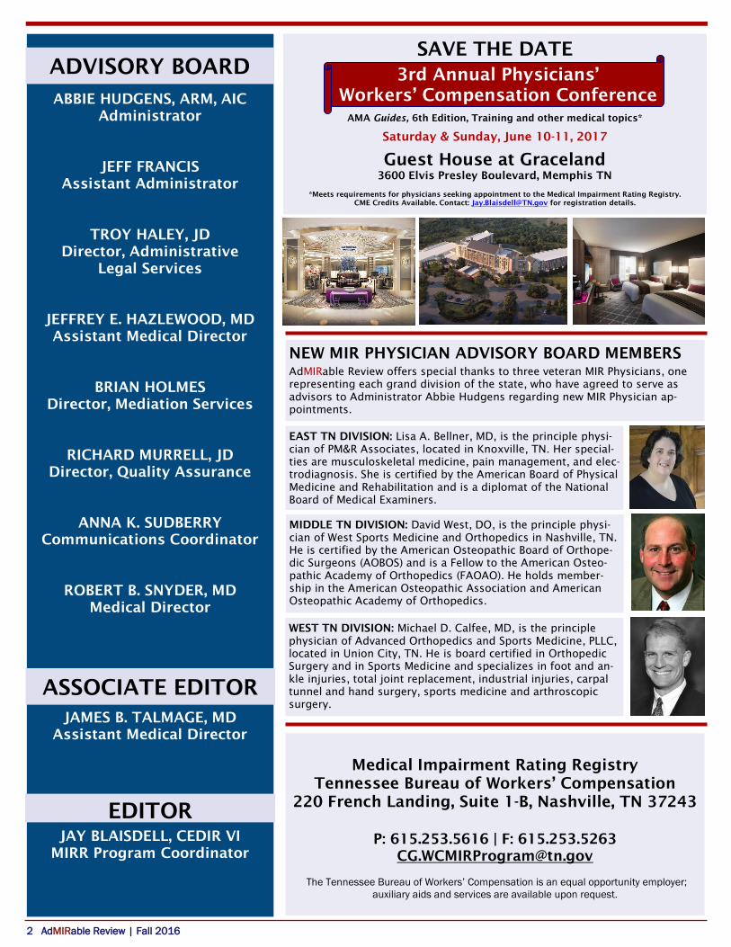
AdMIRable Review offers special thanks to three veteran MIR Physicians, one
representing each grand division of the state, who have agreed to serve as
advisors to Administrator Abbie Hudgens regarding new MIR Physician ap-
pointments.
EAST TN DIVISION: Lisa A. Bellner, MD, is the principle physi-
cian of PM&R Associates, located in Knoxville, TN. Her special-
ties are musculoskeletal medicine, pain management, and elec-
trodiagnosis. She is certified by the American Board of Physical
Medicine and Rehabilitation and is a diplomat of the National
Board of Medical Examiners.
MIDDLE TN DIVISION: David West, DO, is the principle physi-
cian of West Sports Medicine and Orthopedics in Nashville, TN.
He is certified by the American Osteopathic Board of Orthope-
dic Surgeons (AOBOS) and is a Fellow to the American Osteo-
pathic Academy of Orthopedics (FAOAO). He holds member-
ship in the American Osteopathic Association and American
Osteopathic Academy of Orthopedics.
WEST TN DIVISION: Michael D. Calfee, MD, is the principle
physician of Advanced Orthopedics and Sports Medicine, PLLC,
located in Union City, TN. He is board certified in Orthopedic
Surgery and in Sports Medicine and specializes in foot and an-
kle injuries, total joint replacement, industrial injuries, carpal
tunnel and hand surgery, sports medicine and arthroscopic
surgery.
Medical Impairment Rating Registry
Tennessee Bureau of Workers’ Compensation
220 French Landing, Suite 1-B, Nashville, TN 37243
P: 615.253.5616 | F: 615.253.5263
The Tennessee Bureau of Workers’ Compensation is an equal opportunity employer;
auxiliary aids and services are available upon request.
3rd Annual Physicians’
Workers’ Compensation Conference
AdMIRable Review | Fall 2016 3
University in Cleveland, Ohio, in
1983.
“In 2003 we invaded Iraq,” says Dr.
Smith. “I got a form letter from the
United States Army saying they were
in desperate need of orthopedic sur-
geons. But I had—and still do—a very
comfortable life here. I have a won-
derful wife and boys—at the time they
were three-years old. And I came
close to throwing that letter in the
trash, but I kept it on the corner of
my desk for a couple of weeks and
kept thinking it over and thinking it
over. And then I took that letter
home and discussed it with my wife.
And I told her I didn’t want to get into
my senior years and look back and
say I was too comfortable, too lazy,
too woulda-coulda-shoulda, and did-
n’t. My father and his two brothers
served in World War II—one the Army,
one in the Air Force, and my Dad in
the Navy. He was in the reserves after
World War II and he got called up to
Korea. And all three of them had the
same thing in common. None of them
made the military a career. They all
served at the time of our country’s
need. And I felt it was my time.”
In the Spring of 2003, with his wife’s
backing, Dr. Smith called the Army.
“And by October 2003 I was sworn in
as an officer in the U.S. Army Reserve
Corp. I thank the Army for letting me
serve at the three levels of care for
injured soldiers. The FST—the For-
ward Surgical Team—is the smallest
unit that they put a doctor in—in the
Army. Then the combat support hos-
pital in Iraq that I was in is the second
largest unit. And then the Landstuhl
Regional Medical Center is the largest
unit. So I got to serve in all three.
This opportunity to serve our brave
servicemen and women was really a
MIR PHYSICIAN SPOTLIGHT
GALEN R. SMITH, MD
GALEN R. SMITH, MD
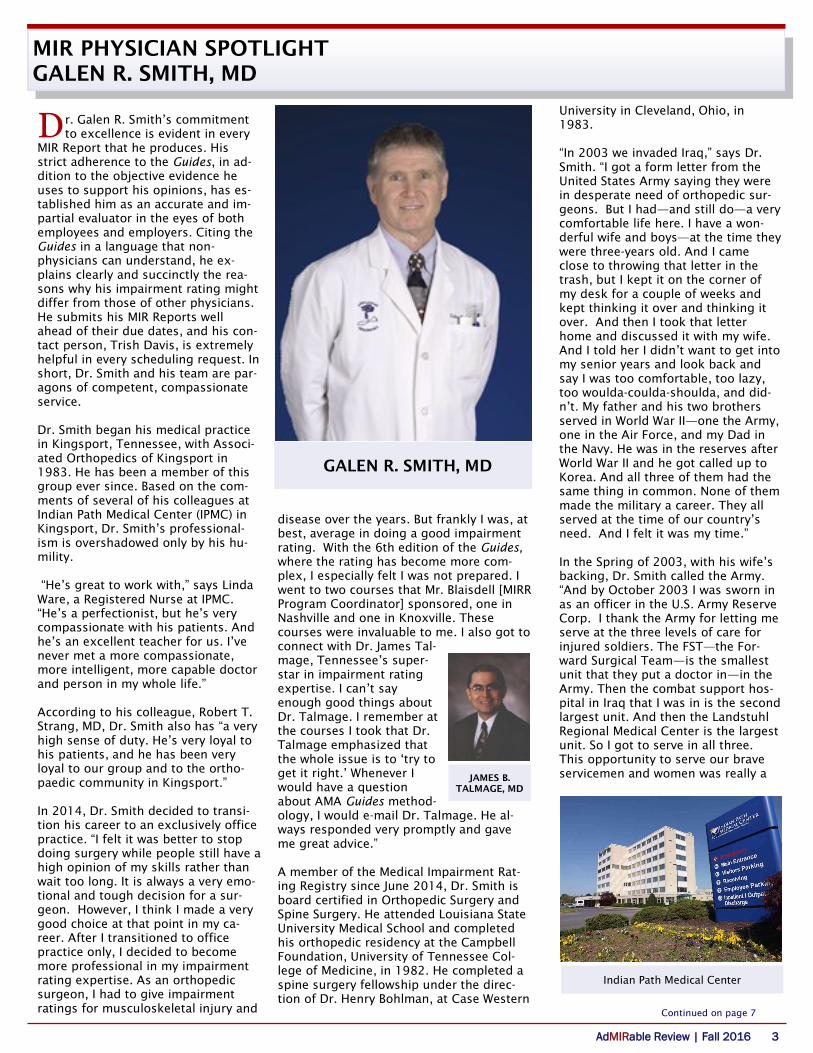
D r. Galen R. Smith’s commitment
to excellence is evident in every
MIR Report that he produces. His
strict adherence to the Guides, in ad-
dition to the objective evidence he
uses to support his opinions, has es-
tablished him as an accurate and im-
partial evaluator in the eyes of both
employees and employers. Citing the
Guides in a language that non-
physicians can understand, he ex-
plains clearly and succinctly the rea-
sons why his impairment rating might
differ from those of other physicians.
He submits his MIR Reports well
ahead of their due dates, and his con-
tact person, Trish Davis, is extremely
helpful in every scheduling request. In
short, Dr. Smith and his team are par-
agons of competent, compassionate
service.
Dr. Smith began his medical practice
in Kingsport, Tennessee, with Associ-
ated Orthopedics of Kingsport in
1983. He has been a member of this
group ever since. Based on the com-
ments of several of his colleagues at
Indian Path Medical Center (IPMC) in
Kingsport, Dr. Smith’s professional-
ism is overshadowed only by his hu-
mility.
“He’s great to work with,” says Linda
Ware, a Registered Nurse at IPMC.
“He’s a perfectionist, but he’s very
compassionate with his patients. And
he’s an excellent teacher for us. I’ve
never met a more compassionate,
more intelligent, more capable doctor
and person in my whole life.”
According to his colleague, Robert T.
Strang, MD, Dr. Smith also has “a very
high sense of duty. He’s very loyal to
his patients, and he has been very
loyal to our group and to the ortho-
paedic community in Kingsport.”
In 2014, Dr. Smith decided to transi-
tion his career to an exclusively office
practice. “I felt it was better to stop
doing surgery while people still have a
high opinion of my skills rather than
wait too long. It is always a very emo-
tional and tough decision for a sur-
geon. However, I think I made a very
good choice at that point in my ca-
reer. After I transitioned to office
practice only, I decided to become
more professional in my impairment
rating expertise. As an orthopedic
surgeon, I had to give impairment
ratings for musculoskeletal injury and
disease over the years. But frankly I was, at
best, average in doing a good impairment
rating. With the 6th edition of the Guides,
where the rating has become more com-
plex, I especially felt I was not prepared. I
went to two courses that Mr. Blaisdell [MIRR
Program Coordinator] sponsored, one in
Nashville and one in Knoxville. These
courses were invaluable to me. I also got to
connect with Dr. James Tal-
mage, Tennessee’s super-
star in impairment rating
expertise. I can’t say
enough good things about
Dr. Talmage. I remember at
the courses I took that Dr.
Talmage emphasized that
the whole issue is to ‘try to
get it right.’ Whenever I
would have a question
about AMA Guides method-
ology, I would e-mail Dr. Talmage. He al-
ways responded very promptly and gave
me great advice.”
A member of the Medical Impairment Rat-
ing Registry since June 2014, Dr. Smith is
board certified in Orthopedic Surgery and
Spine Surgery. He attended Louisiana State
University Medical School and completed
his orthopedic residency at the Campbell
Foundation, University of Tennessee Col-
lege of Medicine, in 1982. He completed a
spine surgery fellowship under the direc-
tion of Dr. Henry Bohlman, at Case Western
JAMES B.
TALMAGE, MD
Continued on page 7
Indian Path Medical Center
4 AdMIRable Review | Fall 2016
I n keeping with the functional assessment philosophies
presented in Chapters 1 and 2 of the 6th
edition of the
Guides, Chapter 12, The Visual System, considers the com-
bined perceptual ability of both eyes in the service of Activi-
ties of Daily Living (ADL). Consequently, the left eye is not
rated without combining it with the right eye, and disfiguring
and other anatomical changes are not considered in this
chapter. They are rated from Section 11.3.
OVERVIEW: TERMS AND METHODOLOGY
Visual impairment is based primarily on objective measure-
ments of visual acuity and visual field. Visual acuity refers to
the sharpness or clarity of vision and is typically measured
by the examinee’s ability to distinguish letters or other sym-
bols arranged in standardized decreasing size and space.
Visual field refers to peripheral vision and may be objectively
measured through a variety of standardized manual or auto-
mated tests while the examinee is focused on a fixed object.
Applying the individual and combined eye results of visual
acuity tests to Table 12-2 on page 288, the rater derives a
Visual Acuity Score (VAS) for right eye (OD), left eye (OS), and
binocular vision (OU) and uses each in Table 12-3 on page
289 to obtain a single Functional Acuity Score (FAS). In simi-
lar fashion, applying the individual and combined eye results
of field vision tests to Table 12-6 on page 296, the rater de-
rives a Visual Field Score (VFS) for right eye, left eye, and
binocular vision and uses each in Table 12-7 on page 297 to
obtain a single Functional Field Score (FFS). The rater then
multiplies the FAS and FFS and divides the result by 100, per
the “Basic Rule” on page 304, to obtain the Functional Vision
Score (FVS). Finally, since the FVS is an ability score (0 = no
ability, 100 = normal ability), the rater subtracts it from 100
to arrive at the Visual System Impairment (VSI). The VSI is
converted to Whole Person Impairment (WPI) using the rule
and formula in the right column of page 306 as demonstrat-
ed by Figure 12-8 on page 307.
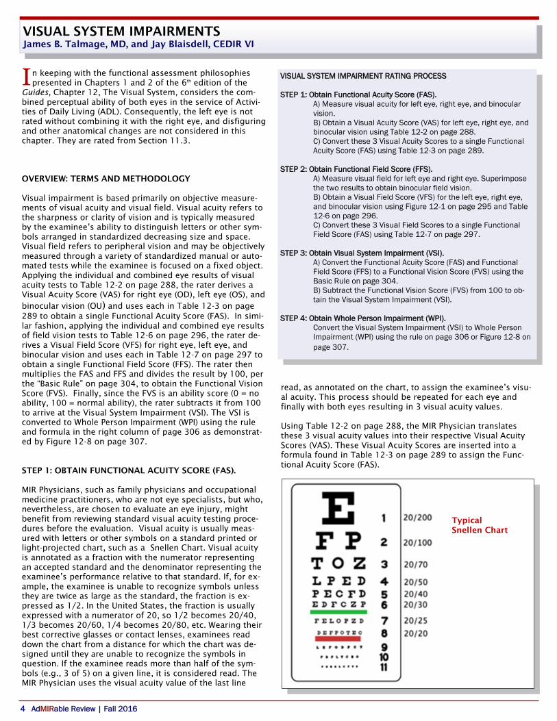
STEP 1: OBTAIN FUNCTIONAL ACUITY SCORE (FAS).
MIR Physicians, such as family physicians and occupational
medicine practitioners, who are not eye specialists, but who,
nevertheless, are chosen to evaluate an eye injury, might
benefit from reviewing standard visual acuity testing proce-
dures before the evaluation. Visual acuity is usually meas-
ured with letters or other symbols on a standard printed or
light-projected chart, such as a Snellen Chart. Visual acuity
is annotated as a fraction with the numerator representing
an accepted standard and the denominator representing the
examinee’s performance relative to that standard. If, for ex-
ample, the examinee is unable to recognize symbols unless
they are twice as large as the standard, the fraction is ex-
pressed as 1/2. In the United States, the fraction is usually
expressed with a numerator of 20, so 1/2 becomes 20/40,
1/3 becomes 20/60, 1/4 becomes 20/80, etc. Wearing their
best corrective glasses or contact lenses, examinees read
down the chart from a distance for which the chart was de-
signed until they are unable to recognize the symbols in
question. If the examinee reads more than half of the sym-
bols (e.g., 3 of 5) on a given line, it is considered read. The
MIR Physician uses the visual acuity value of the last line
read, as annotated on the chart, to assign the examinee’s visu-
al acuity. This process should be repeated for each eye and
finally with both eyes resulting in 3 visual acuity values.
Using Table 12-2 on page 288, the MIR Physician translates
these 3 visual acuity values into their respective Visual Acuity
Scores (VAS). These Visual Acuity Scores are inserted into a
formula found in Table 12-3 on page 289 to assign the Func-
tional Acuity Score (FAS).
VISUAL SYSTEM IMPAIRMENT RATING PROCESS
STEP 1: Obtain Functional Acuity Score (FAS).
A) Measure visual acuity for left eye, right eye, and binocular
vision.
B) Obtain a Visual Acuity Score (VAS) for left eye, right eye, and
binocular vision using Table 12-2 on page 288.
C) Convert these 3 Visual Acuity Scores to a single Functional
Acuity Score (FAS) using Table 12-3 on page 289.
STEP 2: Obtain Functional Field Score (FFS).
A) Measure visual field for left eye and right eye. Superimpose
the two results to obtain binocular field vision.
B) Obtain a Visual Field Score (VFS) for the left eye, right eye,
and binocular vision using Figure 12-1 on page 295 and Table
12-6 on page 296.
C) Convert these 3 Visual Field Scores to a single Functional
Field Score (FAS) using Table 12-7 on page 297.
STEP 3: Obtain Visual System Impairment (VSI).
A) Convert the Functional Acuity Score (FAS) and Functional
Field Score (FFS) to a Functional Vision Score (FVS) using the
Basic Rule on page 304.
B) Subtract the Functional Vision Score (FVS) from 100 to ob-
tain the Visual System Impairment (VSI).
STEP 4: Obtain Whole Person Impairment (WPI).
Convert the Visual System Impairment (VSI) to Whole Person
Impairment (WPI) using the rule on page 306 or Figure 12-8 on
page 307.
Typical
Snellen Chart
VISUAL SYSTEM IMPAIRMENTS
James B. Talmage, MD, and Jay Blaisdell, CEDIR VI

AdMIRable Review | Fall 2016 5
STEP 2: OBTAIN FUNCTIONAL FIELD SCORE (FFS).
Field vision is tested using either manual (kinetic) or auto-
mated (static) perimeters. If the MIR Physician is not an eye
specialist who is able to test field vision, the MIR Physician
should give careful consideration to the reliability, scope, and
date of existing field test results to determine if additional
tests should be scheduled. Static field vision tests are consid-
ered unreliable if either false positives or false negatives ex-
ceed 20% or if fixation errors are greater than 30%. False pos-
itives are the number of times the examinee signifies that
stimuli are seen in the absence of stimuli. False negatives are
the number of times that the examinee fails to respond to
stimuli that should have been seen based on earlier respons-
es. Fixation errors are the number of times that the patient
looks away from the central target. Visual field testing should
be conducted by an ophthalmologist or other licensed and
trained medical doctor with perimetry equipment capable of
detecting deficits well outside the central 60° radius of the
as a gray scale,” which must then be translated into the
“pseudoisopter equivalent to the Goldmann III-4-e isopter” as
demonstrated in Example 12-11 on page 301.
If automated
visual field plots are available in lieu of Goldmann visual field
plots, the MIR Physician should create a pseudoisopter “by
drawing a line surrounding all points with a sensitivity of 10
dB or better, excluding points with less than 10 dB sensitivity
(see Figure 12-5).” 1 (295)
If the MIR Physician is not an eye spe-
cialist and needs assistance converting automated perimeter
results into their pseudoisopter equivalent, the MIR Physician
should contact the Program Coordinator to arrange for addi-
tional payment for consultation with an eye specialist pursu-
ant to TN Rules and Regulations 0800-2-20-.07 (2).
Once the Goldmann III-4-e isopter, or its pseudoisopter
equivalent, has been plotted, the MIR Physician constructs
and applies the testing grid in Figure 12-1 on page 295 in
conjunction with Table 12-6 on page 296 to determine the
examinee’s Visual Field Score (VFS) for the eye in question. A
normal VFS is typically around 100.
The testing grid in Figure 12-2 is constructed by either draw-
ing it on the visual field plot of the III-4-e isopter (or its
equivalent) or by overlaying a transparency of the testing
grid on the field plot. The circular testing grid is divided into
four quadrants (upper left and right, lower left and right)
with 10 meridians drawn from the center of the quadrant, 3
extending in each lower quadrant and 2 extending in each
upper quadrant, at the following degree positions: 25°, 65°,
115°, 155°, 195°, 225°, 255°, 285°, 315°, and 345°.
With the completed testing grid superimposed on the Gold-
mann III-4-e isopter (or its pseudoisopter equivalent), the MIR
Physician should record the peripheral extent (as measured
in degrees from the center of the Field of Vision [FOV]) of
each meridian while consulting Table 12-6. For each meridi-
an, the examinee receives 1 point for stimulus seen at 2° in-
tervals for the first 10°, and then 1 point for each stimulus
seen in the peripheral 20° and beyond. For example, vision
along a meridian extending to a FOV of 8° yields 4 points and
vision along a meridian of 40° yields 8 points. Please note
1Rondinelli R, Genovese E, Katz R, et al. Guides to the Evaluation of Permanent Impairment. 6th ed. Chicago, IL: AMA, 2008
examinee’s field of vision. If additional field tests are need-
ed, the MIR Physician should contact the MIRR Program
Coordinator. If the examinee claims, and the MIR Physician
suspects, that there is no visual field deficit, the MIR Physi-
cian may conduct a simple Confrontation Visual Field test,
as described on page 293, to confirm that the field of vi-
sion appears normal.
The results of Goldmann manual perimetry equipment,
where the test operator manually moves stimuli of various
sizes into the examinee’s field of vision until they are de-
tected, are more direct for impairment purposes because
they are plotted as contour lines, called isopters, which
outline the areas of stimuli perception much like a contour
map. These isopters have different names based on the
stimulus size and intensity. The Goldmann III-4e isopter,
or its equivalent, is necessary to rate visual impairments.
The majority of field vision tests, however, are now con-
ducted on automatic perimeters, such as Humphreys and
Octopus brand machines, in which static stimuli of differ-
ent intensities appear at various locations in the vision
field. Automatic perimetry results are “commonly plotted
Goldmann Manual Perimeter
Humphreys Field Analyzer Automated Perimeter
VISUAL SYSTEM IMPAIRMENTS
(Continued from page 4)
6 AdMIRable Review | Fall 2016
the testing grid, to assign a VFS for binocular vision. The 3
Visual Field Scores (left eye, right, binocular vision) are then
entered into Table 12-7 on page 297 to arrive at a Functional
Field Score (FFS).
STEP 3: OBTAIN VISUAL SYSTEM IMPAIRMENT (VSI).
The process is relatively straightforward once the Functional
Acuity Score (Step 1) and Functional Field Score (Step 2) are
obtained. In accordance with the “Basic Rule” found on page
304, the MIR Physician multiplies the FAS by the FFS and di-
vides the product by 100 to obtain the Functional Vision Score
(FVS). Since the FVS is an ability score, not inability, the MIR
Physician subtracts the FVS from 100 to obtain the Visual Sys-
tem Impairment.
FVS = (FAS x FFS)/100 VSI = 100-FVS
STEP 4: OBTAIN WHOLE PERSON IMPAIRMENT (WPI).
All MIR Reports convert regional and system impairments to
whole person impairments. To convert visual system impair-
that the location on the meridian is rounded to the nearest 2°
within the center 10° of the FOV. Outside 10° of the FOV, the
location on the meridian is rounded to the nearest 10°. If a sco-
toma (blind spot) overlaps a meridian, subtract the radial extent
of the scotoma according to Table 12-6 from the meridians
point value. Finally, with the value of each of the 10 meridians
assigned, the MIR Physician summates the points to arrive at a
total, which is the Visual Field Score (VFS) for the eye that was
tested.
The entire process is repeated to arrive at the VFS for the other
eye, and yet again, with the two monocular fields imposed on
ment to whole person impairment, the MIR Physician fol-
lows the conversion rule on page 306: if the VSI is less than
or equal to 50%, then WPI = VSI; if the VSI is more than 50%,
then WPI = 50 + 0.7 (VSI – 50). This rule is demonstrated
graphically in Figure 12-8 on page 307.
OTHER CONSIDERATIONS
Since standardized measurement analysis is not yet availa-
ble for other aspects of visual impairment such as contrast
sensitivity, photophobia, color vision defects, stereopsis,
and diplopia, the 6th edition of the Guides does not provide
a detailed methodology for considering them. Instead it
allows for the MIR Physician to increase the VSI up to 15
points for “significant factors that affect functional vision
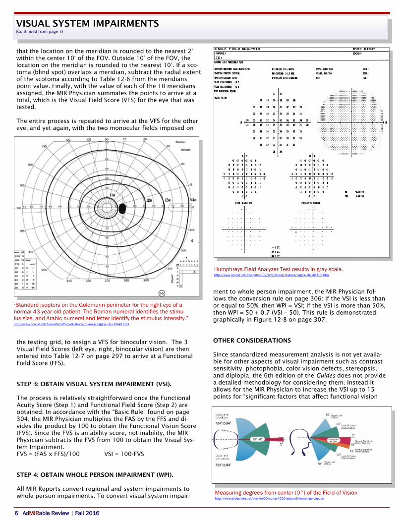
“Standard isopters on the Goldmann perimeter for the right eye of a
normal 43-year-old patient. The Roman numeral identifies the stimu-
lus size, and Arabic numeral and letter identify the stimulus intensity.” http://www.oculist.net/downaton502/prof/ebook/duanes/pages/v3/v3c049.html
Humphreys Field Analyzer Test results in gray scale. http://www.oculist.net/downaton502/prof/ebook/duanes/pages/v8/v8c109.html
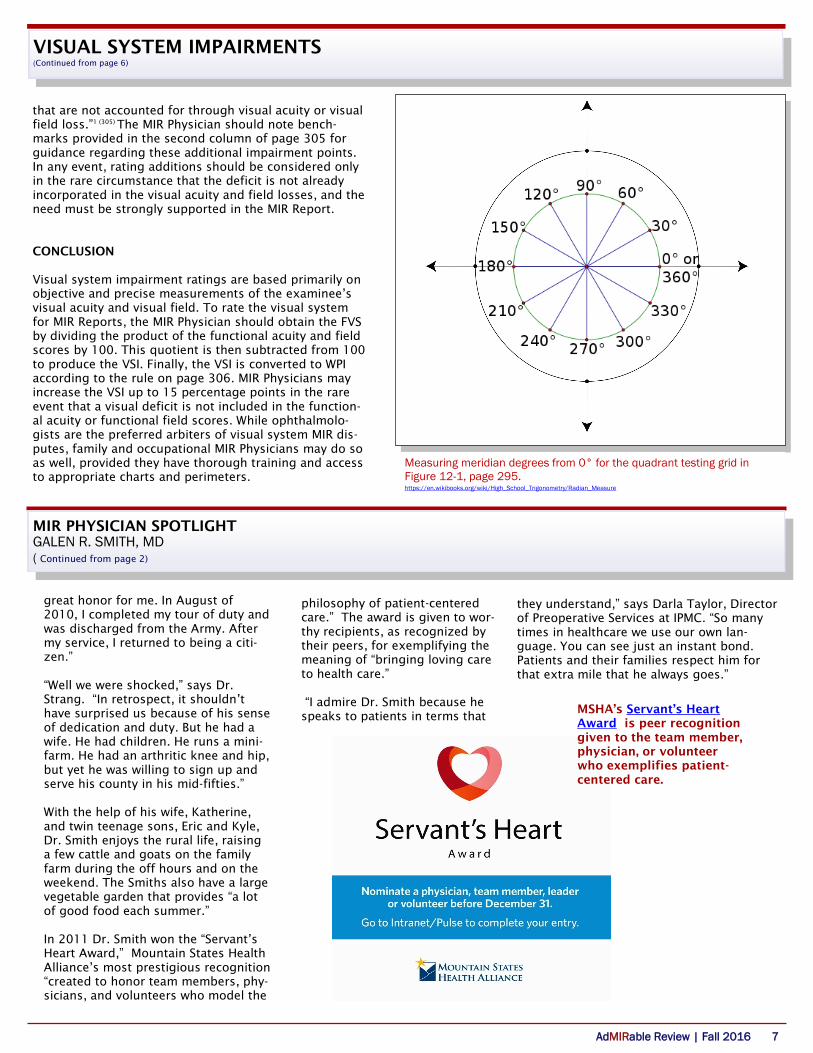
Measuring degrees from center (0°) of the Field of Vision http://www.slideshare.net/marknb00/comp-4010-lecture3-human-perception
VISUAL SYSTEM IMPAIRMENTS
(Continued from page 5)
AdMIRable Review | Fall 2016 7
VISUAL SYSTEM IMPAIRMENTS
(Continued from page 6)
that are not accounted for through visual acuity or visual
field loss.”1 (305)
The MIR Physician should note bench-
marks provided in the second column of page 305 for
guidance regarding these additional impairment points.
In any event, rating additions should be considered only
in the rare circumstance that the deficit is not already
incorporated in the visual acuity and field losses, and the
need must be strongly supported in the MIR Report.
CONCLUSION
Visual system impairment ratings are based primarily on
objective and precise measurements of the examinee’s
visual acuity and visual field. To rate the visual system
for MIR Reports, the MIR Physician should obtain the FVS
by dividing the product of the functional acuity and field
scores by 100. This quotient is then subtracted from 100
to produce the VSI. Finally, the VSI is converted to WPI
according to the rule on page 306. MIR Physicians may
increase the VSI up to 15 percentage points in the rare
event that a visual deficit is not included in the function-
al acuity or functional field scores. While ophthalmolo-
gists are the preferred arbiters of visual system MIR dis-
putes, family and occupational MIR Physicians may do so
as well, provided they have thorough training and access
to appropriate charts and perimeters.
Measuring meridian degrees from 0° for the quadrant testing grid in
Figure 12-1, page 295. https://en.wikibooks.org/wiki/High_School_Trigonometry/Radian_Measure
MIR PHYSICIAN SPOTLIGHT
GALEN R. SMITH, MD ( Continued from page 2)
great honor for me. In August of
2010, I completed my tour of duty and
was discharged from the Army. After
my service, I returned to being a citi-
zen.”
“Well we were shocked,” says Dr.
Strang. “In retrospect, it shouldn’t
have surprised us because of his sense
of dedication and duty. But he had a
wife. He had children. He runs a mini-
farm. He had an arthritic knee and hip,
but yet he was willing to sign up and
serve his county in his mid-fifties.”
With the help of his wife, Katherine,
and twin teenage sons, Eric and Kyle,
Dr. Smith enjoys the rural life, raising
a few cattle and goats on the family
farm during the off hours and on the
weekend. The Smiths also have a large
vegetable garden that provides “a lot
of good food each summer.”
In 2011 Dr. Smith won the “Servant’s
Heart Award,” Mountain States Health
Alliance’s most prestigious recognition
“created to honor team members, phy-
sicians, and volunteers who model the
philosophy of patient-centered
care.” The award is given to wor-
thy recipients, as recognized by
their peers, for exemplifying the
meaning of “bringing loving care
to health care.”
“I admire Dr. Smith because he
speaks to patients in terms that
they understand,” says Darla Taylor, Director
of Preoperative Services at IPMC. “So many
times in healthcare we use our own lan-
guage. You can see just an instant bond.
Patients and their families respect him for
that extra mile that he always goes.”
MSHA’s Servant’s Heart
Award is peer recognition
given to the team member,
physician, or volunteer
who exemplifies patient-
centered care.