Volume 42, Number 3, Pages 43–54 May/June 2005, Supplement ... suppl 1/pdf/kern.pdf · 43 JRRD...
12
43 JRRD JRRD Volume 42, Number 3, Pages 43–54 May/June 2005, Supplement 1 Journal of Rehabilitation Research & Development Muscle biopsies show that FES of denervated muscles reverses human muscle degeneration from permanent spinal motoneuron lesion Helmut Kern, MD; 1 Katia Rossini, DBiol; 2 Ugo Carraro, MD; 2* Winfried Mayr, PhD; 3 Michael Vogelauer, MD; 1 Ursula Hoellwarth, MD; 1 Christian Hofer, DEng 1 1 Ludwig Boltzmann Institute of Electrostimulation and Physical Rehabilitation, Department of Physical Medicine, Wilhelminenspital. A-1171 Vienna, Austria; 2 C.N.R. Institute of Neuroscience, Laboratory of Applied Myology of the Department of Biomedical Science and of the Interuniversity Institute of Myology, University of Padua Medical School, I-35121 Padua, Italy; 3 Center of Biomedical Engineering and Physics, Medical University of Vienna, A-1090 Vienna, Austria Abstract—This paper presents biopsy analyses in support of the clinical evidence of muscle recovery induced by a new system of life-long functional-electrical-stimulation (FES) training in per- manent spinal-motoneuron-denervated human muscle. Not ear- lier than 1 year after subjects experienced complete conus cauda lesion, their thigh muscles were electrically stimulated at home for several years with large skin surface electrodes and an expressly designed stimulator that delivered much longer impulses than those presently available for clinical use. The poor excitability of long-term denervated muscles was first improved by several months of twitch-contraction training. Then, the mus- cles were tetanically stimulated against progressively increased loads. Needle biopsies of vastus lateralis from long-term dener- vated subjects showed severe myofiber atrophy or lipodystrophy beginning 2 years after spinal cord injury (SCI). Muscle biopsies from a group of 3.6- to 13.5-year denervated subjects, who underwent 2.4 to 9.3 years of FES, show that this progressive training almost reverted long-term muscle atrophy/degeneration. Key words: FES, functional electrical stimulation, human, long-duration impulses, long-term denervation, muscle, muscle degeneration, muscle recovery, myofiber regeneration, perma- nent denervation, spinal cord injury, spinal motoneuron lesion. INTRODUCTION Spinal cord injury (SCI) causes a rapid loss of mus- cle mass, which is especially severe when the injury involves spinal motoneurons. While much interest has been shown in the use of functional electrical stimulation (FES) to restore movement of the limbs of subjects para- lyzed by upper motoneuron lesions [1–2], very few clini- cians hope to recover permanent spinal-motoneuron- denervated muscles by means of electrical stimulation training. Indeed, atrophy is especially severe when the injury involves spinal motoneurons, in which long-term Abbreviations: BMCA = Brain Motor Control Assessment, CT = computerized tomography, DDM = denervated and degenerated muscle, EMG = electromyography, EU = European Union, FES = functional electrical stimulation, H-E = hematoxylin-eosin, LMS = lumbosacral magnetic stimulation, SCI = spinal cord injury, TMS = transcranial magnetic stimulation. The work was supported by the European Union (EU) Commission Shared Cost Project RISE (Contract QLG5- CT-2001-02191), the Austrian Ministry of Science, and in part by institutional funds of the Italian C.N.R. Institute of Neuroscience, Unit for Neuromuscular Biology and Physio- pathology, and of the Italian M.U.I.R., Laboratory of Applied Myology, Department of Biomedical Science, Uni- versity of Padua, Italy. * Address all correspondence to Prof. Ugo Carraro, Laboratory of Applied Myology, Department of Biomedical Sciences, Viale G. Colombo 3I-35121 Padova, Italy; +39-049-8276030; fax: +39-049-8276040. Email: [email protected] DOI: 10.1682/JRRD.2004.05.0061
Transcript of Volume 42, Number 3, Pages 43–54 May/June 2005, Supplement ... suppl 1/pdf/kern.pdf · 43 JRRD...

JRRDJRRD Volume 42, Number 3, Pages 43–54
May/June 2005, Supplement 1
Journal of Rehabil itation Research & Development
Muscle biopsies show that FES of denervated muscles reverses human muscle degeneration from permanent spinal motoneuron lesion
Helmut Kern, MD;1 Katia Rossini, DBiol;2 Ugo Carraro, MD;2* Winfried Mayr, PhD;3 Michael Vogelauer, MD;1 Ursula Hoellwarth, MD;1 Christian Hofer, DEng11Ludwig Boltzmann Institute of Electrostimulation and Physical Rehabilitation, Department of Physical Medicine, Wilhelminenspital. A-1171 Vienna, Austria; 2C.N.R. Institute of Neuroscience, Laboratory of Applied Myology of the Department of Biomedical Science and of the Interuniversity Institute of Myology, University of Padua Medical School, I-35121 Padua, Italy; 3Center of Biomedical Engineering and Physics, Medical University of Vienna, A-1090 Vienna, Austria
Abstract—This paper presents biopsy analyses in support of theclinical evidence of muscle recovery induced by a new system oflife-long functional-electrical-stimulation (FES) training in per-manent spinal-motoneuron-denervated human muscle. Not ear-lier than 1 year after subjects experienced complete conus caudalesion, their thigh muscles were electrically stimulated at homefor several years with large skin surface electrodes and anexpressly designed stimulator that delivered much longerimpulses than those presently available for clinical use. The poorexcitability of long-term denervated muscles was first improvedby several months of twitch-contraction training. Then, the mus-cles were tetanically stimulated against progressively increasedloads. Needle biopsies of vastus lateralis from long-term dener-vated subjects showed severe myofiber atrophy or lipodystrophybeginning 2 years after spinal cord injury (SCI). Muscle biopsiesfrom a group of 3.6- to 13.5-year denervated subjects, whounderwent 2.4 to 9.3 years of FES, show that this progressivetraining almost reverted long-term muscle atrophy/degeneration.
Key words: FES, functional electrical stimulation, human,long-duration impulses, long-term denervation, muscle, muscledegeneration, muscle recovery, myofiber regeneration, perma-nent denervation, spinal cord injury, spinal motoneuron lesion.
INTRODUCTION
Spinal cord injury (SCI) causes a rapid loss of mus-cle mass, which is especially severe when the injury
involves spinal motoneurons. While much interest hasbeen shown in the use of functional electrical stimulation(FES) to restore movement of the limbs of subjects para-lyzed by upper motoneuron lesions [1–2], very few clini-cians hope to recover permanent spinal-motoneuron-denervated muscles by means of electrical stimulationtraining. Indeed, atrophy is especially severe when theinjury involves spinal motoneurons, in which long-term
Abbreviations: BMCA = Brain Motor Control Assessment, CT =computerized tomography, DDM = denervated and degeneratedmuscle, EMG = electromyography, EU = European Union, FES= functional electrical stimulation, H-E = hematoxylin-eosin,LMS = lumbosacral magnetic stimulation, SCI = spinal cordinjury, TMS = transcranial magnetic stimulation.The work was supported by the European Union (EU)Commission Shared Cost Project RISE (Contract QLG5-CT-2001-02191), the Austrian Ministry of Science, and inpart by institutional funds of the Italian C.N.R. Institute ofNeuroscience, Unit for Neuromuscular Biology and Physio-pathology, and of the Italian M.U.I.R., Laboratory ofApplied Myology, Department of Biomedical Science, Uni-versity of Padua, Italy.*Address all correspondence to Prof. Ugo Carraro, Laboratoryof Applied Myology, Department of Biomedical Sciences, VialeG. Colombo 3I-35121 Padova, Italy; +39-049-8276030; fax:+39-049-8276040. Email: [email protected]: 10.1682/JRRD.2004.05.0061
43

44
JRRD, Volume 42, Number 3, 2005, Supplement 1
irreversible denervation results in fat substitution andmuscle fibrosis. Although early denervation has beenwidely studied both in animal models and humans, thelong-term effects of denervation have attracted much lessattention since the general belief is that all myofibers dis-appear within several months of denervation [3–5]. Inrats, myofibers exhibit a net loss of nuclear domains for7 months, followed by nuclear groupings, a specific mor-phologic marker of long-term severe muscle atrophy [6].Furthermore, permanent spinal motoneuron denervationhas also demonstrated to be accompanied by a continu-ous production of new myofibers [7–12]. Activated satel-lite cells, myotubes, and regenerated myofibers areconsistently present in atrophic rat muscles, even afteryear-long permanent denervation in both hemidiaphragmand leg muscles [7–9]. Interestingly, we have recentlyextended these observations to human muscle [13]. Allthese events can prolong the period during whichdenervated tissue may be recovered through reinnerva-tion or FES training.
In permanent, complete conus cauda lesions, SCIresults in irreversible loss of the nerve supply to some orall the muscles of the affected limbs, making the markedatrophy of the denervated muscle and the severe second-ary medical problems of skin and bone more difficult totreat successfully (e.g., pressure sores and bone deminer-alization and fracture). Without functional motor nervefibers, activating a sufficient population of myofibers ismore problematic when electric currents are provided bysurface electrodes; 6 months after SCI, long-term spinal-motoneuron-denervated muscle is poorly excitable bystandard clinically approved electrodes and stimulationdevices. Despite these difficulties, pilot studies on FES oflong-term human denervated and degenerated muscles(DDM) have been published [5]. Especially encouragingare some of our previous results, which have shown that anew electrical-stimulation training strategy is effective inrestoring a certain degree of muscle mass and forceproduction even after long-standing complete spinal-motoneuron lesions [14–17]. These clinical works stronglysupported the idea that FES training of denervated mus-cles could restore DDM, a fact that had not previouslybeen recognized, principally because of the lack of cus-tomized technology that can meet demands completelydifferent from those required for motor-nerve stimulation.On the other hand, a stimulator delivering biphasic long-duration impulses with a pulse width between 10 ms and200 ms and amplitudes of up to ±80 V and ±200 mA is
able to elicit DDM twitch contractions via surface elec-trodes [16–17]. Within a few months, such trainingincreases myofiber excitability of the quadriceps muscleto a level that allows tetanic contractions. Finally, thestructural and metabolic characteristics of the tetanicallystimulated muscles are restored to values that allow elec-trically supported standing up and standing. Recently,clinical results by computerized tomography (CT) scanand force measurements during tetanic stimulation of thethigh muscles were promising enough to convince ethicalcommittees and funding agencies to accept our proposalfor a European Union (EU) Trial (“RISE”) for restorationof DDM in conus cauda syndrome by FES training. Withthe support of the EU RISE project, we are using light andelectron microscopy to analyze in detail biopsies ofhuman long-term DDM before and after FES.
We present the preliminary results of morphometricanalyses of DDM in a group of subjects enrolled in theRISE Trial, who were biopsied before FES. We comparethese subjects with a previous group of subjects who hadundergone FES training of denervated muscles and werebiopsied after 2.4 to 9.3 years of FES training that was ini-tiated between 1.2 and 8.7 years after SCI. Our purposewas to present biopsy analyses in support of the clinicalevidence of muscle recovery induced by the new life-longFES training in permanent spinal-motoneuron-denervatedhuman muscle. Microscopic muscle analyses from thesetwo independent groups of subjects support our previousclinical observations [14–15,18]. Besides recovery of theatrophic muscle fibers, sustained regeneration of newmyofibers may explain the observations that in the FES-treated muscle, the fibers are larger than the fibers inuntreated muscles of the group of DDM subjects from theEU RISE Trial [19].
MATERIALS AND METHODS
Characteristics of SubjectsNine subjects (one female and eight male, aged 20 to
49), who had experienced traumatic conus cauda lesions,were assigned to two groups. The first group includedfive DDM subjects who were recently enrolled in the EURISE Trial and were biopsied pre-FES training. Althoughpre- and post-FES-training biopsies of the RISE subjectswere approved by ethical committees and accepted bysubjects, the follow-up was too brief for us to performposttraining bioptic analyses on this group (Table 1).

45
KERN et al. FES recovery of human denervated degenerated muscle
The second group of four subjects included the firstand only worldwide analyses of spinal-motoneuron-dener-vated FES-trained subjects who demonstrated clear clini-cal signs of quadriceps femoris muscle mass and forceimprovements after several years of daily training (see“Training Strategy”). We assessed complete spinal moto-neuron denervation of the quadriceps muscle by electro-physiological testing, i.e., by chronaxie measurements,needle electromyography (EMG), Brain Motor ControlAssessment (BMCA), and transcranial and lumbosacralmagnetic stimulation (TMS and LMS, respectively).Rheobase and chronaxie measurements [20–22] wereperformed with the use of a constant-current stimulatorand two touch electrode probes placed successively tothree different sites of the quadriceps muscle. Chronaxievalues, which in normal innervated muscles are 0.3 to0.7 ms, were over 20 to 30 ms in the denervated group.Excitability of the FES-trained denervated musclesincreased (chronaxie of 5 to 10 ms), but never reachednormal values.
Needle EMG of the quadriceps muscle was accord-ing to Dumitri and Zwarts [23]. The needle was insertedinto the m. rectus femoris, which we examined at differ-ent depths and in four directions. When necessary, weperformed additional needle EMG analyses in the vastuslateralis and medialis muscles. We looked for insertionactivity, spontaneous activity during muscle relaxation(fibrillation potentials, positive sharp waves, and fascicu-lations), and volitional activity. Needle EMG examina-
tion was completed by the stimulation of the femoralnerve at the groin with a supramaximal single pulse toelicit an M-wave. In both denervated and FES-traineddenervated muscles, no EMG responses occurred duringvolitional activation.
We conducted BMCA to rule out any voluntary orreflex innervation of the quadriceps muscle. The BMCAprotocol is a comprehensive multichannel-surface EMGrecording of the limb muscles used to characterize motor-control features in persons with upper and/or spinalmotoneuron dysfunction [24]. Key information is con-tained in the overall temporal pattern of motor-unit activ-ity, observed in the EMG envelope. In both denervatedand FES-trained denervated muscles, no motor outputswere recorded during the entire BMCA protocol.
We recorded stimulus-induced activities of the limbmuscles with multichannel surface EMG according toDimitrijevic et al. [25]. Transcranial stimulation wasconducted with a double-cone coil (MAGSTIM 200, Mag-stim Company Ltd, Wales, UK) placed over the medialcaput. We applied lumbosacral stimulation at vertebral lev-els T12, L2, and L4 with the use of a circular coil. Weincreased the amplitude of the magnetic stimulation from0.4 T to 4 T in steps of 10 percent. In both the denervatedand the FES-trained denervated muscle groups reportedhere, no responses occurred to either TMS or LMS. In thefew cases in which LMS showed some incomplete spinal-motoneuron denervation, the results of the microscopy ofthe quadriceps muscle were not included in this paper.
Table 1.Time intervals between spinal cord injury (SCI) and muscle biopsy, between SCI and onset of functional electrical stimulation (FES), and between start of FES and biopsy in long-term spinal-motoneuron denervation of human vastus lateralismuscle; biopsies taken uni- or bilaterally.
Subject Age/SexTime Intervals (yr) Between
SCI and Muscle Biopsy SCI and FESdm Onset FESdm Onset and Biopsy
Den1&2 20/Male 0.8 — —Den3 37/Male 1.3 — —Den4&5 45/Female 2.9 — —Den6 49/Male 3.3 — —Den7&8 33/Male 19.0 — —FESdm1&2 48/Male 3.6 1.2 2.4FESdm3&4 42/Male 6.3 2.0 4.3FESdm5&6 35/Male 10.6 1.3 9.3FESdm7 30/Male 13.5 8.7 4.8
Den = denervated muscle/myofibers, FESdm = functional electrical stimulation of denervated muscle.

46
JRRD, Volume 42, Number 3, 2005, Supplement 1
Training StrategyThe contractile response of denervated muscle to elec-
trical stimulation depends on the stage of postdenervationmuscle atrophy/degeneration, which in turn depends on thetime between the denervation event and the onset of stimu-lation [15,18,21]. In subjects who had been injured for notless than 1 year, we applied biphasic stimulation impulsesof very long duration and high intensity at the beginning oftreatment [16]. We then adjusted the stimulation parame-ters according to the increasing excitability induced by FEStraining. In short, the several-year-long stimulation strategymay be divided in four phases (see “Phases” 1 to 4). Aphysiotherapist evaluated subjects every 6 to 8 weeks andprogressively modified the stimulation parameters to beused at home 5 days a week. The subjects themselvesapplied the electrode above their thigh muscles, always inthe same positions, and activated the preprogrammedstimulator. The surface electrodes made of conductive sili-cone rubber were applied directly to the skin via a wetsponge cloth (early training) or gel (later on, when theirskin had adapted to the high electrical current). Flexibleelectrodes that fit closely to uneven skin surface providedhomogeneous distribution of the electrical field in the stim-ulated thighs. Every 9 to 12 months, we clinically assessedthe subjects.
Phase 1: Early Twitch StimulationAt least 1.2 years after SCI (and after appropriate
instructions to avoid skin burns related to the high currentlevels to be used), the subjects began a home training pro-gram. Anatomically shaped electrodes of a large surfacearea were strapped to the anterior aspect of subjects’ thighsin proximal and distal positions. First, we assessed theseverely reduced excitability of long-term denervated myo-fibers (long-term complete spinal-motoneuron denervation)by delivering very long biphasic rectangular impulses,which, however, yielded only twitch contractions of thethigh muscles. To activate fibers throughout the quadricepsfemoris muscles in complete conus cauda syndrome, wedelivered biphasic rectangular current pulses of 150 to200 ms duration, up to ±200 mA amplitude, representing animpulse energy of up to 3.2 J. Since no stimulator on themarket could deliver such a high current intensity, we devel-oped a generator of long, high-strength stimuli [16–17].Training was initiated with stimulation at 2 Hz, anddelivered for 15 min per day (a series of 4 s “on,” 2 s“off”), 5 days a week. The duration of these impulses wasapproximately 1,500 times longer than that of the impulses
used in subjects with upper motoneuron lesions. With aninterpulse interval of about 400 ms, the resulting stimula-tion frequency was slightly less than 2 Hz (single twitcheselicited every half second). During the next few months, theprogressively increasing muscle excitability permitted anincrease of the twitch stimulation to series of 5 s “on,” 1 s“off,” 3 to 5 min of stimulation with 1 to 2 min of rest.
Phase 2: Late Twitch StimulationAs the training proceeded, muscle excitability contin-
ued to increase. Thus, during the successive 3 months oftraining, the pulses could be shortened to 80 to 100 ms.After an additional 1 to 2 months, the protocol waschanged to the following tetanic pattern (Phases 3 and 4).
Phase 3: Burst Stimulation for Long-Term Spinal-Motoneuron-Denervated Muscles
The tetanic pattern for DDM [14–16,18] consisted of a40 ms pulse and a 10 ms pause delivered at 20 Hz for 2 s“on,” 2 s “off,” 3 to 5 min of stimulation with 1 min of rest(i.e., 3 to 5 times a session), twice a day, 5 days a week.
Phase 4: Force/Endurance StimulationBetween the 9th and 12th month of FES training of
denervated muscles, force-training sessions were intro-duced by tetanic contractions with 70 to 80 percent ofmaximum load, 8 to 12 repetitions, 4 to 6 sets, with 2 minof rest, twice a week. At first, the leg contracted to fullknee extension without any ankle weight, and later, withan ankle weight of up to 5 kg, in 0.5 kg steps.
With this progressive FES training, the mass andforce of thigh muscles increased to values that allowedelectrical-stimulation-supported standing up and standingexercise [14–16,18].
Analyses of Human Muscle BiopsyUsing Bergström needles (UNIMED 5.00 × 100 mm,
Ref. No. 23.601 500100), we biopsied the right and leftvastus lateralis muscle through a small skin area (at thetimes stated in Table 1). We stained cryosections of frozenbiopsies with hematoxylin-eosin (H-E). The details of fibermorphometry are reported by Rossini et al. [13]. In brief,we collected three 10 µm-thick sections on glass slides andstained them with H-E with the use of conventionaltechniques. We acquired images with a Zeiss microscopeconnected to a Leica DC 300F camera at low magnifica-tion under the same conditions that we used to acquire areference ruler. The minimum transverse diameter of each

47
KERN et al. FES recovery of human denervated degenerated muscle
myofiber was measured against the reference ruler. Wegrouped the myofibers and plotted the relative percentile in10 μm steps. We performed morphometric analysis withScion Image for Windows, version Beta 4.0.2 (by 2000Scion Corporation).*
RESULTS
Light Microscopy of Human Long-Term Denervated Muscle
Denervation of skeletal muscle causes rapid loss incontractile force, which is accompanied by several struc-tural, biochemical, and physiological changes. Despitethe severe atrophy of the thigh, Figure 1 surprisingly dis-plays that 1-year spinal-motoneuron-denervated humanvastus lateralis muscles show more than 70 percent ofthe cryosection area being covered by atrophic myofiberprofiles (Figure 1(a) and (b)). Only 3 years post-SCI, thebiopsies show the expected degeneration of muscle tis-sue. Indeed, fat and loose connective tissue areas prevailon the myofiber area (Figure 1(c) and (d)). The micro-scope images on which Figure 1 is based show that up to1 year after SCI, the denervated myofibers underwentpure atrophy, while adipocytes and collagen sheets arescanty (Figure 2(a)–(b) and (d)–(e), respectively; com-pare in the inset of Figure 2(b) the fiber size of normalhuman muscle). These results are unattended, at least toour knowledge. The severe lipodystrophy of lower moto-neuron-denervated human muscles is shown 3.3 yearsand 19.0 years after SCI, as shown in Figure 2(g)–(h),and (j)–(k), respectively: Here, fat and/or loose andfibrous connective tissues prevail over the atrophic myo-fibers (3- to 19-year denervation percentile myofiber areadecreases to 18.9 mean ± 9.9 standard deviation [SD]versus 76.1 ± 8.6 at 1-year denervation, p < 0.001, by stu-dent’s t-test for independent groups). On the other hand,Table 2 shows that the cumulative value for eightbiopsies of denervated muscle minimum diameter is 16.8 ±4.5 µm without significant differences between early andlate denervation.
*Free software downloaded from the Web site: www.scioncorp.com
Figure 1. Human long-term denervated vastus lateralis muscle. Percentilecryosection area covered by myofibers or connective tissues in long-termspinal-motoneuron denervation of human vastus lateralis muscle:(a) 0.8 yr denervation, (b) 1.3 yr denervation, (c) 3.3 yr denervation, and(d) 19.0 yr denervation. Heavy muscle degeneration is present afterdenervation periods of more than 2 years. (Den = denervated myofibers;numbers after “Den” identify biopsy taken.)

48
JRRD, Volume 42, Number 3, 2005, Supplement 1
Figure 2.Human long-term denervated vastus lateralis muscle: (a) to (c) 0.8 yr denervation, (d) to (f) 1.3 yr denervation, (g) to (i) 3.3 yr denervation, (j) to (l) 19 yrdenervation. Inset in (b) is of normal human muscle. Cryosections are stained with hematoxylin-eosin (H-E). (a), (d), (g), and (j) scale bar: 1 mm; (b),(e), (h), and (k) scale bar: 100 μm. Heavy fat infiltration and increased content of fibrous connective tissue were present in biopsies taken more than2 years after denervation. (c), (f), (i), and (l): fiber size spectra show that in all samples, 50% of myofibers had diameters smaller than 20 μm. (Den =denervated myofibers; numbers after “Den” identify biopsy.)

49
KERN et al. FES recovery of human denervated degenerated muscle
HISTOLOGY OF LONG-TERM DDM WITH FES TRAINING OF DENERVATED MUSCLES
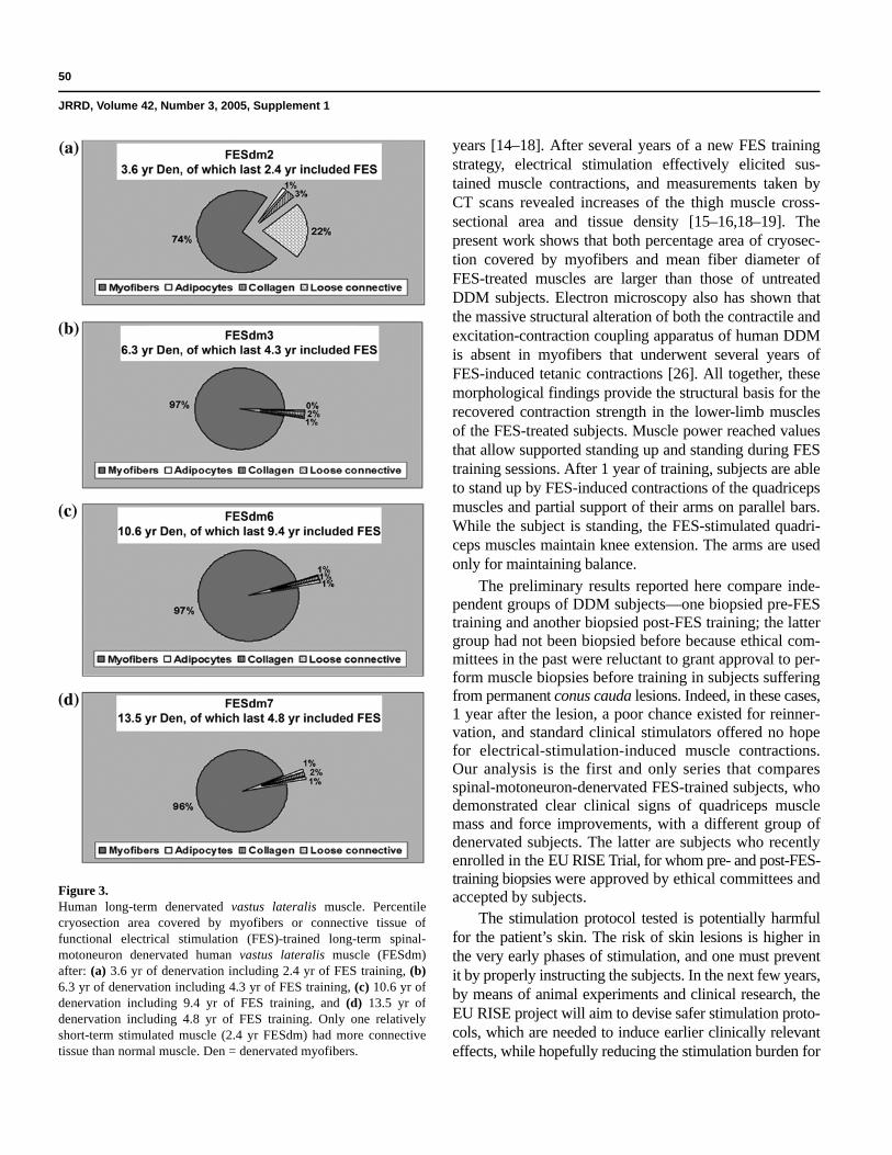
Figures 3 and 4 show four examples of the effects ofFES training on human long-term spinal-motoneuron-denervated vastus lateralis muscles. In Figure 3, morethan 75 percent of the cryosection area is covered bymyofiber profiles, and in Figure 4, the biopsies showround myofibers of heterogeneous size, but with hyper-trophic fibers clearly prevailing.
A residual population of small myofibers is differen-tially present in the electrically stimulated muscles, asexemplified by fiber size spectra in Figure 4(b), (d), (f),and (h). The mean diameter is 26.7 ± 22.9 µm, 45.7 ±18.3 µm, 39.2 ± 18.0 µm, and 58.2 ± 24.1 µm, respec-tively. The cumulative value for seven vastus lateralisbiopsies is 42.2 ± 10.8 µm, which is significantly higherthan the value of 16.8 ± 4.5 µm for the eight long-termdenervated human muscles (p < 0.001 by student’s t-test
for independent groups). Right and left legs in allsubjects have concordant mean fiber diameter and similarprofiles of their fiber diameter spectra, but the two groupsare small (five subjects in the DDM group and foursubjects in the FES-trained denervated muscle group)and heterogeneous to have statistical significance. Inconclusion, after several years of electrically inducedexercise, FES-trained myofibers overcame the myofibersize of normal sedentary adults (37.9 ± 9.5 µm, inset ofFigure 4(a)).
DISCUSSION
Our previous clinical assessments have shown thatmuscle atrophy/degeneration due to long-term spinal-motoneuron denervation can be reversed with the use ofthe FES protocol developed in Vienna during the last few
Table 2.Time intervals between spinal cord injury (SCI) and muscle biopsy, percentile cryosection area covered by myofibers or connective tissues, and meanminimum diameter of denervated myofibers (Den) or of functional electrical stimulation (FES)-trained denervated myofibers (FESdm) in long-termspinal motoneuron denervation of human vastus lateralis.
BiopsyTime (yr) Percentile Area Covered by
Diameter†
(Mean ± SD)Den* FESdm Fibers† Adipocyte‡ Connective Tissue†
Den1 0.8 — 70.0 1.2 28.8 15.6 ± 6.8Den2 0.8 — 86.0 2.4 11.6 19.2 ± 7.8Den3 1.3 — 72.4 3.1 24.5 16.2 ± 10.0Den4 2.9 — 26.4 12.6 61.0 15.9 ± 10.3Den5 2.9 — 11.6 3.1 85.3 22.3 ± 18.0Den6 3.3 — 5.4 44.8 49.8 8.12 ± 4.5Den7 19.0 — 28.4 2.6 69.0 15.3 ± 15.3Den8 19.0 — 22.5 25.5 52.0 21.6 ± 25.3Mean ± SD 6.3 ± 7.9 — 40.3 ± 30.9 11.9 ± 15.6 47.8 ± 24.7 16.8 ± 4.5§
FESdm1 3.6 2.4 97.5 1.3 1.2 30.7 ± 23.7FESdm2 3.6 2.4 73.4 1.3 25.3 26.7 ± 22.9FESdm3 6.3 4.3 97.5 0.2 2.3 45.7 ± 18.3FESdm4 6.3 4.3 98.0 0.5 1.5 46.5 ± 14.3FESdm5 10.6 9.4 97.7 0.5 1.8 48.1 ± 12.2FESdm6 10.6 9.4 97.4 0.5 2.1 39.2 ± 18.0FESdm7 13.5 4.8 96.3 1.2 2.5 58.2 ± 24.1Mean ± SD 7.8 ± 3.8 — 94.0 ± 9.1 0.8 ± 0.5 5.2 ± 8.9 42.2 ± 10.8¶
*Den vs. FESdm, student’s t-test (independent groups), p = 0.324†p < 0.001‡p = 0.042§Cumulative values of 8 Den biopsies¶Cumulative values of 7 FESdm biopsiesSD = standard deviation
50
JRRD, Volume 42, Number 3, 2005, Supplement 1
years [14–18]. After several years of a new FES trainingstrategy, electrical stimulation effectively elicited sus-tained muscle contractions, and measurements taken byCT scans revealed increases of the thigh muscle cross-sectional area and tissue density [15–16,18–19]. Thepresent work shows that both percentage area of cryosec-tion covered by myofibers and mean fiber diameter ofFES-treated muscles are larger than those of untreatedDDM subjects. Electron microscopy also has shown thatthe massive structural alteration of both the contractile andexcitation-contraction coupling apparatus of human DDMis absent in myofibers that underwent several years ofFES-induced tetanic contractions [26]. All together, thesemorphological findings provide the structural basis for therecovered contraction strength in the lower-limb musclesof the FES-treated subjects. Muscle power reached valuesthat allow supported standing up and standing during FEStraining sessions. After 1 year of training, subjects are ableto stand up by FES-induced contractions of the quadricepsmuscles and partial support of their arms on parallel bars.While the subject is standing, the FES-stimulated quadri-ceps muscles maintain knee extension. The arms are usedonly for maintaining balance.
The preliminary results reported here compare inde-pendent groups of DDM subjects—one biopsied pre-FEStraining and another biopsied post-FES training; the lattergroup had not been biopsied before because ethical com-mittees in the past were reluctant to grant approval to per-form muscle biopsies before training in subjects sufferingfrom permanent conus cauda lesions. Indeed, in these cases,1 year after the lesion, a poor chance existed for reinner-vation, and standard clinical stimulators offered no hopefor electrical-stimulation-induced muscle contractions.Our analysis is the first and only series that comparesspinal-motoneuron-denervated FES-trained subjects, whodemonstrated clear clinical signs of quadriceps musclemass and force improvements, with a different group ofdenervated subjects. The latter are subjects who recentlyenrolled in the EU RISE Trial, for whom pre- and post-FES-training biopsies were approved by ethical committees andaccepted by subjects.
The stimulation protocol tested is potentially harmfulfor the patient’s skin. The risk of skin lesions is higher inthe very early phases of stimulation, and one must preventit by properly instructing the subjects. In the next few years,by means of animal experiments and clinical research, theEU RISE project will aim to devise safer stimulation proto-cols, which are needed to induce earlier clinically relevanteffects, while hopefully reducing the stimulation burden for
Figure 3.Human long-term denervated vastus lateralis muscle. Percentilecryosection area covered by myofibers or connective tissue offunctional electrical stimulation (FES)-trained long-term spinal-motoneuron denervated human vastus lateralis muscle (FESdm)after: (a) 3.6 yr of denervation including 2.4 yr of FES training, (b)6.3 yr of denervation including 4.3 yr of FES training, (c) 10.6 yr ofdenervation including 9.4 yr of FES training, and (d) 13.5 yr ofdenervation including 4.8 yr of FES training. Only one relativelyshort-term stimulated muscle (2.4 yr FESdm) had more connectivetissue than normal muscle. Den = denervated myofibers.

51
KERN et al. FES recovery of human denervated degenerated muscle
Figure 4.Effects of functional electrical stimulation (FES) training on histology of long-term human denervated and degenerated muscle: (a) and (b) 3.6 yrof denervation of which 2.4 yr included FES, (c) and (d) 6.3 yr of denervation of which 4.3 yr included FES, (e) and (f) 10.6 yr of denervation ofwhich 9.4 yr included FES, and (g) and (h) 13.5 yr of denervation of which 4.8 yr included FES. Inset in (a) is of normal human muscle.Cryosections are stained with hematoxylin-eosin (H-E). Scale bar: 100 μm. Round myofibers pave biopsy specimens. Residual population of smallmyofibers is differentially present in four electrical stimulated muscles, as also shown by fiber size spectra in (b), (d), (f), and (h). Severelyatrophic myofibers are frequent in only relatively short-term stimulation cases (a) and (b). FESdm = FES-trained denervated myofibers.

52
JRRD, Volume 42, Number 3, 2005, Supplement 1
the subjects. A major improvement in subjects’ quality oflife would be attained if the number of sessions in the life-long stimulation program were successfully reduced to onesession per day. Any successive change in stimulationparameters/modalities reducing the amount of currentdelivered will warrant a second-generation protocol.
An important by-product of our biopsy study is theunexpectedly healthy appearance of the spinal-motoneuron-denervated human muscle 1 year after SCI. If confirmed byindependent studies, this result could open new perspectivesin the management of subjects with spinal-motoneuron-denervated muscle who are awaiting reinnervation and/orFES training.
CONCLUSION
We believe that the effectiveness of FES trainingfor the recovery of irreversibly spinal-motoneuron-denervated human muscles is sufficiently supported byour structural muscle analyses. These results justify fur-ther endeavors in this field on our part and encourageother researchers to independently verify our hypothesesand conclusions.
REFERENCES
1. Graupe D. An overview of the state of the art of non-invasive FES for independent deambulation by thoraciclevel paraplegics. Neurol Res. 2002;24:431–42.
2. Johnston TE, Finson RL, Smith BT, Bonaroti DM, BetzRR, Mulcahey MJ. Functional electrical stimulation foraugmented walking in adolescents with incomplete spinalcord injury. J Spinal Cord Med. 2003;26:390–400.
3. Carraro U, Catani C, Biral D. Selective maintenance ofneurotrophically regulated proteins in denervated rat dia-phragm. Exp Neurol. 1979;63:468–75.
4. Carraro U, Catani C, Dalla Libera L. Myosin light andheavy chains in rat gastrocnemius and diaphragm musclesafter chronic denervation or reinnervation. Exp Neurol.1981;72:401–12.
5. Carraro U. Modulation of trophism and fiber type expres-sion of denervated muscle by different patterns of electricalstimulation. Basic Appl Myol. 2002;12:263–72.
6. Viguie CA, Lu D-X, Huang S-K, Rengen H, Carlson BM.Quantitative study of the effects of long-term denervationon the extensor digitorum longus muscle of the rat. AnatRec. 1997;248:346–54.
7. Carraro U, Morale D, Mussini I, Lucke S, Cantini M, BettoR, Catani C, Dalla Libera L, Danieli Betto B, Noventa D.Chronic denervation of rat diaphragm: maintenance offiber heterogeneity with associated increasing uniformityof myosin isoforms. J Cell Biol. 1985;100:161–74.
8. Mussini I, Favaro G, Carraro U. Maturation, dystrophicchanges and the continuous production of fibers in skeletalmuscle regenerating in the absence of nerve. J NeurophatolExp Neurol. 1987;46:315–31.
9. Carraro U, Catani C, Degani A, Rizzi C. Myosin expressionin denervated fast and slow twitch muscles: fibre modu-lation and substitution. In: Pette D, editor. The dynamicstate of muscle fibres. Berlin (Germany): Walter deGruyter; 1990. p. 247–62.
10. Borisov AB, Dedkov EI, Carlson BM. Interrelations ofmyogenic response, progressive atrophy of muscle fibers,and cell death in denervated skeletal muscle. Anat Rec.2001;264:203–18.
11. Carraro U, Rossini K, Zanin ME, Rizzi C, Mayr W, KernH. Induced myogenesis in long-term permanent denerva-tion: perspective role in functional electrical stimulation ofdenervated legs in humans. Basic Appl Myol. 2002;12:53–64.
12. Carlson BM, Borisov AB, Dedkov EI, Dow D, Kostromi-nova TY. The biology and restorative capacity of long-termdenervated skeletal muscle. Basic Appl Myol. 2002;12:247–54.
13. Rossini K, Zanin ME, Carraro U. To stage and to quantifyregenerative myogenesis in human long-term permanentdenervated muscle. Basic Appl Myol. 2002;12:277–87.
14. Kern H. Funktionelle Elektrostimulation paraplegischerPatienten. Österr Z Phys Med. 1995;5 Heft 1 Supplemen-tum:1–79.
15. Kern H, Hofer C, Strohhofer M, Mayr W, Richter W, Stohr H.Standing up with denervated muscles in humans using func-tional electrical stimulation. Artif Organs. 1999;23:447–52.
16. Hofer C, Mayr W, Stöhr H, Unger E, Kern H. A stimulatorfor functional activation of denervated muscles. ArtifOrgans. 2002;26:276–79.
17. Mayr W, Hofer C, Bijak M, Roflt D, Unger E, Reichel M.Functional Electrical Stimulation (FES) of denervatedmuscles: existing and prospective technological solutions.Basic Appl Myol. 2002;12:287–90.
18. Kern H, Hofer C, Mödlin M, Forstner C, Mayr W, RichterW. Functional electrical stimulation (FES) of long-termdenervated muscles in humans: Clinical observations andlaboratory findings. Basic Appl Myol. 2002;12:291–97.
19. Kern H, Salmons S, Mayr W, Rossini K, Carraro U. Recov-ery of long-term denervated human muscles induced byelectrical stimulation. Muscle Nerve. 2005;31(1):98–101.
20. Edel H. Fibel der Elektrodiagnostik und Elektrotherapie.München (Germany): Müller und Steinicke; 1983. p. 1–325.

53
KERN et al. FES recovery of human denervated degenerated muscle
21. Gutmann E. The denervated muscle. Prague: PublishingHouse of the Czechoslovak Academy of Sciences; 1962.p. 1–486.
22. Jantsch H, Schuhfried F. Niederfrequente Ströme zur Diag-nostik und Therapie. Wien-München (Germany): Maudrich;1981. p. 1–286.
23. Dumitru D, Zwarts M. Needle electromyography. In:Dumitru D, Amato AA, Zwarts M, editors. Electrodiagnos-tic medicine. 2nd ed. Philadelphia (PA): Hanley & BelfusInc.; 2002. p. 257–91.
24. Sherwood AM, McKay B, Dimitrijevic MR. Motor controlafter spinal cord injury: assessment using surface EMG.Muscle Nerve. 1996;19:966–79.
25. Dimitrijevic MR, Kofler M, McKay WB, Sherwood AM,Van der Linden C, Lissens MA. Early and late lower limb
motor evoked potentials elicited by transcranial magneticmotor cortex stimulation. Electroencephalogr Clin Neuro-physiol. 1992;85:365–73.
26. Kern H, Boncompagni S, Rossini K, Mayr W, Fanò G,Zanin ME, Podhorska-Okolow M, Protasi F, Carraro U.Long-term denervation in humans causes degeneration ofboth contractile and excitation-contraction coupling appa-ratus that can be reversed by functional electrical stimula-tion (FES). A role for myofiber regeneration? J Neuro-phatol Exp Neurol. 2004;63:919–31.
Submitted for publication May 25, 2004. Accepted inrevised form January 20, 2005.