Volume 2, Issue 1 (February 2010) - Dr. Babette Gladstein, VMD
84
VOLUME TWO | ISSUE ONE | FEBRUARY 2010 www.journal of prolotherapy.com IN THIS ISSUE BEULAH LAND PRESS ISSN 1944-0421 (print) ISSN 1944-043X (online) ■ ■ ■ ■ ■ ■ ■ ■ ■ ■ ■ ■ ■ ■ ■ ■ Athletes Do Not Stop Before the Finish Line Letters to the Editor from Timothy Speciale, DO & Barbara Young Prolotherapy in South Korea: Interview with Dr. Choi Yung Do An Interview with a Personal Injury Attorney Prolotherapy as an Alternative to Surgery: A Prospective Pilot Study of 34 Patients from a Private Medical Practice Hyperthermia Induces Venous Blood Alkalosis: A Study in Five Ironman Triathletes 69 Year-old Still Running 100-mile Races Thanks to Prolotherapy Prolotherapy for Professional Sports Injuries The Ligament Injury Connection to Osteoarthritis The Acceleration of Articular Cartilage Degeneration in Osteoarthritis by Nonsteroidal Anti-inflammatory Drugs Prolotherapy Case Studies from Veterinarians Review of Free Yourself from Chronic Pain and Sports Injuries By Donna Alderman, DO Prolotherapy Injection Technique of the Elbow Literature Reviews: Prolotherapy for Sports Injuries Introduction of Prolotherapy in the Caribbean Seminars, Training, & Organizations JOURNAL of PROLOTHERAPY VOLUME TWO | ISSUE ONE | FEBRUARY 2010 | PAGES 257-336 BEULAH LAND PRESS THE LIGAMENT-NSAID CONNECTION TO Degenerative Osteoarthritis After Before
Transcript of Volume 2, Issue 1 (February 2010) - Dr. Babette Gladstein, VMD

V O L U M E T W O | I S S U E O N E | F E B R U A R Y 2 0 1 0 w w w . j o u r n a l of p r o l o t h e r a p y . c o m
IN THIS ISSUE
B E U L A H L A N D P R E S S
ISSN 1944-0421 (print)ISSN 1944-043X (online)
■ ■
■
■
■
■
■
■
■ ■
■
■
■ ■ ■
■
Athletes Do Not Stop Before the Finish Line Letters to the Editor from Timothy Speciale, DO & Barbara YoungProlotherapy in South Korea: Interview with Dr. Choi Yung DoAn Interview with a Personal Injury AttorneyProlotherapy as an Alternative to Surgery: A Prospective PilotStudy of 34 Patients from a Private Medical PracticeHyperthermia Induces Venous Blood Alkalosis: A Study in Five Ironman Triathletes69 Year-old Still Running 100-mile Races Thanks to ProlotherapyProlotherapy for Professional Sports Injuries
The Ligament Injury Connection to OsteoarthritisThe Acceleration of Articular Cartilage Degeneration in Osteoarthritis by Nonsteroidal Anti-in�ammatory DrugsProlotherapy Case Studies from VeterinariansReview of Free Yourself from Chronic Pain and Sports Injuries By Donna Alderman, DOProlotherapy Injection Technique of the ElbowLiterature Reviews: Prolotherapy for Sports InjuriesIntroduction of Prolotherapy in the CaribbeanSeminars, Training, & Organizations
Doctors
PatientsT E L L U S Y O U R S T O R I E S
S H A R E Y O U R E X P E R I E N C E
[ for Doctors & Patients]
[ J O U R N A L of P R O L O T H E R A P Y . C O M ] [ 7 0 8 - 8 4 8 - 5 0 1 1 ]
Calling all Prolotherapists! Do you have a Prolotherapy article
you would like published in the Journal of Prolotherapy?
We would love to review it and help you share it with
the world! For information, including submission
guidelines, please log on to the authors’ section
of www.journalofprolotherapy.com.
The Journal of Prolotherapy is unique in that it has a target audience of
both physicians and patients. Help spread the word to other people like
yourself who may benefit from learning about your struggle with
chronic pain, and first-hand experience with Prolotherapy.
For information on how to tell your story in the Journal of
Prolotherapy, please log on to the contact section of
www.journalofprolotherapy.com.
JO
UR
NA
L o
f P
RO
LO
TH
ER
AP
YV
OL
UM
E T
WO
| I
SS
UE
ON
E |
FE
BR
UA
RY
20
10
| P
AG
ES
25
7-
33
6B
EU
LA
H L
AN
D P
RE
SS
T H E L I G A M E N T - N S A I D C O N N E C T I O N T O
DegenerativeOsteoarthritis
AfterBefore

www.amazon.comwww.beulahlandpress.com
and Enhancing Athletic Performance
AVAILABLE AT
CURING SPORTS INJURIES WITH PROLOTHERAPY
&AVAILABLE AT
CURING SPORTS INJURIES WITH PROLOTHERAPY
Just as the original book Prolo Your Pain
Away! a�ected the pain management
�eld, Prolo Your Sports Injuries Away! has
rattled the sports world.
Learn the twenty myths of sports medicine including the myths of:
• anti-inflammatory medications
• why cortisone shots actually weaken tissue
• how ice, rest, & immobilization may actually hurt the athlete
• why the common practice of taping and bracing do not stabilize injured areas
• & why the arthroscope is one of athletes’ worst nightmares!
&

J O U R N A L of P R O L O T H E R A P Y | V O L U M E 2 , I S S U E 1 | F E B R U A R Y 2 0 1 0
I N T H I S I S S U E O F T H E J O U R N A L O F P R O L O T H E R A P Y
G R E A T N E W S C O R N E R
263 AthletesDoNotStopBeforethe FinishLine! Ross A. Hauser, MD
L E T T E R S T O T H E E D I T O R
265 LetterfromTimothySpeciale,DO Timothy Speciale, DO
266 LetterfromBarbaraYoung Barbara Young
I N T H E S P O T L I G H T
267 ProlotherapyinSouthKorea: InterviewwithDr.ChoiYungDo
Ross A. Hauser, MD & Choi Yung Do, MD
269 AnInterviewwithaPersonalInjury Attorney Ross A. Hauser, MD
& Steven A. Crifase, Attorney at Law
F A N T A S T I C F I N D I N G S
272 ProlotherapyasanAlternativeto Surgery:AProspectivePilotStudy
of34PatientsfromaPrivateMedical PracticeRoss A. Hauser, MD, Marion A. Hauser,
MS, RD, Nicole M. Baird, CHFP, & Danielle J. Martin
282 HyperthermiaInducesVenous BloodAlkalosis:AStudyinFive
IronmanTriathletes Ross A. Hauser, MD & Joseph J. Cukla, LPN
R E M A R K A B L E R E C O V E R I E S
290 69Year-oldStillRunning100-mile RacesThankstoProlotherapy
Cathy A. Skinkis, MA
291 ProlotherapyforProfessionalSports InjuriesPaul C. Kramm, MD
W O N D E R W H Y ?
294 TheLigamentInjuryConnectionto OsteoarthritisMark T. Wheaton, MD
& Nichole Jensen
305 TheAccelerationofArticular CartilageDegenerationin
OsteoarthritisbyNonsteroidal Anti-inflammatoryDrugs Ross A. Hauser, MD
F O U R - L E G G E D P R O L O T H E R A P Y
323 ProlotherapyCaseStudiesfrom VeterinariansBabette Gladstein, DVM with
contributions by Roger L. DeHaan, DVM,
& Shaun Fauley, DVM
B O O K R E V I E W S
327 ReviewofFree Yourself from Chronic Pain and Sports InjuriesByDonna
Alderman,DOMark L. Johnson, MD, FACS
T E A C H I N G T E C H N I Q U E S
328 ProlotherapyInjectionTechnique oftheElbowRodney S. Van Pelt, MD
I T ’ S A W I D E W I D E W O R L D
330 LiteratureReviews:Prolotherapyfor SportsInjuriesGary B. Clark, MD, MPA
334 IntroductionofProlotherapyin theCaribbeanJ. V. A. Humphreys, MD
S K I L L E N H A N C E M E N T
336 Seminars,Training,&Organizations
TableofContents

J O U R N A L of P R O L O T H E R A P Y | V O L U M E 2 , I S S U E 1 | F E B R U A R Y 2 0 1 0
J O U R N A L O F P R O L O T H E R A P Y T E A M & S U B S C R I B E R I N F O R M A T I O N
SubscriberInformation
E D I T O R - I N - C H I E F
RossA.Hauser,MDOak Park, IL
E D I T O R I A L B O A R D
DonnaAlderman,DOGlendale, CA
GunterBaehnisch,MDLeipzig, Germany
RobertBanner,MDLondon, Ontario, Canada
JoséEleazarCalderón,MDMonclova, Coahuila, Mexico
GaryB.Clark,MD,MPABoulder, CO
JOPTeamMarkDeLaurentis,MDCherry Hill, NJ
ShaunFauley,DVMNaperville, IL
JoernFunck,MDLuebeck, Germany
BabetteGladstein,DVMNew York, NY
MarkL.Johnson,MD,FACSNashville, TN
GeorgeH.Kramer,MDMinnetonka, MN
JohnNeustadt,NDBozeman, MT
JoanResk,DO,JDRoanoke, VA
JoséHectorSalazar,MDMonterrey Nuevo Leon, Mexico
JudithShoemaker,DVMNottingham, PA
GarrettSwetlikoff,NDKelowna, BC, Canada
RodneyS.VanPelt,MDUkiah, CA
MarkT.Wheaton,MDMinnetonka, MN
R E S I D E N T E D I T O R S
PeterJ.Blakemore,DOWatertown, NY
PatrickM.Hobbins,DOSouth Bend, IN
The Journal of Prolotherapy® is unique in that it has a targetaudience of both physicians and patients. The purpose ofthis journal is to provide the readers with new cutting-edgeinformation on Prolotherapy, as well as provide a forum forphysiciansandpatientsaliketotelltheirstories.
Journal of Prolotherapy®ispublishedquarterly–inFebruary,May,August,andNovemberbyBeulahLandPress,715LakeStreet,Suite600,OakPark,Illinois,60301.©Copyright2010byBeulahLandPress.Allrightsreserved.Noportionofthecontentsmaybereproducedinanyformwithoutwrittenpermissionfromthepublisher.
All subscription inquiries, orders, back issues, claims, andrenewalsshouldbeaddressedtoBeulahLandPress,715LakeSt. Suite 600, Oak Park, IL 60301; phone: 708.848.5011; fax:708.848.0978.Email:[email protected];http://beulahlandpress.com.
P R I C I N G
Annualsubscription:$100/year.Includes4paperissuesandonlineaccesstoallwww.journalofprolotherapy.comwebcontent.
C L A I M S
Claimsforundeliveredcopiesmustbemadenolaterthanonemonthfollowingthemonthofpublication.Thepublisherwillsupply missing copies when losses have been sustained intransitandwhenthereservestockwillpermit.
C H A N G E O F A D D R E S S
Change of address notices should be sent to JOP, 30 days inadvance to: JOP 715 Lake St. Suite 600, Oak Park, IL 60301;phone:708.848.5011;fax:708.848.0978.
C O P Y R I G H T P E R M I S S I O N
Permission requests to photocopy or otherwise reproducecopyrighted material owned by Beulah Land Press should berequestedbycontactingBeulahLandPressat708.848.5011orbyemailinginfo@benuts.com.
D I S C L A I M E R
This publication does not constitute medical or otherprofessional advice and should not be taken as such.To theextent the articles published herein may be used to assist inthe care of patients, this is the result of the sole professionaljudgment of the health professional. The health careprofessional’s judgment is the primary component of qualityhealth care. The information presented in this journal isnot a substitute for the exercise of such judgment by thehealth professional. The opinions expressed in the Journal of Prolotherapy®aretheopinionsoftheauthorsoftheirrespectivearticlesandnotnecessarilythatofthepublisher.Thedecisionson what to do for a specific medical condition or symptomshouldbebasedontheanalysisbytheperson’sprivatehealthcareprofessional.
J O P S T A F F
MarionA.Hauser,MS,RDSenior Editor
NicoleM.Baird,CHFPAssociate Editor
TravisE.MitchellSenior Graphic Designer/Webmaster
DougR.SkinkisAssistant to the Editor-in-Chief/ Assistant Graphic Designer
PatriciaH.MillerAssistant to Media Relations
EnidM.ForsythLay Editor
MediaforDoctors,Inc.Media Relations
ISSN 1944-0421 (print)ISSN 1944-043X (online)

J O U R N A L of P R O L O T H E R A P Y | V O L U M E 2 , I S S U E 1 | F E B R U A R Y 2 0 1 0
J O U R N A L O F P R O L O T H E R A P Y A U T H O R S
Authors
G A R Y B . C L A R K , M D , M P A
GaryB.Clark,MD,MPA iscurrently theMedicalDirectorof theCenter forHealing InjuryandPainandBoulderProlotherapy inBoulder,Colorado.Dr.ClarkearnedaBAandMDfromUniversityofColorado,internedinPediatricsatYale-NewHavenHospital;andcompletedhisresidencyandboardcertificationin Pathology and Neuropathology at Walter Reed Army Medical Center and Armed Forces Instituteof Pathology. Dr. Clark also earned a Masters Degree in Public Administration with Special Study inOrganizationalDevelopmentatGeorgeWashingtonUniversity.HeretiredfromtheUSArmyin1991after23yearsofactiveduty.TocontactDr.Clark:179030thStreet,Suite230,Boulder,Colorado80301;Tel:303.444.5131;www.doctorclark.com.
R O G E R L . D E H A A N , D V M , M T S , C V C
RogerL.DeHaan,DVMgraduatedfromMichiganStateUniversitywithhisdegreeinveterinarymedicinein1967.Followingthat,hewasinvolvedinagriculturalmissionsinColombia,SouthAmericafor12years.HecurrentlyownsHolisticVeterinaryServices,aclinicalpracticeinKingsMountain,NorthCarolina.Dr.DeHaanhaspublishedtwobooksonhealthwithchallenginganswerstopeopleandpets,aswellastheplanet.Thetitlesare“We don’t Die…We Kill Ourselves: Our Foods are Killing US!”and“Restoring the Creation Mandate: HEALING for People, Pets, Plants & the Planet!”Dr. DeHaan may be reached at 105 Police ClubDrive,KingsMountain,NC28086;Tel:704.734.0061;www.aholisticvet.com.
J O S E P H J . C U K L A , L P N
JosephJ.Cukla,LPNreceivedhisBachelorofArtdegreeinEnglishfromPiedmontCollegeinGeorgia.He received his Practical Nurse license at City Colleges of Chicago. He is a full time Practical Nurse atCaring Medical and Rehabilitation Services, 715 Lake St. Suite 600, Oak Park, Illinois;Tel: 708.848.7789;www.caringmedical.com.
N I C O L E M . B A I R D , C H F P
NicoleM.BairdisaCertifiedHolisticFitnessPractitionerandistheMarketingandPublicationsManagerfor Caring Medical & Rehabilitation Services, as well as Beulah Land Corporation, in Oak Park, Illinois.Herpassionforwriting,nutrition,andfood leadherdownthepathofco-authoringThe Hauser Diet: A Fresh Look at Healthy Living!aswellasdevelopingCaringMedical’snutritioncenter.Nicolehasanavidinterestinnutrition,alternativemedicine,exercise,andmedicalresearchandhasusedherknowledgetobecomeaninstrumentalwriterformanyofthepublicationsassociatedwithCaringMedical/BeulahLandCorporation’smanyprintedmaterials,includingpatientbrochures,websitecontent,casestudies,books,e-newsletters, and now the Journal of Prolotherapy®. Nicole is the driving force behind the Journal of Prolotherapy®andistheonewhokeepsthepublicationonscheduleandorganized.NicolecanbereachedatCaringMedical&RehabilitationServices,715LakeSt.Suite600,OakPark,IL60301;Tel:708.848.7789;www.caringmedical.comandwww.journalofprolotherapy.com.

J O U R N A L of P R O L O T H E R A P Y | V O L U M E 2 , I S S U E 1 | F E B R U A R Y 2 0 1 0
J O U R N A L O F P R O L O T H E R A P Y A U T H O R S
M A R I O N A . H A U S E R , M S , R D
MarionA.Hauser,MS,RDreceivedherBachelorofScienceinNutritionfromUniversityofIllinoisandherMasterofScienceinNutritionanddieteticinternshipfromEasternIllinoisUniversity.MarionistheCEOofCaringMedicalandRehabilitationServicesinOakPark,IllinoisandownerofBeulahLandNutritionals.Asa registereddietitian,Marion isalsoawell-knownspeakerandwriteronavarietyof topics relatedtonaturalmedicineandnutrition.Marionhasrecentlyreleased“The Hauser Diet: A Fresh Look at Healthy Living.”Marionco-authoredthenationalbestsellerentitled“Prolo Your Pain Away! Curing Chronic Pain with Prolotherapy”alongwithafour-bookminiseriesofProlotherapybooks,aswellasacomprehensivesportsbookdiscussingtheuseofProlotherapyforsportsinjuries.MarionHausermaybereachedat715LakeSt.Suite600,OakPark,IL60301;Tel:708.848.7789;www.caringmedical.com.
B A B E T T E G L A D S T E I N , D V M
BabetteGladstein,DVMisagraduateoftheUniversityofPennsylvania,SchoolofVeterinaryMedicine.Herpost-doctoralworkhasincludedveterinaryacupunctureattheAmericanAcademyofVeterinaryMedicalAcupunctureatColoradoStateUniversity,aswellaspre-veterinarystudiesatHunterCollegeinNewYorkCity. She is a member of the American Association of Equine Practitioners, the AmericanVeterinarianMedicalAssociation,andtheAmericanHolisticVeterinaryMedicalAssociation.Dr.GladsteinhasalsobeenaffiliatedwithTheNewYorkRacingAssociation,MeadowlandsRaceway,andUSEquestrian.Asalicensedveterinarian inNewYork,NewJersey,Connecticut,Florida,andCalifornia,Dr.Gladstein’streatmentmodalityexpertiseincludesProlotherapy,acupuncture,ultrasound,chiropractic,andmassagetherapy.Sincethemid‘80s, Dr. Gladstein observed and studied the benefits of nutrition and nutritional supplements inanimalcareandtreatmentsandfollowedthiswithinvestigationsintotherapeuticultrasound,massageandacupuncture,aswellasphysical therapy forhorses.Dr.Gladsteinmaybereachedat45East89thStreet,#31D,NY,NY10128;Tel:212.828.5663;www.animalacupuncture.net.
S H A U N F A U L E Y, D V M
ShaunFauley,DVMgrewupinthenorthwestsuburbsofChicago,wherehegraduatedfromStevensonHighSchoolin1980.Hedevelopedaloveofanimalsandscienceearlyonfromspendingsummerswiththefamilyhorseandnumerousotherpetsincludingdogs,cats,guineapigs,andfishtonameafew.ThisinterestcontinuedintocollegewhereDr.FauleygraduatedfromIllinoisStateUniversityin1984withaBSinbiology.Heobtainedhisveterinarydegreein1988fromtheUniversityofIllinois.HeworkedforseveralyearsatareaclinicsbeforeopeningCareAnimalClinic inNaperville, IL in1996.HestartedperformingProlotherapyonanimalsintheearly1990swithDr.RossHauser.ThisnewtechniqueforaddressingchronicpainmanagementwasfoundtobeaseffectiveinfamilypetsasitwasforDr.Hauser’shumanpatients.Since those early years, Dr. Fauley has performed hundreds of Prolotherapy treatments with excellentresults.“Thisisatreatmentthatisseverelyunderutilizedbytheveterinaryprofession,usuallybecauseveryfewpetownershaveevenheardofthetechnique”claimsDr.Fauley.Hecontinuestospreadthewordsothatpeople,aswellastheirpets,can“ProlotheirPainAway!”Dr.FauleymaybereachedatCareAnimalClinic,531West87thSt.,Naperville,IL60565;Tel:630.355.6164.
C H O I Y U N G D O , M D
ChoiYungDo,MDgraduated fromthe InjeUniversityMedicalSchool inPusan,KoreaandcompletedhisinternshipandneurosurgicalresidencyatInjeBaekHospitalinSeoul.AfterhemetDr.Kim,thegreatmasterofacupunctureandmoxacautery,andreceivedspecializedtraining,heswitchedtoalternativemedicineasameansofpainmanagement.HistreatmentsincludeProlotherapy,Acupuncture,Apitoxin,Moxacautery,andNeuraltherapy.HeisamemberoftheKSAT—theKoreanSocietyofApitoxinTherapy—andofIMS,theKoreanSocietyofInterventionalMuscleandSoftTissueStimulationTherapy.HehasbeenusingProlotherapysinceheopenedhisprivateofficeinWonjucity,andhastreatedthousandsofcaseswithgoodoutcomes.HeisaSeventh-dayAdventistandhelpswiththemissionaryworkinKorea.Dr.ChoiYungDomaybereachedatYungKwangClinic,230-10Woosandong,Wonju,GangwonDo,SouthKorea;Tel:+82.33.731.1218;Cell:+82.019.212.1141;E-mail:[email protected].

J O U R N A L of P R O L O T H E R A P Y | V O L U M E 2 , I S S U E 1 | F E B R U A R Y 2 0 1 0
J O U R N A L O F P R O L O T H E R A P Y A U T H O R S
R O S S A . H A U S E R , M D
RossA.Hauser,MDreceivedhisundergraduatedegreefromUniversityofIllinois.HegraduatedfromtheUniversityofIllinoisCollegeofMedicineinChicagoanddidhisresidencyatLoyola/HinesVAinPhysicalMedicine and Rehabilitation. Dr. Hauser is the Medical Director of Caring Medical and RehabilitationServicesinOakPark,IllinoisandispassionateaboutProlotherapyandnaturalmedicine.Dr.HauserandhiswifeMarion,havewrittensevenbooksonProlotherapy,includingthenationalbestseller“Prolo Your Pain Away! Curing Chronic Pain with Prolotherapy,”nowinitsthirdedition,alongwithafour-booktopicalminiseriesofProlotherapybooks.Healsospearheadedthewritingofa900-pageepicsportsbookthatdiscussestheuseofProlotherapyforsportsinjuries,“Prolo Your Sports Injuries Away! Curing Sports Injuries and Enhancing Athletic Performance with Prolotherapy.” Dr.Hausermaybereachedat715LakeSt.,Suite600,OakPark,IL60301;Tel:708.848.7789;www.caringmedical.com.
M A R K L . J O H N S O N , M D , F A C S
Mark L. Johnson, MD received his undergraduate degree from Emory University. After receiving his M.D.fromtheUniversityofAlabamainBirmingham,hecompletedhisGeneralSurgeryinternshipattheUniversityofKentuckyinLexington,andhisUrologyresidencyattheUniversityofIllinoisinChicago.Hehadextensivepostgraduate training in laparoscopic and robotic surgery. He is a member of numerous professionalorganizations including the American College of Osteopathic Sclerotheropeutic Pain Management, theAmericanCollegeofSurgeons,theAmericanUrologicalAssociation,theSocietyofLaparoendoscopicSurgeons,TheTennesseeMedicalAssociation,andtheDavidsonCountyMedicalSociety.Dr.JohnsonretiredfromsurgicalpracticefiveyearsagotopracticeProlotherapyfull-time.Dr.JohnsonmaybereachedatProlotherapyNashville,278FranklinRoad,Suite150,Brentwood,TN37027;Tel:615.506.0536;www.prolotherapynashville.com.
N I C H O L E J E N S E N
NicholeJensenisapre-medicalundergraduatestudentattheUniversityofMinnesota.SheispursuingaBachelorofScienceinKinesiologyandwillgraduateinDecember2009.Sheplanstoattendmedicalschoolaftergraduationandwouldliketostudysportsmedicineandorthopaedicsurgery.Nicholeworksasareceptionist/assistantforDr.MarkWheatonatLakesideSportsandPainClinicinExcelsior,Minnesota.Nicholeenjoysrunningandsoccer,andistheassistantcoachfortheRichfieldHighSchoolsoccerteam.
J . V . A . H U M P H R E Y S , M D
J. V. A. Humphreys, MD (Doctor of Integrative Medicine) is presently pursuing postgraduate medicaltraining(MSc)withtheUniversityofSouthampton(SchoolofMedicine) inAllergyandhascompletedaShortCourse inEpidemiologywiththeLondonSchoolofHygieneandTropicalMedicine(UniversityofLondon).HeisalsoamemberoftheEuropeanAcademyofAllergologyandClinicalImmunology.Dr.Humphreyshasbeenengagedinmanycharityworksanddonationstotheneedyinbothgovernmentandprivatefacilities,aswellasindividually.Dr.HumphreysmaybereachedatOptimumHealthClinic,Ltd.POBoxW1280,St.Johns,Antigua,WestIndies;Tel:718.305.1538;www.drhumphreys.net.
P A U L C . K R A M M , M D
PaulC.Kramm,MDcompletedmedicalschoolandhisspecialtytraininginPhysicalMedicineandRehabilitationattheUniversityofMinnesota.Beingdisillusionedwiththestandardpainmanagementtoolsofnarcotics,cortisoneanddestructionofhealthynerves,hewasconvincedtherehadtobeabetterwaytotreatpain.Hethenreceiveda subspecialty certification in pain management, and before discovering Prolotherapy, had learned to usebotulinumtoxinformanypainconditionsincludingheadache.Whiletravelingthestatesonthelecturecircuitteachingphysicianshowtousebotulinum,hefirstheardaboutProlotherapy.Initiallysoundingtoogoodtobetrue,hesoonbecameaconverttoProlotherapyafterreadingfromabookfoundinDr.MarkWheaton’slobby.Thehandwrittenaccountsofmanypatients’amazingresponsestoProlotherapyleftalastingimpression.Dr.KrammhasaspecialinterestinsportsmedicineandusesProlotherapyforhiscollegiateandprofessionalathleteclientele.HeisactivelyresearchingtheuseofProlotherapyfortheso-calledfunctionaldisorderssuchasirritablebowelsyndrome,acidreflux,esophagealspasmandinterstitialcystitis.Dr.Krammmaybereachedat8595UnitedPlazaBlvd,Suite200,BatonRouge,LA70809;Tel:225.757.5657;[email protected].

J O U R N A L of P R O L O T H E R A P Y | V O L U M E 2 , I S S U E 1 | F E B R U A R Y 2 0 1 0
J O U R N A L O F P R O L O T H E R A P Y A U T H O R S
M A R K T . W H E A T O N , M D
MarkT.Wheaton,MDisboard-certifiedinPhysicalMedicineandRehabilitation,withfellowshiptraininginSportsMedicine,andhasperformedProlotherapysince1996inhisprivatepractice,LakesideSportsandPainClinic,inExcelsior,Minnesota.Dr.WheatonwasacontributingauthortotheHausers’Prolo Your Pain Away!andProlo Your Sports Injuries Away!booksandwasprivilegedtobeavolunteerattheirmedicalmissionary clinic for almost 10 years. He also enjoys his role as a Prolotherapy instructor and lecturer,stating,“IoweagreatdebttoDr.GustavHemwall,whograciouslytaughtthetechniqueofProlotherapytomeandmanyothercurrentProlotherapiststhroughhisbooksandseminars.”Dr.Wheatonalsousesothercomplementary methods such as Neural Therapy, Neurotransmitter Therapy, Electrotherapy, PhysicalTherapy,andManualMuscleTherapyinhispractice.Dr.WheatoncanbereachedatLakesideSportsandPainClinic,21920MinnetonkaBlvd,Excelsior,MN55331;Tel:952.593.0500;www.drmarkwheaton.com.
R O D N E Y S . V A N P E LT , M D
Rodney S.Van Pelt, MD received his medical degree from Loma Linda University Medical School andcompletedhis familypracticeresidency inFlorida.HepracticedfamilymedicineforseveralyearsuntilfallinginlovewiththespecialtyofOrthopedicMedicinewhichusesallthedifferentmodalitiesforpainwithconservativetreatments.Dr.VanPeltthenreceivedspecializedtrainingintheCyriaxtechniqueofOrthopedic Medicine, taking some of his training in Oxford, England. He is one of the few Americanphysicians who is a member of the Society of Orthopedic Medicine of London, England. Dr.Van PeltpracticesfulltimeProlotherapyinnorthernCalifornia.Dr.VanPeltmaybereachedatOrthopedicWellnessCenterPlazaDelSol,776SStateSt.,Ukiah,CA95482;Tel:707.463.1782;www.sfpmg.com.
C A T H Y A . S K I N K I S , M A
CathyA.Skinkis,MAreceivedhermedicaltraininginthemilitaryasanAirForceMedicwheresheservedour country for four years. Cathy is currently the Clinical Manager for Caring Medical & RehabilitationServices,inOakPark,Illinois,andhasbeenworkingintheAlternativeMedicinearenafor16years.Shehasapassionforpassingonherknowledgeinordertohelpotherpeopleachieveoptimalhealth,particularlywith Prolotherapy. Cathy is a regular contributor for the many writings of Caring Medical, including abeevenomstudypublishedin2000,aswellascasereports,webmaterial,andnewsletters.Cathycanbe reachedatCaringMedical&RehabilitationServices,715LakeSt.Suite600,OakPark, IL60301;Tel:708.848.7789;www.caringmedical.com.
D A N I E L L E J . M A R T I N
DanielleJ.MartinwasbornandraisedinNorthwestIndiana.Sinceshewasyoungshehashadavestedinterestinnutritionanditsabilitytopositivelyaffectbothindividualsandthecommunityindailylife.Herpassionfornutritionhas ledhertostudyDieteticsatPurdueUniversity inWestLafayette, Indiana.Herinterestinhealthandnutritionhasallowedhertoutilizehertalentsformedicalwriting.

J O U R N A L of P R O L O T H E R A P Y | V O L U M E 2 , I S S U E 1 | F E B R U A R Y 2 0 1 0 263
G R E A T N E W S C O R N E R : A T H L E T E S D O N O T S T O P B E F O R E T H E F I N I S H L I N E
I don’t know about you, but I love watching the Track and Field Championships. One thing I find fascinating is how many athletes look around to see
where they stand in comparison to their competitors while they are still running the race! The problem with not fully concentrating until you cross the finish line is that the 0.3 seconds it took you to turn your head could cost you the event! Recently one of my athlete patients who reported 90% improvement with Prolotherapy told me, “I’m not stopping Prolotherapy until I cross the finish line!” This is good advice for every athlete. Do not stop treatments until you are back at your sport 100%. For an athlete to completely recover from an injury, the strength of the injured tissue must have fully recovered – 100%. How will an athlete know the injury is 100% cured? The best evidence of a full cure is the athlete’s ability to compete or train fully in his/her sport at pre-injury level without medications! The key to maximizing an athlete’s healing ability is to avoid anything that will hamper healing such as taking nonsteroidal anti-inflammatory medications (NSAIDs). I would encourage all athletes and doctors who treat athletes to please read the article in this issue regarding the comprehensive review describing how NSAIDs accelerate the progression of degenerative arthritis. As they say, one picture is worth a thousand words! NSAIDs cause significant cartilage breakdown like that shown in Figure 1. Whether you have medical training or not, the fraying of articular cartilage is evident in this patient’s knee as seen through an arthroscope. If an injured athlete (or any patient for that matter) takes an anti-inflammatory medication and then competes in an event or training because the pain has been muted, the end result is likely going to be long term acceleration of the arthritic or degenerative process in the injured joint/tissue.
In the short term, the athlete is inhibiting the tissue from repairing by taking NSAIDs. If the tissue involved is a ligament, an unstable joint results. Exercising on an unstable joint will lead to degenerative arthritis very quickly. Dr. Mark Wheaton and Nichole Jensen provide a phenomenal in-depth review revealing how ligament injury is the precursor to degenerative arthritis. In their abstract they write, “Being that ligament injury, excess laxity, joint hypermobility, and clinical instability are known to be major causes of osteoarthritis, any treatment which can address restoration of ligament function would help reduce the incidence, pain, and dysfunction of osteoarthritis.” This provides one of the primary rationales for using Prolotherapy in degenerative joint and spinal disease.
Athletes Do Not Stop Before the Finish Line!
G R E A T N E W S C O R N E R
Ross A. Hauser, MD
Figure 1. A picture is worth 1,000 words. This is a knee from the view of an orthopedic surgeon under arthroscopy—frayed and degenerated.

J O U R N A L of P R O L O T H E R A P Y | V O L U M E 2 , I S S U E 1 | F E B R U A R Y 2 0 1 0264
G R E A T N E W S C O R N E R : A T H L E T E S D O N O T S T O P B E F O R E T H E F I N I S H L I N E
F or the athlete who wants to heal quickly, as well as the athlete who wants to optimize performance, I report with Joe Cukla, LPN on a study involving
blood pH and five ironman triathletes evaluated in a hyperthermia chamber, which presents some interesting points and questions. Could blood pH be the real key to athletic performance? We’ll let our readers decide… The Journal of Prolotherapy is excited to have Babette Gladstein, DVM, join the board and writing team as a regular columnist. There are only a handful of holistic vets in the country, even fewer who do Prolotherapy. While the mission of JOP remains to educate the world about the life-changing effects of Prolotherapy, realize this does not just apply to treating human musculoskeletal ailments. What could prove the effects of Prolotherapy more clearly than Prolotherapy helping injured animals recover their jumping, walking, and stair-climbing abilities? Dr. Gladstein leads a team of veterinarians, Shaun Fauley, DVM and Roger DeHaan, DVM in a presentation of successful Prolotherapy case studies. Thank you, Dr. Gladstein, and welcome to JOP! Gary Clark, MD discusses Prolotherapy and athletic injuries in his literature review column, including a case study of an injured hockey player who, fortunately for his team, found Prolotherapy. In Remarkable Recoveries we feature case study contributions from Paul Kramm, MD on some of his professional athlete patients who have received Prolotherapy. Cathy Skinkis reports on a 69 year-old marathon runner whose only hope at running again was Prolotherapy. Tim Special, DO presents his own personal story of success with Prolotherapy, in Letters to the Editor. Also, a former patient of Dr. Hemwall, Barbara Young, shares her experience with Prolotherapy. Whether a professional athlete with an injury, or a patient suffering with pain from overuse, elbow injuries are a major problem for many people. In this issue, JOP columnist, Rodney Van Pelt, MD guides the practitioner through Prolotherapy to the elbow. JOP reaches the West Indies in a Wide, Wide World article by Dr. J. Humphreys. It is exciting to see Prolotherapy in action in the Caribbean—nice work Dr. J!
I also interview physician Choi Yung Do who is furthering Prolotherapy efforts in South Korea. As you can see, Prolotherapy is changing lives around the globe. The book Prolo Your Pain Away! has even been translated into Korean. (See Figure 2.)
Also included in this issue is an interview with personal injury attorney Steven Crifase regarding his experience with the current legal climate of orthopedic surgeries with poor results. Along those very same lines, we present a study of 34 patients in a private Prolotherapy office who were told by other doctors that surgery was their only option. The results proved to us that Prolotherapy is a very viable option, even when someone gets to the point of surgery being the only option offered by their traditional medicine physician. As you can see, we have a packed special edition issue to kick off our 2010 volume of the Journal of Prolotherapy®! Enjoy! n Until the next injection,
Figure 2. This is the cover of the Korean translation of Prolo Your Pain Away! We are glad a lot of patients in South Korea are doing just that!

J O U R N A L of P R O L O T H E R A P Y | V O L U M E 2 , I S S U E 1 | F E B R U A R Y 2 0 1 0 265
L E T T E R S T O T H E E D I T O R : L E T T E R F R O M T I M O T H Y S P E C I A L E , D O
Letter from Timothy Speciale, DO
L E T T E R S T O T H E E D I T O R
M y name is Timothy L. Speciale, D.O. I am an Osteopathic Physician who specializes in Non-Surgical Orthopaedic Medicine and
Anti-Aging/Regenerative Medicine. I have been actively practicing Prolotherapy since 1992. I have had the good fortune to have been trained and treated by Dr. Gustav Hemwall who was the leading Prolotherapist in the world. At age 19, I tore my medial and lateral meniscus (cartilage) of my left knee playing basketball. The chief of Orthopaedics performed a medial and lateral menisectomy on my left knee. I basically have been walking around bone on bone for the past 38 years. I continued to lead a very active life including running, basketball, racquetball and tennis without pain or limitations. Approximately 25-30 years post injury, my left knee began to swell excessively. I would personally drain 120 ml of fluid off of my knee two to three times per week. I saw three Orthopaedic specialists who all wanted to perform Arthroscopic surgery and considered an eventual total knee replacement. I decided to seek expert advice from Dr. Hemwall. He treated me with Prolotherapy. Dr. Hemwall treated my tibial collateral ligament, coronary ligament, anterior cruciate ligament, and pes anserinus: sartorius, gracilis tendon, and semitendinosus tendon. I improved about 80 percent. About three months later, I was then treated by Dr. Thomas Ravin at a training course. He treated my posterolateral knee which included the fibula collateral ligament and the arcuate ligament. My knee has responded quite well. My knee has full extension and I have only lost about 10 degrees of flexion. I have no pain. I get treated with therapy boosters about two-three times per year as a precaution. I thank God for Dr. Hemwall’s persistence in positively changing so many lives. Like Dr. Hauser, I have personally performed Prolotherapy on literally thousands of cases with incredible results and have very few side effects. I encourage everyone to at least investigate Prolotherapy as a first line of treatment.
C o m m e n t s o n C e r v i C a l P a i n
Many patients, including myself, have had multiple treatment modalities including osteopathic manipulation, chiropractic, physical therapy (including hands-on physical therapy, traction, ultrasound, and electrical muscle stimulation and TENS), accupuncture, Pilates, yoga and cervical epidural injections. I personally have been performing manual therapy since 1976. It is an extremely physical job including pushing and pulling, even though I use proper body mechanics. I have been involved in athletics since childhood including baseball, basketball and I have a black belt in karate. I tell you all of this as a side note that I personally have experienced moderate to severe cervical pain and right-sided radiculopathy on and off for many years. My profession and athletics have all contributed to cervical somatic dysfunction with ligamentous instability. I have been treated successfully with Prolotherapy to my cervical spine by Dr. Tom Ravin and Dr. Mark Cantieri (co-authors of The Principles of Prolotherapy) and Dr. John Finkenstadt. If one decides to have treatment of their cervical spine, it is imperative that they receive their treatment by an experienced Prolotherapist. Periodically, I may need “booster” injections because I continue to aggravate my condition. The blessing I’ve experienced with Prolotherapy is having no side effects and never having any down time…I have been able to continue work full-time with a very active practice in musculoskeletal and anti-aging medicine. Many individuals have had unnecessary cervical surgery because cervical radiculopathies were mistakenly diagnosed as herniated cervical discs, when, in actuality, their diagnosis was cervical ligamentous instability. It is extremely imperative to have a thorough physical examination including deep palpation which involves putting joints under various loads to determine if the ligaments/tendon junctions are compromised. As a final note, proper body mechanics are discussed at every visit. Improper posture at the lumbosacral junction places abnormal stress to the cervical thoracic spine. n
Timothy L. Speciale, D.O.8612 Main Street, Suite 1Williamsville, NY 14221Phone: 716.626.6301Fax: [email protected]

J O U R N A L of P R O L O T H E R A P Y | V O L U M E 2 , I S S U E 1 | F E B R U A R Y 2 0 1 0266
L E T T E R S T O T H E E D I T O R : L E T T E R F R O M B A R B A R A Y O U N G
Letter from Barbara Young
L E T T E R S T O T H E E D I T O R
D ear Dr. Hauser,
I am happy to write you a letter regarding my experiences with Prolotherapy. I have reached the point of being “mad as hell and not being willing to take it anymore.” In fact, I have been contacting people to see about starting a lobby group to fight for some of the proven “alternative” medical protocols. It would seem that you have already done this. I feel that it is criminal to use very expensive surgery to correct situations that could be much more economically handled. I would think that the insurance companies would be interested in this as well. Knowing that there is no such thing as the free lunch, and that we all pay for these things in one way or another, it seems that we have a very corrupt system.
m y o w n e x P e r i e n C e s
Forty years ago, I fell down a flight of concrete stairs and injured my back and sciatic nerve. I was put in the hospital, straightened somewhat and sent home with a back brace and constant pain. I had a baby and two other children under the age of six. I returned to my home in Dearborn, MI and contacted an osteopathic physician that I had had some treatments with previously. He suggested that I try a new treatment that he had been at a conference to learn about. It consisted of a series of injections of sugar water to stimulate the body to heal itself. I was willing to try it and it worked—no more pain!
I moved back to the Chicago area and experienced pain in my back again due to improper lifting. Because Prolotherapy works and it had been seven years, I didn’t immediately think of trying to find a practitioner in IL and went to an orthopedic doctor. Of course, part of the reason is that there are many and they are covered by insurance.
I was talking to a friend and she told me about a treatment she had had on an ankle by a physician named Dr. Hemwall in Oak Park, IL. I immediately realized that she was talking about the same treatment I had had and made an appointment. It worked, No more pain!
Some years later, I was experiencing tendonitis and this time I called Dr. Hemwall and he fixed it. Soon after that, I had something else that was a problem and thought of Dr. Hemwall in Oak Park.
I discovered that Dr. Hemwall had retired and his practice had been taken over by Dr. Hauser. I had a little reservation, but went and was cured again. I think I had two treatments with Dr. Hauser.
Twelve years passed and I began having some orthopedic problems. I was treated with cortisone and therapy which worked until I got bursitis of the hip. It persisted and I was treated with Celebrex and physical therapy. Because of a life threatening case of the hives, I was taken off the Celebrex and the pain came back. I was offered more cortisone and suddenly thought of Prolotherapy. I felt stupid because I had not thought of it first. In any case, I decided to get Prolotherapy and the pain was lessened 80% with the first treatment. I went back for another visit, this time for a pinched nerve and some knee pain as well—not related. I was treated and pretty much cured. I shall probably have to have another treatment on my knee. The pain is caused by osteoarthritis. I have an uneven gait caused by residual polio so am prone to these types of problems.
Over a period of forty years, I have been treated five times by three different practitioners. I can gratefully say that it has always worked.
Sincerely,
Barbara Young
J O U R N A L of P R O L O T H E R A P Y | V O L U M E 2 , I S S U E 1 | F E B R U A R Y 2 0 1 0 267
I N T H E S P O T L I G H T : P R O L O T H E R A P Y I N S O U T H K O R E A : I N T E R V I E W W I T H D R . C H O I Y U N G D O
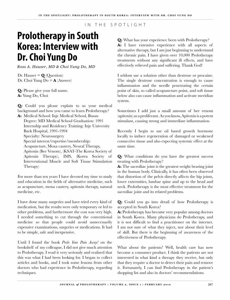
Prolotherapy in South Korea: Interview with Dr. Choi Yung Do
I N T H E S P O T L I G H T
Ross A. Hauser, MD & Choi Yung Do, MD
Dr. Hauser = Q (Question)Dr. Choi Yung Do = A (Answer)
Q: Please give your full name.A: Yung Do, Choi Q: Could you please explain to us your medical background and how you came to learn Prolotherapy?A: Medical School: Inje Medical School, Busan Degree: MD Medical School Graduation: 1991 Internship and Residency Training: Inje University Baek Hospital, 1991-1994 Specialty: Neurosurgery Special interest/expertise/membership: Acupuncture, Moxa cautery, Neural Therapy, Apitoxin (Bee Venom), (KSAT-The Korea Society of Apitoxin Therapy), IMS, (Korea Society of Interventional Muscle and Soft Tissue Stimulation Therapy) For more than ten years I have devoted my time to study and education in the fields of alternative medicine, such as acupuncture, moxa cautery, apitoxin therapy, natural medicine, etc. I have done many surgeries and have tried every kind of medication, but the results were only temporary or led to other problems, and furthermore the cost was very high. I needed something to cut through the conventional medicine so that people could avoid unnecessarily expensive examinations, surgeries or medications. It had to be simple, safe and inexpensive. Until I found the book Prolo Your Pain Away! on the bookshelf of my colleague, I did not give much attention to Prolotherapy. I read it very seriously and realized that this was what I had been looking for. I began to collect articles and books, and I took some lessons from other doctors who had experience in Prolotherapy, regarding techniques.
Q: What has your experience been with Prolotherapy?A: I have extensive experience with all aspects of alternative therapy, but I am just beginning to understand the chronic pain. I have given over 10,000 Prolotherapy treatments without any significant ill effects, and have effectively relieved pain and suffering. Thank God! I seldom use a solution other than dextrose or procaine. The single dextrose concentration is enough to cause inflammation and the needle penetrating the certain point of skin, so called acupuncture point, and soft tissue below also can cause inflammation and activate meridian system. Sometimes I add just a small amount of bee venom (apitoxin) as a proliferant. As you know, Apitoxin is a potent stimulant, causing strong and immediate inflammation. Recently I begin to use oil based growth hormone locally to induce regeneration of damaged or weakened connective tissue and also expecting systemic effect at the same time. Q: What conditions do you have the greatest success treating with Prolotherapy?A: The sacroiliac joint is the greatest weight bearing joint in the human body. Clinically, it has often been observed that distortion of the pelvis directly affects the hip joints, lower extremities, lumbar spine and up to the head and neck. Prolotherapy is the most effective treatment for the sacroiliac joint and its related problems. Q: Could you go into detail of how Prolotherapy is accepted in South Korea?A: Prolotherapy has become very popular among doctors in South Korea. Many physicians do Prolotherapy, and it is not difficult to find a practitioner on the internet. I am not sure of what they inject, nor about their level of skill. But there is the beginning of awareness of the effectiveness of Prolotherapy. What about the patients? Well, health care has now become a consumer product. I think the patients are not interested in what kind a therapy they receive, but only that they require a doctor to detect their pain and remove it. Fortunately, I can find Prolotherapy in the patient’s shopping list and also in doctors’ recommendations.

J O U R N A L of P R O L O T H E R A P Y | V O L U M E 2 , I S S U E 1 | F E B R U A R Y 2 0 1 0268
I N T H E S P O T L I G H T : P R O L O T H E R A P Y I N S O U T H K O R E A : I N T E R V I E W W I T H D R . C H O I Y U N G D O
Q: Is Prolotherapy becoming the standard of care for treating chronic pain in Korea? Why or why not?A: Positively yes, because the therapy is very safe, simple and even more it is very effective. Another reason is that many people have become aware of the harmfulness of the long-term use of steroids and of the fact that chronic pain cannot be controlled by steroids in the long run. At present, Prolotherapy seems to be the only substitute for steroid therapy. Q: What would you like to see for the future in regard to Prolotherapy?A: What would I like to see in the future? I think 3-D image guidance technology will be used in the field of Prolotherapy, and gene or cell therapy will replace traditional prolo solutions. A stem cell itself, or some kind of genetically programmed substances, will be used to repair the damaged tissue or restore the function of the weak ligaments. It will be very exciting. Q: Is there a Prolotherapy society that you know of ? What is their contact information?A: As far as I know, there are no official Prolotherapy societies in Korea. Hence, there are no reliable training or certification systems. Q: As you know, I came across your name because you treated some American Christian missionaries. Do you incorporate faith into your medical practice?A: I think belief has a direct effect on one’s health, and may play a bigger role in the healing process than any other factor. It elicits a relaxation response, a slowed heartbeat, lower blood pressure, and a reduction in stress and anxiety, which have been regarded as a factor affecting health and healing. But people never really think it through. Before, I was possessed by the misconception that modern medicine can exercise control over illness or disease. No, it is not true. Why is it that, as the number of doctors and medical facilities increase, the number of illnesses and patients also increase? Why are so many people suffering from illness and pain in spite of all of the great medical achievements? All I can do, as a doctor, is manage the disease’s symptoms. Why do feel like a fake? Why do I have to pretend to be more than I really am? I am struggling and grappling with all of these questions.
“She had suffered a great deal under the care of many doctors and had spent all she had, yet instead of getting better she grew worse.” Mark. 5:26 I do not have all the answers, but there is one thing I do know. God forgives our sin and heals the sick. We have to acknowledge the power of God that influences every moment of our lives. Our body’s organs are under God’s constant care, and cannot work independently. It’s not the medicines or the surgeries. Only by His grace, do we have the capacity to repair and heal our own cells. We just put a tiny piece of the puzzle in the right place, and walk into the light of God. “I serve and God cures.” Q: Please sum up your feelings about Prolotherapy and its future success in your home town and around the world.A: Over many years of clinical experience, I’ve tried many kinds of therapy. Some were very effective, but not safe, while the cost was high. And some were very safe, but not as effective as I had expected. Of course, there is no master key which can open all doors. Prolotherapy cannot cure all pain. But I’ve found Prolotherapy to be one of the most effective treatments for curing chronic pain. And there is evidence that Prolotherapy offers many advantages over “conventional” therapy in physiologic or functional outcomes. So I think that its future worldwide success is only a question of time. n
Choi Yung Do, MD performing Prolotherapy on a patient.

J O U R N A L of P R O L O T H E R A P Y | V O L U M E 2 , I S S U E 1 | F E B R U A R Y 2 0 1 0 269
I N T H E S P O T L I G H T : A N I N T E R V I E W W I T H A P E R S O N A L I N J U R Y A T T O R N E Y
An Interview with a Personal Injury Attorney
I N T H E S P O T L I G H T
Ross A. Hauser, MD & Steven A. Crifase, Attorney at Law
P atients who receive Prolotherapy may have at one time sustained
an injury that was treated with surgery that achieved a less than optimal result. Prolotherapy physicians hope to be given the opportunity to treat these injuries prior to surgery, as they feel that Prolotherapy is frequently a practical alternative to expensive, time-intensive surgical procedures
and resultant rehabilitation. Steve Crifase was one such person who came to our office in order to avoid knee surgery—and he just happens to be a personal injury attorney. We thought that our readers would find it interesting to hear the opinion of a personal injury attorney related to the cases he has been involved with, and some of the concerns that he has based on what he has seen in his twenty-five years practicing as a personal injury attorney. The interview between Mr. Crifase and Dr. Hauser was recorded and transcribed. Dr. Hauser = Q (Question)Mr. Crifase = A (Answer) Mr. Crifase was asked to begin the interview with a short introduction of himself and his practice. A: I graduated from Loyola University in ’82 and I’ve been doing personal injury and worker’s comp. practice since then. I’m AB rated the top, which represents 10-15% of lawyers in different sub-categories, and is peer review and rated. Q: Please tell us about that rating.A: It’s a Martindale Hubble rating, which is a nationally recognized peer rating. Judges and lawyers rate each other.
Q: What category are you rated in?A: I have it in Personal Injury. Doesn’t mean I know what I’m doing but…(he jokes) Q: Are you an independent practice?A: I’m an independent practice. I’ve been independent since ’84. Q: By yourself ?A: Yes. Well, I’ve got co-counsel that I work with, but my physical plant is just myself and a secretary. Q: Okay. And then you and I had a discussion that you’re concerned about certain kinds of procedures that chronic pain patients get and you’ve had some experience with these various procedures. So I just thought you could just give us an overview.A: You know, from a layman, and I call it an entirely layman standpoint, all my clients come in with traumatic injuries and my concern has always been, and not from a medical malpractice standpoint, but the information, the informed consent that the surgeons do or do not give them. And most of the clients that I represent are laboring and don’t have the exposure to other ideas in terms of treatment alternatives and so on, so many of them will come in with a recommendation that their doctor says they need this surgery or that, and I will always say, “Well, did they give you the odds in terms of a bad result or have you talked about that with them?” The most glaring ones that I’m seeing now are these disc replacements—whether it’s the ProDisc, or this Charite, if I’m pronouncing it correctly. But that seems to be a popular item I’ve gotten. I’ve had four clients that had lumbar disc replacements, two of which needed revisions, the whole apparatus taken out and replaced. Q: With another artificial disc?A: With another artificial disc. Q: I understand. Interesting.A: And I’ve got a guy right now who’s been recommended
Steven A. Crifase,Attorney at Law

J O U R N A L of P R O L O T H E R A P Y | V O L U M E 2 , I S S U E 1 | F E B R U A R Y 2 0 1 0270
I N T H E S P O T L I G H T : A N I N T E R V I E W W I T H A P E R S O N A L I N J U R Y A T T O R N E Y
to have it from a top neurosurgeon out of Loyola. I sat down with him, went through the internet and showed him the results. I guess the problem, and you understand this much better than I do, is their whole theory about how the spine rotates and that’s why the current disc designs don’t work because there’s a premise error there, in terms of how it turns or how it exerts force and I guess they’re coming up with different facet fractures. In any event, that seems to be one of the things that’s coming up. It’s the same thing with the cervical disc surgeries that people come in with. I think I’ve handled fifteen of those in the last ten years and I can only think of four that didn’t need revisions shortly thereafter. Q: Are you talking about cervical disc replacement?A: Fusions with screws and apparatus that came out or bone density issues, whatever. It’s just so many of these people go in thinking they’re going to be fine after surgery and they’re so demoralized and disappointed when the expectations, created in large part by the surgeons, don’t bear out. Q: So the main concern you have in regard to disc replacements or other surgeries is that the likelihood of a bad result is minimized?A: I don’t think it’s even communicated. Q: So in your customers, your clients, you would say that in your experience, because you’ve been in practice twenty-five years now, that your clients note that basically, their understanding is it’s just going to be a positive response. Their surgeons don’t even talk about, you’re saying they don’t talk about post-operative expectations, like long-term arthritis or…?A: Or failure of the theory or procedure at all. No, I’m generally the first one to even raise the question, “Did they talk to you about the consequences that might not be favorable?” And across the board the answer is, “No, they didn’t. They just said I’d be fine. I’d be fine, my leg pain would be gone, I might have some back discomfort, but that would be it.” Q: Okay. So we discussed a little bit the topic of fusions. So your clients have had some bad experiences with fusion or disc replacements. Any other surgeries come to mind?
A: I guess the bigger joints. The shoulder surgeries. Depending on who the surgeon is, there’s certainly a huge variance in outcomes, in terms of who does the work. I’m not going to name names, but there are certainly some people whose work I’ve been hugely impressed with over the years. Other results weren’t as favorable…if they call me and they’re saying this is what the doctor is suggesting, what do you think? I certainly always say, “Schedule a conference and talk to them about the results that might not be as favorable as they’re suggesting and see what they say.” So I just think, and it’s against my interests... I make more money on these cases if there’s surgery. I make a tremendous amount of money on these disc replacement surgeries so I’m hypocritical to criticize the application or the use of those, but I just think that as a rule, and again it doesn’t rise to a level of medical negligence, it’s just a sad situation for people not to at least know that the outcome
might not be what they’d like it to be. And I always joke that builders need to build and bakers need to bake and surgeons need to do surgery. I’ve got any number of friends who are surgeons and they have expenses and crushing overhead and they’re getting chiseled by the insurance carriers so they definitely need to cut. So sometimes I think that economic imperative outweighs some of their better judgment. Q: I see, Steve. Have you had any experience in regard to various
injection techniques to relieve pain?A: I have. My experience has been that they generally don’t work. I’m not talking about the Prolotherapy because my knees feel great. It’s a huge improvement. But the cortisone and even some of the nerve blocks is what I am talking about. Usually some of the guys with the disc injuries will be sent over to pain management first before surgery and they’ll try any number of injections. And I give them credit with respect to the informed consent on those, because they generally tell me the doctor said, “Maybe it will work in three out of ten occasions.” But I don’t think I’ve ever had anybody that…, where it’s arrested the pain for a permanent application. It’s generally been a temporary thing.
Q: So the main concern you have in regard to disc replacements or
other surgeries is that the likelihood of a bad
result is minimized? A: I don’t think it’s even
communicated.

J O U R N A L of P R O L O T H E R A P Y | V O L U M E 2 , I S S U E 1 | F E B R U A R Y 2 0 1 0 271
I N T H E S P O T L I G H T : A N I N T E R V I E W W I T H A P E R S O N A L I N J U R Y A T T O R N E Y
Q: What can people do to protect themselves?A: I don’t know. Do their own research so they can make an informed decision about, and then query their physicians about outcomes. Q: What would be some specific questions you recommend people ask?A: I would recommend that they ask about studies, because most of the stuff is in the studies. …I was hearing about a Columbia Medical School study about, what is it, 70% of the back surgery recipients would have done just as well without. The body would have healed, given the opportunity. I don’t know if that’s the right percentage but I know that one was released four or five years ago. And the carriers that I deal with, because we’re always arguing to get authorization for surgery on some of these work comp cases, were throwing that in my face for years. It’s kind of calmed down, but there are all sorts of studies I’m just remotely aware of but that you guys are intimately familiar with, that confirm that surgery’s not always the viable answer. But it’s a huge industry. I don’t know what the numbers are. I was just reading, I thought I just saw something that said that back treatment is a 38 billion dollar a year industry in the United States. I don’t know that that’s all surgery, but I would just tell them to do studies and question their physicians in a friendly and respectful way. Q: Steve, if you don’t mind me asking, what made you choose Prolotherapy versus, you know, the gamut of treatment options for yourself ?A: I have so many clients that have knee problems and knee tears and this and that and I generally, all day long am dealing with medical records and what not, and I’ve got friends who are ortho surgeons, and really didn’t have a friend who did knees. If I did, maybe I would have gone to him, but I had no interest in going to an orthopod and figured I’d exhaust all the other remedies or avenues first. My son had such a great result with his shoulder and as I did more and more research, and then the New York Times just ran a front page article. Did you see that? On one of the NFL guys that came back after three weeks when they expected it to be an eight week injury. Q: Yes.A: I don’t remember what the details were but I’m assuming it wasn’t just blood spinning. That it was a combination of agents that they put in. And can I ask you a question? Are you guys injecting the hyaluronic acid?
Q: Steve, we have in the past used hyaluronic acid, but currently, we didn’t see it cause regeneration, per se. It was adding expense to the procedure and we just didn’t see the benefit of it. Obviously clients come here to hopefully to get cured of their pain or at least the majority of their pain. We saw it give temporary relief. We weren’t seeing any long term relief.A: Right. Q: That’s why we don’t even have it in the office. Occasionally I’ll get somebody who wants it so we might special order it for them.A: Then do you have, are you going to put a sign in your waiting room, “Screaming patients mean things are working well.” (he laughs) Q: When Doug [our Patient Liaison] meets with them, he kind of goes over that. “You’re going to hear different kinds of screams. This is the bad kind of scream, it sounds like this. This is the good kind of scream, it sounds like that.” (he jokes)A: Right. Hallelujah screams! Right! Q: We appreciate your time.A: Sure. Q: Thank you so much. Can others contact you via email?A: Yes, that’s fine. [email protected]. Q: Thanks Steve.A: Okay. Bye.
t o C o n t a C t m r . C h r i f a s e
Steven Chrifase, Ltd8 South Michigan Suite 2000, Chicago, Illinois 60603Telephone: 312.855.0511Fax: 312.855.0537 www.stevencrifase.com
e d i t o r ’ s n o t e
Moral to the story: if your physician recommends surgery, please make sure to explore all options, ask for risk-benefit information, as well as detailed success statistics and furthermore, explore non-surgical alternatives, such as Prolotherapy, where indicated. n

J O U R N A L of P R O L O T H E R A P Y | V O L U M E 2 , I S S U E 1 | F E B R U A R Y 2 0 1 0272
F A N T A S T I C F I N D I N G S : P R O L O T H E R A P Y A S A N A L T E R N A T I V E T O S U R G E R Y
Prolotherapy as an Alternative to Surgery A Prospective Pilot Study of 34 Patients from a Private Medical Practice
F A N T A S T I C F I N D I N G S
Ross A. Hauser, MD, Marion A. Hauser, MS, RD, Nicole M. Baird, CHFP, & Danielle J. Martin
a B s t r a C t
Thirty-four patients with average musculoskeletal pain duration of 27 months who were told by their medical doctor/surgeon that surgery was needed, including 20 joint replacements and nine arthroscopic procedures, were treated with Hackett-Hemwall dextrose Prolotherapy in lieu of surgery. Patients were followed prospectively and asked questions regarding levels of pain, stiffness, and other physical and psychological symptoms, as well as questions related to activities of daily living before and after their last Prolotherapy treatment.
In this study, Prolotherapy caused a statistically significant improvement in their pain and stiffness. The average starting level of pain was 7.6 and stiffness 7.2, but after Prolotherapy they decreased to 1.3 and 2.5 respectively. Ninety-one percent of patients felt Prolotherapy gave them 50% or greater pain relief, and 71% felt the pain relief was greater than 75%. Upon interview, an average of 10 months after their last Prolotherapy session, this study revealed improvement in patients’ quality of life parameters in addition to pain and stiffness including depression, anxiety, medication usage, as well as range of motion, sleep and exercise ability. Seventy-nine percent felt they had enough pain relief with Prolotherapy that they will not now or in the future need surgery. Four of the remaining seven patients noted 50% or greater pain relief from the Prolotherapy and plan on getting more Prolotherapy in the future.
In this study, Prolotherapy was able to eliminate the need for surgery realistically in 31 out of 34 patients. If Prolotherapy could eliminate 80% of musculoskeletal surgeries in the United States, this procedure alone could make a tremendous dent in cost savings to Medicare, private insurers, and patients. This does not include the money that is lost from productivity and additional expenses that accompany surgery such as future or revision surgeries, rehabilitation, physiotherapy, medications, or disability (from continued pain). Prolotherapy does not
i n t r o d u C t i o n
C hronic pain is a recurring medical dilemma in the United States. It has been estimated that over one third of the American population suffers from
chronic pain, and some studies indicate a much higher incidence of pain experienced regularly.1-3 While chronic pain effects many areas of the body, low back pain is the most common form of chronic pain, with an estimated 80% of people suffering from back pain at some point in their lives.4 After back pain, knee and shoulder pain are the most often reported musculoskeletal complaints according to one study.5 Businesses in the United States alone lose 61.2 billion dollars per year in loss of productivity because of employee disability due to chronic pain.6
This rise in chronic pain is accompanied by an increase in surgical procedures as a pain treatment. Common surgeries that are used to intervene for the pain are knee and shoulder arthroscopy, back, neck or ankle fusion, and knee and hip joint replacement. From the years 1990 to 1996 total hip replacement surgery increased by 23%, one in seven of them were revision surgeries.7
have the risks associated with surgery. Often patients can immediately return to work after receiving Prolotherapy. Since results with Prolotherapy are often permanent, no future treatments are needed. These are reasons enough for patients to consider a Prolotherapy evaluation before undergoing a musculoskeletal surgery.
As this pilot study found such significant improvements in these participants with chronic musculoskeletal pain who were told that surgery was needed, further studies under more controlled circumstances, with larger patient populations, should be done.
Journal of Prolotherapy. 2010;(2)1:272-281.Keywords: alternative to knee replacement, alternative to surgery, arthroscopy, joint replacement, Prolotherapy.

J O U R N A L of P R O L O T H E R A P Y | V O L U M E 2 , I S S U E 1 | F E B R U A R Y 2 0 1 0 273
F A N T A S T I C F I N D I N G S : P R O L O T H E R A P Y A S A N A L T E R N A T I V E T O S U R G E R Y
Figure 1. Projected escalation in number of knee and hip replacements in the United States. By the year 2030 it is estimated that the number of hip replacements performed could reach 1.85 million, and the number of knee replacements as high as 3.48 million.
YEA
R
H IP REPLACEMENTS
20302015
...2005200420032002200120001999199819941982
...1960
0 0.5 1 1.5 2MILLIONS
HipReplacements(Partial&Total)
YEA
R
KNEE REPLACEMENTS
20302015
...20052004200320022001200019991998
...1970
0 0.5 1 1.5 2 2.5 3 3.5 4MILLIONS
TotalKneeReplacementsIn a study looking at total hip and knee replacements performed annually from 2000 to 2004, the number of hip replacements increased from 164,458 to 225,900, and knee replacements increased from 281,534 to 431,485, a jump of 37% and 53% respectively. The same study projected in 2015 that the number of total hip replacement surgeries will reach nearly 600,000, and total knee replacements will reach nearly 1.4 million.8 (See Figure 1.) Another study by Cowen, published in Neurosurgery in 2006 states from 1993 to 2003 spinal fusions rose from the 41st most common inpatient procedure to the 19th most common, with cervical fusions increasing by 89%, thoracolumbar fusions by 31% and lumbar fusions by 134%.9 A definite increasing trend is seen with musculoskeletal surgical procedures. With the increase in surgical procedures comes significant increases in healthcare costs, as a total hip replacement has an average cost of $39,299, while a total knee replacement can cost $35,000 or more.10, 11 Health care costs associated with knee replacement surgery amounts to around $2 billion annually nationwide and if the hospital charges grow with inflation that cost is estimated to amount to nearly $80.2 billion for all primary revised hip/knee replacement surgeries by 2015.12 Spinal-fusion surgery has an average hospital bill of more than $34,000, not including professional fees.13 Surgical cost is only one limiting consideration relating to chronic pain.
While surgery for pain is sometimes a necessary treatment, it carries risk. A relatively common complication associated with surgical procedures is the need for revision surgery. Statistics from The Hospital for Special Surgery showed that in 1973 the need for hip replacement revisions were fewer than 1%, but by 1983 revision rates had risen to 10%.14 A later study published in the same journal saw the revision rate between 1990 and 2002 for total hip arthroplasties increase by 3.7 per 100,000 procedures, along with total knee revision arthroplasties increasing by 5.4 procedures for every 100,000.15 The most common causes of revision total hip arthroplasty are hip instability, mechanical loosening, and infection. (See Figure 2.) Given this trend, it is projected that from the years 2005 to 2030, the hip revision rate will increase by 137% and knee revision rates will have increased by 601%.16
Figure 2. Each revision total hip arthroplasty is over $55,000 in costs just for the hospitalization alone.
PERCENTAGE OF RE VISIONS
Dislocation
Mechanical Loosening
Infection
Implant Failure
Other Mechanical Complications
Periprosthetic Osteolysis
Periprosthetic Fracture
Bearing Surface Water
0.0 5.0 10.0 20.0 25.0
TheMostCommonCausesofRevisionTotalHipArthroplastyintheUnitedStates2005-2006
15.0
22.5
19.7
14.8
9.9
7.2
6.9
6.2
5.0
Source: Bozic KJ, et al. The epidemiology of revision total hip arthroplasty in the United States. J Bone Joint Surg (Am). 2009;91: 128-133.

J O U R N A L of P R O L O T H E R A P Y | V O L U M E 2 , I S S U E 1 | F E B R U A R Y 2 0 1 0274
F A N T A S T I C F I N D I N G S : P R O L O T H E R A P Y A S A N A L T E R N A T I V E T O S U R G E R Y
The doctor that introduced Prolotherapy into mainstream medicine practice was George S. Hackett, MD.48 In a study of 206 traumatic headache patients published by Dr. Hackett and colleagues, 79% were completely relieved of their headaches.49 In regards to low back pain, a survey revealed that 82% of 1,178 patients treated with Prolotherapy considered themselves cured.50
While Prolotherapy has been traditionally used for ligament and tendon injuries, it has a long history of use in osteoarthritis and other degenerative conditions.51-53 Because surgery for degenerative conditions carries risks and complications and often does not totally resolve the patients’ pain or even makes it worse; patients are turning to Prolotherapy as an alternative to surgery. Prior studies on Prolotherapy done at a charity clinic run by the primary author have shown that Prolotherapy eliminates pain even in those patients who have been told by their medical doctor(s) that surgery was the only treatment option for their pain.54-57 (See Table 1.) To further document the success rate of Prolotherapy in helping patients who have been told by an orthopedic surgeon or other physician that surgery was needed to resolve their musculoskeletal pain, this study was undertaken.
Revision surgery is only one risk associated with musculoskeletal surgery. Many patients have concerns about other possible risks that accompany surgery which include peri-operative risks such as deep vein thrombosis, along with more intermediate and long-term risks including loosening and wearing of the prosthesis and pseudarthrosis.17-19 Dislocation is also of concern to hip arthroplasty patients, as it is a regular occurrence and the risk increases with each revision surgery required.20-24 The need for a blood transfusion is common and of concern, as patients may lose a significant enough amount of blood during a joint replacement surgery to require a transfusion.25-27 Spinal fusions are sometimes recommended for back pain, but the fusion success rate is sub-optimal and the patient may still experience post-fusion pain, in addition to a long recovery time.28-30 Artificial discs also present problems by leaving patients with persistent pain symptoms after implantation.31, 32 In addition, lumbar fusion failures have been shown to cause radiculopathy, degeneration in adjacent discs, and nerve injuries.33-36 In relation to the knee, various studies showing arthroscopic debridement and arthroscopy report no benefit for knee osteoarthritis and often leave the patient with chronic pain and complications.37-40 Ankle replacement surgery has been used for patients with ankle pain, but also reports historically high complication rates, along with a number of failures.41-43
Because surgery carries risks and complications and often does not cure pain symptoms, patients are seeking alternatives with the same or greater results. Prolotherapy is one alternative that patients are now turning to. Prolotherapy works by initiating a brief inflammatory response, which causes a reparative cascade to generate new collagen and extra cellular matrix giving connective tissue their strength and ability to handle strain and force.44, 45 This healing cascade produces fibroblasts, which is critical for the repair of tendons and ligaments. Simply put, the affect of Prolotherapy is similar to that of an injury except with Prolotherapy there is no disruption of the architecture of the tissue. High-resolution ultrasounds have been used to confirm that Prolotherapy does indeed stimulate tissue growth.46 One double-blinded animal study by Dr. Liu showed that Prolotherapy increased ligament mass by 44%, ligament thickness by 27%, and ligament bone junction strength by 28%.47
Painful body part where Prolotherapy was performed
Average pain level prior to Prolotherapy
Average pain level after Prolotherapy
Percent of patients who reported greater than 50% pain relief from Prolotherapy
Knee 6.8 3.0 100%
Back 6.0 2.1 96%
Neck 6.6 2.1 90%
Shoulder 7.0 2.6 90%
Hip 7.1 2.4 100%
Table 1. Results from prior studies done on the effects of Prolotherapy for patients whose doctor told them that surgery was the only option for their chronic pain.*
* Hauser R, et al. A retrospective study on dextrose Prolotherapy for unresolved knee pain at an outpatient charity clinic in rural Illinois. JOP. 2009;1:11-21.
Hauser R, et al. A retrospective study on Hackett-Hemwall dextrose Prolotherapy for chronic hip pain at an outpatient charity clinic in rural Illinois. JOP. 2009;2:76-88.
Hauser R, et al. Dextrose Prolotherapy for unresolved low back pain: a retrospective case series study. JOP. 2009;3:145-155.
Hauser R, et al. A retrospective study on Hackett-Hemwall dextrose Prolotherapy for chronic shoulder pain at an outpatient charity clinic in rural Illinois. JOP. 2009;4:205-216.
Hauser R, et al. Dextrose Prolotherapy for unresolved neck pain. Practical Pain Man-agement. 2007;7(8):56-69.

J O U R N A L of P R O L O T H E R A P Y | V O L U M E 2 , I S S U E 1 | F E B R U A R Y 2 0 1 0 275
F A N T A S T I C F I N D I N G S : P R O L O T H E R A P Y A S A N A L T E R N A T I V E T O S U R G E R Y
Hypothesis: Prolotherapy can resolve pain, even in patients who were told by a medical doctor(s) that surgery is needed for their painful condition. Objective: To investigate the outcome of patients who underwent Prolotherapy treatment as an alternative to surgery. Method: In early 2007, unresolved chronic pain patients seeking Prolotherapy at a private medical practice in lieu of surgery were followed prospectively to determine if Prolotherapy treatments resulted in pain relief. Conclusions: In this study, we observed that patients with unresolved musculoskeletal pain had a statistically significant improvement in their pain and stiffness, as well as significant functional gains in other measures in quality of life, including walking ability, after receiving Hackett-Hemwall dextrose Prolotherapy in lieu of surgery.
MethodsP a t i e n t C h a r a C t e r i s t i C s
A total of 34 patients were treated for their chronic pain at Caring Medical, a private Prolotherapy practice in Oak Park, Illinois and followed for their response to Prolotherapy. The average age of patients was 57 years-old with 18 being male and 16 female. All patients were told by a medical doctor(s) that surgery was needed to resolve their pain and 91% were told that surgery was their only option. The patients represented 21 knees, five hips, two wrists, two ankles, two feet, one shoulder, and one lower back. The operations the patients were trying to avoid were 20 joint replacements, nine arthroscopic procedures, three fusions, and four tendon/ligament repairs. The reasons the patients chose not to have surgery varied: 34% natural medicine minded, 18% personal choice, 18% risks, 9% family decision, 3% expense, and 3% fear. Prior bad experience with surgery was not a reason any of the patients received Prolotherapy. Fifty-nine percent of the patients being treated knew of others who had benefited from Prolotherapy. The average length of pain patients reported prior to receiving Prolotherapy was 27 months. The average patient had seen 2.5 physicians prior to receiving Prolotherapy. The average patient was taking 1.1 medications for pain before receiving Prolotherapy. Thirty-two percent of patients were taking one pain
medication per day before receiving Prolotherapy, and 18% were taking two to three pain medications per day. (See Table 2.)
i n t e r v e n t i o n s
The participants received the Hackett-Hemwall technique of Prolotherapy. A 15% dextrose, 10% Sarapin and 0.2% lidocaine solution was used as the base solution. Patients being treated for peripheral joint degeneration also received 2IU of Human Growth Hormone injected into their joints. General inclusion criteria were a history of musculoskeletal pain and being told by a medical doctor/surgeon that surgery was needed, as well as being an appropriate Prolotherapy candidate. Guidelines for the latter included having joint motion at least 50% of normal, motivation to get better, a willingness to stop anti-inflammatory or narcotic medications, and determination to receive the necessary number of visits required for Prolotherapy to resolve or reduce the pain complaint.
o u t C o m e s
An independent data collector (DP) was the sole person obtaining the patient information. The data was obtained before and after the patients had received their Prolotherapy treatments. Follow-up telephone contact was made when it had been at least three months since their last Prolotherapy session. For the analysis of the patient data, patient percentages of the various responses were calculated by another independent data collector (DG), who also had no previous knowledge of Prolotherapy. These responses gathered from patients before Prolotherapy were then compared with the responses to the same questions after Prolotherapy.
Total number of patients treated 34
Percent told surgery was needed 100%
Percent told surgery was only option 91%
Average age of the patients 57
Average number of prior physicians seen 2.5
Average length of pain (in months) 27
Table 2. Patient characteristics at baseline.

J O U R N A L of P R O L O T H E R A P Y | V O L U M E 2 , I S S U E 1 | F E B R U A R Y 2 0 1 0276
F A N T A S T I C F I N D I N G S : P R O L O T H E R A P Y A S A N A L T E R N A T I V E T O S U R G E R Y
Resultst r e a t m e n t o u t C o m e s
Patients received an average of 4.5 Prolotherapy treatment sessions. The average time of follow-up after their last Prolotherapy session was 10 months. Prior to Prolotherapy the average patient was taking 1.1 medications for pain, but this decreased to 0.2 after Prolotherapy. Thirteen patients were able to stop taking medications or decrease them because of Prolotherapy. One of the 26 patients not on pain medications following Prolotherapy had to resume since stopping Prolotherapy.
Patients were asked to rate their levels of crunching, stiffness, and pain on a scale of 0 to 10, with 0 being no crunching/stiffness/pain and 10 being severe/crippling crunching/stiffness/pain. The average starting crunching level was 5.2, the average starting stiffness level was 7.2, and the average starting pain level was 7.6. Following Prolotherapy, patients reported an average ending crunching level of 1.5, an ending stiffness level of 2.5, and an average pain level of 1.3. (See Figure 3.) The before and after Prolotherapy pain levels are seen in Figure 4. Ninety-one percent of patients felt Prolotherapy gave them 50% or greater pain relief, and 71% felt the pain relief was greater than 75%. Of patients who still reported pain, 21% felt they stopped the Prolotherapy sessions too soon. Before Prolotherapy, 6% of patients had normal range of motion, but that increased to 24% after Prolotherapy. Prior to Prolotherapy, 9% of patients had only a slight restriction of motion (75% or greater of normal range of motion), but this increased to 77% after Prolotherapy.
In regard to activities of daily living (ADL), 50% of participants said their overall disability was 50% or greater, due to pain. After Prolotherapy, none of the participants had an overall disability of greater than 50%. Seventeen percent of patients had an overall disability of 25% or less, but after Prolotherapy this increased to 81%. Specifically, before Prolotherapy 23% felt that in regard ADLs (including bathing and dressing self), they were dependent on someone else, but after Prolotherapy 100% of patients were independent in ADLs. Concerning another important quality of life issue, only two (6%) out of the 34 patients reported having normal walking ability prior to treatment, but after treatment this increased to 20 (59%). (See Figure 5.)
Before receiving Prolotherapy, 53% of the patients reported feelings of depression, and 62% reported feelings of anxiety. After receiving Prolotherapy, 94% were no longer depressed and 71% were no longer anxious. Prior to Prolotherapy, 76% of the patients reported hindered sleep due to chronic pain. After receiving Prolotherapy, 79% of patients noticed that their ability to sleep had much improved. When patients were asked if Prolotherapy had changed their life for the better, 91% answered “yes.” Only one out of the 34 patients said Prolotherapy did not help their
Figure 3. Levels reported by patients for crunching, stiffness, and pain before and after Prolotherapy.
BEFORE PROLO
LEV
ELS
REPO
RTED
10
9
8
7
6
5
4
3
2
1
0CRUNCHING STIFFNESS PAIN
AFTER PROLO
Figure 4. Starting and ending pain levels before and after receiving Hackett-Hemwall dextrose Prolotherapy in 34 patients who underwent Prolotherapy treatments as an alternative to surgery.
BEFORE PROLO
NU
MBE
R O
F PA
TIEN
TS
18
16
14
12
10
8
6
4
2
00 1 2 3 4 5 6 7 8 9 10
PAIN LEVEL
PainLevelsBefore&AfterProlotherapy
AFTER PROLO

J O U R N A L of P R O L O T H E R A P Y | V O L U M E 2 , I S S U E 1 | F E B R U A R Y 2 0 1 0 277
F A N T A S T I C F I N D I N G S : P R O L O T H E R A P Y A S A N A L T E R N A T I V E T O S U R G E R Y
pain. Three out of 34 (9%) received less than 25% pain relief with Prolotherapy. Seventy-nine percent of the patients answered “yes” to having enough relief after their Prolotherapy treatment that they felt they will never need surgery. For the seven patients (21%) who answered “no” to that question, three felt they will need surgery. The four remaining patients noted greater than 50% pain relief, but plan to receive additional Prolotherapy treatment in the future. Of interest, is 100% of the patients treated stated that they have recommended Prolotherapy to someone else.
StatisticalAnalysisA matched sample paired t-test was used to calculate the difference in responses between the before and after measures for pain and stiffness. The paired sample t ratio was computed on this pre-post Prolotherapy study. The paired t ratios for all the groups were highly significant, using N pairs minus one as the degrees of freedom. For the entire 34 participants the paired t ratio was significant for pain reduction (t = 16.085, p<.0000001). For the analysis on stiffness resolution, the paired t was also highly significant (t = 11.323, p<.0000001). In summary, for the 34 participants in this study, their pain and stiffness was significantly reduced at the p<.0000001 level by Hackett-Hemwall dextrose Prolotherapy.
DiscussionP r i n C i P l e f i n d i n g s
The results of this prospective, non-controlled, pilot study show that Hackett-Hemwall dextrose Prolotherapy helps decrease pain and improve the quality of life of pain patients who have been told that they need surgery to resolve their musculoskeletal pain. Decreases in pain and stiffness reached statistical significance. On a scale of 0 to 10, the ending pain, stiffness, and crunching (crepitation) levels were 1.3, 2.5, and 1.5 respectively. Nine-one percent of participants received 50% or greater pain relief with Prolotherapy. Seventy-nine percent of patients felt they had enough current pain relief with Prolotherapy that they will never need surgery. Four (12%) of the patients received 50% or greater pain relief with Prolotherapy, but plan to receive additional Prolotherapy in order to avoid surgery. Three of the patients (9%) felt they will still need surgery. Additional noted improvements were seen overall in range of motion, walking ability, depressive and anxious symptoms, sleep and need for pain medication. One-hundred percent of patients recommended Prolotherapy to someone they know.
In regard to the three participants who ended up needing surgery; one had terrible shoulder pain especially with playing sports. He had failed physical therapy, cortisone injections, and medications for an intrasubstance tear of the supraspinatus tendon and impingement syndrome. He stated the two Prolotherapy treatments helped him 15%, but he was and is an active cricket player and decided on
Totally compromised
Severely compromised
Very compromised
Definitely compromised
Somewhat compromised
Not compromised
Figure 5. Starting and ending walking ability before and after receiving Hackett-Hemwall dextrose Prolotherapy.
StartingWalkingAbility EndingWalkingAbility
3%
17%
12%
38% 12%
18%0%
0%
0%59%
38%
3%

J O U R N A L of P R O L O T H E R A P Y | V O L U M E 2 , I S S U E 1 | F E B R U A R Y 2 0 1 0278
F A N T A S T I C F I N D I N G S : P R O L O T H E R A P Y A S A N A L T E R N A T I V E T O S U R G E R Y
surgery. He is back to playing. Of interest is this participant at various times had five other body areas treated with Prolotherapy and responded 100%. The second patient who ended up needing surgery had osteoarthritis of the hip. He had six Prolotherapy treatments and felt he was 90% better in regard to pain from the Prolotherapy. He noted that he was sleeping and walking better since receiving Prolotherapy. Objectively, he had more range of motion with the Prolotherapy, but not enough for his activity level. He had a successful hip replacement. The third participant received two Prolotherapy treatments to her degenerated knee. She stated the Prolotherapy helped 50% with the pain but she was anxious to get back to dancing (her passion), and decided to get a total knee replacement. She is back to dancing. While these three participants would be considered “failures” of Prolotherapy because they needed surgery, on closer examination it is clear that two of the patients did not receive the recommended number of treatments before stopping Prolotherapy. In the experience of the primary author (R.H.), patients who have been told by surgeons that surgery is their only option can often require at least six visits of Prolotherapy, especially if they have joint degeneration to the point of “bone on bone.” These three patients do demonstrate the challenge that doctors who utilize Prolotherapy face daily in active patients, that they want to get better quickly. While surgery is not a quick fix, Prolotherapy does require a patient to go to a doctor’s office and receive the treatment every month, sometimes for six months to a year. While this can be a stumbling block to some patients, for the patient who does not want to have surgery, surely this is a small inconvenience for a lifetime of pain relief.
s t r e n g t h s a n d l i m i t a t i o n s
Our study cannot be compared to a clinical trial in which an intervention is investigated under controlled conditions. Instead, it is aimed to document the response in a private medical practice of patients to Prolotherapy who have been told that surgery is needed to resolve their pain. Clear strengths of the study are the numerous quality of life parameters that were studied. Such quality of life issues as overall disability, walking ability, stiffness, range of motion, activities of daily living, sleep, anxiety and depression, in addition to pain level, are important factors affecting the person with pain. The statistically significant improvement in pain and stiffness levels, as well as improvements in quality of life measured, treated
solely by Prolotherapy, even though subjective, is likely to have resulted from Prolotherapy.
Another strength of this study is that the study population received only Prolotherapy as a treatment for their pain; no other treatment modalities were used. While all 100% of patients were told by a medical doctor(s) that surgery was needed to resolve their pain, 91% were told by a medical doctor(s) that surgery was the only treatment option that would resolve their pain. This is further evidence that the amount of pain and disability suffered by these patients was significant. Patients with this caliber of pain and degeneration typically do not experience spontaneous pain improvement, so resolution of their symptoms most likely resulted from the Prolotherapy they received. A weakness of this study is that there was not a control group. Also the study did not isolate one particular patient population in regard to diagnosis necessitating a specific type of surgery. The lack of X-ray or MRI correlation for diagnosis and response to treatment was also a limitation.
i n t e r P r e t a t i o n o f f i n d i n g s
In 2004, there were 3.4 million operations on the musculoskeletal system necessitating and inpatient hospital stay.58 While advances in technology and surgery are admirable; the cost of the surgeries is astronomical. In 2004 the estimated cost of performing spinal fusions was $17.6 billion and discectomy was $11.25 billion. While over a million hip and knee total joint replacements were performed in 2004 at a cost estimated at $30 billion, there are many reasons for people in chronic pain to forego surgery for their pain including risk with the procedure, lack of results, financial burden, inability to work while recovering, as well as personal preference toward natural healing techniques. While one can debate the efficacy of Prolotherapy versus surgery for specific medical diagnoses and symptoms, the cost comparison between the two is not debatable. Excluding the additional costs of rehabilitation, physiotherapy, repeat procedures, side effects, post-operative medications, and future medical problems caused by the surgeries, Prolotherapy is significantly less expensive than the commonly performed surgeries. (See Figures 6a & 6b.)
In the current study, conservatively 79% of the patients receiving Prolotherapy felt that Prolotherapy did resolve their painful condition to the point that they will not now,

J O U R N A L of P R O L O T H E R A P Y | V O L U M E 2 , I S S U E 1 | F E B R U A R Y 2 0 1 0 279
F A N T A S T I C F I N D I N G S : P R O L O T H E R A P Y A S A N A L T E R N A T I V E T O S U R G E R Y
nor in the future, need the previously recommended surgical procedure. That number increases to 91% if you include the additional four patients who already had 50% or more pain relief with Prolotherapy and plan to get more Prolotherapy. Using a conservative number, such as 80% for the number of surgical procedures that would be eliminated with Prolotherapy, the cost savings if patients received Prolotherapy versus surgery are enormous. For instance, in the United States the number of knee replacements in the year 2015 is estimated to be 1.4 million. If 80% of these could be eliminated by patients receiving Prolotherapy now, the cost savings just in these surgeries alone would be $78 billion in the U.S. Imagine if 80% of the 4 million arthroscopies on the knee could be eliminated. This would save the U.S. health care system another $32 billion per year. If Prolotherapy could eliminate 80% of musculoskeletal surgeries in the United States, this procedure alone could make a tremendous dent on saving Medicare, private insurers, and patients money. (See Table 3.) This again does not include the money that is lost from lost productivity, and additional expenses that accompany surgery such as rehabilitation, physiotherapy, future procedures, medications, and disability (from continued pain).
When a person undergoes Prolotherapy, they often go right back to work after the appointment. There is no lost work productivity except the time it takes to go to the Prolotherapy appointment. After Prolotherapy, the person is instructed not to take narcotic or anti-inflammatory medications, as these decrease the healing with Prolotherapy. Normally no medications are needed after Prolotherapy. It is also quite common with Prolotherapy that no physiotherapy or other pain therapies are needed. Typically results with Prolotherapy are permanent. No future Prolotherapy is needed. These are reasons enough for patients to consider a Prolotherapy evaluation before undergoing a musculoskeletal surgery.
Surgery Type Estimated surgery cost in the year 2015
Estimated number of these surgeries in the year 2015
Dollar savings estimating 80% elimination of these surgeries with Prolotherapy
Dollar savings estimating 90% elimination of these surgeries with Prolotherapy
Knee Replacement $70,000 1.4million $78.4trillion $88.2trillion
Hip Replacement $80,000 600,000 $38.4trillion $43.2trillion
Knee Arthroscopy $10,000 8million $64trillion $72trillion
Spinal Fusion $68,000 500,000 $27.2trillion $30.6trillion
Shoulder Arthroscopy $10,000 1.5million $12trillion $13.5trillion
Table 3. Potential cost savings with Prolotherapy instead of common musculoskeletal surgeries.* * Data extrapolated for the year 2015 to demonstrate if Prolotherapy was done today so these surgeries would not be needed.
Figure 6a. Cost comparison of surgery versus Prolotherapy.
Surgery Type Average Cost of Surgery
CervicalFusion $39,000
HipReplacement $46,000
KneeReplacement $33,000
SpinalFusion $56,000
AnkleFusion $30,000
ArthroscopyKnee $5,000
Prolotherapy Type Average Cost of Treatments
Low/MidBack/Neck* $2,500
Shoulder/Hip* $1,875
Elbow/Knee/Ankle* $1,875
Wrist/Foot/Hand* $1,125
* The average person requires 4 to 6 treatment sessions given at 4 to 6 week intervals. Prices displayed are based on an average of 5 treatment sessions.
Figure 6b. The cost of Prolotherapy is significantly lower as compared to surgical procedures.
SURGERY
CO
ST (U
SD)
60,00055,00050,00045,00040,00035,00030,00025,00020,00015,00010,000
5,0000
TREATMENTS
FinancialComparison:Surgeryvs.Prolotherapy
CERVICAL FUSION
HIP REPLACEMENT
KNEE REPLACEMENT
SPINAL FUSIO
N
ANKLE FUSION
ARTHROSCOPIC KNEE
LOW/MID
BACK/NECK*
SHOULDER/HIP*
ELBOW/KNEE/A
NKLE*
WRIST/FOOT/HAND*
PROLOTHERAPY
* The average person requires 4 to 6 treatment sessions given at 4 to 6 week intervals. Prices displayed are based on an average of 5 treatment sessions.
J O U R N A L of P R O L O T H E R A P Y | V O L U M E 2 , I S S U E 1 | F E B R U A R Y 2 0 1 0280
F A N T A S T I C F I N D I N G S : P R O L O T H E R A P Y A S A N A L T E R N A T I V E T O S U R G E R Y
ConclusionNinety-one percent (31 out of 34) of patients who were told by at least one medical doctor that they needed surgery to resolve their chronic musculoskeletal pain complaint felt Hackett-Hemwall dextrose Prolotherapy changed their life for the better. In this study, Prolotherapy caused a statistically significant improvement in their pain and stiffness. Upon interview, on average 10 months after their last Prolotherapy session, this study revealed improvement in patients’ quality of life parameters in addition to pain and stiffness including depression, anxiety, medication usage, as well as range of motion, sleep and exercise ability. Seventy-nine percent felt they had enough pain relief with Prolotherapy that they will not now or in the future ever need surgery. Four of the remaining seven patients, noted 50% or greater pain relief from the Prolotherapy and plan on getting more Prolotherapy in the future. Since this pilot study found such significant improvements in these participants with chronic musculoskeletal pain who were told that surgery was needed, further studies under more controlled circumstances with larger patient populations should be done. n
a C K n o w l e d g e m e n t s
Doug Puller (D.P.), independent data collector.Dave Gruen (D.G.), independent data analyst from www.bolderimage.com.
B i B l i o g r a P h y
Dennis C, et al. Pain hurts: individuals, significant others, and society. APS Bulletin-Volume 16, Number 1,Winter 2006. Available at: http://www.ampainsoc.org/pub/bulletin/win06/pres1.htm. Accessed: October 21, 2009.
Berry PH, et al. Pain: understanding of assessment, management, and treatments: American Pain Society; 2007. Available at: http://www.ampainsoc.org/ce/downloads/npc/npc.pdf. Accessed November 19, 2009.
Harstall C. How prevalent is chronic pain? Pain: Clinical Updates, X, 1–4. (2003).
Flor H, et al. Etiological theories and treatments for chronic low back pain. Somatic models and interventions. Pain.1984;19:105-121.
Urwin M, et al. Estimating the burden of musculoskeletal disorders in the community: the comparative prevalence of symptoms at different anatomical sites, and the relation to social deprivation. Ann Rheum Dis. 1998;57(11):649-655.
1.
2.
3.
4.
5.
Stewart W, et al. Lost productive time and cost due to common pain conditions in the US workforce. JAMA. 2003;290(18):2443-2454.
Floren M, et al. Trends in Hip Replacement Rates. Springer Berlin Heidelberg. 2009;47-52.
Kim S. Changes in surgical loads and economic burden of hip and knee replacements in the US: 1997-2004. Arthritis Rheum. 2008;59:481-488.
Cowan JA Jr, et al. Neurosurgery. 2006 Jul;59(1):15-20; discussion 15-20.
Hip replacement cost. Available at: http://www.costhelper.com/cost/health/hip-replacement.html. Accessed November 19, 2009.
Knee replacement cost. Available at. http://www.costhelper.com/cost/health/knee-replacement.html. Accessed November 19, 2009.
Cramer J. Strict guides could reduce knee surgery medical cost. The Gazette: The Newspaper of Johns Hopkins University. Available at: http://www.jhu.edu/gazette/aprjun95/apr1095/10knee.html. Accessed: October 20, 2009.
Deyo R, et al. Spinal-fusion surgery – the case for restraint. NEJM. 2004;350:722-726.
Pellicci PM, et al. Long-term results of revision total hip replacement. A follow-up report. J Bone Joint Surg Am. 1985;67:513-516. Available at: http://www.ejbjs.org/cgi/reprint/67/4/513.pdf. Accessed November 19, 2009.
Kurtz S, et al. Prevalence of primary and revision total hip and knee arthroplasty in the United States from 1990 through 2002. J Bone Joint Surg Am. 2005;87-A:1487-1497.
Kurtz S, et al. Projections of primary and revision hip and knee arthroplasty in the United States from 2005 to 2030. J Bone Joint Surg Am. 2007;89:780-785.
Cross MJ. Complications of total knee arthroplasty. 2008. Available at: http://emedicine.medscape.com/article/1250540-overview. Accessed November 19, 2009.
Haas S, et al. Pneumatic sequential-compression boots compared with aspirin prophylaxis of deep-vein thrombosis after total knee arthroplasty. Bone Joint Surg Am. 1990;72:27-31.
Robinson Y. Spine imaging after lumbar disc replacement: pitfalls and current recommendations. Patient Safety in Surg. 2009;Jul 20;3(1):15.
Sanchez-Sotelo J, et al. Hospital cost of dislocation after primary total hip arthroplasty. The Journal of Bone and Joint Surgery (American). 2006;88:290-294.
Pulido L, et al. Late instability following total hip arthroplasty. Clinical Medicine & Research. 2007;5(2):139-142.
Frymoyer JW, et al. A comparison of radiographic findings in fusion and nonfusion patients ten or more years following lumbar disc surgery. Spine. 1979;4:435-440.
Skeels MD. The dislocator, early and late: the role of large heads. Orthopedics. 2009;Sep;32(9).
Barry D, et al. The cumulative long-term risk of dislocation after primary charnley total hip arthroplasty. The Journal of Bone and Joint Surgery (American). 2004;86:9-14.
6.
7.
8.
9.
10.
11.
12.
13.
14.
15.
16.
17.
18.
19.
20.
21.
22.
23.
24.

J O U R N A L of P R O L O T H E R A P Y | V O L U M E 2 , I S S U E 1 | F E B R U A R Y 2 0 1 0 281
F A N T A S T I C F I N D I N G S : P R O L O T H E R A P Y A S A N A L T E R N A T I V E T O S U R G E R Y
Toy PTCY, et al: The pre-operative autologous blood donation study group; Blood loss and replacement in total hip arthroplasty a multicenter study. Transfusion. 1992;32:63-67.
Martin JW, et al. Post-operative blood retrieval and transfusion in cementless total knee arthroplasty. J Arthroplasty. 1992;7:205-210.
Prasad N. Blood loss in total knee arthroplasty. Int Orthop. 2007;31(1):39-44.
McAfee PC. Lumbar spine fusion for degenerative disc disease. Available at: http://www.spine-health.com/treatment/spinal-fusion/lumbar-spine-fusion-degenerative-disc-disease. Accessed on November 24, 2009.
Herkowitz HN, et al. Lumbar spine fusion in the treatment of degenerative conditions: current indications and recommendations. Journal of the American Academy of Orthopaedic Surgeons. 1995;3:123-135.
Lehmann TR. Long-term follow-up of lower lumbar fusion patients. Spine. 1987;Mar;12(2):97-104.
Van Ooij, et al. Findings in 67 patients with recurrent or persistent symptoms after implantation of a disc prosthesis for low back pain. [Article in Dutch.] Ned Tijdschr Geneeskd. 2007; Jul 14;151(28):1577-84. Available at: http://www.ncbi.nlm.nih.gov/pubmed/17715768?itool=EntrezSystem2.PEntrez.Pubmed.Pubmed_ResultsPanel.Pubmed_RVDocSum&ordinalpos=7. Accessed on November 24, 2009.
Van Ooij, et al. Complications of artificial disc replacement: a report of 27 patients with the SB Charité disc. J Spinal Disord Tech. 2003;Aug;16(4):369-83.
Ibid.
Sukovich W. Progress, challenges and opportunities in disc space preparation for lumbar interbody fusion. The Internet Journal of Spine Surgery. 2005;1(2).
Wetzel FT. The failed posterior lumbar interbody fusion. Spine. 1991;16(7):839-845.
Mcafee P. Current concepts review- Interbody fusion cages in reconstructive operations on the spine. Journal of Bone and Joint Surgery (Am). 1999;81:859-880.
Laupattarakasem W, et al. Arthroscopic debridement for knee osteoarthritis. Cochrane Database of Systematic Reviews 2008, Issue 1. Art. No.: CD005118. DOI: 10.1002/14651858.CD005118.pub2. Available at: http://www.cochrane.org/reviews/en/ab005118.html. Accessed November 24, 2009.
Kirkley A, et al. A randomized trial of arthroscopy for osteoarthritis of the knee. NEJM. 2008;359:1097-1107. Available at: http://content.nejm.org/cgi/content/short/359/11/1097. Accessed: October 20, 2009.
Moseley JB, et al. A controlled trial of arthroscopic surgery for osteoarthritis of the knee. NEJM. 2002;347:81-88. Available at: http://content.nejm.org/cgi/content/full/359/11/1097. Accessed: November 24, 2009.
Wylde V. Total knee replacement: is it really an effective procedure for all? Knee. 2007;Dec;14(6):417-23.
Guyer AJ, et al. Current concepts review: total ankle arthroplasty. Foot Ankle Int. 2008;29(2):256-64.
25.
26.
27.
28.
29.
30.
31.
32.
33.
34.
35.
36.
37.
38.
39.
40.
41.
Karantana A, et al. The Scandinavian total ankle replacement: survivorship at 5 and 8 years comparable to other series. Clin Orthop Relat Res. 2009 Jul 16. Available at: http://www.ncbi.nlm.nih.gov/pubmed/19609630?itool=EntrezSystem2.PEntrez.Pubmed.Pubmed_ResultsPanel.Pubmed_RVDocSum&ordinalpos=4. Accessed on November 24, 2009.
Claridge RJ. Intermediate term outcome of the agility total ankle arthroplasty. Foot Ankle Int. 2009;Sep;30(9):824-35.
Reeves, et al. Randomized prospective double-blind placebo-controlled study of dextrose prolotherapy for knee osteoarthritis with or without ACL laxity. Altern Ther Health Med. 2000; Mar;6(2):68-74,77-80.
Dagenais S, et al. Evidence-informed managed chronic low back pain with Prolotherapy. The Spine Journal. 2008;8:203–212.
Fullerton BD. High-resolution ultrasound and magnetic resonance imaging to document tissue repair after prolotherapy. Arch PM&R. 2008;89(2):377-385.
Liu Y. An in situ study of the influence of a sclerosing solution in rabbit medial collateral ligaments and its junction strength. Connective Tissue Res. 1983;2:94-102.
Hackett G. Referral pain and sciatica in low back diagnosis. Journal of the American Medical Association. 1957;163:183.
Hackett G. Prolotherapy for headache. Headache. 1962;April:3-11.
Hackett G. Referred pain from low back ligament disability. AMA Archives of Surgery. 1956;73:878-883.
Ongley M. A new approach to the treatment of chronic low back pain. Lancet. 1987;2(8551):143-147.
Klein R. Proliferant injections for low back pain: histologic changes of injected ligaments and objective measures of lumbar spine mobility before and after treatment. Journal of Neurology, Orthopedic Medicine and Surgery. 1989;10:141-144.
Hackett G. Ligament and Tendon Relaxation Treated by Prolotherapy. 5th ed. Oak Park, IL, Gustav A. Hemwall; 1992.
Hauser R, et al. A retrospective study on dextrose prolotherapy for unresolved knee pain at an outpatient charity clinic in rural Illinois. Journal of Prolotherapy. 2009;1:11-21.
Hauser R, et al. A retrospective study on Hackett-Hemwall dextrose Prolotherapy for chronic hip pain at an outpatient charity clinic in rural Illinois. Journal of Prolotherapy. 2009;2:76-88.
Hauser R, et al. Dextrose prolotherapy for unresolved low back pain: a retrospective case series study. Journal of Prolotherapy. 2009;3:145-155.
Hauser R, et al. Prolotherapy: dextrose Prolotherapy for unresolved neck pain. Practical Pain Management. 2007;7(8):56-69.
Merrill, C. (Thomson Healthcare) et al. (AHRQ). Hospital Stays Involving Musculoskeletal Procedures, 1997–2005. HCUP Statistical Brief #34. July 2007. Agency for Healthcare Research and Quality, Rockville, MD. http://www.hcup-us.ahrq.gov/reports/statbriefs/sb34.pdf.
United States Bone and Joint Decade: The burden of musculoskeletal diseases in the United States. Rosemont, IL: American Academy of Orthopaedic Surgeons;2008.
42.
43.
44.
45.
46.
47.
48.
49.
50.
51.
52.
53.
54.
55.
56.
57.
58.
59.

J O U R N A L of P R O L O T H E R A P Y | V O L U M E 2 , I S S U E 1 | F E B R U A R Y 2 0 1 0282
F A N T A S T I C F I N D I N G S : H Y P E R T H E R M I A I N D U C E S V E N O U S B L O O D A L K A L O S I S : A S T U D Y I N F I V E I R O N M A N T R I A T H L E T E S
Hyperthermia Induces Venous Blood Alkalosis: A Study in Five Ironman Triathletes
F A N T A S T I C F I N D I N G S
Ross Hauser, MD & Joseph J. Cukla, LPN
i n t r o d u C t i o n
B ody temperature regulation is an important component of any exercise or training regimen. This is especially true for the endurance athlete
competing in a high temperature environment. If the ambient temperature becomes too high, the athlete reaches a point where elevated body temperature and dehydration ensue. As a result, symptoms such as cramping, nausea, dizziness, and weakness appear. At this time,
a B s t r a C t
To study the relationship between athletic performance in extreme heat and blood chemistry abnormalities, five Ironman triathletes were subjected to a hyperthermic chamber for one hour. The goal was to simulate the excessive heat and the feelings they experienced during their suboptimal athletic performances in the Ironman Triathlon. The hypothesis of the study was that accompanying the hyperthermia was extreme blood alkalosis and this, not dehydration or electrolyte abnormalities, was responsible for these five athletes’ suboptimal performances during their various Ironman races.
One of the subjective feelings that the participants self-rated during this experiment was their perceived ability to run. This feeling of “ability to run” steadily decreased during their time in the hyperthermic chamber. As their core temperatures increased in the chamber, so did their venous serum blood pH levels, with all participants sustaining extreme degrees of venous blood alkalosis. It was this blood alkalosis that correlated closest to their feelings of an inability to run and other unpleasant feelings that they experienced during their recent Ironman Triathlon races.
Journal of Prolotherapy. 2010;(2)1:282-289.Keywords: alkalosis, athlete, athletic performance, blood ph, hyperthermia.
athletic performance also begins to decrease.1-4 Fortney and Vroman, in their study on exercise performance and temperature control said, “…the effect of high ambient temperatures on exercise performance is most evident in prolonged submaximal exercise,”5 as is the case in Ironman Triathlons. These researchers and others have primarily examined the effects of body core temperature in athletes and how it relates to decreased blood volume and dehydration, the shunting of core blood reserves to the athlete’s peripheral surface and hypothalamic thermal regulation. It has been concluded that optimal athletic performance, especially in endurance activities such as running and cycling, is achieved in moderate external temperatures.6, 7 A study featured in the New York Times showed that runners perform best in temperatures ranging from 41 to 50 degrees Fahrenheit.8 Nielsen, et al, in their paper on heat acclimation, measured core temperatures in eight athletes during 90 minute exercise periods and their experiment showed that in a cool environment of 18-20 degrees Celsius (64.4 to 68 degrees Fahrenheit) core temperature remained steady at 37.8 degrees C (100 degrees Fahrenheit); but in a 40 degrees C (104 degrees F) environment core temperatures rose to nearly 40 degrees C (104 degrees F) during the exercise period, with a corresponding decrease in performance.9 It has been documented that as external temperatures rise, so does an individual’s body temperature.10-12 Consequently, as exercise in high temperatures persist and basal body temperatures continue to rise, pace and performance begin to decrease.13, 14 There is debate as to what physiological parameters in the blood cause levels to decrease in athletic performance with elevated temperature. Febbraio and Snow, in their study on the effect of heat stress on muscle energy metabolism during exercise, showed that sustained maximal voluntary muscle contraction with leg extensions attenuated in hyperthermic conditions.15 Galloway and Maughan, in

J O U R N A L of P R O L O T H E R A P Y | V O L U M E 2 , I S S U E 1 | F E B R U A R Y 2 0 1 0 283
F A N T A S T I C F I N D I N G S : H Y P E R T H E R M I A I N D U C E S V E N O U S B L O O D A L K A L O S I S : A S T U D Y I N F I V E I R O N M A N T R I A T H L E T E S
their paper on the effects of ambient temperatutre on the capacity to perform prolonged exercise, said that reduced performance at 31 degrees C (88 degrees F) would most likely result from a reduction in central blood volume.16 This occurs as the body shunts blood peripherally for more efficient cooling. Several physiological parameters are affected by a rise in basal body temperature. Studies have examined various blood and urine test results and how they are affected by hyperthermia. Some have looked at how hyperthermia can result in a breakdown of electrolytes and can increase the use of muscle glycogen stores, likely resulting in decreased ability and increased fatigue.17-19 Another study has shown hyperthermia to deplete intercellular glutathione content, thus possibly affecting immune response.20 While the endurance athlete competing in high heat is at risk for dehydration, for the athlete not dehydrated, the etiological basis for a decline in the athlete’s performance with hyperthermia is not known. The high external temperature fluctuations of the Ironman were simulated with the use of an infrared heating chamber in a controlled environment so that objective lab tests and subjective surveys could be administered to the participants.
m a t e r i a l s & m e t h o d s
This study examined how various biochemical parameters were affected in five Ironman triathletes subjected to hyperthermia via a hyperthermia chamber. The focus of this study was to look at several venous blood markers, including venous serum blood pH, osmolality and electrolytes, among others, to see which correlated best with the athletes’ hyperthermia and survey questions, including “body achiness” and “ability to run,” among others.
s u B j e C t s
The subjects were five athletes, four male and one female, who had completed an Ironman Triathlon in 2005. The race is comprised of a 2.4 mile swim, 112 mile bike, and a 26.2 mile run with a cut-off time of 17 hours to complete the course. The five participants in this study each raced in extreme heat and humidity in the summer of 2005, and had sub-optimal performances leading to an inability to run
during the marathon portion of the Ironman Triathlon. Each athlete had finishing times much higher than were anticipated because they had to walk an average 15-18 miles of the marathon because of being overheated. (See Table 1.)
The triathletes were asked to lay in a Far infrared (FIR) hyperthermia chamber within five months of completing their individual race event. The chamber was to simulate the conditions that caused them to stop running in the Ironman Triathlon. (See Figure 1.) Venous blood pH and electrolytes were measured every 15 minutes during the study and other variables at the beginning and the end of the study. The chamber used for the study was a BioTherm with 90+% Far infrared (FIR), 5-14 microns, peak 9.25-10.2 with an analogue controller. The study design included a blood and urine test analysis of 17 different biochemical parameters and 13 self-reported survey questions pertaining to physical changes such as perceived temperature, mental clarity, nausea, energy level, and ability to run. The patients arrived at the clinic well-hydrated and having eaten a few hours prior to the study. Temperature, vital signs, blood tests, and survey questions were administered at the beginning and end of the 60 minute experiment, as well as every 15 minutes during the time that the participants spent in the chamber. Urine was only collected at the beginning and end of the trial, while blood was collected every 15 minutes via a venous catheter that remained in place. The Nova machine used to test the venous serum pH and electrolytes was a Model 8 NOVA CRT machine designed by NOVA Biomedical Corporation. This laboratory machine at the primary author’s office read normal venous serum pH as 7.50-7.52.*
* The NOVA 8 CRT machine used in 2005 to analyze the venous blood pH used serum where the normal venous serum blood values were 7.50 to 7.52. Currently, the primary author’s lab now uses a NOVA Model CCX laboratory machine to analyze pH and it uses 7.36 to 7.38 as normal values, using whole venous blood instead of serum.
Subject Sex Age
2005 Ironman
Event Weather ConditionsProjected
TimeActual Time
AP M 37 Wisconsin 98ºFahrenheit(F)with95%humidity 13hours 14hr44min
KH F 27 Wisconsin 98ºFahrenheit(F)with95%humidity 13hours 14hr22min
JC M 44 Wisconsin 98ºFahrenheit(F)with95%humidity 13hours 14hr44min
TK M 37 Canada 87ºFahrenheit(F)with85%humidity 11hours 12hr42min
RH M 42 Canada 87ºFahrenheit(F)with85%humidity 12hours 13hr15min
Table 1. Demographics, race conditions, and athletes’ projected vs. actual race times.

J O U R N A L of P R O L O T H E R A P Y | V O L U M E 2 , I S S U E 1 | F E B R U A R Y 2 0 1 0284
F A N T A S T I C F I N D I N G S : H Y P E R T H E R M I A I N D U C E S V E N O U S B L O O D A L K A L O S I S : A S T U D Y I N F I V E I R O N M A N T R I A T H L E T E S
The blood samples were drawn into a marble-top tube (SST), allowed to coagulate for 30 minutes, then spun down in a centrifuge for 15 minutes. The serum was drawn off and immediately tested as the serum pH will change soon after being exposed to air.
The following biochemical variables were analyzed: venous blood pH, urine pH, glutathione rbc, glutathione plasma, anti-oxidant assay, (peroxidase, catalase and superoxide dismutase) cortisol, serum osmolality, urine specific gravity, white blood count, hemoglobin, hematocrit, platelets, ferritin, C-reactive protein, magnesium, potassium and calcium.
r e s u l t s
The five athletes consented to have their data collected for scientific research. As stated, four were men (80%) and one was a woman (20%). All were friends and training partners during their preparation to complete an Ironman event in 2005. Averages for the different biochemical parameters were taken before and after each subject spent 60 minutes in the infrared heat chamber. The average starting glutathione red blood cells (glut-rbc) was 250, and the average ending 218.8. Glutathione plasma (Glut-plasma) began at an average of 142.4, and increased to 190.4. The average
Anti-oxidant assay (AOA) level started at 1.1, and ended at 1.3. Cortisol levels began at 12.7 and increased to 19.0 following exposure to the extreme heat. (See Table 2.)
Figure 1. One of the athletes in the hyperthermia chamber during the one hour study.
Glut-rbc Glut-plasma AOA Cortisol
Average Before 250 142.4 1.1 12.7
Average After 218.8 190.4 1.3 19.0
Table 2. Antioxidant and cortisol levels before and after hyperthermia.
Serum osmolality (Osmo) averaged 296.2 at the start of the study and rose to 302.2 at the end. The average white blood cell (WBC) count was 7.26 before and 8.08 after. Hemoglobin (HGB) began at 13.6 and ended at 15.9. Hematrocrit (HCT) starting levels averaged 43.28 and finished at 46.72. Platelets (PLT) began at 278.4 and increased to 303.8. Urine specific gravity (UASG) began at 1.018 and ended at 1.0162. Urine pH (UAPH) started at an average of 6.4 and ended at 6.5. There was only a small change in C-reactive protein (CRP) as well; levels began at 0.62 and ended at 0.64. Finally, Ferritin (Fer) levels averaged 106 prior to the experiment and rose to 114 after. (See Table 3.)
Osmo WBC HGB HCT PLT UASG UAPH CRP Fer
Average Before
296.2 7.26 13.6 43.3 278.4 1.0 6.4 0.62 106
Average After
302.2 8.08 15.9 46.7 303.8 1.0 6.5 0.64 114
Table 3. Blood and urine chemistries before and after hyperthermia.
The average potassium level began at 4.35. After 30 minutes it dropped to 4.20, and ended at 4.36. Calcium panels began at an average of 4.83. Thirty minutes into the experiment it increased to 4.87, and ended at 5.01. Magnesium panels began at an average of 1.21. Midway levels were documented at 1.23, with an ending average of 1.31. Sodium levels began at an average of 141.0, after 30 minutes averaged 142.3, and ended at 143.8. (See Table 4.)
Calcium Magnesium Potassium Sodium
Average Before 4.83 1.21 4.35 141.0
Average After 30 minutes
4.87 1.23 4.20 142.3
Average After 1 hour
5.01 1.31 4.36 143.8
Table 4. Electrolyte levels before, during, and after hyperthermia.

J O U R N A L of P R O L O T H E R A P Y | V O L U M E 2 , I S S U E 1 | F E B R U A R Y 2 0 1 0 285
F A N T A S T I C F I N D I N G S : H Y P E R T H E R M I A I N D U C E S V E N O U S B L O O D A L K A L O S I S : A S T U D Y I N F I V E I R O N M A N T R I A T H L E T E S
In terms of pH, normal levels range from 7.50-7.52 when samples are run on the Model 8 NOVA. Figure 2 depicts the actual result for the five athletes. Note all participants experienced increases in all blood pH values as their time in the hyperthermia chamber increased. (See Figure 2.) The average venous serum blood pH for the five athletes began at 7.55, increased to 7.59 after 30 minutes, and ended at 7.67 after 60 minutes.
Figure 2. Venous serum blood pH changes over time. The venous serum blood pH of all five study participants became more alkaline as time in the hyperthermia chamber increased.
pH
TIME
VenousSerumBloodpHOverTime
Normal core body temperature in humans is 98.6 degrees Fahrenheit. In this study, beginning body temperature averaged 97.5 degrees, and it rose to 99.7 degrees after 30 minutes in the heating chamber, and after 60 minutes was 101.4 degrees. (See Figure 3.)
When the actual body temperature of the five athletes was plotted against their changes in blood pH, it becomes even clearer that extreme body temperatures correlated with rises in venous serum blood pH. (See Figure 4.)
Subjects were asked to rate their answers to the survey questions at the beginning, as well as after every 15 minutes for the hour they were lying in the chamber. All question used an interval scale of 0 to 10. Ten indicated the most positive subjective response with 0 indicating the most negative. Not all of the reported responses began with a rating of 10 because if one subject did not feel “optimal” at the start of the experiment, the overall starting average for that variable would be less than 10. For example, the
Figure 3. Body temperature changes over time. Body temperatures rose in each of the five athletes when placed in the hyperthermia chamber for 60 minutes.
TEM
PERA
TURE
TIME
BodyTemperatureOverTime
Figure 4. Actual body temperature compared to venous blood pH in five athletes. A direct correlation is seen in increased body temperature and alkalosis.
AC
TUA
L BO
DY
TEM
P
VENOUS BLOOD pH
ActualBodyTempvs.VenousBloodpH
subjects were asked to rate their mental clarity during the experiment. At the beginning of the proceedings, the self-rated average mental clarity was 9.8. Halfway through the trial, the average mental clarity dropped to 7.6, and upon completion of an hour in the hyperthermia chamber, the subjects rated their average mental clarity as 3.2.

J O U R N A L of P R O L O T H E R A P Y | V O L U M E 2 , I S S U E 1 | F E B R U A R Y 2 0 1 0286
F A N T A S T I C F I N D I N G S : H Y P E R T H E R M I A I N D U C E S V E N O U S B L O O D A L K A L O S I S : A S T U D Y I N F I V E I R O N M A N T R I A T H L E T E S
“Overall feeling” was a subjective ranking, best explained by the question and answer, “How do you feel? Answers: “Great, good, fair, or awful.” By assigning numeric rankings to this question, 10 was the average starting response, 7.2 the average at 30 minutes, and 5.6 at the end of the 60 minutes in the chamber. Averages answers to all of the survey questions can be seen in Table 5, including thirst, ability to breathe, amount of sweat, comfort/temperature perception, stomach feeling/ache, headache/head pressure, appetite, and body achiness.
Survey Questions
Starting Ranking
Halfway Ranking (after 30 minutes)
Ending Ranking (after 60 minutes)
Ability to breathe 10 8.0 7.4
Appetite 10 7.6 5.8
Body achiness 10 8.6 7.4
Comfort 10 6.2 1.8
Headache/pressure 10 8.6 7.2
Mental clarity 9.8 7.6 3.2
Nausea 10(none) 8.4 5.4(worsening)
Overall feeling 10 7.2 5.6
Perceived energy 9.8 7.6 3.2
Sweating 10(notsweating) 5.2 4.2(profuse)
Stomach feeling 10 8.4 5.4
Thirst 10 6.6 4.0
Table 5. Average answers to all of the survey questions. (10= ideal/great/normal. 0= worst feeling/terrible).
Lastly, the subjects were asked about their perceived ability to run at different intervals throughout the experiment. The athletes’ starting ability to run averaged 6.2. They did not start out with a higher average due to the participants having varying degrees of injuries, illness, and pretrial fatigue, which they perceived would affect their ability to run as compared to how they would run if at peak condition (peak condition being a 10 on the scale used in this study). A rating of three meant the athlete felt he or she would only be able to walk, and a rating of two meant the athlete felt he or she would only be able to walk very slowly. A rating of one meant barely able to walk at all, and a rating of 0 would be completely stopped. After 30 minutes, the average rated ability to run was 2.8, and at the end of the trial it had further decreased to 0.2. When the athletes’ perceived ability to run is plotted against venous serum blood pH, a direct negative correlation is seen. (See Figure 5.)
Figure 5. Perceived ability to run versus venous serum blood pH. As the blood pH rises a subsequent perceived inability to run is seen.
PERC
EIV
ED A
BILI
TY T
O R
UN
VENOUS SERUM BLOOD pH
AveragePerceivedAbilitytoRunvs.VenousSerumBloodpH
d i s C u s s i o n
In this study of five triathletes, their experience in the hyperthermia chamber clearly replicated the overheated feeling, exhaustion, and nausea that occurred during their Ironman event. During the one hour study, serum osmolality levels did increase, as did electrolyte levels, but not to the degree to signify dehydration. Cortisol levels increased drastically, and large changes in glutathione levels were also observed, which confirmed that the high body core temperatures were causing significant distress to the participants. Venous blood alkalosis is the most likely blood parameter responsible for the unpleasant feelings associated with the hyperthermia. While there were some changes in the various blood biochemical parameters, the most notable changes were in venous serum blood pH. As time elapsed and core body temperature increased, the participants’ blood pH climbed from its initial average of 7.55 to 7.67 after one hour in the hyperthermia chamber. Blood pH and its relationship to exercise have been studied by looking at opposite ends of the spectrum. Anaerobic exercise, short duration, high-intensity exercise, the type sprinters and power-lifters perform, for example, produces some level of metabolic acidosis in the athlete, due in part to the production of lactic acid in the muscles. Aerobic exercise, the type that triathletes and marathon runners

J O U R N A L of P R O L O T H E R A P Y | V O L U M E 2 , I S S U E 1 | F E B R U A R Y 2 0 1 0 287
F A N T A S T I C F I N D I N G S : H Y P E R T H E R M I A I N D U C E S V E N O U S B L O O D A L K A L O S I S : A S T U D Y I N F I V E I R O N M A N T R I A T H L E T E S
perform, causes the athlete to lose large amounts of sweat containing electrolytes, particularly chloride, leading to metabolic alkalosis. This anion, chloride, is lost in large amounts (salty taste of sweat) during long-duration aerobic exercise. Severe metabolic alkalosis is feared in pediatrics, especially in neonatal settings, because sweating in infants can lead to excessive chloride wasting,21 whether due to high ambient temperatures or diseases like cystic fibrosis, where one symptom of CF is very salty sweat because of inordinate amounts of sodium chloride being lost through the skin.22 Alkalosis is also monitored in the veterinary arena, especially in performance animals like horses. Prolonged slow work causes heavy sweat loss and pH rises, causing poor performance, nervousness, and muscle cramping.23 Thus a thoroughbred horse sprinting in a short race would be at risk of acidosis, while a show horse in dressage would be subject to alkalosis. Venous blood pH, which seemed to have the most influence on the subjects’ performance, or their “ability to run,” was measured because the body’s enzymes work optimally within a narrow range of blood pH. These enzymes are the catalysts which speed up the reactions in the oxidative phosphorylation process by which the body produces energy in the mitochondria of the cells.24 Basically all metabolic processes in the body are run by a series of enzymes, all of which function at an optimum pH. As with all enzymes, extremely high or low pH values can lead to a complete or partial loss of activity of a particular enzyme.25 An animal study using frogs looked at the enzyme phosphofructokinase, involved in the one of the rate-limited steps during oxidative phosphorylation in reptilian and human metabolism. It was found that a small shift in pH caused this enzyme to lose its ability to function, thereby dramatically slowing down metabolism.26 When pH fluctuates outside its very narrow optimal range, enzyme activity slows down. As enzyme activity slows, the body’s ability to make energy is also slowed and energy reserves suffer.27 Energy production and athletic performance go hand-in-hand. If energy production wanes for whatever reason, athletic performance will logically drop. Anyone who has watched an Ironman Triathlon race on television, spectated one, or completed one knows that this is one of the toughest one-day endurance events in the world. Obviously an athlete’s physiology during a 10 to 17 hour event like the Ironman will be severely challenged. As such, multiple parameters were evaluated
in this study. Endurance athletics cause drastic fluctuations in mineral and hydration levels. Mineral levels such as potassium, sodium, magnesium, and calcium were evaluated in this study. These changed very little in the five athletes we studied, so changes in mineral levels could not have accounted for the drastic changes in the athletes’ demeanor and feelings in the hyperthermia chamber. To evaluate hydration levels, urine specific gravity and serum osmolality were checked. These both increased (along with hemoglobin and hematocrit), suggesting the athletes were starting to get water depleted but stayed within the normal range, thereby eliminating dehydration as the cause of these participants’ symptoms subjected to hyperthermia. Urine pH was looked at because it can give insight as to whether or not a subject is experiencing metabolic or respiratory acidosis if the urine is too acidic. It may also give an indication of respiratory alkalosis due to hyperventilation if the urine is too alkaline.28 As cellular respiration is increased dramatically during high-level athletics, urine pH was measured in this study. Urine pH generally stayed the same, even though the blood pH became very alkaline. Additional cellular damage occurs with the increase of cellular respiration in endurance events, challenging the athletes’ antioxidant reserves. Glutathione levels in the red blood cells and plasma were studied, along with the anti-oxidant assay, measuring the enzymes glutathione peroxidase, catalase, and superoxide dismutase. The anti-oxidant assay results before and after hyperthermia exposure were relatively unchanged, while glutathione levels were drastically affected. Glutathione in the red blood cells and plasma were measured because of the role of glutathione in preventing cellular damage caused by free radicals produced during cellular respiration.29, 30 Glutathione is also involved in the detoxification of harmful compounds, in the formation and maintenance of disulfide bonds in proteins and in transport of amino acids across cell membranes.31 The large change in glutathione levels, along with cortisol, the main stress hormone, give credence to the notion that the five athletes, truly were stressed and enduring excessive tissue damage in the hyperthermia chamber. As the Ironman Triathlon event is extremely stressful, hormones such as cortisol will be secreted. Cortisol is the predominant glucocorticoid in the body. It is an essential component of adaptation to severe stress. The action which supports this stress reaction is gluconeogenesis,32 the synthesis of glucose from molecules that are not carbohydrates, such as amino

J O U R N A L of P R O L O T H E R A P Y | V O L U M E 2 , I S S U E 1 | F E B R U A R Y 2 0 1 0288
F A N T A S T I C F I N D I N G S : H Y P E R T H E R M I A I N D U C E S V E N O U S B L O O D A L K A L O S I S : A S T U D Y I N F I V E I R O N M A N T R I A T H L E T E S
and fatty acids.33 This is important in endurance events. Since muscle tissue is damaged and causes inflammation, C-reactive protein (CRP) was also checked. The CRP test is a sensitive and quantitative measurement used to detect low-grade inflammatory responses, evaluating the severity and course of an inflammatory process; it is an abnormal protein, virtually absent from the blood of healthy people,34 or those not participating in some type of endurance event. In this study, the CRP values changed very little, which may be due to the fact that the athletes did not receive enough tissue damage to change this value. Hyperthermia is, of course, related to a spectrum of heat illnesses, with the most severe being heat stroke. Severe heat stroke denatures proteins, destabilizes phospholipids and lipoproteins, liquefies membrane lipids, leading to cardiovascular collapse, multi-organ failure, and ultimately, death.35 The level of heat illness experienced by the study participants, however, did not approach heatstroke status, either in competition, or in the infrared chamber. The ambient temperature in the races and the generated heat in the chamber were factors in the subjects’ blood becoming alkaline, but we must not forget the heat generated by their bodies during an actual competition. Strenuous physical activity can increase heat production more than 10-fold to levels exceeding 1000kcal/h.36 Skin is the major heat-dissipating organ. At high ambient temperatures, evaporation, through sweating, becomes the most effective means of heat loss. So while an athlete can avoid acute heatstroke through proper hydration before and during a race, and hopefully having an efficient eccrine system, the negative effects on performance from rising blood pH levels are something else for the athlete to consider. In this study as temperature and pH increased, mental clarity decreased 20.2% after 30 minutes, and 60.6% after 60 minutes in the chamber. Nausea increased by 10.6% midway through the experiment, and ended with an overall deficit of 40.6%. Running ability decreased by 60% midway through, and by over 95% at the conclusion of the study. While the exact chemical cause of these symptoms can not be proved in this study involving five people, venous blood alkalosis is one factor to consider. Basic biology and chemistry notes that the farther away from “optimum pH” for a particular enzyme, the less efficient that enzyme will work,37 which most likely will result in a suboptimal performance for the athlete involved in high-level competition.
Future studies testing venous blood alkalosis would necessitate a larger sample size and would ideally involve athletes before and after actual competition in the heat. An apparent application of these results would be the control of blood alkalosis to enhance athletic performance for the athlete competing in the heat.
C o n C l u s i o n
This pilot study has shown that venous serum blood alkalosis increases in five triathletes exposed to extreme heat. As core body temperature increased and blood pH became more alkaline, symptomatic factors influencing athletic performance including mental clarity, nausea, and running ability were negatively impacted. Therefore, one can infer that blood pH plays an important role in athletic performance and should be considered when undertaking training programs for endurance activities. Future studies are needed to see if measures can be taken to lower an athlete’s blood pH prior to or during an event through diet and/or supplements to help ensure alkalinity does not rise to such an extent as to impact performance. n
B i B l i o g r a P h y
Werner J. Temperature regulation during exercise: an overview. In: Gisolfi CV, Lamb Dr, Nadel ER, eds. Exercise, Heat, and Thermoregulation. Dubuque, IA: Brown and Benchmark; 1993:49-77.
Noakes TD. Fluid and electrolyte disturbances in heat illness. International Journal of Sports Medicine. 1998;19:146-149.
Bouchama A, et al. Heat stroke. The New England Journal of Medicine. 2002;346:1978-1988.
Sawka M, et al. Physiological responses to acute exercise-heat stress. CSA. 2006;12:6-15.
Fortney SM, et al. Exercise, performance and temperature control: temperature regulation during exercise and implications for sports performance and training. Sports Medicine. 1985;2:9-20.
Noakes TD. Fluid and electrolyte disturbances in heat illness. International Journal of Sports Medicine. 1998;19:146-149.
Lars N, et al. Hyperthermia and central fatigue during prolonged exercise in humans. Journal of Applied Physiology. 2001;91:1055-1060.
Kolata G. Everything you know about marathons is wrong. New York Times. November 3, 2006.
Nielsen B, et al. Human circulatory and thermoregulatory adaptations with heat acclimation and exercise in a hot, dry environment. Journal of Physiology. 1993;460:467-485.
Reilly T, et al. Investigation of diurnal variation in sustained exercise performance. Ergonomics. 1998;41:1085-1094.
Gonzalez-Alonso J, et al. Metabolic and thermodynamic responses to dehydration-induced reductions in muscle blood flow in exercising humans. The Journal of Physiology. 1999;520.2:577-589.
1.
2.
3.
4.
5.
6.
7.
8.
9.
10.
11.

J O U R N A L of P R O L O T H E R A P Y | V O L U M E 2 , I S S U E 1 | F E B R U A R Y 2 0 1 0 289
F A N T A S T I C F I N D I N G S : H Y P E R T H E R M I A I N D U C E S V E N O U S B L O O D A L K A L O S I S : A S T U D Y I N F I V E I R O N M A N T R I A T H L E T E S
Gonzalez-Alonso J, et al. Influence of body temperature on the development of fatigue during prolonged exercise in heat. Journal of Applied Physiology. 1999;86:1032-1039.
Fortney SM, et al. Exercise, performance and temperature control: temperature regulation during exercise and implications for sports performance and training. Sports Medicine. 1985;2:9-20.
Sawka M, et al. Physiological responses to acute exercise-heat stress. CSA. 2006;12:6-15.
Febbraio RJ, et al. Effect of heat stress on muscle energy metabolism during exercise. Journal of Applied Physiology. 1994; 77:(6)2827-2831.
Galloway SDR, et al. Effect of ambient temperature on the capacity to perform prolonged exercise in man. Journal of Physiology.1995;489:35-36.
Deswal K, et al. Effects of hyperthermia on enzymes and electrolytes in blood and cerebrospinal fluid. International Journal of Biometeorology. 2005;25:227-233.
Murray R. Dehydration, hyperthermia and athletes: Science and practice. Journal of Athletic Performance. 1996;31:248-252.
Febbraio MA, et al. Metabolic indices of fatigue in prolonged exercise at different ambient temperatures. Abstracts of poster presentations: Dehydration, Re-hydration, and Exercise in Heat. Nottingham, England. 1995:17.
Anderstam B, et al. Lipid peroxide levels in murine adenocarcinoma exposed to hyperthermia: The role of glutathione depletion. Radiat Res. 1992;132:296-300.
Ferry R, et al. Hypochloremic Alkalosis. 2008. Available at: http://www.emedicine.com/ped/TOPIC1114.HTM. Accessed September 28, 2009.
Mayo Foundation for Medical Education and Research. Cystic Fibrosis. MayoClinic.com, 2008.
Acidosis, Dehydration and Myopathy. Ranvet. Available at: http://www.ranvet.com.au/acidosis_dehydration_myopathy.htm. Accessed September 28, 2009.
Hauser M, et al. The Hauser Diet. Beulah Land Press. Oak Park, IL. 2007;1:8.
Worthington, Introduction to Enzymes. 1972. Available at: http://www.worthington-biochem.com/introbiochem/Enzymes.pdf. Accessed October 23, 2009.
Trivedi B, et al. Effect of pH on the kinetics of frog muscle phosphofructokinase. Journ Biol Chem. 1966;241(17):4110-4114.
May R, et al. Chronic metabolic acidosis accelerated whole body proteolysis and Oxidation in Awake Rats. Kidney Intl. 1992; 41:1535-1542.
Fischbach F. Common Laboratory and Diagnostic Tests. Lippincott. 3rd Ed., 2002;706.
Ibid.
Murray R, et al. Harper’s Biochemistry. Appleton and Lange. 24th Ed. 1996:118,737.
Dorland’s Pocket Medical Dictionary. W.B. Saunders Company. Philadelphia, PA. 1995;354.
Murray R, et al. Harper’s Biochemistry. Appleton and Lange. 24th Ed. 1996;247.
12.
13.
14.
15.
16.
17.
18.
19.
20.
21.
22.
23.
24.
25.
26.
27.
28.
29.
30.
31.
32.
Dorland’s Pocket Medical Dictionary. W.B. Saunders Company. Philadelphia, PA. 1995;352.
Fishbach F. Common Laboratory and Diagnostic Tests. Lippincott. 3rd Ed; 2002;219.
Helman R, et al. Heat stroke. Available at: http://emedicine.medscape.com/article/166320-overview. Accessed September 28, 2009.
Ibid.
Sheppard T. Enzymes. 2005. Available at: http://www.blobs.org/science/article.php?article=24#1. Accessed September 28, 2009.
33.
34.
35.
36.
37.

J O U R N A L of P R O L O T H E R A P Y | V O L U M E 2 , I S S U E 1 | F E B R U A R Y 2 0 1 0290
R E M A R K A B L E R E C O V E R I E S : 6 9 Y E A R - O L D S T I L L R U N N I N G 1 0 0 - M I L E R A C E S T H A N K S T O P R O L O T H E R A P Y
69 Year-old Still Running 100-mile Races Thanks to Prolotherapy
R E M A R K A B L E R E C O V E R I E S
Cathy A. Skinkis, MA
S am Rizk is a 69 year-old man who came to Caring Medical in June of 2008 with complaints of left low back and ischial tuberosity (sitz bone) pain for
the past six years. The pain was aggravated by running, so he had ceased running for three months prior to seeing us. Sam is an endurance runner who picked up running at the age of 53. He had tried chiropractic and physical therapy, but achieved only temporary relief of his pain. He had also tried Prolotherapy with another doctor which also gave him temporary relief. When his pain returned with continued running, he decided to give Prolotherapy another try, and this time at Caring Medical. At his first visit Sam had tenderness in the left L3-S1, left SI and left ischial tuberosity regions. Prolotherapy was administered that same day to the left low back and left ischial tuberosity. We did not see Sam again until February of 2009 when he returned for another Prolotherapy treatment. He reported that his one treatment the prior year gave him “marked improvement” and he was able to complete two marathons in June 2008. He returned for another Prolotherapy treatment due to a flare up of low back pain after doing some heavy training for two upcoming races, one in March 2009 and one in April 2009. He was determined to get back on his training path, so we treated him once again with Prolotherapy. He sent us a picture in May 2009 saying that Prolotherapy not only helped him with pain, but it enabled him to set his personal record and complete his third hundred mile running race. On July 23, 2009, he returned to Caring Medical because he had injured his right low back and his shoulder while doing some speed work and heavy lifting. Sam received Prolotherapy to all of the painful areas and is now feeling 100% again. He is currently continuing marathon and ultramarathon training.
To date, Sam has completed 18 marathons, three 100-mile endurance races, two 50-mile endurance races, and numerous 5K and 10K races. He is a believer in Prolotherapy, its healing ability and its ability to get people back to an active life. n
Sam Rizk running a 100-mile race thanks to Prolotherapy.

J O U R N A L of P R O L O T H E R A P Y | V O L U M E 2 , I S S U E 1 | F E B R U A R Y 2 0 1 0 291
R E M A R K A B L E R E C O V E R I E S : P R O L O T H E R A P Y F O R P R O F E S S I O N A L S P O R T S I N J U R I E S
Prolotherapy for Professional Sport Injuries
R E M A R K A B L E R E C O V E R I E S
Paul C. Kramm, MD
A professional athlete’s career frequently depends on the ability to bounce back from injuries that are an unfortunate but routine part of their
sport. All too often we hear of a lengthy interruption in a professional athlete’s participation or the premature ending of an athlete’s career due to injury. It is especially disheartening when their desire to play is still very strong and the only thing lacking is cooperation from the parts of their body that have unhealed injuries. The mind is willing but the body is not. These injuries are begging to be repaired. These tissues are begging for Prolotherapy. Prolotherapy works simply and elegantly by targeting the injury site and then stimulating the amazing regenerative potential that our own immune system possesses. More and more professional athletes are happy to discover that Prolotherapy can not only eliminate the pain from their new or lingering injuries but will also improve the strength, quickness and agility they feared were gone for good. This is only possible with adequate tissue repair. At present, most athletic injuries are initially treated with strategies designed to reduce pain and inflammation at the injured site. Because the temporary swelling and discomfort from injuries can be a nuisance to athletic performance, athletic trainers have utilized many methods to reduce or eliminate inflammation. Unfortunately, this presents a dilemma that continues to be poorly recognized from the little leagues right up to most professional sports organizations. Simply put, methods that suppress inflammation may actually be too successful for the athlete’s own good. In a worst case scenario, an injury that would naturally heal completely if left alone becomes a lingering, chronic one if inflammation (i.e. repair) is suppressed too much. Yes, the body’s natural process of repair is called inflammation. Inflammation isn’t a side effect of the repair process, it is the repair process. This needs to be kept in mind when deciding whether to use anti-inflammatory methods to treat athletic injuries.
Fortunately, as the following athletes have come to realize, if unrepaired tissue is the limiting factor in returning to their former greatness, the brief inflammation they endure as a result of the Prolotherapy repair process is a miniscule price to pay to return to the sport they love. Debbie Parris-Thymes placed fourth in the 1996 Olympics in the 400-meter hurdles. The world-class runner from Jamaica also won a gold medal in the 2001 track and field world championships. However, by 2003 injuries were taking their toll and her times were suffering. Like a lot of athletes she nevertheless continued to train, enduring the increasing pain until it became too much to bear. She eventually knew that she would have to obtain significant relief from what she initially thought was “just a hamstring injury” or her career would come to a premature end. Naturally, she consulted medical experts who gave her bad news. Not only did the doctors tell her she would need spine surgery, but that whether she had surgery or not it would be unlikely for her to effectively compete in a sport where fractions of a second mean the difference between winning or not. Fortunately, a physical therapist she consulted knew about the benefits of Prolotherapy and recommended that she consider this treatment instead of surgery. She was more than happy to see if Prolotherapy could help her recover when she heard there was very little down time, no surgery or hardware implanted in her body, there was no lengthy and painful rehabilitation period and it worked purely by stimulating the body’s own repair mechanism. At our first visit she pointed to the location where her right hamstring muscles attach to the part of her pelvic bone called the ischium and was convinced that she only needed that area injected. After a very modest amount of improvement, round two of injections also included her right sacroiliac joint. After a third round of injections done only to a part of her spine called the thoracolumbar junction, she became pain-free for the first time in many months and stayed that way for years to follow. She felt so

J O U R N A L of P R O L O T H E R A P Y | V O L U M E 2 , I S S U E 1 | F E B R U A R Y 2 0 1 0292
R E M A R K A B L E R E C O V E R I E S : P R O L O T H E R A P Y F O R P R O F E S S I O N A L S P O R T S I N J U R I E S
good that she decided to return to professional track. She later wrote: “As a result of the Prolotherapy treatment I found that it helped me to be able to continue competing for a few more years whereas without treatment I would have had to retire a lot earlier from competition because I was usually in so much pain.” So instead of retiring in 2003, she won her 2004 and 2005 national championship events, had a top ten finish in the 2004 Olympics, and narrowly missed the finals of the 2005 World championships. Michael Clayton ended his stellar collegiate football career at Louisiana State University with the school record for most career touchdowns scored. As a wide receiver he helped LSU win the NCAA national championship in 2003 and then became a first round draft pick by the Tampa Bay Buccaneers in 2004. In his first year as a pro, he led his team and all NFL rookies with 80 receptions for 1193 yards and 7 touchdowns. Unfortunately, by the last game of the second season he had to sit out because of the intense pain from an injury known as “turf toe.” Usually occurring to tendons and ligaments at the base of the big toe this injury occurs less often than an ankle sprain, however, frequently being more painful, it is responsible for more lost playing time in football players. This injury usually occurs when the injured player is struck on back of the lower leg when his forefoot is planted on the ground tearing soft tissues under the ball of the foot. Without adequate repair of these tendons and ligaments, turf toe can be a recurring problem. Michael’s case wasn’t unusual. It first happened to him in college and now recurred as a pro. Typically, flare-ups of turf toe are treated by rest and anti-inflammatory treatments better suited for acutely decreasing symptoms than by inducing the rigorous tissue repair needed to eliminate recurrence. While it is true that resting an injury may give a respite in pain, rest alone rarely, if ever, initiates the body’s healing response. Adding anti-inflammatory drugs and techniques may decrease even further the likelihood of significant repair of these tissues since again, the body’s repair mechanism is inflammation.
After one round of Prolotherapy to the tendons and ligaments of this part of his forefoot, Michael’s turf toe pain was completely gone and hasn’t returned since. Kirston Pittman had the unique distinction of being the first player in college football history to own two BCS championship rings. In 2003, as a defensive end he earned freshman All-SEC and honorable mention All-American honors helping LSU win their national championship that year. The second championship in 2007 was especially sweet since he had missed the entire 2006 season with an Achilles tendon rupture that occurred during pre-season training. The part of his tendon that had ruptured healed well following surgical repair. However, the place where his now somewhat shorter Achilles tendon attached to his heel bone became a new problem called enthesopathy, or chronic tendinosis. For one and a half years following this injury every step he took was painful, including the entire 2007 championship season. During this period his heel was repeatedly treated with the usual modalities used by athletic trainers and also injected with cortisone. Because the Achilles tendon attachment to the bone was in a weakened state, a bone spur developed and grew larger over that year and a half. Then, he began Prolotherapy to the heel and soon became pain-free for the first time in almost two years. Prolotherapy also arrests the process of bone spur formation since the much stronger tendon attachment no longer pulls away from the bone, which is the stimulus for spur formation. He was looking forward to a pro career when he signed a two-year contract with the St. Louis Rams and began the 2009 training camp pain-free. With the rigorous training the heel pain started to creep back in and he
faced a dilemma. Although Prolotherapy had proved itself to Kirston, the team’s orthopaedic surgeon recommended a different approach. Kirston was told he only had two choices. Either he would undergo surgery to remove the heel spur and be out for the entire season or face immediate retirement before his career started. Prolotherapy wasn’t one of the options given to him, raising a point. Occasionally, Prolotherapists and orthopaedic surgeons will view sports injuries somewhat differently leading to different treatment approaches. For
Dr. Kramm treating one of his athlete patients.

J O U R N A L of P R O L O T H E R A P Y | V O L U M E 2 , I S S U E 1 | F E B R U A R Y 2 0 1 0 293
R E M A R K A B L E R E C O V E R I E S : P R O L O T H E R A P Y F O R P R O F E S S I O N A L S P O R T S I N J U R I E S
example, in the case of bone spurs a Prolotherapist sees the culprit as a weak tendon or ligament attachment to the bone. When the weakened tendon slowly pulls away from bone, the bone will naturally grow into the gap thus created. The bone spur is therefore a byproduct of this process rather than the sole source. Since Prolotherapy creates stronger tendon and ligament attachments to bone the problem is solved without requiring the bone spur to be removed. No spur removal means no lengthy and painful recovery period before they return to action. An orthopaedic surgery approach will often implicate the bone spur seen on X-ray as the source of the problem necessitating spur removal to achieve relief. Orthopaedic surgeons who don’t perform Prolotherapy unfortunately don’t have the opportunity to witness what is commonly seen by the Prolotherapist: the complete resolution of pain and return to full function following Prolotherapy without actual spur removal. We’ll never know if all Kirston needed was a “touch-up” of Prolotherapy injections to the heel, which may have allowed him to play the entire 2009 season that he will now miss. Prolotherapy has saved many a professional athlete’s career. It would save many more if it was routinely considered in the treatment regimen considered by the athletic trainers and orthopaedic surgeons charged with bringing these talented individuals back to professional competitiveness. I have personally observed that many of the professional athletes who have been treated by Prolotherapy retrospectively considered it a stroke of luck to have heard about it. Thankfully, this is changing as word is spreading about the many advantages of Prolotherapy to the professional athlete seeking full return to competitiveness in the least amount of time and in the least amount of pain. n

J O U R N A L of P R O L O T H E R A P Y | V O L U M E 2 , I S S U E 1 | F E B R U A R Y 2 0 1 0294
W O N D E R W H Y ? T H E L I G A M E N T I N J U R Y C O N N E C T I O N T O O S T E O A R T H R I T I S
The Ligament Injury Connection to OsteoarthritisW O N D E R W H Y ?
Mark T. Wheaton, MD & Nichole Jensen
a B s t r a C t
Osteoarthritis (OA) or degenerative joint disease (DJD) is more common than all the other types of arthritis combined. It is well-established that injury to a joint increases the chances that the joint will develop osteoarthritis over time. Precipitating causes include sudden impact or trauma, overuse or repetitive motion injuries, biomechanical abnormalities (congenital or acquired), ligamentous injury, joint hypermobility, obesity, intra-articular or systemic corticosteroids, avascular necrosis, and hereditary factors. Osteoarthritis, though the accepted term used to describe degenerative joint disease, is misleading because it primarily relates to cartilage, not bone, and involves degeneration, not inflammation. A lack of understanding about the development of osteoarthritis has resulted in a broad array of symptom-based treatment options such as rest, ice, heat, analgesics, anti-inflammatories, narcotics, braces and wraps, physical therapy and exercise, chiropractic, viscosupplementation, corticosteroid injections, and surgery. While advances have been made in joint replacement, cartilage repair, cartilage replacement, and spinal procedures, treatments to limit or even reverse articular cartilage breakdown have been lacking. Being that ligament injury, excess laxity, joint hypermobility, and clinical instability are known to be major causes of osteoarthritis, any treatment which can address restoration of ligament function would help reduce the incidence, pain, and dysfunction of osteoarthritis.
Journal of Prolotherapy. 2010;(2)1:294-304.Keywords: cartilage, degeneration, hypermobility, instability, ligaments, osteoarthritis.
with joint injuries. There are intrinsic causes for OA (defined as primary OA) which have a genetic and/or biomechanical etiology and extrinsic causes (defined as secondary OA) which are caused by external factors. Secondary OA is caused by sudden impact, direct trauma, overuse or repetitive motion injuries, avascular necrosis, corticosteroids, obesity, and ligamentous injury with resultant joint hypermobility and instability. The ligamentous causes of OA will be the primary focus of this article. OA can appear in synovial joints, which are composed of cartilage, bone, and joint fluid contained within the joint capsule.1, 2 Examples of synovial joints are the knees, hips, shoulders and fingers. (See Figure 1.) Osteoarthritis can also be found in the non-synovial joints of the cervical, thoracic, and lumbar spine regions. There are no standard treatment options which have been able to decrease or eliminate pain due to osteoarthritis, much less arrest the development of the disease. Progression of degeneration often eventually leads to joint replacement or spinal fusion. As a last resort, surgery is agreed upon by surgeon and patient when the pain, disability, and imaging studies are determined to be of sufficient degree to warrant it. While many joint and spine surgeries have
i n t r o d u C t i o n
O steoarthritis (OA) is the most common form of arthritis and is typically found in the older population. With the aging of the active
“baby-boomer” generation, the number of people who suffer from OA is expected to skyrocket. Also, there has been a rise in the number of reported cases in the younger adult populations and it is frequently associated
Figure 1. A synovial joint. The knee is an example of a synovial joint. Used with permission from Hauser, R. et al. Prolo Your Sports Injuries Away! Beulah Land Press, Oak Park, IL. 2001.

J O U R N A L of P R O L O T H E R A P Y | V O L U M E 2 , I S S U E 1 | F E B R U A R Y 2 0 1 0 295
W O N D E R W H Y ? T H E L I G A M E N T I N J U R Y C O N N E C T I O N T O O S T E O A R T H R I T I S
It is estimated that 15% of the world’s population also experiences pain and joint degeneration due to the presence of osteoarthritis.8 The number of hospitalizations as a result of osteoarthritis has doubled in the last 15 years. In 1993, there were 322,000 hospitalizations, and in 2006 the number rose to 735,000.9
m e d i C a l C o s t s
The cost of treatment for osteoarthritis can put a large burden on both the patient and the health care system alike. Medications, even if effective in reducing pain, exact a great cost over the long-term, both in the costs of the medications themselves but also relative to the side effects, complications, and secondary medical problems (morbidity and mortality). The many treatment options that are regularly used to treat OA will be discussed later in this article but some perspective should be given here as to the financial burden associated with OA considering both medical/surgical (direct) costs and work-loss (indirect) costs.
One report estimated the total cost of bilateral knee joint replacements
at over $85,000. This included the hospital stay, surgeon fees, anesthesiologist fees, a 5-day stay in an inpatient rehabilitation center, and a pathologist visit. However, this did not include outpatient physical therapy because the length of treatment is unknown. Luckily for this patient, much of the expenses were covered by insurance.10 The cost of hip and knee replacements have risen from about $7,000 in 1997 to an average of $32,000 for the knee and $37,000 for the hip in 2003.11 Another option for joint replacement is to travel overseas. Vibrant Medicare reported hip joint replacement costs in India to be between $5900 and $7300 (US currency), while in the UK the costs were between $13,700 and $19,800 (US currency). An estimated $7.9 billion were spent on hip and knee replacements in the United States in 1997.12
The average out-of-pocket expense as a direct result of osteoarthritis was approximately $2,600 per person per year with a total annual disease cost of $5,700.13, 14 Job-related osteoarthritis costs were estimated to be between
a successful outcome, there are an alarming number of surgeries that aren’t successful, usually not due to poor surgical technique, but rather due to an improper determination that degenerative joint cartilage and spinal discs are the only sources of a patient’s pain. Much of this can be attributed to the surgeon exclusively relying on imaging studies, such as X-rays and magnetic resonance imaging (MRI), which do not reveal the significant pain generators of ligaments, joint capsules, muscles, and tendons. Therefore, because these soft tissues (connective tissues) are not considered in the diagnosis and alternative interventions are not presented in the discussion, many unnecessary surgeries are performed.
P r e v a l e n C e
The number of reported cases of osteoarthritis have been on the rise in the past quarter century. In 1995 it was projected that approximately 21 million Americans suffered from osteoarthritis. (See Figure 2.) As of 2005, based on data collected from The National Health and Nutrition Examination Survey I (NHANES I), osteoarthritis affected 27 million of the 46 million people in the United States that suffer from arthritis. Also, recent data shows that one out of two Americans are at risk for knee osteoarthritis over their lifetime.4 Hip osteoarthritis occurs in 0.7 to 4.4% of adults and knee osteoarthritis occurs in approximately 5% of the American population between the ages of 35 to 54.3, 5-7
While many joint and spine surgeries have
a successful outcome, there are an alarming number of surgeries
that aren’t successful, usually not due to poor
surgical technique, but rather due to an
improper determination that degenerative joint
cartilage and spinal discs are the only sources of a
patient’s pain.
Figure 2. Projected amount of Americans with osteoarthritis.
YEA
R
AFFECTED AMERICANS
AmericanswithOsteoarthritis
1998
2009
2020
2030
0 10 20 30 40 50 60 70MILLIONS

J O U R N A L of P R O L O T H E R A P Y | V O L U M E 2 , I S S U E 1 | F E B R U A R Y 2 0 1 0296
W O N D E R W H Y ? T H E L I G A M E N T I N J U R Y C O N N E C T I O N T O O S T E O A R T H R I T I S
$3.4 and $13.2 billion per year. Other studies reported average annual direct medical, drug, and indirect work loss costs were $8,601, $2,941, and $4,603, respectively.15 Logically, the primary goal going forward for the health care field regarding osteoarthritis would be to utilize the most effective treatments available that are also the most cost-effective.
e t i o l o g y
There are many causes of joint injury reported in the literature as well as associated risk factors which increase the likelihood of joint degeneration. It may be caused by a systemic (genetic) predisposition or by local (mechanical) factors. For some the cause is known (secondary), but for others the cause is unknown (primary). For example, a person may have an inherited predisposition to develop the disease, but it may only materialize when a biomechanical insult (such as a knee injury) has occurred.16 It should be emphasized at the outset that osteoarthritis is primarily a degenerative process, not an inflammatory one as the name implies. A more appropriate term would be osteoarthrosis or degenerative joint disease. Ligament damage or weakness is one cause of joint degeneration. Joint subluxations, dysplasia, and incongruity prevent the normal distribution of weight and stresses on the articular surfaces of the joint leading to cartilage injury and joint degeneration. The disruption of ligaments and joint capsules, causing increased joint laxity, increases the risk of articular cartilage injury because the joint motion is no longer stabilized by the ligament structure.10 These mechanical abnormalities cause changes in the areas of contact on opposing surfaces and increase the intensity of impact loading and shear and compression forces on some regions of cartilage. (See Figure 3.) The mechanical properties of articular cartilage depend on the macromolecular framework consisting of collagens and aggregating proteoglycans and the water within the macromolecular
framework. The collagens give the tissue its strength, while the interaction of the proteoglycans with water gives the tissue its stiffness (resistance) to compression, resilience, and durability.18, 19 The cartilage is the thickest in areas where contact pressure is greatest. After a ligament injury, joint motion becomes greater and may offset the contact surface to regions where the cartilage may be thinner and less able to support the applied stresses.17 The loss of sensory innervations of the joint and surrounding muscles also increases the susceptibility of joint degeneration because of an increase in the instability of the joint.18 When the load is applied slowly, the muscles are able to contract and absorb much of the energy and stabilize the joint. However, if the load is sudden, the muscles do not have time to respond to stabilize the joint and decrease the forces applied to the cartilage surfaces. Even normal levels of joint use may cause articular surface injury and degeneration in unstable, subluxed, or malaligned joints and in joints that do not have normal innervation.20 Genetic hypermobility such as Ehlers-Danlos Syndrome and non-genetic hypermobility (Benign Hypermobility Syndrome) where trauma or injury is absent increase the likelihood of OA development. Further prospective studies are needed to study the effects of non-traumatic hypermobility as it relates to OA.
Direct trauma is a second cause of joint degeneration and is typically associated with athletic participation. The articular surface can be damaged by single or repetitive impact from a direct blow to the joint or bones that form
STRAIN ON JOINT
Figure 3. Ligament laxity can cause instability of the joint. The result is stretched ligaments and misaligned joints.
LIGAMENT LAXITY

J O U R N A L of P R O L O T H E R A P Y | V O L U M E 2 , I S S U E 1 | F E B R U A R Y 2 0 1 0 297
W O N D E R W H Y ? T H E L I G A M E N T I N J U R Y C O N N E C T I O N T O O S T E O A R T H R I T I S
the joint. It can also be damaged by torsional loading resulting from twisting or turning of joint surfaces that are relative to each other. The rate of loading also affects the type of damage that may be caused by sudden impact axial compression or torsional strain. During slow impact loading, the movement of fluid within the cartilage allows it to deform and decrease the forces applied to the matrix macromolecular framework. In sudden or high impact loading, the matrix macromolecular framework suffers a greater level of stress because the loading occurs too fast to allow for adequate fluid movement and tissue deformation.20 One study performed a 36 year follow-up of 141 participants that had sustained a hip or knee injury after 22 years of age and found that, due to the deleterious effects of trauma that had compromised the structural integrity of the joint, 96 (68%) of the participants had developed osteoarthritis in the injured joint.21 Another study showed that 80% of American football players with a history of knee injury showed signs of osteoarthritis 10 to 30 years after retiring.22
Soccer players also have an increased incidence rate of osteoarthritis in the lower extremity joints, mainly the knee, when compared to a control group of the same age. The most common types of injuries are sprains and strains, which are usually caused by excessive forces applied to a joint in an abnormal direction. This leads to a high number of meniscal and ligamentous injuries that ultimately translate to an increased instability within the joint.23, 24 While direct trauma or compression to the cartilage surfaces can alone can cause OA over time, it is unquestionably the concomitant ligament injury in the majority of these cases which sets the joint up for OA development. When cartilage wear and degradation outpace cartilage repair, the wheels are set in motion for joint degeneration.
A third cause of joint degeneration is overuse. This can be seen in jobs involving manual labor with repetitive motions such as farming, construction work, and lifting heavy loads. Heavy manual labor and stresses in the work environment were major predictors in development of hip osteoarthritis.25 Hip osteoarthritis was diagnosed in 41 subjects (4.9%) after a 22-year follow-up study of 840 participants. Baseball players also have an increased risk of developing osteoarthritis in their shoulders and elbows due to the repetitive motion of pitching and throwing.26, 27 The average Major League Baseball pitcher throws over 3,000 pitches per season with little rest between games. Excess joint loading forces at the extremes of motion repeated many times over contribute to joint and
connective tissue wear and degeneration. (See Figure 4.) A biomechanically sound shoulder and elbow joint, strong and well-conditioned muscles, excellent pitching technique and mechanics, and adequate rest afford the athlete the best case scenario for avoiding overuse injuries leading to degeneration. When all of these things are in place and injury still occurs, could it be that subtle, unrecognized ligament deficiency is responsible for overuse injuries? Other risk factors for joint degeneration are above-average body weight, supported by the fact that for every 1 pound increase in weight, the overall force across the knee in a single-leg stance increases 2-3 pounds.16, 18 Failure to accurately realign fractures, leaving room for abnormal movement and deviation;28 car accidents, which subject the body to sudden impacts may cause injury to ligaments and muscles and lead to pain and weakness in the spine and extremities; poor posture, age, abnormal joint anatomy or alignment,18 associated diseases, and genetics are other considerations leading to OA. Genetic factors account for 50% of cases of osteoarthritis in the hand and hip and a smaller percentage in the knees.16
Figure 4. The pitcher’s nightmare. Most pitchers experience this sequence of events to some degree. Shoulder joint laxity is the underlying etiology of the pitcher’s shoulder pain.Used with permission from Hauser, R. et al. Prolo Your Sports Injuries Away! Beulah Land Press, Oak Park, IL. 2001.
CommonLocationsforOsteoarthritist h e K n e e
Knee joints are particularly susceptible to direct trauma and ligament injury because they are located between the two longest lever arms in the body, the tibia and

J O U R N A L of P R O L O T H E R A P Y | V O L U M E 2 , I S S U E 1 | F E B R U A R Y 2 0 1 0298
W O N D E R W H Y ? T H E L I G A M E N T I N J U R Y C O N N E C T I O N T O O S T E O A R T H R I T I S
femur, and they experience high repetitive impact loads.29
(See Figure 5.) Because of their inherent vulnerability in different planes and joint angles, they are more likely to develop osteoarthritis after injury. Meniscal tears, which are the result of traumatic impact or torsional loading, are a cause of osteoarthritis. Meniscal tears are believed to cause osteoarthritis because of decreased joint stability and the alteration of biomechanical forces. The primary function of the meniscus is to distribute the forces evenly across the knee joint. When significant tears of the meniscus occur or when meniscal tissue is removed with surgery, the contact forces increase over a smaller area of the cartilage leading to cartilage loss which is accelerated further by an acquired varus or valgus deformity. Research has shown that 13% to 43% of subjects that had meniscal damage and/or underwent a partial meniscectomy developed clinical symptoms associated with osteoarthritis.30-32 An injury to the meniscus during middle-age, defined as a horizontal tear, is associated with degeneration and is likely a result of an already existent osteoarthritic process in the knee.33, 34
Osteoarthritis also has a high rate of incidence in both male and female soccer players who had a torn anterior cruciate ligament (ACL). One study found that 82% of female soccer players had radiographic changes in their knees 12 years after tearing their ACL, and 51% of those individuals met the criteria for radiographic
Figure 5. Mechanism of anterior cruciate ligament injury in agility sports. When trying to pivot around an opponent, an athlete decelerates and pivots on a planted foot, causing the ACL injury.Used with permission from Hauser, R. et al. Prolo Your Sports Injuries Away! Beulah Land Press, Oak Park, IL. 2001.
knee osteoarthritis.35 Another study found that 78% of male soccer players had radiographic changes in their injured knees 14 years after a torn ACL, and 41% of those individuals had more advanced changes.36 Other studies report ranges from 12% up to 50 to 60% of patients 5 years post-ACL reconstruction displaying signs of osteoarthritis.37 Instability of the joint caused by ACL tears also increases the chances of the development of osteoarthritis due to changes in the molecular structures. Cartilage and synovial fluid samples obtained post-ACL injury revealed a rapid onset of damage to type II collagen and an initial increase in proteoglycan content associated with osteoarthritis.38 After ACL reconstruction, stability may be restored in one plane of motion, but it may not fix it in all other planes of motion because of graft structure, intra-articular graft placement, and initial graft tension.39, 40 The development of osteoarthritis following ACL tear has not been clearly determined, but those with chronic ACL deficiency are at a significantly higher risk of secondary meniscal damage.37 The combination of meniscal injuries at the time of ACL injury is most frequently associated with knee osteoarthritis.41
Other factors that play a role in the development of osteoarthritis in the knee are medial joint laxity, higher BMI (Body Mass Index) values, lesser quadriceps femoral strength, lesser knee flexion, greater knee adduction, and greater co-contraction of the quadriceps femoris and gastrocnemius muscles.42, 43
t h e h i P
The hip joint is inherently more stable than the knee joint due to its ball-and-socket configuration and surrounding musculature. High load-bearing with or without joint trauma is the primary association with hip osteoarthritis. It is commonly associated with heavy manual labor and major musculoskeletal injuries. A 22-year follow-up study of adult Finns diagnosed 4.9% of subjects with hip osteoarthritis after working jobs that involved heavy manual labor. Men with high exposure to heavy lifting were at a higher risk of developing hip osteoarthritis and the risk increased as the weight of the loads increased. Also, a higher risk was associated with lifting heavier loads before the age of 30. Occupations of farming and construction work showed increased incidence rates of hip osteoarthritis due to superolateral migration of the femoral head.25, 44-46 Similar results were also found in women who experienced high levels of physical work in their occupation and at home. Increased risk factors

J O U R N A L of P R O L O T H E R A P Y | V O L U M E 2 , I S S U E 1 | F E B R U A R Y 2 0 1 0 299
W O N D E R W H Y ? T H E L I G A M E N T I N J U R Y C O N N E C T I O N T O O S T E O A R T H R I T I S
include frequent stair climbing, physically demanding tasks outside of their occupation, and high-intensity sports activity.47 Female physical education teachers had a higher prevalence of osteoarthritis in the hip when compared to a similar-aged control group.48 Damage as a consequence of musculoskeletal injuries also was an independent predictor for the development of hip osteoarthritis.25 Specific risks include high loads, sudden or irregular impact, preexisting abnormalities such as hip dysplasia, and labral tears.
Athletes are prone to hip injuries and later development of OA. Professional soccer players have a 10-fold risk of developing hip osteoarthritis compared to that of the normal population, even with the lack of an injury.49 Similar findings emerged among former National Football League (NFL) players with 55.6% reporting arthritic problems in an NFL Players Association Survey in 2001.50, 51 Repetitive low-grade impact from sport-related stresses can be enough to damage the soft tissue and surrounding ligament structure, weakening the joint, and starting the arthritic process.52
t h e s h o u l d e r
By virtue of its shallow socket (glenoid) and great range of motion, the shoulder is very susceptible to connective tissue injury and instability leading to osteoarthritis. Osteoarthritis seen in the shoulder and elbow can be traced back to direct trauma or repetitive usage. Multiple studies have shown that repetitive high-stress activities involving the throwing arm in youth baseball players have led to the development of osteochondritis of the head of the radius and the capitulum of the humerus. Because of the presence of loose bodies floating in the joint, pain and eventual development of osteoarthritis can occur.26, 50, 53 Recurrent dislocations, especially anteriorly, can also cause development of instability and osteoarthritis in the shoulder.
The development of glenohumeral osteoarthritis occurs at a point of maximum joint-reaction force where the humeral head meets the glenoid and when the arm is abducted 90 degrees. This wear and tear causes the glenoid to become flattened and eroded posteriorly and may increase the likelihood of posterior subluxation. The combination of years of dislocations and surgery tighten the joint capsule and produce fixed subluxations in the opposite direction of the dislocations, resulting in severe cases of degenerative arthritis.54 Anterior
instability has also been associated with the development of osteoarthritis. One study found shoulder osteoarthritis in the radiographs of 11.3% of subjects and CT scans revealed arthritic changes in 31.2%.55 The number and frequency of dislocations and/or subluxations were significantly higher in the osteoarthritic joints when compared to the non-osteoarthritic joints. Rheumatoid arthritis, rotator cuff tears, and Lyme disease also increase the chances for development of osteoarthritis in the glenohumeral joint.56
t h e a n K l e
The most common injury to the ankle is the ligamentous lesion to the lateral ligament complex as a result of an inversion ankle sprain. Ankle sprains have been shown to occur more frequently in individuals with clinical instability and are more common in those with previous ankle sprains.57 Between 10% and 30% of patients that experience inversion sprains experience chronic ankle instability.58, 59 One study from 1979 reported osteoarthritis in 78% of subjects associated with ankle instability after 10 years, but other research has shown that osteoarthritis does not result until 26 years after a single severe sprain and 38 years in recurrent ankle sprains.60, 61 Post-traumatic osteoarthritis is the cause of more than 70% of the arthritis cases in the ankles.60 The incidence rates of osteoarthritis in recent years have increased, in part due to an increase in sports injuries.
Subtalar instability is believed to be one cause for chronic functional instability in the foot and ankle. One study reported that damage to the bifurcate ligament results in a significant increase in both plantarflexion and dorsiflexion, while injury to the inferior extensor retinaculum resulted in a significant increase in inversion and eversion. Also, dissection of the calcaneofibular ligament increased the degree of internal and external rotation and also produced significant kinematic changes in all degrees of motion in the subtalar joint.62 Other contributing factors that result in the development of osteoarthritis in the ankle are malleolar fractures, tibial pilon fractures, talus fractures, and distal tibial fractures.60 Poor ankle biomechanics also increase the likelihood of the development of osteoarthritis. There is a strong association of OA with abnormal pronation and external rotation during heel-strike, as well as abnormal supination and internal rotation during the acceleration phase during the gait cycle.63

J O U R N A L of P R O L O T H E R A P Y | V O L U M E 2 , I S S U E 1 | F E B R U A R Y 2 0 1 0300
W O N D E R W H Y ? T H E L I G A M E N T I N J U R Y C O N N E C T I O N T O O S T E O A R T H R I T I S
The connection between ankle ligament injury and instability with osteoarthritis is clear from these studies and, as with other joints, the incidence of OA is expected to increase with the aging of a more active population.
t h e w r i s t a n d h a n d
Osteoarthritis of the wrist is associated with traumatic injuries and is frequently seen in the athletic population. Scapholunate interosseous ligament injury is the most common form of carpal instability and is caused by excessive wrist extension and ulnar deviation in collision and contact sports.64, 65 Without a proficient scaphoid ligament, the scaphoid falls into a flexed position that alters the articular contact areas and stress patterns within the wrist.
Osteoarthritis can also develop in a scaphoid non-union with advanced collapse because the “hump-back” deformity that results over time causes changes in the kinematic patterns that result in dorsal instability.50 Distal radial fractures also have been linked to the development of osteoarthritis, especially in the younger populations. Failure to properly realign distal radial fractures caused 65% to 68% of subjects to develop post-traumatic osteoarthritis in 7 to 34 years following injury due to increased instability and weakness within the joint.28, 50, 66 There was also an observed relationship between the narrowing of the joint space and extra-articular malunion. The reported number of cases of OA increases significantly when the displacement of intra-articular fractures are greater than two millimeters.67
Osteoarthritis is also very common in the joints of the hands, predominately the first carpometacarpal (CMC) joint and the distal interphalangeal (DIP) joints. Though these are not weight-bearing joints, the first CMC joint, in particular, is very mobile and therefore subject to cartilage breakdown from overuse or excessive forces. It is less clear whether hypermobility apart from injury is responsible for OA of the DIP joints where multiple and bilateral involvement is the norm. This would likely focus more attention to a genetic or heritable source for OA of the hands.
t h e n e C K a n d l o w B a C K
Osteoarthritis can also be found in the cervical spine and lumbar spine, which have both synovial and non-synovial elements. Causes are multifactorial and, like the
appendicular joints, the axial joints possess many pain generators, including the disc annulus, the periosteum, the dura, muscles, tendons, ligaments, capsules, and the nerves when compressed or stretched. The eventual development of OA in the form of degenerative disc disease (DDD), degenerative facet joint disease (DJD), or spinal stenosis is the end-stage of these unresolved pain generators.
Osteoarthritis of the spine tends to first appear during the third decade of life and can be related to the general aging process or related to a person’s type of work. Gender can also affect incidence rates of osteoarthritis with a higher prevalence in post-menopausal women, an indication that hormones play a role. Excessive weight also increases the likelihood of development of the disease because of the increased stress the joints must support in the lumbar spine. Excessive abdominal weight is almost entirely a biomechanical problem since the lordotic configuration of the lumbar spine is further taxed by an anterior shift in the center of gravity. The cycle of a sedentary lifestyle and weakened abdominal and spinal muscles, causes further strain on the spine, discs, and facet joint capsules. The ligament component of spinal stability is related to the support, health, and proper function of these tissues and often overlooked as a major, if not the major, source of back pain and ultimate degeneration. The case can be made that excess use or even complete dependence on the MRI has focused too much attention on the intervertebral disc and the vertebrae themselves to the exclusion of the ligaments and facet joint capsules. Ligaments do not often show themselves on MRI to be damaged in the way a disc would and, therefore, the history and physical examination are of ultimate importance to determine the presence of pain, injury, and dysfunction involving these connective tissue structures. (See Figure 6.)
Figure 6. Ligament injury can produce diverse symptomatogy.
• swelling• vertebral subluxations• weakness
• Balance difficulties• decreased joint motion• dizziness• joint Cracking
PossibleSignsandSymptomsofLigamentInjury:
• joint instability• muscle spasm• numbness• Pain

J O U R N A L of P R O L O T H E R A P Y | V O L U M E 2 , I S S U E 1 | F E B R U A R Y 2 0 1 0 301
W O N D E R W H Y ? T H E L I G A M E N T I N J U R Y C O N N E C T I O N T O O S T E O A R T H R I T I S
The iliolumbar ligament is the ligament of primary importance in the lumbar spine. It is the major stabilizing component between the vertebral spine and the pelvis. However, it is also the weakest of the three stabilizing ligaments and without an intact iliolumbar ligament there would be decreased stability of the vertebral column in relation to the pelvis and excess motion of both the sacrum and the vertebral column. Also, due to its attachment angle, this ligament has an increased susceptibility to injury, especially during flexion and lateral bending. Repetitive microtrauma to the iliolumbar ligament, due to poor posture, obesity or faulty physical mechanics, can push it past its physiologic limits and induce low back pain.68 According to George Hackett, M.D., ligamentous laxity is caused by acute and/or repetitive trauma and this laxity puts tension on the intrinsic nerve fibers, causing pain.69
Repetitive strains from accidents, surgery, poor posture, and injuries increase the risk of development of osteoarthritis of the spine. Genetics, such as family history of osteoarthritis and congenital defects of joints and the spine, as well as leg abnormalities, can also play a role its development. Spinal osteoarthritis occurs between the facet joints in the posterior spinal column, as it does in any other synovial joints in the body, and often leads to mechanically-induced pain because of inflammation and induced frictional pain.70 One study researched the prevalence of facet joint osteoarthritis in conjunction with lower back pain across age groups. The highest reported cases of osteoarthritis were reported in the 60-69 year old age group with 88.9% of males and 89.5% of females with reported lower back pain also showing signs of osteoarthritis on CT scans.71 (See Figure 7.) The L4-L5 spinal level had significantly higher levels of osteoarthritis and is commonly associated with degenerative spondylolisthesis.72 This may be due to increased stresses and forces which the low back is subjected to when lifting objects.52 A gender difference was discovered in the Kalichman study, showing a significant difference in the prevalence of facet joint osteoarthritis between males and females at the L4-L5 level. Women had a higher prevalence and were found to be at a higher risk for the development of osteoarthritis in the spine, hands, and knees because cartilage is a sex-hormone-sensitive tissue.73 The L5-S1 level is also vulnerable to facet degeneration due to its location at the base of the spinal column and greater angulation. This is also the reason for a greater incidence of degenerative disc disease at the L4-L5 and L5-S1 levels.
Figure 7. Incidence of osteoarthritis. Note the frequency of osteoarthritis involvement found on post-mortem examination in people of different ages. The underlying cause is typically ligament injury leading to excessive joint mobility.Adapted from Osteoarthritis: Is it an Arrestable or Reversable Disease? By John H. Bland. Advances in Inflammation Research, Vol. 11, I.O. Merness, Raven Press, NY, ©1986, pp. 177-187.
Used with permission from Hauser, R. et al. Prolo Your Sports Injuries Away! Beulah Land Press, Oak Park, IL. 2001.
A consequence of spinal instability is the growth of bone spurs (osteophytes) at the entheses. Bone spurs are seen by some as part of the normal aging process and may not cause pain, but without question, instability is the most common etiology for spurs. These growths of bone are best thought of as traction spurs whereby repeated traction at ligament insertions result in microscopic tearing and bleeding. They can appear on the facet joints and on the spinal vertebrae and are the body’s attempt to re-stabilize the joint. With continued growth they can cause irritation and even entrapment of nerves passing through the spinal structure due to foraminal narrowing.70
The cervical spine is also at risk for the development of osteoarthritis from various mechanisms of injury, including whiplash, fractures, dislocations, sprains and strains, repetitive stress, poor posture, all of which threaten the stability of the cervical spine and its neural contents. (See Figure 8.) The causes are similar to injuries of the lumbar spine but vary in degree in that lifting injuries and obesity, for example, are less common causes in the neck than the low back while motor vehicle accidents (whiplash) causes more neck injuries. With over 5.5 million car crashes in the United States every year, it is no surprise the most common mechanism of injury is whiplash.

J O U R N A L of P R O L O T H E R A P Y | V O L U M E 2 , I S S U E 1 | F E B R U A R Y 2 0 1 0302
W O N D E R W H Y ? T H E L I G A M E N T I N J U R Y C O N N E C T I O N T O O S T E O A R T H R I T I S
By using high-speed technology, it was discovered that the cervical spine undergoes a sigmoid deformation as it is compressed by the rising trunk, with the lower segments undergoing extension while the upper segments flex around an abnormally located axis of rotation. There is also an observed anterior rotation to the upper elements of the cervical spine and a posterior rotation to its lower elements. Instead of the articular processes gliding by one another, the inferior processes chisel into the superior articular processes of the supporting vertebra.74 This pattern of movement may lead to impaction fractures of the articular cartilage or articular processes, intervertebral discs may be torn or avulsed, and soft-tissue injuries may occur due to the abnormal separation of the vertebrae of the cervical spine, causing uneven forces to be applied to the surrounding joints. Also, altered joint mechanics and collagen fiber disorganization of and around the cervical facet joint capsule may imply ligament damage that has the potential to alter nerve fiber signaling and produce strained physiologic modifications, leading to pain and the development of osteoarthritis.75
C o n C l u s i o n
The relationship of ligament injury and osteoarthritis is a convincing one. When there is insufficient ligament support to stabilize joint motion, the resultant increase in joint laxity leads to the development and acceleration of articular cartilage injury. The biomechanical
abnormalities caused by joint instability greatly increase impact loading via increased shear and compression forces across areas of contact on opposing cartilage surfaces. Even with early recognition of ligament injury and deficiency, traditional medical interventions do not treat the etiology of the disease. It is for this reason that the prevalence of osteoarthritis will increase as will the number of joint replacements. n
B i B l i o g r a P h y
McKinley M, et al. Human Anatomy. McGraw-Hill, Debuque, Iowa. 2006.
Zakhary B, et al. Joint Trauma and Osteoarthritis. Arthritis MD, retrieved from: http://www.arthritismd.com/joint-trauma_and_osteoarthritis.html. 2005.
Lawrence R, et al. Estimates of the prevalence of selected arthritis and musculoskeletal diseases in the United States. Arthritis and Rheumatism. 1998;41(5):778-799.
Lawrence R, et al. Estimates of the prevalence of arthritis and other rheumatic conditions in the United States, part II. Arthritis and Rheumatism. 2008;58(1):26-35.
Lawrence R, et al. Estimates of the prevalence of selected arthritis and musculoskeletal diseases in the United States. Journal of Rheumatology. 1989;16(4):427-441.
Felson D, et al. Osteoarthritis: new insights. Part 1: the disease and its risk factors. Annals of Internal Medicine. 2000;133(8):635-646.
Petersson I, et al. Radiographic osteoarthritis of the knee classified by the Ahlback and Kellgren & Lawrence systems for the tibiofemoral joint in people aged 35-54 years with chronic knee pain. Annals of the Rheumatic Diseases. 1997;56(8):493-496.
Felson D, et al. The incidence and natural history of knee osteoarthritis in the elderly. The Framingham Osteoarthritis Study. Arthritis and Rheumatism. 1995;38(10):1500-1505.
HCUP Facts and Figures, 2006: AHRQ analysis of arthritis hospitalizations, retrieved from Hospitalizations for Osteoarthritis Rising Sharply, USA, Medical News Today, September 5, 2008, http://www.medicalnewstoday.com/article/120308.php.
Brody JE. Personal Health; A New Set of Knees Comes at a Price: A Whole Lot of Pain. New York Times. 2008. published February 8, 2005.
Revolution Health Group: How to cover – and Cut – the rising cost of osteoarthritis care, Updated July 17, 2007.
Lethbridge-Cejku M, et al. Hospitalizations for arthritis and other rheumatic conditions: data from the 1997 National Hospital Discharge Survey. Medical Care. 2003;41(12):1367-1373.
Gabriel SE. Direct medical costs unique to people with arthritis. Journal of Rheumatology. 1997;24(4):719-725.
Maetzel A, et al. Community Hypertension and Arthritis Project Study Team. The economic burden associated with osteoarthritis, rheumatoid arthritis, and hypertension: a comparative study. Annals of Rheumatic Diseases. 2004;63(4):395-401.
White AG, et al. Direct and indirect costs of pain therapy for osteoarthritis in an insured population in the United States. 2008.
1.
2.
3.
4.
5.
6.
7.
8.
9.
10.
11.
12.
13.
14.
15.
Figure 8. X-ray of the neck. This X-ray shows excessive degeneration causing neural foramina encrouchment.

J O U R N A L of P R O L O T H E R A P Y | V O L U M E 2 , I S S U E 1 | F E B R U A R Y 2 0 1 0 303
W O N D E R W H Y ? T H E L I G A M E N T I N J U R Y C O N N E C T I O N T O O S T E O A R T H R I T I S
Felson DT, et al. Osteoarthritis: new insights. Part 1: The disease and its risk factors. Annals of Internal Medicine. 2000;133 (8), 635-46.
Andriacchi TP, et al. A framework for the in vivo pathomechanics of osteoarthritis at the knee. Annals of Biomedical Engineering. 2004;32(3):447-457.
Buckwalter JA, et al. Athletics and osteoarthritis. The American Journal of Sports Medicine. 1997;25(6):873-881.
Buckwalter JA, et al. Joint injury, repair, and remodeling: roles in post-traumatic osteoarthritis. Clinical Orthopaedics and Related Research. 2004;(423):7-16.
Buckwalter JA. Sports, joint injury, and posttraumatic osteoarthritis. The Journal of Orthopaedic and Sports Physical Therapy. 2003;33(10):578-588.
Gelber AC, et al. Joint injury in young adults and risk for subsequent knee and hip osteoarthritis. Annals of Internal Medicine. 2000;133(5):321-328.
Rall KL, et al. A study of long term effects of football injury to the knee. Missouri Medicine. 1984;Jun;61:435-438.
Kujala UM, et al. Osteoarthritis of weight bearing joints of lower limbs in former elite male athletes. BMJ (Clinical Research Ed.). 1994;308(6923):231-234.
Kujala UM, et al. Knee osteoarthritis in former runners, soccer players, weight lifters, and shooters. Arthritis and Rheumatism. 1995;38(4):539-546.
Juhakoski R, et al. Risk factors for the development of hip osteoarthritis: a population-based prospective study. Rheumatology (Oxford, England). 2009;48(1):83-87.
Adams JE. Injury to the throwing arm. A study of traumatic changes in the elbow joints of boy baseball players. California Medicine. 1965;102:127-132.
Bennett GE. Shoulder and elbow lesions of professional baseball pitcher. JAMA. 1941;117: 510-514.
Forward DP, et al. Do young patients with malunited fractures of the distal radius inevitable develop symptomatic post-traumatic osteoarthritis? The Journal of Bone and Joint Surgery, British Volume. 2008;90(5):629-637.
Fleming BC, et al. Ligament injury, reconstruction, and osteoarthritis. Current Opinion in Orthopaedics. 2005;16(5):354-362.
Hart AJ, et al. Assessment of osteoarthritis after reconstruction of the anterior cruciate ligament: a study using single-photon emission computed tomography at ten years. The Journal of Bone and Joint Surgery, British Volume. 2005;87(11):1483-1487.
Liden M, et al. Osteoarthritic changes after anterior cruciate ligament reconstruction using bone-patellar tendon-bone or hamstring tendon autografts: a retrospective, 7 year radiographic and clinical follow-up study. Arthroscopy. 2008;24(8):899-908.
Neuman P, et al. Prevalence of tibiofemoral osteoarthritis 15 years after non-operative treatment of anterior cruciate ligament injury: a prospective cohort study. The American Journal of Sports Medicine. 2008;36(9):1717-1725.
Roos EM. Joint injury causes knee osteoarthritis in young adults. Current Opinion in Rheumatology. 2005;17(2):195-200.
Englund M. Meniscal tear – a feature of osteoarthritis. Acta Orthopaedica Scandinavia Supplementum. 2004;75(312):1-45.
16.
17.
18.
19.
20.
21.
22.
23.
24.
25.
26.
27.
28.
29.
30.
31.
32.
33.
34.
Lohmander LS, et al. High prevalence of knee osteoarthritis, pain, and functional limitations in female soccer players twelve years after anterior cruciate ligament injury. Arthritis and Rheumatism. 2004;50(10):3145-3152.
Von Porat A, et al. High prevalence of osteoarthritis 14 years after an anterior cruciate ligament tear in male soccer players: a study of radiographic and patient relevant outcomes. Arthritis and Rheumatism. 2004;63(3):269-273.
Nebelung W, et al. Thirty-five years of follow-up of anterior cruciate ligament-deficient knees in high-level athletes. Arthroscopy: The Journal of Arthroscopic and Related Surgery. 2005;21 (6); 696-702.
Nelson F, et al. Early post-traumatic osteoarthritis-like changes in human articular cartilage following rupture of the anterior cruciate ligament. Osteoarthritis and Cartilage/OARS, Osteoarthritis Research Society. 2006;14(2):114-119.
Jonsson H, et al. Positive pivot shift after ACL reconstruction predicts later osteoarthrosis: 63 patients followed 5-9 years after surgery. Acta Orthopaedic Scandinavica. 2004;75(5):594-599.
Logan MC, et al. Tibiofemoral kinematics following successful anterior cruciate ligament reconstruction using dynamic multiple resonance imaging. American Journal of Sports Medicine. 2004;32(4):984-992.
Øiestad BE, et al. Knee osteoarthritis after anterior cruciate ligament injury. The American Journal of Sports Medicine. 2009;37(7), 1434-43.
Rudolph KS, et al. Age-related changes in strength, joint laxity, and walking patterns: are they related to knee osteoarthritis? Physical Therapy. 2007;87(11):1422-1432.
Schmitt LC, et al. Instability, Laxity, and physical Function in patients with medial knee osteoarthritis. Physical Therapy. 2008;88(12):1506-1516.
Bierma-Zeinstra SM, et al. Risk factors and prognostic factors of hip and knee osteoarthritis. Nature Clinical Practice, Rheumatology. 2007;3(2):78-85.
Hoaglund FT, et al. Primary osteoarthritis of the hip: etiology and epidemiology. The Journal of the American Academy of Orthopaedic Surgeons. 2001;9(5):320-327.
Jensen LK. Hip osteoarthritis: influence of work with heavy lifting, climbing stairs or ladders, or combining kneeling/squatting with heavy lifting. Occupational and Environmental Medicine. 2008;61(1):6-19.
Vingard E, et al. Osteoarthritis of the hip in women and its relation to physical load at work and in the home. Annals of the Rheumatic Diseases. 1997;56(5):293-298.
White JA, et al. Relationships between habitual physical activity and osteaoarthrosis in aging women. Public Health. 1993;107(6):459-470.
Shepard GJ, et al. Ex-professional association footballers have an increased prevalence of osteoarthritis of the hip compared with age matched controls despite not having sustained notable hip injuries. British Journal of Sports Medicine. 2003;37(1):80-81.
Koh J, et al. Osteoarthritis in other joints (hip, elbow, foot, ankle, toes, wrist) after sports injuries. Clinics in Sports Medicine. 2005;24(1):57-70.
35.
36.
37.
38.
39.
40.
41.
42.
43.
44.
45.
46.
47.
48.
49.
50.

J O U R N A L of P R O L O T H E R A P Y | V O L U M E 2 , I S S U E 1 | F E B R U A R Y 2 0 1 0304
W O N D E R W H Y ? T H E L I G A M E N T I N J U R Y C O N N E C T I O N T O O S T E O A R T H R I T I S
Callahan LF, et al. Osteoarthritis in retired National Football League players: The role of injuries and playing position. Arthritis and Rheumatism. 2002;(46):S415.
Hauser R, et al. Prolo Your Sports Injuries Away. Oak Park, IL: Beulah Land Press. 2001.
Stubbs MJ, et al. 3rd. Osteochondritis dissecans of the elbow. Clinics in Sports Medicine. 2001;20(1):1-9.
Neer CS, et al. Recent Experience in Total Shoulder Replacement. Journal of Bone and Joint Surgery. 1982;64 (3); 319-337.
Ogawa K, et al. Osteoarthritis in shoulders with traumatic anterior instability: preoperative survey using radiography and computed tomography. Journal of Shoulder and Elbow Surgery. 2006;15(1):23-29.
Woodward TW, et al. The painful shoulder: part II. Acute and chronic disorders. American Family Physician. 2000;61(11)3291-3300.
Dvorak J, et al. Football injuries and physical symptoms. The American Journal of Sports Medicine. 2000;28,S-3-S-9.
Hintermann B, et al. Medial ankle instability: an exploratory, prospective study of fifty-two cases. American Journal of Sports Medicine. 2004;32(1):183-190.
Karlsson J, et al. Chronic lateral instability of the ankle in athletes. Sports Medicine (Aukland, N.Z.). 1993;16(5):355-365.
Valderrabano V, et al. Ligamentous posttraumatic ankle osteoarthritis. The American Journal of Sports Medicine. 2006;34(4):612-620.
Harrington KD. Degenerative arthritis of the ankle secondary to long-standing lateral ligament instability. The Journal of Bone and Joint Surgery, American Volume. 1979;61(3)354-361.
Weindel S, et al. Subtalar instability: a biomechanical cadaver study. Archives of Orthopedic and Traumatic Surgery. 2008;October 7, 2008 [Epub ahead of print].
Hashimoto T, et al. A kinematic study of ankle joint instability due to rupture of the lateral ligaments. Foot and Ankle International. 1997;18(11):729-734.
Jones WA. Beware the sprained wrist. The incidence and diagnosis of scapholunate instability. The Journal of Bone and Joint Surgery, British Volume. 1988;70(2):293-297.
Rettig AC. Epidemiology of hand and wrist injuries in sports. Clinics in Sports Medicine. 1998;17(3):401-406.
Knirk JL, et al. Intraarticular fractures of the distal end of the radius in young adults. The Journal of Bone and Joint Surgery, American Volume. 1986;68 (5):647-59.
Bradway JK, et al. Open reduction and internal fixation of displaced, comminuted intra-articular fractures of the distal radius. The Journal of Bone and Joint Surgery. 1989;71A:839-47.
Sims JA, et al. The role of the iliolumbar ligament in low back pain. Medical Hypotheses. 1996;46:511-15.
Hackett G. Ligament and Tendon Relaxation Treated by Prolotherapy, Third Edition. Springfield, IL: Charles S. Thomas. 1958.
Ray CD. Understanding Osteoarthritis of the Spine. Spine Health: Trusted Information for Pain Relief, retrieved from: http://www.spine-health.com/conditions/arthritis/understanding-osteoarthritis-spine. 2005.
51.
52.
53.
54.
55.
56.
57.
58.
59.
60.
61.
62.
63.
64.
65.
66.
67.
68.
69.
70.
Kalichman L, et al. Facet joint osteoarthritis and low back pain in the community-based population. Spine. 2008;33(23):2560-2565.
Vogt MT, et. al. Lumbar olisthesis and lower back symptoms in elderly white women. The Study of Osteoporotic Fractures. Spine. 1998;23(23):2640-2647.
Rosner IA, et al. Estrogens and osteoarthritis. Clinical Orthopaedics and Related Research. 1986;Dec:(213):77-83.
Bogduk N, et al. Biomechanics of the cervical spine part 3: minor injuries. Clinical Biomechanics. 2001;16:267-75.
Quinn KP, et al. Structural changes in the cervical facet capsular ligament: potential contributions to pain following subfailure loading. Stapp Car Crash Journal. 2007;51:169-87.
71.
72.
73.
74.
75.

J O U R N A L of P R O L O T H E R A P Y | V O L U M E 2 , I S S U E 1 | F E B R U A R Y 2 0 1 0 305
W O N D E R W H Y ? T H E A C C E L E R A T I O N O F A R T I C U L A R C A R T I L A G E D E G E N E R A T I O N I N O S T E O A R T H R I T I S
The Acceleration of Articular Cartilage Degeneration in Osteoarthritis by
Nonsteroidal Anti-inflammatory Drugs
W O N D E R W H Y ?
Ross A. Hauser, MD
Nonsteroidal anti-inflammatory drugs (NSAIDs) are among the most commonly used drugs in the world for the treatment of osteoarthritis (OA)
symptoms,1 and are taken by 20-30% of elderly people (defined as people over the age of 64 years) in developed countries.2 The worldwide pain management prescription drug market totaled approximately $24 billion in 2002 and passed $30 billion by 2006. Celebrex (celecoxib) led the pack with nearly $4 billion in sales in 2002.3 Each year,
a B s t r a C t
Nonsteroidal anti-inflammatory drugs (NSAIDs) are among the most commonly used drugs in the world for the treatment of osteoarthritis (OA) symptoms, and are taken by 20-30% of elderly people in developed countries. Because of the potential for significant side effects of these medications on the liver, stomach, gastrointestinal tract and heart, including death, treatment guidelines advise against their long term use to treat OA. One of the best documented but lesser known long-term side effects of NSAIDs is their negative impact on articular cartilage.
In the normal joint, there is a balance between the continuous process of cartilage matrix degradation and repair. In OA, there is a disruption of the homeostatic state and the catabolic (breakdown) processes of chondrocytes. It is clear from the scientific literature that NSAIDs from in vitro and in vivo studies in both animals and humans have a significantly negative effect on cartilage matrix which causes an acceleration of the deterioration of articular cartilage in osteoarthritic joints. The preponderance of evidence shows that NSAIDs have no beneficial effect on articular cartilage in OA and accelerate the very disease for which they are most often used and prescribed. Some of the effects of NSAIDs on the articular cartilage in OA include inhibition of chondrocyte proliferation, synthesis of cellular matrix components, glycosaminoglycan synthesis, collagen synthesis and proteoglycan synthesis. The net effect of all or some of the above is an acceleration of articular cartilage breakdown.
In human studies, NSAIDs have been shown to accelerate the radiographic progression of OA of the knee and hip. For those using NSAIDs compared to the patients who do not use them, joint replacements occur earlier and more quickly and frequently. The author notes that massive NSAID use in osteoarthritic patients since their
introduction over the past forty years is one of the main causes of the rapid rise in the need for hip and knee replacements, both now and in the future.
While it is admirable for the various consensus and rheumatology organizations to educate doctors and the lay public about the necessity to limit NSAID use in OA, the author recommends that the following warning label be on each NSAID bottle:
The use of this nonsteroidal anti-inflammatory medication has been shown in scientific studies to accelerate the articular cartilage breakdown in osteoarthritis. Use of this product poses a significant risk in accelerating osteoarthritis joint breakdown. Anyone using this product for the pain of osteoarthritis should be under a doctor’s care and the use of this product should be with the very lowest dosage and for the shortest duration of time.
If NSAID use continues, then most likely the exponential rise in degenerative arthritis and subsequent musculoskeletal surgeries, including knee and hip replacements as well as spine surgeries, will continue to rise as well.
Journal of Prolotherapy. 2010;(2)1:305-322.Keywords: accelerating articular cartilage degeneration, articular cartilage, cox-2 inhibition, non-steroidal anti-inflammatory medication, nsaid, osteoarthritis, prostaglandin.
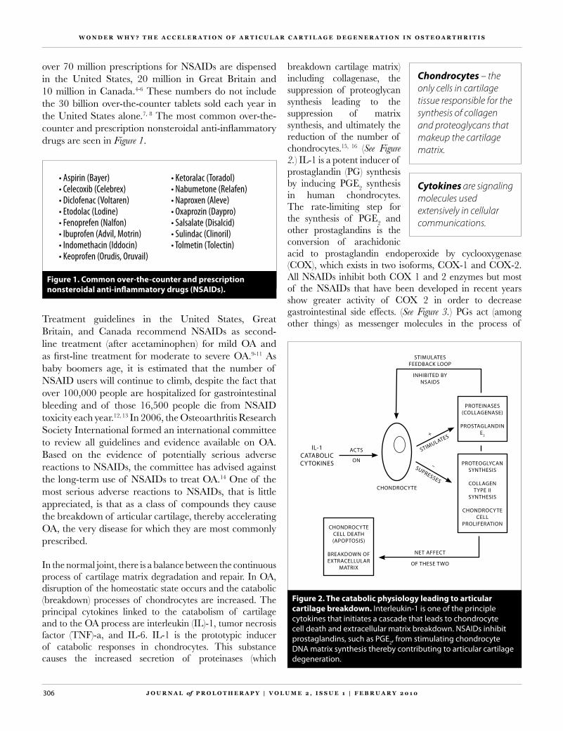
J O U R N A L of P R O L O T H E R A P Y | V O L U M E 2 , I S S U E 1 | F E B R U A R Y 2 0 1 0306
W O N D E R W H Y ? T H E A C C E L E R A T I O N O F A R T I C U L A R C A R T I L A G E D E G E N E R A T I O N I N O S T E O A R T H R I T I S
breakdown cartilage matrix) including collagenase, the suppression of proteoglycan synthesis leading to the suppression of matrix synthesis, and ultimately the reduction of the number of chondrocytes.15, 16 (See Figure 2.) IL-1 is a potent inducer of prostaglandin (PG) synthesis by inducing PGE2 synthesis in human chondrocytes. The rate-limiting step for the synthesis of PGE2 and other prostaglandins is the conversion of arachidonic acid to prostaglandin endoperoxide by cyclooxygenase (COX), which exists in two isoforms, COX-1 and COX-2. All NSAIDs inhibit both COX 1 and 2 enzymes but most of the NSAIDs that have been developed in recent years show greater activity of COX 2 in order to decrease gastrointestinal side effects. (See Figure 3.) PGs act (among other things) as messenger molecules in the process of
Figure 2. The catabolic physiology leading to articular cartilage breakdown. Interleukin-1 is one of the principle cytokines that initiates a cascade that leads to chondrocyte cell death and extracellular matrix breakdown. NSAIDs inhibit prostaglandins, such as PGE2, from stimulating chondrocyte DNA matrix synthesis thereby contributing to articular cartilage degeneration.
IL-1CATABOLICCYTOKINES
CHONDROCYTE
ACTS
ON
+
STIMULATES
–SUPRESSES
PROTEOGLYCANSYNTHESIS
COLLAGENTYPE II
SYNTHESIS
CHONDROCYTECELL
PROLIFERATION
PROTEINASES(COLLAGENASE)
PROSTAGLANDINE2
STIMULATESFEEDBACK LOOP
INHIBITED BY NSAIDS
CHONDROCYTE CELL DEATH(APOPTOSIS)
BREAKDOWN OFEXTRACELLULAR
MATRIX
NET AFFECT
OF THESE TWO
over 70 million prescriptions for NSAIDs are dispensed in the United States, 20 million in Great Britain and 10 million in Canada.4-6 These numbers do not include the 30 billion over-the-counter tablets sold each year in the United States alone.7, 8 The most common over-the-counter and prescription nonsteroidal anti-inflammatory drugs are seen in Figure 1.
Figure 1. Common over-the-counter and prescription nonsteroidal anti-inflammatory drugs (NSAIDs).
• aspirin (Bayer) • Celecoxib (Celebrex) • diclofenac (voltaren) • etodolac (lodine) • fenoprefen (nalfon) • ibuprofen (advil, motrin)• indomethacin (iddocin)• Keoprofen (orudis, oruvail)
• Ketoralac (toradol) • nabumetone (relafen)• naproxen (aleve) • oxaprozin (daypro)• salsalate (disalcid) • sulindac (Clinoril) • tolmetin (tolectin)
Treatment guidelines in the United States, Great Britain, and Canada recommend NSAIDs as second-line treatment (after acetaminophen) for mild OA and as first-line treatment for moderate to severe OA.9-11 As baby boomers age, it is estimated that the number of NSAID users will continue to climb, despite the fact that over 100,000 people are hospitalized for gastrointestinal bleeding and of those 16,500 people die from NSAID toxicity each year.12, 13 In 2006, the Osteoarthritis Research Society International formed an international committee to review all guidelines and evidence available on OA. Based on the evidence of potentially serious adverse reactions to NSAIDs, the committee has advised against the long-term use of NSAIDs to treat OA.14 One of the most serious adverse reactions to NSAIDs, that is little appreciated, is that as a class of compounds they cause the breakdown of articular cartilage, thereby accelerating OA, the very disease for which they are most commonly prescribed. In the normal joint, there is a balance between the continuous process of cartilage matrix degradation and repair. In OA, disruption of the homeostatic state occurs and the catabolic (breakdown) processes of chondrocytes are increased. The principal cytokines linked to the catabolism of cartilage and to the OA process are interleukin (IL)-1, tumor necrosis factor (TNF)-a, and IL-6. IL-1 is the prototypic inducer of catabolic responses in chondrocytes. This substance causes the increased secretion of proteinases (which
Cytokines are signaling molecules used extensively in cellular communications.
Chondrocytes – the only cells in cartilage tissue responsible for the synthesis of collagen and proteoglycans that makeup the cartilage matrix.

J O U R N A L of P R O L O T H E R A P Y | V O L U M E 2 , I S S U E 1 | F E B R U A R Y 2 0 1 0 307
W O N D E R W H Y ? T H E A C C E L E R A T I O N O F A R T I C U L A R C A R T I L A G E D E G E N E R A T I O N I N O S T E O A R T H R I T I S
as proteoglycans, collagen, fibronectin, integrins, and other adhesive proteins which are needed to maintain the high tensile strength and low compressibility under load of the articular cartilage.19, 20 Type II collagen is the predominant collagen type in the extracellular matrix with proteoglycan (PRG) macromolecules dispersed throughout. They contain highly negatively charged carboxyl and sulfate groups (keratin and chondroitin sulfate) on the glycosaminoglycans, giving them a high affinity for water. (See Figure 4.) The nature of the high density of negative charges imparts the physical properties to PRGs. Because of their attraction and binding of water, PRGs are viscous, making them ideal for lubricating fluid in joints. The charges repel each other, which gives them an open structure and is space-filling. These biochemical traits contribute to the mechanical properties of PRGs in articular cartilage, such as absorption and distribution of compressive weight, protecting structures in the joints from mechanical damage.21 The normal synthesis and breakdown of the PRGs and their component molecules,
Figure 4. The proteoglycan structure of articular cartilage. The high content of water in proteoglycans help the cartilage act as a shock absorber.
WATER
PROTEOGLYCAN WATER
WATERWATER
WATER
COLLAGEN FIBRILS
COREPROTEIN
GLYCOSAMINOGLYCAN GAG CHAINS –
FIXED NEGATIVE CHARGES
FLUID FLOW
WATER
WATER
WATER
WATER
LOAD-INDUCEDCOMPRESSION(REVERSIBLE)
WATER
WATER
WATER
COREPROTEIN
ELECTRICAL FIELD STREAMING
POTENTIAL CURRENTS
HowCartilageActsasaShockAbsorber
inflammation. It was hoped that the use of NSAIDs would decrease the catabolic program in OA, thereby having a disease-modifying effect. Research, unfortunately is showing PGs, like PGE2, stimulate chondrocyte proliferation and subsequent synthesis of cellular matrix components.17 The net result of their blockade and other NSAID effects is the acceleration of articular cartilage degeneration. To show how this occurs and to what extent, a basic understanding of articular cartilage anatomy is needed.
a r t i C u l a r C a r t i l a g e a n a t o m y
Articular cartilage functions as a wear-resistant, smooth, nearly frictionless, load-bearing surface. The composition and physiochemical properties of articular cartilage, the fundamental organization of the collagen network, and the molecular organization of collagen and proteoglycans all have profound effects on the intrinsic mechanical properties of the extracellular matrix.18 Cartilage is composed of a complex extracellular matrix of collagen and elastic fibers within a hydrated gel of glycosaminoglycans and proteoglycans. This extracellular matrix, which makes up 98% of the articular cartilage volume, is synthesized by the chondrocytes which comprise the other 2% of the cartilage tissue. It is well known that chondrocytes can synthesize the extracellular matrix such
Figure 3. Inhibition of cyclooxygenase 1 and 2 by NSAIDs. Studies have shown that, although most NSAIDs inhibit both COX-1 and COX-2, it is the inhibition of COX-2 that is responsible for the anti-inflammatory effects of NSAIDS. On the other hand, inhibition of COX-1 by these agents causes damage to the GI tract. This has led to the development of a new generation of NSAIDs that specifically inhibit COX-2.
TRAUMA, SPORTS INJURY, CHEMICAL OR INFLAMMATION
STIMULI
CELL MEMBRANE
PHOSPHOLIPASE A2
ARACHIDONIC ACID
COX-2 MEDIATEDCOX-1 MEDIATED
CONVENTIONAL NSAIDs
COX-2 SPECIFIC NSAIDs
POTENTIAL FOR SIGNIFICANT
GASTOINTESTINAL TRACT DAMAGE
(BLEEDING)
POTENT INHIBITION OF
INFLAMMATION

J O U R N A L of P R O L O T H E R A P Y | V O L U M E 2 , I S S U E 1 | F E B R U A R Y 2 0 1 0308
W O N D E R W H Y ? T H E A C C E L E R A T I O N O F A R T I C U L A R C A R T I L A G E D E G E N E R A T I O N I N O S T E O A R T H R I T I S
separation, as long as the inciting issue (NSAID use) continues. (See Figure 5.) These changes alter the intrinsic mechanical properties of articular cartilage and produce swelling.25 The articular cartilage, having lost some of its compressive ability under load, further degenerates. As the surface fibrillation progresses, the articular defects penetrate deeper into the cartilage until the cartilage is lost. The increased pressure on the subchondral bone causes it to thicken. Often bone cysts form deep to the eburnated areas. Eventually, bony nodules or osteophytes form at the periphery of the cartilage surface. All of these changes account not only for the pathology found on radiographs or histologically (findings under the microscope), but also for the joint pain, tenderness, loss of motion and stiffness of OA.26 It is the relief of some of these clinical manifestations that accounts for the widespread use of NSAIDs not only in the United States, but around the world.
t h e e x t e n t o f t h e P r o B l e m
In 2006, the Center for Disease Control combined data from the National Health Interview Survey years 2003-2005 Sample Adult Core components to estimate the average annual arthritis prevalence in the U.S. population aged 18 years and older. Overall, 21.6% (46.4 million) of adults reported arthritis or another rheumatic condition diagnosed by a doctor, with 27 million Americans having osteoarthritis, up from 21 million in 1990.27, 28 By the year
including glycosaminoglycans, is mediated by the indigenous chondrocytes. Glycosaminoglycans turn over several times as rapidly as the fibrillar collagen. If any part of this complex system is disrupted, the normal properties of articular cartilage are jeopardized, leading to joint degeneration. It is the extracellular matrix of articular cartilage that is the primary target of osteoarthritic cartilage degeneration and the accelerating effects of this breakdown by NSAIDs.
One of the earliest features of the development of osteoarthritis is degeneration of the articulating surfaces of the joint. This is characterized by fibrillation of the articular cartilage, in which the mesh of collagen fibers is disrupted. Degeneration of type II collagen is seen, as well as a decrease in the extracellular matrix.22 Loss of proteoglycan from the matrix is characteristic. The loss of aggrecan, the predominant PRG in articular cartilage imposes an increasing load on the collagen fibrils, causing further breakdown.23 Early in the course of OA, the tissue mounts an attempt at repair. Chondrocytes proliferate and there is an increase in matrix synthesis.24 However, if this repair process is disrupted for any reason including the use of NSAIDs, degradative enzymes overwhelm the synthetic capability and the repair fails. Particular compositional, molecular, and structural changes will continue to occur within the articular cartilage including decreased proteoglycan and increased water content, collagen fibril network disorganization, and proteoglycan
Figure 5. The pathogenesis of osteoarthritis accelerated by NSAIDs. NSAID use inhibits the body’s repair processes, leading to decreased proteoglycan and extracellular matrix content and function, which ultimately leads to articular cartilage breakdown.
NORMAL JOINT ARTHRITIC JOINT
CHONDROCYTESSTIMULATED
INCREASE IN
MATRIX SYNTHESIS
TISSUE MOUNTS
A REPAIR
TRUAMAINJURY
NSAIDSUSE
TISSUE REPAIRINHIBITED
DECREASE PROTEOGLYCAN
SYNTHESIS + STRUCTURE
PRESSURE ONARTICULARCARTILAGE
ARTICULARCARTILAGE
BREAKDOWN

J O U R N A L of P R O L O T H E R A P Y | V O L U M E 2 , I S S U E 1 | F E B R U A R Y 2 0 1 0 309
W O N D E R W H Y ? T H E A C C E L E R A T I O N O F A R T I C U L A R C A R T I L A G E D E G E N E R A T I O N I N O S T E O A R T H R I T I S
2030, an estimated 67 million (25% of the projected total adult population) adults aged 18 years and older will have doctor-diagnosed arthritis with two-thirds of those with arthritis being women. (See Figure 6.) The impact of this arthritis on individuals is significant. Almost 41% report severe limitations in their usual activities and 31% report being limited in work due to the arthritis.29 (See Figure 7.) The average direct cost (medications, assistive devices) of OA is approximately $2,600 per year per person living with OA, but the total annual cost (including lost wages, loss of productivity) of OA per person living with OA is at the low end $5,700 but in the high end over $10,000.30-32 The question remains as to why is there this alarming increase in osteoarthritis to the point that between 1997 and 2005 the number of knee surgeries climbed by 69% from 328,800 to 555,800, hip replacements rose 32% from 290,700 to 383,500, and spinal fusion surgeries increased by 73% from 202,100 procedures to 349,400 per year?33
Nonsteroidal anti-inflammatory drugs (NSAIDs) are one of the most commonly used classes of medications. Ibuprofen was the first NSAID available by prescription in the United States in 1974, under the brand names Motrin and Rufen. It rose to be the fifth-largest selling prescription drug and in 1984 was the first new entrant in the non-prescription pain reliever market in nearly 30 years. For the last thirty plus years, NSAIDs are among the most frequently used drugs in the United States. From 1973 to 1983, for instance, the number of NSAID prescriptions dispensed by retail pharmacies tripled, rising from 28 million to around 70 million by the early 1980s. (See Figure 8.) What are the long-term effects of this NSAID use? Could it be that the massive widespread use of NSAID twenty and thirty plus years ago is the reason that there is currently an epidemic of disabling osteoarthritis resulting in a slew of spine and joint replacement operations? By 1983, five of the 50 drug products most often dispensed were NSAIDs, representing over 4% of the total prescription market.34 To put a practical visual on these numbers in percentage terms, enough NSAIDs were purchased in the United States by drugstores and hospitals to treat 1.29% of the entire civilian population each day in 1983. The number one use for these NSAIDs in 1983 was osteoarthritis. While the prescribing patterns for specific NSAIDs have changed over the years, as drugs like ibuprofen and naproxen became available over-the-counter, an NSAID is still the number one medication
Figure 7. Percent of adults with doctor-diagnosed arthritis with ”arthritis attributable” activity and work limitations in 2002.Data Source: www.cdc.gov, 2002 National Health Interview Survey.
Hootman J, et al. Prevalence of doctor-diagnosed arthritis and arthtitis-attributed activity limitation–United States, 2003-2005. MMWR. 2006;55(40):1089-1092.
Bolen J, et al. Racial/Ethnic differences in the prevelance and impact of doctor-diagnosed arthritis–United States, 2002. MMWR. 2005;54(5):119-123.
PERC
ENT
OF
AD
ULT
S W
ITH
D
OC
TOR-
DIA
GN
OSE
D A
RTH
RITI
S
AR THRITIS-AT TRIBUTABLE L IMITATION T YPE
100
90
80
70
60
50
40
30
20
10
0ACTIVITY LIMITATION WORK LIMITATION
40.9%(18.9 million
adults)
30.6%(8.3 million
adults)
(Among 42.7 million persons ≥ 18 years w/ doctor-diagnosed arthritis)1
(Among 42.7 million persons 18-64 years w/ doctor-diagnosed arthritis)2
prescribed by physicians for osteoarthritis. For instance, 80% of rheumatologists noted they frequently prescribe NSAIDs for symptomatic hip and knee osteoarthritis, while for the same group of clients, 65% of primary care physicians use an NSAID.35, 36 Even when physicians were educated on guidelines based on the European League Against Rheumatism, American College of Rheumatology, and The Arthritis Society guidelines for OA treatment,
Figure 6. Projected prevalence of doctor-diagnosed arthritis, in US adults 18 and older, 2005-2030.Data Source: www.cdc.gov
Hootman JM, et al. Projections of U.S. prevelance of arthritis and associated activity limitations. Arthritis Rheum. 2006;54(1):226-229.
Hootman J, et al. Prevalence of doctor-diagnosed arthritis and arthtitis-attributed activity limitation–United States, 2003-2005. MMWR. 2006;55(40):1089-1092.
WOMEN
PRO
JEC
TED
NU
MBE
R O
F A
DU
LTS
WIT
H
DO
CTO
R-D
IAG
NO
SED
ART
HRI
TIS
(IN
MIL
LIO
NS)
YEAR
50
40
30
20
10
02005 2010 2015 2020 2025 2030
MEN
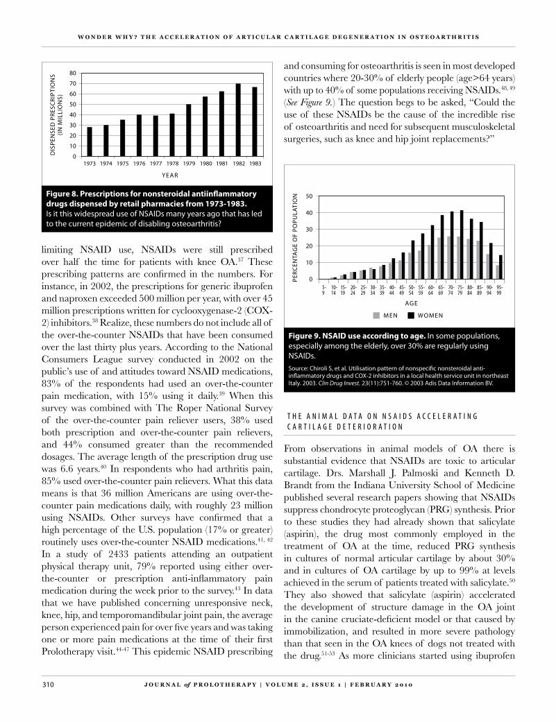
J O U R N A L of P R O L O T H E R A P Y | V O L U M E 2 , I S S U E 1 | F E B R U A R Y 2 0 1 0310
W O N D E R W H Y ? T H E A C C E L E R A T I O N O F A R T I C U L A R C A R T I L A G E D E G E N E R A T I O N I N O S T E O A R T H R I T I S
t h e a n i m a l d a t a o n n s a i d s a C C e l e r a t i n g C a r t i l a g e d e t e r i o r a t i o n
From observations in animal models of OA there is substantial evidence that NSAIDs are toxic to articular cartilage. Drs. Marshall J. Palmoski and Kenneth D. Brandt from the Indiana University School of Medicine published several research papers showing that NSAIDs suppress chondrocyte proteoglycan (PRG) synthesis. Prior to these studies they had already shown that salicylate (aspirin), the drug most commonly employed in the treatment of OA at the time, reduced PRG synthesis in cultures of normal articular cartilage by about 30% and in cultures of OA cartilage by up to 99% at levels achieved in the serum of patients treated with salicylate.50 They also showed that salicylate (aspirin) accelerated the development of structure damage in the OA joint in the canine cruciate-deficient model or that caused by immobilization, and resulted in more severe pathology than that seen in the OA knees of dogs not treated with the drug.51-53 As more clinicians started using ibuprofen
limiting NSAID use, NSAIDs were still prescribed over half the time for patients with knee OA.37 These prescribing patterns are confirmed in the numbers. For instance, in 2002, the prescriptions for generic ibuprofen and naproxen exceeded 500 million per year, with over 45 million prescriptions written for cyclooxygenase-2 (COX-2) inhibitors.38 Realize, these numbers do not include all of the over-the-counter NSAIDs that have been consumed over the last thirty plus years. According to the National Consumers League survey conducted in 2002 on the public’s use of and attitudes toward NSAID medications, 83% of the respondents had used an over-the-counter pain medication, with 15% using it daily.39 When this survey was combined with The Roper National Survey of the over-the-counter pain reliever users, 38% used both prescription and over-the-counter pain relievers, and 44% consumed greater than the recommended dosages. The average length of the prescription drug use was 6.6 years.40 In respondents who had arthritis pain, 85% used over-the-counter pain relievers. What this data means is that 36 million Americans are using over-the-counter pain medications daily, with roughly 23 million using NSAIDs. Other surveys have confirmed that a high percentage of the U.S. population (17% or greater) routinely uses over-the-counter NSAID medications.41, 42 In a study of 2433 patients attending an outpatient physical therapy unit, 79% reported using either over-the-counter or prescription anti-inflammatory pain medication during the week prior to the survey.43 In data that we have published concerning unresponsive neck, knee, hip, and temporomandibular joint pain, the average person experienced pain for over five years and was taking one or more pain medications at the time of their first Prolotherapy visit.44-47 This epidemic NSAID prescribing
and consuming for osteoarthritis is seen in most developed countries where 20-30% of elderly people (age>64 years) with up to 40% of some populations receiving NSAIDs.48, 49 (See Figure 9.) The question begs to be asked, “Could the use of these NSAIDs be the cause of the incredible rise of osteoarthritis and need for subsequent musculoskeletal surgeries, such as knee and hip joint replacements?”
Figure 8. Prescriptions for nonsteroidal antiinflammatory drugs dispensed by retail pharmacies from 1973-1983. Is it this widespread use of NSAIDs many years ago that has led to the current epidemic of disabling osteoarthritis?
DIS
PEN
SED
PRE
SCRI
PTIO
NS
(IN
MIL
LIO
NS)
YEAR
80
70
60
50
40
30
20
10
01973 1974 1975 1976 1977 1978 1979 1980 1981 1982 1983
Figure 9. NSAID use according to age. In some populations, especially among the elderly, over 30% are regularly using NSAIDs.Source: Chiroli S, et al. Utilisation pattern of nonspecific nonsteroidal anti-inflammatory drugs and COX-2 inhibitors in a local health service unit in northeast Italy. 2003. Clin Drug Invest. 23(11):751-760. © 2003 Adis Data Information BV.
PERC
ENTA
GE
OF
POPU
LATI
ON
AGE
50
40
30
20
10
05-9
10-14
15-19
20-24
25-29
30-34
35-39
40-44
45-49
50-54
55-59
60-64
65-69
70-74
75-79
80-84
85-89
90-94
95-99
MEN WOMEN

J O U R N A L of P R O L O T H E R A P Y | V O L U M E 2 , I S S U E 1 | F E B R U A R Y 2 0 1 0 311
W O N D E R W H Y ? T H E A C C E L E R A T I O N O F A R T I C U L A R C A R T I L A G E D E G E N E R A T I O N I N O S T E O A R T H R I T I S
to say that the medications can “induce progressive joint degeneration within three to four months.”62 Animal studies have also shown the effects of NSAIDs on proliferation, cell cycle kinetics, cytotoxicity, and cell death of chondrocytes. In one study the NSAIDs indomethacin, ketorolac, diclofenac, piroxicam, and celecoxib inhibited thymidine incorporation of chondrocytes at therapeutic concentrations. NSAIDs also arrested chondrocytes in their cell cycles, thus inhibiting chondrocyte cell replication. Upon 24 hour exposure to indomethacin, ketorolac, diclofenac, and piroxicam, chondrocyte cell death (both apoptosis and necrosis) was induced in cell cultures.63 One mechanism by which NSAIDs are toxic to chondrocytes is by inhibiting PGE2 synthesis by chondrocytes.64 PGE2 elicits differentiation of chondrocytes and is an important contributor to cartilage formation and promotes DNA and matrix synthesis in chondrocytes.65, 66 PGE2 has a growth stimulatory effect on chondrocytes, thereby increasing chondrocyte DNA synthesis.67, 68 NSAIDs inhibit the enzyme cyclooxygenase which is responsible for PGE2 release in chondrocytes.69
h u m a n s t u d i e s
In 1991, Kenneth D. Brandt, MD, one of the main researchers on NSAIDs’ effect on cartilage wrote, “No clinical evidence exists today, however, to support the contention that NSAIDs favorably influence the progression of joint degeneration in man.”70 While this author will not refute this statement, an addition to it is warranted …but much evidence exists that NSAID use accelerates articular cartilage degeneration. This issue is extremely important since 30 billion over-the-counter doses of NSAIDs are sold annually in the United States.71
While the condition known as osteoarthritis has other names, including degenerative joint disease, the name is actually misleading; the words do not accurately describe the pathophysiology of the condition. The term osteoarthritis literally means inflammation of a bony joint but the most common clinical presentation of the condition is one of articular cartilage breakdown without joint swelling, heat, or any other markers of inflammation. The more appropriate term for osteoarthrosis or degenerative joint disease is understood as a non-inflammatory degenerative process. The notion of treating a non-inflammatory condition with an anti-inflammatory medication is bound to have long-term detrimental effects.
and other NSAIDs, instead of aspirin for OA, Drs. Palmoski and Brandt studied the effects these drugs had on canine articular cartilage. Specifically they found that fenoprofen and ibuprofen inhibited net PRG synthesis in a concentration-dependent fashion. At concentrations in the culture medium comparable to plasma concentrations seen in patients after oral administration of NSAIDs in humans, net PRG synthesis in the presence of these drugs averaged 72% and 86% of the control values, respectively (P<0.01).54-56 (See Figure 10.) In another study on canine articular cartilage, these researchers found that the inhibitory effect of the NSAID indomethacin was greater when the articular cartilage was depleted of glycosaminoglycans.57 In other words, there is a greater inhibition of PRG synthesis in osteoarthritic cartilage than normal cartilage. Other researchers have confirmed these findings that NSAIDs consistently suppress proteoglycan and glycosaminoglycan synthesis.58-60 This depletion of matrix proteoglycans has been shown to be one cause of the increased degeneration of cartilage chondrocytes from the use of NSAIDs.61 Taken to the extreme, one researcher put it this way, “…depending on dose and at concentrations that in many cases correspond to therapeutic plasma levels, these drugs may lead to a pronounced reduction or complete blockade of synthesis of the proteoglycans and collagen of the cartilage matrix.” They went on
P-value is the sense of statistical significance. P<0.01 means that there is a less than 1 in 100 (1%) chance that the results occurred by chance. The lower the p-value, the more significant the result.
Figure 10. Net synthesis of 35S-glycosaminoglycans by normal canine knee cartilage cultured in the presence of ibuprofen. Ibuprofen inhibited glycosaminoglycan synthesis by cartilage cells at doses that are commonly achieved by those taking this medication.
GLY
CO
SAM
INO
GLY
CA
IN S
YNTH
ESIS
(PER
CEN
T O
F C
ON
TRO
L)
I BUPROFEN CONCENTRATION (µg/ml)
100
90
80
70
60
50
40
30
20
10
012 35 70 140

J O U R N A L of P R O L O T H E R A P Y | V O L U M E 2 , I S S U E 1 | F E B R U A R Y 2 0 1 0312
W O N D E R W H Y ? T H E A C C E L E R A T I O N O F A R T I C U L A R C A R T I L A G E D E G E N E R A T I O N I N O S T E O A R T H R I T I S
was measured on 245 osteoarthritic patients and 80 normal patients’ cartilage organ cultures subjected to various NSAIDs. The commonly used NSAIDs indomethacin, ibuprofen, and naproxen were shown to significantly inhibit (from 40 to 70%) glycosaminoglycan synthesis in patients’ cartilage.76 (See Figure 12.) Notice that paracetamol (acetaminophen or Tylenol) did not inhibit GAG synthesis. The researchers noted that caution must be exercised in extrapolation from in-vitro (lab) to in-vivo (person) effects of NSAIDs, but it seems possible that some highly effective anti-inflammatory agents may produce adverse effects on cartilage integrity when employed during long-term treatment.77 Other researchers have confirmed NSAIDs’ inhibitory effect on proteoglycan synthesis and have commented that “…any drug that suppresses proteoglycan synthesis and impairs the ability of the chondrocyte to repair its damaged extracellular matrix, could potentially accelerate the breakdown of the articular tissue.”78, 79
n s a i d s i n h i B i t P r o s t a g l a n d i n s y n t h e s i s
One way in which NSAIDs stop the chondrocytes from repairing themselves is by the inhibition of the synthesis of Prostaglandin E2 (PGE2). Prostaglandins (PG) are produced by most human cell types (including chondrocytes) and have a variety of physiologic functions. PG synthesis is initiated by the mobilization of arachidonic acid from cell membrane phospholipids as a result of the enzyme phospholipase A2. The enzyme
Figure 12. Some of the most commonly used NSAIDs, Indomethacin, Ibuprofen, & Naxopren, were found to decrease GAG synthesis, whereas Paracetamol (acetaminophen) did not.
% O
F TO
TAL
SHO
WIN
G
INC
REA
SED
GA
G S
YNTH
ESIS
60
40
20
0
-20
-40
-60
-80Paracetamol
10–100
TheeffectofNSAIDsonGAGsynthesis
Idomethacin30
Ibuprofen30
Naproxen50–100
NSAID & CONC. RANGE TESTED (µg/ml)
(32) (34) (64) (60)
% O
F TO
TAL
SHO
WIN
GD
ECRE
ASE
D G
AG
SYN
THES
IS
# OF PATIENTS
At present no quantitative non-invasive method for determining the anabolic (building up) and catabolic (breaking down) activity of NSAIDs on human cartilage in vivo exists. Most information on the effects of NSAIDs on the turnover of extra-cellular matrix macromolecules comes from short-term organ culture studies. Initial evaluations into the pathophysiology of osteoarthritis concentrated on the effects of NSAIDs on glycosaminoglycan synthesis. It was established that in all but the most severe cases of osteoarthritis, the chondrocyte response to proteoglycan depletion was an increase in glycosaminoglycan synthesis.72, 73 One of the first to show that NSAIDs diminished glycosaminoglycan synthesis in aged human cartilage cells (taken during hip surgery) in vitro was a research group from the University of Sydney in 1976.74 J.T. Dingle, led several of the follow-up studies on the effects of NSAIDs on human cartilage metabolism. The initial studies revealed significant declines in glycosaminoglycan synthesis in both normal and osteoarthritic human cartilages.75 (See Figure 11.) In a follow-up study, the same research group, took femoral head articular cartilage from non-arthritic and osteoarthritic patients post-operatively after total hip replacement. The relative human cartilage metabolism
in vivo – experimentation using whole, living organisms or live isolated cells. Once the cells are disrupted and the individual parts are treated or analyzed this is known as in vitro.
Figure 11. The effect of NSAIDs on GAG synthesis in humans (both non-arthritic and osteoarthritic) is shown. Concentrations of NSAIDs are similar to those obtained in serum/synovial fluid during treatment.
PERC
ENT
CO
NTR
OL
GA
G S
YNTH
ESIS 100
90
80
70
60
50
40
30
20
10
0Ibuprofen100 µg/ml
TheeffectofNSAIDsonGAGsynthesisinhumancartilageinpeoplewithandwithoutosteoarthritis
Indomethacin10 µg/ml
Diclofenac0.3 µg/ml
Aspirin30 µg/ml
Naproxen300 µg/ml
NORMAL WITH OA

J O U R N A L of P R O L O T H E R A P Y | V O L U M E 2 , I S S U E 1 | F E B R U A R Y 2 0 1 0 313
W O N D E R W H Y ? T H E A C C E L E R A T I O N O F A R T I C U L A R C A R T I L A G E D E G E N E R A T I O N I N O S T E O A R T H R I T I S
other cells at sites of inflammation. Prostaglandins, whose synthesis involves COX-1, are responsible for maintenance and protection of the gastrointestinal tract, while prostaglandins, whose synthesis involves COX-2, are responsible for inflammation and pain. One of the main prostaglandins involved in this inflammatory reaction is PGE2. Researchers have shown that the PGE2 levels correlate with the amount of COX-2 expression in chondrocytes.92 (See Figure 14.) Also well established is that this PGE2 release can easily be inhibited by the use of NSAIDs.93, 94 (See Figure 15.) Since the over expression of the COX-2 protein plays an important role in many pathophysiologic states, including inflammation, cancer, angiogenesis, Alzheimer’s disease, and several forms of inflammatory arthritis, NSAIDs especially those that
cyclooxygenase along with other enzymes converts arachidonic acid to five primary prostaglandins: PGD2, PGE2, PGI2 (Prostacyclin), PGF2a, and TXA2 (thomboxane). (See Figure 13.) These PGs have a variety of functions including the mediation of inflammation, calcium movement, sensitization of spinal neurons to pain, blood clotting, blood pressure, circulation, control of blood flow in kidneys, hormone regulation, protection of gastrointestinal lining, and the control of cell growth.80, 81 Chondrocytes and synovial fibroblasts produce PGE2. PGE2 levels are increased to an impact load on articular cartilage or during cartilage degeneration.82, 83 PGE2 is reported to have anabolic effects on cartilage: increasing proteoglycan and DNA and collagen synthesis,84, 85 stimulating proliferation and proteoglycan aggrecan synthesis,86, 87 and, at low concentrations, stimulating type II collagen synthesis.88
Human chondrocytes express two forms of the cyclooxygenase enzyme, known as the COX-1 and COX-2 isoforms. Unstimulated human chondrocytes do not contain detectable COX-2.89 COX-1 is present in most cells under physiological conditions, whereas COX-2 is induced by some cytokines presumably in pathological conditions such as joint trauma, degeneration, or osteoarthritis.90, 91 Put another way, COX-2 is undetectable in most normal tissues, is an inducible enzyme, becoming abundant in activated macrophages (immune cells) and
Figure 14. Correlation analysis of COX-2 expression and PGE2 levels by chondrocytes. The isoform COX-2 enzyme levels correlate directly with PGE2 levels.
110
100
90
80
70
60
50
40
30
20
10
0
-10-500 0 500 1000 1500 2000 2500 3000 3500
PGE 2 LE VELS (pg/ml)
PERC
ENT
CO
X2
EXPR
ESSI
ON
Figure 15. PGE2 released into culture medium, as a function of log-NSAID dose (M). Results are expressed as % of control values. l — l ASA; — TA. NSAIDs at physiologic concentrations are potent inhibitors of PGE2 synthesis.
100%
75%
50%
25%
0%2 5 10 20 40
NSAID (10 –5 M) CONCENTRATION
PGE 2 L
EVEL
S (%
OF
CO
NTR
OL)
PHOSPHOLIPIDS
Figure 13. Biosynthesis of prostaglandins. The enzyme cyclooxygenase is the key enzyme in the formation of the five primary prostaglandins including PGE2. NSAIDs inhibit prostaglandin synthesis by inhibiting the enzyme cyclooxygenase.
Phosphol ipase A2
ARACHIDONICACID
Cycloox ygenase
PGH2
TXA2 PGF2a PGD2 PGE2 PGI2
Speci f ic terminal synthases

J O U R N A L of P R O L O T H E R A P Y | V O L U M E 2 , I S S U E 1 | F E B R U A R Y 2 0 1 0314
W O N D E R W H Y ? T H E A C C E L E R A T I O N O F A R T I C U L A R C A R T I L A G E D E G E N E R A T I O N I N O S T E O A R T H R I T I S
Figure 16. Chondrocytes produce PGE2 in response to injury. NSAIDs, especially those that block COX-2 inhibit PGE2 synthesis in chondrocytes thereby stalling the body’s main inflammatory repair mechanism. Long-term, this will accelerate degenerative osteoarthritis of the joint.
NORMAL JOINT (NO PGE2 PRODUCED) INJURED JOINT
TRUAMAINJURY
NSAIDSUSE
NSAIDSBLOCK
HEALING
JOINT HEALING STIMULATED
JOINT REPAIRED
CHONDROCYTESPRODUCE PGE2
inhibit COX-2, are used for many of these conditions. In regard to joint inflammation, one author notes, “…by inhibiting joint conditions, they (NSAIDs) may indirectly be beneficial to cartilage, specifically when inflammation is primary in the cause of cartilage damage, as in the case for rheumatoid arthritis.” However, in OA, in which inflammation may contribute to but is not primarily responsible for cartilage damage, adverse direct effects of NSAIDs on cartilage with long-term treatment may have an important impact on long-term outcome.”94 In other words PGE2 can exert catabolic or anabolic effects depending on the microenvironment.95
Since normal articular chondrocytes produce very little PGE2 and osteoarthritic chondrocytes produce a lot of it through the COX-2 enzyme, it would make sense from a traditional medical point of view to attack arthritis pain from this angle. This is especially true since the over expression of the COX-2 protein (and thus increased PGE2 levels) plays an important role in many pathophysiolgic states, including systemic inflammation, fever, cancer, angiogenesis, Alzheimer’s disease, and inflammatory arthritis.96 Yes, in certain conditions inflammation is harmful, but it is a big leap to assume everywhere there is PGE2 it is harming tissue. The articular chondrocytes make PGE2 in response to injury to stimulate healing. Osteoarthritic cartilage spontaneously releases PGE2 in levels at least 50-fold higher than normal cartilage and
18-fold higher than normal cartilage stimulated with cytokines and endotoxin.97-100 The inflammation that occurs through PGE2 when a normal or osteoarthritic joint is injured is the body’s immune system response to try and get the joint injury repaired.101 When a person uses medications that block this response, while pain may be improved, the repair mechanisms for the joint are inhibited. The long-term consequences, of course can be an acceleration of the degenerative osteoarthritic process. (See Figure 16.) Long-term NSAID treatment not only blocks PGE2 production by direct inhibition of COX-2 activity but by down-regulating COX-2 synthesis.102
n s a i d s a C C e l e r a t e t h e r a d i o g r a P h i C P r o g r e s s i o n o f o s t e o a r t h r i t i s o f t h e K n e e a n d h i P
The suggestion that indomethacin accelerates the bone destruction in osteoarthritis of the hip was first made by Coke in 1967;103 subsequent reports have been numerous that provide further clinical evidence of the damaging effects of non-steroidal anti-inflammatory drugs on osteoarthritic hips.104-107 In one retrospective investigation of the relationship between the use of non-steroidal anti-inflammatory drugs on hip destruction in primary osteoarthritis of the hip, 70 hips were studied in 64 patients. Cranial acetabular migration, a measure of acetabular destruction, was present in 37 hips and absent in 33. Regular intake of NSAIDs was noted for

J O U R N A L of P R O L O T H E R A P Y | V O L U M E 2 , I S S U E 1 | F E B R U A R Y 2 0 1 0 315
W O N D E R W H Y ? T H E A C C E L E R A T I O N O F A R T I C U L A R C A R T I L A G E D E G E N E R A T I O N I N O S T E O A R T H R I T I S
31 of the 37 migrating hips. In regard to the other six, three took NSAIDs on and off and only three of the 37 did not take NSAIDs. Those patients with serious hip destruction when compared with those who did not have the acetabular destruction did not differ in sex, age, pain grading, or walking ability. The only significant difference was the amount of NSAIDs taken. According to the researchers, NSAID use was associated with progressive formation of multiple small acetabular and femoral subcortical cysts and subchondral bone thinning. They concluded, “The association of acetabular bone destruction with regular NSAID intake in patients with osteoarthritis of the hip adds further evidence to the clinical and experimental observations on the powerful and potentially harmful effects of these drugs on cartilage and bone.”108 In this study the NSAIDs used regularly and associated with acetabular migration in this series were indomethacin (14 hips); ibuprofen (8 hips); naproxen (3 hips); sulindac, aspirin, and piroxicam (2 hips each); flurbiprofen, azapropazone, diclofenac, fenclofenac, and ketoprofen (1 hip each). The authors noted, “This study suggests caution in the widespread use of NSAIDs for osteoarthritis of the hip…”
Researchers in Norway studied the course of osteoarthritis in 294 hips of 186 patients with radiographs over a three year period. The development of the disease in patients treated with an NSAID was compared with that in a control group (no NSAID). In the NSAID group the OA disease progressed at a level of statistical significance more frequently and severely. Specifically the researchers found that in the three year period of the study, the osteoarthritic hips treated with the NSAID had more cysts, altered bone structure, and overall hip destruction. The way they put it was, “In the present study, loss of trabecular structure in the subchondral bone seems to be a characteristic feature in ‘indomethacin joint destruction’ as well as disappearance of normal joint contours and multiple small cysts.”109 Solomon reported similar destruction in osteoarthritic hip joints as “new events” during treatment with NSAIDs. He performed further investigations on the extirpated (cut out from surgery) femoral heads with examination of cut surface, slab radiographs, and
histology. Many of the heads, and especially those with changes attributable to NSAIDs, were found to have microscopic fragmentation of the bony trabeculae giving the appearance of a jammed marrow space.110 To see if these sort of damaging changes occurred with NSAID use on osteoarthritic knees the Longitudinal Investigation
of Nonsteroidal Anti-inflammatory Drugs in Knee Osteoarthritis (LINK) study group was formed in England. They did a large study to compare the rate of radiographic progression in knee osteoarthritis comparing indomethacin (NSAID) with placebo. The study involved 20 rheumatology clinics in the United Kingdom. Patients received indomethacin 25mg three times daily or a placebo. The average person in each group was around 60 years of age and had osteoarthritis in the knee for over five years. The study involved 85 clients in the indomethacin group and 85 in the placebo group. Radiographic analysis was done yearly and the radiographic
grade was judged by two observers using a six point scale. The average length of follow-up was three years. By the third year of the study, the results were so dramatic demonstrating the acceleration of the degeneration of the articular cartilage in the knee osteoarthritic patient treated with indomethacin that this part of the study had to be stopped. There were more than twice as many patients showing deterioration in the indomethacin group as the placebo. The difference between the two groups was highly statistically significant (p=0.009). The authors noted that the risk of deterioration within a one year period in patients taking indomethacin relative to placebo was 2.1 (risk ratio).111 The authors concluded firmly, “Our study confirms beyond a reasonable doubt that indomethacin increases the rate of radiological deterioration of osteoarthritic knees.”
What actually happens to patients who take NSAIDs on a regular basis? If NSAIDs, by inhibiting pain and inflammation in osteoarthritic joints, cause people with OA to overuse a damaged joint, this should result in accelerating joint degeneration and joint replacements at an earlier time or, alternatively, if treatment with NSAIDs alters cartilage metabolism and inhibits joint healing, an acceleration of articular cartilage degeneration should be
By the third year of the study, the results were so dramatic demonstrating
the acceleration of the degeneration of the articular cartilage in
the knee osteoarthritic patient treated with
indomethacin that this part of the study had
to be stopped.

J O U R N A L of P R O L O T H E R A P Y | V O L U M E 2 , I S S U E 1 | F E B R U A R Y 2 0 1 0316
W O N D E R W H Y ? T H E A C C E L E R A T I O N O F A R T I C U L A R C A R T I L A G E D E G E N E R A T I O N I N O S T E O A R T H R I T I S
seen. Numerous studies have shown that non-steroidal anti-inflammatory drugs, particularly indomethacin, increase the rate of progression of osteoarthritis of the hip and knee.112-117 Statistically significant progression of hip radiographs in osteoarthritic patients can be seen within one year of those patients taking NSAIDs. In one study, the authors noted, “…a statistically significant correlation between the NSAID consumption score and changes in the radiological parameter (p=0.0001). This statistically significant difference was retained when the percentage of days taking NSAIDs was added (p = 0.0004).”118
In a recent landmark study, Dutch researchers studied more than 1600 subjects with hip OA and 635 with knee OA. The researchers evaluated radiographs of the hip and knee at baseline and follow-up. The researchers assessed the associations between different types of NSAIDs and the progression of OA. The mean follow-up period was 6.6 years. They found that long-term use of the NSAID diclofenac was associated with a more than twofold increase in radiologic progression of hip osteoarthritis and a threefold increase in progression observed in the knee. Ibuprofen use was also shown to be associated with a statistically significant increase in progression of the users’ knee and hip OA.119 The interesting point of this study is that the study population was healthy. The authors noted that this may have resulted in an underestimation of the reported associations. Their conclusion noted,“…these data suggest that diclofenac may induce accelerated progression of hip and knee OA. Whether this occurs because of a true deleterious effect on cartilage or because of excessive mechanical loading on a hip or knee following pain relief, remains to be investigated.” Another study comparing diclofenac with placebo, as seen in Figure 17, accelerated OA in knees as evidenced by a greater decline in joint space width on X-rays compared to placebo.120
n s a i d s i n C r e a s e t h e n e e d f o r j o i n t r e P l a C e m e n t
It is important to remember that pain has a physiologic function: if a joint produces pain when it is used, it is a signal for the body to use that joint less or else the structure eliciting the pain will be further damaged. One study focused on a group of patients with hip osteoarthritis who needed to have a joint replacement in the not-too-distant future. They were randomly prescribed an NSAID, aspirin-like drug, or acetaminophen. Over the next months, the patients were asked about their joint pain, and radiographs of their hips were taken. The patients given the NSAID
had more progression of their hip radiographs and needed to have joint replacements performed in half the time as the group given acetaminophen. The authors speculated why this occurred. They noted that the NSAID might have prevented normal cartilage turnover and repair, and accelerated the joint degeneration; or, more likely, the potent medication decreased joint pain and those subjects were therefore more active. This has led to the suggestion that potent NSAIDs can lead to “analgesic joint,” which can develop when pain is relieved by the NSAID, thus increasing the joint use and subsequent load on the joint, causing accelerated joint degeneration and ultimate need for joint replacement, especially if the excessive joint load continues.121 This latter notion has actually been studied: patients who take NSAIDs for knee OA put increased joint forces on their knees with walking because of pain relief, compared to those who do not have pain relief taking nothing, or just a placebo. As one researcher put it, “Of particular concern is the fact that anti-inflammatory or analgesic relief may actually be associated with an increase in joint forces.”122, 123 Other researchers have confirmed that the same type of knee joint loads that cause knee osteoarthritis are increased significantly during walking with NSAID use.124-126 The net effect of increased pressure on the damaged joint would be accelerated osteoarthritis and need for knee or hip replacement. One research team confirmed that NSAID use increases the risk of getting a hip replacement due to primary osteoarthritis by 50% during a two year period.127 These researchers raised the
Figure 17. Graph of the mean (SD) change in joint space width at each 6-month visit in knees with late stage osteoarthritis (joint space width <50% of that in normal healthy knees) in patients receiving either diclofenac ( ) or placebo (l).Source: Buckland-Wright JC, et al. Quantitative microfocal radiography detects changes in OA knee joint space width in patients in placebo-controlled trial of NSAID therapy. J Rheumatol. 1995;22:937-43.
0.2
0
-0.2
-0.4
-0.6
-0.80 6 12 18
TIME (MONTHS)
CH
AN
GE
IN J
SW (m
m)

J O U R N A L of P R O L O T H E R A P Y | V O L U M E 2 , I S S U E 1 | F E B R U A R Y 2 0 1 0 317
W O N D E R W H Y ? T H E A C C E L E R A T I O N O F A R T I C U L A R C A R T I L A G E D E G E N E R A T I O N I N O S T E O A R T H R I T I S
question of the deleterious effect on cartilage resulting from NSAID intake in osteoarthritis. Other researchers have also confirmed that NSAID users need total joint replacements sooner than those who do not take them.128
o v e r a l l e f f e C t s o f n s a i d s o n o s t e o a r t h r i t i C j o i n t s
It is clear from the scientific literature that NSAIDs from in vitro and in vivo studies in both animals and humans have a significant negative effect on cartilage matrix which causes an acceleration of the deterioration of articular cartilage in osteoarthritic joints. The preponderance of evidence shows that NSAIDs have no beneficial effect on articular cartilage and accelerate the very disease for which they are most used and prescribed. While the rapid deterioration of joints after long-term NSAID treatment can be from a loss of proactive pain sensations, it is much more likely that it is a direct effect of NSAIDs on cartilage. (See Figure 18.) Some of these effects can be seen in Figure 19 and include inhibition of chondrocyte proliferation, synthesis of cellular matrix components, glycosaminoglycan synthesis, collagen synthesis, and proteoglycan synthesis. Clinically this is manifested as an accelerated progression of the knee or hip osteoarthritis as seen by standard radiographs. The long-term consequence of the deterioration of the joint is a need for joint replacement. This author notes that massive NSAID use in osteoarthritic patients since their introduction over the past forty years is one of the main causes of the rapid rise in the need for hip and knee replacements both now and in the near future.
r e C o m m e n d a t i o n s o n t h e u s e o f n s a i d s i n o s t e o a r t h r i t i s
The preponderance of scientific evidence shows that NSAIDs damage articular cartilage. Various scientific papers and consensus groups have stated that there is no convincing data to show that the widely used NSAIDs and recommended selective COX-2 inhibitors have favorable effects on cartilage.129-131 Even the main consensus paper from the International Cartilage Repair Society and Osteoarthritis Research Society International stated that NSAID use has to be limited to the short term. Specifically the recommendation was as follows: In patients with symptomatic hip or knee osteoarthritis, non-steroidal anti-inflammatory drugs (NSAIDs) should be used at the lowest effective dose but their long-term use should be avoided if possible.132 They also noted that NSAIDs should not be first-line therapy for joint OA. Other groups have raised similar sentiments. The committees of the International League Against Rheumatism and the World Health Organization came up with guidelines for the testing of new drugs in osteoarthritis. The consensus from these meetings resulted in recommendations by The European Group for the Respect of Ethics and Excellence in Science (GREES) for governmental registration and approval of new drugs used in the treatment of OA and have added the requirement that the drug not have a deleterious effect on the diseased and non-diseased contra lateral joint; i.e., no deleterious effect on osteoarthritic or normal cartilage.133 If this latter recommendation were followed, the vast majority, if not all NSAIDs, would be immediately taken off the market and no new ones would get approved.
Figure 18. Effects of NSAIDs on articular cartilage. A typical X-ray showing cartilage deterioration. Studies have shown that taking NSAIDs not only accelerates this process, but makes it more likely the person will need a joint replacement.
Figure 19. NSAIDs taken long term have a negative effect on joint physiology and ultimately lead to degenerative arthritis.
• acceleration of radiographic progression of osteoarthritis• decreased joint space width• increased joint forces/loads• increased risk of joint replacement• inhibition of chondrocyte proliferation• inhibition of collagen synthesis• inhibition of glycosaminoglycan synthesis• inhibition of prostaglandin synthesis• inhibition of proteoglycan synthesis• inhibition of synthesis of cellular matrix components
TheeffectofNSAIDsonjoints

J O U R N A L of P R O L O T H E R A P Y | V O L U M E 2 , I S S U E 1 | F E B R U A R Y 2 0 1 0318
W O N D E R W H Y ? T H E A C C E L E R A T I O N O F A R T I C U L A R C A R T I L A G E D E G E N E R A T I O N I N O S T E O A R T H R I T I S
While it is admirable for the various consensus and rheumatology organizations to educate doctors and the lay public about the necessity to limit NSAID use in OA, this author (RH) feels the warnings are not enough. Within the last year, for instance, the FDA has again implemented new rules requiring stronger and more extensive label warnings (in addition to the heart disease risks) regarding the risk of liver damage and stomach bleeding for people taking common over-the-counter pain relievers. As for NSAIDs, the new regulations require front labels to instruct users to see new warnings that say, “This product contains a nonsteroidal anti-inflammatory drug (NSAID), which may cause severe stomach bleeding. The chance is higher if you are age 60 or older, have had stomach ulcers or bleeding problems, take a blood thinning or steroid drug, take other drugs containing prescription or nonprescription NSAIDs, have three or more alcoholic drinks every day using this product, take more or for a longer time than directed.”134
The lay public for whom NSAIDs are prescribed and recommended by both health care professionals and drug manufacturers should be aware that long-term NSAID use is detrimental to articular cartilage. Specifically, be informed that NSAIDs will likely worsen the OA disease for which it is prescribed. Physicians, allied health care professionals, and drug manufacturers should be required to inform the lay public that NSAID use can accelerate OA articular cartilage degeneration. A strict warning label on these medications should read as follows: The use of this nonsteroidal anti-inflammatory medication has been shown in scientific studies to accelerate the articular cartilage breakdown in osteoarthritis. Use of this product poses a significant risk in accelerating osteoarthritis joint breakdown. Anyone using this product for the the pain of osteoarthritis should be under a doctor’s care and use of this product should be with the very lowest dose and for the shortest possible duration of time. One of the basic tenants of medicine is stated in the Hippocratic oath, “I will prescribe regimens for the good of my patients according to my ability and my judgment and never do harm to anyone.”135 For doctors to uphold this statement in the treatment of their OA patients, it would necessitate the almost complete banning of the use of NSAIDs for this condition. If this does not occur, then most likely the exponential rise in degenerative arthritis and subsequent musculoskeletal surgeries, including knee and hip replacements, as well as spine surgeries, will continue for decades to come. n
B i B l i o g r a P h y
MacDonald TM. Epidemiology and pharmacoeconomic implications of non-steroidal anti-inflammatory drug-associated gastrointestinal toxicity. Rheumatology. 2000;2:13-30.
Barat I, et al. The consumption of drugs by 75-year-old individuals living in their own homes. European Journal of Clinical Pharmacology. 2000;56:501-509.
Seget S. Theta Reports. Pain Management: World Prescription Drug Markets. Report No. 1217, March 2003. PJB Medical Publications Inc. New York, NY.
Gabriel SE, et al. The cost-effectiveness of misoprostol for nonsteroidal anti-inflammatory drug-associated adverse gastrointestinal events. Arthritis and Rheum. 1993;36:447-59.
Nonsteroidal anti-inflammatory drugs and serious gastrointestinal adverse reactions-2. British Medical Journal (Clin Res Ed). 1986;292:1190-1.
National Totals for Prescription of Antiarthritic Drug Therapies in Canada. Montreal: IMS Canada; 1997.
Brooks P, et al. Nonsteroidal anti-inflammatory drugs – differences and similarities. The New England Journal of Medicine. 1991;324:1716-1725.
Green GA. Understanding NSAIDS: from aspirin to COX-2. Clin Cornerstone. 2001;3:50-59.
Tannenbaum H, et al. An evidence-based approach to prescribing NSAIDs in the treatment of osteoarthritis and rheumatoid arthritis: the Second Canadian Consensus Conference. Canadian Journal of Clinical Pharmacology. 2000; 7(Suppl A):4A-16A.
American College of Rheumatology Subcommittee on Osteoarthritis Guidelines. Recommendations for the medical management of osteoarthritis of the hip and knee: 2000 update. Arthritis Rheum. 2000;43:1905-1915.
National Institute of Health and Clinical Excellence (NICE). Guidelines on the use of Cox II selective inhibitors. Technology Appraisal No. 27. London, NICE; 2001.
Graham D. NSAID ulcers: prevalence and prevention. Modern Rheumatology. 2000;10:2-7.
Brooks P, et al. Nonsteroidal anti-inflammatory drugs – differences and similarities. New England Journal of Medicine. 1991;324:1716-1725.
Zhang W, et al. OARSI recommendations for the management of hip and knee osteoarthritis. Part II: OARSI evidence-based, expert consensus guidelines. Osteoarthritis and Cartilage. 2008;16:137-162.
Garcia F. Catabolic events in osteoarthrtitis cartilage. Osteoarthritis and Cartilage. 1999;7:308-309.
Blanco FJ, et al. Chondrocytes in OA die by apoptosis: a possible explanation for the etiopathogenesis of OA. Arthritis and Rheumatism. 1998;38:540-545.
Geng Y, et al. Regulation of cyclooxygenase-2 expression in normal human articular chondrocytes. Journal of Immunology. 1995;155:796-801.
1.
2.
3.
4.
5.
6.
7.
8.
9.
10.
11.
12.
13.
14.
15.
16.
17.

J O U R N A L of P R O L O T H E R A P Y | V O L U M E 2 , I S S U E 1 | F E B R U A R Y 2 0 1 0 319
W O N D E R W H Y ? T H E A C C E L E R A T I O N O F A R T I C U L A R C A R T I L A G E D E G E N E R A T I O N I N O S T E O A R T H R I T I S
Carey-Beth J, et al. A review of articular cartilage pathology and the use of glucosamine sulfate. Journal of Athletic Training. 2001;36:413-419.
Ishiguro N, et al. Mechanism of cartilage destruction in osteoarthritis. Nagoya Journal of Medical Science. 2002;65:73-84.
Poole AR, et al. Composition and structure of articular cartilage: a template for tissue repair. Clinical Orthopedics. 2001;391:S26-33.
Vidal BC, et al. Articular cartilage: collagen II—proteoglycans interactions. Availability of reactive groups. Variation in birefringence and differences as compared to collagen I. Acta Histochem. 1988;83:189-205.
Aigner T, et al. Mechanisms of disease: role of chondrocytes in the pathogenesis of osteoarthritis-structure, chaos and senescence. Nat Clin Pract Rhematol. 2007;3:391-399.
Felson DT, et al. Osteoarthritis: new insights. Part 1: the disease and its risk factors. Annals of Internal Medicine. 2000;133:635-646.
Reife RA, et al. Pathological cartilage degradation in human arthritides. In: Woessner J, et al. Joint Cartilage Degradation, Basic and Clinical Aspects. New York: Informa Health Care; 1995.
Bollet AJ, et al. Biochemical findings in normal and osteoarthritic articular cartilage, II: chondroitin sulfate concentration and chain length, water, and ash content. Journal of Clinical Investigation. 1966;45:1170-1177.
Kempson GE. Relationship between tensile properties of articular cartilage from the human knee and age. Annals of Rheumatic Diseases. 1982;41:508-511.
Theis KA, et al. Arthritis burden and impact are greater among U.S. women than men: intervention opportunities. Journal of Women’s Health. 2007;16:441-453.
Hootman JM, et al. Prevalence of doctor-diagnosed arthritis and arthritis-attributable activity-limitation-United States, 2003-2005. MMWR. 2006;55:1089-1092.
Hootman JM, et al. Projections of U.S. prevalence of arthritis and associated activity limitations. Arthritis and Rheumatism. 2006;54:226-229.
Garbiel SE, et al. Direct medical costs unique to people with arthritis. Journal of Rheumatology. 1997; 24:719-725.
Maetzel A, et al. The economic burden associated with osteoarthritis, rheumatoid arthritis, and hypertension: a comparison study. Annals of Rheumatic Diseases. 2004;63:395-401.
Gupta S, et al. The economic burden of disabling hip and knee osteoarthritis from the perspective of individuals living with the condition. Rheumatology. 2005;44:1531-1537.
Merrill C. Hospital Stays Involving Musculoskeletal Procedures, 1997-2005. Health Care Cost and Utilization Project. Available at: http://www.hcup-us.ahrq.gov/reports/statbriefs/sb34.jsp. Accessed on October 2, 2009.
Baum C, et al. Utilization of nonsteroidal anti-inflammatory drugs. Arthritis and Rheumatism. 1985;28:686-692.
Linsell L, et al. Prospective study of elderly people comparing treatments following first primary care consultation for a symptomatic hip or knee. Family Practice. 2005;22:118-125.
18.
19.
20.
21.
22.
23.
24.
25.
26.
27.
28.
29.
30.
31.
32.
33.
34.
35.
Hochberg MC, et al. Preferences in the management of osteoarthritis of the hip and knee: results of a survey of community-based rheumatologists in the United States. Arthritis Care and Research. 1996;9:170-176.
DeHann MN, et al. Knee osteoarthritis clinical practice guidelines-how are we doing? Journal of Rheumatology. 2007;34:2099-2105.
Wilcox CM, et al. Patterns of use and public perception of over-the-counter pain relievers: focus on nonsteroidal anti-inflammatory drugs. Journal of Rheumatology. 2005;32:2218-2224.
Wilcox CM, et al. Patterns of use and public perception of over-the-counter pain relievers: focus on nonsteroidal anti-inflammatory drugs. Journal of Rheumatology. 2005;32:2218-2224.
Singh G. Gastrointestinal complications of prescription and over-the-counter anti-inflammatory drugs: a view from the ARAMIS data bank. Arthritis, Rheumatism, and Aging Medical Information System. American Journal Ther. 2000;7:115-121.
Kaufman DW, at al. Recent patterns of medication use in the ambulatory adult population in the United States. The Slone Survey. JAMA. 2002;287:337-44.
Curhan GC, et al. Frequency of use of acetaminophen, nonsteroidal anti-inflammatory drugs, and aspirin in US women. Pharmacoepidemiol Drug Saf. 2002;11:687-693.
Biossonnault WG, et al. Risk factors for anti-inflammatory-drugs- or aspirin-induced gastrointestinal complications in individuals receiving outpatient physical therapy services. J Orthop Sports Phys Ther. 2002;32:510-517.
Hauser R, et al. An observational study of patients with unresolved neck pain who were treated with dextrose prolotherapy at an outpatient charity clinic in rural Illinois. Practical Pain Management. 2007;7(8):56-69.
Hauser R, et al. A retrospective study on dextrose prolotherapy for unresolved knee pain at an outpatient charity clinic in rural Illinois. Journal of Prolotherapy. 2009;1:11-21.
Hauser R. et al. A retrospective study on Hackett-Hemwall dextrose prolotherapy for chronic hip pain at an outpatient charity clinic in rural Illinois. Journal of Prolotherapy. 2009;2:76-88.
Hauser, R, et al. Dextrose prolotherapy and pain of chronic TMJ dysfunction. Practical Pain Management. 2007;7(9):49-57.
Barat I, et al. The consumption of drugs by 75-year-old individuals living in their own homes. European Journal of Clinical Pharmacology. 2000;56:501-509.
Chiroli S, et al. Utilization pattern of nonspecific nonsteroidal anti-inflammatory drugs and COX-2 inhibitors in a local health service unit in northeast Italy. Clin Drug Invest. 2003;23:751-760.
Palmoski MJ, et al. Effect of salicylate on proteoglycan metabolism in normal canine articular cartilage in vitro. Arthritis and Rheumatism. 1979;22:746-754.
Palmoski MJ, et al. Aspirin aggravates the degeneration of canine joint cartilage caused by immobilization. Arthritis and Rheumatism. 1982;25:1333-1342.
Palmoski MJ, et al. Effect of salicylate and indomethacin on glycosaminoglycan and prostaglandin E2 synthesis in intact canine cartilage ex vivo. Arthritis Rheum. 1984;27:398-403.
36.
37.
38.
39.
40.
41.
42.
43.
44.
45.
46.
47.
48.
49.
50.
51.
52.

J O U R N A L of P R O L O T H E R A P Y | V O L U M E 2 , I S S U E 1 | F E B R U A R Y 2 0 1 0320
W O N D E R W H Y ? T H E A C C E L E R A T I O N O F A R T I C U L A R C A R T I L A G E D E G E N E R A T I O N I N O S T E O A R T H R I T I S
Slowman-Kovacs SD, et al. Effects of salicylates on chondrocytes from osteoarthritic and contralateral knees of dogs with unilateral anterior cruciate ligament transaction. Arthritis and Rheumatism. 1989;32:486-489.
Palmoski MJ, et al. Marked suppression by salicylate of the augmented proteoglycan synthesis in osteoarthritis cartilage. Arthritis and Rheumatism. 1980;23:83-91.
Palmoski MJ, et al. Effects of some nonsteroidal anti-inflammatory drugs on proteoglycan metabolism and organization in canine articular cartilage. Arthritis and Rheumatism. 1980;23:1010-1020.
Brandt KD, et al. Nonsteroidal anti-inflammatory drugs in the treatment of osteoarthritis. Clinical Orthopedics. 1986;213:84-91.
Palmoski MJ, et al. Relationship between matrix proteoglycan content and the effects of salicylate and indomethacin on articular cartilage. Arthritis and Rheumatism. 1983;26:528-531.
Mckenzie LS, et al. Osteoarthrosis: uncertain rationale for anti-inflammatory drug therapy. Lancet.1:908-912.
Dekel S, et al. The effect of anti-inflammatory drugs on glycosaminoglycan sulphation in pig cartilage. Prostaglandins Med. 1980;4:133-140.
Mitrovic D, et al. Anti-inflammatory drugs, prostanoid and proteoglycan production by cultured bovine articular chondrocytes. Prostaglandins. 1984;28:417-434.
Serni U, et al. Is there preliminary in-vivo evidence for an influence of nonsteroidal anti-inflammatory drugs on progression in osteoarthritis? Part II-evidence from animal models. Osteoarthritis and Cartilage. 1999;7:351-352.
Kalbhen DA. The inhibitory effects of steroidal and non-steroidal antirheumatic drugs on articular cartilage in osteoarthrosis and its counteraction by a biological GAG-peptide complex. Z Rheumatol. 1982;41:202-211.
Chang JK, et al. Effects of non-steroidal anti-inflammatory drugs on cell proliferation and death in cultured epiphyseal-articular chondrocytes of fetal rats. Toxicology. 2006;228:111-123.
Matsuda K, et al. Celecoxib inhibits nitric oxide production in chondrocytes of ligament-damaged osteoarthritic rat joints. Rheumatology International. 2006;26:991-995.
Schwartz Z, et al. The effect of prostaglandin E2 on costochondral chondrocyte differentiation is mediated by cyclic adenosine 3’, 5’-monophosphate and protein kinase C. Endocrinology. 1998;139:1825-1834.
O’Keefe RJ, et al. Influence of prostaglandins on DNA and matrix synthesis in growth plate chondrocytes. J Bone Miner Res. 1992;7:397-404.
Miyamoto M, et al. Simultaneous stimulation of EP2 and EP4 is essential to the effect of prostaglandin E2 in chondrocyte differentiation. Osteoarthritis and Cartilage. 2003;11:644-652.
Clark CA, et al. Differential regulation of EP receptor during chondrogenesis and chondrocyte maturation. Biochem Biophys Res Commun. 2005;328:764-776.
Brochhausen C, et al. Cyclooxygenases and prostaglandin E2 in growth plate chondrocytes in vitro and in situ – Prostaglandin E2 dependent proliferation of growth plate chondrocytes. Arthritis Res Ther. 2006;8:1-21.
53.
54.
55.
56.
57.
58.
59.
60.
61.
62.
63.
64.
65.
66.
67.
68.
69.
Brandt K. The mechanism of action of nonsteroidal anti-inflammatory drugs. Journal of Rheumatology. 1991;13:120-121.
Green GA. Understanding NSAIDs: from aspirin to Cox-2. Clin Cornerstone. 2001;3:50-59.
Maroudas A, et al. Cartilage of the hip joint. Topographical variation of glycosaminoglycan content in normal and fibrillated tissue. Annals of Rheumatologic Diseases. 1973;32:1-8.
Mankin HJ, et al. Biochemical and metabolic abnormalities in articular cartilage from osteoarthritic human hips. Journal of Bone and Joint Surgery. 1970;52A:424-431.
McKenzie LS, et al. Effect of anti-inflammatory drugs on sulphated glycosaminoglycan synthesis in aged human articular cartilage. Annals of Rheumatologic Diseases. 1976;35:487-497.
Dingle JT. Prostaglandins in human cartilage metabolism. Journal of Lipid Mediators. 1993;6:303-312.
Dingle JT. The effect of nonsteroidal anti-inflammatory drugs on human articular cartilage glycosaminoglycan synthesis. Osteoarthritis and Cartilage. 1999;7:313-314.
Dingle JT. The effects of NSAIDs in human articular cartilage GAG synthesis. European Journal of Rheumatology. 1996;16:47-52.
Manicourt DH, et al. Effect of tenoxicam and aspirin on the metabolism of proteoglycans and hyaluronan in normal and osteoarthritic human articular cartilage. British Journal of Pharmacology. 1994;113:1113-1120.
Rainsford KD, et al. Effect of meloxicam, compared with other NSAIDs on cartilage proteoglycan metabolism, synovial prostaglandin E2, and production of interleukins 1, 6 and 8, in human and porcine explants in organ cultures. Pharm Pharmacol. 1997;49:991-998.
Atkins D, et al. Role of prostaglandins in bone metabolism: a review. Journal of the Royal Society of Medicine. 1979;72:27-34.
Wallace JL, Tigley AW. Review article: new insights into prostaglandins and mucosal defense. Aliment Pharmacol Ther. 1995;9:227-235.
Notoya K, et al. The induction of cell death in human osteoarthritis chondrocytes by nitric oxide is related to the production of prostaglandin E2 via the induction of cyclooxygenase-2. Journal of Immunology. 2000;165:3402-3410.
Jeffrey JE, et al. Cyclooxygenase inhibition lowers prostaglandin E2 release from articular cartilage and reduces apoptosis but not proteoglycan degradation following an impact load in vitro. Arthritis Research & Therapy. 2007;9:R129-R130.
DiBattista JA, et al. Prostaglandin E2 stimulates insulin-like growth factor binding protein-4 expression and synthesis in cultured human articular chondrocytes: possible mediation by Ca++calmodulin regulated processes. Journal of Cell Biochemistry. 1997;65:408-419.
O’Keefe RJ, et al. Influence of prostaglandins on DNA and matrix synthesis in growth plate chondrocytes. J Bone Miner Res. 1992;7:397-404.
Lowe GN, et al. Effects on deoxyribonucleic acid and aggrecan synthesis in the RCJ 3.1C5.18 chondrocyte cell line: role of secondary messengers. Endocrinology. 1996;137:2208-2216.
70.
71.
72.
73.
74.
75.
76.
77.
78.
79.
80.
81.
82.
83.
84.
85.
86.

J O U R N A L of P R O L O T H E R A P Y | V O L U M E 2 , I S S U E 1 | F E B R U A R Y 2 0 1 0 321
W O N D E R W H Y ? T H E A C C E L E R A T I O N O F A R T I C U L A R C A R T I L A G E D E G E N E R A T I O N I N O S T E O A R T H R I T I S
DiBattista JA, et al. Prostaglandin E2 stimulates incorporation of proline into collagenase digestible proteins in human articular chondrocytes: identification of an effector autocrine loop involving insulin-like growth factor I. Mol Cell Endocrinol. 1996;123:27-35.
DiBattista JA, et al. Prostaglandin E2 up-regulates insulin-like growth factor binding-3-expression and synthesis in human articular chondrocytes by a cAMP-independent pathway: Role of calcium and protein kinase A and C. Journal of Cell Biochemistry. 1996;62:1-14.
Geng Y, et al. Regulation of cyclooxygenase-2 expression in normal human articular chondrocytes. Journal of Immunology. 1995;155:796-801.
Vane JR, et al. Inducible isoforms of cycooxygenase and nitric-oxide synthase in inflammation. Proceedings of the National Academy of Science USA. 1994;91:2046-2050.
Ristimaki A, et al. Induction of cyclooxygenase-2 by interleukin-1. Journal of Biol Chem. 1994;269:11769-11775.
Blanco F, et al. Effect of anti-inflammatory drugs on COX-1 and COX-2 activity in human articular chondrocytes. The Journal of Rheumatology. 1999;26:1366-1373.
Baasleer CT, et al. Effects of tiaprofenic acid and acetylsalicylic acid on human articular chondrocytes in 3-dimensional culture. Journal of Rheumatology. 1992;19:1433-1438.
Mastbergen SC, et al. Differential direct effects of cyclooxygenase–1/2 inhibition on proteoglycan turnover of human osteoarthritic cartilage: an in vitro study. Arthritis Research & Therapy. 2006;8:R2-13.
Goldring MB, et al. The regulation of chondrocyte function by proinflammatory mediators: prostaglandins and nitric oxide. Clin Orthop Relat Res. 2004;427:Suppl:S37-46.
Dubois RN, et al. Cyclooxygenase in biology and disease. FASEB J. 1998;12:1063-1073.
Martel-Pelletier J, et al. Cyclooxygenase-2 and prostaglandin in articular tissues. Seminars in Arthritis and Rheumatism. 2003;33:155-167.
Amin AR, et al. Superinduction of cyclooxygenase-2 in activity in human osteoarthritis-affected cartilage: Influence of nitric oxide. Journal of Clinical Invest. 1997;99:1231-1237.
Pelletier JP, et al. Diacerhein and rhein reduce the interleukin 1 beta stimulated inducible nitric oxide synthesis level and activity while stimulating cyclooxygenase-2 synthesis in human osteoarthritic chondrocytes. Journal of Rheumatology. 1998;25:2417-2424.
Geng Y, et al. Regulation of cyclooxygenase-2 expression in normal human articular chondrocytes. Journal of Immunology. 1995;155:796-801.
Aoyama T, et al. PGE2 signal through EP2 promotes the growth of articular chondrocytes. J Bone Miner Res. 2005;20:377-389.
Alvarez-Soria MA, et al. Long-term NSAID treatment directly decreases COX-2 and mPGES-1 production in the articular cartilage of patients with osteoarthritis. Osteoarthritis and Cartilage. 2008; Dec;16(12):1484-93.
Coke H. Long term indomethacin therapy of coxarthrosis. Ann Rheum Dis. 1967;26:346-350.
87.
88.
89.
90.
91.
92.
93.
94.
95.
96.
97.
98.
99.
100.
101.
102.
103.
Milner JC. Osteoarthritis of the hip and indomethacin. J Bone Joint Surg (Br). 1972;54B:752-756.
Ronningen H, et al. Indomethacin hips. Acta Orthop Scan. 1977;48:556-561.
Serup J, et al. Salicylate arthropathy: accelerated coxarthrosis during long-term treatment with acetyl salicylic acid. Praxis. 1981;70:359-362.
McKenzie LS, et al. Osteoarthrosis: uncertain rationale for anti-inflammatory drug therapy. Lancet. 1976;i:908-909.
Newman NM, et al. Acetabular bone destruction related to non-steroidal anti-inflammatory drugs. The Lancet. 1985; July 6:11-13.
Ronningen H, et al. Indomethacin treatment in osteoarthritis of the hip joint. Acta Orthop Scand. 1979;50:169-174.
Solomon L. Drug-induced arthropathy and necrosis of the femoral head. J Bone and Joint Surgery. 1973;55B: 246-261.
Huskisson HC, et al. Effects of anti-inflammatory drugs on the progression of osteoarthritis of the knee. Journal of Rheumatology. 1995;22:1941-1946.
Ledingham J, et al. Radiographic progression of hospital referred osteoarthritis of the hip. Annals of Rheumatic Diseases. 1993;52:263-267.
Ronningen H, et al. Indometacin treatment in osteoarthritis of the hip joint. Acta Orthop Scand. 1979;50:169-174.
Newman NM, et al. Acetabular bone destruction related to non-steroidal anti-inflammatory drugs. Lancet. 1958;2:11-14.
Coke H. Long term indometacin therapy of coxarthrosis. Annals of Rheumatic Diseases. 1967;26:346-347.
Arora JS, et al. Indocid arthropathy of hips. Proc R Soc Med. 1968;61:669-672.
Rashad S, et al. Effect of non-steroidal anti-inflammatory drugs on the course of osteoarthritis. Lancet. 1989;2:519-522.
Dougados M, et al. Radiological progression of hip osteoarthritis: definition, risk factors and correlations with clinical status. Annals of Rheumatic Diseases. 1996;55:356-362.
Reijman M, et al. Is there an association between the use of different types of nonsteroidal anti-inflammatory drugs and radiologic progression of osteoarthritis? The Rotterdam Study. Arthritis Rheum. 2005;52:3137-3142.
Buckland-Wright JC, et al. Quantitative microfocal radiography detects changes in OA knee joint space width in patients in placebo-controlled trial of NSAID therapy. Journal of Rheumatology. 1995;22:937-943.
Lane NE, et al. All About Osteoarthritis. Oxford University Press, Oxford England; 2002: 19.
Andriacchi TP, et al. Methods for evaluating the progression of osteoarthritis. Journal of Rehabilitation Research & Development. 2000;37:163-170.
Schnitzer T, et al. Effect of NSAIDs on knee loading in patients with osteoarthritis. Arthritis Rheum. 1990;33:S92-S97.
Schnitzer T, et al. Effect of piroxicam on gait in patients with osteoarthritis of the knee. Arthritis Rheum. 1993;36:1207-1213.
104.
105.
106.
107.
108.
109.
110.
111.
112.
113.
114.
115.
116.
117.
118.
119.
120.
121.
122.
123.
124.

J O U R N A L of P R O L O T H E R A P Y | V O L U M E 2 , I S S U E 1 | F E B R U A R Y 2 0 1 0322
W O N D E R W H Y ? T H E A C C E L E R A T I O N O F A R T I C U L A R C A R T I L A G E D E G E N E R A T I O N I N O S T E O A R T H R I T I S
Blin O, et al. Quantitative analysis of walking in patients with knee osteoarthritis: a method of assessing the effectiveness of non-steroidal anti-inflammatory treatment. Ann Rheum Dis. 1990;49:990-993.
Sharma L, et al. Knee adduction moment, serum hyaluronic acid level, and disease severity in medial tibiofemoral osteoarthritis. Arthritis Rheum. 1998;41:1233-1240.
Gossec L, et al. Predictive factors of total hip replacement due to primary osteoarthritis: a prospective 2 year study of 505 patients. Annals of Rheumatic Diseases. 2005;64:1028-1032.
Rashad S, et al. Effect of non-steroidal anti-inflammatory drugs on the course of osteoarthritis. Lancet. 1989;2:519-522.
Shield MJ. Anti-inflammatory drugs and their effects on cartilage synthesis and renal function. European Journal of Rheumatology and Inflammation. 1993;13:7-16.
Ding C. Do NSAIDs affect the progression of osteoarthritis? Inflammation. 2002;26:139-142.
125.
126.
127.
128.
129.
130.
Courtney P, et al. Key questions concerning paracetamol and NSAIDs for osteoarthritis. Anna Rheum Dis. 2002;61:767-773.
Zhang W, et al. OARSI recommendations for the management of hip and knee osteoarthritis, Part II: OARSI evidence-based, expert consensus guidelines. Osteoarthritis and Cartilage. 2008;16:137-162.
Group for the Respect of Ethics and Excellence in Science (GREES). Recommendations for the registration of drugs used in the treatment of osteoarthritis. Annals of the Rheumatic Diseases. 1996;55:552-557.
Hendrick B. FDA Issues New Warnings for Painkillers. WEB MD, Available at: http://www.webmd.com/pain-management/news/20090428/fda-issues-new-warnings-painkillers. Accessed May 13, 2009.
Helidonis E, et al. The contribution of Hippocratic oak in third millennium medical practice. American Journal of Otolaryngology. 2001;22:303-305.
131.
132.
133.
134.
135.
“This book tells the true story of a little known but highly-effective treatment for chronic pain—Prolotherapy. An estimated 80% of patients show substantial or complete elimination of pain.”-K.DeanReeves,MD,ClinicalAssociateProfessor,UNIVERSIT Y OF K ANSAS
“For six years I had pain like daggers twisting in my knees. After failed surgeries, I tried Prolotherapy. Now, I no longer have pain! Prolotherapy works!”-AlekJakich, CHICAGO, ILL INOIS
Thebook,The Cure for Chronic Pain: End the Pain of Arthritis, Sports Injury, and Other Join Problemsgivesyouthefactsonhowtogetpermanentrelieffromcripplingback,neck,elbow,knee,andotherformsofjointandmusclepainwithProlotherapy.Activateyournaturalhealingprocesssoyoucanenjoywork,sportsandlifeagain.
Discoverhow:• Totakecontrolofyourhealth.• Prolotherapytreatsthecauses ofpain.• Somedrugsandsurgeriesmay contributetoyourpain.
Nancy S. Moore, 808 NW 9th Street, Redmond, Oregon 97756, [email protected]
NancyS.Moore,PhD,RN,coauthorofPatient-Focused Healing: Integrating Caring and Curing in Health Careandnumerouspublicationsonhealingandalternativetherapies,isahealthadvocate.HerworkwithotherstopromotehealthcarethathealsaswellascureshasgainednationalrecognitionbytheNormanCousin’sAward,NurseWeek Magazine,theAssociationofHealingHealthCareProjects,andothers.
ORDER TODAY for just $19.95
by calling 541.504.2858, or log onto www.natural-healing-health.com

J O U R N A L of P R O L O T H E R A P Y | V O L U M E 2 , I S S U E 1 | F E B R U A R Y 2 0 1 0 323
F O U R - L E G G E D P R O L O T H E R A P Y : P R O L O T H E R A P Y C A S E S T U D I E S F R O M V E T E R I N A R I A N S
results in about 10 days. Now, Luigi could touch his toe to the ground and bear a little weight on the leg. Although he exhibited mild pain in the hip joint on examination, he was no longer favoring the right hind leg and holding it off the ground. Luigi received two more Prolotherapy treatments to the hip and knee, after intervals of two weeks and one month. By now, he was able to bear normal weight on the right hind, and pain was only evident when the hip was pulled back. The joint laxity had resolved and Luigi, clearly in better spirits, was able to start a physical rehab program that included balancing exercises and long walks on a harness. It was very apparent that Luigi was regaining muscle strength. Despite Luigi’s progress, the orthopedic surgeon assigned to his treatment remained concerned about the little dog’s radiographs. (See Figures 1a & 1b.) There was visual and clinical evidence of improvement, but the radiographs of his hip remained unchanged. It was determined that an FHO (Femoral Head Osteotomy) was necessary, although he would still be supported by Prolotherapy treatments. As of writing, Luigi had his first post-surgery Prolotherapy treatment. This will be followed up with a course of Acell, the attenuated pig bladder cells that act like stem cells, and we will report on his progress. In summary, Luigi received a total of five Prolotherapy sessions two to three weeks apart. The solution injected was equal parts of 2% lidocaine, 50% dextrose, vitamin B12, and Heel’s liquid Traumeel. A total of 7cc was used during the procedure. Luigi was injected at the dorsal and lateral aspect of the hip in four locations into and around the articular capsule surrounding the femoral head. He was also injected three different times at four injections sites: the lateral tibial collateral ligament, under the infrapatellar bursa, into the tendon of long digital extensor, and deeply into joint space under the patellar ligament.
Prolotherapy Case Studies from VeterinariansBabette Gladstein, DVM with contributions by Roger L. DeHaan, DVM & Shaun Fauley, DVM
F O U R - L E G G E D P R O L O T H E R A P Y
j u s t l u i g i ’ s l u C K
T hings were going very badly for the tiny, five pound poodle. At just under one year-old, Luigi had been surrendered to the Humane Society
because his family could no longer afford the care he needed. He had been diagnosed with hip dysplasia and was now profoundly lame, unable to bear any of his weight on the right hind leg. Palpation showed that both the knee and the hip were implicated. Further physical examination found that Luigi had palpable laxity of the right hip as well as moderate draw at the right knee. Both areas were extremely painful. Medical staff at the Humane Society gave the go-ahead for a course of Prolotherapy treatment. Luigi’s initial course included Prolotherapy treatment started at the hip only, along with therapeutic ultrasound three times a week, and Adequan® Injections. Within 10 days of starting this program, Luigi was able to touch his toe to the ground. It was intermittent, but it was a good start. There was also a diminution of pain at this time. Luigi received his second Prolotherapy treatment a month after the first, and this time it was administered to both the hip and the knee. Again, the treatment produced
Luigi, a tiny five pound poodle.

J O U R N A L of P R O L O T H E R A P Y | V O L U M E 2 , I S S U E 1 | F E B R U A R Y 2 0 1 0324
F O U R - L E G G E D P R O L O T H E R A P Y : P R O L O T H E R A P Y C A S E S T U D I E S F R O M V E T E R I N A R I A N S
n i n e s t o r i e s , o n e C a t ’ s l i f e
In a freak accident, Precious, the cat, fell nine stories from her high-rise home and was lucky to survive. Unfortunately, another crash—of the economy—meant her family could not provide the medical care she needed. She was taken to the Humane Society where she was examined and found to have escaped the incident without internal injury. Her left front paw, however, had obviously taken the brunt of the impact. It was swollen to about two inches in diameter and the metacarpal bones looked, on X-ray, like broken crockery in considerable disarray. (See Figures 2a & 2b.) Although her injuries could have been much worse, it was still a very painful and debilitating condition for this domestic shorthair cat. Despite receiving excellent acute medical care, the poor cat’s pain from the injury persisted, and she remained unable to bear weight on the paw for several months. About seven months after the fall, those in charge of her care scheduled laser and therapeutic ultrasound for her. These treatments were administered three times a week with Adequan® injections. After the regimen, Precious seemed more comfortable with less swelling in her paw, but she was still non-weight bearing on the limb.
At her next Prolotherapy session, a month later, Precious’ left front wrist and elbow were treated, as well as her paw. This time, the subsequent improvement seemed more pronounced with Precious exhibiting less pain on palpation. Precious received two more Prolotherapy treatments, each one producing further improvements. In fact, after the third follow-up session, Precious could occasionally bear weight on the limb. Radiographs were taken three weeks later. These films showed improvement and bone healing with the exception of the total non-union of the middle metacarpal. Precious received her most recent Prolotherapy treatment 10 months after her fall. Following her session, it was apparent her level of pain had dramatically subsided, as had the swelling. Her paw was now back to a normal size of about one inch in diameter. After another four weeks, Precious received injections of Acell
Figure 1a & 1b. Luigi’ s before and after Prolotherapy X-rays. Luigi has shallow hip sockets and a femoral head luxation, which is more pronounced on the right hip. Luxation refers to the misplacement of the top of the femur out of the hip socket. Many small dogs are prone to this type of disorder.
Figure 2a. X-ray of Precious’s front paw six months before the accident, during an exam.
Figure 2b. X-ray of Precious’s front paw after the accident, before treatment. You can see the bones look like broken crockery.

J O U R N A L of P R O L O T H E R A P Y | V O L U M E 2 , I S S U E 1 | F E B R U A R Y 2 0 1 0 325
F O U R - L E G G E D P R O L O T H E R A P Y : P R O L O T H E R A P Y C A S E S T U D I E S F R O M V E T E R I N A R I A N S
in the paw, wrist, elbow, and shoulder area. On physical examination a few days later, Precious was pain free in all the injected areas, showed no pain on palpation and had no swelling. She was moving freely, coping with stairs and jumping up, but not down. She had started to bear full weight on the injured paw, although still limped slightly. In total, Precious received five Prolotherapy sessions two to four weeks apart. The solution injected was equal parts of 2% lidocaine, 50% dextrose, vitamin B12, and Heel’s liquid Traumeel. A total of 5cc was used during the procedure. Precious was injected in the joint spaces between the proximal phalanges and the metacarpal bones as well as the joint spaces between the distal carpals and the metacarpals and in between the actual metacarpal spaces. All injections were done dorsal to palmar. The intent was to cause ankylosis, but actual healing did occur as seen on X-ray. (See Figure 3.)
with very small amounts of Telazol 0.3 to 0.4ml intramuscularly. They were woken up by administration of acupuncture after the procedures.
s u B m i t t e d B y r o g e r l . d e h a a n , d v m
P et owner John Lee drives his two Bernese mountain dogs almost 60 miles one way from his Greer, S.C., home to see Dr. DeHaan. He found
out about DeHaan’s practice as one would a speakeasy in the 1920s, through word of mouth, after dozens of vet visits and thousands of dollars of surgery didn’t alleviate the genetic hip problem that made it hard for five year-old pooch Mack to walk. Mack, who weighs 140 pounds, couldn’t make it up the stairs at home and the pain he suffered made it difficult to go in and out of the house to use the bathroom. He took medication that masked his pain, and his playful personality, but didn’t treat it. So, Lee tracked down DeHaan and brought his beloved family pet to A Holistic Veterinary Office, where DeHaan utilizes unconventional methods like chiropractic, acupuncture, bio-magnetics, and Prolotherapy to treat a wide variety of animal issues. During a recent Prolotherapy treatment at A Holistic Veterinary Office, Mack sat patiently without the slightest bark or whimper as DeHaan injected a non-drug Prolotherapy solution at the sites of pain and weakness. The intent of the injections was to stimulate the animal’s own natural healing mechanism to repair and rebuild injured tissue and alleviate pain. “It’s worked for Mack,” his owner said. “He’s been getting around a lot better. He’s upstairs on a regular basis and he’s much more mobile. It’s not cheap—some visits can run in the neighborhood of $150—and some treatments require a significant time investment, but it’s worth it,” said Lee. “We want to do everything we can non surgically to improve (his) quality of life. If we can extend his life and make his life more comfortable and enjoyable, then that’s what we want to do.” Dr. DeHaan’s Prolotherapy solution consists of 50% dextrose diluted down to 15% plus equal amounts of lidocaine, vitamin B12, Sarapin and a homeopathic German solution named Discus Comp by HEEL. In Mack’s case, this was injected into the left ACL as well as the first and second lumbar vertebrae, the sacrum, and both hip joints.
Figure 3. Precious’s X-rays showed that there was normal bone healing after the Prolotherapy sessions were started. After the last Prolotherapy treatment, all but one metacarpal bone had healed. That bone was a classic non-union, which will probably never heal.
Precious is back to her old self again, thanks in large part to Prolotherapy.
Three injections at the elbow were done on the lateral aspect of the olecranon, two below and one above. The shoulder was injected at the lateral gleno-humeral junction. Precious has made an almost complete recovery from her traumatic experience. Both Luigi and Precious were anesthetized for all Prolotherapy treatments

J O U R N A L of P R O L O T H E R A P Y | V O L U M E 2 , I S S U E 1 | F E B R U A R Y 2 0 1 0326
F O U R - L E G G E D P R O L O T H E R A P Y : P R O L O T H E R A P Y C A S E S T U D I E S F R O M V E T E R I N A R I A N S
Response was gradual because the problem was chronic. At one year of age, by X-ray, Mack was diagnosed with bilateral hip dysplasia. Months later he dislocated one hip so the ball of the hip joint was surgically removed. He also developed arthritis on his left front elbow. Although a well built 140 pound dog, one could also say he was a bag of rickety bones, or in professional terms we call Mack “ligamentous.” Ligamentous signifies chronic ligament degeneration as a result of genetic and dietary predispositions. In Mack’s case, the plan was to give six Prolotherapy treatments. The first three were scheduled a week apart, and the final three were approximately monthly. Supportive treatments were Mega C Plus, a form of buffered vitamin C for ligament health; Thyro Plus, a raw thyroid glandular to support his thyroid; plus a homeopathic combination for pain, arthritis and kidney support, to facilitate detox and regeneration. Mack’s recovery was nothing short of miraculous. Immediately he was happier, stronger and more active. By the forth treatment, Mack was off all pain medications and was going up and down 13 steps in the home. It is important to treat every pet as an individual. Diet is a huge key because the average commercial diet is pro-inflammatory. In other words, corn, wheat, soy and dairy and their by-products are all proven pro-inflammatory ingredients that cause or aggravate inflammatory conditions. In fact, Mack has more recently been transitioned to a “raw” meat BARF diet with benefit. Dr. DeHaan has treated hundreds of dogs and given thousands of Prolotherapy injections over the past 15 years. He considers Prolotherapy one of his “silver-bullet” treatments. Because of the success of Prolotherapy, clients often drive great distances, like Mack’s owner, for relief that has evaded them by all other means.
s u B m i t t e d B y s h a u n f a u l e y , d v m
“Skipper” is a 12 year-old, male, 60 pound, mix-breed Shepard with a six month history of acute lameness on the right rear leg. The original vet diagnosed a partially torn ACL, discussed surgery, and dispensed anti-inflammatory medication. The lameness progressed over the first several months then seemed to “level out” over the final two to four months before the owner decided to pursue other options. Skipper’s owner called my office to discuss the
benefits of Prolotherapy. I pinioned that Prolotherapy could strengthen the joint capsule sufficiently that a fully-ruptured ACL could probably be avoided. They agreed to try and Skipper was scheduled for his first treatment. To help minimize movement and the mild discomfort the Prolotherapy injections may cause, Domitor was used for sedation. This was then reversed with Antiseden when the procedure was finished and the sedation was quickly neutralized. A total of four procedures were performed three to six weeks apart. During each treatment the joint capsule was infused with the Prolotherapy solution on both sides of the stifle. By the third treatment, much thicker tissue was evident surrounding the joint, which one could clearly feel with the syringe as each injection site was penetrated. Skipper made great improvement in his mobility and endurance by the fourth treatment, with the greatest changes noticed after the second and third. My experience is that improvements can still happen even with the fifth or sixth treatment, but the “jumps” are not as great and may not justify the cost. We finished Skipper’s program after the fourth visit, at which time he was doing much better with minimal discomfort and a much stronger and secure gait. The owners understand the ACL could still rupture at any time without warning but hopefully we have made that a more remote possibility. I still recommend periodic Prolotherapy injections to maintain a strong joint capsule and minimize recurrence. The owners will check in regularly and we will perform injections as needed, but so far Skipper is doing quite well. n
Skipper, a 12 year-old, male, 60 pound, mix-breed Shepard.

J O U R N A L of P R O L O T H E R A P Y | V O L U M E 2 , I S S U E 1 | F E B R U A R Y 2 0 1 0 327
B O O K R E V I E W S : R E V I E W O F F R E E Y O U R S E L F F R O M C H R O N I C P A I N A N D S P O R T S I N J U R I E S B Y D O N N A A L D E R M A N , D O
this book may be purchased at www.familydoctorpress.com, and by calling 818.957.3000 for just $19.95 (plus tax where applicable). Cases of books are available for resale at a reduced price!
Review of FREE YOURSELF from
Chronic Pain and Sports Injuries
By Donna Alderman, DO
B O O K R E V I E W S
Mark L. Johnson, MD, FACS
D r. Alderman’s recent book is a “must read” addition to the growing body of literature about the medical treatment technique, Prolotherapy
(also referred to as Regenerative Injection Therapy—RIT). Drawing on her 15 years of experience employing the technique, her substantial knowledge of the medical literature, and wisdom imparted to her by elders in the field, she has produced a book that is accessible and informative for both patients and practitioners. This book leaves one with the strong impression that, first and foremost, Dr. Alderman cares about her patients. She organizes her book around a large number of actual patient stories. While her book has a strong scientific basis (heavily footnoted and annotated), she starts with real problems in real people to explain why the science is important. She does a particularly skillful job of providing enough references to the scientific literature to give a practitioner confidence that her viewpoints are solidly based on science, while at the same time, not taxing those readers who are not trained in the healing arts—those who are just trying to find a cure for pain and need a layman’s understanding of Prolotherapy. She thoroughly, but concisely, introduces the reader to the basic diagnostic concepts and treatment strategies utilized by Prolotherapists. Through the course of the book the diagnostic dilemma posed by pain originating in connective tissue is highlighted and explained—many patients come to Prolotherapists with one or many incorrect “diagnoses.” She highlights various disease conditions that can be treated successfully. Dr. Alderman explains the technique of Prolotherapy, again emphasizing what a person will actually experience while undergoing treatment.
She relates her own story—how she was frustrated by the lack of results using “traditional” approaches to various joint and body pains, and how exciting it was to find a treatment technique that consistently provided outstanding results in these same patients. She deals with the issue of skepticism that many people feel examining a treatment that is not universally practiced—her own mother referred to her as the “voodoo doctor”—until Prolotherapy healed her mother’s knee pain. Many people would be helped by this book. It would be an excellent resource in a Prolotherapist’s waiting room, or a nice gift for any health professional. It would be particularly suited for our friends and relatives who are suffering needlessly with connective tissue pain. n

J O U R N A L of P R O L O T H E R A P Y | V O L U M E 2 , I S S U E 1 | F E B R U A R Y 2 0 1 0328
T E A C H I N G T E C H N I Q U E S : P R O L O T H E R A P Y I N J E C T I O N T E C H N I Q U E O F T H E E L B O W
T E A C H I N G T E C H N I Q U E S
P rolotherapy injections into and around the elbow produce very rewarding results with a 90% success rate at eliminating or greatly reducing pain. We
will first review some elbow anatomy. The elbow contains three separate joints; the humeroulnar, humeroradial, and radioulnar joints. The osseous stability of these joints is reinforced by the medial and lateral ligament complexes. It is the stimulation of these ligament complexes with Prolotherapy that is often the key to eliminating chronic elbow pain. The medial ligament complex, or ulnar collateral ligament complex, provides valgus (or medial) stability. The lateral ligament complex provides rotational and varus (or lateral) stability. The annular ligament encircles the head of the radius, stabilizing it. (See Figures 1a & 1b.) Typically when a patient is referred for Prolotherapy of the elbow, they carry the diagnosis of lateral epicondylitis, or tennis elbow. I prefer the term epicondylosis, signifying a pain at the lateral epicondyle of the elbow and the lack of evidence of inflammation in the area for most patients who present with lateral elbow pain. Lateral epicondylosis is seven to ten times more common than medial epicondylosis; it involves the dominant arm 75% of the time.1 In my practice, lateral epicondylosis is the single most successfully treated diagnosis. During a careful examination of the elbow, typically the lateral epicondyle is very tender along with the annular ligament on the radial head. The Prolotherapy technique of injecting the lateral elbow involves first having the patient sit on the edge of the exam table with the elbow bent, the palm resting on the thigh. Next the lateral epicondyle is identified and solution is “peppered” here. While the normal dextrose Prolotherapy solution can be utilized, if needed, the
Rodney S. Van Pelt, MD
Prolotherapy Injection Technique of the Elbow
proliferant may be augmented with sodium morrhuate. Depending on the solution used, at least 1cc of solution is utilized at the common extensor tendon attachment at the lateral epicondyle. Typically the supra-condylar ridge and radial head are also tender and these are injected with the same solution. (See Figure 2.) It is important not to
Figure 1a. Lateral view of elbow.
radial head
lateral epicondyle annular ligament
Figure 1b. Medial view of elbow.
medial epicondyle
ulnar collateral ligament
site of ulnar nerve

J O U R N A L of P R O L O T H E R A P Y | V O L U M E 2 , I S S U E 1 | F E B R U A R Y 2 0 1 0 329
T E A C H I N G T E C H N I Q U E S : P R O L O T H E R A P Y I N J E C T I O N T E C H N I Q U E O F T H E E L B O W
have the needle go superior to the radius, as it is possible to hit the radial nerve. Generally, at least 3cc of solution are utilized.*
Medial epicondylosis, or golfer’s elbow, is straight forward to treat. The area involved, like its lateral counterpart is readily identified. The patient, while seated on the edge of an exam table, places the palm on the crown of the head. This leaves the elbow bent to 90 degrees with the medial epicondyle facing anterior. At least 1cc of Prolotherapy solution is infiltrated at the point of injury at the common flexor tendon attachment onto the medial epicondyle. In this area, caution is exercised for the ulnar nerve, which runs immediately posterior in the ulnar grove (the space between the medial epicondyle and the olecranon process). Additionally, the median nerve runs anteriorly to the medial epicondyle. As always, the Prolotherapist must be familiar with the anatomy of the region. (See Figure 3.)
Figure 2. Injection to supra-condylar ridge.
Figure 3. Injection of the medial epicondyle.
A less common cause of pain in the elbow is osteoarthritis. To treat this, the patient is put in the prone position with the elbow extended, palm down. (See Figure 4.) The humeroradial joint is identified and the skin cleansed.** The joint is then injected with 2cc of 25% dextrose solution. Afterward, the joint is flexed and extended several times to distribute the fluid throughout the joint.
One of the main ligaments of the elbow that frequently needs Prolotherapy is the ulnar collateral ligament, which runs from the medial epicondyle to the medial edge of the olecranon and coronoid process. The patient is positioned as for medial epicondylosis. The fibroosseous junctions of the ligament are peppered with Prolotherapy solution again exercising caution regarding the ulnar nerve. On the lateral side of the elbow a similar structure exists, the radial collateral ligament. This extends from the lateral epicondyle to the annular ligament of the radius. To treat this ligament, the solution is again peppered over the injured segments from the lateral epicondyle to the annular ligament. In an experienced Prolotherapist’s hands, treatment of elbow pain including medial and lateral epicondylosis is very successful. Prolotherapy can often get tennis players and golfers back on the courts and course very quickly! n
Leach RE, et al. Lateral and medial epicondylitis of the elbow. Clinical Sports Medicine. 1987;6:259-72.
* Some physicians will utilize a lot more solution.
** It is assumed before all injections that the area is cleaned.
1.
Figure 4. Intra-articular injection of the humeroradial joint.
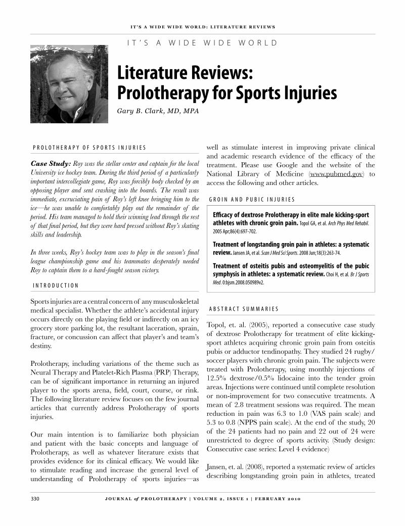
J O U R N A L of P R O L O T H E R A P Y | V O L U M E 2 , I S S U E 1 | F E B R U A R Y 2 0 1 0330
I T ’ S A W I D E W I D E W O R L D : L I T E R A T U R E R E V I E W S
Gary B. Clark, MD, MPA
Literature Reviews: Prolotherapy for Sports Injuries
I T ’ S A W I D E W I D E W O R L D
P r o l o t h e r a P y o f s P o r t s i n j u r i e s
Case Study: Roy was the stellar center and captain for the local University ice hockey team. During the third period of a particularly important intercollegiate game, Roy was forcibly body checked by an opposing player and sent crashing into the boards. The result was immediate, excruciating pain of Roy’s left knee bringing him to the ice—he was unable to comfortably play out the remainder of the period. His team managed to hold their winning lead through the rest of that final period, but they were hard pressed without Roy’s skating skills and leadership. In three weeks, Roy’s hockey team was to play in the season’s final league championship game and his teammates desperately needed Roy to captain them to a hard-fought season victory.
i n t r o d u C t i o n
Sports injuries are a central concern of any musculoskeletal medical specialist. Whether the athlete’s accidental injury occurs directly on the playing field or indirectly on an icy grocery store parking lot, the resultant laceration, sprain, fracture, or concussion can affect that player’s and team’s destiny. Prolotherapy, including variations of the theme such as Neural Therapy and Platelet-Rich Plasma (PRP) Therapy, can be of significant importance in returning an injured player to the sports arena, field, court, course, or rink. The following literature review focuses on the few journal articles that currently address Prolotherapy of sports injuries. Our main intention is to familiarize both physician and patient with the basic concepts and language of Prolotherapy, as well as whatever literature exists that provides evidence for its clinical efficacy. We would like to stimulate reading and increase the general level of understanding of Prolotherapy of sports injuries—as
well as stimulate interest in improving private clinical and academic research evidence of the efficacy of the treatment. Please use Google and the website of the National Library of Medicine (www.pubmed.gov) to access the following and other articles.
g r o i n a n d P u B i C i n j u r i e s
Efficacy of dextrose Prolotherapy in elite male kicking-sport athletes with chronic groin pain. topol ga, et al. Arch Phys Med Rehabil. 2005 apr;86(4):697-702.
Treatment of longstanding groin pain in athletes: a systematic review. jansen ja, et al. Scan J Med Sci Sports. 2008 jun;18(3):263-74.
Treatment of osteitis pubis and osteomyelitis of the pubic symphysis in athletes: a systematic review. Choi h, et al. Br J Sports Med. 0:bjsm.2008.050989v2.
a B s t r a C t s u m m a r i e s
Topol, et. al. (2005), reported a consecutive case study of dextrose Prolotherapy for treatment of elite kicking-sport athletes acquiring chronic groin pain from osteitis pubis or adductor tendinopathy. They studied 24 rugby/soccer players with chronic groin pain. The subjects were treated with Prolotherapy, using monthly injections of 12.5% dextrose/0.5% lidocaine into the tender groin areas. Injections were continued until complete resolution or non-improvement for two consecutive treatments. A mean of 2.8 treatment sessions was required. The mean reduction in pain was 6.3 to 1.0 (VAS pain scale) and 5.3 to 0.8 (NPPS pain scale). At the end of the study, 20 of the 24 patients had no pain and 22 out of 24 were unrestricted to degree of sports activity. (Study design: Consecutive case series: Level 4 evidence) Jansen, et. al. (2008), reported a systematic review of articles describing longstanding groin pain in athletes, treated

J O U R N A L of P R O L O T H E R A P Y | V O L U M E 2 , I S S U E 1 | F E B R U A R Y 2 0 1 0 331
I T ’ S A W I D E W I D E W O R L D : L I T E R A T U R E R E V I E W S
with rest/restricted activity, passive or active physical therapy, corticosteroid injection, dextrose Prolotherapy, or surgery. Although mentioning Prolotherapy as an optional treatment, the report focused on surgical results and provided little insight into the effect of the more conservative treatments. (Study design: Systematic review: Level 4 evidence) Choi, et. al. (2008), reported a systematic review of 25 journal articles that consisted of case reports or case series; there were no random controlled trials. One hundred and ninety-four athletes were cited as being diagnosed with osteitis pubis and treated with some combination of conservative treatment, e.g., rest, nonsteroidal anti-inflammatories, physical therapy (six case reports); local anesthetic injection; corticosteroid injection (four case reports); dextrose Prolotherapy (one case series); antibiotic therapy (ten case reports/series of osteomyelitis of the pubic symphysis); or surgery (six case reports/series). Without any clinical studies available providing direct comparison of treatment modalities, the authors graded the quality of evidence at Level 4—they could not determine the comparative effectiveness of the various treatments based on the studies currently existing. They deemed that further, more scientifically rigorous study is necessary. (Study design: Systematic review: Level 4 evidence)
a C h i l l e s t e n d i n o s i s
Prolotherapy injections and eccentric loading exercises for painful Achilles tendinosis: a randomized trial. yelland mj, et al. Br J Sports Med. Published online: 22 june 2009. doi:10.1136/bjsm. 2009.057968.
a B s t r a C t s u m m a r y
Yelland, et. al., compared Prolotherapy of Achilles tendinosis to the efficacy of eccentric loading exercises (ELE). Subjects were randomly selected for one of three groups: 1) a 12-week program of hypertonic glucose Prolotherapy injections (n=14); 2) a 12-week program of ELE (n=15), and 3) a 12-week program of combined Prolotherapy and ELE treatment (n=14). Prolotherapy was administered by delivering injections of hypertonic glucose diluted with lignocaine and delivered alongside (parallel to) the affected tendon. Long term outcome was assessed over the ensuing 12 months by measuring the proportions of participants achieving a minimum clinically important change (MCIC) for VISA-A questionnaire
scores. The symptoms of pain, stiffness, and limitation of activity, along with treatment costs, were also periodically assessed over that 12 month period. At 12 months, Prolotherapy (compared to ELE) demonstrated earlier reductions of stiffness and activity limitation. Combined treatment demonstrated even earlier reductions of pain, stiffness, and activity limitation—as well as lowest incremental cost. (Study design: Single-blinded, randomized clinical trial: Level 1 evidence)
j o P C o m m e n t a r y
Currently, there are relatively few clinical reports on the use of Prolotherapy specifically as treatment for sports injuries. And ... most of those reports are at the lowest level of quality of evidence. Evidence-based medicine has become a cornerstone of modern medical practice, attempting to apply the best available scientific evidence to medical decision making. Good evidence is the fundamental basis upon which therapies are judged as most appropriate and necessary by the medical community (i.e., clinically effective and efficient) and deemed reimbursable by insurance programs.1
Existing clinical evidence can be collected and ascertained for quality by scientific, engineering, and statistical methods. Several evidence grading systems are available. One example is that of the New Zealand Guidelines Group (NZGG), which has developed a system for grading the quality of evidence and is often referenced. These levels of evidence (or levels of confidence) are summarized in Table 1.2
If there were a level 5, it would incorporate anecdotal reports that might be based on empirical experience but are undocumented, totally hearsay, and subjective, providing no weight of evidence, whatsoever. It has been pointed out by many observers, including the National Institutes of Health Center for Complementary and Alternative Medicine,3 that the beneficial effects of Prolotherapy may be much more complex than first thought. Aside from the inflammatory stimulation of fibroblastic ligament or tendon regeneration, the individual elements of Prolotherapy may have their own specific effects. The needling, itself, may have some acupuncture effect on pain. The anesthetic (i.e., procaine,

J O U R N A L of P R O L O T H E R A P Y | V O L U M E 2 , I S S U E 1 | F E B R U A R Y 2 0 1 0332
I T ’ S A W I D E W I D E W O R L D : L I T E R A T U R E R E V I E W S
Reliability Level of Evidence Source of the Evidence
1.Evidencethathasahighdegreeofprovenreliabilityleavinglittlequestiontodebate.
Trialstudiesthatusewell-testedmethods(includingcomparablecontrolgroups)tomakecomparisonsinafairwayandproduceresultsthatleaveverylittleroomforuncertainty.Example:UsuallyLevelIevidenceisfrom1)systematicreviewsormeta-analyseswithconsistentfindingsor2)large,high-qualityrandomizedcontrolledstudies.
2.Evidencethathassignificantreliabilitybutisstillopentosomedebate.
Trialstudiesthatusewell-testedmethods(includingcomparablecontrolgroups)tomakecomparisonsinafairwaybutwheretheresultsleavesomeroomforuncertainty.Forexample,thesizeofthestudymaybesmallenoughtocausesignificantlossestofollow-uportheexperimentaldesignprecludesadequateselectionofgroupsforcomparison.Example:UsuallyLevel2evidenceisfrom1)systematicreviewswithoutconsistentfindings,2)randomizedcontrolledtrialsinwhichsignificantnumbersofsubjectsarelost,or3),smallrandomizedcontrolledstudies.
3.Evidencethathassomeweightofclinicalsignificancebutiswithoutahighdegreeofprovenreliability.
Trialstudiesthatuseanexperimentaldesignthatdoesnotguaranteethatfaircomparisonscanbemade,thus,producingresultsthataredoubtful.Example:UsuallyLevel3evidenceisfrom1)systematicreviewsofcase-controlstudiesor2)individualcase-controlstudies.
4.Evidencethathassomeweightofclinicalsignificancebutisbasedonreportsofempiricalexperiencewithoutanycomparablegroups.
Trialstudiesthatuseanexperimentaldesignwithoutcomparable(control)groupsthatproduceahighprobabilityofresultsbeingduetochanceorbecausethegroupscomparedweredifferentattheoutsetofthestudy.Example:UsuallyLevel4evidenceisfrom1)cohortorcase-controlstudieswherethegroupswerenotreallycomparableor2)totallyuncontrolledcase-seriesreports.
Table 1. New Zealand Guidelines Group (NZGG) levels of evidence of medical efficacy.2
research clinic. Well-designed case studies can be a very beneficial contribution to the total body of evidence—though not as weighty as randomized controlled studies. Currently, the body of Level 1 and 2 evidence of Prolotherapy efficacy is still meager. But, the vastly more preponderant Level 3 and 4 private-practice therapeutic evidence still provides an unwavering weight of overall support for the efficacy of Prolotherapy in treating injuries of all types, including those that are sport-related. There are many articles on sports injuries of all types that do not address Prolotherapy as a major therapeutic consideration. Instead, there is a plethora of reports on failed physical, medical, and surgical treatments that permeate throughout the professional channels of information. We have to show that Prolotherapy can make a difference. However, to prove the truth to the medical community at-large and the healthcare insurance companies, we need more Level 1 and 2 evidence. The work of Scarpone, et. al., and Rabago, et. al., are most recent examples of the quality of work that needs to be accomplished.4, 5
Yelland, et. al., cited earlier, have provided an excellent example of Level 1 evidence for the efficacy of Prolotherapy. An “efficacious” treatment, by definition, is both therapeutically effective in resolving the cause of the patient’s symptoms and, also, therapeutically efficient, i.e., cost-effective. As time goes on, there will be more and more Level 1 and 2 evidence proclaiming the therapeutic efficacy of Prolotherapy. However, it will take disciplined and concerted effort, time, and money to reach the needed preponderance of compelling clinical evidence to convince Medicare and private healthcare insurance companies to reimburse for Prolotherapy.
Case Study (continued): Over the evening, Roy’s knee had been put at rest, iced, wrapped (compressed) and elevated (i.e., R.I.C.E. therapy). The very next day following his injury, he visited a local musculoskeletal pain specialist for diagnosis and treatment. Physical examination revealed a clearly demarcated, swollen medial collateral ligament (MCL), which was exquisitely sensitive to palpation at its proximal and distal attachments. There were no findings of meniscal, coronary ligament, cruciate ligament, or other structural damage upon specific physical testing. Due to joint stability and isolation of a discrete injury, radiography was not necessary.
lidocaine, lignocaine) used for diluting the dextrose and other proliferant components may have a Neural Therapy effect on local sympathetic enervation and lymphedema. Additionally, we have yet to prove the differences between the various proliferant combinations that are in use around the country. Varying concentrations of dextrose (glucose) are being used. Other proliferant solution constituents are being used, including phenol and glycerin (P2G solution is phenol, glucose and glycerin), sodium morrhuate, and testosterone. Also, some say that tendons deserve different proliferant concentrations and constituents compared to ligaments. All of the variations of Prolotherapy and their respective effects need to be carefully documented and analyzed in the private treatment room as well as the academic

J O U R N A L of P R O L O T H E R A P Y | V O L U M E 2 , I S S U E 1 | F E B R U A R Y 2 0 1 0 333
I T ’ S A W I D E W I D E W O R L D : L I T E R A T U R E R E V I E W S
Initial treatment consisted of Neural Therapy,6 performing intradermal injections of procaine circumferentially around the edge of the clearly demarcated medial collateral swelling. The edematous swelling began to subside during the injections and was essentially gone by the time that the injections were completed. Roy reported significant pain relief. He was asked to return in one week and urged to continue R.I.C.E. as much as possible and crutches until all pain had subsided—along with restriction from the skating rink. He was placed on a mild physical therapy program to maintain strength and range of motion. At Roy’s second visit the next week, his MCL was nonswollen but still slightly tender at both the proximal and distal MCL attachments. P2G Prolotherapy was performed at both the proximal and distal MCL. Roy was taken off crutches, continued on a moderate physical therapy program, and cautioned to participate in only mild skating. Any physical activity was to be limited by the occurrence of any MCL pain.
Roy’s team trainer claimed that his recovery was “miraculous!” ...“What is this Prolotherapy thing?!” Three weeks following the accident, Roy played in the final game. The coach saved him for the third period. His team broke a tie with Roy’s winning a hat trick in the last five seconds. His last clinic visit in the following week showed full recovery of the MCL. n
B i B l i o g r a P h y
Evidence–based medicine. Wikepedia. http://en.wikepedia.org.
Levels of Evidence. New Zealand Complementary and Alternative Medicine. www.cam.org.nz/levelsofevidence.
Prolotherapy. National Center for Complementary and Alternative Medicine. http://nccam.nih.gov.
Scarpone M, et al. The efficacy of prolotherapy for lateral epicondylosis: a pilot study. Clinical Journal of Sports Medicine. 2008 May;18(3):248-54.
Rabago D, et al. The systematic review of four injection therapies for lateral epicondylosis: prolotherapy, polidocanol, whole blood and platelet rich plasma. British Journal of Sports Medicine. 2009 Jan 21.
Neural Therapy. Wikepedia. http://en.wikepedia.org.
1.
2.
3.
4.
5.
6.

J O U R N A L of P R O L O T H E R A P Y | V O L U M E 2 , I S S U E 1 | F E B R U A R Y 2 0 1 0334
I T ’ S A W I D E W I D E W O R L D : I N T R O D U C T I O N O F P R O L O T H E R A P Y I N T H E C A R I B B E A N
Introduction of Prolotherapy in the Caribbean
I T ’ S A W I D E W I D E W O R L D
J. V. A. Humphreys, MD
I n January 2009, I introduced Prolotherapy in the twin island state of Antigua and Barbuda. Optimum Health Clinic Ltd. (my private practice), situated at
Belmont (Surgical and Medical) Complex was the site where this dynamic and extremely effective therapy was birthed for the first time in the West Indies. (See Figure 1.) I am happy to report that Prolotherapy is growing in popularity on the islands. I will share two success stories with you.
Pete, a 47 year-old man that I have been working with for three years for obesity, diabetes and hypertension, had been experiencing moderate to severe left hip pain for about two years. Pete worked in the field as a construction engineer and fell from a ladder. Pete benefited greatly from Prolotherapy, reporting one month after one treatment of Prolotherapy that the pain that had been his constant companion for two years is no longer present. He remarked that minutes to days after treatment he felt “a pulling of muscles” and greater range of motion as time progressed. Pete was given a solution of Sarapin and dextrose proliferants. A second treatment was given within a month. He has continued to be pain free since his last treatment a few months ago. Pete has been discharged thanks to Prolotherapy. By the way, Pete has lost almost 100 pounds and both his hypertension and
diabetes are under control without m e d i c a t i o n . “Congratulations Pete! I have found in my experience that dextrose does very well as a stand alone proliferant but benefits are augmented in some instances with the addition of other proliferants. Prolotherapy is no doubt a valuable
form of pain management. It has changed the landscape of medical practice in the Caribbean. Invariably, it has proven more effective than oral medication therapy (NSAIDs) and steroidal therapy, which studies now show hamper the biosynthesis and gene expression of collagen and proteoglycan of chondrocytes.1 (See Figure 3.)
Figure 1. Optimum Health Clinic Ltd., where Prolotherapy recently got its start in the West Indies.
My first patient, Charmaine a 51 year-old perimenopausal, primiparous woman, had severe degenerative disease of the knees (bilaterally) for a number of years. The pain was refractory to NSAIDs and she had even had local steroid injections done; which too was ineffective. Charmaine did a course of Prolotherapy on her knees. (See Figure 2.) One day while clearing my post box, I saw Charmaine leaving work carrying a pile of books. She limped slowly across the car park. Being concerned, I inquired from her the reason for her limp. She smiled and said, “Doc, I have no pain. My only problem is that I have to keep reminding myself that I have no pain and that I need to get rid of this programmed habit to limp.” She then showed me her limp free “stage” walk. With the type of degeneration Charmaine had, she couldn’t fake it, even if she wanted to. I smiled and thought to myself, “Amazing!” This was what I needed as a practitioner …a vote of confidence.
Figure 2. Dr. Humphreys performing Prolotherapy to the lateral knee.

J O U R N A L of P R O L O T H E R A P Y | V O L U M E 2 , I S S U E 1 | F E B R U A R Y 2 0 1 0 335
I T ’ S A W I D E W I D E W O R L D : I N T R O D U C T I O N O F P R O L O T H E R A P Y I N T H E C A R I B B E A N
Prolotherapy has even caused a stir in the medical community in Antigua and Barbuda with increased referrals from physicians. As a matter of fact, while preparing this article, my medical assistant informed me that the president of the local medical association has made an appointment for his first Prolotherapy treatment.
I must express profound thanks to my Prolotherapy instructor, Dr. Ross Hauser, whose remarkable teaching skills both theoretically and clinically proved invaluable. (See Figure 4.) I commend Dr. Ross Hauser and the staff from Caring Medical and the other forerunners in Prolotherapy…Antigua thanks you…the Caribbean thanks you! n
B i B l i o g r a P h y
Fujii K, et al. Effects of NSAID on collagen and proteoglycan synthesis of cultured chondrocytes. J-Rheumatol-Suppl. 1989 Aug; 1828-31.
e d i t o r ’ s n o t e
As you will read in this patient’s letter, Antigua is already experiencing the power of Prolotherapy in the Caribbean.
a l e t t e r o f t h a n K s f r o m P a t i e n t t o d r . j . v . a . h u m P h r e y s i n a n t i g u a
Hello Dr. Humphreys, It is now four weeks after my first treatment and I would like to take this opportunity to thank you for introducing me to Prolotherapy treatment. As you know, when I came to your office three months ago I complained of severe back pain, pain radiating down my left leg and tingling in my toes of my left foot. I have had this problem for over six months. I tried chiropractic care, acupuncture and massage with no resolution of my herniated disc problems.
1.
Figure 3. Prolotherapy brings renewed hope for pain patients in the West Indies.
Figure 4. Dr. Ross Hauser and Dr. Humphreys at Caring Medical, Oak Park, Illinois in 2008.
I was unable to perform any activity, let alone exercise. Walking, standing, or sitting in the same place for a long period of time caused an increase in my pain. My doctor prescribed several different types of pain killers, which I had to take every day, was the only thing that gave me some relief. As you are aware, these medications are known stimulators of connective tissue breakdown and cause my condition to get worse. My doctor also advised that I may have to do surgery to cure my herniated disc. However, because of your treatment, I will not have to do the surgery. I am happy to report that after three weeks of my first Prolotherapy treatment I am about 90% cured! Two weeks before my first treatment I discontinued taking all anti-inflammatory drugs and I haven’t taken any since. My lower back pain is much better and I don’t have any pain radiating down my leg or tingling in my toes. I am now able to get back to my usual activities of walking, standing, exercising and dancing! I am so excited about this treatment that I am telling everyone I know that is having problems with back pain, or any other joint paint, about the Prolotherapy treatment. Once again, thank you so much for helping me and with the help of God you will do wonders with this procedure. Thank you God bless you, C.T. Antigua

J O U R N A L of P R O L O T H E R A P Y | V O L U M E 2 , I S S U E 1 | F E B R U A R Y 2 0 1 0336
S K I L L E N H A N C E M E N T : S E M I N A R S , T R A I N I N G , & O R G A N I Z A T I O N S
Seminars, Training, & OrganizationsS K I L L E N H A N C E M E N T
American Holistic Veterinary Medial Association2218OldEmmortonRoadBelAir,MD21015Phone:410.569.0795Fax:410.569.2346www.ahvma.org
The International Veterinary Acupuncture Society2625RedwingRd.Suite160FortCollins,CO80526Phone:970.266.0666Fax:970.266.0777www.ivas.org
a P r i l 8 – 1 1 , 2 0 1 0
San Diego, CAThe American College of Osteopathic Sclerotherapeutic Pain Management’sSpring2010TrainingSeminar“Prolotherapy;AComprehensiveApproach”.Thisconferencewillincludemorning lectures on prolotherapy, a 3-D Anatomy laband an intensive 3 day afternoon hands on workshopwith 20 stations to practice actual injection practice. Inaddition,NeuralTherapy,Mesotherapy,PRPandStemCellinjections will be introduced. Finally, billing and codingandassetprotectionwillbediscussed.
For more information: http://acospm.com
Notice to meeting organizers: If you are sponsoring a Prolotherapy meeting or training session, please email: [email protected] for a free listing of your meeting.
o r g a n i z a t i o n s
American Association of Orthopedic Medicine (AAOM)600PembrookDrive,WoodlandPark,CO80863Phone:888.687.1920Fax:719.687.5184www.aaomed.org
GetProlo.comBeulahLandCorporation715LakeSt.Suite400OakPark,IL60301Phone:708.848.5011Fax:708.848.8053www.getprolo.com
The American Academyof Osteopathy3500DePauwBlvd,Suite1080Indianapolis,IN46268Phone:317.879.1881Fax:317.879.0563www.academyofosteopathy.org
The Hackett Hemwall Foundation2532BaldenStreet,Madison,WI53705USAwww.HackettHemwall.org
American College of Osteopathic Sclerotherapeutic PainManagement, Inc.303S.IngramCt.Middletown,DE19709Phone:302.376.8080TollFree:800.471.6114Fax:302.376.8081www.acopms.com
American Osteopathic Academy of Sports Medicine (AOASM)2810CrossroadsDrive,Suite3800Madison,WI53718Phone:608.443.2477Fax:608.443.2474www.aoasm.org
n o v e m B e r 6 – 1 3 , 2 0 1 0
Guadalajara, MexicoThe American Association of Orthopaedic Medicine isoffering a Hands-on Prolotherapy Course in CiudadGuzman, Guadalajara in Mexico. This course willinclude daily lectures, instruction in patient evaluationand diagnosis, solutions, needle placement, injectiontechniqueandhands-onpatienttreatment.
For more information: http://www.aaomed.org
a P r i l 2 8 – m a y 1 , 2 0 1 0
Amelia Island, FLThe American Association of Orthopaedic Medicine 2010Annual Conference titled“The Athlete and OrthopaedicMedicine” will be highlighted by several nationallyand internationally renowned orthopaedic medicineauthoritiesspeakingonavarietyofdifferenttopics.
For more information: http://www.aaomed.org

www.amazon.comwww.beulahlandpress.com
and Enhancing Athletic Performance
AVAILABLE AT
CURING SPORTS INJURIES WITH PROLOTHERAPY
&AVAILABLE AT
CURING SPORTS INJURIES WITH PROLOTHERAPY
Just as the original book Prolo Your Pain
Away! a�ected the pain management
�eld, Prolo Your Sports Injuries Away! has
rattled the sports world.
Learn the twenty myths of sports medicine including the myths of:
• anti-inflammatory medications
• why cortisone shots actually weaken tissue
• how ice, rest, & immobilization may actually hurt the athlete
• why the common practice of taping and bracing do not stabilize injured areas
• & why the arthroscope is one of athletes’ worst nightmares!
&

V O L U M E T W O | I S S U E O N E | F E B R U A R Y 2 0 1 0 w w w . j o u r n a l of p r o l o t h e r a p y . c o m
IN THIS ISSUE
B E U L A H L A N D P R E S S
ISSN 1944-0421 (print)ISSN 1944-043X (online)
■ ■
■
■
■
■
■
■
■ ■
■
■
■ ■ ■
■
Athletes Do Not Stop Before the Finish Line Letters to the Editor from Timothy Speciale, DO & Barbara YoungProlotherapy in South Korea: Interview with Dr. Choi Yung DoAn Interview with a Personal Injury AttorneyProlotherapy as an Alternative to Surgery: A Prospective PilotStudy of 34 Patients from a Private Medical PracticeHyperthermia Induces Venous Blood Alkalosis: A Study in Five Ironman Triathletes69 Year-old Still Running 100-mile Races Thanks to ProlotherapyProlotherapy for Professional Sports Injuries
The Ligament Injury Connection to OsteoarthritisThe Acceleration of Articular Cartilage Degeneration in Osteoarthritis by Nonsteroidal Anti-in�ammatory DrugsProlotherapy Case Studies from VeterinariansReview of Free Yourself from Chronic Pain and Sports Injuries By Donna Alderman, DOProlotherapy Injection Technique of the ElbowLiterature Reviews: Prolotherapy for Sports InjuriesIntroduction of Prolotherapy in the CaribbeanSeminars, Training, & Organizations
Doctors
PatientsT E L L U S Y O U R S T O R I E S
S H A R E Y O U R E X P E R I E N C E
[ for Doctors & Patients]
[ J O U R N A L of P R O L O T H E R A P Y . C O M ] [ 7 0 8 - 8 4 8 - 5 0 1 1 ]
Calling all Prolotherapists! Do you have a Prolotherapy article
you would like published in the Journal of Prolotherapy?
We would love to review it and help you share it with
the world! For information, including submission
guidelines, please log on to the authors’ section
of www.journalofprolotherapy.com.
The Journal of Prolotherapy is unique in that it has a target audience of
both physicians and patients. Help spread the word to other people like
yourself who may benefit from learning about your struggle with
chronic pain, and first-hand experience with Prolotherapy.
For information on how to tell your story in the Journal of
Prolotherapy, please log on to the contact section of
www.journalofprolotherapy.com.
JO
UR
NA
L o
f P
RO
LO
TH
ER
AP
YV
OL
UM
E T
WO
| I
SS
UE
ON
E |
FE
BR
UA
RY
20
10
| P
AG
ES
25
7-
33
6B
EU
LA
H L
AN
D P
RE
SS
T H E L I G A M E N T - N S A I D C O N N E C T I O N T O
DegenerativeOsteoarthritis
AfterBefore