Volume 2, Chapter 3-1: Slime Molds: Biology and Diversity
34
Glime, J. M. 2019. Slime Molds: Biology and Diversity. Chapt. 3-1. In: Glime, J. M. Bryophyte Ecology. Volume 2. Bryological Interaction. Ebook sponsored by Michigan Technological University and the International Association of Bryologists. Last updated 18 July 2020 and available at <https://digitalcommons.mtu.edu/bryophyte-ecology/>. 3-1-1 CHAPTER 3-1 SLIME MOLDS: BIOLOGY AND DIVERSITY TABLE OF CONTENTS What are Slime Molds? ....................................................................................................................................... 3-1-2 Identification Difficulties ...................................................................................................................................... 3-1- Reproduction and Colonization ........................................................................................................................... 3-1-5 General Life Cycle ....................................................................................................................................... 3-1-6 Seasonal Changes......................................................................................................................................... 3-1-7 Environmental Stimuli ............................................................................................................................... 3-1-13 Light.................................................................................................................................................... 3-1-13 pH and Volatile Substances ................................................................................................................ 3-1-14 Water................................................................................................................................................... 3-1-15 Reproduction in Myxomycetes .................................................................................................................. 3-1-15 Dispersal............................................................................................................................................................ 3-1-16 Habitat Needs .................................................................................................................................................... 3-1-19 Moisture ..................................................................................................................................................... 3-1-19 Latitude ...................................................................................................................................................... 3-1-20 Food and Light ........................................................................................................................................... 3-1-20 Role of Bryophytes as Slime Mold Habitats ..................................................................................................... 3-1-20 Slime Mold Effects on Bryophytes ................................................................................................................... 3-1-24 Bryophytes Known to Grow on Slime Molds ................................................................................................... 3-1-25 Epizooites .......................................................................................................................................................... 3-1-25 Potential for Symbiosis? ................................................................................................................................... 3-1-26 Interactions with Invertebrates .......................................................................................................................... 3-1-26 Summary ........................................................................................................................................................... 3-1-32 Acknowledgments ............................................................................................................................................. 3-1-33 Literature Cited ................................................................................................................................................. 3-1-33
Transcript of Volume 2, Chapter 3-1: Slime Molds: Biology and Diversity

Glime, J. M. 2019. Slime Molds: Biology and Diversity. Chapt. 3-1. In: Glime, J. M. Bryophyte Ecology. Volume 2. Bryological Interaction. Ebook sponsored by Michigan Technological University and the International Association of Bryologists. Last updated 18 July 2020 and available at <https://digitalcommons.mtu.edu/bryophyte-ecology/>.
3-1-1
CHAPTER 3-1 SLIME MOLDS: BIOLOGY AND DIVERSITY
TABLE OF CONTENTS
What are Slime Molds? ....................................................................................................................................... 3-1-2 Identification Difficulties ...................................................................................................................................... 3-1- Reproduction and Colonization ........................................................................................................................... 3-1-5 General Life Cycle ....................................................................................................................................... 3-1-6 Seasonal Changes ......................................................................................................................................... 3-1-7 Environmental Stimuli ............................................................................................................................... 3-1-13 Light .................................................................................................................................................... 3-1-13 pH and Volatile Substances ................................................................................................................ 3-1-14 Water ................................................................................................................................................... 3-1-15 Reproduction in Myxomycetes .................................................................................................................. 3-1-15 Dispersal ............................................................................................................................................................ 3-1-16 Habitat Needs .................................................................................................................................................... 3-1-19 Moisture ..................................................................................................................................................... 3-1-19 Latitude ...................................................................................................................................................... 3-1-20 Food and Light ........................................................................................................................................... 3-1-20 Role of Bryophytes as Slime Mold Habitats ..................................................................................................... 3-1-20 Slime Mold Effects on Bryophytes ................................................................................................................... 3-1-24 Bryophytes Known to Grow on Slime Molds ................................................................................................... 3-1-25 Epizooites .......................................................................................................................................................... 3-1-25 Potential for Symbiosis? ................................................................................................................................... 3-1-26 Interactions with Invertebrates .......................................................................................................................... 3-1-26 Summary ........................................................................................................................................................... 3-1-32 Acknowledgments ............................................................................................................................................. 3-1-33 Literature Cited ................................................................................................................................................. 3-1-33

3-1-2 Chapter 3-1: Slime Molds: Biology and Diversity
CHAPTER 3-1 SLIME MOLDS: BIOLOGY AND DIVERSITY
Figure 1. Orange slime mold on moss, Blue Lake Creek valley, Washington, USA. Photo by Matt Goff, Sitka Nature, with permission.
What are Slime Molds?
Slime mold or slime mould is an informal name given to three kinds of unrelated eukaryotic organisms. While the bryophytes were undergoing classification changes at the familial and ordinal levels, Protista were jumping to new kingdoms and phyla. Hence, anyone whose knowledge about these organisms is as old as mine needs a road map to understand who now belongs where. Slime molds are no longer considered fungi, but instead seem to be protozoa.
The protozoa have been joined by other groups to form the current concept of the paraphyletic kingdom Protista, also known as Protozoa, a grouping that is one of convenience. One such group to join them is the slime molds (Figure 1). Once classified as fungi, they have been booted out of that kingdom due to their lack of chitin and their feeding by engulfing food. They are now considered Protista due to their motile stages that look and behave like protozoa. Within the Protozoa, we will consider here the phylum known as Eumycetozoa or Amoebozoa (Shadwick et al. 2009; Kang et al. 2017).
The slime molds are comprised of more than 1000 species from all seven continents (Lloyd 2011). The life cycle is one reason for their current classification position. They can live freely as single cells, but in dictyostelids they
can later aggregate to form multicellular reproductive structures.
Using 18S rDNA and cladistics, Leontyev et al. (2019) revised the classification of the Myxomycetes. Noting that "Myxomycetes show a higher within-group genetic divergence than true fungi, higher animals, or vascular plants," they divide the slime molds into three classes, giving the groups taxonomic status according to the International Code of Nomenclature: • CLASS MYXOMYCETES (Figure 2-Figure 9)
The Myxomycetes, also known as Myxogastria, are the acellular slime molds, referring to the plasmodium that is multinucleate with no cell separation. These form the largest group of slime molds and contain almost all of the slime molds that associate with bryophytes. Based on the list of genera in nomen.eumycetozoa.com (5 May 2019), I have found all but three of the genera with at least one species that has been found on bryophytes to be in this class. The plasmodium (Figure 22, Figure 24) moves by amoeboid movement with rapidly streaming protoplasm, reaching speeds up to 1.35 mm per second (Alexopoulos 1962, 1964). The mass can migrate when it streams to an advancing position and

Chapter 3-1: Slime Molds: Biology and Diversity 3-1-3
withdraws its protoplasm from the rear area. When food becomes scarce, this mass will migrate to the surface of the substrate and form its rigid fruiting bodies. These produce spores that hatch into amoebae to continue the life cycle (Ling 1999).
Figure 2. Physarum decipiens young fruiting bodies on leafy and thallose liverworts. Photo by David Mitchell, from The Eumycetozoan Project, DiscoverLife.org, with permission.
Figure 3. Physarum decipiens mature fruiting bodies on leafy liverwort. Photo by Alain Michaud, from The Eumycetozoan Project, DiscoverLife.org, with permission.
Figure 4. Physarum cinereum immature fruiting bodies. Photo from Denver Botanical Garden, from The Eumycetozoan Project, DiscoverLife.org, with permission.
Figure 5. Physarum cinereum mature fruiting bodies. Photo by David Mitchell, from The Eumycetozoan Project, DiscoverLife.org, with permission.
Figure 6. Physarum globuliferum with immature fruiting bodies. Photo by Ray Simons, from The Eumycetozoan Project, DiscoverLife.org, with permission.
Figure 7. Physarum globuliferum with mature fruiting bodies releasing spores. Photo by Dmitry Leontyev, from The Eumycetozoan Project, DiscoverLife.org, with permission

3-1-4 Chapter 3-1: Slime Molds: Biology and Diversity
Figure 8. Physarum leucophaeum with immature fruiting bodies. Photo by Denver Botanical Garden, from The Eumycetozoan Project, DiscoverLife.org, with permission.
Figure 9. Physarum leucophaeum with mature fruiting bodies emitting spores. Photo by Alain Michaud, from The Eumycetozoan Project, DiscoverLife.org, with permission.
• CLASS DICTYOSTELIOMYCETES (Figure 12) Dictyostelids are cellular slime molds. I have found only two genera with any species reported on these slime molds. The Dictyosteliomycetes do not form huge plasmodia (Figure 22, Figure 24) and remain as individuals, feeding on microorganisms. When they run out of food, they form fruiting bodies, first releasing signal molecules that enable them to find each other and
then aggregating as swarms. They join to form a tiny multicellular coordinated slug-like creature (Figure 10). They can aggregate about 100,000 cells in Dictyostelium discoideum (Figure 11-Figure 12) (Kessin et al. 1996). This aggregate crawls to an open place in the light to form a fruiting body (Kakiuchi et al. 2001). While some of the amoeboid cells actually become spores, others become part of the dead stalk that lifts the spores upward. About 20% of the cells of the Dictyostelium discoideum die as they form the stalk (Kessin et al. 1996). This group is largely unrecorded from bryophytes. The only record I found was for Dictyostelium quercibrachium from the margin of a small bog in Ohio, USA (Cavender et al. 2005), and it is not clear if was actually on a moss.
Figure 10. Dictyostelium mucoroides pseudoplasmodial slug on agar. Note their slug-like appearance. Photo by Dmitry Leontyev, through Creative Commons.
Figure 11. Dictyostelium discoideum development. Photo by Usman Bashir, through Creative Commons.
Figure 12. Dictyostelium discoideum fruiting in an open place. Photo by Usman Bashir, through Creative Commons.
Chapter 3-1: Slime Molds: Biology and Diversity 3-1-5
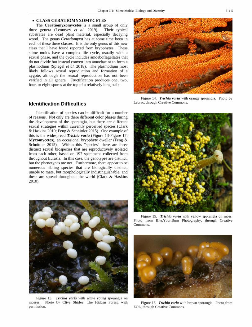
• CLASS CERATIOMYXOMYCETES The Ceratiomyxomycetes is a small group of only
three genera (Leontyev et al. 2019). Their typical substrates are dead plant material, especially decaying wood. The genus Ceratiomyxa has at some time been in each of these three classes. It is the only genus of this new class that I have found reported from bryophytes. These slime molds have a complex life cycle, usually with a sexual phase, and the cycle includes amoeboflagellates that do not divide but instead convert into amoebae or to form a plasmodium (Spiegel et al. 2018). The plasmodium most likely follows sexual reproduction and formation of a zygote, although the sexual reproduction has not been verified in all genera. Fructification produces one, two, four, or eight spores at the top of a relatively long stalk.
Identification Difficulties
Identification of species can be difficult for a number of reasons. Not only are there different color phases during the development of the sporangia, but there are different sexual strategies within currently perceived species (Clark & Haskins 2010; Feng & Schnittler 2015). One example of this is the widespread Trichia varia (Figure 13-Figure 17; Myxomycetes), an occasional bryophyte dweller (Feng & Schnittler 2015). Within this "species" there are three distinct sexual biospecies that are reproductively isolated from each other, based on 197 specimens collected from throughout Eurasia. In this case, the genotypes are distinct, but the phenotypes are not. Furthermore, there appear to be numerous sibling species that are biologically distinct, unable to mate, but morphologically indistinguishable, and these are spread throughout the world (Clark & Haskins 2010).
Figure 13. Trichia varia with white young sporangia on mosses. Photo by Clive Shirley, The Hidden Forest, with permission.
Figure 14. Trichia varia with orange sporangia. Photo by Lebrac, through Creative Commons.
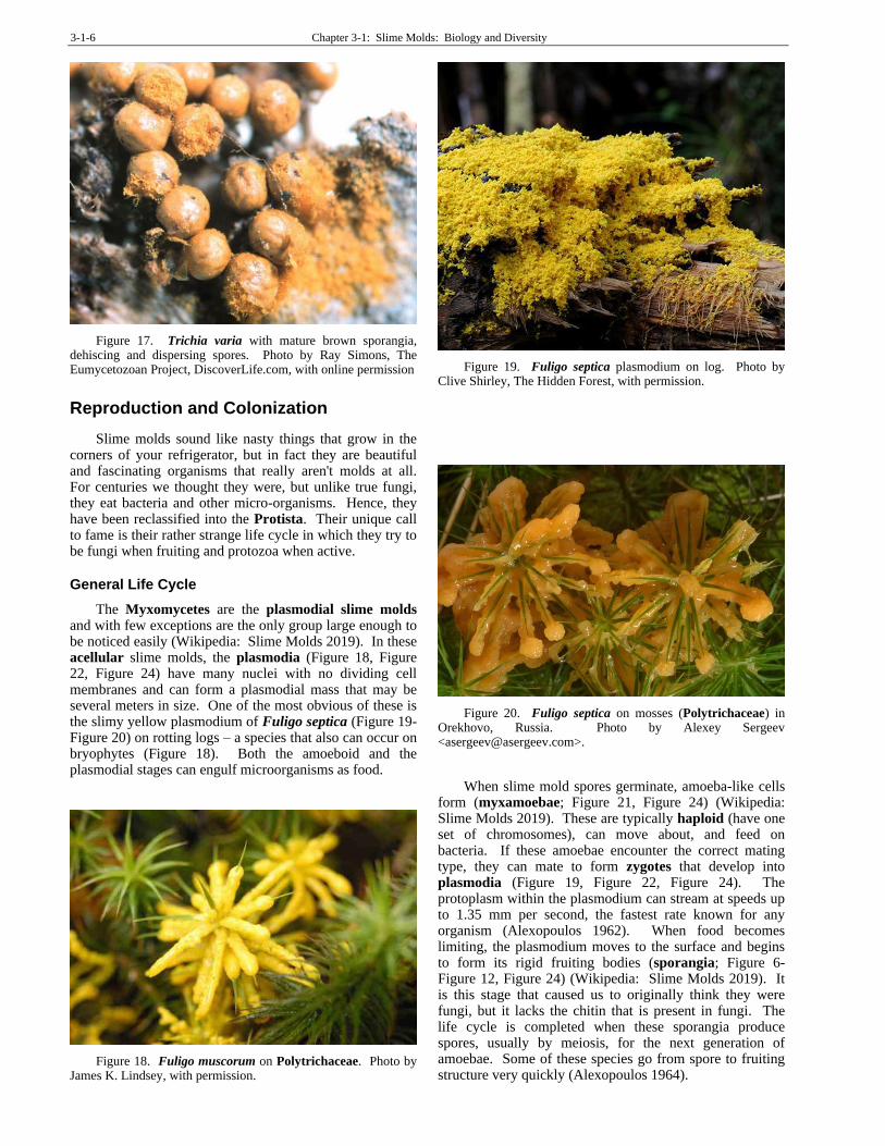
Figure 15. Trichia varia with yellow sporangia on moss. Photo from Bite.Your.Bum Photography, through Creative Commons.
Figure 16. Trichia varia with brown sporangia. Photo from EOL, through Creative Commons.
3-1-6 Chapter 3-1: Slime Molds: Biology and Diversity
Figure 17. Trichia varia with mature brown sporangia, dehiscing and dispersing spores. Photo by Ray Simons, The Eumycetozoan Project, DiscoverLife.com, with online permission
Reproduction and Colonization
Slime molds sound like nasty things that grow in the corners of your refrigerator, but in fact they are beautiful and fascinating organisms that really aren't molds at all. For centuries we thought they were, but unlike true fungi, they eat bacteria and other micro-organisms. Hence, they have been reclassified into the Protista. Their unique call to fame is their rather strange life cycle in which they try to be fungi when fruiting and protozoa when active.
General Life Cycle
The Myxomycetes are the plasmodial slime molds and with few exceptions are the only group large enough to be noticed easily (Wikipedia: Slime Molds 2019). In these acellular slime molds, the plasmodia (Figure 18, Figure 22, Figure 24) have many nuclei with no dividing cell membranes and can form a plasmodial mass that may be several meters in size. One of the most obvious of these is the slimy yellow plasmodium of Fuligo septica (Figure 19-Figure 20) on rotting logs – a species that also can occur on bryophytes (Figure 18). Both the amoeboid and the plasmodial stages can engulf microorganisms as food.
Figure 18. Fuligo muscorum on Polytrichaceae. Photo by James K. Lindsey, with permission.
Figure 19. Fuligo septica plasmodium on log. Photo by Clive Shirley, The Hidden Forest, with permission.
Figure 20. Fuligo septica on mosses (Polytrichaceae) in Orekhovo, Russia. Photo by Alexey Sergeev <[email protected]>.
When slime mold spores germinate, amoeba-like cells
form (myxamoebae; Figure 21, Figure 24) (Wikipedia: Slime Molds 2019). These are typically haploid (have one set of chromosomes), can move about, and feed on bacteria. If these amoebae encounter the correct mating type, they can mate to form zygotes that develop into plasmodia (Figure 19, Figure 22, Figure 24). The protoplasm within the plasmodium can stream at speeds up to 1.35 mm per second, the fastest rate known for any organism (Alexopoulos 1962). When food becomes limiting, the plasmodium moves to the surface and begins to form its rigid fruiting bodies (sporangia; Figure 6-Figure 12, Figure 24) (Wikipedia: Slime Molds 2019). It is this stage that caused us to originally think they were fungi, but it lacks the chitin that is present in fungi. The life cycle is completed when these sporangia produce spores, usually by meiosis, for the next generation of amoebae. Some of these species go from spore to fruiting structure very quickly (Alexopoulos 1964).

Chapter 3-1: Slime Molds: Biology and Diversity 3-1-7
Figure 21. Didymium myxamoebae hatched from spores. Brown structures are spores. Photo by George Barron, modified, with permission.
Figure 22. Fuligo aurea plasmodium. Photo through Creative Commons.
If free water is available, myxamoebae (Figure 21) form swarm cells (Figure 24) by developing flagella – one long and one very short (Myxomycota 2019). Some species mate as myxamobae (Figure 24) and others as swarm cells. Although adjoined myxamoebae are ready to mate, they generally cannot mate with the same strain, i.e. no sibling mating.
If conditions become too dry for the plasmodium (Figure 22), it will form a sclerotium (Figure 23, Figure 24), which is a dry dormant state (Wikipedia: Slime Molds 2019) and sometimes resembles the slime left by a slug. When this sclerotium once again becomes moist, it returns to the active plasmodium state. An alternative to this is that some species can form a microcyst (Figure 24) (Myxomycota 2019). This stage occurs when the amoeboid cells or swarm cells round up and form a thin wall, then become dormant, surviving unfavorable conditions.
Figure 23. Sclerotium. Photo courtesy of Steve Stephenson.
The multinucleate, diploid plasmodium (Figure 22) moves and feeds until conditions are right (or wrong) and it reorganizes into sporangia (Myxomycota 2019). The spores that are produced generally undergo meiosis to produce four nuclei. Three of these abort, leaving a single haploid nucleus, in a cell that becomes the haploid spore.
Figure 24. Generalized slime mold life cycle. Modified from Hoppe & Schwippert 2014.
Some species can produce diploid (having 2 sets of chromosomes) amoeboflagellates (includes flagellated cells and amoeboid cells) that develop directly into the plasmodium (Figure 22) without having any crossing with another cell (Clark & Haskins 2010). This appears to be the result of a failure of meiosis, resulting in diploid spores (apomixis). Thus a single spore of some species can complete a life cycle without any mating occurring.
Seasonal Changes
Reproduction in the Myxomycetes is typically
seasonal. Eliasson (1980) recorded the times of
fructification (producing sporangia) in several Swedish
species over the course of four years. Those Myxomycetes
fruiting in May-June include Amaurochaete atra (Figure
3-1-8 Chapter 3-1: Slime Molds: Biology and Diversity
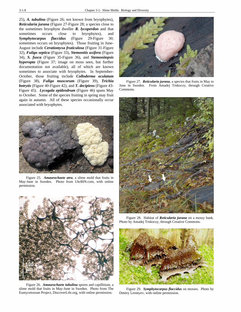
25), A. tubulina (Figure 26; not known from bryophytes),
Reticularia jurana (Figure 27-Figure 28; a species close to
the sometimes bryophyte dweller R. lycoperdon and that
sometimes occurs close to bryophytes), and
Symphytocarpus flaccidus (Figure 29-Figure 30;
sometimes occurs on bryophytes). Those fruiting in June-
August include Ceratiomyxa fruticulosa (Figure 31-Figure
32), Fuligo septica (Figure 33), Stemonitis axifera (Figure
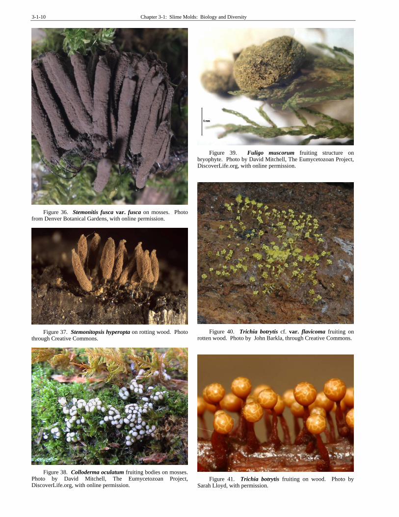
34), S. fusca (Figure 35-Figure 36), and Stemonitopsis
hyperopta (Figure 37; image on moss seen, but further
documentation not available), all of which are known
sometimes to associate with bryophytes. In September-
October, those fruiting include Colloderma oculatum
(Figure 38), Fuligo muscorum (Figure 39), Trichia
botrytis (Figure 40-Figure 42), and T. decipiens (Figure 43-
Figure 45). Lycogala epidendrum (Figure 46) spans May
to October. Some of the species fruiting in spring may fruit
again in autumn. All of these species occasionally occur
associated with bryophytes.
Figure 25. Amaurochaete atra, a slime mold that fruits in May-June in Sweden. Photo from UkrBIN.com, with online permission.
Figure 26. Amaurochaete tubulina spores and capillitium, a slime mold that fruits in May-June in Sweden. Photo from The Eumycetozoan Project, DiscoverLife.org, with online permission.
Figure 27. Reticularia jurana, a species that fruits in May to June in Sweden. From Amadej Trnkoczy, through Creative Commons.
Figure 28. Habitat of Reticularia jurana on a mossy bank. Photo by Amadej Trnkoczy, through Creative Commons.
Figure 29. Symphytocarpus flaccidus on mosses. Photo by Dmitry Leontyev, with online permission.

Chapter 3-1: Slime Molds: Biology and Diversity 3-1-9
Figure 30. Symphytocarpus flaccidus with maturing capsules. Photo by Thomas Laxton, through Creative Commons.
Figure 31. Ceratiomyxa fruticulosa fruiting bodies on bryophytes. Photo by David Mitchell, The Eumycetozoan Project, DiscoverLife.org, with online permission.
Figure 32. Ceratiomyxa fruticulosa fruiting bodies. Photo by David Mitchell, The Eumycetozoan Project, DiscoverLife.org, with online permission.
Figure 33. Fuligo septica plasmodia growing on mosses at the base of a tree. Photos by David Mitchell, The Eumycetozoan Project, DiscoverLife.org, with online permission.
Figure 34. Stemonitis axifera fruiting bodies growing on moss. Photo by David Mitchell, The Eumycetozoan Project, DiscoverLife.org, with online permission.
Figure 35. Stemonitis fusca fruiting bodies on log. Photo from Encyclopedia of Life, through Creative Commons.
3-1-10 Chapter 3-1: Slime Molds: Biology and Diversity
Figure 36. Stemonitis fusca var. fusca on mosses. Photo from Denver Botanical Gardens, with online permission.
Figure 37. Stemonitopsis hyperopta on rotting wood. Photo through Creative Commons.
Figure 38. Colloderma oculatum fruiting bodies on mosses. Photo by David Mitchell, The Eumycetozoan Project, DiscoverLife.org, with online permission.
Figure 39. Fuligo muscorum fruiting structure on bryophyte. Photo by David Mitchell, The Eumycetozoan Project, DiscoverLife.org, with online permission.
Figure 40. Trichia botrytis cf. var. flavicoma fruiting on rotten wood. Photo by John Barkla, through Creative Commons.
Figure 41. Trichia botrytis fruiting on wood. Photo by Sarah Lloyd, with permission.

Chapter 3-1: Slime Molds: Biology and Diversity 3-1-11
Figure 42. Trichia botrytis old and dry fruiting structures on wood. Photo by Bernard Dupont, through Creative Commons.
Figure 43. Trichia decipiens young fruiting bodies. Photo by David Mitchell, The Eumycetozoan Project, DiscoverLife.org, with online permission.
Figure 44. Trichia decipiens. Mature fruiting bodies. Photo by Alain Michaud, The Eumycetozoan Project, DiscoverLife.org, with online permission.
Figure 45. Trichia decipiens empty fruiting bodies. Photo by Alain Michaud, The Eumycetozoan Project, DiscoverLife.org, with online permission.
Figure 46. Fruiting bodies of Lycogala epidendrum (wolf's milk; toothpaste slime) on mosses. The plasmodia are composed of small, red amoeboid cells (Wikipedia: Lycogala epidendrum 2019). When the conditions change, these rarely seen cells find each other by chemical signals and aggregate into the fruiting body, as seen here. Photos by David Mitchell, The Eumycetozoan Project, DiscoverLife.org, with online permission.
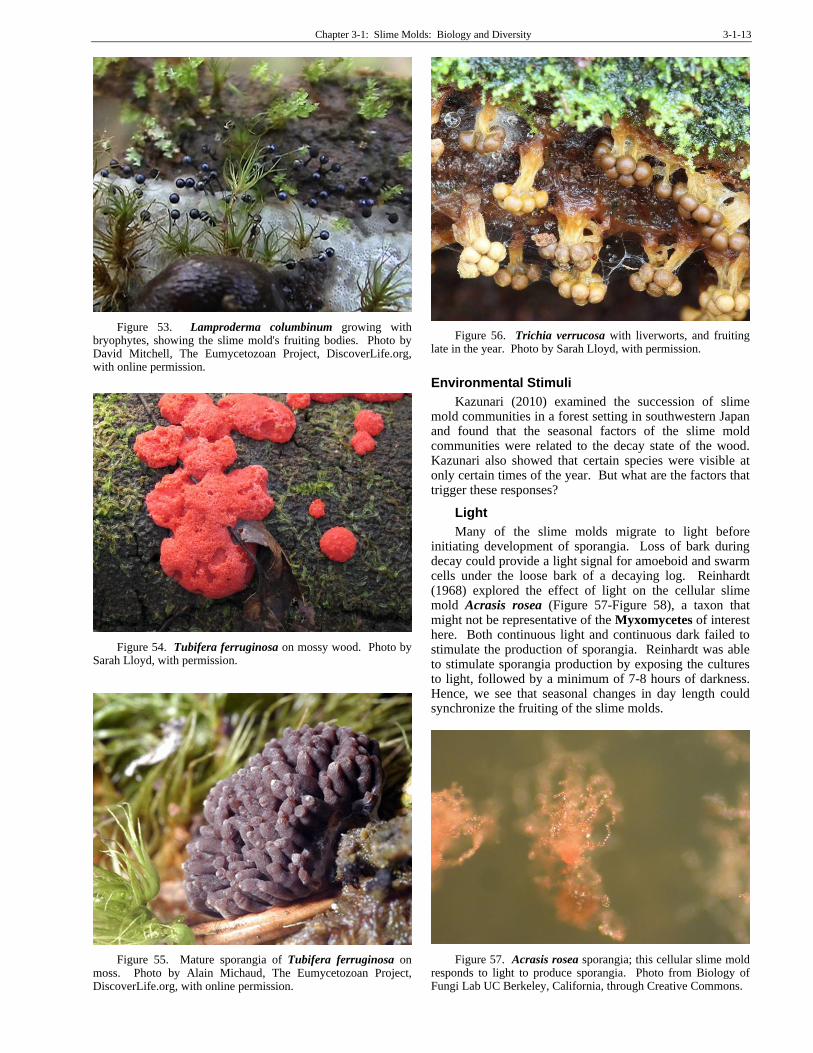
Some of the other seasonal records for the occasional Myxomycetes bryophyte dwellers include Arcyria ferruginea (Figure 47; known from bryophytes – based on photos by Iyp-tala at <https://hiveminer.com/Tags/arcyria>; Dawn & Jim at <https://hiveminer.com/Tags/arcyria>), A. obvelata (Figure 48; known from bryophytes – based on photo from <https://www.alamy.com/stock-photo-arcyria-obvelata-slime-mold-73514471.html>, Collaria arcyrionema (Figure 49; syn=Lamproderma arcyrionema; known from bryophytes – Ranade et al. 2012), and Physarum viride (Figure 50; known from bryophytes – Stephenson & Studlar 1985), all of which appeared early in the year. Stemonitopsis hyperopta (Figure 37; known from bryophytes based on online image; attribution not available), Cribraria intricata (Figure 51; known from mosses – Ranade et al. 2012), Cribraria cribrarioides (Figure 52; on bryophytes in photograph), Lamproderma columbinum (Figure 53; known from bryophytes – Stephenson & Studlar 1985), Tubifera ferruginosa (Figure

3-1-12 Chapter 3-1: Slime Molds: Biology and Diversity
54-Figure 55; known from bryophytes – Stojanowska & Panek 2004), and Trichia verrucosa (Figure 56; known from bryophytes based on image) appeared later in the year.
Figure 47. Arcyria ferruginea fruiting bodies. Photo by Alain Michaud, The Eumycetozoan Project, DiscoverLife.org, with online permission.
Figure 48. Arcyria obvelata, a species that has been photographed elsewhere growing on bryophytes. Photo by Patrick Schifferli, through Creative Commons.
Figure 49. Collaria arcyrionema fruiting, a species reported from bryophytes. Photo by Guang-Bao Xiang and Quan-Nian Jun, through Creative Commons.
Figure 50. Physarum viride fruiting bodies. Photo by Dmitry Leontyev, The Eumycetozoan Project, DiscoverLife.org, with online permission.
Figure 51. Cribraria intricata, a species known to grow on bryophytes. Photo by Clive Shirley, The Hidden Forest <www.hiddenforest.co.nz>, with permission.
Figure 52. Cribraria cribrarioides on bryophytes, and fruiting late in the year. Photo from Myxotropic, through Creative Commons.
Chapter 3-1: Slime Molds: Biology and Diversity 3-1-13
Figure 53. Lamproderma columbinum growing with bryophytes, showing the slime mold's fruiting bodies. Photo by David Mitchell, The Eumycetozoan Project, DiscoverLife.org, with online permission.
Figure 54. Tubifera ferruginosa on mossy wood. Photo by Sarah Lloyd, with permission.
Figure 55. Mature sporangia of Tubifera ferruginosa on moss. Photo by Alain Michaud, The Eumycetozoan Project, DiscoverLife.org, with online permission.
Figure 56. Trichia verrucosa with liverworts, and fruiting late in the year. Photo by Sarah Lloyd, with permission.
Environmental Stimuli
Kazunari (2010) examined the succession of slime mold communities in a forest setting in southwestern Japan and found that the seasonal factors of the slime mold communities were related to the decay state of the wood. Kazunari also showed that certain species were visible at only certain times of the year. But what are the factors that trigger these responses?
Light
Many of the slime molds migrate to light before initiating development of sporangia. Loss of bark during decay could provide a light signal for amoeboid and swarm cells under the loose bark of a decaying log. Reinhardt (1968) explored the effect of light on the cellular slime mold Acrasis rosea (Figure 57-Figure 58), a taxon that might not be representative of the Myxomycetes of interest here. Both continuous light and continuous dark failed to stimulate the production of sporangia. Reinhardt was able to stimulate sporangia production by exposing the cultures to light, followed by a minimum of 7-8 hours of darkness. Hence, we see that seasonal changes in day length could synchronize the fruiting of the slime molds.
Figure 57. Acrasis rosea sporangia; this cellular slime mold responds to light to produce sporangia. Photo from Biology of Fungi Lab UC Berkeley, California, through Creative Commons.

3-1-14 Chapter 3-1: Slime Molds: Biology and Diversity
Figure 58. Acrasis rosea amoebae, a cellular slime mold, emerging from spores. Photo by Chirley Chio at Mushroom Observer, California, through Creative Commons.
Kakiuchi et al. (2001) demonstrated the role of the colors of light in the initiation of reproduction in the Myxomycetes slime mold Physarum polycephalum (Figure 59). Light initiates the breakup of the plasmodium (Figure 22) into equal-sized spherical pieces within about five hours. Blue and far-red light both initiate this behavior, whereas red light (but not blue) inhibits the far-red induction. These fragments develop the sporangia and spores. When it is time to develop sporangia, plasmodia can creep out from under bark or the bases of bryophytes and seek higher ground and more light.
Figure 59. Physarum polycephalum on leafy liverworts. Photo by Bernard Spragg, through Creative Commons.
pH and Volatile Substances
Researchers have found that bark pH is important in determining slime mold distribution on bark, but that it might be masked by geographic location (Everhart et al. 2008; Keller & Everhard 2010). It is reasonable to ask, then, if substrate pH is important in the reproductive cycle.
Early work by Reinhardt (1968) demonstrated that pH was important for fruiting in Acrasis rosea (Figure 57-Figure 58); a cellular slime mold in an entirely different clade), with growth occurring at pH 3.5-7.6, but fruiting only at 5.0-6.6. Such differences in pH could occur as a result of changes in the decay state of a log or litter. Of course this is only one species, and not even in the Myxomycetes, but it illustrates the mechanisms that might be used by other slime molds as well.
Gray (1939) found that temperature and pH are closely interrelated, at least in the Myxomycetes slime mold Physarum polycephalum (Figure 59). When pH remains constant, the time required for fruiting varies directly with the temperature, requiring longer times at higher temperatures. Furthermore, the higher the temperature, the fewer cultures produce fruiting bodies. When pH also varies, higher temperatures require greater acidity to produce fruiting bodies. At a constant temperature, the greatest fruiting occurs at pH 3.0. The maximum temperature at which this species will produce sporangia is 32.5º-35.0ºC. Sclerotia will not form at low temperatures (8º-12ºC) or high temperatures (32.5º-35.0ºC). Light still seems to be necessary for fruiting at all temperatures.
While the change in pH could be a seasonal phenomenon, research by Newell et al. (1969) suggests a different relationship. In the slime mold Dictyostelium
discoideum (Figure 11-Figure 12; Dictyosteliomycetes), a dweller of shallow soil, also known from bryophytes and litter, the amoebae form multicellular aggregates from which they are able to form fruiting bodies with stalks and spores. This change of state may occur at the same microsite, or it can change its structure into a form that can migrate to a more favorable location. This migration can be stimulated by the accumulation of metabolites from the slime mold or a low ionic strength in its substrate. This migration is inhibited by the presence of a buffer or overhead illumination. In an unbuffered system, the stimulus for fruiting is "appreciably volatile." In the presence of a buffer, the slime molds transformed from a migrating slug (Figure 10) and sat still, producing fruiting bodies on that spot. The strong base NaOH was completely ineffective in preventing the formation of the moving slug. Furthermore, the transformation into a moving slug was inversely related to the density of the slime mold cells, indicating that it was something produced by the slime mold that signalled the migration. Others (Bonner et al. 1950; Francis 1964) have observed that this species moves toward heat, following a very low temperature change gradient (as little as 0.05º C per cm). This behavior could decrease the volatile substance produced by the slime mold – an indicator that it is not too dense a population. But a heat gradient also would lead the moving slug form toward the light, which would then stop the migration and cause it to form the fruiting bodies.
Using the unicellular slime mold Dictyostelium
mucoroides (Figure 10; Dictyosteliomycetes), Filosa
(1979) similarly demonstrated the presence of a volatile
substance by using charcoal as an absorbent. In the dark,
this species produced macrocysts (encysted, resting
plasmodium), but in the light it produced fruiting bodies.
But if the dark cultures were grown over activated
charcoal, they likewise would form fruiting bodies. When
grown in light with KOH (a CO2 absorbent), they produced
macrocysts, but if activated charcoal was added, they again
only produced fruiting bodies.
All of these responses to heat, light, pH, and an
exudate from the slime molds themselves could optimize
their reproductive potential. These stimuli cause the slime
molds to move to a location where spores are more easily
dispersed and will have less competition for space during
fruiting and food for the next generation.

Chapter 3-1: Slime Molds: Biology and Diversity 3-1-15
Water
In the cellular slime molds, surface water is a key factor as well (Bonner et al. 1982). When the plasmodial slug tip reaches above the water film, it usually causes the slime mold to shift gears and produce the fruiting structures. Among the cellular slime molds, light seems to be less important, promoting fructification only in those phototactic slugs that orient away from the surface.
Reproduction in Myxomycetes
Some slime molds are particularly associated with bryophytes (Ing 1994), and almost all of these are in the Myxomycetes, the acellular or plasmodial slime molds. Myxo means slime. They gain their energy by engulfing and digesting bacteria, yeasts, fungal spores, and decaying material in their amoeboid stage (Wikipedia: Slime Molds 2019), food sources that are often available on bryophytes. Spores are formed in a capsule-like structure. When the spores germinate, they release the amoeboid cells, referred to as the myxamoebae (Figure 21). If there is sufficient water for swimming, the myxamoeba may develop flagella and become a swarm cell. This process can be reversed, the flagellum retracted, and the amoeboid stage returned.
Unlike the Dictyosteliomycetes, the Myxomycetes are sexual. When two different mating strains find each other, they join to form a zygote. Even in forming the plasmodium (Figure 22), the Myxomycetes differ from
the Dictyosteliomycetes. In Myxomyceyes, the zygote does not form an amoeba, but instead divides only its nucleus. These nuclei continue to divide to form the plasmodium – a large, multinucleate body composed of a single cell. In their plasmodium (Figure 22) stage, the Myxomycetes can flow like an amoeba, feeding as they traverse their substrate (Wikipedia: Slime Molds 2019). The plasmodium prefers darkness, and when it ventures into the light it is likely to go into its sclerotium (Figure 23, Figure 24) stage – a dormant stage that can remain so for years; this stage is also imitated by drying conditions. That shiny dry covering that looks like a slug's slime trail on the surface of a moss might be a sclerotium. The sclerotium is particularly likely to form if the plasmodium dries out. If, on the other hand, it runs short on food first, it goes into its fruiting stage. Such factors as light and temperature can induce the plasmodium to transform into fruiting structures (Figure 61 that produce meiospores, hence returning the organism to its 1n state (having only one set of chromosomes). The subsequent spores may germinate into flagellated cells or amoeboid cells that multiply vegetatively and engulf food to gain energy.
Figure 60. Didymium squamulosum sporangia. Photo by Ray Simons, The Eumycetozoan Project, DiscoverLife.org, with online permission.
Figure 61. Trichia subfusca mature fruiting bodies on bark. Photo by Alain Michaud, The Eumycetozoan Project, DiscoverLife.org, with online permission.
Temperature plays an important role in maintaining the active state of the amoeboid stage, and any habitable site must have sufficient moisture, making bryophytes necessary for survival of any that venture onto rocks (Ing 1994). The behavior of the slime mold under adverse conditions is reminiscent of the bryophytes and many of the fauna found there. When the going gets rough, they sleep like Rip Van Winkle! For the slime molds, it is the sclerotium (Figure 23, Figure 24); for many fauna it is a cyst; and for the bryophytes it is a simple dormancy without any change of state.
The Physarales (Figure 2-Figure 9; Figure 60-Figure 68), and especially Diderma (Figure 62-Figure 68), frequently fruit extensively where bryophytes and lichens cover the bark (Brooks et al. 1977). We know substrate is important for finding food in the mobile stages, but is it important for fruiting? Do the bryophytes offer the advantage of a higher perch for dispersal of these tiny beings?
Figure 62. Diderma sp. on liverwort. Ken-ichi Ueda, through Creative Commons.
3-1-16 Chapter 3-1: Slime Molds: Biology and Diversity
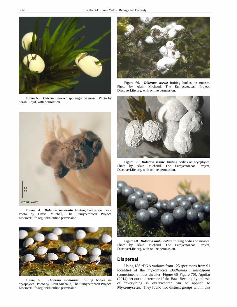
Figure 63. Diderma cinerea sporangia on moss. Photo by Sarah Lloyd, with permission.
Figure 64. Diderma imperialis fruiting bodies on moss. Photo by David Mitchell, The Eumycetozoan Project, DiscoverLife.org, with online permission.
Figure 65. Diderma montanum fruiting bodies on bryophytes. Photo by Alain Michaud, The Eumycetozoan Project, DiscoverLife.org, with online permission.
Figure 66. Diderma sessile fruiting bodies on mosses. Photo by Alain Michaud, The Eumycetozoan Project, DiscoverLife.org, with online permission.
Figure 67. Diderma sessile. fruiting bodies on bryophytes. Photo by Alain Michaud, The Eumycetozoan Project, DiscoverLife.org, with online permission.
Figure 68. Diderma umbilicatum fruiting bodies on mosses. Photo by Alain Michaud, The Eumycetozoan Project, DiscoverLife.org, with online permission.
Dispersal
Using 18S rDNA variants from 125 specimens from 91 localities of the myxomycete Badhamia melanospora (sometimes a moss dweller; Figure 69-Figure 70), Aguilar (2014) set out to determine if the Baas-Becking hypothesis of "everything is everywhere" can be applied to Myxomycetes. They found two distinct groups within this

Chapter 3-1: Slime Molds: Biology and Diversity 3-1-17
species: one group comprises all populations from Argentina and Chile; the other is formed by populations from North America together with human-introduced populations from other parts of the world. For this species, they concluded that everything is not everywhere. Instead, the taxon consists of a complex that has at least two cryptic species that probably diverged as allopatric (having non-overlapping distributions) in North and South America. But as will be seen in this chapter, many of the slime molds do have widespread distributions on several continents.
Figure 69. Badhamia melanospora, a species that sometimes grows on bryophytes. Photo from The Eumycetozoan Project, DiscoverLife.org, with online permission.
Figure 70. Badhamia melanospora spore SEM. Can it travel around the world? The Eumycetozoan Project, DiscoverLife.org, with permission.
It appears that some slime molds occur in the same
places for multiple years, but their propensity for living on
logs and even living trees means that at some time they
must disperse to survive. Schnittler and Tesmer (2008)
asked if the habitat colonization model for spore-dispersed
organisms works for slime molds. They found spore
numbers per sporangium ranging from 1 to 106. Average
spore size ranges 10.3 µm to 14.8 µm in the studied taxa.
Culture data suggest that the number of spores required to
create the observed frequencies (as a percent of
successfully colonized habitat islands) is generally three
orders of magnitude higher. Species with sexual
reproductive systems typically produce more spores than
do asexual ones.
The presence of individual species is limited not by
dispersal, which seems to be efficient, but by suitable
substrate (Ing 1994). We have seen that the species are
seasonal, but as we might expect, the time of year for the
conspicuous fruiting varies with climatic zone. The
dispersal is primarily tied to the onset of rain after a long
warm period. This is typically autumn in the temperate
regions, whereas in parts of the tropics it begins with the
monsoon season. Dispersal does not determine species
presence, except perhaps among the corticolous species.
Rather, it is suitable substrates that determine presence.
One factor in dispersal of the spores is their surface
structure. Three types exist in the Myxomycetes: spiny,
reticulate, and smooth surfaces (Hoppe & Schwippert
2014). Using spores from 17 species, including
Metatrichia floriformis (Figure 71) (reticulate; see Figure
72), Fuligo septica (Figure 33) (spiny; see Figure 73), and
Licea parasitica (smooth; see Figure 74) as well as
Ceratiomyxa fruticulosa (Figure 31-Figure 32;
Ceratiomyxomycetes) (smooth) (all known from
bryophytes as well as other substrata), they determined the
wettability of the spores. Spiny spores would half sink into
the water but nevertheless they floated. Reticulate spores
are superhydrophobic and float on the surface tension of
the water. Spores with no ornamentation sink to the bottom
rather quickly.
Figure 71. Metatrichia floriformis sporangia. Photo by Clive Shirley, The Hidden Forest, with permission.

3-1-18 Chapter 3-1: Slime Molds: Biology and Diversity
Figure 72. Stemonitis fusca, sometimes a moss dweller, reticulate spores. SEM photo courtesy of Yuri Novozhilov.
Figure 73. Physarum notabile, sometimes a moss dweller, spiny spores. SEM photo courtesy of Yuri Novozhilov.
Figure 74. Licea deplanata, not a known bryophyte dweller, smooth spore. SEM photo courtesy of Yuri Novozhilov.
Dispersal by wind seems to predominate (Keller & Smith 1978). Underlying bryophytes can become covered in spores (Figure 75). Dispersal may be aided by the capillitium (Figure 76) that in some species twists in response to changing moisture conditions. The capillitium also is likely to act like a salt shaker, doling out a few spores at a time instead of releasing all of them in a single
burst of wind, a function similar to that of the peristome in mosses.
Figure 75. Tubulifera ferruginosa. Photo by David Mitchell, The Eumycetozoan Project, DiscoverLife.org, with online permission.
Figure 76. Trichia varia spores and capillitium. Photo by Alain Michaud, The Eumycetozoan Project, DiscoverLife.org, with online permission.
In some species, insects and mites seem to be important dispersal agents (Keller & Smith 1978; Eliasson 1977). Beetles are abundant on Amaurochaete (Figure 25) species and spores that cling to the body and legs would get a free ride for dispersal (Eliasson 1977).
Eliasson (1980) indicated that invertebrates are important in the dispersal of several species of slime molds. This is sometimes accomplished through predation by snails and insects that carry the spores on their bodies or in their digestive tracts (Ing 1967; Angela Newton, Bryonet, 20 November 2006).
The isopod Philoscia muscorum (Figure 77) appears to spread the cellular slime mold Didymium bahiense (Figure 78) (Ing 1004). Huss (1989) verified the potential of dispersal by earthworms (Figure 79) and pillbugs (Isopoda; Figure 77). Some of these invertebrate species are bryophyte dwellers, although typically not the ones used in the experiments. These invertebrates were fed both spores and myxamoebae of slime molds. Although percentages of both survived, the spores survived better

Chapter 3-1: Slime Molds: Biology and Diversity 3-1-19
than the myxamoebae. When invertebrate feces were cultivated, the species the invertebrates had eaten developed in the cultures.
Figure 77. The isopod Philoscia muscorum, a likely dispersal agent for the cellular slime mold Didymium bahiense. Photo by Malcolm Storey, through Creative Commons.
Figure 78. Didymium bahiense on bryophytes. Photo by Alain Michaud, The Eumycetozoan Project, DiscoverLife.org, with online permission.
Figure 79. The earthworm Octolasion cyaneum; some species in this genus ingest slime molds and disperse them. Photo by Chih-Han Chang, through Creative Commons.
A similar relationship was found between the cellular slime mold Dictyostelium discoideum (Figure 11-Figure
12; Dictyosteliomycetes), an occasional bryophyte dweller, and the nematode Caenorhabditis elegans (Figure
80) (Kessin et al. 1996). This nematode is an inhabitant of the moss Sphagnum (Figure 81) (Glatzer & Ahlf 2001) and feeds on slime molds, including consumption of the spores. It kills the amoeboid stage, but the spores survive the digestive tract, making this another organism capable of moving the spores from one place to a new location for germination.
Figure 80. Caenorhabditis elegans, a nematode that seems to benefit from some properties of Sphagnum, and that also can disperse slime molds living there. Photo by Kbradnam, through Creative Commons.
Figure 81. Sphagnum recurvum, in a genus that is home for the nematode Caenorhabditis elegans. Photo by Malcolm Storey, DiscoverLife.org, with online permission.
Habitat Needs
Publications on slime molds are in no short supply. Gray and Alexopoulos (1968) published a treatise on the biology. Martin and Alexopoulos (1969) wrote a general treatise on the group. Ing (1994) summarized the phytosociology, arranged according to major vegetation types. Rollins and Stephenson (2011) summarized the global distribution and ecology.
As of 2011, Sarah Lloyd reported that only 1000 species of slime molds had been described. Their greatest abundance is in temperate forests, where they occur on living and dead trees and rotting wood, but also in some unusual habitats, including on dung and on living animals (Stephenson & Rojas 2017).
Moisture
Ing (1994) related the slime molds to their habitat factors, surmising that temperature is an important limiting factor in tropical, subtropical, Mediterranean, and alpine species. There is a consistent distinction between the corticolous, lignicolous, and epiphyllous species, and the lignicolous species have a preference for either conifers or deciduous trees. Ing even referred to bryophyte associations, noting that a few slime molds are particularly

3-1-20 Chapter 3-1: Slime Molds: Biology and Diversity
associated with them. This may be due to water relations, with Ing noting that water and water-retaining substrates are of prime importance. The presence of fruiting structures (sporangia) is dependent on the arrival of rain after a prolonged warm period, making their presence most common in autumn in temperate regions. In the tropics, capsules form when the monsoon season begins. Fruiting seems to be independent of substrate.
Eliasson (1980) noted that species that have large plasmodia (Figure 22) typically are rare under arid conditions. This would suggest that the slime molds on bryophytes are the larger species in most habitats because of the moisture-holding capacity of the bryophytes.
On the other hand, Schnittler et al. (2013), based on observations in Xinjiang Province, China, concluded that corticolous Myxomycetes are some of the most drought-tolerant organisms in that habitat. They are opportunistic, permitted by their ability to survive in a dormant state for decades and to complete their life cycles in a few days of appropriate conditions.
Latitude
Stephenson et al. (1993) found recognizable patterns in the latitudinal variation of slime molds. The species assemblages in the tropical-subtropical regions is distinctly different from that found in temperate regions. Furthermore, the species differ in their substrate usage at different latitudes. Some species that are rare outside the Arctic and subArctic can be relatively common in these northern regions (Stephenson et al. 2000).
Food and Light
Naturally, available food is of importance in the location of active slime molds. Slime molds frequently make "decisions" for location based on the quality of food available. The common Myxomycetes slime mold Physarum polycephalum (Figure 59, Figure 82; sometimes a bryophyte dweller), in its amoeboid phase and if both locations are shaded, will choose the higher food quality 100% of the time (Latty & Beekman 2010). When a much higher quality food is in the light, it is selected, but when the difference in quality is small, the slime mold will select the shade over the light location, even if its food is of lesser quality.
Figure 82. Physarum polycephalum plasmodium or rotting wood. Photo by Frankenstoen, through Creative Commons
Role of Bryophytes as Slime Mold Habitat
Stephenson and Studlar (1985) found representatives of all six orders (at that time) of slime molds, exclusive of the Labyrinthulomycota and the Plasmodiophorids in their study of bryophyte-dwellers in the United States and Canada. The Physarales (Figure 2-Figure 9; Figure 60-Figure 68) (38% of all collections) were the most abundant, but members of the Stemonitales (Figure 34-Figure 37) (23%), Trichiales (Figure 13, Figure 40-Figure 45) (18%), and Liceales (Figure 83-Figure 84) (17%) were also commonly bryophyte associates. The order Echinosteliales (Figure 99-Figure 100) and the class Ceratomyxomycetes (Figure 31-Figure 32) comprised only 4% and 1%, respectively. All four of the major types of slime mold fruiting bodies (sporangia, aethalia, plasmodiocarps, and pseudoaethalia) were represented in their 170 collections.
Figure 83. Licea floriformis fruiting bodies on moss leaves. Photo by David Mitchell, The Eumycetozoan Project, DiscoverLife.org, with online permission.
Figure 84. Licea retiformis plasmodium. Photo by David Mitchell, The Eumycetozoan Project, DiscoverLife.org, with online permission.
But are these slime molds preferential colonists of bryophytes? Stephenson and Studlar (1985) set out to try to answer this question. By examining 170 collections throughout North America, they found that three species were particularly common: Fuligo septica (Figure 33), Stemonitis axifera (Figure 34), and S. fusca (Figure 35).
Chapter 3-1: Slime Molds: Biology and Diversity 3-1-21
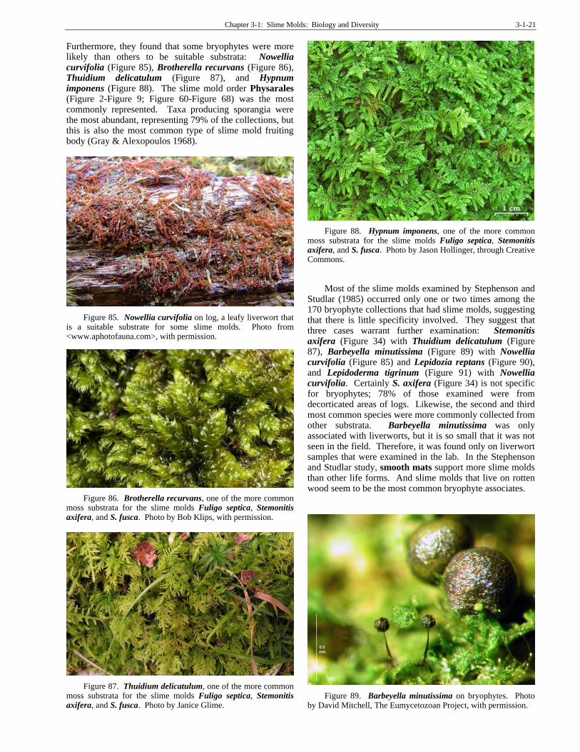
Furthermore, they found that some bryophytes were more likely than others to be suitable substrata: Nowellia curvifolia (Figure 85), Brotherella recurvans (Figure 86), Thuidium delicatulum (Figure 87), and Hypnum imponens (Figure 88). The slime mold order Physarales (Figure 2-Figure 9; Figure 60-Figure 68) was the most commonly represented. Taxa producing sporangia were the most abundant, representing 79% of the collections, but this is also the most common type of slime mold fruiting body (Gray & Alexopoulos 1968).
Figure 85. Nowellia curvifolia on log, a leafy liverwort that is a suitable substrate for some slime molds. Photo from <www.aphotofauna.com>, with permission.
Figure 86. Brotherella recurvans, one of the more common moss substrata for the slime molds Fuligo septica, Stemonitis axifera, and S. fusca. Photo by Bob Klips, with permission.
Figure 87. Thuidium delicatulum, one of the more common moss substrata for the slime molds Fuligo septica, Stemonitis axifera, and S. fusca. Photo by Janice Glime.
Figure 88. Hypnum imponens, one of the more common moss substrata for the slime molds Fuligo septica, Stemonitis axifera, and S. fusca. Photo by Jason Hollinger, through Creative Commons.
Most of the slime molds examined by Stephenson and
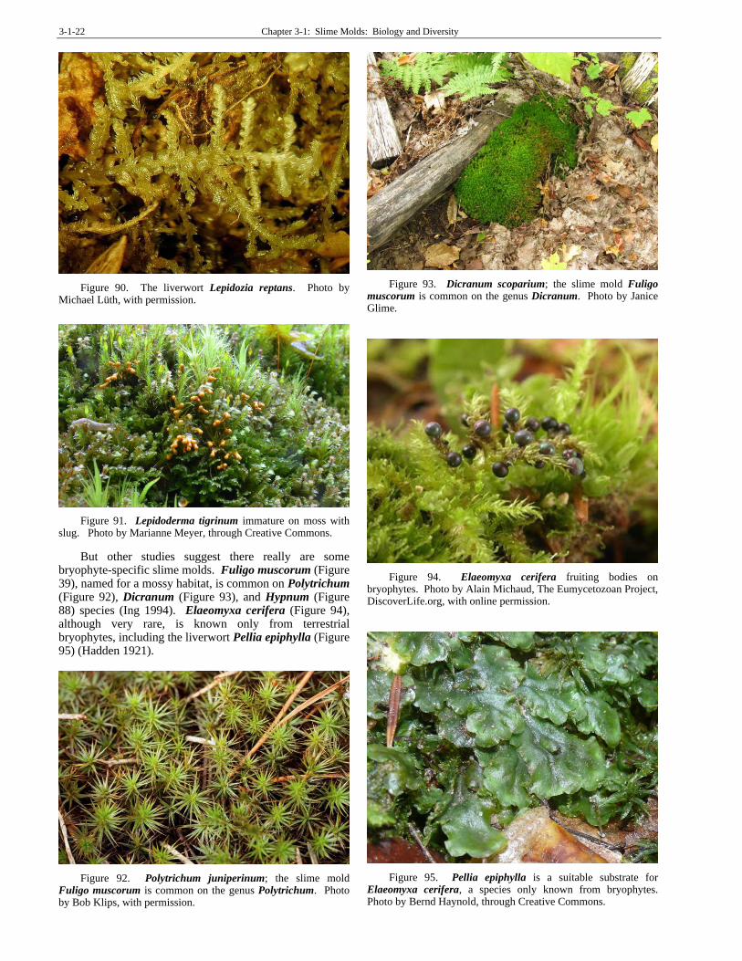
Studlar (1985) occurred only one or two times among the 170 bryophyte collections that had slime molds, suggesting that there is little specificity involved. They suggest that three cases warrant further examination: Stemonitis axifera (Figure 34) with Thuidium delicatulum (Figure 87), Barbeyella minutissima (Figure 89) with Nowellia curvifolia (Figure 85) and Lepidozia reptans (Figure 90), and Lepidoderma tigrinum (Figure 91) with Nowellia curvifolia. Certainly S. axifera (Figure 34) is not specific for bryophytes; 78% of those examined were from decorticated areas of logs. Likewise, the second and third most common species were more commonly collected from other substrata. Barbeyella minutissima was only associated with liverworts, but it is so small that it was not seen in the field. Therefore, it was found only on liverwort samples that were examined in the lab. In the Stephenson and Studlar study, smooth mats support more slime molds than other life forms. And slime molds that live on rotten wood seem to be the most common bryophyte associates.
Figure 89. Barbeyella minutissima on bryophytes. Photo by David Mitchell, The Eumycetozoan Project, with permission.
3-1-22 Chapter 3-1: Slime Molds: Biology and Diversity
Figure 90. The liverwort Lepidozia reptans. Photo by Michael Lüth, with permission.
Figure 91. Lepidoderma tigrinum immature on moss with slug. Photo by Marianne Meyer, through Creative Commons.
But other studies suggest there really are some bryophyte-specific slime molds. Fuligo muscorum (Figure 39), named for a mossy habitat, is common on Polytrichum (Figure 92), Dicranum (Figure 93), and Hypnum (Figure 88) species (Ing 1994). Elaeomyxa cerifera (Figure 94), although very rare, is known only from terrestrial bryophytes, including the liverwort Pellia epiphylla (Figure 95) (Hadden 1921).
Figure 92. Polytrichum juniperinum; the slime mold Fuligo muscorum is common on the genus Polytrichum. Photo by Bob Klips, with permission.
Figure 93. Dicranum scoparium; the slime mold Fuligo muscorum is common on the genus Dicranum. Photo by Janice Glime.
Figure 94. Elaeomyxa cerifera fruiting bodies on bryophytes. Photo by Alain Michaud, The Eumycetozoan Project, DiscoverLife.org, with online permission.
Figure 95. Pellia epiphylla is a suitable substrate for Elaeomyxa cerifera, a species only known from bryophytes. Photo by Bernd Haynold, through Creative Commons.

Chapter 3-1: Slime Molds: Biology and Diversity 3-1-23
If bryophytes are indeed a preferred substrate for some species, the next question is why. Stephenson and Studlar (1985) suggest that bryophytes serve as spore traps, increasing the chances of the trapped species becoming residents here. The bryophytes then provide a moist habitat, again favoring growth of slime molds. These same conditions provide a habitat for numerous protozoa and bacteria, providing food for the slime molds, and even the detritus produced by tardigrades, annelids, and arthropods can serve as food sources (Gerson 1969, 1982; Richardson 1981).
In a single study, Bovee (1979) reported 68 species of protozoa (particularly shelled amoebae and ciliates) among mosses, mostly the mosses Brachythecium salebrosum (Figure 96), Plagiomnium cuspidatum (Figure 97), and Pylaisiella selwynii (Figure 98) on a rotten log in Minnesota. Many of these protozoa provide suitable food for the slime molds in their mobile phase.
Figure 96. Brachythecium salebrosum, home of many protozoa. Photo by Michael Lüth, with permission.
Figure 97. Plagiomnium cuspidatum, home of many protozoa. Photo by Janice Glime.
Bryophytes may provide a preferred location for forming sporangia. Slime molds migrate to the highest position available before making sporangia (Stephenson &
Studlar 1985), and bryophytes on a log could very well be that place.
Figure 98. Pylaisia selwynii, home of many protozoa. Photo by Jan-Peter Frahm, with permission.
In any case, the slime molds, like the tardigrades, rotifers, and protozoa, seem to be well-adapted to the poikilohydric (having no mechanism to prevent desiccation) existence of living among bryophytes (Gerson 1982). When the bryophyte and the slime mold dry out, the myxamoebae and swarm cells of the slime mold can form microcysts; plasmodia (Figure 22) are able to form sclerotia (Figure 23, Figure 24). These structures are all resistant and survive well under desiccating conditions. They can quickly resume activity when water becomes available. The tolerance of slime molds to alternate wetting and drying that typically accompanies the bryophytes provides us with another reason to suspect that they can live within, as well as sporulate upon, bryophyte clumps.
But not all slime molds benefit from the moist environment of the bryophytes. The genus Echinostelium (Figure 99-Figure 100) is comprised of tiny slime molds that live on bark (Keller & Brooks 1976). But in areas that support the growth of algae, mosses, and leafy liverworts, larger aphano- and phaneroplasmodial slime molds are favored. Keller and Brooks surmised that the tiny protoplasmodial Echinostelium species were unable to compete.
Figure 99. Echinostelium minutum, a tiny species that is probably unable to compete. Photo by Satyendra Rajguru, The Eumycetozoan Project, DiscoverLife.org, with online permission.
3-1-24 Chapter 3-1: Slime Molds: Biology and Diversity
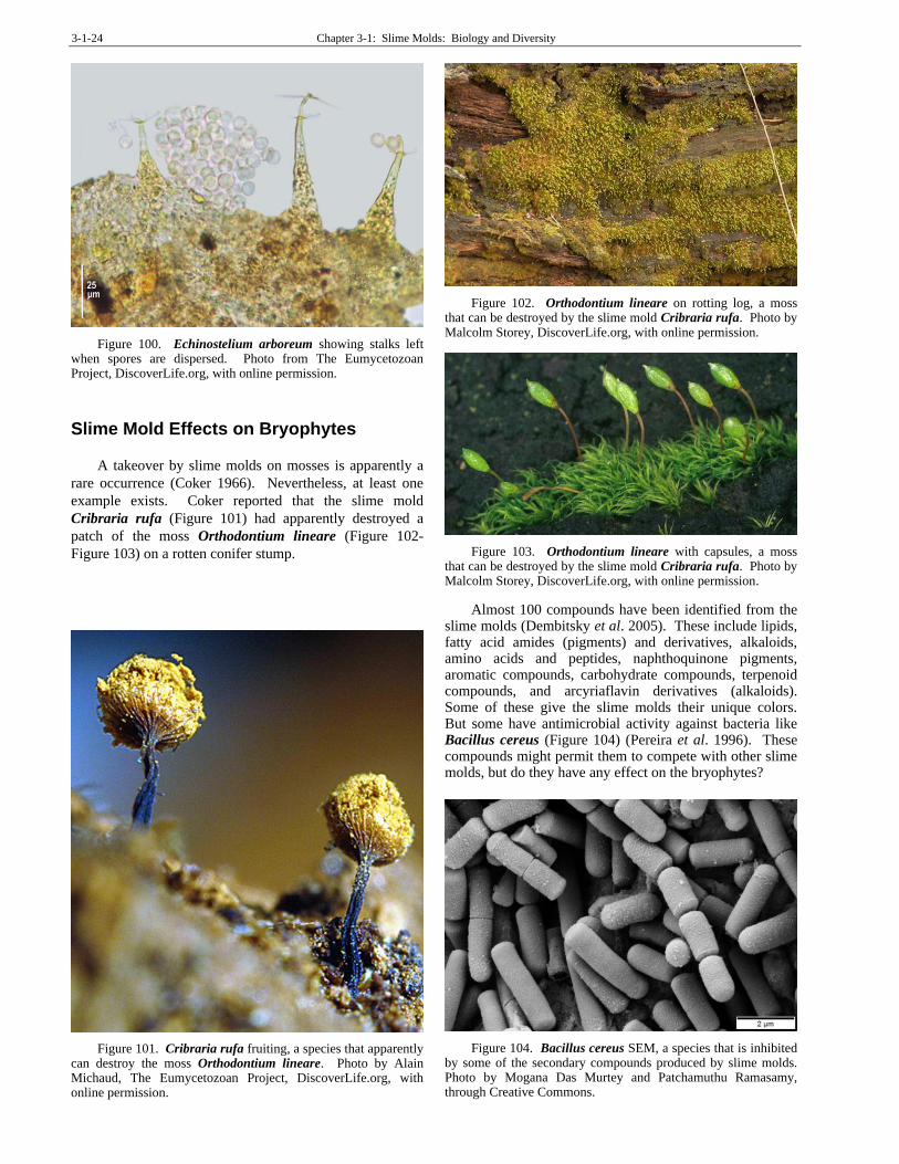
Figure 100. Echinostelium arboreum showing stalks left when spores are dispersed. Photo from The Eumycetozoan Project, DiscoverLife.org, with online permission.
Slime Mold Effects on Bryophytes
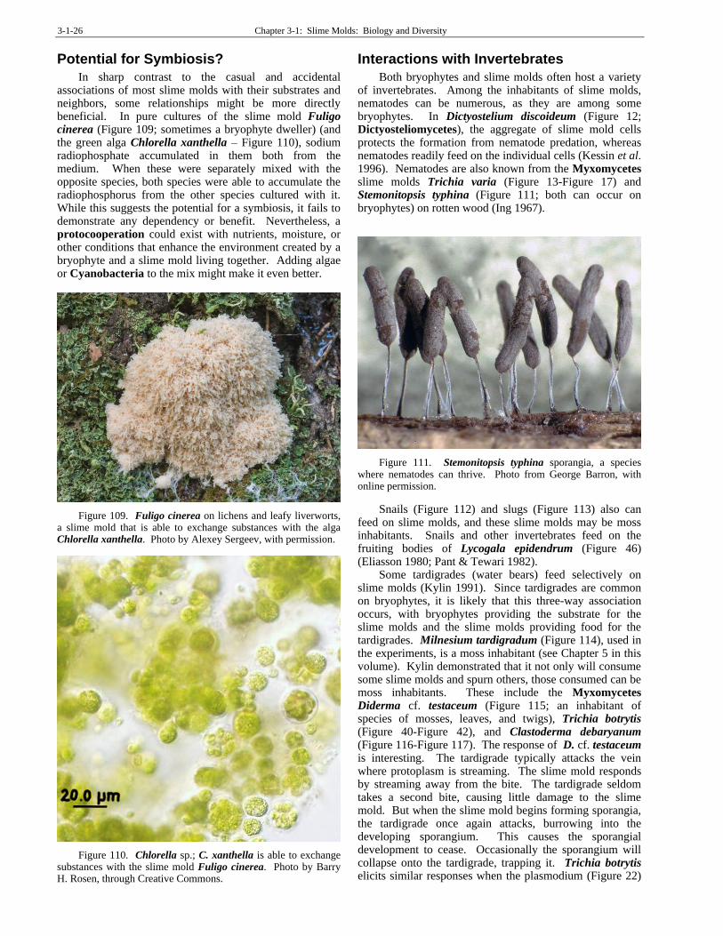
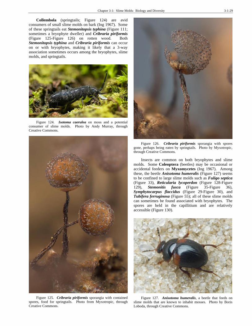
A takeover by slime molds on mosses is apparently a
rare occurrence (Coker 1966). Nevertheless, at least one
example exists. Coker reported that the slime mold
Cribraria rufa (Figure 101) had apparently destroyed a
patch of the moss Orthodontium lineare (Figure 102-
Figure 103) on a rotten conifer stump.
Figure 101. Cribraria rufa fruiting, a species that apparently can destroy the moss Orthodontium lineare. Photo by Alain Michaud, The Eumycetozoan Project, DiscoverLife.org, with online permission.
Figure 102. Orthodontium lineare on rotting log, a moss that can be destroyed by the slime mold Cribraria rufa. Photo by Malcolm Storey, DiscoverLife.org, with online permission.
Figure 103. Orthodontium lineare with capsules, a moss that can be destroyed by the slime mold Cribraria rufa. Photo by Malcolm Storey, DiscoverLife.org, with online permission.
Almost 100 compounds have been identified from the slime molds (Dembitsky et al. 2005). These include lipids, fatty acid amides (pigments) and derivatives, alkaloids, amino acids and peptides, naphthoquinone pigments, aromatic compounds, carbohydrate compounds, terpenoid compounds, and arcyriaflavin derivatives (alkaloids). Some of these give the slime molds their unique colors. But some have antimicrobial activity against bacteria like Bacillus cereus (Figure 104) (Pereira et al. 1996). These compounds might permit them to compete with other slime molds, but do they have any effect on the bryophytes?
Figure 104. Bacillus cereus SEM, a species that is inhibited by some of the secondary compounds produced by slime molds. Photo by Mogana Das Murtey and Patchamuthu Ramasamy, through Creative Commons.

Chapter 3-1: Slime Molds: Biology and Diversity 3-1-25
Slime molds do not usually appear to be any threat to
the bryophytes. However, in some cases, it appears that
the slime molds are aggressive enough to overgrow and
destroy the bryophytes (Coker 1966). Fuligo intermedia
(Figure 105) seems to be harmful (Pant & Tewari 1982),
most likely due to its density of fruiting bodies that can
cover patches several centimeters in diameter. Such
growths would deprive the moss of light and may interfere
with gas exchange.
Figure 105. Fuligo intermedia fruiting bodies on bryophytes. Photo by David Mitchell, The Eumycetozoan Project, DiscoverLife.org, with online permission.
Bryophytes Growing on Slime Molds
In some species, the fruiting bodies of slime molds can
persist. That can lead to a reverse relationship with
bryophytes. It gives the bryophytes sufficient time to grow
over the slime molds, as observed by Sarah Lloyd (2011).
She found a growth of leafy liverworts on the stalk of a
slime mold on decaying wood, undoubtedly a very rare
occurrence.
Epizooites
One of the most unusual habitats for slime molds is on
living lizards, Corytophanes cristatus (Figure 106), in
Mexico and Costa Rica (Lloyd 2011). This lizard is a sit-
and-wait predator and therefore moves around little. It uses
its head to dig its nest and often has residual soil in the
scoop on the top of its head. This microenvironment is
home to the tiny liverwort Lejeunea obtusangula (see
Figure 107) (Gradstein & Equihua 1995). But this lizard is
also sometimes home to the slime mold Physarum
pusillum (Figure 108). The co-occurrence of the liverwort
and the slime mold, if at all, is most likely one of chance
resulting from the scooping behavior of the lizard.
Figure 106. Corytophanes cristatus, the crested lizard that sometimes has the slime mold Physarum pusillum or the leafy liverwort Lejeunea obtusangula growing on it. Photo by Simon J. Tonge, through Creative Commons.
Figure 107. Lejeunea sp. from the Neotropics; L. obtusangula sometimes occurs on the lizard Corytophanes cristatus. Photo by Michael Lüth, with permission.
Figure 108. Physarum pusillum fruiting bodies, a species known to live on the lizard Corytophanes cristatus. Photo by Gustavo F. Morejón J., through Creative Commons.
3-1-26 Chapter 3-1: Slime Molds: Biology and Diversity
Potential for Symbiosis?
In sharp contrast to the casual and accidental associations of most slime molds with their substrates and neighbors, some relationships might be more directly beneficial. In pure cultures of the slime mold Fuligo cinerea (Figure 109; sometimes a bryophyte dweller) (and the green alga Chlorella xanthella – Figure 110), sodium radiophosphate accumulated in them both from the medium. When these were separately mixed with the opposite species, both species were able to accumulate the radiophosphorus from the other species cultured with it. While this suggests the potential for a symbiosis, it fails to demonstrate any dependency or benefit. Nevertheless, a protocooperation could exist with nutrients, moisture, or other conditions that enhance the environment created by a bryophyte and a slime mold living together. Adding algae or Cyanobacteria to the mix might make it even better.
Figure 109. Fuligo cinerea on lichens and leafy liverworts, a slime mold that is able to exchange substances with the alga Chlorella xanthella. Photo by Alexey Sergeev, with permission.
Figure 110. Chlorella sp.; C. xanthella is able to exchange substances with the slime mold Fuligo cinerea. Photo by Barry H. Rosen, through Creative Commons.
Interactions with Invertebrates
Both bryophytes and slime molds often host a variety of invertebrates. Among the inhabitants of slime molds, nematodes can be numerous, as they are among some bryophytes. In Dictyostelium discoideum (Figure 12; Dictyosteliomycetes), the aggregate of slime mold cells protects the formation from nematode predation, whereas nematodes readily feed on the individual cells (Kessin et al. 1996). Nematodes are also known from the Myxomycetes slime molds Trichia varia (Figure 13-Figure 17) and Stemonitopsis typhina (Figure 111; both can occur on bryophytes) on rotten wood (Ing 1967).
Figure 111. Stemonitopsis typhina sporangia, a species where nematodes can thrive. Photo from George Barron, with online permission.
Snails (Figure 112) and slugs (Figure 113) also can feed on slime molds, and these slime molds may be moss inhabitants. Snails and other invertebrates feed on the fruiting bodies of Lycogala epidendrum (Figure 46) (Eliasson 1980; Pant & Tewari 1982).
Some tardigrades (water bears) feed selectively on slime molds (Kylin 1991). Since tardigrades are common on bryophytes, it is likely that this three-way association occurs, with bryophytes providing the substrate for the slime molds and the slime molds providing food for the tardigrades. Milnesium tardigradum (Figure 114), used in the experiments, is a moss inhabitant (see Chapter 5 in this volume). Kylin demonstrated that it not only will consume some slime molds and spurn others, those consumed can be moss inhabitants. These include the Myxomycetes Diderma cf. testaceum (Figure 115; an inhabitant of species of mosses, leaves, and twigs), Trichia botrytis (Figure 40-Figure 42), and Clastoderma debaryanum (Figure 116-Figure 117). The response of D. cf. testaceum is interesting. The tardigrade typically attacks the vein where protoplasm is streaming. The slime mold responds by streaming away from the bite. The tardigrade seldom takes a second bite, causing little damage to the slime mold. But when the slime mold begins forming sporangia, the tardigrade once again attacks, burrowing into the developing sporangium. This causes the sporangial development to cease. Occasionally the sporangium will collapse onto the tardigrade, trapping it. Trichia botrytis elicits similar responses when the plasmodium (Figure 22)

Chapter 3-1: Slime Molds: Biology and Diversity 3-1-27
is attacked, usually feeding for about 12 hours, but has a sporangium that is too small for the tardigrade to burrow into it. Clastoderma debaryanum is a much smaller slime mold and the tardigrade usually consumes the entire plasmodium.
Figure 112. Fruiting bodies of Arcyria stipata with one of its enemies – a snail. Photo by David Mitchell, The Eumycetozoan Project, DiscoverLife.org, with online permission.
Figure 113. Slug and the slime mold Lamproderma on mosses. Photo by Keller, through Creative Commons.
Figure 114. Milnesium tardigradum SEM, a species that feeds on the moss-inhabiting slime molds Diderma cf. testaceum, Trichia botrytis, and Clastoderma debaryanum. Photo from Schokraie et al. 2012, through Creative Commons.
Figure 115. Diderma testaceum fruiting structures, with lichens, a slime mold that serves as food for the tardigrade Milnesium tardigradum. Masse (1892) indicated that this species grows on leaves, mosses, and twigs. Photo by James K. Lindsey, through Creative Commons.
Figure 116. Clastoderma debaryanum on moss, a slime mold that serves as food for the tardigrade Milnesium tardigradum. Photo from Myxotropic, through Creative Commons.
Figure 117. Clastoderma debaryanum fruiting body on moss, a slime mold that serves as food for the tardigrade Milnesium tardigradum. Photo from Myxotropic, through Creative Commons.

3-1-28 Chapter 3-1: Slime Molds: Biology and Diversity
Isopods are common inhabitants on bryophytes and will readily consume them (Hames & Hopkin 1989). They likewise can occur on slime molds (Ing 1967). They eat both plasmodia (Figure 22) and fruiting bodies of the Myxomycetes slime molds. The isopods Trichoniscus pusillus (Figure 118) and Oniscus asellus (Figure 119) feed on the slime molds Trichia varia (Figure 13-Figure 17) and Arcyria denudata (Figure 120). The isopod Androniscus dentiger (Figure 121) eats both plasmodia and sporangia of Didymium iridis (Figure 122), at the same time dispersing this species across the substrate. Spores have been found in the isopod digestive tracts undigested. All of these three slime molds are known from bryophytes.
Figure 118. Trichoniscus pusillus, an isopod that feeds on the slime molds Trichia varia and Arcyria denudata. Photo by Malcolm Storey, EOL, through Creative Commons.
Figure 119. Oniscus asellus with moss on log, an isopod that feeds on the slime molds Trichia varia and Arcyria denudata. Photo by Kurt Kulac, through Creative Commons.
Figure 120. Arcyria denudata fruiting bodies. Photo by Kim Fleming, The Eumycetozoan Project, DiscoverLife.org, with online permission.
Figure 121. Androniscus dentiger, an isopod that feeds on the slime mold Didymium iridis. Photo by Gilles San Martin, through Creative Commons.
Figure 122. Didymium iridis sporangia, food for the isopod Androniscus dentiger. Photo by through Creative Commons.
Millipedes are likely known from both bryophytes and slime molds. The millipede Cylindroiulus punctatus (Figure 123) consumes the sporangia of the slime mold Trichia varia (Figure 13-Figure 17) on wet, rotten wood (Ing 1967).
Figure 123. Cylindroiulus punctatus, a millipede that feeds on the slime mold Trichia varia. Photo by Saxifraga-Ab H Baas, through Creative Commons.
Chapter 3-1: Slime Molds: Biology and Diversity 3-1-29
Collembola (springtails; Figure 124) are avid consumers of small slime molds on bark (Ing 1967). Some of these springtails eat Stemonitopsis typhina (Figure 111; sometimes a bryophyte dweller) and Cribraria piriformis (Figure 125-Figure 126) on rotten wood. Both Stemonitopsis typhina and Cribraria piriformis can occur on or with bryophytes, making it likely that a 3-way association sometimes occurs among the bryophytes, slime molds, and springtails.
Figure 124. Isotoma caerulea on moss and a potential consumer of slime molds. Photo by Andy Murray, through Creative Commons.
Figure 125. Cribraria piriformis sporangia with contained spores, food for springtails. Photo from Myxotropic, through Creative Commons.
Figure 126. Cribraria piriformis sporangia with spores gone, perhaps being eaten by springtails. Photo by Myxotropic, through Creative Commons.
Insects are common on both bryophytes and slime molds. Some Coleoptera (beetles) may be occasional or accidental feeders on Myxomycetes (Ing 1967). Among these, the beetle Anisotoma humeralis (Figure 127) seems to be confined to large slime molds such as Fuligo septica (Figure 33), Reticularia lycoperdon (Figure 128-Figure 129), Stemonitis fusca (Figure 35-Figure 36), Symphytocarpus flaccidus (Figure 29-Figure 30), and Tubifera ferruginosa (Figure 55); all of these slime molds can sometimes be found associated with bryophytes. The spores are held in the capillitium and are relatively accessible (Figure 130).
Figure 127. Anisotoma humeralis, a beetle that feeds on slime molds that are known to inhabit mosses. Photo by Boris Loboda, through Creative Commons.

3-1-30 Chapter 3-1: Slime Molds: Biology and Diversity
Figure 128. Pink Reticularia lycoperdon on mossy log. Photo by David Mitchell, The Eumycetozoan Project, DiscoverLife.org, with online permission.
Figure 129. White Reticularia lycoperdon on mossy bark. Photo by Marion Zãller, through Creative Commons.
Figure 130. Capillitium of sporangium of Stemonitis. Photo by Janice Glime.
Some beetles even seem to be obligate feeders on slime molds (Dudka & Romanenko 2006). Lawrence and Newton (1980) reported on about 35 beetle species, mostly from North American, that feed on slime mold spores. Dudka and Romanenko (2006) found that slime mold spores occurred in 19 of the 25 beetle (Latridiidae) guts they examined from Crimea. These included Latridius
hirtus (Figure 131), Enicmus rugosus (Figure 132), and E. fungicola (Figure 133) as obligate slime mold feeders. On the other hand Corticarina truncatella (Figure 134) is a facultative slime mold feeder. The most common 13 species of slime molds, including Fuligo septica (Figure 33), Mucilago crustacea (Figure 135), Stemonitis axifera (Figure 34), S. fusca (Figure 35), and S. splendens (Figure 136), were inhabited by five species of Latridiidae; all of these slime molds can occur on bryophytes.
Figure 131. Latridius hirtus adult, a beetle that feeds on slime mold spores. Photo by Stefan Schmidt, through Creative Commons.
Figure 132. Enicmus rugosus adult, a beetle that feeds on slime mold spores. Photo from Zoologische Staatssammlung Muenchen, through Creative Commons.
Figure 133. Enicmus fungicola adult, a beetle that feeds on slime mold spores. Photo by Tim Faasen, with permission.

Chapter 3-1: Slime Molds: Biology and Diversity 3-1-31
Figure 134. Corticarina truncatella adult, a beetle that facultatively feeds on slime mold spores. Photo from Zoologische Staatssammlung Muenchen, through Creative Commons.
Figure 135. Mucilago crustacea on mosses. Photo by Drew Henderson, through Creative Commons.
Figure 136. Stemonitis splendens, one of the slime molds eaten by the beetle family Latridiidae. Photo by Dan Molter, through Creative Commons.
Some Coleoptera (beetles) in the Leiodidae can be considered slime mold beetles (Wheeler & Miller 2005). Stetholiodes sp. (Figure 137) is a slime mold beetle that was originally described from moss in northern Indiana (Blatchley 1910). Several species of Agathidium (Figure 138) are known moss inhabitants, including A. brevisternum, A. rhinocerellum, and A. cavisternum
(Figure 139) (Wheeler & Miller 2005). The only known host for Agathidium rhinocerellum is the Myxomycetes slime mold Fuligo septica (Figure 33, Figure 140), a widespread generalist species that includes bryophytes among its substrates. It is likely that other moss dwellers in this family also feed on slime molds.
Figure 137. Stetholiodes laticollis adult; some members of this genus are slime mold beetles that live on mosses. Photo by Museum of Comparative Zoology, Harvard University, through Creative Commons.
Figure 138. Agathidium sp. adult; some members of this genus are both moss and slime mold inhabitants. Photo by Joyce Gross, with permission.
Figure 139. Agathidium cavisternum, a moss dweller and possible slime mold feeder. Photo from Museum of Comparative Zoology, Harvard University, through Creative Commons.

3-1-32 Chapter 3-1: Slime Molds: Biology and Diversity
Figure 140. Fuligo septica on moss, a slime mold that is host for the beetle Agathidium rhinocerellum. Photo by David Mitchell, The Eumycetozoan Project, DiscoverLife.org, with online permission.
Some Diptera larvae live on the slime mold plasmodia (Figure 22) and feed on them, with some remaining there as pupae. Bradysia (Figure 141) species feed on plasmodia of Fuligo septica (Figure 33) and sporangia of Lycogala epidendrum (Figure 46) and Arcyria incarnata (Figure 142-Figure 143), all occasional bryophyte dwellers. In fact, some flies can be reared on slime molds as their only food.
Figure 141. Bradysia larvae, a species that feeds on slime mold plasmodia of Fuligo septica and sporangia of Lycogala epidendrum and Arcyria incarnata. Photo by David Cappaert, through Creative Commons.
Figure 142. Arcyria incarnata fruiting bodies, food for Bradysia. Photo by Stu's Images, through Creative Commons.
Figure 143. Arcyria incarnata fruiting bodies on mosses, food for Bradysia. Photo by Dan Molter, through Creative Commons.
Summary
Slime molds are really not molds, but protozoa, with an amoeboid feeding stage and a spore-producing, non-feeding stage. They also lack chitin, a compound found in true molds. The bryophyte dwelling members are included in the Eumycetozoa or Amoebozoa and classified into the classes Myxomycetes, Dictyosteliomycetes, and Ceratiomyxomycetes.
The life cycle has a dormant spore that will germinate when adequate water is available and develop into swarm cells or amoeboid cells. This stage feeds like an amoeba. In Myxomycetes, either of these cell types can form a zygote that divides to form a plasmodium. This stage likewise feeds on bacteria, algae, and protozoa. It can dry out to form a sclerotium that can remain dormant for years, or move to higher ground in the light to form sporangia and spores. Either stage can occur on bryophytes, but the plasmodium stage is likely to be unnoticed. The life cycle is usually keyed to seasons, with autumn being the more favorable fruiting season for most species. Dispersal is most likely primarily by wind, but animals are also dispersal vectors, either by carrying spores on the outside or by digesting them or plasmodia and dispersing them in the feces.
The slime molds respond to light, pH, volatile substances, temperature, and water availability to trigger fruiting. We know most slime molds seek higher positions with more light before forming sporangia. Do bryophytes provide a more suitable location for that event? Do slime molds benefit in their dispersal by the activities of moss fauna?
The slime molds known to associate with mosses are predominantly in the Myxomycetes. The mosses may provide prolonged moisture and a place to get above the prevailing substrate for better dispersal, or they may be dispersed by some of the invertebrates living among the bryophytes. Little is known about the effect the slime molds have on the bryophytes. Some slime molds live on animals, and these may be the same animals that have bryophytes growing on them. The potential for symbiosis exists, but little evidence supports any symbiotic relationship.

Chapter 3-1: Slime Molds: Biology and Diversity 3-1-33
Acknowledgments
Thank you to Sarah Lloyd for challenging my organization of this subchapter and to Steve Stephenson for providing me with recent literature that helped me untangle the classification of these slime molds. The Eumycetozoan website has been invaluable for checking current nomenclature and finding suitable images. Thank you to Yuri Novozhilov for providing me with images to illustrate the three spore surface types.
Literature Cited Aguilar, M., Fiore-Donno, A. M., Lado, C., and Cavalier-Smith,
T. 2014. Using environmental niche models to test the 'everything is everywhere' hypothesis for Badhamia. ISME J. 8: 737-745.
Alexopoulos, C. J. 1962. Introductory Mycology. John Wiley and Sons, 613 pp.
Alexopoulos, C. J. 1964. The rapid sporulation of some Myxomycetes in moist chamber culture. Southwest. Nat. 9: 155-159.
Blatchley, W. S. 1910. An illustrated descriptive catalogue of the Coleoptera or beetles (exclusive of the Rhyncophora) known to occur in Indiana. Nature Publishing, Indianapolis, 1386 pp.
Bonner, J. T., Clarke, W. W. Jr., Neely, C. L. Jr., and Slifkin, M. K. 1950. The orientation to light and the extremely sensitive orientation to temperature gradients in the slime mold Dictyostelium discoideum. J. Cell. Comp. Physiol. 36: 149-158.
Bonner, J. T., Davidowski, T. A., Hsu, W. L., Lapeyrolerie, D. A., and Suthers, H. L. B. 1982. The role of surface water and light on differentiation in the cellular slime molds. Differentiation 21: 123-126.
Bovee, E. C. 1979. Protozoa from acid-bog mosses and forest mosses of the Lake Itasca region (Minnesota, USA). Univ. Kans. Sci. Bull. 51: 615-629.
Brooks, T. E., Keller, H. W., and Chassain, M. 1977. Corticolous Myxomycetes, VI. A new species of Diderma. Mycologia 69: 179-184.
Cavender, J. C., Vadell, E., Landolt, J. C., and Stephenson, S. L. 2005. New species of small dictyostelids from the Great Smoky Mountains National Park. Mycologia 97: 493-512.
Clark, J. and Haskins, E. F. 2010. Reproductive systems in the Myxomycetes: A review. Mycosphere 1: 337-353.
Coker, P. D. 1966. The destruction of bryophytes by lichens, fungi, Myxomycetes and algae. Trans. Brit. Bryol. Soc. 5: 142-143.
Dembitsky, V. M., Rezanka, T., Spizek, J., and Hanus, L. O. 2005. Secondary metabolites of slime molds (Myxomycetes). Phytochemistry 66: 747-769.
Dudka, I. O. and Romanenko, K. O. 2006. Co-existence and interaction between Myxomycetes and other organisms in shared niches. Acta Mycol. 41: 99-112.
Eliasson, U. 1977. Ecological notes on Amaurochaete Rost. (Myxomycetes). Bot. Not. 129: 419-425.
Eliasson, U. 1980. Patterns of occurrence of Myxomycetes in a spruce forest in South Sweden. Ecography 4: 20-31.
Everhart, S. E., Keller, H. W., and Ely, J. S. 2008. Influence of bark pH on the occurrence and distribution of tree canopy myxomycete species. Mycologia 100: 191-204.
Feng, Y. and Schnittler, M. 2015. Sex or no sex? Independent marker genes and group I introns reveal the existence of three sexual but reproductively isolated biospecies in Trichia varia (Myxomycetes). Org. Divers. Evol. 15: 631-650.
Filosa, M. F. 1979. Macrocyst formation in the cellular slime mold Dictyostelium mucoroides: Involvement of light and volatile morphogenetic substance(s). J. Exper. Zool. 207: 491-495.
Francis, D. W. 1964. Some studies on phototaxis of Dictyostelium. J. Cell. Comp. Physiol. 64: 131-138.
Gerson, U. 1969. Moss-arthropod associations. Bryologist 72: 495-500.
Gerson, U. 1982. Bryophytes and invertebrates, Chapt. 9. In: Smith, A. J. E. (ed.). Bryophyte Ecology. Chapman and Hall, New York, pp. 291-332.
Glatzer, H. and Ahlf, W. 2001. Adjustment of a formulated sediment for sediment testing with Caenorhabditis elegans (Nematoda). Acta Hydrochem. Hydrobiol. 29: 41-46.
Gradstein, S. R. and Equihua, C. 1995. An epizoic bryophyte and algae growing on the lizard Corytophanes cristatus in Mexican rain forest. Biotropica 27: 265-268.
Gray, W. D. 1939. The relation of pH and temperature to the fruiting of Physarum polycephalum. Amer. J. Bot. 26: 709-714.
Gray, W. D. and Alexopoulos, C. J. 1968. Biology of the Myxomycetes. The Ronald Press, Co., New York.
Hadden, N. G. 1921. Mycetozoa at Porlock in October 1920. Trans. Brit. Mycol. Soc. 7: 13-16.
Hames, C. A. C., and Hopkin, S. P. 1989. The structure and function of the digestive system of terrestrial isopods. J. Zool. 217: 599-627.
Hoppe, T. and Schwippert, W. W. 2014. Hydrophobicity of myxomycete spores: An undescribed aspect of spore ornamentation. Mycosphere 5: 601-606.
Hurley, B. P. 2006. Fungus gnats in forestry nurseries and their possible role as vectors of Fusarium circinatum. Ph.D. dissertation, University of Pretoria, Pretoria, South Africa.
Huss, M. J. 1989. Dispersal of cellular slime molds by two soil invertebrates. Mycologia 81: 677-682.
Ing, B. 1967. Myxomycetes as food for other organisms. Proc. S. London Entomol. Nat. Hist. Soc. 1967: 18-23.
Ing, B. 1994. The phytosociology of Myxomycetes. Tansley Review No. 62. New Phytol. 126: 175-201.
Kakiuchi, Y., Takahashi, T., Murakami, A., and Ueda, T. 2001. Light irradiation induces fragmentation of the plasmodium, a novel photomorphogenesis in the true slime mold Physarum polycephalum: Action spectra and evidence for involvement of the phytochrome. Photochem. Photobiol. 73: 324-329.
Kang, S., Tice, A. K., Spiegel, F. W., Silberman, J. D., Pánek, T., and Cepicka, I. 2017. Between a pod and a hard test: The deep evolution of Amoebae. Molec. Biol. Evol. 34: 2258-2270.
Kazunari, T. 2010. Succession in myxomycete communities on dead Pinus densiflora wood in a secondary forest in southwestern Japan. Ecol. Res. 25: 995-1006.
Keller, H. W. and Brooks, T. E. 1976. Corticolous Myxomycetes V: Observations on the genus Echinostelium. Mycologia 68: 1204-1220.
Keller, H. W. and Everhart, S. E. 2010. Importance of Myxomycetes in Biological Research and Teaching. Fungi 3: 13-27.
Keller, H. W. and Smith, D. M. 1978. Dissemination of myxomycete spores through the feeding activities (ingestion-defecation) of an acarid mite. Mycologia 70: 1239-1241.
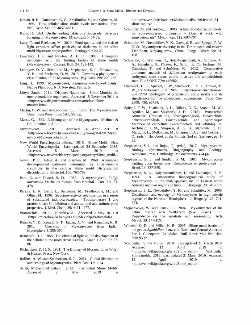
3-1-34 Chapter 3-1: Slime Molds: Biology and Diversity
Kessin, R. H., Gundersen, G. G., Zaydfudim, V., and Grimson, M. 1996. How cellular slime molds evade nematodes. Proc. Natl. Acad. Sci. 93: 4857-4861.
Kylin, H. 1991. On the feeding habits of a tardigrade: Selective foraging on Myxomycetes. Mycologist 5: 54-55.
Latty, T. and Beekman, M. 2010. Food quality and the risk of light exposure affect patch‐choice decisions in the slime mold Physarum polycephalum. Ecology 91: 22-27.
Lawrence, J. F. and Newton, A. F. Jr. 1980. Coleoptera associated with the fruiting bodies of slime molds (Myxomycetes). Coleopt. Bull. 34: 129-143.
Leontyev, D. V., Schnittler, M., Stephenson, S. L., Novozhilov, Y. K., and Shchepin, O. N. 2019. Towards a phylogenetic classification of the Myxomycetes. Phytotaxa 399: 209-238.
Ling, H. 1999. Myxomycetes, commonly overlooked plants. Native Plant Soc. N. J. Newslett. Fall, p. 5.
Lloyd, Sarah. 2011. Disjunct Naturalists. Slime Moulds: the most remarkable organisms. Accessed 18 November 2011 at <http://www.disjunctnaturalists.com/articles1/slime-moulds.htm>.
Martin, G. W. and Alexopoulos, C. J. 1969. The Myxomycetes. Univ. Iowa Press, Iowa City, 560 pp.
Masse, G. 1892. A Monograph of the Myxogastres. Methuen & Co., London, p. 211.
Myxomycota. 2019. Accessed 14 April 2019 at <http://www.botany.hawaii.edu/faculty/wong/Bot201/Myxomycota/Myxomycota.htm>.
New World Encyclopedia editors. 2015. Slime Mold. New World Encyclopedia. Last updated 24 September 2015. Accessed 5 March 2019 at <http://www.newworldencyclopedia.org/entry/Slime_mold>.
Newell, P. C., Telser, A., and Sussman, M. 1969. Alternative developmental pathways determined by environmental conditions in the cellular slime mold Dictyostelium discoideum. J. Bacteriol. 100: 763-768.
Pant, G. and Tewari, S. D. 1982. A myxomycete, Fuligo intermedia Macbr. on mosses from Nainital. Curr. Sci. 51: 478.
Pereira, E. R., Belin, L., Sancelme, M., Prudhomme, M., and Ollier, M. 1996. Structure–activity relationships in a series of substituted indolocarbazoles: Topoisomerase I and protein kinase C inhibition and antitumoral and antimicrobial properties. J. Med. Chem. 39: 4471-4477.
Protostelida. 2010. Microbewiki. Accessed 2 May 2019 at <https://microbewiki.kenyon.edu/index.php/Protostelida>.
Ranade, V. D., Korade, S. T., Jagtap, A. V., and Ranadive, K. R. 2012. Checklist of Myxomycetes from India. Mycosphere 3: 358-390.
Reinhardt, D. J. 1968. The effects of light on the development of the cellular slime mold Acrasis rosea. Amer. J. Bot. 55: 77-86.
Richardson, D. H. S. 1981. The Biology of Mosses. John Wiley & Halsted Press, New York.
Rollins, A. W. and Stephenson, S. L. 2011. Global distribution and ecology of Myxomycetes. Plant Biol. 12: 1-14.
Saleh, Muhammad Fahad. 2012. Plasmodial Slime Molds. Accessed 2 May 2019 at
<https://www.slideshare.net/MuhammadSaleh9/lecture-24-slime-molds>.
Schnittler, M. and Tesmer, J. 2008. A habitat colonisation model for spore-dispersed organisms – Does it work with eumycetozoans? Mycol. Res. 112: 697-707.
Schnittler, M., Novozhilov, Y. K., Carvajal, E., and Spiegel, F. W. 2013. Myxomycete diversity in the Tarim basin and eastern Tian-Shan, Xinjiang prov., China. Fungal Divers. 59: 91-108.
Schokraie, E., Warnken, U., Hotz-Wagenblatt, A., Grohme, M. A., Hengherr, S., Förster, F., Schill, R. O., Frohme, M., Dandekar, T., and Schnölzer, M. 2012. Comparative proteome analysis of Milnesium tardigradum in early embryonic state versus adults in active and anhydrobiotic state. PLoS ONE 7(9): e45682.
Shadwick, L. L., Spiegel, F. W., Shadwick, J. D. L., Brown, M. W., and Silberman, J. D. 2009. Eumycetozoa = Amoebozoa?: SSUrDNA phylogeny of protosteloid slime molds and its significance for the amoebozoan supergroup. PLoS One. 2009; 4(8): e6754.
Spiegel, F. W., Shadwick, L. L., Ndiritu, G. G., Brown, M. W., Aguilar, M., and Shadwick, J. D. 2018). Protosteloid Amoebae (Protosteliida, Protosporangiida, Cavosteliida, Schizoplasmodiida, Fractoviteliida, and Sporocarpic Members of Vannellida, Centramoebida, and Pellitida). In: Archibald, J. M., Simpson, A. G. B., Slamovits, C. H., Margulis, L., Melkonian, M., Chapman, D. J., and Corliss, J. O. (eds.). Handbook of the Protists, 2nd ed. Springer, pp. 1-38.
Stephenson, S. L. and Rojas, C. (eds.). 2017. Myxomycetes: Biology, Systematics, Biogeography, and Ecology. Academic Press, Cambridge, Massachusetts, 474 pp.
Stephenson, S. L. and Studlar, S. M. 1985. Myxomycetes fruiting upon bryophytes: Coincidence or preference? J. Bryol. 13: 537-548.
Stephenson, S. L., Kalyanasundaram, I., and Lakhanpal, T. N. 1993. A Comparative biogeographical study of Myxomycetes in the mid-Appalachians of Eastern North America and two regions of India. J. Biogeogr. 20: 645-657.
Stephenson, S. L., Novozhilov, Y. K., and Schnittler, M. 2000. Distribution and ecology of Myxomycetes in high-latitude regions of the Northern Hemisphere. J. Biogeogr. 27: 741-754.
Stojanowska, W. and Panek, E. 2004. Myxomycetes of the nature reserve near Wałbrzych (SW Poland). II. Dependence on the substrate and seasonality. Acta Mycol. 39: 147-159.
Wheeler, Q. D. and Miller, K. B. 2005. Slime-mold beetles of the genus Agathidium Panzer in North and Central America, Part I. Coleoptera: Leiodidae. Bull. Amer. Mus. Nat. Hist. 290: 95 pp.
Wikipedia: Slime Molds. 2019. Last updated 21 March 2019. Accessed 12 April 2019 at <https://en.wikipedia.org/wiki/Slime_mold>. Wikipedia: Slime molds. 2019. Last updated 21 March 2019. Accessed 12 April 2019 at <https://en.wikipedia.org/wiki/Slime_mold>.