Voltage Clamp Analysis of Repetitive Firing in Frog - Gpb.sav.sk
12
Gen. Physiol. Biophys. (1983), 2, 95—106 95 Voltage Clamp Analysis of Repetitive Firing in Frog Atrial Trabeculae A. K. FILIPPOV and V. I. POROTIKOV Institute for Biological Testing of Chemical Compounds, Kirova 23, 142450 Kupavna, Moscow Region, USSR Abstract. The membrane currents responsible for induced pace-maker activity in frog atrial muscle were investigated using a double sucrose gap technique. The trabecula was clamped to the potential from which pace-making started (35—40 mV positive to resting potential) and immediately after that a test pulse was applied and the membrane inward current was measured. When the duration of the conditioning pulse was increased from 0.02 s to 1 s the inward current increased and reached an asymptotic value. Such a change in inward current seems to be important for the induction of repetitive firing. Mn 2+ blocked the inward current suggesting the involvement of slow Ca 2+ /Na + membrane channels. A strong elevation of constant depolarizing current prevented repetitive firing, however this effect was reversed by a 3—4 fold increase in potassium ion concentration [K + ] D in the external medium. The outward time-independent membrane current was shown to be increased when [K + ] 0 was elevated. Dimedrole, 10~ 6 —5x 10~ 5 mol.l -1 , which was observed to decrease the outward time-inde- pendent current without affecting fast and slow inward currents, prevented repetitive firing. To a lesser extent this effect was also observed with procainamide, quinidine and oxylidine. These results show that an increase in outward transmem- brane time-independent current can induce repetitive firing and a decrease in outward current may prevent it. The inhibition of repetitive firing described above may be due to the inability of a small outward membrane current to repolarize the membrane perfectly so that the repetitive activation of calcium inward current is not possible. Some antiarrhythmic drugs can probably act in this manner. Key words: Frog atrial trabecula — Repetitive firing — Voltage clamp — Calcium channel — Background current — Antiarrhythmic drugs Introduction It is known that, in frog atrial trabeculae, repetitive activity (pace-making) is often observed when small long-lasting depolarizing currents are applied across the membrane (Brown and Noble 1969; Lenfant et al. 1972; Brown et al. 1972;
Transcript of Voltage Clamp Analysis of Repetitive Firing in Frog - Gpb.sav.sk

Gen. Physiol. Biophys. (1983), 2, 95—106 95
Voltage Clamp Analysis of Repetitive Firing in Frog Atrial Trabeculae
A. K. FILIPPOV and V. I. POROTIKOV
Institute for Biological Testing of Chemical Compounds, Kirova 23, 142450 Kupavna, Moscow Region, USSR
Abstract. The membrane currents responsible for induced pace-maker activity in frog atrial muscle were investigated using a double sucrose gap technique. The trabecula was clamped to the potential from which pace-making started (35—40 mV positive to resting potential) and immediately after that a test pulse was applied and the membrane inward current was measured. When the duration of the conditioning pulse was increased from 0.02 s to 1 s the inward current increased and reached an asymptotic value. Such a change in inward current seems to be important for the induction of repetitive firing. Mn2+ blocked the inward current suggesting the involvement of slow Ca2+/Na+ membrane channels. A strong elevation of constant depolarizing current prevented repetitive firing, however this effect was reversed by a 3—4 fold increase in potassium ion concentration [K+]D in the external medium. The outward time-independent membrane current was shown to be increased when [K+]0 was elevated. Dimedrole, 10~6 —5x 10~5 mol.l - 1 , which was observed to decrease the outward time-independent current without affecting fast and slow inward currents, prevented repetitive firing. To a lesser extent this effect was also observed with procainamide, quinidine and oxylidine. These results show that an increase in outward transmembrane time-independent current can induce repetitive firing and a decrease in outward current may prevent it. The inhibition of repetitive firing described above may be due to the inability of a small outward membrane current to repolarize the membrane perfectly so that the repetitive activation of calcium inward current is not possible. Some antiarrhythmic drugs can probably act in this manner.
Key words: Frog atrial trabecula — Repetitive firing — Voltage clamp — Calcium channel — Background current — Antiarrhythmic drugs
Introduction
It is known that, in frog atrial trabeculae, repetitive activity (pace-making) is often observed when small long-lasting depolarizing currents are applied across the membrane (Brown and Noble 1969; Lenfant et al. 1972; Brown et al. 1972;

96 Filippov and Porotikov
Brown et al. 1976a). This repetitive firing is very similar to the activity of the cardiac sinus pace-maker. In most recent reports concerning repetitive firing the authors investigated ionic currents responsible for the phase of slow diastolic depolarization (Brown and Noble 1969 ; Lenfant et al. 1972; Brown et al. 1972; 1976a). The decrease of potential in this phase was shown to be related to a slow inactivation of outward potassium current. It is clear, however, that this is not the only transmembrane current controlling the appearence of repetitive firing. In particular a great number of data demonstrate that both sinus pace-making and induced repetitive firing seem to be related to a function of membrane channels carrying the slow Ca2+/Na+ current (Brown and Noble 1969; Brooks and Lu 1972; Lenfant et al. 1972; Saxon et al. 1976). However, a direct analysis of membrane currents to confirm this suggestion has not been carried out.
In the present study the slow inward transmembrane current was shown to be responsible for the generation of repetitive activity. The appearance of oscillations was found to depend greatly on the size of the outward membrane current as well. By regulating the size of the outward current with some agents one may induce or prevent repetitive firing.
Methods
Preparation and perfusion system
Trabeculae having an approximate diameter of 0.05—0.2 mm were dissected from the endocardial surface of the atria of Rana ndibunda. The length of the preparations was 4—5 mm. The contraction of trabeculae still proximally attached to the atrial wall did improve with time after dissection, suggesting that a „healing over" process (Baldwin 1970; Déléze 1970) was operating both rapidly and effectively. The electrical properties of these preparations were investigated half an hour after dissection using a double sucrose gap technique (Rougier et al. 1968). As shown diagramatically in Fig. 1, five-chambered Perspex perfusion baths were used. A test gap (compartment 3) was 0.2 mm wide, adjacent compartments (compartments 2 and 4) were 0.8 mm wide, the walls were 0.2 mm wide. Grease seals separated the solutions in adjacent chambers preventing mixing of the solutions and potential jump on the sucrose-Ringer junction (Blaustein and Goldman 1966). The sucrose resistance was sufficiently high (50 MÍ2 in the absence of the preparation). The two end compartments of the bath (compartment 1 and 5) were perfused with normal Ringer's solution. Successful monophasic action potentials with an amplitude 100—120 mV and a duration of 350—500 ms were obtained with this system indicating that leakage currents through the sucrose were small. Substitution of Ringer's solution in compartments 1 and 5 by high potassium Ringer's solution (KC1 110 mmol.r' was substituted for NaCI of the normal Ringer's solution) did not change the size and shape of action potentials, but faster membrane depolarization and a decrease in action potential amplitude were observed over the course of the experiment in this case probably due to a leakage of K* into adjacent chambers during long pulses (McGuigan 1974). In view of this, all of the experiments were conducted with normal Ringer's solution in compartments 1 and 5. This did not permit an accurate measurement of the absolute value of the resting potential so it was assumed to be zero under the control conditions, i.e. 10 to 15 min after beginning of perfusion with normal Ringer's solution. However, changes in resting potential induced by various treatments e.g. by elevating the external potassium ion concentration, could be measured

Repetitive Firing in the Heart 97
St
Os
Q
~~}Si
V A Q A ^ > 1
Sj
&1M1&1Ä r<=h H s i t
CA Fig. 1. Schematic diagram of the perfusion bath and electronic apparatus. Abbreviations: R — Ringer's solution; S — isotonic sucrose solution; VF — voltage follower; CA — current amplifier; OA — operational clamp amplifier; St — stimulator; Si, S2 — switch selecting constant voltage or current clamp mode; R — resistance 50 MÍ3 for constant current experiments; Os — oscilloscope. The perfusion bath compartments are numbered 1—5.
satisfactorily. The normal electrical activity of the preparation was maintained for a period of between 1.5 and 2.5 h.
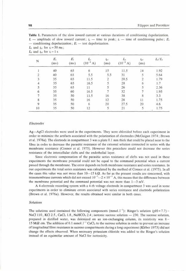
The frog atrial trabecula is a multicellular preparation. Since the diameter of the preparation and therefore the number of cells in each trabecula varied, the currents measured differed considerably from trabecula to trabeula (e.g. compare current values in Table 1, Fig. 7 and Fig. 8). The number of cells in the test gap could not be determined, therefore current density was not calculated. This made it difficult to obtain the mean values of parameters, therefore, individual values are presented (Table 1).
Electronic apparatus
A block diagram of the electronic apparatus used to generate rectangular pulses and apply them to the preparation is shown in Fig. 1. The voltage electrode in compartment 1 was connected to the input stage of a field effect transistor voltage follower VF (input resistance — 10" S2) and the voltage output from this was observed on an oscilloscope. The amplified voltage output was also connected to the inverting input of a voltage clamp amplifier OA (maximum output ± 100 V). In the voltage clamp mode the membrane potential was compared with a command potential applied to the non inverting input of the amplifier. The output of the amplifier (open loop gain 10 000) was applied to the current electrode in compartment 5 of the perfusion bath. For constant current experiments the output of the square voltage generator was applied to the same electrode through a high resistance (R = 50 Mfl). This resistance was high enough to keep current flow nearly constant during changes in the impedance and potential of the preparation.
The current flowing across the membrane was recorded using an electrode in the central chamber and, after amplification in a current amplifier CA (input resistance of amplifier 100 Q, gain 600), the signal was observed on an oscilloscope.
98 Filippov and Porotikov
Table 1. Parameters of the slow inward current at various durations of conditioning depolarization. I, — amplitude of slow inward current; tp — time to peak; U — time of conditioning pulse; Ec
— conditioning depolarization; E, — test depolarization. Li and fpl for fc = 50 ms; 1,2 and /p2 for U = 1 s
N
1 2 3 4 5 6 7 8 9 10
Ec (mv)
40 40 35 35 35 35 35 35 35 35
E, (mv)
65 65 65 65 65 60 50 50 50 50
I,i
(10~8A)
6 5.5 11.5
16.5 11 16.5
11.5 16 6 12
fP. (ms)
15 5.5 2 5 5 7 16 12 20 5
1,2
(10"8 A)
11.5 31 20.5 28 26 32 38 28 27.5 21
'p2
(ms)
14 5 2 6 5 7 8 11 20 5
/,2/Z.l
1.92
5.64
1.79
1.7 2.36 1.95
3.3 1.75 4.6 1.75
Electrodes
Ag—AgCl electrodes were used in the experiments. They were chlorided before each experiment in order to minimize the artifacts associated with the polarization of electrodes (McGuigan 1974; Brown et al. 1976a). The electrode in compartment 3 was a plate 0.1 mm thick that could be placed near to the fibre in order to decrease the parasitic resistance of the external solution connected in series with the membrane resistance (Connor et al. 1975). However this procedure could not decrease the series resistance of the intercellular clefts and the endothelial layer.
Since electronic compensation of the parasitic series resistance of clefts was not used in these experiments the membrane potential could not be equal to the command potential when a current passed through the membrane. The error depends on both membrane resistance and series resistance. In our experiments the total series resistance was calculated by the method of Connor et al. (1975). In all the cases this value was not more than 10—15 k£2. As far as the present results are concerned, with transmembrane currents which did not exceed 10~7—2 x 10~7 A, this means that the difference between the membrane potential and the command potential was not more than 1—3 mV.
A 4-eIectrode recording system with a 4-th voltage electrode in compartment 3 was used in some experiments in order to eliminate errors associated with series resistance and electrode polarization (Brown et al. 1976a). However, the results obtained were similar in both cases.
Solutions
The solutions used contained the following components (mmol.T1): Ringer's solution (pH = 7.7)-NaCl 110, KC1 2.5 , CaCh 1.8, NaHCOj 2.4; isotonic sucrose solution — 230. The sucrose solution, prepared in distilled water, was deionized on an ion-exchanging column, its resistivity was 8 -15 Mfl.cm. The addition of 0.1 mmol.r1 CaCh to the sucrose solution in order to prevent an increase of longitudinal fibre resistance in sucrose compartments during a long experiment (Kléber 1973) did not change the effects observed. When necessary potassium chloride was added to the Ringer's solution instead of an equimolar amount of NaCl for isotonicity.

Repetitive Firing in the Heart 99
8
J i ' — r 1s 20 ms
Fig. 2.A: Typical oscillations of membrane potential (upper trace) when a long depolarization current (lower trace) is applied to the frog atrial fiber. B: Voltage-clamp records of membrane current on the test pulse when the membrane was previously clamped for 1 s to that potential from which repetitive firing started (40 mV positive to resting potential). Upper trace: membrane potential; lower trace: membrane current. Traces started when the conditioning pulse was switched off. a: control; b: Mn2+, 10 mmol.r1, 5 min. Holding potential equals zero (resting potential).
The following drugs were used: procainamide, quinidine, the derivative of quinidine — 3-benzoyl-oxyquinuclidine hydrochloride (oxylidine), dimedrole (Minmedprom).
All of the experiments were conducted at room temperature (18—20 °C).
Results
Repetitive activity in most of the experiments (41 preparations) started from potential of 35 to 40 mV and above, positive to the resting potential (Fig. 2 A). For this reason the membrane potential was clamped to 35—40 mV (conditioning pulse) and then to 65—70 mV (test pulse) positive to the resting potential in order to study the inward current controlling repetitive firing. The inward ionic current arising from a test pulse was shown to be blocked by Mn2+ (2 — 10 mmol.r1) and had a time of activation (~10 ms) and inactivation ( ~ 3 0 ms) typical for a slow inward Ca2+/Na+ current (Fig. 2B).
A novel slow inward current phenomenon was observed in the present experiments. When the duration of a conditioning pulse was increased from 0.02 s to I s , the amplitude of the inward current, measured from peak to the steady-state value, was seen to increase to an asymptotic level (Fig. 3). A more than 5-fold increase was obtained in some experiments. The time to the peak of the slow inward current remained unchanged (Table 1).
It is clear that in order to make repetitive activity possible, the membrane must be repolarized to some extent in order to decrease the inactivation of slow inward current. For example, an increase in depolarizing current was observed to prevent repetitive activity (Fig. 4 top): successful repolarization could not be obtained and

100 Filippov and Porotikov
0.02 0.1 0.51 3 6 s 8
01
0.2
03 iA
— i — i — I I i i -
\ \
\ X — X — * — x
Fig. 3.A: Records of transmembrane current on the test pulse when the duration of the conditioning pulse varied from 50 ms (a) to 100 ms (b), 250 ms (c) and 500 ms (d). Note the increase of peak inward current when the duration of prior clamping (40 mV positive to resting potential) was increased. Holding potential equals zero. Traces started when the conditioning pulse was switched off. B: The relationship between magnitude of peak inward current (ordinate) measured during the test pulse (65 mV positive to resting potential) and the duration of the conditioning pulse (40 mV positive to resting potential). Logarithmic time-scale.
slow inward current remained inactivated after a first spike (data not shown). In such a case one may suggest that agents serving to increase outward membrane repolarizing current might initiate repetitive firing. In order to test this hypothesis we studied the effect of potassium ions on repetitive firing. It is known that an elevation of potassium ion concentration in the external solution increases outward time-independent (back-ground) potassium current measured at 100 ms jumps of potential on frog atrial trabeculae (Noble 1976). In all of our experiments an elevation of K+ in external media from 2.5 mol.P1 to 8 - 10 mmol.r1 was shown to induce repetitive firing (Fig. 4). An increase in time-independent outward current at potentials from which repetitive firing started was also observed (Fig. 5).

Repetitive Firing in the Heart 101
J < a.
1s
Fig. 4. Responses of a preparation to long depolarizing current pulses. Upper trace: membrane potential; lower trace: membrane current. A: control, [K+]„ = 2.5 mmol.r1; B: [K+] = 8 mmol.r , 3 min.
75 mV
Fig. 5. Current-voltage plot of the time-independent (background) current measured at 100 ms rectangular jumps of membrane potential. Crosses (x ): control, [K+]0 = 2.5 mmol.T1, triangles (v): [K*]„ = 8 mmol.l-1, 3 min. Holding potential is equal to zero.

102 Filippov and Porotikov
B
-40-
-80 nA
rs^^KAJ\a
25 SO 75 100 mV * \ . b k
Fig. 6A: Dependence of the background current on the membrane potential under control conditions (x ) and with dimedrole 10~5 mol.T1, 10 min (v). Holding potential is equal to zero. B: Dependence of the peak values of slow inward current on the membrane potential under control conditions (x ) and with dimedrole 10"5 mol.ľ, 20 min, (v)-Holding potential is equal to 40 mV (positive to resting potential). C: Records of membrane potential when long depolarizing current pulses (4 X 10"s A) were applied to the preparation, a: control; b: dimedrole, 10~5 mol.ľ,10 min; c: washing out, 10 min.
The question arises whether agents serving to decrease outward repolarizing current can prevent repetitive firing. We have found that drugs with known antiarrhythmic effects: namely procainamide (Vaughan Williams 1970); oxylidine (Zaitseva and Mashkovsky 1970); and dimedrole (Angelakos and Hagnauer 1959 • Mendez and Kabela 1970), decreased background outward current and did not change fast and slow inward currents in a certain range of concentrations. Dimedrole (10~6 —5x 10~5 m o l . ľ 1 ) was observed to have the most dramatic effects on the time-independent outward current (Fig. 6A). Repetitive activity was also prevented (Fig. 6C), however no decrease of slow inward current was seen with dimedrole (Fig. 6B). No changes in fast inward current were obtained. Similar but weaker effects were observed with procainamide (1.8 x 10~4 — 3.7 x 10"4 mol .ľ 1 ) , quinidine (7 x 10"5 mol . ľ 1 ) and oxylidine (1.5 x 10~ 5-3.7 X 10~5 mol.ľ 1 ) (Table 2). The antiarrhythmic agent quinidine was found to be less specific in its action on membrane outward current. There was no concentration of quinidine at which the drug only decreased the time-independent outward current, quinidine also decreased fast inward current but to less extent than the time-independent outward current (Table 2). The considerable decrease of fast inward current proposed for this type of drug (Vaughan Williams 1970) was observed in our experiments at higher concentrations of all agents (procainamide — 2.6 x 10"3 m o l . ľ 1 ; oxylidine — 1.8 x 10"4 m o l . ľ 1 ; quinidine — 3 x 10"4 m o l . ľ 1 ; dimedrole— 10"4 mol . ľ 1 ) .

Repetitive Firing in the Heart 103
Table 2. Mean changes of fast (I,) and slow (/,) inward currents and of background outward current (Z,i) induced by antiarrhythmic drugs. Values of background current corresponding to the depolarizing pulse + 35 mv and peak values of current-voltage curves I,(E) and Ii(E) were compared. Mean changes (in %) of the currents „ ± " s.e.m. are presented; „ + " corresponds to increase of current; „ - " corresponds to decrease of current when the drug was added. N — the number of experiments
Drugs
Procainamide 1.8xl0" 4-3.7xl0" 4mol.l" 1
(N = 7)
Oxylidine 1 . 5 x l 0 - 5 - 3 x l 0 - 5 m o l . ľ
(N = 6)
Quinidine 7 x l 0 " 5 m o l . ľ
(N = 8)
Dimedrole 1 0 " 6 - 5 x l 0 - 5 m o l . ľ
(N = 5)
/„i (% of changes)
-49±10
- 5 8 ± 1 5
-55±15
-73120
(% of changes)
+ 5±10
+ 8 ±10
-35 + 5
0±4
L (% of changes)
0±10
0±8
0 + 10
+ 5±15
Discussion
Our results suggest that a slow Ca 2 +/Na+ current (but not a fast Na+ current) is responsible for a repetitive firing in frog atrial trabeculae. This possibility is supported by the following observations: fast sodium current was fully inactivated at potential (Reuter 1973); verapamil and D-600 but not TTX blocked an inward current measured on the test pulse when a conditioning depolarization up 35 mV to 40 mV was applied to a cardiac fibre (Kohlhardt et al. 1972); and Mn2 + blocked the repetitive firing on frog atrial fibres whereas TTX — did not (Lenfant et al. 1972).
The increase of slow inward current when the duration of the conditioning pulse was increased (Fig. 3) is particularly interesting. This result is not expected from classical Hodgkin-Huxley kinetics and could possibly be explained if the fibre was not properly clamped during the conditioning pulse. It is possible that an uncontrolled action potential, elicited by the conditioning pulse and lasting for about 200 ms, could make the slow channel temporally less available for passing current. However, it is unlikely that an action potential could appear in the 0.2 mm test gap during voltage clamping when the space constant A of frog atrial trabeculae

104 Filippov and Porotikov
has been shown to be approximately 0.7 mm (Brown et al. 1976b). The effect described was also observed on the 4-electrode recording system allowing a minimization of errors associated with series resistance and electrode polarization. Additionally, special calculations have indicated that the space uniformity of the membrane potential is not a serious problem for the study of slow Ca2+/Na+
current and outward membrane currents in frog atrial trabeculae (Brown et al. 1976b; Giles and Noble 1976). The observed effect did not depend on fibre diameter. Therefore, this phenomenon can not be considered to be a mechanical artifact related to the pull of more cells in the test compartment during contraction. Another possible error in determining the slow inward current could be related to a contribution of time-dependent outward membrane current, since slow inward current was measured as the difference between the maximum inward current and the current level at the end of the test pulse. Taking the minimal time-constant of time-dependent outward current to be 350 ms in papillary muscle McDonald et al. (1975) demonstrated the error in estimating the slow inward current in this way to be no more than 10 % when the duration of the test pulse was 300 ms. The same time-constant has been demonstrated for time-dependent outward current in frog atrial trabeculae (Brown et al. 1976a). One can see that with the 50 ms duration of test pulse in our experiments, the error in estimating the slow inward current was much less than 10 %, while a 5-fold increase in slow inward current was obtained in some experiments (Table 1). Tne leakage and time-independent currents are, of course, excluded when slow inward current is estimated from the peak to the steady-state level. In conclusion, it seems more reasonable to assume that the observed increase of slow inward current (Fig. 3) is not an artifact and reflects the reaction of cellular systems to long depolarizing pulses. It was not the purpose of this paper to study the mechanism of the effect described. Current research in our laboratory is focused on the investigation of this mechanism. In this paper however, attention is drawn to conditions necessary for the appearance of repetitive firing. Since the slow inward current supports repetitive firing one may assume that the observed increase of slow inward current with prolonged depolarization is one of the factors responsible for the appearance of this pattern of activity.
Another way to induce repetitive firing is to increase the outward time-independent potassium current; this was demonstrated in our experiments with increased [K+]„. Elevation of outward potassium current seems to accelerate membrane repolarization, making slow calcium channels more available to activation.
One can assume, however, that a small steady-state part of the slow inward current can interfere with outward time-independent current. For example, an increase of the outward current measured (Fig. 5) could occur if the slow inward current decreased. Nevertheless, the inhibition of slow inward current does not induce repetitive firing, but prevents it (Lenfant et al. 1972).

Repetitive Firing in the Heart 105
Johnson and Lieberman (1971) reported that with a 0.3—1 mm test gap fast membrane current may be registered with serious errors due to poor voltage-control. However, they concluded that the conditions for properly clamping the fibre during a fast current were better when the width of the test gap and the diameter of the preparation were decreased to 0.2—0.1 mm. Therefore we hypothesised that qualitative analysis of the action of drugs on the fast inward current may be carried out. This suggestion is confirmed by our observation that class I antiarrhythmic drugs procainamide, quinidine, oxylidine and dimedrole at high concentrations decrease fast inward current in agreement with the results of Vaughan Williams (1970) and the general consensus of opinion.
The results obtained allow us to conclude that repetitive firing should be prevented by both decrease of slow inward current and/or decrease of outward background current. This conclusion can be considered in relation to the analysis of antiarrhythmic action. One may suggest that there are conditions promoting repetitive firing between the normal and depolarized ischemic myocardial zones where the local depolarizing currents can be registered (Szekeres and Papp 1971). These currents could probably induce repetitive action potentials. The elevated [K+]0 observed under conditions of inhibited oxygen supply (Case 1972; Coelho 1957; McDonald and McLeod 1972) increases potassium efflux and can also promote the repetitive firing in ischemic zones. The appearance of such an ectopic center of automaticity similar to a sinus pace-maker should disrupt normal cardiac rythm. If so, the known antiarrhythmic properties of dimedrole, procainamide, oxylidine and quinidine may be related to some extent to the ability of these drugs to decrease the outward background potassium current across the membrane. Such a suggestion is supported by the finding that dimedrole, procainamide, oxylidine, and to a lesser extent quinidine, do not change fast and slow inward currents in the range of concentrations that can be considered as therapeutic.
References
Angelakos K. T., Hagnauer A. H. (1959): Pharmacological agents for the control of spontaneous ventricular fibrillation under progressive hypothermia. J. Pharmacol. Exp. Therap. 127, 137—145
Baldwin K. M. (1970): The fine structure and electrophysiology of heart muscle cell injury. J. Cell. Biol. 46, 455^t77
Blaustein M. P., Goldman D. E. (1966): Origin of axon membrane hyperpolarization under sucrose gap. Biophys. J. 6, 453—470
Brooks C. McC, Lu H. H. (1972): The Sino-Atrial Pacemaker of the Heart (Ed. Ch. C. Thomas), Springfield, Illinois
Brown H. F., Noble S. J. (1969): Membrane currents underlying delayed rectification and pace-maker activity in frog atrial muscle. J. Physiol. (London) 204, 717—736
Brown H. F., Clark A., Noble S. J. (1972): Pacemaker current in frog atrium. Nature New Biology. 235, 30—31

106 Filippov and Porotikov
Brown H. F., Clark A., Noble S. J. (1976a): Identification of the pace-maker current in frog atrium. J. Physiol. (London) 258, 521—545
Brown H. F., Noble D., Noble S. J. (1976b): The influence of non-uniformity on the analysis of potassium current in heart muscle. J. Physiol. (London) 258, 615—629
Case R. B. (1972): Myocardiology: Recent Advances in Studies of Cardiac Structure and Metabolism. Vol. 1, University Park Press
Coelho R. R. (1957): The exchange of potassium in frog nerve: role of aerobic metabolism and glycolysis. J. Cell. Comp. Physiol. 49, 261
Connor J., Barr L., Jakobsson E. (1975): Electrical characteristics of frog atrial trabeculae in the double sucrose gap. Biophys. J. 15, 1047—1067
Déléze J. (1970): The recovery of resting potential and input resistance in sheep heart injured by knife or laser. J. Physiol. (London) 208, 547—562
Giles W., Noble S. J. (1976): Changes in membrane current in bullfrog atrium produced by acetylcholine. J. Physiol. (London) 261, 103—123
Johnson E. A., Lieberman M. (1971): Heart: Excitation and contraction. Annu. Rev. Physiol. 33, 479—532
Kléber A. G. (1973): Effects of sucrose solution on the longitudinal tissue resistivity of trabecular muscle from mammalian heart. Pfliigers. Arch. 345, 195—205
Kohlhardt M., Bauer B., Krause H, Fleckenstein A. (1972): Differentiation of the transmembrane Na and Ca channels in mammalian cardiac fibres by the use of specific inhibitors. Pfliigers Arch. 335, 309—322
Lenfant J., Mironneau J., Aka J. K. (1972): Activité repetitive de la fibre sino-auriculaire de grenouille. J. Physiol. (Paris) 64, 5—18
McDonald T. F., McLeod D. P. (1972): The effect of 2,4-dinitrophenol on electrical and mechanical activity, metabolism and ion movements in guinea-pig ventricular muscle. Brit. J. Pharmacol. 44, 711—722
McDonald T. F., Nawrath H., Trautwein W. (1975): Membrane currents and tension in cat ventricular muscle treated with cardiac glycosides. Circ. Res. 37, 674—682
McGuigan J. A. S. (1974): Some limitations of the double sucrose gap and its use in the study of the slow outward current in mammalian ventricular muscle. J. Physiol. (London) 240, 775—806
Mendez R., Kabela E. (1970): Cardiac pharmacology. Annu. Rev. Pharmacol. 10, 291 Noble S. J. 1976: Potassium accumulation and depletion in frog atrial muscle. J. Physiol. (London) 258,
579—613 Reuter H. (1973): Divalent cations as charge carriers in excitable membranes. Progress in Biophysics
and Mol. Biology. 26, 1 ^ 3 Rougier O., Vassort G, Stämpfli R. (1968): Voltage clamp experiments of frog atrial heart muscle
fibres with the sucrose gap technique. Pfliigers Arch. 301, 91—108 Saxon M. E., Kukushkin N. I., Tsintsadze M. A. (1976): Ionic nature of repeated responses of
myocardium fibres. Biofizika 21, 703—708 (in Russian) Szekeres L., Papp G J. (1971): Experimental Cardiac Arrhythmias and Antiarrhythmic Drugs, pp.
9—23, Akadémiai kiadó, Budapest Vaughan Williams E. H. (1970): Classification of antiarrhythmic drugs, p. 449. In: Symposium on
Cardiac Arrhythmias, Elsinore, Denmark Zaitseva K. A., Mashkovsky M. D. (1970): Antiarrhythmic activity of oxylidine. Pharmacol, and
Toxicol. 3, 305—309
Received June 28, 1982/Accepted October 25, 1982