Vol. XXI, No. 2 REPORT NO. 371 5 January 1962 A ... · is paratusin severe lesions a segmental...
9
Vol. XXI, No. 2 REPORT NO. 371 5 January 1962 A CYTOCHEMICAL STUDY OF THE NEURONAL GOLGI APPARATUS IN EXPERIMENTAL DIPHTHERITIC ENCEPHALITIS IN RATS by Norwin H. Becker, LCDR, MC, USNR, Alex B. Novikoff, Ph.D., and Sidney Goldfischer, M.D. Bureau of Medicine and Surgery, Navy Department, Research Project MR005.14-3002-8.03
-
Upload
trinhxuyen -
Category
Documents
-
view
214 -
download
0
Transcript of Vol. XXI, No. 2 REPORT NO. 371 5 January 1962 A ... · is paratusin severe lesions a segmental...

Vol. XXI, No. 2 REPORT NO. 371 5 January 1962
A CYTOCHEMICAL STUDY
OF THE NEURONAL GOLGI APPARATUS
IN EXPERIMENTAL DIPHTHERITIC ENCEPHALITIS IN RATS
by
Norwin H. Becker, LCDR, MC, USNR,
Alex B. Novikoff, Ph.D.,
and
Sidney Goldfischer, M.D.
Bureau of Medicine and Surgery, Navy Department, Research Project MR005.14-3002-8.03

SUMMARY PAGE
THE PROBLEM
To investigate the cytochemical alterations of the neuronal Golgi apparatus in toxic states (i.e. diphtheritic encephalitis induced in rats).
FINDINGS
The neuronal Golgi apparatus becomes fragmented and lost within 24-48 hours after administration of diphtheria toxin.
APPLICATION
The alterations of the Golgi apparatus in diphtheritic encephalitis suggests its use- fulness as an index of early neuronal damage in central nervous system toxicology.
ADMINISTRATIVE INFORMATION This investigation was undertaken as a part of Bureau of Medicine and Surgery
Research Task MR005.14-3100-8, A Study of Alterations in Cytological Organelles as an Index of Early Nerve Cell Damage. The present report is No. 3 on this Subtask,—the two previous reports being NMRL Numbers 362 and 367 (1961).

1 Reprinted from the Archives of Neurology
November 1961, Vol. 5, pp. 497-503 Copyright 1961, by American Medical Association
497
A Cytochemical
Study of the
Neuronal Golgi
Apparatus
LCDR NORWIN H. BECKER,
MC, USNR
ALEX B. NOVIKOFF, Ph.D.
AND
SIDNEY GOLDFISCHER, M.D.
NEW LONDON, CONN.
Experimental Diphtheritic Encephalitis in Rats
Further investigations of enzyme activi- ties in the rat brain that survive fixation in cold dilute formaldehyde* have demon- strated high levels of nudeosiclediphospha- tase activity in the Golgi apparatus of neurons.2,3 The classical metal impregna- tion methods for staining the Golgi appara- tus are generally tedious and fickle, and they preclude parallel enzymological stud- ies.4 The method used in this study3
rapidly visualizes the Golgi apparatus in frozen sections and permits cytochemical studies of other cell organelles in the same or parallel sections.3
It has been shown that acid phophatase- rich granules (lysosomes) undergo changes early in the course of neuronal necrobiosis produced by anoxia ° and diphtheria toxin.7
With the availability of a cytochemical method for demonstrating the Golgi ap- paratus and its nucleosidediphosphatase
Received for publication June 19, 1961. From the Naval Medical Research Laboratory,
Groton, Conn., and the Pathology Department, Albert Einstein College of Medicine, New York.
activity, it is now possible to compare altera- tions in these 2 related organelles during neuronal pathology.
This paper describes the neuronal Golgi apparatus, as seen in nucleosidediphospha- tase preparations, and illustrates the applica- bility of these cytochemical methods in neuropathology by a study of diphtheritic encephalitis in rats. The observations on the nucleosidediphosphatase activity consti- tute the first step in mapping the brain for an enzyme localized in the Golgi apparatus, an organelle reaching extroardinary de- velopment in most neurons.
Methods Male and female Sprague-Dawley rats weigh-
ing 150-300 gm. were used. They were killed by crushing of the cervicothoracic spinal cord; the cranial vault was rapidly reflected and the intact brain removed. The brain was divided into 2-3 mm. thick coronal and longitudinal sections and placed in ice-cold formol-calcium.8 Twenty normal rat brains were processed for examination of lysosomes and Golgi apparatus.
Pathological alterations in the neuronal Golgi apparatus and lysosomes were demonstrated in the
51

498 ARCHIVES OF NEUROLOGY
extensive lesions produced by the intracerebral in- jection of 0.01 cc. diphtheria toxin." Euch of two groups of 24 rats was given either 500-1,000 guinea pig minimum lethal dose (M.L.D.) or 50 guinea pig M.L.D. of purified toxin.* They were killed 6-36 hours and 12-72 hours, respectively to the group, after inoculation. Six control rats, given an intracerebral injection of 0.01 cc. isotonic saline, were killed after 6, 12, and 48 hours. In all cases, sections contralateral to the site of in- jection also were taken for study.
After fixation for 18-24 hours, the blocks were rinsed in cold water for 2-4 hours and placed in ice-cold 0.88M sucrose-1% gum acacia10 for 24- 48 hours to facilitate sectioning and storage. Frozen sections 10-15,11 thick were cut with a Sartorius freezing microtome into ice-cold dis- tilled water. These sections were stored in water at 2-4 C for 24 to 72 hours and then incubated for enzyme activity. No diminution in enzyme activity was noted as a result of this (and con- siderably longer) storage.
♦Provided by Dr. C. G. Pope, Wellcome Re- search Laboratories, Beckenham, England.
Fig. 1,—Neocortex, laj'er VI. Nucleosidediphos- phatase activity, with IDP as substrate. The well- stained Golgi lamellae (G) are pleomorphic. The capillaries (V) and glial fibers (F) are also stained. Reduced about 11% from mag. X 700.
Nucleosidediphosphatase activity was demon- strated by incubating sections for 20 minutes at 37 C in a modified Gomori medium containing inosinediphosphate (IDP) or thiamine pyrophos- phate as substrate and manganese' ions as acti- vator.3 Sections from the same block were stained for acid phosphatase-rich lysosomes by incubation for 90 minutes at 37 C in the glycerophosphatc medium of Gomori.11 In some instances the same sections were incubated first for acid phosphatase activity and then for nucleosidediphosphatase ac- tivity.
Control sections incubated in the absence of sub- strate were essentially negative in ail instances.
Blocks of tissue adjacent to those taken for frozen sections were fixed in the same fashion, dehydrated in alcohols, imbedded in paraffin, and stained with hematoxylin-eosin and thionin ("par- affin Nissl").
Results
Normal Animals.—The Golgi apparatus is sharply delineated in the nucleosidedi-
Fig. 2.—Motor nucleus NV. Nucleosidediphos- phatase activity, with IDP as substrate. The heav- ily stained Golgi lamellae (G) show a complex "reticular" nature. Arrow points to extensions in the dendrite. Glial fibers are seen at F. Reduced about 11% from mag. X 700.
52 Vol. 5, Nov., 1961

NEURONAL COLGI APPARATUS 499
Fig. 3.—Purkinje cells. Nucleosidediphosphatase activity, with IDP as substrate. The elongated nature of the Golgi lamellae is evident only occa- sionally in this photograph (arrows). Most lamellae happen to be cut transversely, and thus appear ovoid. Reduced about 11% from mag. X 700.
phosphatase preparations (Figs. 1-3) be- cause, as shown by electron, microscopy,3,6
in the neuron the reaction product of the enzyme reaction is restricted to the sub- microscopic paired membranes 12"u of this organelle. Under the light microscope, the aggregates of these membranes, covered with reaction product, are generally visual- ized as elongated, often branching and sometimes anastomosing, threads. More careful study shows the threads in these and other cells to be ribbons, or "lamellae."12
In general, the larger neurons contain ex- tensive and branching Golgi lamellae which stain intensely: the smaller neurons have less complex lamellae, which stain less intensely. Long and delicate lamellae are frequently seen extending into the dendritic processes, but are absent from the axons (Fig. 2). Becker ct al. 53
The neuronal Golgi apparatus of the large motor neurons of the neocortex (Fig. 1), the hippocampus, Purkinje cells (Fig. 3), and cerebellar and cranial nerve nuclei (Fig. 2) are heavily stained. Those of the thala- mus, hypothalamus, cerebellar granule cells, and remaining neocortex are moderately stained. The neurons of the basal ganglia and remaining brain-stem neurons are gen- erally lightly stained.
It should be noted that within each archi- tectonic zone considerable variations are present in both form and staining intensity of the Golgi apparatus. These variations are not due to technical factors, such as penetration of reagents, but reflect anar tomical and probably functional differences among the neurons. Because they are the same in all untreated animals, departures from the normal in the Golgi lamellae of pathological tissue are readily detected.
The cell membranes of neuroglia, endo- thelial cells of capillaries, and smooth muscle of arterioles also split the substrates used to demonstrate the Golgi apparatus, as do the basement membranes of capil- laries. Inosinediphosphate is readily hydro- lyzed, while thiamine pyrophosphate is split slowly. Nucleosidetriphosphates, as pre- viously reported with adenosinetriphosphate, are also hydrolyzed by the cell membranes of blood vessels and neuroglia.
Even at short incubation times the cell membranes of the astrocytes stain so heavily for nucleosidephosphatase activity that suf- ficiently adequate resolution of the cell in- terior is difficult (Fig. 4A). The cell membranes of the oligodendroglia in the white matter are stained (Fig. 4B), while the perineuronal satellite cells of the gray matter are unstained. A few perinuclear filaments resembling Golgi lamellae are seen in some of the oligodendroglia.
The Golgi lamellae of the choroidal and ependymal cells appear as extensive delicate filaments lying near the luminal side of the nuclei.
The distribution of acid phosphatase activity in the lysosomes of the normal rat brain has been described in an earlier pub-

Fig. A,~~A, protoplasmic astrocytes. B, oligo- dendroglia. Nucleosidediphosphatase activity, with IDP as substrate. The glial staining is localized to the cell membranes. The membranes are difficult to resolve in the photograph. Capillaries (V) stain heavily. Reduced about 11% from mag. X 700.
lication.1 Staining of parallel or identical sections for acid phosphatase and nucleo- sidediphosphatase activities shows that many lysosomes lie very close to and upon the Golgi lamellae.
Diphtheritic Encephalitis.—The chrono- logical sequence of clinical signs and the distribution of the neuronal lesions pro- duced by the diphtheria toxin are similar to those originally described.9 In the routine histological sections prepared from the 36 of the 48 animals that survived the procedure, there is evidence of neuronal degeneration and loss in all 12 rats permitted to survive 24 hours or longer (Fig. 5). Occasional zones of spongy necrosis and edema are evident in the neuropi!. The Purkinje cells, granule cells, and the neurons of the neocor- tex are most frequently involved. Occa-
Fig. 5. — Neocortex. Diphtheritic encephalitis, 24 hours after 1,000 M.L.D. The neurons (JV) are swollen or pyknotic and show variable staining. Spongy degeneration is present in the neuropil (P). Nissl stain; reduced about 11% from mag. X 200.
sional lesions are seen in the basal ganglia, thalamus, and the cerebellar and brain-stem nuclei. The same distribution of the lesions is readily seen in the cytochemical prepara- tions.
In the nucleosidediphosphatase prepara- tions, no abnormalities are seen in the Golgi apparatus prior to 18 hours survival. On the other hand, swelling of the acid phos- phatase-rich lysosomes ("cytolysome" for- mation) 15 is regularly seen 12 to 18 hours after both dosage levels (Fig. 8). Lysoso- mal swelling is most marked in the Purkinje cells and in the neocortex.
In the group given a 500-1,000 M.L.D. dose, a progressive loosening and loss of the Golgi lamellae appears after 18 hours. The altered lamellae that remain are rounded and show low levels of enzyme activity (Fig. 6). The loss of the fragmented
54 Vol. 5, November, 1961
NEURONAL G0LG1 APPARATUS. 501
Fig. 6.—Purkinje cells, diphtheritic encephalitis, 18 hours after 500 M.L.D. Nucleosidediphosphatase activity, with IDP as substrate. The elements of the Golgi apparatus (G) are sparse and rounded and show little nucleosidediphosphatase activity. Reduced about 11% from mag. X 700.
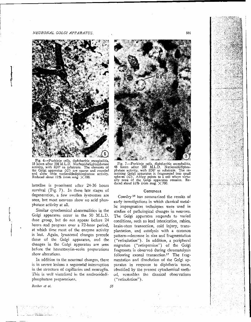
lamellae is prominent after 24-36 hours survival (Fig. 7). In these late stages of degeneration, a few swollen lysosomes are seen, but most neurons show no acid phos- phatase activity at all.
Similar cytochemical abnormalities in the Golgi apparatus occur in the SO M.L.D. dose group, but do not appear before 24 hours and progress over a 72-hour period, at which time most of the enzyme activity is lost. Again, lysosomal changes precede those of the Golgi apparatus, and the changes in the Golgi apparatus are seen before the hematoxylin-eosin preparations show alterations.
In addition to the neuronal changes, there is in severe lesions a segmental interruption in the structure of capillaries and neuroglia. This is well visualized in the nucleosidedi- phosphatase preparations.
Becker et al.
Fig. 7.—Purkinje cells, diphtheritic encephalitis, 48 hours after 500 M.L.D. Nucleosidediphos- phatase activity, with IDP as substrate. The re- maining Golgi apparatus is fragmented into small spheres (67). Arrow points to a cell where virtu- ally none of the Golgi apparatus remains. Re- duced about 11% from mag. X 700.
Comment Cowdry16 has summarized the results of
early investigations in which classical metal- lic impregnation techniques were used in studies of pathological changes in neurons. The Golgi apparatus responds to varied conditions, such as lead intoxication, rabies, brain-stem transection, cold injury, trans- plantation, and autolysis with a common pattern—decrease in size and fragmentation ("retisolution"). In addition, a peripheral migration ("retispersion") of the Golgi fragments is observed during chromatolysis following axonal transection.17 The frag- mentation and dissolution of the Golgi ap- paratus in response to diphtheria toxin, identified by the present cytochemical meth- od, resembles the classical observations ("retisolution").
55
;■:■£■■'*.v. J;

502 ARCHIVES OF NEUROLOGY
' l;.~■■■Mit.•*■*■+■■ ■-*■>■ "tJ!i':
■*m>.
u mM m -> .- \
Fig. 8.—Purkinje cells, diphtheritic encephalitis, 12 hours after 500 M.L.D. Acid phosphatase activ- ity, with glycerophosphate as substrate. Note the acid phosphatase-rich granules or Iysosomes. Ar- rows point to some of the abnormal swollen Iysosomes ("cytolysomes"). Reduced about 11% from mag. X 700.
The swelling of Iysosomes prior to changes in the Golgi apparatus is consistent with the view that they may serve as sensi- tive indicators of early necrobiosis, in cells generally 15-18 and in neurons.'6,7
The close spatial relation between man}' Iysosomes and the Golgi lamellae that is observed in neurons is paralleled in many, but not all, cells.5 This is consistent with the tentative suggestion that Iysosomes, in- volved in pinocytosis or secrefion or both, may fuse with or arise from the Golgi "vac- uoles." 18'19 Such speculations may be use- ful in suggesting studies with normal and pathological nervous tissue. Recently, Ogawa et al.20 have concluded that the "neutral red granules" of cultured fibroblasts and as- trocytes of cerebellar cortex are, in reality, iysosomes.
The availability of a simple, rapid, and reproducible means of demonstrating the Golgi apparatus in frozen sections may help reawaken interest of neurologists in this cell organelle. Its impressive size in neu- rons implies an important role, but this remains elusive. The finding of a specific nucleosidediphosphatase activity in the Goigi apparatus of neurons and all other cells tested2'3 may suggest approaches to the in- vestigation of its specific biochemistry. The ability to stain parallel or the same sections for activities of this specific phosphatase and for acid phosphatase activity associated with Iysosomes may be used to elucidate the origin of neurosecretory granules. Electron micrographs suggest that secretory granules in many cell types,12"14 including some neurosecretory granules of neurons,21 arise in the Golgi apparatus. The granules ap- pear in the spaces ("cisternae") enclosed within the paired membranes, and when they separate from the apparatus they are surrounded by Golgi membrane.
The ease of making such cytochemical preparations should enable the neuropathol- ogist to study more readily the pathological alterations in the Golgi apparatus and to relate these to changes in other cell or- ganelles such as Iysosomes and mitochondria.
Summary
The distribution of nucleosidediphospha- tase activity in the rat brain is described and its ability to visualize the neuronal Golgi apparatus is illustrated. Fragmenta- tion and dissolution of the Golgi apparatus, as seen in the cytochemical preparations, is described in neurons injured by diphtheria toxin. Marked changes in the Iysosomes occur before those in the Golgi apparatus.
The usefulness in neurology and neuro- pathology of the cytochemical staining pro- cedures for the Golgi apparatus is briefly discussed.
The technical assistance of James F. Galvin HM1 and Miss Mary Catogas is acknowledged. We wish to thank Mr. George Dominguez for assistance with the photography.
56 Vol. 5, Nov., 1961

NEURONAL GOLGI APPARATUS 503
LCDR Norwin H. Becker, U.S. Naval Medical Research Laboratory, U.S. Naval Submarine Base, New London, Conn.
REFERENCES
1. Becker,'N. H.; Goldfischer, S.; Shin, W. Y., and Novikoff, A. B.: The Localization of Enzyme Activities in the Rat Brain, J. Biophys. Biochem. Cytol. 8:649-663, 1960.
2. Novikoff, A. B., and Goldfischer, S.: Nucleo- sidediphosphatase Activity in the Golgi Apparatus, 5th International Congress of Biochemistry, Mos- cow, 1961, to be published.
3. Novikoff, A. B., and Goldfischer, S.: Nucleo- sidediphosphatase Activity in the Golgi Apparatus and Its Usefulness for Cytological Studies, Proc. Nat. Xcad. Sei. (U.S.A.), to be published.
4. Lillie, R. D.: Histopathologic Technique and Practical Histochemistry, New York, Blakiston Company, 1954, pp. 323-328.
5. Novikoff, A. B.; Goldfischer, S.; Essner, E., and Iaciofano, P.: The Relations Between Acid Phosphatase-Rich Lysosomes and the Golgi Ap- paratus, J. Histochem. Cytochem., to be published.
6. Becker, N. H., and Barron, K. D.: The Cytochemistry of Anoxic and Anoxic-Ischemic Encephalopathy in Rats: I. Alterations in Neu- ronal Lysosomes Identified by Acid Phosphatase Activity, Amer. J. Path. 38:161-175, 1961.
7. Barron, K. D.: A Histochemical Study of Neuronal Necrosis Produced by Intracerebral In- jection of Diphtheria Toxin, Neurology, to be published.
8. Baker, J. R.: The Histochemical Recogni- tion of Lipine, Quart. J. Micro. Sei. 87:441-470, 1946.
9. Agarwal, S. C, and Pryce, D. M.: Experi- mental Diphtheritic Paralysis in Rats, J. Path. Bact. 78:171-177, 1959.
10. Holt, S. J.: Factors Governing the Valid- ity of Staining Methods for Enzymes, and Their
Bearing Upon the Gomori Acid Phosphatase Tech- nique, Exp. Cell Res. Suppl. 7:1-27, 1959.
11. Gomori, G.: Microscopic Histochemistry; Principles and Practice, Chicago, University of Chicago Press, 1952, p. 273.
12. Pollister, A. W„ and Pollister, P. F.: The Structure of the Golgi Apparatus, Int. Rev. Cytol. 6:85-106, 1957.
13. Palay, S. L.: The Morphology of Secre- tion, in Frontiers of Cytology, edited by S. L. Palay, New Haven, Conn., Yale University Press, 1958, pp. 305-342.
14. Dalton, A. J.: The Golgi Apparatus and Secretory Granules, in The Cell, edited by J. Brächet and A. E. Mirsky, New York, Academic Press, Inc., 1961, pp. 603-619.
15. Novikoff, A. B.: Biochemical and Staining Reactions of Cytoplasmic Constituents, in Develop- ing Cell Systems and Their Control, edited by D. Rudnick, New York, The Ronald Press Com- pany, 1960, pp. 167-203.
16. Cowdry, E. V.: Cytological Constituents— Mitochondria, Golgi Apparatus and Chromidial Substance, in General Cytology, edited by E. V. Cowdry, Chicago, University of Chicago Press, 1924, pp. 311-382.
17. Penfield, W. G.: Alterations of the Golgi Apparatus in Nerve Cells, Brain 43:290-305, 1920.
18. Novikoff, A. B.: Lysosomes and Related Particles, in The Cell, edited by J. Brächet and A. E. Mirsky, New York, Academic Press, Inc., 1961, pp. 423-488.
19. Novikoff, A. B., and Essner, E.: Nucleo- sidephosphatase Activities of Cytomembranes, Exp. Cell. Res., to be published.
20. Ogawa, K.; Mizuno, N., and Okamoto, M.: Lysosomes in Cultured Cells, J. Histochem. Cyto- chem. 9:202, 1961.
21. Palay, S. L.: The Fine Structure of Se- cretory Neurons in the Preoptic Nucleus of the Goldfish (Carassius Auratus), Anat. Rec, 138: 417-443, 1960.
::'\
Becker et cd. Printed and Published in the Tl™;i.ed States of America


![POSSIBLE ROLES OF VERTEBRATE NEUROGLIA IN POTASSIUM DYNAMICS, SPREADING DEPRESSION AND ...ucgbarg/res/jeb_95_111.pdf · 2009-07-15 · Vertebrate neuroglia in potassium dynamics HighEC[K+]](https://static.fdocuments.us/doc/165x107/5f14071992d3e603436d6ccc/possible-roles-of-vertebrate-neuroglia-in-potassium-dynamics-spreading-depression.jpg)